and in liver and muscle: glycogen lectures/biochemistry/glycogen-2.pdf · 2 glycogen –12 topics...
TRANSCRIPT
Carbohydrate Storage and Synthesis in Liver and Muscle: Glycogen

2
Glycogen – 12 topics Carbohydrate MetabolismInvesting for the future Outline of Topics
Introduction Structure of Glycogen – highly branced ‐glucose polymerGlycogenesis – Glc incorporated into glycogen (liver & muscle, kidney)Glycogenolysis –Glucose mobilized from glycogen in liver and muscleHormonal regulation of hepatic glycogenesis vs. glycogenolysis – insulin vs. glucagonMechanisms of glucagon action – Signals phosphorylations, pathways flipGlycogenolysis in liver – plasma glycemia maintenance: acute vs. postabsorbtiveGlycogenolysis in muscle – Mobilizing glucose for ATP contraction activityRegulation of glycogenesis – replenish glycogen stores vs. immediate needsGluconeogenesis – de novo (new) glucose from non carbohydrate carbon skeletonsRegulation of gluconeogenesis – De novo glucose synthesis fueled by fat oxidation Interconversions of fructose/galactose/mannose/glucose – glycoproteins, etc., … Inborn errors of metabolism – glycogen storage diseases
Introduction Structure of Glycogen – highly branced ‐glucose polymerGlycogenesis – Glc incorporated into glycogen (liver & muscle, kidney)Glycogenolysis –Glucose mobilized from glycogen in liver and muscleHormonal regulation of hepatic glycogenesis vs. glycogenolysis – insulin vs. glucagonMechanisms of glucagon action – Signals phosphorylations, pathways flipGlycogenolysis in liver – plasma glycemia maintenance: acute vs. postabsorbtiveGlycogenolysis in muscle – Mobilizing glucose for ATP contraction activityRegulation of glycogenesis – replenish glycogen stores vs. immediate needsGluconeogenesis – de novo (new) glucose from non carbohydrate carbon skeletonsRegulation of gluconeogenesis – De novo glucose synthesis fueled by fat oxidation Interconversions of fructose/galactose/mannose/glucose – glycoproteins, etc., … Inborn errors of metabolism – glycogen storage diseases
Glucose Fuel Storage and mobilization for oxidatonGlucose Fuel Storage and mobilization for oxidaton

3
Metabolic Fate of Glucose Glycogen MetabolismIntroduction
Red cells and the brain – Have an absolute requirement for blood glucose for their energy metabolism.
These cells consume about 80% of the glucose (200 g, 1.1 mol, ca. 1500 kcal) consumed per day by a 70 kg human, in good health.
Blood and extracellular fluid volume contains about 10 g glucose – must be replenished constantly.
Assumes a blood volume = 7 L, hematocrit = 45%, and no other distribution system operates.
Normally, blood [glucose] range is between 4 – 6.5 mM = glycemia(about 80 – 120 mg/dL)
4
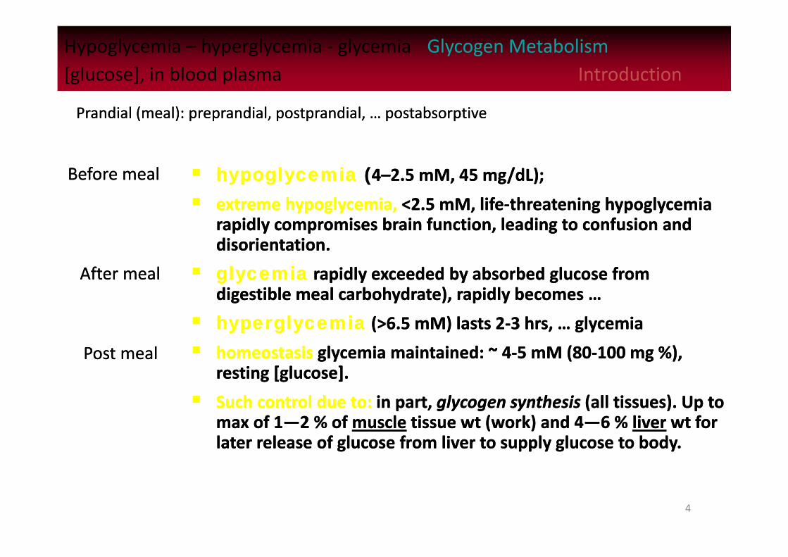
Hypoglycemia – hyperglycemia ‐ glycemia Glycogen Metabolism[glucose], in blood plasma Introduction
hypoglycemia (4–2.5 mM, 45 mg/dL);
extreme hypoglycemia, <2.5 mM, life‐threatening hypoglycemiarapidly compromises brain function, leading to confusion and disorientation.
glycemia rapidly exceeded by absorbed glucose from digestible meal carbohydrate), rapidly becomes …
hyperglycemia (>6.5 mM) lasts 2‐3 hrs, … glycemia
homeostasis glycemia maintained: ~ 4‐5 mM (80‐100 mg %), resting [glucose].
Such control due to: in part, glycogen synthesis (all tissues). Up to max of 1—2 % of muscle tissue wt (work) and 4—6 % liver wt for later release of glucose from liver to supply glucose to body.
Before mealBefore meal
After mealAfter meal
Post mealPost meal
Prandial (meal): preprandial, postprandial, … postabsorptivePrandial (meal): preprandial, postprandial, … postabsorptive
5
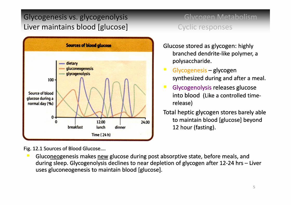
Glycogenesis vs. glycogenolysis Glycogen MetabolismLiver maintains blood [glucose] Cyclic responsesGlycogenesis vs. glycogenolysis Glycogen MetabolismLiver maintains blood [glucose] Cyclic responses
Fig. 12.1 Sources of Blood Glucose….Fig. 12.1 Sources of Blood Glucose….
Glucose stored as glycogen: highly branched dendrite‐like polymer, a polysaccharide.
Glycogenesis – glycogen synthesized during and after a meal.
Glycogenolysis releases glucose into blood (Like a controlled time‐release)
Total heptic glycogen stores barely able to maintain blood [glucose] beyond 12 hour (fasting).
Gluconeogenesis makes new glucose during post absorptive state, before meals, and during sleep. Glycogenolysis declines to near depletion of glycogen after 12‐24 hrs – Liver uses gluconeogenesis to maintain blood [glucose].
Gluconeogenesis makes new glucose during post absorptive state, before meals, and during sleep. Glycogenolysis declines to near depletion of glycogen after 12‐24 hrs – Liver uses gluconeogenesis to maintain blood [glucose].

6
Glycogen Storage Carbohydrate MetabolismVarious Tissues StructureGlycogen Storage Carbohydrate MetabolismVarious Tissues Structure
Blood glucose = 10 g, tissues needs easily deplete.
Glycogen degraded to glucose‐1P G6P for oxidative metabolism in tissues to synthesize ATP.
Liver: G6P G + P, by G6P phosphatase. Muscle lacks G6P phosphatase.
Blood glucose = 10 g, tissues needs easily deplete.
Glycogen degraded to glucose‐1P G6P for oxidative metabolism in tissues to synthesize ATP.
Liver: G6P G + P, by G6P phosphatase. Muscle lacks G6P phosphatase.
Fig. 12.2 Tissue distribution of carbohydrate energy reserves(70 kg adult).
Fig. 12.2 Tissue distribution of carbohydrate energy reserves(70 kg adult).
Fig. 12.3 Close‐up of glycogen structure.
Fig. 12.3 Close‐up of glycogen structure.
Highly branched dendritic polymerHighly branched dendritic polymer
7
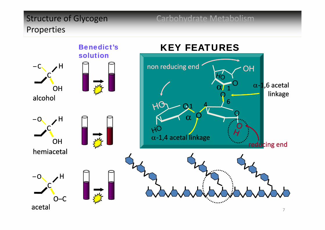
Structure of Glycogen Carbohydrate MetabolismProperties
KEY FEATURESKEY FEATURES
non reducing endnon reducing end
‐1,6 acetal linkage
‐1,6 acetal linkage
‐1,4 acetal linkage‐1,4 acetal linkage
OO
OO
O 11
6611 44
CC–O–O
OHOH
HH
CC–O–O
O–CO–C
HH
reducing endreducing endhemiacetalhemiacetal
acetalacetal
Benedict’s solutionBenedict’s solution
CC–C–C
OHOH
HH
alcoholalcohol
8
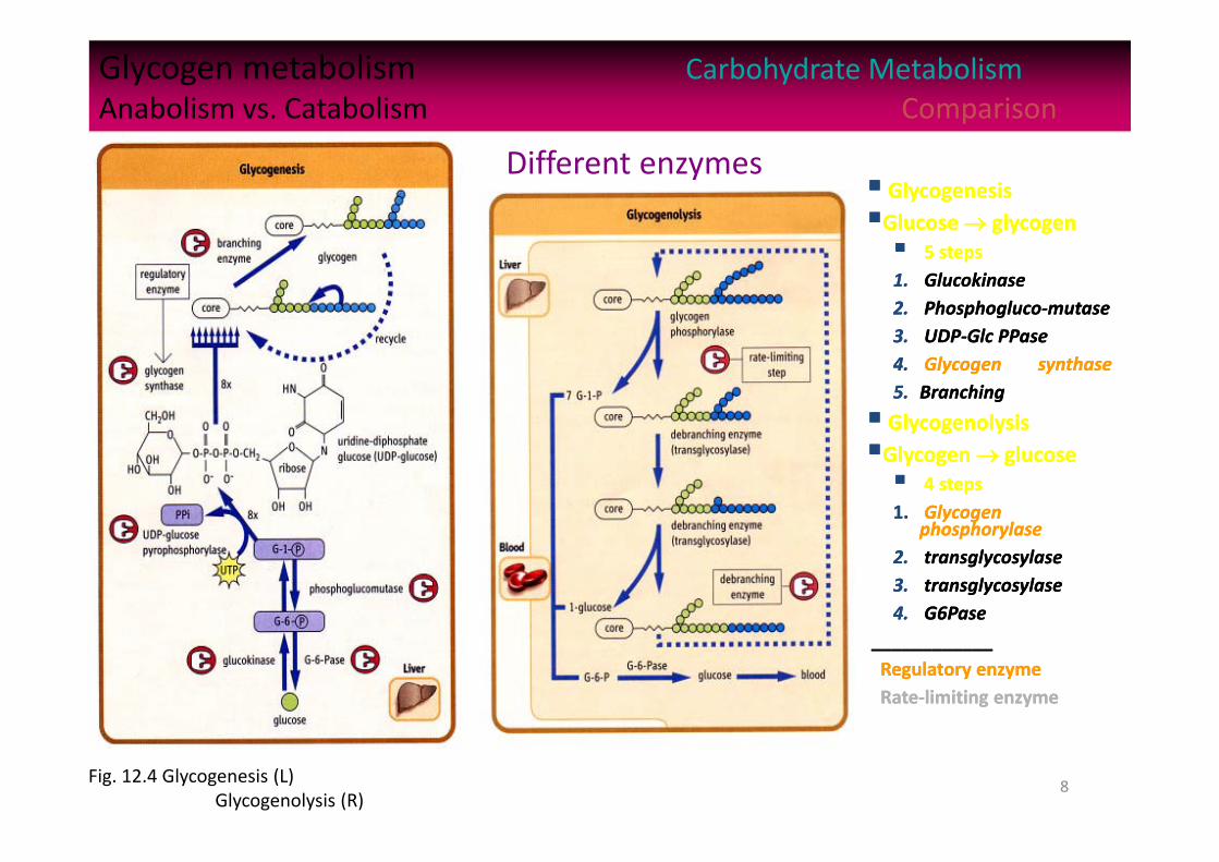
Glycogen metabolism Carbohydrate MetabolismAnabolism vs. Catabolism Comparison
GlycogenesisGlucose glycogen 5 steps1. Glucokinase2. Phosphogluco‐mutase3. UDP‐Glc PPase4. Glycogen synthase5. Branching
GlycogenolysisGlycogen glucose 4 steps1. Glycogen
phosphorylase2. transglycosylase3. transglycosylase4. G6Pase
____________Regulatory enzymeRate‐limiting enzyme
GlycogenesisGlucose glycogen 5 steps1. Glucokinase2. Phosphogluco‐mutase3. UDP‐Glc PPase4. Glycogen synthase5. Branching
GlycogenolysisGlycogen glucose 4 steps1. Glycogen
phosphorylase2. transglycosylase3. transglycosylase4. G6Pase
____________Regulatory enzymeRate‐limiting enzyme
Fig. 12.4 Glycogenesis (L) Glycogenolysis (R)
Different enzymes
9
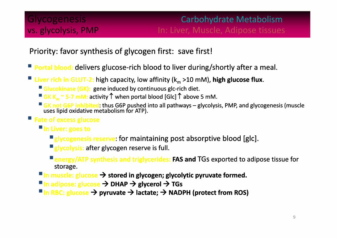
Glycogenesis Carbohydrate Metabolismvs. glycolysis, PMP In: Liver, Muscle, Adipose tissues
Portal blood: delivers glucose‐rich blood to liver during/shortly after a meal.
Liver rich in GLUT‐2: high capacity, low affinity (km >10 mM), high glucose flux.Glucokinase (GK): gene induced by continuous glc‐rich diet.GK Km ~ 5‐7 mM: activity when portal blood [Glc] above 5 mM.GK not G6P inhibited: thus G6P pushed into all pathways – glycolysis, PMP, and glycogenesis (muscle uses lipid oxidative metabolism for ATP).
Fate of excess glucoseIn Liver: goes toglycogenesis reserve: for maintaining post absorptive blood [glc].glycolysis: after glycogen reserve is full.energy/ATP synthesis and triglycerides: FAS and TGs exported to adipose tissue for storage.
In muscle: glucose stored in glycogen; glycolytic pyruvate formed.In adipose: glucose DHAP glycerol TGsIn RBC: glucose pyruvate lactate; NADPH (protect from ROS)
Priority: favor synthesis of glycogen first: save first! Priority: favor synthesis of glycogen first: save first!
©Copyright 1999‐2004 by Gene C. Lavers 10
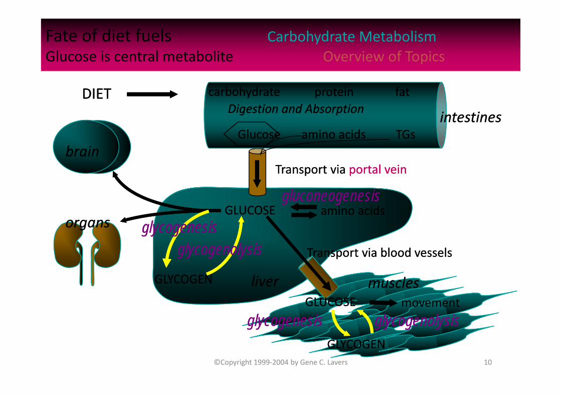
Fate of diet fuels Carbohydrate MetabolismGlucose is central metabolite Overview of Topics
DIETDIETDigestion and AbsorptionDigestion and Absorption
GlucoseGlucose
carbohydratecarbohydrate fatfatproteinprotein
amino acidsamino acids TGsTGs
liverliver
Transport via portal veinTransport via portal vein
intestinesintestines
amino acidsamino acids
GLYCOGENGLYCOGEN musclesmusclesmovement
gluconeogenesisgluconeogenesis
brainbrain
glycogenolysisglycogenolysis
GLUCOSEGLUCOSE
GLUCOSEGLUCOSE
GLYCOGENGLYCOGEN
organsorgans
glycogenesisglycogenesis glycogenolysisglycogenolysis
glycogenesisglycogenesisTransport via blood vessels Transport via blood vessels

11
Hormonal control Carbohydrate MetabolismGlycogenolysis In Liver Comparison
Glycogenolysis: response to low blood [glc] from: Post absorptive utilization. Response to stress.
3 hormones — activation mode:
Glucagon—3.5 kd peptide, from ‐cells of endocrine pancreas; main function: activate hepatic glycogenolysis to maintain normoglycemia.
Epinephrine—tyrosine derivative, a catecholamine from adrenal medulla activates glycogenolysis in response to acute stress.
Cortisol—adrenocortical steroid varies diurnally in plasma, but may be chronically elevated under continuously stressful conditions.
Glucagon, Epinephrine, Cortisol, InsulinGlucagon, Epinephrine, Cortisol, Insulin
Fig. 12.5 Hormones involved in control of glycogenolysis.
Fig. 12.5 Hormones involved in control of glycogenolysis.
12
Glucagon Carbohydrate Metabolism In Liver Hormonal Regulation of GlycogenolysisGlucagon Carbohydrate Metabolism In Liver Hormonal Regulation of Glycogenolysis
Glucagon – 3500 MW protein (29‐aa): secreted by ‐cells of endocrine pancreas, activates glycogenolysis to maintain normal glycemia, when blood [glucose] becomes hypoglycemic.
Glucagon t/2 ~ 5 minutes. (removal from blood by receptor binding, renal filtration, proteolytic inactivation in liver.)
Elevated blood [glucagon]: between meals; chronically elevated during fasting or low‐carbohydrate diet.
Decreased blood [glucagon]: decreases during and soon after a meal ([glucose] is very high).
Glucagon, epinephrine (adrenalin), cortisol, insulinGlucagon, epinephrine (adrenalin), cortisol, insulin

Acute & Chronic Stress Carbohydrate MetabolismGlycogenolysis Activation
Acute & Chronic Stress Carbohydrate MetabolismGlycogenolysis Activation
Physiologic ‐‐ in response to increased blood glucose utilization during prolonged exercise.
Pathologic ‐‐ as a result of blood loss.
Psychological ‐‐ in response to acute or chronic threats.
Acute stress (regardless of source): activates glycogenolysis through the action of catecholamine hormone, epinephrine (released by the adrenal medula).
During prolonged exercise: both glucagon and epinephrine contribute to stimulation of glycogenolysis.
Glycogenolysis is activated in response to stress Glycogenolysis is activated in response to stress
14
Insulin Carbohydrate MetabolismHormonal regulation Inhibition of GlycogenolysisInsulin Carbohydrate MetabolismHormonal regulation Inhibition of Glycogenolysis
Insulin secreted by pancreas ‐cells when blood [glucose] is high.
Synthesized as single peptide chain zymogen: proinsulin.
In secretory granules, selective proteolysis releases an internal peptide and a 2‐chained (via 2 ‐S–S‐ ) insulin hormone.
Insulin elicits uptake and intracellular use or storage of glucose, an anabolic hormone.
Hyperglycemia results in elevated blood [insulin] associated with fed state.
Hyperinsulinism associated with “insulin resistance” and if chronic can lead to diabetes type‐2 and related pathologies.
Antagonist of glucagon, epinephrine (adrenalin), cortisol Antagonist of glucagon, epinephrine (adrenalin), cortisol
15
Glycogen Carbohydrate MetabolismSignal Transduction Regulation Mechanism
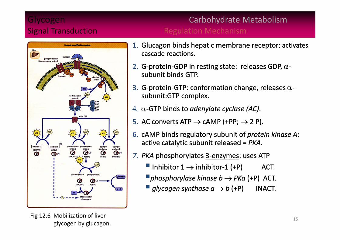
Fig 12.6 Mobilization of liver glycogen by glucagon.
1. Glucagon binds hepatic membrane receptor: activates cascade reactions.
2. G‐protein‐GDP in resting state: releases GDP, ‐subunit binds GTP.
3. G‐protein‐GTP: conformation change, releases ‐subunit:GTP complex.
‐GTP binds to adenylate cyclase (AC).
5. AC converts ATP cAMP (+PP; 2 P).
6. cAMP binds regulatory subunit of protein kinase A: active catalytic subunit released = PKA.
7. PKA phosphorylates 3‐enzymes: uses ATP
Inhibitor 1 inhibitor‐1 (+P) ACT.
phosphorylase kinase b PKa (+P) ACT. glycogen synthase a b (+P) INACT.
16
Glycogen Carbohydrate MetabolismSignal Transduction Regulation Mechanism
Fig 12.6 Mobilization of liver glycogen by glucagon.
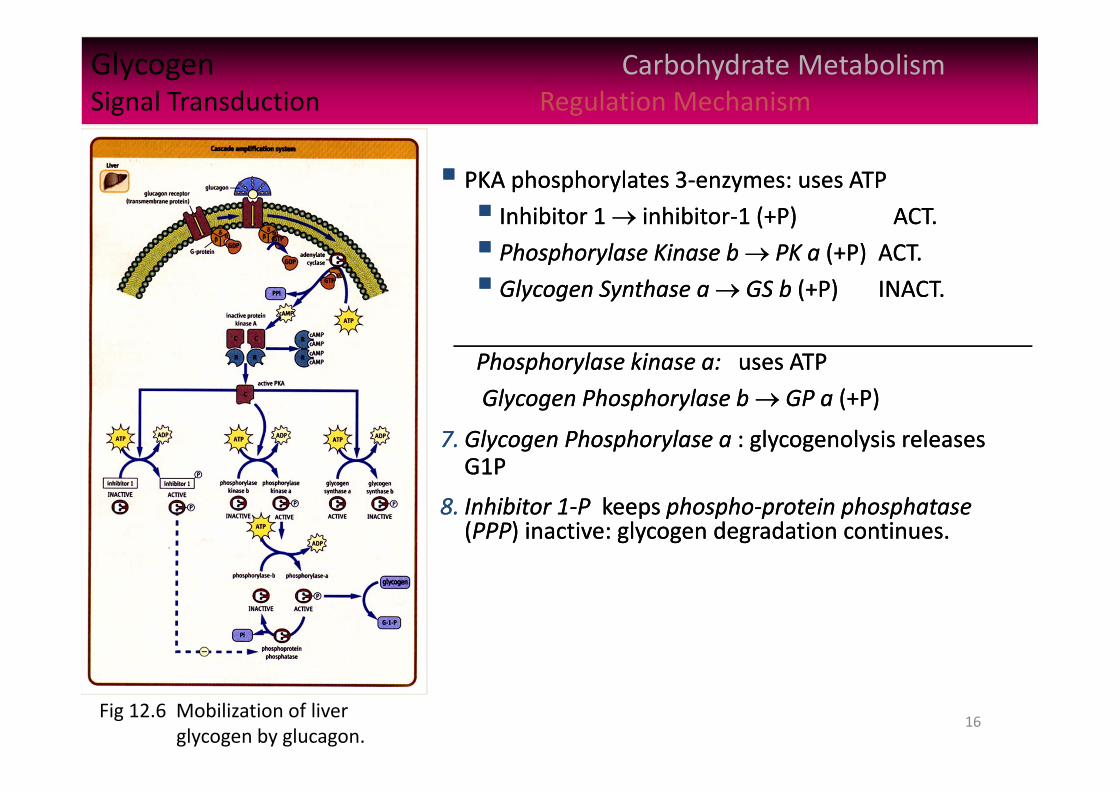
PKA phosphorylates 3‐enzymes: uses ATP
Inhibitor 1 inhibitor‐1 (+P) ACT.
Phosphorylase Kinase b PK a (+P) ACT.
Glycogen Synthase a GS b (+P) INACT.
Phosphorylase kinase a: uses ATP
Glycogen Phosphorylase b GP a (+P)
7. Glycogen Phosphorylase a : glycogenolysis releases G1P
8. Inhibitor 1‐P keeps phospho‐protein phosphatase(PPP) inactive: glycogen degradation continues.
17
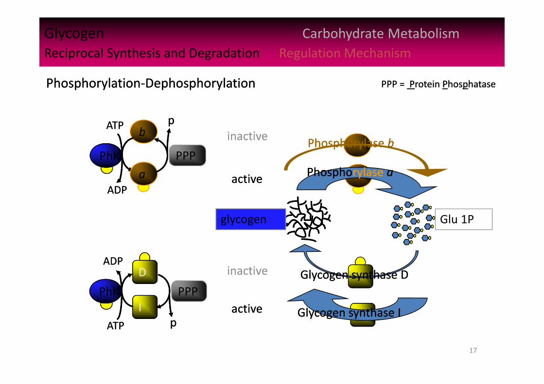
Glycogen Carbohydrate MetabolismReciprocal Synthesis and Degradation Regulation MechanismGlycogen Carbohydrate MetabolismReciprocal Synthesis and Degradation Regulation Mechanism
Glu 1P
Phosphorylase aPhosphorylase a
Glycogen synthase IGlycogen synthase I
Glycogen synthase DGlycogen synthase D
Phosphorylase bPhosphorylase bb
PhKPhK
ATPATP
ADPADPa
PPP
pp
IPhKPhK
ADPADP
ATPATP
D
PPP
pp
inactiveinactive
activeactive
inactiveinactive
activeactive
glycogen
Phosphorylation‐DephosphorylationPhosphorylation‐Dephosphorylation PPP = Protein PhosphatasePPP = Protein Phosphatase

18
Balancing Pathway Activities Carbohydrate MetabolismAvoiding futile cycles Inhibiting glucose
Prandial glucose used up, glycemia falls into hypoglycemia.
Glucagon’s enzyme cascade amplification turns on liver glycogenolysis – balanced inhibition of glycogenesis. Also produces inhibition of … Protein synthesis – uses considerable ATP and GTP
Cholesterol synthesis – uses ATP
Fatty acid (FA) synthesis – uses ATP to activate acetyl CoA (malonyl CoA)
Triglyceride (TGs) synthesis from glycolytic DHAP derived from glucose
Glucose synthesis (gluconeogenesis) – uses GTP
Glucose utilization (glycolysis) – uses ATP
Key enzymes phosphorylated in opposing pathways, avoids futile cycles.
Glucagon shifts liver metabolism to keep blood [glc] glycemic to maintain vital body functions (see Ch 20).
Glycogenolysis floods system with G1P, G6P, and glucose Glycogenolysis floods system with G1P, G6P, and glucose
19
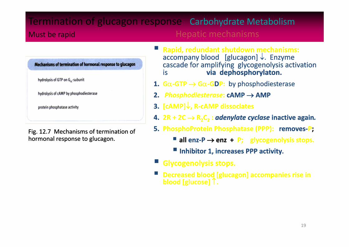
Termination of glucagon response Carbohydrate MetabolismMust be rapid Hepatic mechanisms
Rapid, redundant shutdown mechanisms: accompany blood [glucagon] . Enzyme cascade for amplifying glycogenolysis activation is via dephosphorylaton.
1. G‐GTP G‐GDP: by phosphodiesterase2. Phosphodiesterase: cAMP AMP
3. [cAMP], R‐cAMP dissociates
4. 2R + 2C R2C2 : adenylate cyclase inactive again.
5. PhosphoProtein Phosphatase (PPP): removes‐P;
all enz‐P enz + P; glycogenolysis stops.
Inhibitor 1, increases PPP activity. Glycogenolysis stops. Decreased blood [glucagon] accompanies rise in
blood [glucose] .
Fig. 12.7 Mechanisms of termination of hormonal response to glucagon. Fig. 12.7 Mechanisms of termination of hormonal response to glucagon.
20
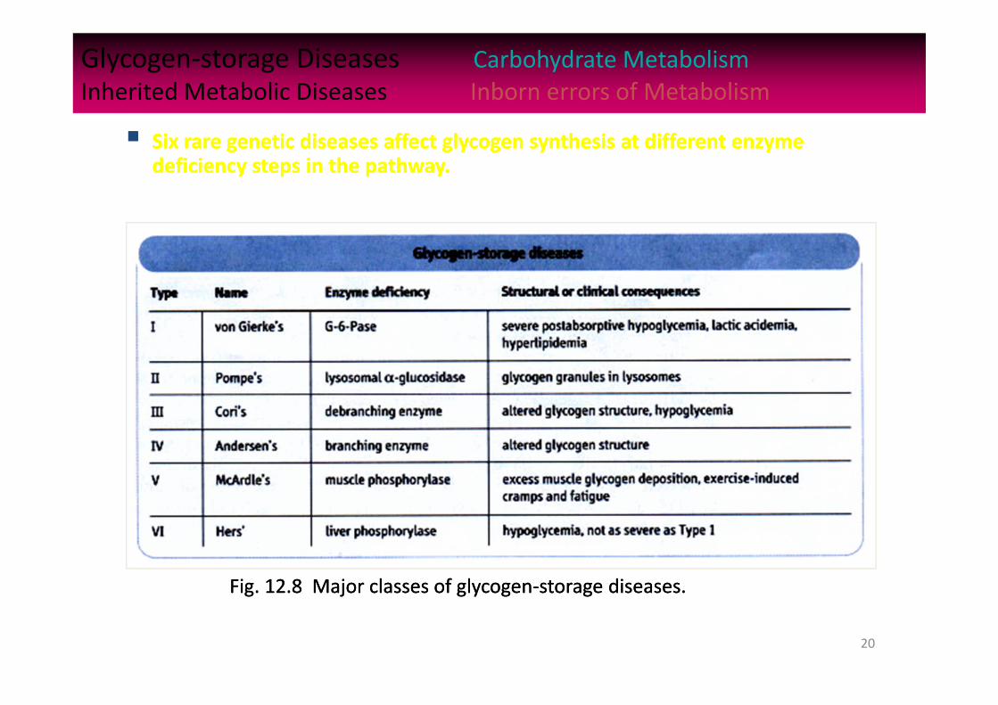
Glycogen‐storage Diseases Carbohydrate MetabolismInherited Metabolic Diseases Inborn errors of Metabolism
Six rare genetic diseases affect glycogen synthesis at different enzyme deficiency steps in the pathway. Six rare genetic diseases affect glycogen synthesis at different enzyme
deficiency steps in the pathway.
Fig. 12.8 Major classes of glycogen‐storage diseases.Fig. 12.8 Major classes of glycogen‐storage diseases.
21
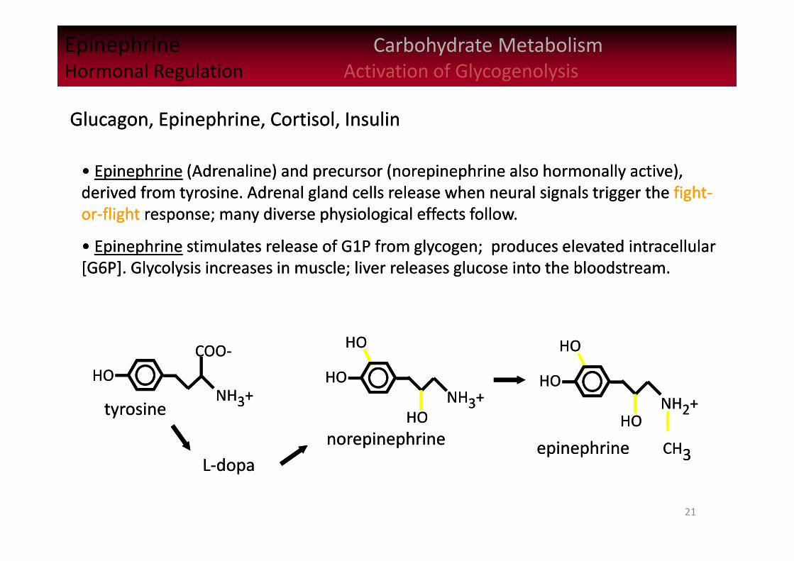
Epinephrine Carbohydrate MetabolismHormonal Regulation Activation of GlycogenolysisEpinephrine Carbohydrate MetabolismHormonal Regulation Activation of Glycogenolysis
• Epinephrine (Adrenaline) and precursor (norepinephrine also hormonally active), derived from tyrosine. Adrenal gland cells release when neural signals trigger the fight‐or‐flight response; many diverse physiological effects follow.
• Epinephrine stimulates release of G1P from glycogen; produces elevated intracellular [G6P]. Glycolysis increases in muscle; liver releases glucose into the bloodstream.
Glucagon, Epinephrine, Cortisol, InsulinGlucagon, Epinephrine, Cortisol, Insulin
HOHONH3+NH3+
COO‐COO‐
HOHONH2+NH2+
CH3CH3
HOHO
HOHOtyrosinetyrosine
L‐dopaL‐dopa
HOHONH3+NH3+
HOHO
HOHO
epinephrineepinephrinenorepinephrinenorepinephrine
22
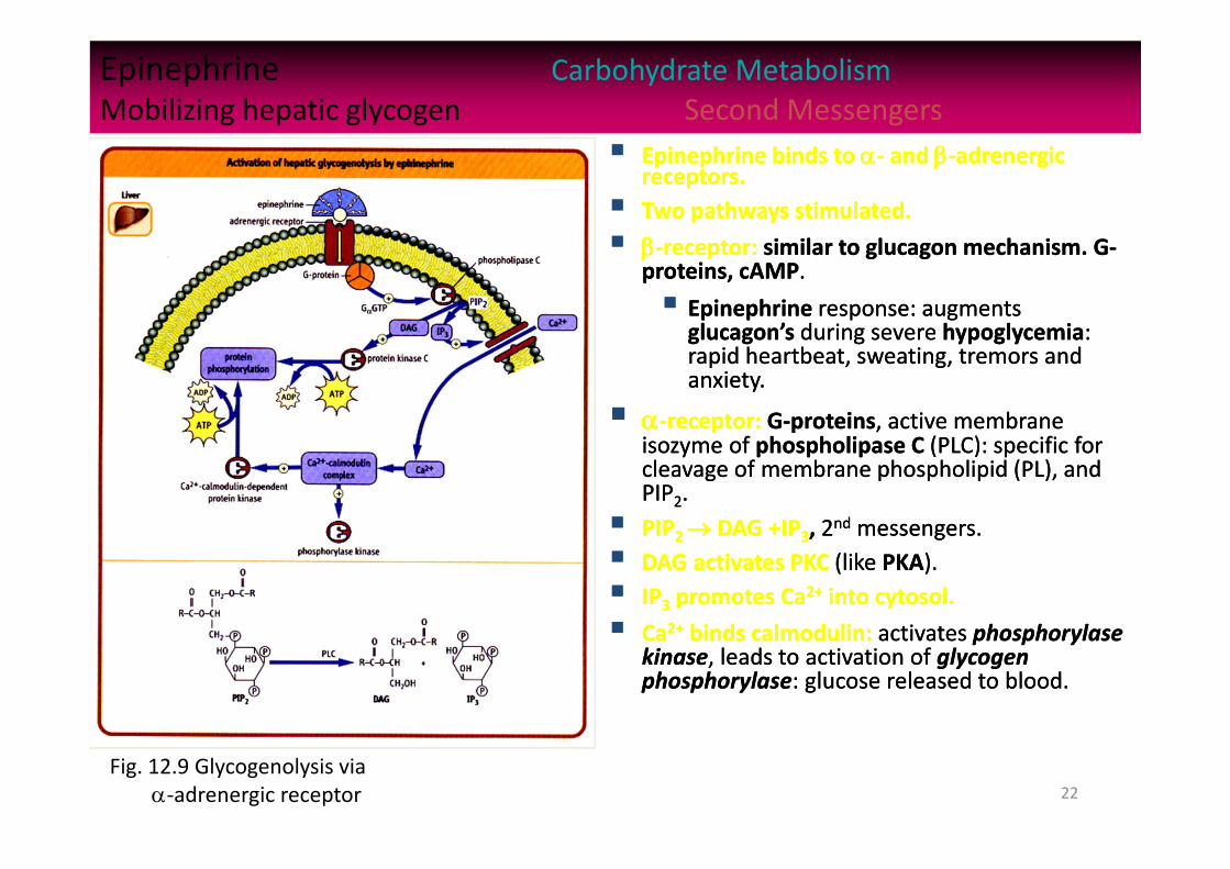
Epinephrine Carbohydrate MetabolismMobilizing hepatic glycogen Second Messengers
Epinephrine binds to ‐ and ‐adrenergic receptors. Two pathways stimulated.
‐receptor: similar to glucagon mechanism. G‐proteins, cAMP.
Epinephrine response: augments glucagon’s during severe hypoglycemia: rapid heartbeat, sweating, tremors and anxiety.
‐receptor: G‐proteins, active membrane isozyme of phospholipase C (PLC): specific for cleavage of membrane phospholipid (PL), and PIP2. PIP2 DAG +IP3, 2nd messengers. DAG activates PKC (like PKA). IP3 promotes Ca2+ into cytosol.
Ca2+ binds calmodulin: activates phosphorylase kinase, leads to activation of glycogen phosphorylase: glucose released to blood.
Fig. 12.9 Glycogenolysis via ‐adrenergic receptor
23
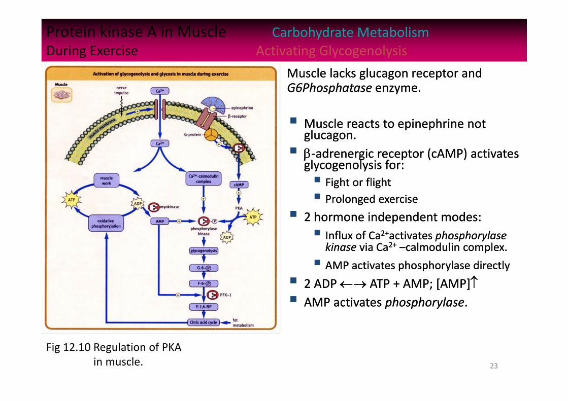
Protein kinase A in Muscle Carbohydrate MetabolismDuring Exercise Activating Glycogenolysis
Muscle reacts to epinephrine not glucagon. ‐adrenergic receptor (cAMP) activates
glycogenolysis for: Fight or flight Prolonged exercise
2 hormone independent modes: Influx of Ca2+activates phosphorylase kinase via Ca2+ –calmodulin complex.
AMP activates phosphorylase directly
2 ADP ATP + AMP; [AMP] AMP activates phosphorylase.
Muscle lacks glucagon receptor and G6Phosphatase enzyme. Muscle lacks glucagon receptor and G6Phosphatase enzyme.
Fig 12.10 Regulation of PKAin muscle.

24
Regulatory effects by Insulin Carbohydrate MetabolismReceptor dimerization Glycogenesis
Insulin’s 2 main functions:
lowers blood glucose by reversing the effect of glucagon’s phosphorylation of enzymes and proteins.
Stimulates gene expression of carbohydrate metabolism enzymes.
Insulin’s 2 main functions:
lowers blood glucose by reversing the effect of glucagon’s phosphorylation of enzymes and proteins.
Stimulates gene expression of carbohydrate metabolism enzymes.
Fig 12.11 Regulatory effects of insulin onhepatic and muscle carbo metab.

25
Gluconeogenesis (GNG) Carbohydrate MetabolismGlucose from non carbohydrates Cytosol‐Mitochondrion
Gluconeogenesis: essential during fasting and starvation, when hepatic glycogen depleted, to maintain blood glucose. Energy and carbon source required: oxidation of FA
released from adipose tissue provides ATP; carbons from 3‐sources. Lactate from RBC and active muscle. Large muscle mass: major source of glucogenic
amino acids; transamination. Glycerol from TGs: DHAP via glycerol‐3P. 3 glycolytic irreversible reactions: PK, PFK‐1, GK
bypassed by phosphatases: FBPase, and G6Pase after PEPCKase 1,3BPG 3PG is reversible, G similar. Lactate cycle: Cori cycle (ch 20). Muscle lactate and
pyr liver‐GNG glc, to muscle‐glycolysislactate Glucose‐alanine cycle: [muscle: glc pyr ala]
[liver: GNG glc] [muscle: glc pyr ala]…
3‐Sources: Lactate, amino acids, glycerol 3‐Sources: Lactate, amino acids, glycerol
Fig 12.12 Pathways of gluconeogenesis.Fig 12.12 Pathways of gluconeogenesis.

26
Regulating gluconeogenesis Carbohydrate MetabolismHormonal mechanisms Glycolysis vs. Gluconeogenesis
Gluconeogenesis vs. glycolysis: avoid a futile cycle; activeGNG—inhibit glycolysis Enz‐P or inactive GNG—active glycolysis. Enz F26BP: allosteric (+) regulator of F16BP. Made by: PFK2: F6P F26BP; enhances glycolysis. F26BPase: F6P F26BP; enhances GNG. PFK2/F26BPase: a bifunctional, with ‘P’ switch: PFK2/F26BPase PFK2/F26BPase‐P PFK1: F6P F16BP; F26BP Rx rate! F16BPase: F6P F16BP; F26BP inhibits GNG! [acetyl CoA]: slows TCA; act. PC [OAA]Glc
Glucagon: promotes phosphorylation (PK, inact.)
Insulin: promotes de‐phosphorylation (PK act.)
During fasting: glucagon, PK‐P inact, GNG, EM Eat Carbo meal: insulin, PK act, GNG, EM
Control: liver PFK1 and F1,6BPaseControl: liver PFK1 and F1,6BPase
Fig. 12.13 Gluconeogenesisregulated by heptic[F26BP] and[acetyl CoA]
Fig. 12.13 Gluconeogenesisregulated by heptic[F26BP] and[acetyl CoA]
27
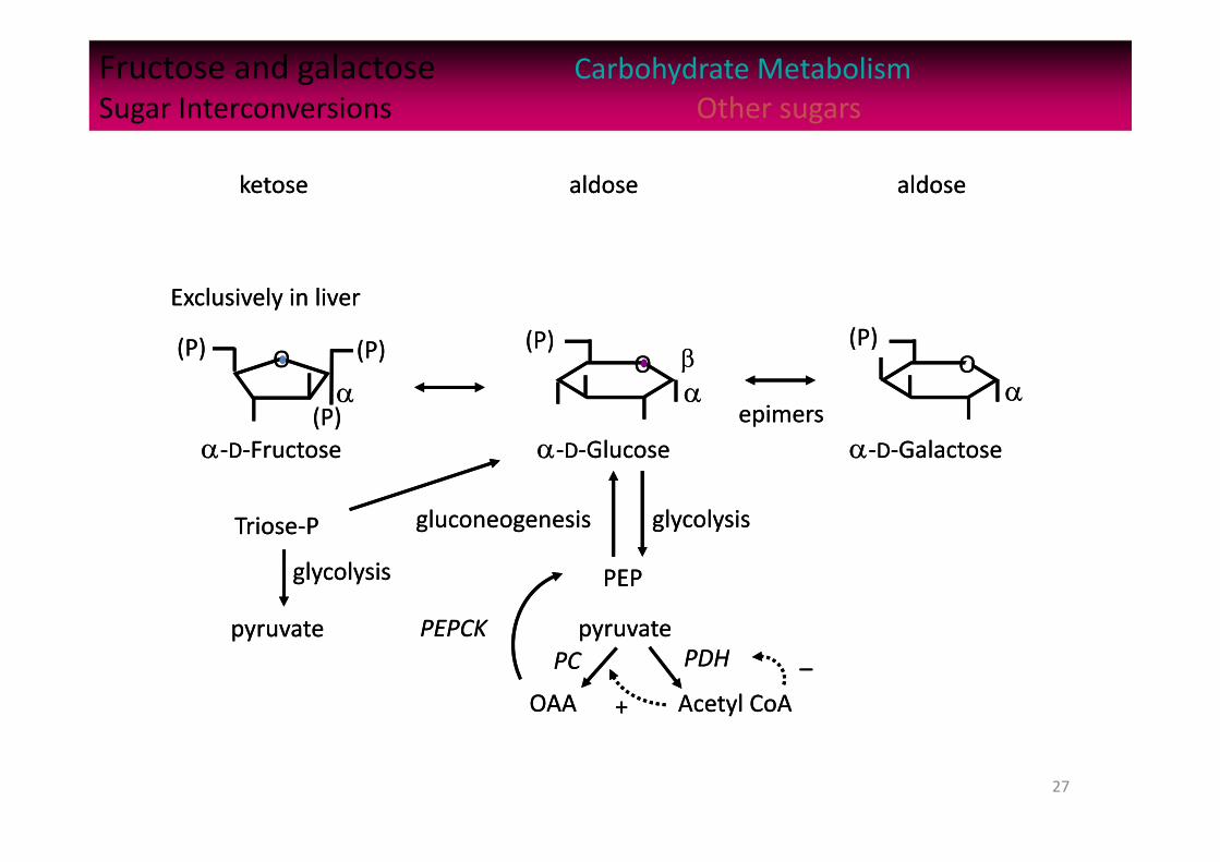
Fructose and galactose Carbohydrate MetabolismSugar Interconversions Other sugars
‐D‐Glucose‐D‐Glucose
Triose‐PTriose‐P
OOOO OO
‐D‐Galactose‐D‐Galactose‐D‐Fructose‐D‐Fructose
Exclusively in liverExclusively in liver
pyruvatepyruvate
glycolysisglycolysis
(P)(P) (P)(P)(P)(P) (P)(P)
(P)(P)
pyruvatepyruvate
glycolysisglycolysisgluconeogenesisgluconeogenesis
OAAOAA
PCPC
Acetyl CoAAcetyl CoA
PDHPDH
epimersepimers
++
––
PEPPEP
PEPCKPEPCK
aldosealdose aldosealdoseketoseketose
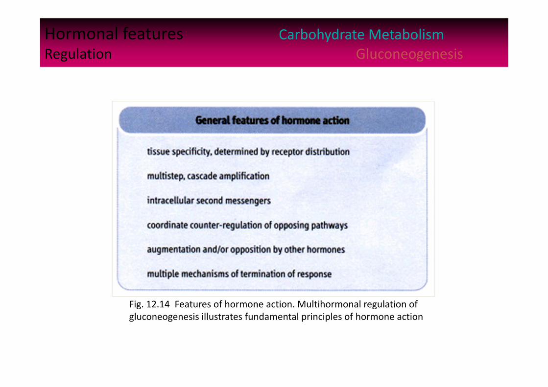
Hormonal features Carbohydrate MetabolismRegulation Gluconeogenesis
Fig. 12.14 Features of hormone action. Multihormonal regulation of gluconeogenesis illustrates fundamental principles of hormone action

29
Metabolic Fate of Glucose Glycogen Metabolism
Introduction
Red cells and the brain – Have an absolute requirement for blood glucose for their energy metabolism.
These cells consume about 80% of the glucose (200 g, 1.1 mol, ca. 1500 kcal) consumed per day by a 70 kg human, in good health.
Blood and extra cellular fluid volume contains about 10 g glucose, which must be replenished constantly.
Assumes a blood volume = 7 L, hematocrit = 45%, and no other distribution system operates.
Normally, blood [glucose] range is between 4 – 6.5 mM (about 80 – 120 mg/dL)
30
Gluconeogenesis Glycogen Metabolism A
backup system – makes new glucose Introduction
• Liver can synthesize glucose from non carbohydrate precursors.
• Amino acids supply carbon skeletons, as does glycerol.
• During starvation*, liver uses degraded muscle protein as the primary precursor of glucose; also lactate (from glycolysis) and glycerol (from fat).
• Fatty acids from triacylglycerides (TAGs) mobilzed (from adipose tissue**) provide the energy for gluconeogenesis.
__________________________* Metabolically may begin about 12 hours after the last meal. ** During well-fed states, excess glucose is converted to triacylglycerides
(TGs) in adipose cells.
©Copyright 1999‐2004 by Gene C. Lavers 31
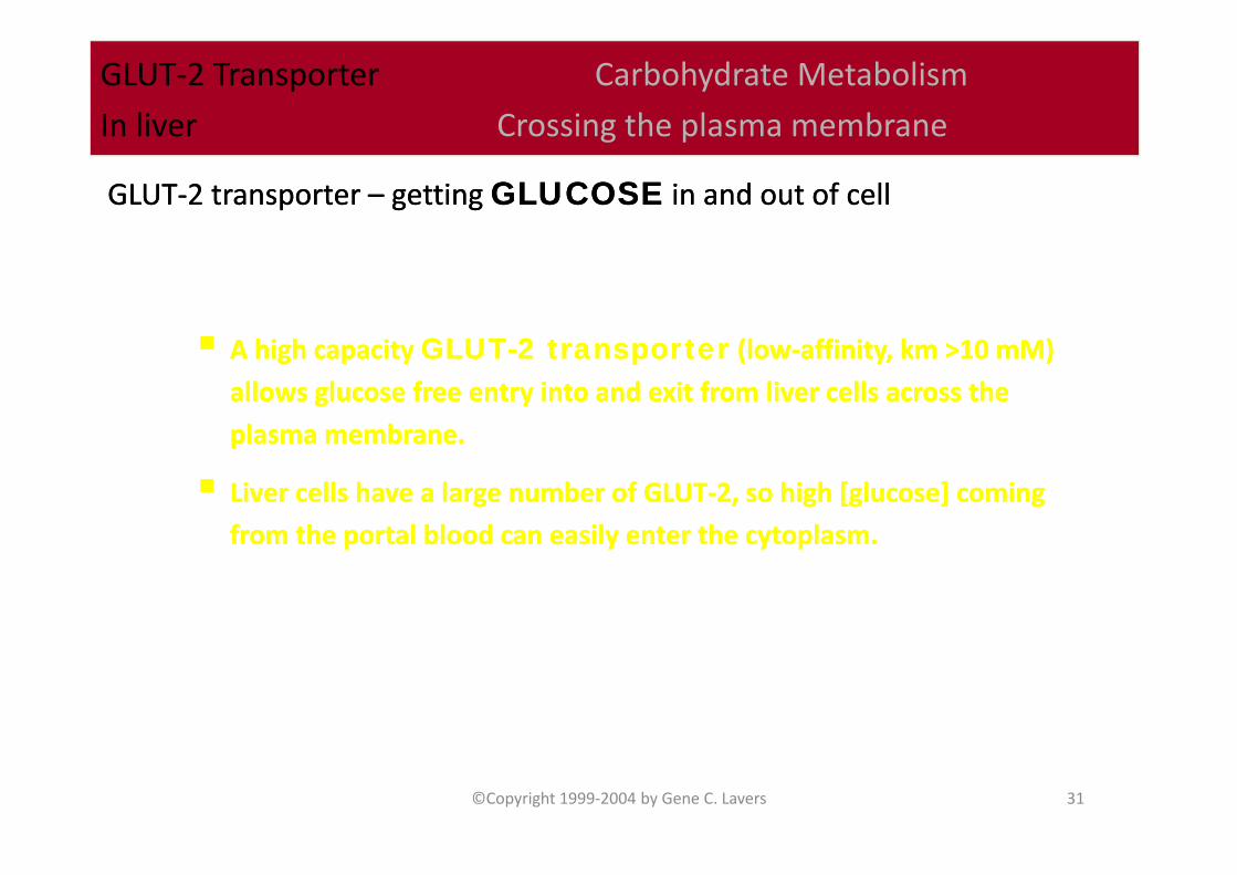
GLUT‐2 Transporter Carbohydrate Metabolism
In liver Crossing the plasma membrane
A high capacity GLUT-2 transporter (low‐affinity, km >10 mM)
allows glucose free entry into and exit from liver cells across the
plasma membrane.
Liver cells have a large number of GLUT‐2, so high [glucose] coming
from the portal blood can easily enter the cytoplasm.
A high capacity GLUT-2 transporter (low‐affinity, km >10 mM)
allows glucose free entry into and exit from liver cells across the
plasma membrane.
Liver cells have a large number of GLUT‐2, so high [glucose] coming
from the portal blood can easily enter the cytoplasm.
GLUT‐2 transporter – getting GLUCOSE in and out of cellGLUT‐2 transporter – getting GLUCOSE in and out of cell
32
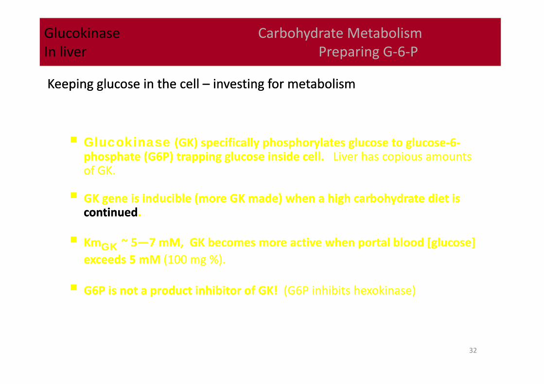
Glucokinase Carbohydrate MetabolismIn liver Preparing G‐6‐P
Glucokinase (GK) specifically phosphorylates glucose to glucose‐6‐phosphate (G6P) trapping glucose inside cell. Liver has copious amounts of GK.
GK gene is inducible (more GK made) when a high carbohydrate diet is continued.
KmGK ~ 5—7 mM, GK becomes more active when portal blood [glucose] exceeds 5 mM (100 mg %).
G6P is not a product inhibitor of GK! (G6P inhibits hexokinase)
Keeping glucose in the cell – investing for metabolismKeeping glucose in the cell – investing for metabolism
33
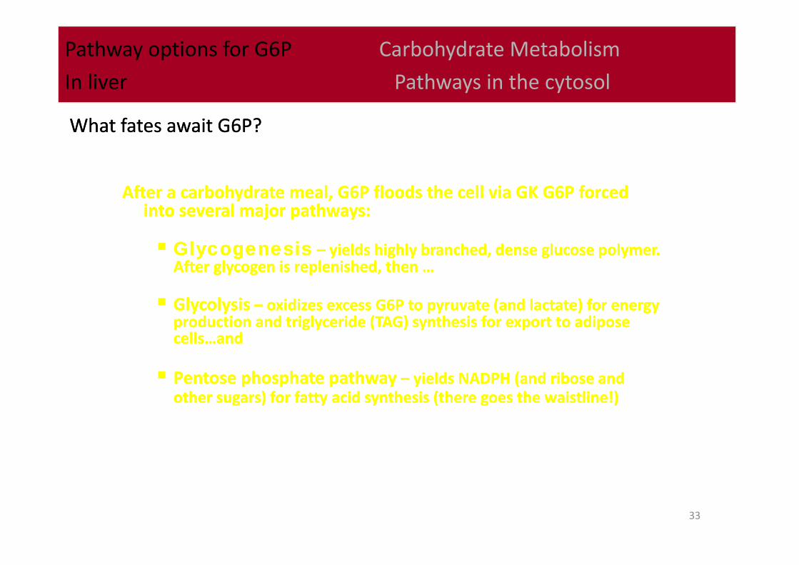
Pathway options for G6P Carbohydrate Metabolism
In liver Pathways in the cytosol
After a carbohydrate meal, G6P floods the cell via GK G6P forced into several major pathways:
Glycogenesis – yields highly branched, dense glucose polymer. After glycogen is replenished, then …
Glycolysis – oxidizes excess G6P to pyruvate (and lactate) for energy production and triglyceride (TAG) synthesis for export to adipose cells…and
Pentose phosphate pathway – yields NADPH (and ribose and other sugars) for fatty acid synthesis (there goes the waistline!)
What fates await G6P?What fates await G6P?
34
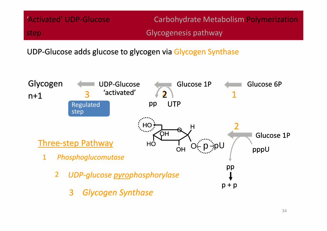
‘Activated’ UDP‐Glucose Carbohydrate Metabolism Polymerization
step Glycogenesis pathway
‘Activated’ UDP‐Glucose Carbohydrate Metabolism Polymerization
step Glycogenesis pathway
UDP‐Glucose adds glucose to glycogen via Glycogen SynthaseUDP‐Glucose adds glucose to glycogen via Glycogen Synthase
Glucose 6PGlucose 6PGlucose 1PGlucose 1PUDP‐GlucoseUDP‐GlucoseGlycogen n+1Glycogen n+1
UDP‐glucose pyrophosphorylaseUDP‐glucose pyrophosphorylase
112233
11
22
33 Glycogen SynthaseGlycogen Synthase
PhosphoglucomutasePhosphoglucomutase
Three‐step PathwayThree‐step Pathway
UTPUTPpppp
HOHOOO
O– pOHOHHOHO
OHOHHH
–pU pppUpppU
pppp
22
22
Glucose 1PGlucose 1P
Regulatedstep
‘activated’‘activated’
p + pp + p
35
Glycogenin Carbohydrate Metabolism
Glycogenesis
Glycogenin Carbohydrate Metabolism
Glycogenesis
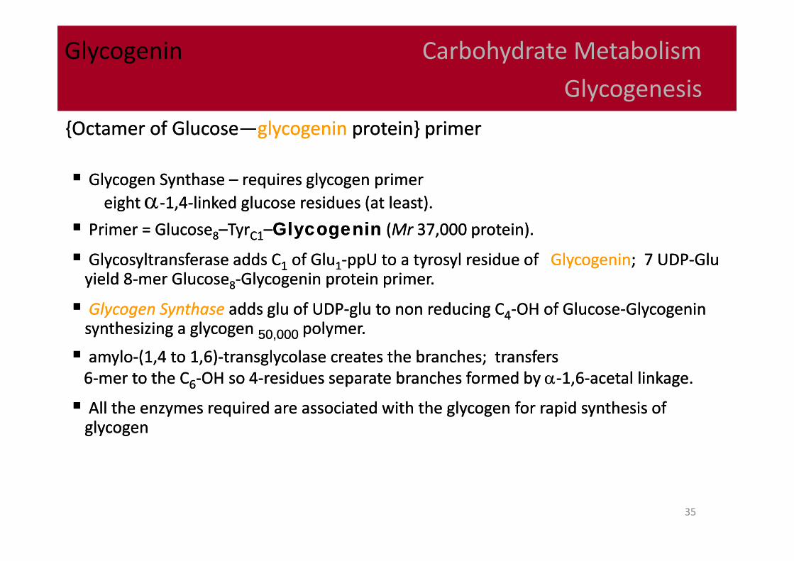
{Octamer of Glucose—glycogenin protein} primer {Octamer of Glucose—glycogenin protein} primer
Glycogen Synthase – requires glycogen primer eight ‐1,4‐linked glucose residues (at least).
Primer = Glucose8–TyrC1–Glycogenin (Mr 37,000 protein).
Glycosyltransferase adds C1 of Glu1‐ppU to a tyrosyl residue of Glycogenin; 7 UDP‐Glu yield 8‐mer Glucose8‐Glycogenin protein primer.
Glycogen Synthase adds glu of UDP‐glu to non reducing C4‐OH of Glucose‐Glycogenin synthesizing a glycogen 50,000 polymer.
amylo‐(1,4 to 1,6)‐transglycolase creates the branches; transfers 6‐mer to the C6‐OH so 4‐residues separate branches formed by ‐1,6‐acetal linkage.
All the enzymes required are associated with the glycogen for rapid synthesis of glycogen
36
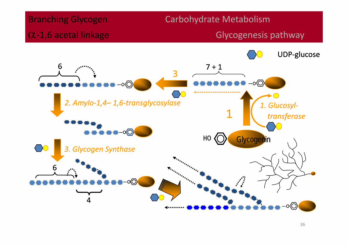
Branching Glycogen Carbohydrate Metabolism
‐1,6 acetal linkage Glycogenesis pathway
Branching Glycogen Carbohydrate Metabolism
‐1,6 acetal linkage Glycogenesis pathway
UDP‐glucoseUDP‐glucose
GlycogeninGlycogeninHOHO
1. Glucosyl‐transferase
1. Glucosyl‐transferase
2. Amylo‐1,4– 1,6‐transglycosylase2. Amylo‐1,4– 1,6‐transglycosylase
3. Glycogen Synthase3. Glycogen Synthase
—O—O
11
7 + 17 + 1
—O—O
66
44
—O—O
—O—O
66
—O—O
33
37
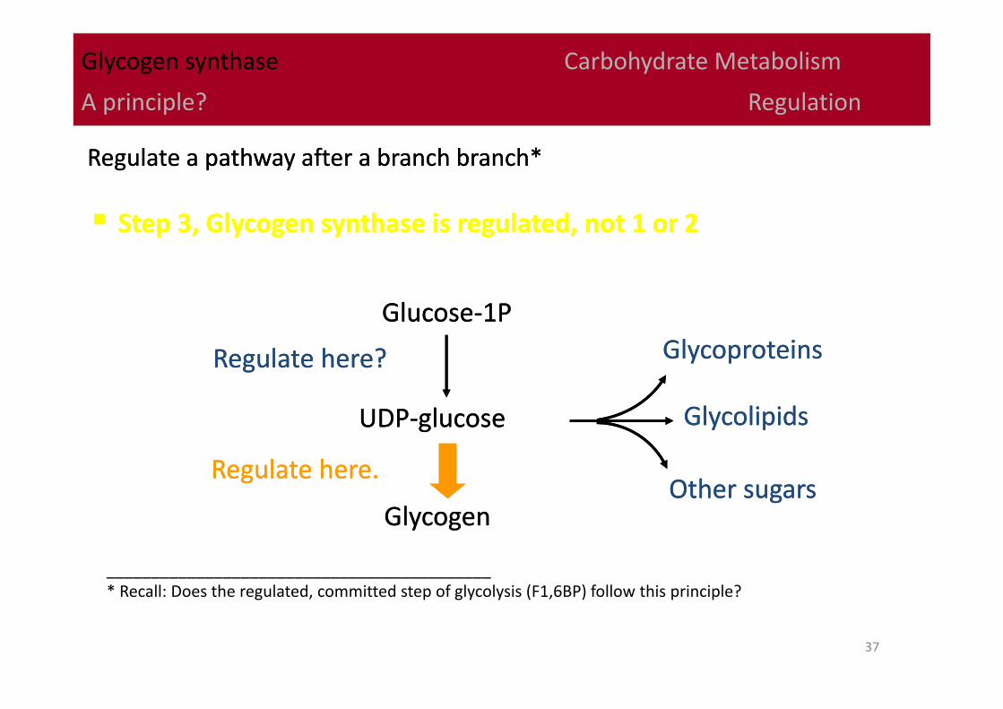
Glycogen synthase Carbohydrate Metabolism
A principle? Regulation
Step 3, Glycogen synthase is regulated, not 1 or 2 Step 3, Glycogen synthase is regulated, not 1 or 2
Regulate a pathway after a branch branch*Regulate a pathway after a branch branch*
UDP‐glucoseUDP‐glucose
GlycoproteinsGlycoproteins
GlycolipidsGlycolipids
Other sugarsOther sugarsGlycogenGlycogen
Glucose‐1PGlucose‐1P
Regulate here?Regulate here?
Regulate here.Regulate here.
___________________________________________* Recall: Does the regulated, committed step of glycolysis (F1,6BP) follow this principle?

38
Polysaccharide Phosphorylases Carbohydrate Metabolism
In Liver Glycogenolysis
Polysaccharide Phosphorylases Carbohydrate Metabolism
In Liver Glycogenolysis
Using phosphate to cleave C—O bonds: phosphorolysisUsing phosphate to cleave C—O bonds: phosphorolysis
* Glucose stored as glycogen or starchanimals plants
* Glucose stored as glycogen or starchanimals plants
GlycogenGlycogen
MuscleMuscle
LiverLiver
PP OOOO
OO
OO
HOHOOO
OPOPOHOHHOHO
OHOHHH HOHO
OO
OR-1OR-1OHOH
HOHOOHOH
HH
+
Glucose 1PGlucose 1P Glycogenn‐1Glycogenn‐1
PP
++
4‐residues from branch
~~ribosomeribosome
Non‐reducing endNon‐reducing end
PP
PP
Branch point
39
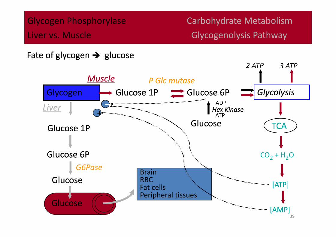
Brain RBC Fat cellsPeripheral tissues
Glycogen Phosphorylase Carbohydrate Metabolism
Liver vs. Muscle Glycogenolysis Pathway
Glycogen Phosphorylase Carbohydrate Metabolism
Liver vs. Muscle Glycogenolysis Pathway
Glycogen Glucose 1PGlucose 1P
[ATP][ATP]
––
MuscleMuscle
LiverLiver
Glucose 6PGlucose 6P GlycolysisGlycolysis
TCATCAGlucose 1PGlucose 1P
Glucose 6PGlucose 6P
GlucoseGlucose
GlucoseGlucose
Fate of glycogen glucoseFate of glycogen glucose
++
CO2 + H2O
[AMP][AMP]
3 ATP
GlucoseGlucose
Hex KinaseHex Kinase
2 ATP
P Glc mutaseP Glc mutase
G6Pase
ATP
ADP