and harry a. dailey...b1476 handbook of porphyrin science 2nd reading porphyrin and heme trafficking...
TRANSCRIPT

b1476 Handbook of Porphyrin Science 2nd Reading
Porphyrin and Heme Traffickingin Metazoans
Iqbal Hamza*,‡ and Harry A. Dailey†
*Department of Animal & Avian Sciences and Department of Cell Biology & Molecular Genetics,University of Maryland, College Park, Maryland, MD 20742, USA†Biomedical and Health Sciences Institute,Department of Microbiology and the Department of Biochemistry andMolecular Biology, University of Georgia, Athens, GA 30602, USA‡University of Maryland, College Park, 2413 ANSC, Bldg 142,College Park, Maryland, MD 20742, USA
List of Abbreviations 000I. Introduction 000II. Heme Biosynthesis 000
A. Synthesis of 5-Aminolevulinate 000B. Synthesis of the Monopyrrole, Porphobilinogen 000C. Assembly of the Linear Tetrapyrrole 000D. Cyclization of the Tetrapyrrole to Form Uroporphyrinogen 000E. Decarboxylation of Uroporphyrinogen 000F. Formation of the Vinyl Groups of Protoporphyrinogen IX 000
G. Oxidation of the Porphyrinogen to Protoporphyrin IX 000H. Insertion of Iron to Form Protoheme IX 000I. Possible Role of Supramolecular Protein Assemblies
in Heme Synthesis 000III. Regulation of Metazoan Heme Biosynthesis 000IV. Heme Transport and Trafficking 000
A. Heme Import in Animals 000B. Heme Export in Animals 000C. Intracellular Heme Transport 000
1
00
b1476_Hamza-Dailey.qxd 5/10/2013 6:55 AM Page 1

b1476 Handbook of Porphyrin Science
D. Contradictions 000E. Heme Transfer between Organelles 000F. Heme Reductases 000
V. Cytoplasmic Heme-binding Proteins 000VI. Extracellular Heme-binding Proteins 000
VII. Intercellular Heme Transport 000VIII. Parasitic Worms and Heme 000
IX. Heme Transport in Insects 000X. Heme Transport in Yeast 000
XI. Conclusions and Future Directions 000XII. Acknowledgements 000
XIII. References 000
List of Abbreviations
ABC ATP-binding cassetteAIP acute intermittent porphyriaALA 5-aminolevulinic acidALAS ALA synthaseALAS-1 housekeeping ALASALAS-2 erythroid ALASBH3 Bcl-2 homology 3CP carrier proteinCPOX coproporphyrinogen oxidaseDcytb duodenal cytochrome bDLP dynamin-like proteineNOS endothelial NOSEPP erythropoietic protoporphyriaER endoplasmic reticulumERMES ER-mitochondria encounter structureFABP fatty acid-binding proteinFECH ferrochelataseFLVCR feline leukemia virus subclass-C receptorGAPDH glyceraldehyde phosphate dehydrogenaseGPI glycosylphosphatidylinositolGST glutathione S-transferaseHb hemoglobinHCP1 heme carrier proteinHeLp heme lipoprotein
2nd Reading
2 Hamza and Dailey
b1476_Hamza-Dailey.qxd 5/10/2013 6:55 AM Page 2

b1476 Handbook of Porphyrin Science
HMB hydroxymethylbilaneHMBS hydroxymethylbilane synthaseHO heme oxygenaseHRG heme-regulated geneHRM heme regulatory motifHAS human serum albuminIM inner membraneIMS intermembrane spaceiNOS inducible NOSIRE iron regulatory elementIRP iron regulatory proteinMAPL mitochondria-anchored protein ligaseMARE Maf recognition elementMDV mitochondrial-derived vesicleMEL murine erythroleukemiamRNA messenger ribonucleic acidMTF mitoferrinNMR nuclear magnetic resonancenNOS neuronal NOSNO nitric oxideNOS nitric oxide synthaseOM outer membraneOPA1 optic atrophyPBG porphobilinogen PBGD PBG deaminasePBGS porphobilinogen synthasePf-GST P. falciparum GSTPPIX protoporphyrin IXPPOX protoporphyrinogen oxidasePUG1 protoporphyrin uptake gene 1RBC red blood cellSLC mitochondrial solute transporterSteap3 six transmembrane epithelial antigen of the prostate 3SUCLG1 succinyl CoA synthetaseUROD uroporphyrinogen decarboxylaseUROS uroporphyrinogen III synthaseV-ATPase vacuolar proton ATPaseXLPP X-linked protoporphyria
2nd Reading
00/Porphyrin and Heme Trafficking in Metazoans 3
b1476_Hamza-Dailey.qxd 5/10/2013 6:55 AM Page 3

b1476 Handbook of Porphyrin Science
I. Introduction
Heme, as a cofactor in a variety of proteins, is widely acknowledged to be essen-tial for gas transport, respiration, xenobiotic detoxification, peroxide productionand destruction, fatty acid desaturation, prostaglandin synthesis, and a variety ofone-electron transfer reactions.4,5 Over the past decade, the number of roles iden-tified for heme has grown substantially. In addition to heme’s role as a cofactor, itis now also recognized as a regulatory ligand. Among the list of biologicalprocesses for which higher eukaryotic heme-binding proteins have now beenimplicated are regulation of circadian rhythm,6,7 adipogenesis,8,9 glucose homeo-stasis,8–10 microRNA processing,11 intracellular proteolysis12 and regulation ofexocrine peptidases,13 gas sensing,6,14 control of ion channels,15 tRNA charging,16
and intracellular and intercellular signal transduction.17,18
Given the number of regulatory networks modulated by heme, it is not sur-prising that in individuals with significant impairment in the ability to synthesizeheme, one finds developmental abnormalities and chronic disease manifesta-tions.19 Even in single-allele mutation acute porphyrias, it could be imagined thatsome symptoms of the neurological attack may be attributable to dysregulation ofheme-requiring regulatory network systems resulting from altered cellular hemelevels. Interestingly, dysfunctional circadian rhythm, whose regulation involvesheme-binding transcription factors (see Ref. 12), has been suggested to be associ-ated with psychiatric disorders such as bipolar disorder and schizophrenia,20,21 andhuman diseases such as breast cancer,22 non-Hodgkin’s lymphoma,23 metabolicsyndrome,24 and atherosclerosis.25 It has even been suggested that heme deficiencymay be a factor in the mitochondrial and neuronal decay of aging.26
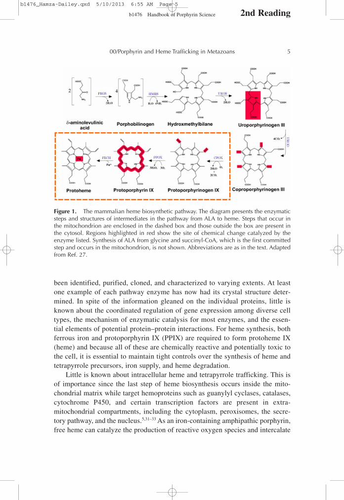
The heme biosynthetic pathway is ancient and, with a few notable excep-tions, is highly conserved. The pathway as found in metazoans is depicted inFigure 1. It differs from that found in plants and most bacteria in that the firstcommitted intermediate, 5-aminolevulinic acid (ALA) is synthesized by ALAsynthase (ALAS) using succinyl-CoA and glycine as substrates.27 In plants,Archaea and most prokaryotes, ALA is formed from glutamyl-tRNA by twoenzymes, glutamyl-tRNA reductase and glutamate-1-semialdehyde aminotrans-ferase.28 For organisms that possess hemoproteins, the inability to synthesize orobtain heme is ultimately lethal. Few organisms do not synthesize heme andamong eukaryotes, Caenorhabditis elegans and other Helminths29 and the cattletick Boophilus microplus30 are the only well-characterized heme-requiringorganisms. The observation that these organisms possess hemoproteins in allcells, but yet can only acquire heme from their diet demonstrates that a robustintercellular and intracellular heme trafficking network must exist. Over the past50 years, all of the enzymes of the eukaryotic heme biosynthetic pathway have
2nd Reading
4 Hamza and Dailey
b1476_Hamza-Dailey.qxd 5/10/2013 6:55 AM Page 4
b1476 Handbook of Porphyrin Science
been identified, purified, cloned, and characterized to varying extents. At leastone example of each pathway enzyme has now had its crystal structure deter-mined. In spite of the information gleaned on the individual proteins, little isknown about the coordinated regulation of gene expression among diverse celltypes, the mechanism of enzymatic catalysis for most enzymes, and the essen-tial elements of potential protein–protein interactions. For heme synthesis, bothferrous iron and protoporphyrin IX (PPIX) are required to form protoheme IX(heme) and because all of these are chemically reactive and potentially toxic tothe cell, it is essential to maintain tight controls over the synthesis of heme andtetrapyrrole precursors, iron supply, and heme degradation.
Little is known about intracellular heme and tetrapyrrole trafficking. This isof importance since the last step of heme biosynthesis occurs inside the mito-chondrial matrix while target hemoproteins such as guanylyl cyclases, catalases,cytochrome P450, and certain transcription factors are present in extra-mitochondrial compartments, including the cytoplasm, peroxisomes, the secre-tory pathway, and the nucleus.5,31–33 As an iron-containing amphipathic porphyrin,free heme can catalyze the production of reactive oxygen species and intercalate
2nd Reading
00/Porphyrin and Heme Trafficking in Metazoans 5
Figure 1. The mammalian heme biosynthetic pathway. The diagram presents the enzymaticsteps and structures of intermediates in the pathway from ALA to heme. Steps that occur inthe mitochondrion are enclosed in the dashed box and those outside the box are present inthe cytosol. Regions highlighted in red show the site of chemical change catalyzed by theenzyme listed. Synthesis of ALA from glycine and succinyl-CoA, which is the first committedstep and occurs in the mitochondrion, is not shown. Abbreviations are as in the text. Adaptedfrom Ref. 27.
b1476_Hamza-Dailey.qxd 5/10/2013 6:55 AM Page 5

b1476 Handbook of Porphyrin Science
into lipid bilayers and is, thus, unlikely to diffuse freely within the cell.34,35
Instead, specific molecules and pathways must exist to facilitate heme delivery todistinct cellular destinations. These issues are of significance since dysfunctionalsubstrate/product handling may lead to ineffective heme synthesis, hemoproteinassembly, and/or toxicity to the cell.
II. Heme Biosynthesis
All of the enzymes of heme biosynthesis are nuclear encoded and cytoplasmicallysynthesized. The overall reaction to synthesize heme in metazoan cells is:8 glycine + 8 succinyl CoA + 5 O2 + Fe2+ → Heme + 8 CoA + 4 NH4 + 14 CO2 +3 H2O2 + 2 H+ + 11 H2O. Of the products, 8 CoA, 8 CO2, 2 H+, and heme are gene-rated in the mitochondrial matrix and 2 CO2 + 3 H2O2 + and 2 H2O are generatedin the mitochondrial inner membrane (IM) space. All remaining products arecytosolic.
A. Synthesis of 5-Aminolevulinate
The first committed step in heme biosynthesis is the formation of 5-aminolevulinate(ALA).3,36–38 In the metazoa, the enzyme ALAS (E.C. 2.3.1.37), which is locatedin the mitochondrial matrix, catalyzes the condensation of glycine with succinylCoA to form ALA and CO2. ALAS is a homodimeric, pyridoxal phosphate-containing enzyme that is a member of the large and well-characterized α-oxoaminesynthase family. At present, only the crystal structure of ALAS from the bacteriumRhodobacter capsulatus has been determined,39 but it is clear that the matureeukaryotic ALAS is highly similar to the bacterial enzyme except that it possessesan additional carboxyl-terminal sequence of approximately 25 residues. Much isknown about the mammalian enzyme from kinetic and site-directed mutagenesisstudies.37 In vertebrates, two isozymes of ALAS exist, one specific fordifferentiating erythroid cells (ALAS-2 or ALAS-E) and the other expressed in allother cell types (ALAS-1 or ALAS-N). The genes for ALAS-1 and -2, as for allheme biosynthetic enzymes, are nuclear and synthesized in the cytosol, althoughthe final destination for both ALAS-1 and -2 is the mitochondrion. Both ALAS-1and -2 possess mitochondrial targeting sequences that are cleaved as part of thetranslocation of ALAS into the mitochondrial matrix.40–42
B. Synthesis of the Monopyrrole, Porphobilinogen
ALA is exported out of the mitochondrial matrix (see below) to reach the secondpathway enzyme, porphobilinogen synthase (PBGS) (E.C. 4.2.1.24) (previously
2nd Reading
6 Hamza and Dailey
b1476_Hamza-Dailey.qxd 5/10/2013 6:55 AM Page 6

b1476 Handbook of Porphyrin Science
named ALA dehydratase).43 This enzyme catalyzes the condensation of two mole-cules of ALA to form one molecule of the monopyrrole porphobilinogen (PBG).The cytosolic homo-octomer can best be described as a tetramer of homodimerswith one divalent metal atom per subunit.36,43 In humans and yeast, this metal iszinc, while in bacteria, one may also find magnesium. Four metal atoms are essen-tial for catalysis and four are involved in stabilization of tertiary structure. Thesezinc ions may be replaced by lead in lead poisoning resulting in an inactiveenzyme. The protein has been crystallized from multiple sources and kineticallycharacterized (see Refs. 38 and 44). Interestingly, PBGS exists in alternate qua-ternary structures named morpheeins.45 One morpheein is the well-characterizedoctomer which possesses high activity and another is a hexamer that has loweractivity. While this change in quaternary structure is proposed to be the basis ofallosteric regulation of PBGS, it is something of an enigma, given that ALAS isconsidered rate-limiting to heme synthesis.
C. Assembly of the Linear Tetrapyrrole
Four molecules of PBG are linked head to tail by the cytosolic enzyme hydroxy-methylbilane synthase (HMBS, previously called PBG deaminase or PBGD)(E.C. 2.5.1.61). The reaction results in the formation of the linear tetrapyrrolehydroxymethylbilane (HMB) and releases four molecules of ammonium.43 Partlybecause a decrease in HMBS activity leads to the human disease acute intermit-tent porphyria (AIP),46,47 this enzyme became the early focus of much attention byresearchers. HMBS has been purified from a variety of sources and the crystalstructures of the Escherichia coli48 and human49 enzymes have been determined.The enzyme is a monomer that is synthesized as an apoprotein. In its first com-plete catalytic cycle, it synthesizes a covalently bound hexameric linear polypyr-role (E-S6). From this, the distal linear tetrapyrrole HMB is cleaved resulting information of the holoenzyme HMBS with a covalently bound dipyrromethanewhich serves as a cofactor for future turnovers and the product HMB.
D. Cyclization of the Tetrapyrrole to Form Uroporphyrinogen
Conversion of HMB to the physiological uroporphyrinogen III isomer requires theaction of uroporphyrinogen III synthase (UROS) (E.C. 4.2.1.75).43 HMB is chemi-cally reactive and will spontaneously cyclize to form uroporphyrinogen I in theabsence of the next pathway enzyme. As uroporphyrinogen I cannot be convertedinto PPIX, it is essential to produce the III isomer. The reaction catalyzed byUROS is the “flipping” or inversion of the final, or D, ring of HMB followed bycyclization to yield the III isomer of uroporphyrinogen. UROS is a monomeric
2nd Reading
00/Porphyrin and Heme Trafficking in Metazoans 7
b1476_Hamza-Dailey.qxd 5/10/2013 6:55 AM Page 7

b1476 Handbook of Porphyrin Science
protein whose structure has been determined for both human50 and Thermus ther-mophilus.51 It does not possess a cofactor and has an unusual tertiary structure thatis composed of two distinct domains connected by a flexible linker region. Crystalstructures have been obtained for a variety of domain orientations suggesting thatthe molecule is highly flexible in solution.
E. Decarboxylation of Uroporphyrinogen
The final cytosolic enzyme in the pathway is uroporphyrinogen decarboxylase(UROD) (E.C. 4.1.1.37).28 This homodimeric enzyme has been crystallized52 andits catalytic mechanism well studied.53 Each subunit contains an independentactive site and the enzyme contains no cofactors or metal ions. UROD catalyzesthe decarboxylation of the four pyrrole acetic acid side chains of uroporphyrino-gen to yield the four methyl pyrrole side chains of coproporphyrinogen and fourmolecules of CO2. UROD will utilize both uroporphyrinogen I and III, and in thepresence of high concentrations of substrate, UROD will decarboxylaterandomly.43 However, it is believed that in situ the reaction starts with the decar-boxylation of the D ring acetate and proceeds sequentially in a clockwise fashion(i.e., D, A, B, C).52,53
F. Formation of the Vinyl Groups of Protoporphyrinogen IX
Coproporphyrinogen III produced by UROD is transported into the mitochondrialintermembrane space (IMS) where the antepenultimate enzyme coproporphyrino-gen oxidase (CPOX) (E.C. 1.3.3.3) is located.54,55 This protein is synthesized in thecytosol with an unusually long (~120 amino acids) mitochondrial leader sequencethat targets it to the IM space.40 The mature protein is a homodimer without boundcofactor. The structures of both human56 and yeast (which is a cytosolic protein)57
CPOX have been solved and were found to possess unique folds. The reaction cat-alyzed is an unusual oxidative decarboxylation of the A and B ring propionates toyield the vinyl groups of protoporphyrinogen IX. CPOX will utilize only thecoproporphyrinogen III isomer and proceeds in a stepwise fashion that requirestwo molecules of molecular oxygen and generates two molecules of CO2. Thereaction catalyzed by CPOX has been extensively studied both experimentally andin silico, but at present no definitive mechanism has been identified.58,59
G. Oxidation of the Porphyrinogen to Protoporphyrin IX
The penultimate step is the oxidation of protoporphyrinogen IX to PPIX. This iscatalyzed by protoporphyrinogen oxidase (PPOX) (E.C. 1.3.3.4) which requires
2nd Reading
8 Hamza and Dailey
b1476_Hamza-Dailey.qxd 5/10/2013 6:55 AM Page 8

b1476 Handbook of Porphyrin Science
three molecules of molecular oxygen and generates three molecules of hydrogenperoxide.54,55 Crystal structures of the plant,1 bacterial,60,61 and human62 enzymeshave been determined and show that the protein is a homodimer with one non-covalently bound FAD per subunit. PPOX is synthesized in the cytosol in itsmature size and is translocated to the outer surface of the inner mitochondrialmembrane via a mechanism that requires an internal mitochondrial targetingsequence.40,63,64 Interestingly, the active site is proposed to be situated in the mid-dle of a tunnel that passes through the protein.
H. Insertion of Iron to Form Protoheme IX
The terminal step of heme synthesis is the insertion of ferrous iron into the proto-porphyrin macrocycle to produce protoheme IX (heme). This is catalyzed by theenzyme ferrochelatase (FECH) (E.C. 4.99.1.1).65 In metazoans, this enzyme issynthesized in the cytosol as a preprotein and is translocated to the mitochondrialmatrix where it is associated with the inner mitochondrial membrane. The mature,processed protein is a homodimer with each subunit possessing a [2Fe-2S]cluster.66 Similar [2Fe-2S] clusters are not found in plant FECHs but are found inthe actinobacterial enzymes and some Gram-negative bacterial FECHs.65,67 Thereis no evidence to suggest that the cluster participates directly in catalysis, but inits absence enzyme activity is significantly reduced leading to speculation that itserves a role as a de facto iron sensor in vivo. Shah et al. have presented evidencesuggesting that the cluster itself plays a regulatory role during catalysis in vivo.68
The crystal structures of human FECH with and without substrate and producthave been determined and it has been shown that the molecule undergoes conside-rable active site remodeling during its catalytic cycle.3,69
I. Possible Role of Supramolecular Protein Assemblies in Heme Synthesis
Of significance to the current review is the intracellular distribution of heme syn-thesis pathway enzymes and the manner in which pathway intermediates aremoved between enzymes. The chemical intermediates in tetrapyrrole synthesis arerelatively reactive and can be cytotoxic. The physiological proof of this statementis amply demonstrated by the fact that a deficiency in any one of the pathwayenzymes results in a clinically distinct disease in animals,47,70,71 and inhibition ofsome later steps can result in photo-induced death of plants or microorganisms.Other than a defect in ALAS-2 which results in X-linked sideroblastic anemia, theclinical manifestations of these disorders, named porphyrias, are generally notanemia but result from the accumulation of pathway intermediates upstream from
2nd Reading
00/Porphyrin and Heme Trafficking in Metazoans 9
b1476_Hamza-Dailey.qxd 5/10/2013 6:55 AM Page 9

b1476 Handbook of Porphyrin Science
the genetic block. These diseases and their molecular basis have been well stud-ied and frequently reviewed by others (see Refs. 46, 47, 70, and 71).
Given the reactivity of the pathway intermediates, it is highly unlikely that theintracellular concentrations of substrate or products attain the µM concentrationsof the measured enzyme Kms. Thus, it is reasonable to assume that “free interme-diates” do not exist in the cell although various porphyrin intermediates are rou-tinely found in the extracellular medium of mammals. This suggested that thepathway was somehow compartmentalized so as to allow direct transfer of product/substrate between enzymes. This topic was first approached experimentally in the1980s when data were presented in support of the hypothesis that the terminalthree pathway enzymes, which are all mitochondrial membrane-associated, format least a transient complex to facilitate the transfer of intermediates.55,72,73
Biochemical data clearly demonstrated that while obligate, tight substrate chan-neling such as occurs in tryptophan synthesis does not exist, under normalcircumstances equilibration of products/substrates with the bulk medium does notoccur. Data gleaned from PPOX and FECH crystal structures provide goodsupport for possible interactions.54,69
Other than data for the terminal, membrane-associated enzymes, there existslittle experimental evidence to support multienzyme complexes of earlier pathwayenzymes. For the first step, an interaction of ALAS-2 with succinyl-CoA syn-thetase (SUCLG1) on the inner mitochondrial membrane has been demonstratedby two groups.74,75 With the identification of mitochondrial solute transporter-25A38 (SLC25A38) as the putative glycine/ALA transporter,76 it is reasonable toanticipate the existence of at least transient complexes among ALAS, SUCLG1,and SLC25A38 on the mitochondrial IM (Figure 2). The possibility for a stable,
2nd Reading
10 Hamza and Dailey
Figure 2. Proposed model for components involved in heme synthesis. Details are discussedin the text and where specific data exist for a particular component, its name is shown.Components that are currently unidentified experimentally are denoted with “?”.Abbreviations and conventions are given in the text. Adapted from Ref. 27.
b1476_Hamza-Dailey.qxd 5/10/2013 6:55 AM Page 10

b1476 Handbook of Porphyrin Science
rigid complex for these and most other heme synthesis enzymes seems unlikelygiven that they possess active site pockets with a single entrance/exit (see Ref. 38).The necessity for substrate(s) and product(s) to enter and exit via a single routewould require movement of one enzyme between donor and acceptor moleculesin the complex, much as cytochrome c physically cycles between electron donorsand acceptors. The only enzyme for which this may not be the case is PPOX whichappears to have a channel through the protein with the active site located withinthis feature.1,60,62
For the synthesis of uroporphyrinogen III from PBG, it has long been thoughtthat close proximity of HMBS and UROS would be probable, given the chemicalreactivity of the linear tetrapyrrolic intermediate, HMB. However, no publisheddata exist to support the presence of a multienzyme complex involving PBGS andHMBS. The possibility that a HMB-binding chaperone exists to move HMB fromHMBS to UROS, while possible, also has no supporting data either in vitro orin vivo. Since ALA must be exported out and coproporphyrinogen imported intothe mitochondria, it is reasonable to anticipate that PBGS, HMBS, UROS, andUROD exist in a supramolecular complex that is spatially close to the mitochon-drion (Figure 2). To date, high-resolution microscopic studies identifying theintracellular distribution of these enzymes is lacking. Approaches that rely uponcell disruption and physical isolation of complexes or in vitro reconstitution ofcomplexes from isolated components requires the strength of protein–proteininteraction in solution that may not exist and may not be necessary in the highlyconcentrated cytosolic milieu. One intriguing possibility is that PBGS, whichexists as either an octomer or hexamer, could, because of its size, serve as a scaf-fold for a multienzyme complex. The observation that PBGS can assume multiplemorpheein forms38,45 may hint at its plastic role as a supramolecular assemblynucleation site. Resolution of these issues will require masterful research, but theresults will be illuminating.
The presence and nature of multienzyme complexes involving the terminalthree membrane-associated enzymes is considerably advanced over what isknown, or not known, about the earlier pathway enzymes but is still extremelyrudimentary. Little is known about possible interactions between CPOX andPPOX other than substrate channeling experiments.73 Radiolabeling experimentsemploying isolated mitochondrial fragments clearly support transient interactionsin situ, although reconstitution of this process with purified components was notaccomplished, which suggests that additional participants other than just CPOX,PPOX, and FECH are required. In silico structural studies show that an interac-tion across the inner mitochondrial membrane to transport protoporphyrin fromPPOX to FECH is feasible and highly likely1 (Figure 3). Interestingly, in thedocked PPOX–FECH complex, the opening of the active site tunnels of the
2nd Reading
00/Porphyrin and Heme Trafficking in Metazoans 11
b1476_Hamza-Dailey.qxd 5/10/2013 6:55 AM Page 11

b1476 Handbook of Porphyrin Science
PPOX dimer not only coincided with the position of the openings to the activesite pockets of the dimeric FECH but is also spatially juxtaposed to surface-bound porphyrin molecules observed in some FECH crystal structures. Theobservation that binding of substrate and product induces changes in the surfacecontour and charge distribution around the active site pocket opening of FECH3,69
provide an explanation for how PPOX and heme-accepting proteins recognize theappropriate form of FECH with which to interact. Given the need to acquirecoproporphyrinogen from the cytosol and movement of at least some heme outof the mitochondrion, the existence of a complex of an outer membrane (OM)coproporphyrinogen transporter, CPOX, PPOX, the IM iron transporter mitofer-rin (MTF1),77 FECH, and a heme chaperone/OM heme transporter at a mitofilin-mediated junction between outer and inner mitochondrial membranes64 is anintriguing possibility.
2nd Reading
12 Hamza and Dailey
Figure 3. Cartoon of proposed interaction between PPOX and FECH across the inner mito-chondrial membrane. The initial PPOX–FECH docking model was published by Koch et al.1
for plant PPOX and human FECH. The current model was derived from PDB files, 3NKS(PPOX) and 2QD1 (FECH). FECH is colored green and PPOX is colored violet. Porphyrinatoms are depicted as red colored solid spheres and the PPOX inhibitor is shown in solidblack. Phospholipids are presented as sticks that are colored wheat. Of note is that the crys-tal structures of FECH with bound porphyrin possess one protoporphyrin bound in the activesite and a second one associated with the outer edge of the active site lip. This is proposed tobe the entry route of porphyrin into the active site pocket.2,3 It is proposed that FECH andPPOX interact transiently across the membrane to facilitate transfer of protoporphyrin toFECH. Following this transfer FECH undergoes spatial alterations that are proposed to lowerthe affinity between FECH and PPOX so that their interaction ceases, thereby, allowing FECHto interact with MTF1 and later a heme-accepting protein. Adapted from Ref. 27.
b1476_Hamza-Dailey.qxd 5/10/2013 6:55 AM Page 12

b1476 Handbook of Porphyrin Science
Three significant issues remain unanswered: (i) transport of coproporphyrino-gen into the mitochondrion, (ii) mitochondrial supply of iron for heme synthesis,and (iii) transport of heme away from its site of synthesis. A series of studies pro-posed that ABCB6, a putative mitochondrial OM ATP-dependent transporter, isresponsible for coproporphyrinogen transport (see below).78,79 At present,however, this claim must be viewed with skepticism since only the transport of thefully conjugated, planar macrocycle coproporphyrin, not the physiologically rele-vant non-planar coproporphyrinogen, has ever been experimentally demonstrated.In addition, and as described below, ABCB6 has been localized to multiple mem-branes and identified as being involved in non-heme synthesis-related disorders.However, given that ABCB6 has been observed to be one of a relatively few genesinduced at high levels during erythropoiesis,80 it seems likely that it plays somerole in erythropoiesis. Given the size of coproporphyrinogen, the possibility thatit transits the OM via a porin rather than a specific transporter cannot be ruled outby current data.
Although the coordinate regulation of tetrapyrrole synthesis and iron metabo-lism must exist to prevent their inappropriate accumulation, there have been lim-ited studies that address this issue at either the genetic or molecular level. The roleof the iron-responsive element system62 and the cellular machinery for iron sulfurcluster assembly has garnered significant interest,81 and our knowledge of wholebody iron trafficking is relatively mature.82 However, there has been significantadvancement in identifying participants essential to the iron supply for heme syn-thesis. This came with the identification of MTF1 as the mitochondrial IM irontransporter that supplies iron to FECH for heme synthesis.77 MTF1 is responsiblefor iron transport for heme synthesis during erythropoiesis, but the presence ofanother IM transport protein, Abcb10, is required to stabilize MTF1.83 The exactrole that Abcb10 plays has yet to be elucidated. MTF1 has been found to form acomplex with FECH which might make possible a direct transfer of transportedferrous iron for heme synthesis to FECH,83 but the actual site of iron entry forFECH remains undefined experimentally. One model proposes that mammalianFECH acquires iron from MTF1 or frataxin via the extended π-helix of FECH84
and enters the active site via the main “mouth”.85,86 However, this model is basedupon the structure of a single monomer of FECH, not the physiologically signifi-cant homodimer, and the putative membrane binding surface of the model is,in fact, the dimer interface. The crystal structures of dimeric human FECH areconsistent with a spatial orientation where the π-helix unwinds into the membraneand not back up into the matrix. All available data suggest that the helix extensionis related to heme release and in the absence of product there is nothing to stabi-lize the extended helix. Additionally, the simultaneous exit of heme and entranceof iron via the same path is not supported by structural or kinetic studies.87,88
2nd Reading
00/Porphyrin and Heme Trafficking in Metazoans 13
b1476_Hamza-Dailey.qxd 5/10/2013 6:55 AM Page 13

b1476 Handbook of Porphyrin Science
Alternatively, structural and site-directed mutagenesis data obtained for humanFECH along with its orientation relative to the IM are all consistent with ironentering the active site of FECH via a solvent-filled channel whose entrance is onthe rear of the enzyme (Medlock et al., unpublished data). It will be of interest tolearn the molecular details of this process and how it is regulated.
Transit of heme away from its site of synthesis remains an unexplored terri-tory. In vitro, the release of heme from the enzyme post-metalation is the rate-limiting step.88 This probably reflects the absence of the native heme acceptor acell-free assay. Given that heme is utilized throughout the cell in a variety of com-partments, it is clear that multiple and specific heme chaperones may exist. Fortransit from the mitochondrion to other membranous compartments, it appearspossible that transfer occurs at direct contact points rather than by diffusionthrough the cytoplasm,31,89 but even that requires first moving heme from FECHto the site of transfer between membranes. As yet unknown is what molecule firstaccepts heme from FECH and whether there is a single unique protein that inter-acts with FECH or multiple pathway-specific proteins. Given its location on theIM, it would seem that for respiratory cytochrome maturation direct transfer fromFECH to cytochrome assembly machinery is possible, although definitive data arestill lacking. For cytoplasmic hemoproteins, including hemoglobin (Hb) and myo-globin, there are no identified players. Putative heme/porphyrin binding/carrierproteins (CPs) have been suggested and even biochemically characterized,90,91 butnone of these is supported by data as in vivo participants in heme trafficking (seeSections IV and V).
III. Regulation of Metazoan Heme Biosynthesis
As noted, five pathway proteins possess either organic cofactors or essential met-als. To date, no pathway enzymes has been shown to be glycosylated and onlymouse liver FECH has been reported to be phosphorylated,92 albeit on residuesthat appear to be on inaccessible sites within the protein. Interestingly these inves-tigators also reported that phosphorylated FECH is located on the mitochondrialOM, something never reported by any other group previously.
Regulation of the pathway is tissue- and cell-specific, subject to modulationby a myriad of developmental and environmental pressures through diverse tran-scriptional factors and is beyond the scope of the current review.3,71,93 In general,however, synthesis of the first committed intermediate, ALA, is considered rate-limiting. Among red blood cell (RBC)-producing metazoans, there are cleardistinctions between regulation of heme synthesis in non-erythroid cells (so-calledhousekeeping heme synthesis) and in differentiating erythroid precursor cells. Thehousekeeping ALAS-1 gene is regulated by diverse factors frequently associated
2nd Reading
14 Hamza and Dailey
b1476_Hamza-Dailey.qxd 5/10/2013 6:55 AM Page 14

b1476 Handbook of Porphyrin Science
with xenobiotic, hormone and drug metabolism and is subject to tissue-specificregulation.71,94 ALAS-1 has an estimated half-life of only a few hours and may besignificantly induced in some cells.95,96 ALAS-1 is present in erythroid precursorcells before they begin erythroid differentiation, but the gene is turned off whenALAS-2 is turned on as these cells begin to synthesize heme for Hb. Thus, ALAS-1cannot substitute for a deficiency of ALAS-2.97,98
ALAS-2 is transcriptionally regulated by erythroid-specific factors such asGATA-1 and possesses an iron regulatory element (IRE) located in the messengerribonucleic acid (mRNA) 5′ untranslated region that allows for translational regu-lation by cellular “free” iron.94,99,100 As with similarly regulated systems, such asfor ferritin synthesis, the IRE binds the iron-free form of the iron regulatory pro-tein (IRP), thereby, preventing translation of the ALAS-2 message when cellulariron concentrations are insufficient for optimal heme synthesis. Thus, maximalALAS-2 activity requires gene induction by erythroid-specific transcription fac-tors as well as sufficient cellular iron levels to support heme synthesis. Studies bySchranzhofer et al.101 with differentiating erythroblasts demonstrated that ALAS-2translational regulation becomes “uncoupled” from that of other IRE-regulatedproteins such as transferrin receptors (which are increased, not decreased duringerythropoiesis) and ferritin (whose protein levels do not increase). This is reason-able during the later accelerated hemoglobinization phase of erythropoiesis, sinceunder “standard” IRE-IRP regulation, elevated iron would diminish transferrinreceptor expression and increase ferritin synthesis, both of which would limit ironavailability for heme synthesis. Additionally, it was postulated that during maxi-mal hemoglobinization, ALAS-2 mRNA increases disproportionately to IRP syn-thesis, thus, circumventing the IRE-IRP regulatory mechanism. The “kiss and run”hypothesis,102 which proposes direct vesicle-mediated transfer of iron to mito-chondria, may offer another possible explanation to this. In this hypothesis, duringterminal erythropoiesis, vesicular iron acquired by receptor-mediated endocytosisis targeted directly to the mitochondrion, thereby, bypassing the cytosolic ironpool and the IRE-IRP system.
Multiple studies and reviews have presented data suggesting that pathwayenzymes after ALAS are in excess, although relative enzyme amounts vary con-siderably.4,47,71 However, these calculations are based on in vitro assays of indi-vidual enzymes under what are considered optimal conditions for each enzymeand then extrapolated back to intact cell conditions. There are no comprehensivestudies that examine the induction of biosynthetic pathway enzymes other thanALAS-1 in non-transformed non-erythroid cells, but the fact that all of the path-way genes possess diverse transcription factor-binding sites is compatible withregulation at multiple sites.90,97,103–110 Indeed, in cancer cells, it is clear that at leastCPOX and FECH are upregulated and downregulated, respectively, and this is the
2nd Reading
00/Porphyrin and Heme Trafficking in Metazoans 15
b1476_Hamza-Dailey.qxd 5/10/2013 6:55 AM Page 15

b1476 Handbook of Porphyrin Science
basis for the effectiveness of photodynamic therapy.111,112 It has been demonstratedthat there is upregulation of all pathway enzymes during erythroid differentiation(see Ref. 111). Even so, current dogma relegates pathway regulation during ery-thropoiesis to ALAS-2 alone.3,93 Re-evaluation of this model may be in order withthe discovery that some mutations in the carboxyl-terminus of ALAS-2 result in ahyperactive enzyme and this, in turn, causes an accumulation of free protopor-phyrin as is found in the disease erythropoietic protoporphyria (EPP),113 as well ashigh concentrations of zinc protoporphyrin. The name currently given to this dis-order is X-linked protoporphyria (XLPP) to reflect the X chromosome location ofALAS-2. This disorder is, thus, similar, but not identical, to that found whenALAS-2 is overproduced in Irp2−/− mice.114 The observation of protoporphyrinaccumulation in XLPP is particularly interesting since levels of FECH are gener-ally assumed to be present in considerable excess in the cell. Indeed, normal EPPoccurs only when FECH levels drop to approximately one-fourth of normal.70 Thissuggests that either the erythroid pathway is designed so that the amount of FECHand/or iron transport mechanisms for heme synthesis are closely linked to normal,maximal amounts of ALAS-2 activity or that additional regulatory steps or limit-ing mechanisms exist at the end of the pathway that we currently do not recognizeor understand.
For both ALAS-1 and ALAS-2, one finds alternative mRNA splice variants.For ALAS-1 two known splice variants occur in the untranslated region ofexon 1.115 These variants possess altered sensitivity to heme-mediated decay of themessage. For human ALAS-2, one splice variant has been described in whichexon 4 is absent.74 This variant represents 35–45% of total ALAS-2 mRNA in thecell and encodes a protein with slightly reduced activity. This variant ALAS-2 istranslocated into the mitochondrial matrix where it was shown to interact withSUCLG1, just as the full length enzyme does and contributes to erythroid hemesynthesis.
Interestingly, both ALAS-1 and -2 possess three heme regulatory motifs(HRMs) composed of a canonical Cys-Pro sequence.102 Two HRMs are found inthe targeting leader sequence and the third is near the amino terminus of themature processed ALAS.40 While such motifs have been shown to serve as a redoxsensitive switch for heme oxygenase (HO)-2,69 there is no evidence to suggestsuch a role for these motifs in ALAS. Indeed, evidence from multiple groups hasshown that heme binds to the ALAS HRMs in vitro41 and in vivo,40,42 and a seriesof mutagenesis experiments clearly demonstrated the significance of all threeALAS HRMs in the heme responsive translocation of the protein into mitochon-dria in vivo.40 While such heme regulation of apoprotein translocation into themitochondria of non-erythroid cells can easily be rationalized, a similar occur-rence in differentiating erythroid cells where massive quantities of heme are being
2nd Reading
16 Hamza and Dailey
b1476_Hamza-Dailey.qxd 5/10/2013 6:55 AM Page 16

b1476 Handbook of Porphyrin Science
synthesized in a relatively short period of time is less easily justified. Several sig-nificant pieces of information that would address this issue are lacking. Amongthese is an identification of the fate of both cytoplasmic ALAS precursor apo-protein and any heme bound to the HRMs. The possibility that the HRM-containingleader peptide or apoprotein HRM serves as a heme chaperone for globinassembly is intriguing, but no data exist to support or rule out this proposition.
While two distinct genes on separate chromosomes exist for ALAS-1 and -2,only single genes exist for the remaining pathway enzymes. However, for PBGS,HMBS, UROS, and FECH, housekeeping versus erythroid-specific promoterregions drive expression that leads to tissue-specific alternate mRNA splicing.Only the HMBS alternate splice variants, however, give rise to isoenzymes ofHMBS with alternative amino-terminal segments.104 Here, the mRNA of the ery-throid form of HMBS skips exon 1 and starts with the non-coding exon 2. Thetranslation start site employed for the erythroid HMBS is in exon 3. The house-keeping HMBS mRNA starts with exon 1, which contains an internal start site andcoding region and skips the non-coding exon 2. The result is that the housekeep-ing HMBS has an additional 17 amino acid residues at the amino terminus that arelacking in the erythroid form.
Human PBGS possesses two non-coding exons, 1A and 1B.105 The transla-tional start site for housekeeping PBGS mRNA is exon 1A, while erythroid PBGSmRNA starts in exon 1B. Thus, an additional 5′ untranslated region is present inthe mRNA for the housekeeping variant that is lacking in the erythroid splice vari-ant. Since these variants are in the non-coding region, the housekeeping and ery-throid forms of the PBGS enzyme are identical. Splice variants also exist forUROS.103
The gene for UROS possesses spatially distinct erythroid and housekeepingpromoters with the erythroid promoter elements located in the intron betweenexons 1 and 2. This promoter drives transcription from an initiation site in exon 2.The housekeeping promoter region is present upstream of exon 1 and drives tran-scription from an initiation site in exon 1. However, since exon 1 is non-coding,both housekeeping and erythroid UROS proteins are identical. For mouse FECH,there appears to be an alternate splice site that gives rise to additional 3′ untrans-lated nucleotides.116
While the existence of splice variants is not a new observation, any experi-mentally supported rationale for a physiological role for these variants is lacking.However, studies on the role of RNA in the spatial regulation of protein synthe-sis117 and/or in stabilization of multiprotein complexes118 raise the intriguing pos-sibility that the alternative splicing for mammalian housekeeping versus erythroidforms of PBGS, HMBS, and UROS are not random but may serve for sequestra-tion of mRNA primed for function.
2nd Reading
00/Porphyrin and Heme Trafficking in Metazoans 17
b1476_Hamza-Dailey.qxd 5/10/2013 6:55 AM Page 17

b1476 Handbook of Porphyrin Science
It has been proposed that during the course of normal erythropoiesis, hemesynthesis in developing erythroid cells overproduces heme to an extent that it istoxic to the cell unless it is exported by the plasma membrane heme transporterfeline leukemia virus subtype-C receptor (FLVCR) (see Section IV.D.).119 While itis clear that disabling mutations in FLVCR result in cell death, presumably due tothe toxic effects of excessive heme, it seems counterintuitive that evolution wouldhave designed such intricate regulatory mechanisms for erythroid heme synthesis(i.e., IRE-IRP, erythroid-specific transcription factors, and complex iron supplyregulatory schemes) that would permit excessive synthesis of heme necessitatingheme export. Indeed, given the “cost” of heme synthesis, it seems more likely thatthe “excessive” heme produced is a planned synthesis of heme by erythroid cells(which must be considered the ultimate heme synthesizing factories in the body)for orderly export and transit to other organs and cells whose heme synthesizingcapabilities may be physiologically limited. The presence of specific heme carri-ers, such as hemopexin,120 and the observation that exogenous administration ofheme to human porphyria patients downregulates heme synthesis, and is subse-quently utilized for hemoproteins by the patient receiving the infusion,46 providesupport for this hypothesis.
IV. Heme Transport and Trafficking
A. Heme Import in Animals
The existence of a receptor-mediated endocytic pathway for heme uptake in duo-denal enterocytes has long been suspected. Studies have hinted at the existence ofsuch heme receptors and uptake proteins in the microvilli of the upper small intes-tine, as well as in culture enterocytes and non-intestinal cells.121,122 HRG-1 (heme-responsive gene-1; systematically named solute carrier 48A1, SLC48A1) wasidentified in Caenorhabditis elegans in a screen for genes that are transcriptionallyregulated by heme (Table 1).123 C. elegans requires heme for growth and reproduc-tion; however, it is unique among metazoans for being a heme auxotroph.29 Thisnutritional requirement for heme, coupled with the inability of C. elegans tosynthesize its own heme, negates several of the confounding variables that haveprevented the previous identification of heme transporters, including transport ofnon-heme porphyrins and feedback regulation in heme biosynthesis. In worms,hrg-1 and its paralog hrg-4 are both intestinal transmembrane permeases. Depletionof either of these molecules results in disrupted heme sensing and aberrantresponses to heme analogs.123
HRG-1 function appears to be conserved across phyla. Transient knockdownof the zebrafish homolog, hrg-1, results in erythropoietic defects; ectopic
2nd Reading
18 Hamza and Dailey
b1476_Hamza-Dailey.qxd 5/10/2013 6:55 AM Page 18

b1476 Handbook of Porphyrin Science
expression of human hrg-1 in murine erythroleukemia (MEL) cells led toincreased accumulation of heme analogs. When expressed in Xenopus oocytes,both human and worm hrg-1 result in heme-dependent transport across theplasma membrane.123 While only a single copy of the hrg-1 gene is present inhumans, the C. elegans genome encodes hrg-1 as well as three additionalparalogs.124 These redundant heme acquisition pathways likely evolved to com-pensate for the worm’s inability to synthesize heme.
Human HRG-1 mRNA is highly expressed in the brain, kidney, heart, andskeletal muscle and moderately expressed in the liver, lung, placenta, and smallintestine.123 When expressed in human embryonic kidney cells, HRG-1 localizesto endolysosomes. Furthermore, the interaction between HRG-1 and hemerequires low pH conditions, biochemical evidence that in vivo HRG-1 may func-tion in the acidic lysosomal microenvironment.123 HRG-1 has been shown to inter-act with the c subunit of the vacuolar proton ATPase (V-ATPase) pump andenhance endosomal acidification.125 It is very important to note that the direction-ality of heme import across the plasma membrane is topologically identical toheme import from a membrane-bound cellular compartment; in both cases, thesubstrate is being carried from the exoplasmic space in the cytoplasm.Mechanistic studies using a yeast system have identified conserved amino acidson the exoplasmic, cytoplasmic, and membrane-spanning regions of HRG-1 thatare required for heme transport across membranes.124
Also reinforcing the role of HRG-1 in heme metabolism are microarray andChIP-Seq studies showing that HRG-1 is a target of BACH1.126 BACH1, a basicleucine zipper transcription factor, heterodimerizes with Maf to bind to Maf recog-nition elements (MAREs) in the promoters of target genes such a globins and HO-1.The BACH1–Maf complex represses transcription of these genes until heme bind-ing to BACH1 relieves the repression.127–129 In summary, HRG-1 is (1) capable of
2nd Reading
00/Porphyrin and Heme Trafficking in Metazoans 19
Table 1. Heme/porphyrin transporters.
Protein name Proposed function Location of function Reference
HRG-1 (SLC48A1) Transport heme Vesicles 123FLVCR (MFSD7B) Export heme RBC plasma membrane 131PCFT (formerly HCP1) Import heme? Plasma membrane 137,138ABCG2 (BCRP) Export heme RBC plasma membrane 268ABCB6 Transport Plasma membrane, 79
coproporphyrinogen III Mitochondria OM, GolgiABC-me (M-ABC2/ Transport heme IM 269,270ABCB10) intermediatesSLC25A38 Transport glycine and ALA IM 76PUG1p Exports heme and imports Yeast PM 267
PPIX in yeast
b1476_Hamza-Dailey.qxd 5/10/2013 6:55 AM Page 19

b1476 Handbook of Porphyrin Science
transporting heme in a variety of systems, (2) transcriptionally regulated by hemein worms and mammalian cells, (3) distributed in cell types relevant to heme andiron metabolism, (4) localized to endolysosomes, and (5) required forhematopoiesis in zebrafish. Given these results, HRG-1 is a compelling candidateheme transporter relevant to heme metabolism in intestinal cells, RBCs, andmacrophages.
B. Heme Export in Animals
There are two main reasons for conjecture over the existence of a heme exporter:(1) heme efflux is a simple solution to the problem of heme detoxification;(2) cellular heme export would facilitate recycling and intercellular transfer ofheme. In support of this, studies in macrophages have indicated that, followingthe phagocytosis of senescent RBCs and degradation of Hb, a portion of theresulting iron in indeed exported in toto.130
One transporter implicated in heme efflux is the major facilitator superfamilyprotein, FLVCR1. Cats infected with feline leukemia virus, subgroup-C (FeLV-C)develop pure red cell aplasia in which erythroid progenitor cells fail to maturefrom burst-forming units into the colony-forming units of erythroid cells.Suppression of FLVCR expression by the virus in embryonic fibroblasts resultedin significantly increased cellular heme content, while ectopic expression ofFLVCR in renal epithelial cells reduced intracellular heme levels.131 These resultswere further confirmed by heme export assays using a fluorescent heme analogand 55Fe-heme in renal epithelial and K562 cells. In mammals, FLVCR is highlyexpressed in hematopoietic cells, and heme efflux mediated by FLVCR is essen-tial for erythroid differentiation.131,132 Knockout mice died at mid-gestation, withno erythropoiesis observed.133 Deletion of FLVCR also perturbed the heme effluxfrom macrophages, blocking the recycling of heme iron from senescent RBCs.133
Heme export by FLVCR is facilitated by hemopexin which binds to a 69 aminoacid peptide (residues 132–201) residing between two exoplasmic loops and twotransmembrane domains.61
It has been reported that FLVCR2, a homolog of FLVCR1, is capable ofimporting heme in mammalian cells and that the basis for the vascular defect inFowler Syndrome may result from defective FLCVR2 function.134 In yeast assays,alongside HRG proteins, FLVCR2 was not observed to import heme.124 However,given the high level of homology between FLVCR1 and FLVCR1 (includingsimilar membrane topology and several conserved heme-binding ligands in cyto-plasmic and exoplasmic loops), it is possible that these proteins have conservedfunctional mechanisms. It is also possible that FLVCR2 may be exporting hemevia intracellular compartments or require a cofactor for optimal function.
2nd Reading
20 Hamza and Dailey
b1476_Hamza-Dailey.qxd 5/10/2013 6:55 AM Page 20

b1476 Handbook of Porphyrin Science
C. Intracellular Heme Transport
Whether heme is synthesized within a cell or imported from outside the cell,heme must be transported across membrane for storage, sequestration, or inser-tion into hemoproteins. This aspect of heme metabolism still remains poorlyunderstood. The ATP-binding cassette (ABC) transporter ABCB6 has beenproposed to play a role in heme movement between the cytoplasm and mito-chondria. ABCB6 was initially identified as a mammalian ortholog for yeastATM1, a mitochondrial iron transporter important for Fe-S cluster biogenesis.135
However, in ABCB6-expressing cells, 55Fe-heme was transported into the mito-chondria in an energy-dependent manner. Another tetrapyrrole compound,coproporphyrin III, competed with ABCB6 for heme binding and inhibited hemeuptake into mitochondria. These results lead Krishnamurthy et al. to proposethat ABCB6 was more likely involved in porphyrin/heme transport intomitochondria, than in Fe-S cluster biogenesis.79 This hypothesis raises a numberof questions. First, it is puzzling that an ABCB6 homolog is present in theC. elegans genome, given that worms do not synthesize heme. Second and mostimportantly, if mitochondria are the terminal site of heme biosynthesis, what isthe physiological relevance of a mitochondrial heme importer? Given the broadsubstrate specificity of ABC transporters, the true physiological substrate forABCB6 remains unclear.
Notably, two distinct molecular weight forms of ABCB6 have been identi-fied.136 Using specific ABCB6 antibodies, it was shown that while the light form(79 kDa) localized to the mitochondrial OM, the heavy form (104 kDa) predomi-nantly resided on the plasma membrane. Overexpression of the plasma membraneform of ABCB6 reduced the cellular accumulation of another tetrapyrrolic com-pound (pheophorbide A), but did not affect heme levels. It is possible that the twoABCB6 forms have distinct functions at different subcellular locations, but furtherstudies are required to pinpoint their precise physiological roles in the cell.
D. Contradictions
Taking advantage of the gradient of heme absorption along the intestine, suppres-sion substractive hybridization between ileal and duodenal cDNA samples wasused to initially identify heme carrier protein (HCP1).137 Expression of this mem-brane-bound protein in Xenopus oocytes resulted in a two- to three-fold increasein heme uptake with an apparent Km of 125 µM.137 Further studies, however,demonstrated that HCP1 was mutated in patients with congenital folate deficiency.This genetic evidence, coupled with direct measurements indicating that HCP1transports folate with high affinity (Km ~1.3 µM at pH 5.5), points to folate, rather
2nd Reading
00/Porphyrin and Heme Trafficking in Metazoans 21
b1476_Hamza-Dailey.qxd 5/10/2013 6:55 AM Page 21

b1476 Handbook of Porphyrin Science
than heme, as the primary endogenous substrate for HCP1.138 Whether HCP1 alsocontributes to the absorption of heme in the intestine is uncertain at this time.139
The ABC transporter, ABCG2 (also known as BCRP), was originally identi-fied as a drug resistance protein in breast cancer cells, but evidence pointed to arole for ABCG2 in heme biology. ABCG2-null mice had 10 times higher levelsof PPIX in erythrocytes than did wild-type mice.140 This suggested that ABCG2may be involved in exporting porphyrins compounds. Krishnamurthy et al.268
used hemin–agarose pull-down assays to demonstrate that ABCG2 interacts withheme; however, no direct evidence has confirmed that heme is a physiologicalsubstrate for ABCG2. ABCG2 has been also identified as the mutated generesulting in the Junior blood system group.141 Members of this blood group (i.e.,ABCG2-null humans) have no apparent clinical phenotype, aside from transfu-sion incompatibility.
Likewise, humans with null mutations in ABCB6, the putative mitochondrialheme importer, have no readily apparent defects in erythropoiesis or heme metab-olism, but instead constitute a distinct blood group system named Langereis.141
While this suggests that, at minimum, a portion of ABCB6 resides on the plasmamembrane of RBCs, the lack of erythropoietic defects in these people contradictsthe hypothesis that ABCB6 plays a significant role in human heme biology. Theonly known human genetic defect associated with ABCB6 is the L811V allele,which causes ocular coloboma, a defect in the closure of the optic fissure.142
Genetic experiments in mice, indeed, reveal a hematological defect consistentwith a role for FLVCR1 as a heme transporter. A mitochondrial isoform ofFLVCR1 (termed FLVCRb) has been identified that may mediate heme exportfrom the mitochondria into the cytoplasm. The original FLVCR knockout mousegenerated by Keel et al. resulted in the deletion of both isoforms.133 Knockoutmice which lacked FLVCR1, yet retained expression of FLVCRb, died asembryos, showing impaired skeletal development and vasculature formation.143
Surprisingly, these mice did not show defects in erythropoiesis. This suggestedthat the FLVCR null phenotype may be due in part to cytosolic heme deficiencyrather than increased cellular heme levels and that FLVCRb may function toexport heme from the mitochondria.
E. Heme Transfer between Organelles
Long thought to be static organelles, a growing body of work has shown that mito-chondria are dynamic, capable of changing morphology, and joining with andsplitting from other mitochondria.144–146 One of the many proteins involved infusion and fission of mitochondria is mitofusin 2, a large dynamin-like GTPasethat spans the mitochondrial OM.147 Mitofusin 2 has been shown to be involved in
2nd Reading
22 Hamza and Dailey
b1476_Hamza-Dailey.qxd 5/10/2013 6:55 AM Page 22

b1476 Handbook of Porphyrin Science
fusing the ER membrane with the mitochondria.148,149 Although the authors of thestudies were interested in this interorganellar connection because of the ramifica-tions in Ca2+ transfer and signaling, this melding of membranes was intriguingbecause of its implication for heme transfer. It is feasible for heme to be passeddirectly from the mitochondria to the ER much like Ca+2 is transferred during IP3-mediated signaling. Since mitofusin 2 is on the mitochondrial OM, heme wouldstill need to cross the mitochondrial IM.
Other data have brought to light that the dynamin-like protein (DLP)mitofilin/optic atrophy 1 (OPA1) is essential for the maintenance of the propermorphology of the cristae — folds of the inner mitochondrial membrane.150 Theconcerted action of an OM protein, an IMS protein,151 and an IM protein could beable to control the flow of heme from the matrix to the endoplasmic reticulum(ER). Conversely, if DLPs tether the IM and OM at specific points in themitochondria, similar to OPA1 tethering cristae to form intracristae region forstorage of cytochrome c, then CPOX, PPOX, and FECH could be placed in closeproximity to each other for rapid transfer of porphyrin intermediates for hemesynthesis.
It is possible that cytosolic heme chaperones are not required for delivery ofnewly synthesized heme from a mitochondrial exporter to cytosolic target pro-teins. Using RBCs, endosome-mediated transfer of iron-loaded transferrin to themitochondria has been demonstrated.152 If part of the OM of the mitochondriawere contiguous with the secretory pathway, it is easy to envisage that many typesof mitochondrial components could be transported by cargo vesicles betweencompartments. Transfer of heme via such an intermediate compartment (e.g., vesi-cle) would not only minimize release of cytotoxic heme-iron but also facilitatehighly regulated movement of sequestered heme required for specific cellularprocesses.
In Saccharomyces cerevisiae, Mmm1, Mdm10, Mdm12, and Mdm34 havebeen identified as the components of a protein complex that forms a moleculartether between ER and mitochondria.153 It was demonstrated that this complex,referred to as the ER-mitochondria encounter structure (ERMES), functions as amolecular zipper between the ER and the mitochondria and that mutation of a sin-gle component disrupts normal assembly of the complex. A synthetic biologyscreen was conducted by analyzing the interaction patterns of 1493 genes in over700,000 double mutants. These studies resulted in the identification of twounknown genes, GEM1 and PSD1, which showed strong correlation to everyERMES gene. Based on this work and others,154 the ERMES complex is proposedto be part of a large macromolecular assembly responsible for recruiting other pro-teins that facilitate the exchange of phospholipids and other materials (e.g., heme)between the ER and the mitochondria.
2nd Reading
00/Porphyrin and Heme Trafficking in Metazoans 23
b1476_Hamza-Dailey.qxd 5/10/2013 6:55 AM Page 23

b1476 Handbook of Porphyrin Science
Additionally, the mitochondria have been shown to give rise to single-membrane- and double-membrane-bound mitochondrial-derived vesicles (MDVs)that are 70–100 nm in diameter and often contain mitochondria-anchored proteinligase (MAPL). MDVs can also fuse with peroxisomes, and a subset of peroxi-somes also possess MDV markers.155,156 Interestingly, MDVs that contain MAPLlack the outer mitochondrial membrane protein TOM20, while MDVs that containTOM20 lack MAPL. The observations suggest that MDVs are able to discriminatethe cargo loaded within.155,157 Indeed, steady-state MDVs were shown to carryselected cargos to the lysosomes as an early response to oxidative stress butdistinct from mitophagy.158
In the early 1960s, several papers from Wildman et al. and Spencer andWildman demonstrated that the chloroplasts of wild-type plants can develop tubu-lar structures and protrusions.159–161 These findings were largely forgotten untilKohler et al. “rediscovered” these structures and named them stromules, forstroma-filled tubules.162,163 Stromules can form between and connect plastids, andboth fluorescently tagged chloroplast protein complexes and green fluorescentprotein have been observed to move through stromules from one plastid toanother.164 In the stroma, proteins traffic by diffusion as well as by an activeprocess of directional travel, whose mechanism remains unknown. It is thoughtthat, besides functioning to aid in the exchange of materials between plastids, stro-mules play a role in facilitating exchange of materials between plastids and otherorganelles.165–167
F. Heme Reductases
Metalloreductases have been demonstrated to play a critical role in the transportof metals. Six transmembrane epithelial antigen of the prostate 3 (Steap3) andduodenal cytochrome b (Dcytb), for example, are ferric reductases required forefficient iron import into cells.168–170 Dcytb and other Steap family proteins havealso been shown to serve cupric reductases.171,172
Studies have indicated that before it is covalently linked to apocytochrome c,hemin (heme with an oxidized iron atom) must be reduced.173,174 Evidence indicatesthat the protein Cyc2p can perform this function in yeast. Using NAD(P)H as cofac-tor, Cyc2p reduces hemin to heme in the IMS of the yeast mitochondria.175,176 Cyc2pfunctions in concert with cytochrome heme lyases for covalent attachment of hemeto apocytochromes c and c1. Given that heme synthesis occurs within the mitochon-drial matrix and FECH produces reduced heme, the model for heme reduction in theIMS by Cyc2p implies that heme is oxidized in that compartment by an unknownmechanism. Other proposed heme reductases include the cytochrome c synthetase,CcmF, of Gram-negative bacteria, which may function as a quinol:heme
2nd Reading
24 Hamza and Dailey
b1476_Hamza-Dailey.qxd 5/10/2013 6:55 AM Page 24

b1476 Handbook of Porphyrin Science
oxidoreductase, and the lipocalin α1-microglobulin, which can reduce hemin incytochrome c and methemoglobin.174 However, a plasma membrane-associatedhemin reductase that plays a role in heme transport has yet to be found.
Another possible candidate heme reductase is HRG-2, a type Ia membrane pro-tein, which localizes to the ER in the C. elegans hypodermis (Table 2).177 Wormsdeficient in HRG-2 show abnormal distribution of soluble hemoproteins andaltered expression of several cytochrome genes. Direct evidence for a reductaseactivity was not demonstrated because of the inability to isolate purified protein.Nevertheless, the topology of HRG-2 is identical to that of microsomal cytochromeP450s and is reminiscent of the heme-binding protein Dap1p in yeast and its humanortholog PGRMC1 which can interact with cytochrome P450s and increase theiractivities by an uncharacterized heme delivery system.178,179 This conserved topol-ogy, along with the presence of a thioredoxin-like fold, implies that HRG-2 mayfunction as a reductase necessary for efficient utilization of heme.
V. Cytoplasmic Heme-binding Proteins
Glutathione S-transferases (GSTs) are a class of enzymes that conjugate glu-tathione to a variety of electrophilic substrates during the process of xenobiotic
2nd Reading
00/Porphyrin and Heme Trafficking in Metazoans 25
Table 2. Heme-binding proteins.
Protein name Proposed function Location of function Reference
HRG-3 Bind heme Extracellular in C. elegans 225HRG-2 Bind heme Hypodermis in C. elegans 177FABP Bind heme/tetrapyrroles Cytosol 205GST Bind heme/tetrapyrroles Cytosol 181HBP23 Bind heme/tetrapyrroles Cytosol 194SOUL/p22HBP family Bind heme/tetrapyrroles Cytosol 192,271Haptoglobin Bind extracellular Hb Blood 209Hemopexin Bind extracellular heme Blood 217HDL and LDL Bind extracellular heme Blood 224HeLp Bind extracellular heme Hemolymph in insects 251,252Vitellogenin Bind heme for heme Extracellular in insects 246,254,258
transferGAPDH Heme insertion into iNOS Cytosol 200HSP90 Heme insertion into Cytosol 202,203
iNOS/nNOSCyc2p Heme reductase Mitochondria IMS 175,176Dap1p/PGRMC1 Heme transfer to ER but also detected in 178,179,272
Cytochrome P450 cytosol, PM andsecretory pathway
Dcytb Ferric reductase Enterocytes 169HSA Bind extracellular heme Blood 221
b1476_Hamza-Dailey.qxd 5/10/2013 6:55 AM Page 25

b1476 Handbook of Porphyrin Science
detoxification. GSTs have long been known for their ability to bind a wide varietyof ligands in the cytoplasm; GSTs were first identified in rate liver as ligandinsthat selectively bind bilirubin, steroids, and organic anions.180 Subsequent studiesshowed that GSTs can interact with heme and porphyrins.181–183 GSTs are highlyabundant in the cytoplasm of malaria parasites and helminthes. A P. falciparumGST (Pf-GST) interacts with heme and was later shown to contain both high- andlow-affinity heme binding sites.184,185 Heme but no PPIX can inhibit GST functionin the rodent malaria parasite, Plasmodium berghei, and an inverse correlationbetween heme levels and GST activities has been shown both in vitro and inP. berghei.186,187 Both a novel Haemonchus contortus GST (Hc-GST-1) and ahomologous protein in Ancylostoma caninum, AC-GST-1, have been shown tointeract with heme.188,189 GSTs have been postulated to play a critical role in thetransport and sequestering of heme in these organisms.184,188,190
The ubiquitously expressed murine protein, p22HBP, was identified as acytosolic heme-binding protein.90 This protein is highly expressed in the liver andis induced during erythroid differentiation in MEL cells; knockdown of p22HBPin MEL cells reduced heme content of these cells. Mouse and human forms ofp22HBP bind heme and other porphyrins, including PPIX, via a hydrophobiccleft.90,91,109 Another murine protein, SOUL, has 27% sequence identity to p22HBPand early studies indicated that it binds heme, forming a hexamer in the presenceof heme and a dimer in the absence of heme.191,192 However, evidence from sur-face plasmon resonance, crystal structure, and NMR studies clearly demonstratesthat SOUL is a monomer that does not bind heme. SOUL also possesses a Bcl-2homology 3 (BH3) domain and likely functions in apoptosis and/or oxidativestress rather than heme trafficking. The Arabidopsis thaliana encodes twop22HBP homologs, cHBP1 and cHBP2, which also bind tetrapyrroles reversiblyin vitro.193 While cHBP1 is highly expressed in leaves, cHBPs is expressed inroots, suggesting that these two HBPs may have similar functions in differentplant tissues.193
HBP23, also known as mouse stress-inducible 23 kDa protein or proliferation-associated gene product, was originally purified on hemin–agarose using chro-matography. HBP23 belongs to the peroxiredoxin family of peroxidases and wasshown to have a high binding affinity for heme.194 HBP23 is highly expressed inthe cytosol of the liver, and is also present in the kidney, spleen, small intestine,and heart.195 Its expression is upregulated by heme and other metalloporphyrins inprimary rat hepatocytes; furthermore, incubation of rat liver HBP23 with hemeinhibited its antioxidant activity.196 Two hydrophobic regions, both containing his-tidine residues, on the surface of HBP23 may be responsible for heme binding.197
Another class of soluble hemoproteins includes the nitric oxide synthases(NOS), which play a role in cellular signaling by enzymatically converting the
2nd Reading
26 Hamza and Dailey
b1476_Hamza-Dailey.qxd 5/10/2013 6:55 AM Page 26

b1476 Handbook of Porphyrin Science
amino acid L-arginine to citrulline and nitric oxide (NO). Mammals have threeforms of NOS — endothelial (eNOS), inducible (iNOS), and neuronal (nNOS) —and NOS function depends on heme insertion into the active homodimeric form ofthe protein. Studies from the Stuehr group have found that, in an intriguing feed-back mechanism, NO blocks heme insertion into extramitochondrial hemoproteins(including NOS) without changes in intracellular heme levels.198,199
Interestingly, the glycolytic enzyme glyceraldehyde phosphate dehydroge-nase (GAPDH) has been implicated in mediating heme insertion into iNOS.200
Though traditionally viewed as a cytoplasmic housekeeping protein, potentialnew roles have emerged for GAPDH in DNA repair, membrane fusion,cytoskeletal remodeling, and cell survival.201 A role for GAPDH in heme biologyis, thus, less surprising. Alternatively, it has been reported that heme insertioninto NOS is facilitated by the stress response protein, HSP90; HSP90 ATPaseactivity in the presence of a currently unidentified heme chaperone (postulated tobe GAPDH) is required for insertion of heme into iNOS and nNOS.202,203 It mustbe cautioned, however, that HSP90 is a highly abundance protein localized tomany cellular compartments (nucleus, mitochondria, and extracellular matrix)and is known to associate with a host proteins with exposed hydrophobic surfacesor low stability.204
One other class of cytosolic proteins known to be capable of binding heme arefatty acid-binding proteins (FABPs).205,206 However, the in vivo function of heme-binding proteins remains unclear.
VI. Extracellular Heme-binding Proteins
During physiological events, including the destruction of senescent RBCs and theenucleation of erythroblasts, free heme and Hb can be released into the plasma.More severe hemolysis is also induced during pathological conditions such ashemoglobinopathies, physical trauma, and infection. In order to protect tissuesfrom the toxicity associated with free heme, to prevent heme iron from being uti-lized by pathogenic microorganisms, and to enable the recycling of heme iron, avariety of heme- and Hb-binding proteins are secreted into the circulation.
Haptoglobin (from the Greek hapto — “bind to”) is a plasma glycoproteinsecreted primarily from hepatocyte that is capable of binding Hb. There are threemajor subtypes of haptoglobin, which can all form soluble complexes with Hbdimers (Kd ~10−12 M) in an equimolar ratio.207–209 The CD163 receptor on the sur-face of monocytes and macrophages recognizes the haptoglobin–Hb complex,which is subsequently endocytosed.210 Receptors for the haptoglobin–Hb complexalso exist on the membrane of hepatocytes and hepatoma cell lines.211,212 Uponendocytosis, the protein complex is degraded in lysosomes, and the unbound heme
2nd Reading
00/Porphyrin and Heme Trafficking in Metazoans 27
b1476_Hamza-Dailey.qxd 5/10/2013 6:55 AM Page 27

b1476 Handbook of Porphyrin Science
is degraded to free iron.213–215 Thus, haptoglobin-binding serves a physiologicalrole which results in the conservation and recycling of Hb and heme iron.215,216
Another plasma protein involved in heme scavenging and recycling is hemo-pexin, which directly binds heme with high affinity (Kd ~10−13 M).217 Hemopexin–heme complexes are recognized by LRP/CD91 (LDL receptor-related protein, alsotermed CD91) on the surface a variety of cells, including hepatocytes,macrophages, and syncytiotrophoblasts.218 Following recognition and binding, thehemopexin–heme complex is endocytosed. Heme is subsequently released in thelysosome for degradation or possibly recycling, while hemopexin (unlike hapto-globin) may be recycled back to the circulation.218,219 In hemopexin-null mice,increased oxidative stress and altered regulation of ferritin and HO-1 is observed,supporting a vital role for hemopexin in heme detoxification.220
Human serum albumin (HSA), the most abundant protein in human plasma, isa 66 kDa protein that is known to bind an assortment of proteins, including heme(Kd ~10−8 M).221,222 The crystal structure, as determined by Zunszain et al.223
reveals how heme is coordinated by HSA. Three basic residues at the entrance toa hydrophobic cleft form charge pair interactions with the propionate side chainsof heme. Inside the cleft, the iron atom within the heme molecule is coordinatedby a tyrosine residue.223 Two other serum proteins known to bind heme are high-and low-density lipoproteins (HDL and LDL). Both HDL and LDL bind hemefaster than hemopexin or HSA, with an affinity that is higher than even that ofHSA (Kd ~10−11 M).221,224 It is thought that this rapid binding is critical to preventdamage by heme during the initial release of heme and provides a buffer periodfor hemopexin and HSA to steadily but tightly bind heme. Eventually, hemopexinand HSA remove all but a residual amount of heme from the lipoproteins.224
VII. Intercellular Heme Transport
Microarrays in C. elegans identified over 200 HRGs. As mentioned, the mem-brane-bound permeases HRG-1 and HRG-4 are responsible for importing dietaryheme into the cytosol of the worm intestine.123,124 However, as C. elegans cannotsynthesize heme, there remains a need for transporters and chaperones to deliverheme to extraintestinal tissues, including neurons, muscle, hypodermal cells, andto develop germ cells and fertilized embryos. HRG-3, an 8 kDa peptide secretedfrom the maternal intestine, delivers heme to developing oocytes, ensuring thatsufficient heme is available to support embryonic development.225 HRG-3 mutantworms die during embryogenesis or developmentally arrest post-hatching — phe-notypes that can be rescued by maternal but not zygotic hrg-3 expression, as wellas by supplementation with dietary heme. HRG-3 was shown to bind both ferrousand ferric heme with a stoichiometry of two HRG-3 monomers to a single heme
2nd Reading
28 Hamza and Dailey
b1476_Hamza-Dailey.qxd 5/10/2013 6:55 AM Page 28

b1476 Handbook of Porphyrin Science
molecule.225 While these data suggest that HRG-3 functions as an intercellularheme chaperone, a number of questions persist: (1) How is heme loaded into thecomplex before secretion? (2) What is the nature of recognition and internaliza-tion of the HRG-3–heme complex into oocytes and other extraintestinal tissues?(3) How is heme released from the complex when it reaches its target tissue?
While a system for intercellular heme transport is required in an animal unableto synthesize heme, experimental evidence suggests that such pathways might alsoexist in vertebrates. First, studies in human colon-derived Caco-2 cells and mousemacrophage suggest that a portion of heme derived from dietary sources or senes-cent RBCs is released into the blood as an intact metalloporphyrin.130,226 Second,when human patients with acute attacks of porphyrias are treated therapeuticallywith intravenously administered heme, heme-dependent enzyme activities in liverincrease, indicating that heme infused into the bloodstream can be redistributed andused in to by peripheral tissues.47,227 Third, loss of function mutations in heme syn-thesis is embryonically lethal in mice; however, null embryos survive at least untilE3.5. This hints at the existence of heme stores that can support embryonic devel-opment for a short period of time.228,229 Similarly, deletion of heme synthesis is lethalto zebrafish, but mutant embryos can survive from 10 to 25 days post-fertilization.These embryos may either contain maternal-derived mRNA for heme synthesisenzymes or, intriguingly, stores of deposited maternal heme.13,230 Consistent withthis notion, FLVCR1 and HRG-1 may function to redistribute heme during organo-genesis and development. Indeed, FLVCR1 is expressed substantially in the yolk sacand placenta, perhaps to mobilize maternal heme for fetal development.133,231
Together, these results lead us to propose the existence of a functional intercellularpathway to facilitate the targeted delivery and redistribution of heme during verte-brate development. We deem it worthwhile to look at trafficking and redistributionof metals such as iron or copper as heuristic models of micronutrient homeostasis.
VIII. Parasitic Worms and Heme
Parasitic worms a major cause of chronic infections in humans, livestock, andcrops. More than one-fourth of the world’s population is infected with one or moreof 20 common parasites, including Ascaris, Ancylostoma, Trichuris, Onchocerca,and Schistosome.232,233 In addition, animal parasites and plant parasites cause enor-mous economic damage due to loss of agricultural production each year. As tradi-tional anthelmintics become less effective due to a rise in drug-resistant worms,new drug targets will become increasingly important.234,235
The growth, reproduction, and pathogenicity of parasitic worms are alldependent on heme and iron. Iron supplementation stimulates the growth of thefluke Schistosoma mansoni in vitro, and in its animal hosts, S. mansoni ingests
2nd Reading
00/Porphyrin and Heme Trafficking in Metazoans 29
b1476_Hamza-Dailey.qxd 5/10/2013 6:55 AM Page 29

b1476 Handbook of Porphyrin Science
large quantities of blood.236 Because the availability of iron in the host’s circu-lation is highly regulated, it is possible that S. mansoni acquires the bulk of itsnutritional iron in the form of heme.237 Similarly, host iron status has beenshown to mediate pathogenicity in the hookworm, Ancylostoma ceylanicum.Animals fed with an iron-restricted diet and then challenged with this hookwormshowed a significant reduction in intestinal worm load compared to controlanimals.238
Mechanisms used by parasitic worms to release heme or iron from ingestedblood would be appropriate targets for anthelmintic drugs. The canine hookworm,A. caninum, breaks down Hb via a cascade of degradation reactions involvingaspartic proteases, cysteine proteases, and metalloproteases.239 Vaccination ofdogs with APR-1, an A. caninum aspartic protease, resulted in the generation ofneutralizing antibodies which actively reduced hookworm loads, fecal egg counts,and host blood loss in response to challenge with A. caninum larvae.189
A number of parasitic worms, including Strongyloides, Ancylostoma,Haemonchus, Trichuris, and Ascaris, have been shown to lack the heme biosyn-thetic pathway.29 The genomes of these worms encode a number of hemoproteins(including globins, cytochrome P450s, catalases, and guanylate cyclases) and thuslikely require heme for survival. Like C. elegans, these worms must, therefore,have evolved an efficient system for intestinal absorption of nutritional heme, aswell as storage and distribution of this heme to extraintestinal tissues. Thus,worm-specific molecules involved in heme homeostasis, especially in hemeuptake, could be potential drug targets for helminthic control.
IX. Heme Transport in Insects
Hematophagous insects ingest massive amounts of blood in a single meal.240 In themosquito Aedes aegypti, heme is the source of over 98% of iron in the insect bodyand eggs.241 Accordingly, Hb is undoubtedly the major source of iron for blood-feeding arthropods. To minimize the toxicity of free heme released by Hb diges-tion, these insects have evolved efficient strategies to excrete, transport, andsequester heme.
Insects’ first line of defense occurs at the level of heme absorption in the gut.First, insects have unique mechanisms to sequester and detoxify heme in the intes-tinal lumen. Ferrous heme can be oxidized to ferric heme, which aggregates intoan insoluble crystal compound called hemozoin.242 Hemozoin has been detected inthe malaria parasite Plasmodium falciparum, the parasitic worm Schistosomamansoni, the parasitic protozoan Haemoproteus columbae, and the kissing bugRhodnius prolixus.242–244 Inhibition of hemozoin formation by chloroquine leads toincreased levels of free heme and increased lipid peroxidation.245 In vitro studies
2nd Reading
30 Hamza and Dailey
b1476_Hamza-Dailey.qxd 5/10/2013 6:55 AM Page 30

b1476 Handbook of Porphyrin Science
further confirmed that, compared with free heme, hemozoin generated fewer freeradicals, caused less lipid peroxidation, and did not lead to the lysis of RBCs.246,247
The mosquito A. aegypti does not form hemozoin and relies on alternatemechanism for protection from heme toxicity. A study in A. aegypti showed that87% of total ingested heme was excreted before the end of the first gonotrophiccycle, indicating that only a small percentage of ingested heme is actuallyabsorbed in the intestine.248 A. aegypti has a layer of peritrophic matrix coveringthe intestinal epithelium that separates intestinal cells from the food. This matrixhas been shown to be associated with high levels of heme after feeding and con-tains a heme-binding protein, A. aegypti intestinal mucin 1, that is likely a majorheme sequestration molecule.249
The pathways of heme uptake have been well characterized using hemeanalogs and fluorescently labeled Hb in the cattle tick Boophilus microplus. Whilemost eukaryotes synthesize their own heme, B. microplus has been shown to lackthe heme biosynthetic pathway.30 Hb is absorbed via receptor-mediated endocyto-sis into specialized digestive cells and transported to primary lysosomes, where itis degraded250 The released heme is sequestered in specialized intracellularorganelles called hemosomes, where it is sequestered and prevented from formingfree radicals.240,250
Heme is transported from the intestine to target organs via the hemolymph-filled open circulatory system — the hemocoel. Contained in the hemolymph area variety of heme-binding proteins involved in sequestration and delivery of heme.Heme lipoprotein (HeLp) is a major component of B. microplus hemolymph thatis expressed in male and female insects after host-attachment and blood feeding.251
HeLp is likely to play a role in delivering heme to tissues. It has been shown to bebound to two heme moieties, and is capable of binding six more. Additionally,injection of HeLp labeled with 55Fe-heme resulted in a decrease of radioactivity inthe hemolymph and a concomitant increase of radioactivity within oocytes, indi-cating that heme bound to HeLp can ultimately be transported to a target tissue.252
The HeLp homolog, hemolymph CP, has also been characterized as a majorhemolymph protein involved in heme sequestration in the American dog tick,Dermacentor variabilis.251,253,254 In vitro assays indicated that phospholipidsincurred less oxidative damage in the presence of HeLp/CP-bound heme whencompared to free heme, further implicating a role for these proteins in minimizingheme toxicity.255
Besides HeLp/CP, other proteins, such as Rhodnius heme-binding protein,may bind heme and decrease toxicity of free heme in insect hemolymph.256,257
Similarly, in ticks and other insects, the major yolk protein, vitellogenin, is capa-ble of binding heme. The binding of heme by vitellogenin strongly inhibited lipidperoxidation induced by free heme.246 Insect vitellogenin also likely plays a role
2nd Reading
00/Porphyrin and Heme Trafficking in Metazoans 31
b1476_Hamza-Dailey.qxd 5/10/2013 6:55 AM Page 31

b1476 Handbook of Porphyrin Science
in transferring heme to eggs. Its expression is induced in the fat body and midgutof female ticks after blood feeding, after which it transferred to developingoocytes.254,258 To this end, an important role has been found for endopeptidasessuch as vitellin-degrading cysteine endopeptidase, Boophilus yolk cathepsin, andtick heme-binding aspartic proteinase, which are all involved in releasing boundheme in the developing eggs.259,260
X. Heme Transport in Yeast
Pathogenic fungi must extract a number of nutrients from their hosts and havebeen shown to be adept at utilizing both nutritional iron and heme. Weissmanet al. first revealed that Candida albicans can use a pathway distinct from ironuptake pathways to utilize nutritional heme and Hb.261 Studies of heme uptakekinetics in C. albicans showed a fast early binding phase followed by a slow grad-ual uptake phase.262 A genome-wide screen in C. albicans revealed a Rbt5 andRbt51 to be candidate genes involved in heme uptake.263 Expression of RBT5 ishighly upregulated in C. albicans under iron-limited conditions, and deletion ofthis gene results in impaired heme utilization. Both Rbt5 and its close homolog,Rbt51, are glycosylphosphatidylinositol (GPI)-anchored proteins transientlylocated on the plasma membrane to serve as receptors for heme and Hb; uponbinding to heme and Hb, the complex is endocytosed and delivered to the vacuole,where heme can be degraded by HO.264 The HO enzyme CaHMX1 of C. albicansis positively regulated by heme and Hb and has been characterized both in vivoand in vitro.262,265
In contrast with fungal pathogens, the budding yeast Saccharomyces cere-visiae lacks high-affinity heme transport systems and, thus, utilizes exogenousheme very poorly. However, an energy-dependent pathway for heme uptake inS. cerevisiae has been detected specifically under conditions of heme starvation orhypoxia.266 Microarray analysis of S. cerevisiae grown under heme-poor condi-tions showed transcriptional upregulation of protoporphyrin uptake gene 1(PUG1).267 Overexpression of PUG1 in both wild type and heme synthesis-deficient strains resulted in increased cellular PPIX levels and increased hemecontent, suggesting that PUG1 is involved in PPIX import and heme export.However, heme uptake is not altered in a PUG1∆ strain.267 The mechanism andcomponents of the S. cerevisiae heme uptake system remain unknown.
XI. Conclusions and Future Directions
Because of its vivid color and near ubiquitous presence in biology, heme has longbeen an object of scientific inquiry. For almost a century, the enzymes of heme
2nd Reading
32 Hamza and Dailey
b1476_Hamza-Dailey.qxd 5/10/2013 6:55 AM Page 32

b1476 Handbook of Porphyrin Science
biosynthesis have been identified and characterized, yet knowledge of heme trans-port, trafficking, and assembly into hemoproteins has clearly lagged. With theavailability of advanced genetic, microscopic, and biochemical tools, as well asparadigms set by researchers studying the transport of biologically relevant nutri-ents, such as metals and sterols, it is hoped that the players involved in theseprocesses will be identified and unified into a holistic model for porphyrins andheme metabolism.
XII. Acknowledgments
This work is supported by funding from the National Institutes of Health(R01DK74797 and R01DK85035 to I. H.) and the Roche Foundation for AnemiaResearch (I. H.). H. A. D. has been supported by funding from the NationalInstitutes of Health (R01DK32303 and R01DK96501). We thank T. Dailey andT. Korolnek for helpful discussions and critical review of this manuscript.
XIII. References1. Koch, M.; Breithaupt, C.; Kiefersauer, R.; Freigang, J.; Huber, R.; Messerschmidt, A. EMBO
J. 2004, 23, 1720–1728.2. Medlock, A.; Swartz, L.; Dailey, T. A.; Dailey, H. A.; Lanzilotta, W. N. Proc. Natl. Acad. Sci.
USA 2007, 104, 1789–1793.3. Medlock, A. E.; Carter, M.; Dailey, T. A.; Dailey, H. A.; Lanzilotta, W. N. J. Mol. Biol. 2009,
393, 308–319.4. Ponka, P. Am. J. Med. Sci. 1999, 318, 241–256.5. Severance, S.; Hamza, I. Chem. Rev. 2009, 109, 4596–4616.6. Dioum, E. M.; Rutter, J.; Tuckerman, J. R.; Gonzalez, G.; Gilles-Gonzalez, M. A.; McKnight, S. L.
Science 2002, 298, 2385–2387.7. Kaasik, K.; Lee, C. C. Nature 2004, 430, 467–471.8. Raghuram, S.; Stayrook, K. R.; Huang, P.; Rogers, P. M.; Nosie, A. K.; McClure, D. B.; Burris, L. L.;
Khorasanizadeh, S.; Burris, T. P.; Rastinejad, F. Nat. Struct. Mol. Biol. 2007, 14, 1207–1213.9. Yin, L.; Wu, N.; Curtin, J. C.; Qatanani, M.; Szwergold, N. R.; Reid, R. A.; Waitt, G. M.;
Parks, D. J.; Pearce, K. H.; Wisely, G. B.; Lazar, M. A. Science 2007, 318, 1786–1789.10. Burris, T. P. Mol. Endocrinol. 2008, 22, 1509–1520.11. Faller, M.; Matsunaga, M.; Yin, S.; Loo, J. A.; Guo, F. Nat. Struct. Mol. Biol. 2007, 14, 23–29.12. Hu, R. G.; Wang, H.; Xia, Z.; Varshavsky, A. Proc. Natl. Acad. Sci. USA 2008, 105, 76–81.13. Wang, H.; Zhou, Q.; Kesinger, J. W.; Norris, C.; Valdez, C. Exp. Biol. Med. 2007, 232, 1170–1180.14. Reinking, J.; Lam, M. M.; Pardee, K.; Sampson, H. M.; Liu, S.; Yang, P.; Williams, S.;
White, W.; Lajoie, G.; Edwards, A.; Krause, H. M. Cell 2005, 122, 195–207.15. Lopez-Barneo, J.; Castellano, A. J. Gen. Physiol. 2005, 126, 1–5.16. Wakasugi, K. Biochem. 2007, 46, 11291–11298.17. Hou, S.; Reynolds, M. F.; Horrigan, F. T.; Heinemann, S. H.; Hoshi, T. Acc. Chem. Res. 2006,
39, 918–924.
2nd Reading
00/Porphyrin and Heme Trafficking in Metazoans 33
b1476_Hamza-Dailey.qxd 5/10/2013 6:55 AM Page 33

b1476 Handbook of Porphyrin Science
18. Kimura, I.; Nakayama, Y.; Yamauchi, H.; Konishi, M.; Miyake, A.; Mori, M.; Ohta, M.; Itoh, N.;Fujimoto, M. J. Biol. Chem. 2008, 283, 4323–4331.
19. Corrigall, A. V.; Hift, R. J.; Davids, L. M.; Hancock, V.; Meissner, D.; Kirsch, R. E.;Meissner, P. N. Mol. Genet. Metab. 2000, 69, 323–330.
20. Mansour, H. A.; Monk, T. H.; Nimgaonkar, V. L. Ann. Med. 2005, 37, 196–205.21. Mansour, H. A.; Wood, J.; Logue, T.; Chowdari, K. V.; Dayal, M.; Kupfer, D. J.; Monk, T. H.;
Devlin, B.; Nimgaonkar, V. L. Genes Brain Behav. 2006, 5, 150–157.22. Chen, S. T.; Choo, K. B.; Hou, M. F.; Yeh, K. T.; Kuo, S. J.; Chang, J. G. Carcinogenesis 2005,
26, 1241–1246.23. Zhu, Y.; Zheng, T. Med. Hypotheses 2008, 70, 788–792.24. Turek, F. W.; Joshu, C.; Kohsaka, A.; Lin, E.; Ivanova, G.; McDearmon, E.; Laposky, A.;
Losee-Olson, S.; Easton, A.; Jensen, D. R.; Eckel, R. H.; Takahashi, J. S.; Bass, J. Science2005, 308, 1043–1045.
25. Wang, J.; Yin, L.; Lazar, M. A. J. Biol. Chem. 2006, 281, 33842–33848.26. Atamna, H. Ageing Res. Rev. 2004, 3, 303–318.27. Hamza, I.; Dailey, H. A. Biochim. Biophys. Acta 2012, 1823, 1617–1632.28. Shoolingin-Jordan, P. M. In The Porphyrin Handbook; Kadish, K. M., Smith, K. M., Guilard, R.,
Eds. Academic Press: San Diego, 2003, Vol. 12; pp. 33–92.29. Rao, A. U.; Carta, L. K.; Lesuisse, E.; Hamza, I. Proc. Natl. Acad. Sci. USA 2005, 102,
4270–4275.30. Braz, G. R.; Coelho, H. S.; Masuda, H.; Oliveira, P. L. Curr. Biol. 1999, 9, 703–706.31. Schultz, I. J.; Chen, C.; Paw, B. H.; Hamza, I. J. Biol. Chem. 2010, 285, 26753–26759.32. Hamza, I. ACS Chem. Biol. 2006, 1, 627–629.33. Severance, S.; Chen, C.; Hamza, I. In Handbook of Porphyrin Science; Kadish, K. M.,
Smith, K. M., Guilard, R., Eds. World Scientific Publishing Co.: Singapore, 2011, Vol. 15,Biochemistry of Tetrapyrroles; pp. 1–48.
34. Balla, G.; Vercellotti, G. M.; Muller-Eberhard, U.; Eaton, J.; Jacob, H. S. Lab. Invest. 1991, 64,648–655.
35. Jeney, V.; Balla, J.; Yachie, A.; Varga, Z.; Vercellotti, G. M.; Eaton, J. W.; Balla, G. Blood 2002,100, 879–887.
36. Ajioka, R. S.; Phillips, J. D.; Kushner, J. P. Biochim. Biophys. Acta 2006, 1763, 723–736.37. Hunter, G. A.; Ferreira, G. C. Biochim. Biophys. Acta 2011, 1814, 1467–1473.38. Layer, G.; Reichelt, J.; Jahn, D.; Heinz, D. W. Protein Sci. 2010, 19, 1137–1161.39. Astner, I.; Schulze, J. O.; van den Heuvel, J.; Jahn, D.; Schubert, W. D.; Heinz, D. W. EMBO
J. 2005, 24, 3166–3177.40. Dailey, T. A.; Woodruff, J. H.; Dailey, H. A. Biochem. J. 2005, 386, 381–386.41. Lathrop, J. T.; Timko, M. P. Science 1993, 259, 522–525.42. Yamauchi, K.; Hayashi, N.; Kikuchi, G. J. Biol. Chem. 1980, 255, 1746–1751.43. Schubert, H. L.; Erskine, P. T.; Cooper, J. B. In Tetrapyrroles: Birth, Life and Death;
Warren, M. J., Smith, A. G., Eds. Landes Bioscience: Austin, Texas, 2009; pp. 43–73.44. Erskine, P. T.; Senior, N.; Awan, S.; Lambert, R.; Lewis, G.; Tickle, I. J.; Sarwar, M.; Spencer, P.;
Thomas, P.; Warren, M. J.; Shoolingin-Jordan, P. M.; Wood, S. P.; Cooper, J. B. Nat. Struct.Biol. 1997, 4, 1025–1031.
45. Jaffe, E. K.; Lawrence, S. H. Arch. Biochem. Biophys. 2012, 519, 144–153.46. Anderson, K. E.; Bloomer, J. R.; Bonkovsky, H. L.; Kushner, J. P.; Pierach, C. A.;
Pimstone, N. R.; Desnick, R. J. Ann. Intern. Med. 2005, 142, 439–450.
2nd Reading
34 Hamza and Dailey
b1476_Hamza-Dailey.qxd 5/10/2013 6:55 AM Page 34

b1476 Handbook of Porphyrin Science
47. Puy, H.; Gouya, L.; Deybach, J. C. Lancet 2010, 375, 924–937.48. Louie, G. V.; Brownlie, P. D.; Lambert, R.; Cooper, J. B.; Blundell, T. L.; Wood, S. P.;
Warren, M. J.; Woodcock, S. C.; Jordan, P. M. Nature 1992, 359, 33–39.49. Gill, R.; Kolstoe, S. E.; Mohammed, F.; Al, D. B. A.; Mosely, J. E.; Sarwar, M.; Cooper, J. B.;
Wood, S. P.; Shoolingin-Jordan, P. M. Biochem. J. 2009, 420, 17–25.50. Mathews, M. A.; Schubert, H. L.; Whitby, F. G.; Alexander, K. J.; Schadick, K.; Bergonia, H. A.;
Phillips, J. D.; Hill, C. P. EMBO J. 2001, 20, 5832–5839.51. Schubert, H. L.; Phillips, J. D.; Heroux, A.; Hill, C. P. Biochemistry 2008, 47, 8648–8655.52. Phillips, J. D.; Whitby, F. G.; Kushner, J. P.; Hill, C. P. EMBO J. 2003, 22, 6225–6233.53. Lewis, C. A., Jr.; Wolfenden, R. Proc. Natl. Acad. Sci. USA 2008, 105, 17328–17333.54. Akhtar, M. In The Porphyrin Handbook, Kadish, K. M., Smith, K. M., Guilard, R., Eds.
Academic Press: San Diego, 2003, Vol. 12; pp. 75–92.55. Dailey, H. A. In Biosynthesis of Heme and Chlorophylls, Dailey, H. A., Ed. McGraw-Hill:
New York, 1990; pp. 123–161.56. Lee, D. S.; Flachsova, E.; Bodnarova, M.; Demeler, B.; Martasek, P.; Raman, C. S. Proc. Natl.
Acad. Sci. USA 2005, 102, 14232–14237.57. Phillips, J. D.; Whitby, F. G.; Warby, C. A.; Labbe, P.; Yang, C.; Pflugrath, J. W.; Ferrara, J. D.;
Robinson, H.; Kushner, J. P.; Hill, C. P. J. Biol. Chem. 2004, 279, 38960–38968.58. Lash, T. D. Bioorg. Med. Chem. Lett. 2005, 15, 4506–4509.59. Silva, P. J.; Ramos, M. J. J. Phys. Chem. B 2011, 115, 1903–1910.60. Corradi, H. R.; Corrigall, A. V.; Boix, E.; Mohan, C. G.; Sturrock, E. D.; Meissner, P. N.;
Acharya, K. R. J. Biol. Chem. 2006, 281, 38625–38633.61. Qin, X.; Sun, L.; Wen, X.; Yang, X.; Tan, Y.; Jin, H.; Cao, Q.; Zhou, W.; Xi, Z.; Shen, Y.
J. Struct. Biol. 2010, 170, 76–82.62. Qin, X.; Tan, Y.; Wang, L.; Wang, Z.; Wang, B.; Wen, X.; Yang, G.; Xi, Z.; Shen, Y. FASEB J.
2011, 25, 653–664.63. Morgan, R. R.; Errington, R.; Elder, G. H. Biochem. J. 2004, 377, 281–287.64. von und zu Fraunberg, M.; Nyroen, T.; Kauppinen, R. J. Biol. Chem. 2003, 278, 13376–13381.65. Dailey, H. A.; Dailey, T. A. In The Porphyrin Handbook, Kadish, K. M., Smith, K. M.,
Guilard, R., Eds. Academic Press: San Diego, 2003, Vol. 12; pp. 93–122.66. Wu, C. K.; Dailey, H. A.; Rose, J. P.; Burden, A.; Sellers, V. M.; Wang, B. C. Nat. Struct. Biol.
2001, 8, 156–160.67. Shepherd, M.; Dailey, T. A.; Dailey, H. A. Biochem. J. 2006, 397, 47–52.68. Shah, D. I.; Takahashi-Makise, N.; Cooney, J. D.; Li, L.; Schultz, I. J.; Pierce, E. L.; Narla, A.;
Seguin, A.; Hattangadi, S. M.; Medlock, A. E.; Langer, N. B.; Dailey, T. A.; Hurst, S. N.;Faccenda, D.; Wiwczar, J. M.; Heggers, S. K.; Vogin, G.; Chen, W.; Chen, C.; Campagna, D. R.;Brugnara, C.; Zhou, Y.; Ebert, B. L.; Danial, N. N.; Fleming, M. D.; Ward, D. M.;Campanella, M.; Dailey, H. A.; Kaplan, J.; Paw, B. H. Nature 2012, 491, 608–612.
69. Dailey, H. A.; Wu, C. K.; Horanyi, P.; Medlock, A. E.; Najahi-Missaoui, W.; Burden, A. E.;Dailey, T. A.; Rose, J. Biochemistry 2007, 46, 7973–7979.
70. Elder, G. H.; Gouya, L.; Whatley, S. D.; Puy, H.; Badminton, M. N.; Deybach, J. C. Cell. Mol.Biol. 2009, 55, 118–126.
71. Kappas, A.; Sassa, S.; Galbraith, R. A.; Nordmann, Y. In The Metabolic and Molecular Basisof Inherited Disease, Scriver, C. R., Beaudet, A. L., Sly, W. S., Valle, D., Eds. McGraw-Hill:New York, 1995; pp. 2103–2159.
72. Ferreira, G. C.; Andrew, T. L.; Karr, S. W.; Dailey, H. A. J. Biol. Chem. 1988, 263, 3835–3839.
2nd Reading
00/Porphyrin and Heme Trafficking in Metazoans 35
b1476_Hamza-Dailey.qxd 5/10/2013 6:55 AM Page 35

b1476 Handbook of Porphyrin Science
73. Proulx, K. L.; Woodard, S. I.; Dailey, H. A. Protein Sci. 1993, 2, 1092–1098.74. Cox, T. C.; Sadlon, T. J.; Schwarz, Q. P.; Matthews, C. S.; Wise, P. D.; Cox, L. L.; Bottomley,
S. S.; May, B. K. Int. J. Biochem. Cell Biol. 2004, 36, 281–295.75. Furuyama, K.; Sassa, S. J. Clin. Invest. 2000, 105, 757–764.76. Guernsey, D. L.; Jiang, H.; Campagna, D. R.; Evans, S. C.; Ferguson, M.; Kellogg, M. D.;
Lachance, M.; Matsuoka, M.; Nightingale, M.; Rideout, A.; Saint-Amant, L.; Schmidt, P. J.;Orr, A.; Bottomley, S. S.; Fleming, M. D.; Ludman, M.; Dyack, S.; Fernandez, C. V.;Samuels, M. E. Nat. Genet. 2009, 41, 651–653.
77. Shaw, G. C.; Cope, J. J.; Li, L.; Corson, K.; Hersey, C.; Ackermann, G. E.; Gwynn, B.;Lambert, A. J.; Wingert, R. A.; Traver, D.; Trede, N. S.; Barut, B. A.; Zhou, Y.; Minet, E.;Donovan, A.; Brownlie, A.; Balzan, R.; Weiss, M. J.; Peters, L. L.; Kaplan, J.; Zon, L. I.;Paw, B. H. Nature 2006, 440, 96–100.
78. Krishnamurthy, P.; Schuetz, J. D. Curr. Pharm. Biotechnol. 2011, 12, 647–655.79. Krishnamurthy, P. C.; Du, G.; Fukuda, Y.; Sun, D.; Sampath, J.; Mercer, K. E.; Wang, J.; Sosa-
Pineda, B.; Murti, K. G.; Schuetz, J. D. Nature 2006, 443, 586–589.80. Nilsson, R.; Schultz, I. J.; Pierce, E. L.; Soltis, K. A.; Naranuntarat, A.; Ward, D. M.;
Baughman, J. M.; Paradkar, P. N.; Kingsley, P. D.; Culotta, V. C.; Kaplan, J.; Palis, J.;Paw, B. H.; Mootha, V. K. Cell Metab. 2009, 10, 119–130.
81. Lill, R.; Kispal, G. Trends Biochem. Sci. 2000, 25, 352–356.82. Sheftel, A. D.; Mason, A. B.; Ponka, P. Biochim. Biophys. Acta 2012, 1820, 161–187.83. Chen, W.; Dailey, H. A.; Paw, B. H. Blood 2010, 116, 628–630.84. Medlock, A. E.; Dailey, T. A.; Ross, T. A.; Dailey, H. A.; Lanzilotta, W. N. J. Mol. Biol. 2007,
373, 1006–1016.85. Hunter, G. A.; Ferreira, G. C. J. Biol. Chem. 2010, 285, 41836–41842.86. Hunter, G. A.; Al-Karadaghi, S.; Ferreira, G. C. J. Porphyrins Phthalocyanines 2011, 15,
350–356.87. Davidson, R. E.; Chesters, C. J.; Reid, J. D. J. Biol. Chem. 2009, 284, 33795–33799.88. Hoggins, M.; Dailey, H. A.; Hunter, C. N.; Reid, J. D. Biochemistry 2007, 46, 8121–8127.89. Zhang, A. S.; Sheftel, A. D.; Ponka, P. Blood 2005, 105, 368–375.90. Blackmon, J. B.; Dailey, T. A.; Lianchun, X.; Dailey, H. A. Arch. Biochem. Biophys. 2002, 407,
196–201.91. Dias, J. S.; Macedo, A. L.; Ferreira, G. C.; Peterson, F. C.; Volkman, B. F.; Goodfellow, B. J.
J. Biol. Chem. 2006, 281, 31553–31561.92. Sakaino, M.; Ishigaki, M.; Ohgari, Y.; Kitajima, S.; Masaki, R.; Yamamoto, A.; Taketani, S.
FEBS J. 2009, 276, 5559–5570.93. May, B. K.; Dogra, S. C.; Sadlon, T. J.; Bhasker, C. R.; Cox, T. C.; Bottomley, S. S. Prog.
Nucleic Acid Res. Mol. Biol. 1995, 51, 1–51.94. Dierks, P. In Biosynthesis of Heme and Chlorophylls, Dailey, H. A., Ed. McGraw-Hill:
New York, 1990; pp. 201–233.95. May, B. K.; Bawden, M. J. Semin. Hematol. 1989, 26, 150–156.96. Tian, Q.; Li, T.; Hou, W.; Zheng, J.; Schrum, L. W.; Bonkovsky, H. L. J. Biol. Chem. 2011,
286, 26424–26430.97. Lake-Bullock, H.; Dailey, H. A. Mol. Cell. Biol. 1993, 13, 7122–7132.98. Yin, X.; Dailey, H. A. Blood Cells, Mol., Dis. 1998, 24, 41–53.99. Bhasker, C. R.; Burgiel, G.; Neupert, B.; Emery-Goodman, A.; Kuhn, L. C.; May, B. K. J. Biol.
Chem. 1993, 268, 12699–12705.
2nd Reading
36 Hamza and Dailey
b1476_Hamza-Dailey.qxd 5/10/2013 6:55 AM Page 36

b1476 Handbook of Porphyrin Science
100. Melefors, O.; Goossen, B.; Johansson, H. E.; Stripecke, R.; Gray, N. K.; Hentze, M. W. J. Biol.Chem. 1993, 268, 5974–5978.
101. Schranzhofer, M.; Schifrer, M.; Cabrera, J. A.; Kopp, S.; Chiba, P.; Beug, H.; Mullner, E. W.Blood 2006, 107, 4159–4167.
102. Zhang, L.; Guarente, L. EMBO J. 1995, 14, 313–320.103. Aizencang, G.; Solis, C.; Bishop, D. F.; Warner, C.; Desnick, R. J. Genomics 2000, 70,
223–231.104. Grandchamp, B.; De Verneuil, H.; Beaumont, C.; Chretien, S.; Walter, O.; Nordmann, Y. Eur.
J. Biochem. 1987, 162, 105–110.105. Kaya, A. H.; Plewinska, M.; Wong, D. M.; Desnick, R. J.; Wetmur, J. G. Genomics 1994, 19,
242–248.106. Magness, S. T.; Tugores, A.; Brenner, D. A. Blood 2000, 95, 3568–3577.107. Magness, S. T.; Tugores, A.; Diala, E. S.; Brenner, D. A. Blood 1998, 92, 320–328.108. Romana, M.; Dubart, A.; Beaupain, D.; Chabret, C.; Goossens, M.; Romeo, P. H. Nucleic Acids
Res. 1987, 15, 7343–7356.109. Takahashi, S.; Taketani, S.; Akasaka, J. E.; Kobayashi, A.; Hayashi, N.; Yamamoto, M.;
Nagai, T. Blood 1998, 92, 3436–3444.110. Tugores, A.; Magness, S. T.; Brenner, D. A. J. Biol. Chem. 1994, 269, 30789–30797.111. Anand, S.; Wilson, C.; Hasan, T.; Maytin, E. V. Cancer Res. 2011, 71, 6040–6050.112. Takahashi, K.; Ikeda, N.; Nonoguchi, N.; Kajimoto, Y.; Miyatake, S.; Hagiya, Y.; Ogura, S.;
Nakagawa, H.; Ishikawa, T.; Kuroiwa, T. Neuro. Oncol. 2011, 13, 1234–1243.113. Whatley, S. D.; Ducamp, S.; Gouya, L.; Grandchamp, B.; Beaumont, C.; Badminton, M. N.;
Elder, G. H.; Holme, S. A.; Anstey, A. V.; Parker, M.; Corrigall, A. V.; Meissner, P. N.;Hift, R. J.; Marsden, J. T.; Ma, Y.; Mieli-Vergani, G.; Deybach, J. C.; Puy, H. Am. J. Hum.Genet. 2008, 83, 408–414.
114. Cooperman, S. S.; Meyron-Holtz, E. G.; Olivierre-Wilson, H.; Ghosh, M. C.; McConnell, J. P.;Rouault, T. A. Blood 2005, 106, 1084–1091.
115. Roberts, A. G.; Elder, G. H. Biochim. Biophys. Acta 2001, 1518, 95–105.116. Chan, R. Y.; Schulman, H. M.; Ponka, P. Biochem. J. 1993, 292 (Pt 2), 343–349.117. Lecuyer, E.; Yoshida, H.; Krause, H. M. Curr. Opin. Cell Biol. 2009, 21, 409–415.118. Hyman, A. A.; Brangwynne, C. P. Dev. Cell 2011, 21, 14–16.119. Khan, A. A.; Quigley, J. G. Biochim. Biophys. Acta 2011, 1813, 668–682.120. Shipulina, N.; Smith, A.; Morgan, W. T. J. Protein Chem. 2000, 19, 239–248.121. Latunde-Dada, G. O.; Simpson, R. J.; McKie, A. T. Trends Biochem. Sci. 2006, 31, 182–188.122. Wyllie, J. C.; Kaufman, N. Lab. Invest. 1982, 47, 471–476.123. Rajagopal, A.; Rao, A. U.; Amigo, J.; Tian, M.; Upadhyay, S. K.; Hall, C.; Uhm, S.; Mathew,
M. K.; Fleming, M. D.; Paw, B. H.; Krause, M.; Hamza, I. Nature 2008, 453, 1127–1131.124. Yuan, X.; Protchenko, O.; Philpott, C. C.; Hamza, I. J. Biol. Chem. 2012, 287, 4914–4924.125. O’Callaghan, K. M.; Ayllon, V.; O’Keeffe, J.; Wang, Y.; Cox, O. T.; Loughran, G.; Forgac, M.;
O’Connor, R. J. Biol. Chem. 2010, 285, 381–391.126. Warnatz, H. J.; Schmidt, D.; Manke, T.; Piccini, I.; Sultan, M.; Borodina, T.; Balzereit, D.;
Wruck, W.; Soldatov, A.; Vingron, M.; Lehrach, H.; Yaspo, M. L. J. Biol. Chem. 2011, 286,23521–23532.
127. Mense, S. M.; Zhang, L. Cell. Res. 2006, 16, 681–692.128. Oyake, T.; Itoh, K.; Motohashi, H.; Hayashi, N.; Hoshino, H.; Nishizawa, M.; Yamamoto, M.;
Igarashi, K. Mol. Cell. Biol. 1996, 16, 6083–6095.
2nd Reading
00/Porphyrin and Heme Trafficking in Metazoans 37
b1476_Hamza-Dailey.qxd 5/10/2013 6:55 AM Page 37

b1476 Handbook of Porphyrin Science
129. Kitamuro, T.; Takahashi, K.; Ogawa, K.; Udono-Fujimori, R.; Takeda, K.; Furuyama, K.;Nakayama, M.; Sun, J.; Fujita, H.; Hida, W.; Hattori, T.; Shirato, K.; Igarashi, K.; Shibahara, S.J. Biol. Chem. 2003, 278, 9125–9133.
130. Knutson, M.; Oukka, M.; Koss, L.; Aydemir, F.; Wessling-Resnick, M. Proc. Natl. Acad. Sci.USA 2005, 102, 1324–1328.
131. Quigley, J. G.; Yang, Z.; Worthington, M. T.; Phillips, J. D.; Sabo, K. M.; Sabath, D. E.;Berg, C. L.; Sassa, S.; Wood, B. L.; Abkowitz, J. L. Cell 2004, 118, 757–766.
132. Quigley, J. G.; Burns, C. C.; Anderson, M. M.; Lynch, E. D.; Sabo, K. M.; Overbaugh, J.;Abkowitz, J. L. Blood 2000, 95, 1093–1099.
133. Keel, S. B.; Doty, R. T.; Yang, Z.; Quigley, J. G.; Chen, J.; Knoblaugh, S.; Kingsley, P. D.;De Domenico, I.; Vaughn, M. B.; Kaplan, J.; Palis, J.; Abkowitz, J. L. Science 2008, 319,825–828.
134. Duffy, S. P.; Shing, J.; Saraon, P.; Berger, L. C.; Eiden, M. V.; Wilde, A.; Tailor, C. S. Mol. Cell.Biol. 2010, 30, 5318–5324.
135. Mitsuhashi, N.; Miki, T.; Senbongi, H.; Yokoi, N.; Yano, H.; Miyazaki, M.; Nakajima, N.;Iwanaga, T.; Yokoyama, Y.; Shibata, T.; Seino, S. J. Biol. Chem. 2000, 275, 17536–17540.
136. Paterson, J. K.; Shukla, S.; Black, C. M.; Tachiwada, T.; Garfield, S.; Wincovitch, S.; Ernst, D. N.;Agadir, A.; Li, X.; Ambudkar, S. V.; Szakacs, G.; Akiyama, S.; Gottesman, M. M. Biochemistry2007, 46, 9443–9452.
137. Shayeghi, M.; Latunde-Dada, G. O.; Oakhill, J. S.; Laftah, A. H.; Takeuchi, K.; Halliday, N.;Khan, Y.; Warley, A.; McCann, F. E.; Hider, R. C.; Frazer, D. M.; Anderson, G. J.; Vulpe, C. D.;Simpson, R. J.; McKie, A. T. Cell 2005, 122, 789–801.
138. Qiu, A.; Jansen, M.; Sakaris, A.; Min, S. H.; Chattopadhyay, S.; Tsai, E.; Sandoval, C.; Zhao, R.;Akabas, M. H.; Goldman, I. D. Cell 2006, 127, 917–928.
139. Laftah, A. H.; Latunde-Dada, G. O.; Fakih, S.; Hider, R. C.; Simpson, R. J.; McKie, A. T. Br. J.Nutr. 2009, 101, 1150–1156.
140. Jonker, J.; Buitelaar, M.; Wagenaar, E.; van der Valk, M.; Scheffer, G.; Scheper, R.; Plosch, T.;Kuipers, F.; Elferink, R.; Rosing, H.; Beijnen, J.; Schinkel, A. Proc. Natl. Acad. Sci. USA 2002,99, 15649–15654.
141. Helias, V.; Saison, C.; Ballif, B. A.; Peyrard, T.; Takahashi, J.; Takahashi, H.; Tanaka, M.;Deybach, J. C.; Puy, H.; Le Gall, M.; Sureau, C.; Pham, B. N.; Le Pennec, P. Y.; Tani, Y.;Cartron, J. P.; Arnaud, L. Nat. Genet. 2012, 44, 170–173.
142. Wang, L.; He, F.; Bu, J.; Liu, X.; Du, W.; Dong, J.; Cooney, J. D.; Dubey, S. K.; Shi, Y.;Gong, B.; Li, J.; McBride, P. F.; Jia, Y.; Lu, F.; Soltis, K. A.; Lin, Y.; Namburi, P.; Liang, C.;Sundaresan, P.; Paw, B. H.; Li, D. Y.; Phillips, J. D.; Yang, Z. Am. J. Hum. Genet. 2012, 90,40–48.
143. Chiabrando, D.; Marro, S.; Mercurio, S.; Giorgi, C.; Petrillo, S.; Vinchi, F.; Fiorito, V.;Fagoonee, S.; Camporeale, A.; Turco, E.; Merlo, G. R.; Silengo, L.; Altruda, F.; Pinton, P.;Tolosano, E. J. Clin. Invest. 2012, 122, 4569–4579.
144. Herzig, S.; Martinou, J. Curr. Mol. Med. 2008, 8, 131–137.145. Santel, A.; Fuller, M. T. J. Cell Sci. 2001, 114, 867–874.146. Chen, H.; Chan, D. C. Curr. Top. Dev. Biol., 2004, 59, 119–144.147. Rojo, M.; Legros, F.; Chateau, D.; Lombes, A. J. Cell Sci. 2002, 115, 1663–1674.148. de Brito, O. M.; Scorrano, L. Nature 2008, 456, 605–610.149. Merkwirth, C.; Langer, T. Cell 2008, 135, 1165–1167.150. John, G. B.; Shang, Y.; Li, L.; Renken, C.; Mannella, C. A.; Selker, J. M. L.; Rangell, L.;
Bennett, M. J.; Zha, J. Mol. Biol. Cell 2005, 16, 1543–1554.
2nd Reading
38 Hamza and Dailey
b1476_Hamza-Dailey.qxd 5/10/2013 6:55 AM Page 38

b1476 Handbook of Porphyrin Science
151. Wong, E. D.; Wagner, J. A.; Scott, S. V.; Okreglak, V.; Holewinske, T. J.; Cassidy-Stone, A.;Nunnari, J. J. Cell Biol. 2003, 160, 303–311.
152. Sheftel, A. D.; Zhang, A.-S.; Brown, C.; Shirihai, O. S.; Ponka, P. Blood 2007, 110, 125–132.153. Kornmann, B.; Currie, E.; Collins, S. R.; Schuldiner, M.; Nunnari, J.; Weissman, J. S.; Walter, P.
Science 2009, 1175088.154. Ghaemmaghami, S.; Huh, W.-K.; Bower, K.; Howson, R. W.; Belle, A.; Dephoure, N.;
O’Shea, E. K.; Weissman, J. S. Nature 2003, 425, 737–741.155. Neuspiel, M.; Schauss, A. C.; Braschi, E.; Zunino, R.; Rippstein, P.; Rachubinski, R. A.;
Andrade-Navarro, M. A.; McBride, H. M. Curr. Biol. 2008, 18, 102–108.156. Andrade-Navarro, M. A.; Sanchez-Pulido, L.; McBride, H. M. Curr. Opin. Cell Biol. 2009, 21,
560–567.157. Schumann, U.; Subramani, S. Trends Cell Biol. 2008, 18, 253–256.158. Soubannier, V.; McLelland, G. L.; Zunino, R.; Braschi, E.; Rippstein, P.; Fon, E. A.;
McBride, H. M. Curr. Biol. 2012, 22, 135–141.159. Wildman, S.; Hongladarom, T.; Honda, S. Science 1962, 138, 434–436.160. Spencer, D.; Wildman, S. G. Aust. J. Biol. Sci. 1962, 15, 599–610.161. Spencer, D.; Wildman, S. Biochemistry 1964, 954–959.162. Köhler, R.; Cao, J.; Zipfel, W.; Webb, W.; Hanson, M. Science 1997, 276, 2039–2042.163. Köhler, R.; Schwille, P.; Webb, W.; Hanson, M. J. Cell Sci. 2000, 113, 3921–3930.164. Kwok, E.; Hanson, M. J. Exp. Bot. 2004, 55, 595–604.165. Hanson, M.; Sattarzadeh, A. Plant Cell Environ. 2008, 31, 646–657.166. Kwok, E.; Hanson, M. Plant Cell Rep. 2004, 23, 188–195.167. Hanson, M. R.; Sattarzadeh, A. Plant Physiol. 2011, 155, 1486–1492.168. McKie, A. T. Biochem. Soc. Trans. 2008, 36, 1239–1241.169. McKie, A. T.; Barrow, D.; Latunde-Dada, G. O.; Rolfs, A.; Sager, G.; Mudaly, E.; Mudaly, M.;
Richardson, C.; Barlow, D.; Bomford, A.; Peters, T. J.; Raja, K. B.; Shirali, S.; Hediger, M. A.;Farzaneh, F.; Simpson, R. J. Science 2001, 291, 1755–1759.
170. Ohgami, R. S.; Campagna, D. R.; Greer, E. L.; Antiochos, B.; McDonald, A.; Chen, J.;Sharp, J. J.; Fujiwara, Y.; Barker, J. E.; Fleming, M. D. Nat. Genet. 2005, 37, 1264–1269.
171. Ohgami, R. S.; Campagna, D. R.; McDonald, A.; Fleming, M. D. Blood 2006, 108,1388–1394.
172. Wyman, S.; Simpson, R. J.; McKie, A. T.; Sharp, P. A. FEBS Lett. 2008, 582, 1901–1906.173. Nicholson, D. W.; Neupert, W. Proc. Natl. Acad. Sci. USA 1989, 86, 4340–4344.174. Richard-Fogal, C. L.; Frawley, E. R.; Bonner, E. R.; Zhu, H.; San Francisco, B.; Kranz, R. G.
EMBO J. 2009, 28, 2349–2359.175. Corvest, V.; Murrey, D. A.; Hirasawa, M.; Knaff, D. B.; Guiard, B.; Hamel, P. Mol. Microbiol.
2012, 83, 968–980.176. Bernard, D. G.; Quevillon-Cheruel, S.; Merchant, S.; Guiard, B.; Hamel, P. P. J. Biol. Chem.
2005, 280, 39852–39859.177. Chen, C.; Samuel, T. K.; Krause, M.; Dailey, H. A.; Hamza, I. J. Biol. Chem. 2012, 287,
9601–9612.178. Hughes, A. L.; Powell, D. W.; Bard, M.; Eckstein, J.; Barbuch, R.; Link, A. J.; Espenshade, P. J.
Cell Metab. 2007, 5, 143–149.179. Mallory, J. C.; Crudden, G.; Johnson, B. L.; Mo, C.; Pierson, C. A.; Bard, M.; Craven, R. J.
Mol. Cell. Biol. 2005, 25, 1669–1679.180. Litwack, G.; Ketterer, B.; Arias, I. M. Nature 1971, 234, 466–467.181. Harvey, J. W.; Beutler, E. Blood 1982, 60, 1227–1230.
2nd Reading
00/Porphyrin and Heme Trafficking in Metazoans 39
b1476_Hamza-Dailey.qxd 5/10/2013 6:55 AM Page 39

b1476 Handbook of Porphyrin Science
182. Tipping, E.; Ketterer, B.; Christodoulides, L.; Enderby, G. Biochem. J. 1976, 157, 461–467.183. Tipping, E.; Ketterer, B.; Koskelo, P. Biochem. J. 1978, 169, 509–516.184. Harwaldt, P.; Rahlfs, S.; Becker, K. Biol. Chem. 2002, 383, 821–830.185. Hiller, N.; Fritz-Wolf, K.; Deponte, M.; Wende, W.; Zimmermann, H.; Becker, K. Protein Sci.
2006, 15, 281–289.186. Srivastava, P.; Puri, S. K.; Kamboj, K. K.; Pandey, V. C. Trop. Med. Int. Health 1999, 4,
251–254.187. Platel, D. F.; Mangou, F.; Tribouley-Duret, J. Mol. Biochem. Parasitol. 1999, 98, 215–223.188. van Rossum, A. J.; Jefferies, J. R.; Rijsewijk, F. A.; LaCourse, E. J.; Teesdale-Spittle, P.;
Barrett, J.; Tait, A.; Brophy, P. M. Infect. Immun. 2004, 72, 2780–2790.189. Loukas, A.; Bethony, J. M.; Mendez, S.; Fujiwara, R. T.; Goud, G. N.; Ranjit, N.; Zhan, B.;
Jones, K.; Bottazzi, M. E.; Hotez, P. J. PLoS Med. 2005, 2, e295.190. Senjo, M.; Ishibashi, T.; Imai, Y. J. Biol. Chem. 1985, 260, 9191–9196.191. Sato, E.; Sagami, I.; Uchida, T.; Sato, A.; Kitagawa, T.; Igarashi, J.; Shimizu, T. Biochemistry
2004, 43, 14189–14198.192. Zylka, M. J.; Reppert, S. M. Mol. Brain Res. 1999, 74, 175–181.193. Takahashi, S.; Ogawa, T.; Inoue, K.; Masuda, T. Photochem. Photobiol. Sci., 2008, 7,
1216–1224.194. Iwahara, S.-I.; Satoh, H.; Song, D.-X.; Webb, J.; Burlingame, A. L.; Nagae, Y.; Muller-
Eberhard, U. Biochemistry 1995, 34, 13398–13406.195. Immenschuh, S.; Nell, C.; Iwahara, S.-i.; Katz, N.; Muller-Eberhard, U. Biochem. Biophys.
Res. Commun. 1997, 231, 667–670.196. Ishii, T.; Kawane, T.; Taketani, S.; Bannai, S. Biochem. Biophys. Res. Commun. 1995, 216,
970–975.197. Hirotsu, S.; Abe, Y.; Okada, K.; Nagahara, N.; Hori, H.; Nishino, T.; Hakoshima, T. Proc. Natl.
Acad. Sci. USA 1999, 96, 12333–12338.198. Albakri, Q. A.; Stuehr, D. J. J. Biol. Chem. 1996, 271, 5414–5421.199. Waheed, S. M.; Ghosh, A.; Chakravarti, R.; Biswas, A.; Haque, M. M.; Panda, K.; Stuehr, D. J.
Free Radical Biol. Med. 2010, 48, 1548–1558.200. Chakravarti, R.; Aulak, K. S.; Fox, P. L.; Stuehr, D. J. Proc. Natl. Acad. Sci. USA 2010, 107,
18004–18009.201. Tristan, C.; Shahani, N.; Sedlak, T. W.; Sawa, A. Cell. Signalling 2011, 23, 317–323.202. Ghosh, A.; Chawla-Sarkar, M.; Stuehr, D. J. FASEB J. 2011, 25, 2049–2060.203. Billecke, S. S.; Draganov, D. I.; Morishima, Y.; Murphy, P. J.; Dunbar, A. Y.; Pratt, W. B.;
Osawa, Y. J. Biol. Chem. 2004, 279, 30252–30258.204. Taipale, M.; Jarosz, D. F.; Lindquist, S. Nat. Rev. Mol. Cell Biol. 2010, 11, 515–528.205. Vincent, S. H.; Muller-Eberhard, U. J. Biol. Chem. 1985, 260, 14521–14528.206. Stewart, J. M.; Slysz, G. W.; Pritting, M. A.; Muller-Eberhard, U. Biochem. Cell Biol. 1996, 74,
249–255.207. Okazaki, T.; Yanagisawa, Y.; Nagai, T. Clin. Chim. Acta 1997, 258, 137–144.208. Tong, Y.; Guo, M. Arch. Biochem. Biophys. 2009, 481, 1–15.209. Wejman, J. C.; Hovsepian, D.; Wall, J. S.; Hainfeld, J. F.; Greer, J. J. Mol. Biol. 1984, 174, 319–341.210. Kristiansen, M.; Graversen, J. H.; Jacobsen, C.; Sonne, O.; Hoffman, H.-J.; Law, S. K. A.;
Moestrup, S. K. Nature 2001, 409, 198–201.211. Kino, K.; Tsunoo, H.; Higa, Y.; Takami, M.; Hamaguchi, H.; Nakajima, H. J. Biol. Chem.
1980, 255, 9616–9620.
2nd Reading
40 Hamza and Dailey
b1476_Hamza-Dailey.qxd 5/10/2013 6:55 AM Page 40

b1476 Handbook of Porphyrin Science
212. Okuda, M.; Tokunaga, R.; Taketani, S. Biochim. Biophys. Acta 1992, 1136, 143–149.213. Bratosin, D.; Mazurier, J.; Tissier, J. P.; Estaquier, J.; Huart, J. J.; Ameisen, J. C.; Aminoff, D.;
Montreuil, J. Biochimie 1998, 80, 173–195.214. Delaby, C.; Pilard, N.; Puy, H.; Canonne-Hergaux, F. Biochem. J. 2008, 411, 123–131.215. Fagoonee, S.; Gburek, J.; Hirsch, E.; Marro, S.; Moestrup, S. K.; Laurberg, J. M.;
Christensen, E. I.; Silengo, L.; Altruda, F.; Tolosano, E. Am. J. Pathol. 2005, 166, 973–983.216. Langlois, M. R.; Delanghe, J. R. Clin. Chem. 1996, 42, 1589–1600.217. Hrkal, Z.; Vodrázka, Z.; Kalousek, I. Eur. J. Biochem. 1974, 43, 73–78.218. Hvidberg, V.; Maniecki, M. B.; Jacobsen, C.; Hojrup, P.; Moller, H. J.; Moestrup, S. K. Blood
2005, 106, 2572–2579.219. Delanghe, J. R.; Langlois, M. R. Clin. Chim. Acta 2001, 312, 13–23.220. Vinchi, F.; Gastaldi, S.; Silengo, L.; Altruda, F.; Tolosano, E. Am. J. Pathol. 2008, 173,
289–299.221. Adams, P.; Berman, M. Biochem. J. 1980 191, 95–102.222. Dockal, M.; Carter, D. C.; Ruker, F. J. Biol. Chem. 1999, 274, 29303–29310.223. Zunszain, P.; Ghuman, J.; Komatsu, T.; Tsuchida, E.; Curry, S. BMC Struct. Biol. 2003, 3, 6.224. Miller, Y. I.; Shaklai, N. Biochim. Biophys. Acta, Mol. Basis Dis. 1998, 1454, 153–164.225. Chen, C.; Samuel, T. K.; Sinclair, J.; Dailey, H. A.; Hamza, I. Cell 2011, 145, 720–731.226. Allhorn, M.; Klapyta, A.; Akerstrom, B. Free Radical Biol. Med. 2005, 38, 557–567.227. Bonkovsky, H. L.; Healey, J. F.; Lourie, A. N.; Gerron, G. G. Am. J. Gastroenterol. 1991, 86,
1050–1056.228. Magness, S. T.; Maeda, N.; Brenner, D. A. Blood 2002, 100, 1470–1477.229. Okano, S.; Zhou, L.; Kusaka, T.; Shibata, K.; Shimizu, K.; Gao, X.; Kikuchi, Y.; Togashi, Y.;
Hosoya, T.; Takahashi, S.; Nakajima, O.; Yamamoto, M. Genes Cells 2010, 15, 77–89.230. Dooley, K. A.; Fraenkel, P. G.; Langer, N. B.; Schmid, B.; Davidson, A. J.; Weber, G.;
Chiang, K.; Foott, H.; Dwyer, C.; Wingert, R. A.; Zhou, Y.; Paw, B. H.; Zon, L. I. Exp.Hematol. 2008, 36, 1132–1142.
231. Chiabrando, D.; Tolosano, E. Adv. Hematol. 2010, 2010, 790632.232. Chan, M. S.; Medley, G. F.; Jamison, D.; Bundy, D. A. Parasitology 1994, 109 (Pt 3), 373–387.233. Hotez, P. J.; Brindley, P. J.; Bethony, J. M.; King, C. H.; Pearce, E. J.; Jacobson, J. J. Clin.
Invest. 2008, 118, 1311–1321.234. Fuller, V.; Lilley, C.; Urwin, P. New Phytol. 2008, 180, 27–44.235. Jasmer, D. P.; Goverse, A.; Smant, G. Ann. Rev. Phytopathol. 2003, 41, 245–270.236. Clemens, L. E.; Basch, P. F. J. Parasitol. 1989, 75, 417–421.237. Correa Soares, J. B.; Maya-Monteiro, C. M.; Bittencourt-Cunha, P. R.; Atella, G. C.; Lara, F. A.;
d’Avila, J. C.; Menezes, D.; Vannier-Santos, M. A.; Oliveira, P. L.; Egan, T. J.; Oliveira, M. F.FEBS Lett. 2007, 581, 1742–1750.
238. Held, M. R.; Bungiro, R. D.; Harrison, L. M.; Hamza, I.; Cappello, M. Infect. Immun. 2006,74, 289–295.
239. Williamson, A. L.; Lecchi, P.; Turk, B. E.; Choe, Y.; Hotez, P. J.; McKerrow, J. H.; Cantley, L. C.;Sajid, M.; Craik, C. S.; Loukas, A. J. Biol. Chem. 2004, 279, 35950–35957.
240. Lara, F. A.; Lins, U.; Paiva-Silva, G.; Almeida, I. C.; Braga, C. M.; Miguens, F. C.; Oliveira,P. L.; Dansa-Petretski, M. J. Exp. Biol. 2003, 206, 1707–1715.
241. Zhou, G.; Kohlhepp, P.; Geiser, D.; Frasquillo Mdel, C.; Vazquez-Moreno, L.; Winzerling, J. J.J. Insect Physiol. 2007, 53, 1169–1178.
242. Egan, T. J. J. Inorg. Biochem. 2008, 102, 1288–1299.
2nd Reading
00/Porphyrin and Heme Trafficking in Metazoans 41
b1476_Hamza-Dailey.qxd 5/10/2013 6:55 AM Page 41

b1476 Handbook of Porphyrin Science
243. Chen, M. M.; Shi, L.; Sullivan, D. J., Jr. Mol. Biochem. Parasitol. 2001, 113, 1–8.244. Oliveira, M. F.; Silva, J. R.; Dansa-Petretski, M.; de Souza, W.; Lins, U.; Braga, C. M.;
Masuda, H.; Oliveira, P. L. Nature 1999, 400, 517–518.245. Oliveira, M. F.; Silva, J. R.; Dansa-Petretski, M.; de Souza, W.; Braga, C. M.; Masuda, H.;
Oliveira, P. L. FEBS Lett. 2000, 477, 95–98.246. Logullo, C.; Moraes, J.; Dansa-Petretski, M.; Vaz, I. S.; Masuda, A.; Sorgine, M. H.; Braz, G. R.;
Masuda, H.; Oliveira, P. L. Insect Biochem. Mol. Biol. 2002, 32, 1805–1811.247. Oliveira, M. F.; Timm, B. L.; Machado, E. A.; Miranda, K.; Attias, M.; Silva, J. R.; Dansa-
Petretski, M.; de Oliveira, M. A.; de Souza, W.; Pinhal, N. M.; Sousa, J. J.; Vugman, N. V.;Oliveira, P. L. FEBS Lett. 2002, 512, 139–144.
248. Pascoa, V.; Oliveira, P. L.; Dansa-Petretski, M.; Silva, J. R.; Alvarenga, P. H.; Jacobs-Lorena, M.;Lemos, F. J. Insect Biochem. Mol. Biol. 2002, 32, 517–523.
249. Devenport, M.; Alvarenga, P. H.; Shao, L.; Fujioka, H.; Bianconi, M. L.; Oliveira, P. L.;Jacobs-Lorena, M. Biochemistry 2006, 45, 9540–9549.
250. Lara, F. A.; Lins, U.; Bechara, G. H.; Oliveira, P. L. J. Exp. Biol. 2005, 208, 3093–3101.251. Donohue, K. V.; Khalil, S. M.; Mitchell, R. D.; Sonenshine, D. E.; Roe, R. M. Insect Mol. Biol.
2008, 17, 197–208.252. Maya-Monteiro, C. M.; Daffre, S.; Logullo, C.; Lara, F. A.; Alves, E. W.; Capurro, M. L.;
Zingali, R.; Almeida, I. C.; Oliveira, P. L. J. Biol. Chem. 2000, 275, 36584–36589.253. Gudderra, N. P.; Neese, P. A.; Sonenshine, D. E.; Apperson, C. S.; Roe, R. M. Insect Biochem.
Mol. Biol. 2001, 31, 299–311.254. Gudderra, N. P.; Sonenshine, D. E.; Apperson, C. S.; Roe, R. M. J. Insect Physiol. 2002, 48,
161–170.255. Maya-Monteiro, C.; Alves, L.; Pinhal, N.; Abdalla, D.; Oliveira, P. Insect Biochem. Mol. Biol.
2004, 34, 81–88.256. Oliveira, P. L.; Kawooya, J. K.; Ribeiro, J. M.; Meyer, T.; Poorman, R.; Alves, E. W.;
Walker, F. A.; Machado, E. A.; Nussenzveig, R. H.; Padovan, G. J.; et al. J. Biol. Chem. 1995,270, 10897–10901.
257. Machado, E. A.; Oliveira, P. L.; Moreira, M. F.; de Souza, W.; Masuda, H. Arch. InsectBiochem. Physiol. 1998, 39, 133–143.
258. Thompson, D. M.; Khalil, S. M. S.; Jeffers, L. A.; Sonenshine, D. E.; Mitchell, R. D.;Osgood, C. J.; Michael Roe, R. Insect Biochem. Mol. Biol. 2007, 37, 363–374.
259. Pohl, P. C.; Sorgine, M. H.; Leal, A. T.; Logullo, C.; Oliveira, P. L.; Vaz Ida, S., Jr.; Masuda, A.Comp. Biochem. Physiol. Part B, Biochem. Mol. Biol. 2008, 151, 392–399.
260. Seixas, A.; Leal, A. T.; Nascimento-Silva, M. C.; Masuda, A.; Termignoni, C.; da Silva Vaz, I.;Jr. Vet. Immunol. Immunopathol. 2008, 124, 332–340.
261. Weissman, Z.; Shemer, R.; Kornitzer, D. Mol. Microbiol. 2002, 44, 1551–1560.262. Santos, R.; Buisson, N.; Knight, S.; Dancis, A.; Camadro, J. M.; Lesuisse, E. Microbiology
2003, 149, 579–588.263. Weissman, Z.; Kornitzer, D. Mol. Microbiol. 2004, 53, 1209–1220.264. Weissman, Z.; Shemer, R.; Conibear, E.; Kornitzer, D. Mol. Microbiol. 2008, 69, 201–217.265. Kim, D.; Yukl, E. T.; Moenne-Loccoz, P.; Montellano, P. R. Biochemistry 2006, 45,
14772–14780.266. Protchenko, O.; Rodriguez-Suarez, R.; Androphy, R.; Bussey, H.; Philpott, C. C. J. Biol. Chem.
2006, 281, 21445–21457.267. Protchenko, O.; Shakoury-Elizeh, M.; Keane, P.; Storey, J.; Androphy, R.; Philpott, C. C.
Eukaryotic Cell 2008, 7, 859–871.
2nd Reading
42 Hamza and Dailey
b1476_Hamza-Dailey.qxd 5/10/2013 6:55 AM Page 42

b1476 Handbook of Porphyrin Science
268. Krishnamurthy, P.; Ross, D. D.; Nakanishi, T.; Bailey-Dell, K.; Zhou, S.; Mercer, K. E.;Sarkadi, B.; Sorrentino, B. P.; Schuetz, J. D. J. Biol. Chem. 2004, 279, 24218–24225.
269. Shirihai, O.; Gregory, T.; Yu, C.; Orkin, S.; Weiss, M. EMBO J. 2000, 19, 2492–2502.270. Zhang, F.; Hogue, D. L.; Liu, L.; Fisher, C. L.; Hui, D.; Childs, S.; Ling, V. FEBS Lett. 2000,
478, 89–94.271. Taketani, S.; Adachi, Y.; Kohno, H.; Ikehara, S.; Tokunaga, R.; Ishii, T. J. Biol. Chem. 1998,
273, 31388–31394.272. Szczesna-Skorupa, E.; Kemper, B. Mol. Pharmacol. 2011, 79, 340–350.
2nd Reading
00/Porphyrin and Heme Trafficking in Metazoans 43
b1476_Hamza-Dailey.qxd 5/10/2013 6:55 AM Page 43

b1476 Handbook of Porphyrin Science2nd Readingb1476_Hamza-Dailey.qxd 5/10/2013 6:55 AM Page 44