anatomy & physiology of cornea
TRANSCRIPT

DEPT. OF OPTHALMOLOGY SHER-E-BANGLA MEDICAL COLLEGE HOSPITAL,
BARISAL.
DR. MD. NURUL ISLAM
DO STUDENT
SESSION – JULY, 2013
23-12-2014 sbj

The Cornea
The cornea is a transparent avascular tissue with a smooth, convex outer surface and concave inner surface, which resembles a small watch-glass.
To meet the diverse functional demands the cornea must be:
- Transparent
- Refract light
- Contain the intraocular pressure
- Provide a protective interface
sbj
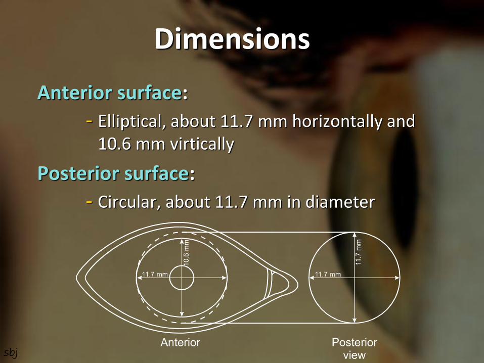
Dimensions
Anterior surface:
- Elliptical, about 11.7 mm horizontally and 10.6 mm virtically
Posterior surface:
- Circular, about 11.7 mm in diameter
sbj

Contd…
Thickness:
- Centrally about 0.52 mm
- Peripherally about 0.67 mm
Surface area:
- About 1.3 cm² (one-sixth of the globe)
Optical zone:
- Cornea is almost a sphere, the central 1/3rd is called optical zone about 5.4 mm
sbj

Contd…
Radius of curvature:
- Anterior surface – about 7.8 mm
- Post. Surface – about 6.5 mm
Refractive power: +43.1 D (Air-tear = +43.6 D, Tear-cornea = +5.3 D, Cornea-aqueous = -5.8 D)
Refractive index: 1.376
sbj

Contd…
Topography:
- Shape of cornea is important for fitting of contact lenses
- Small spherical zone of ant. curvature (2-4 mm) is decentered up & outwards with visual axis, but correctly centered for pupillary aperture is termed as corneal apex or cap
- Curvature varies from apex to limbus, greater flattening in nasally and superiorly
- Cornea is flatter in men than in women
- Cornea flattens slightly on convergence
sbj

Composition of human cornea
Water: 78 %
Collagen: 15 % of which: Type-I : 50-55 %
Type-III : 1 %
Type-IV : 8-10 %
Type-VI : 25-30 %
Other protein: 5 %
Keratan sulphate: 0.7 %
Condroitin/dermatan sulphate: 0.3 %
Hyaluronic acid: +
Salts: 1 % sbj
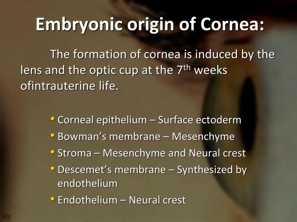
Embryonic origin of Cornea:
The formation of cornea is induced by the lens and the optic cup at the 7th weeks ofintrauterine life.
• Corneal epithelium – Surface ectoderm
• Bowman’s membrane – Mesenchyme
• Stroma – Mesenchyme and Neural crest
• Descemet’s membrane – Synthesized by endothelium
• Endothelium – Neural crest
sbj

Structure Behind the precorneal tear film there are five layers of cornea:
1. Epithelium
2. Bowman’s layer
3. Stroma
4. Descemet’s membrane
5. Endothelium
sbj
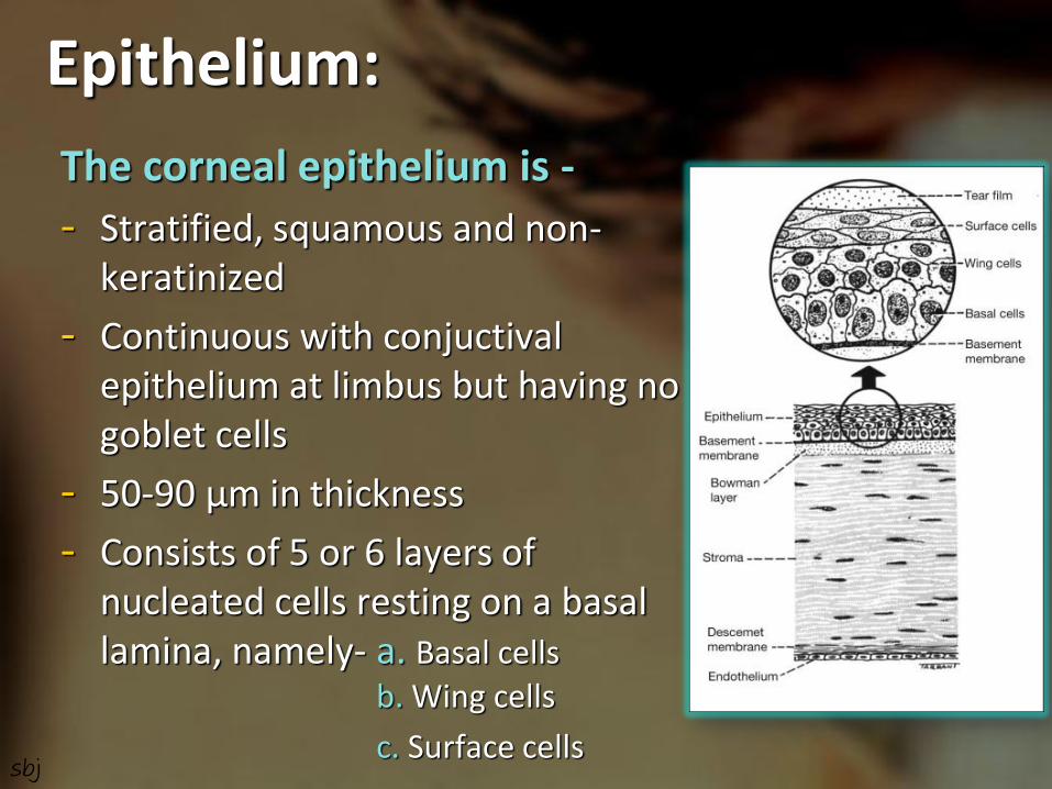
Epithelium:
The corneal epithelium is -
- Stratified, squamous and non-keratinized
- Continuous with conjuctival epithelium at limbus but having no goblet cells
- 50-90 µm in thickness
- Consists of 5 or 6 layers of nucleated cells resting on a basal lamina, namely- a. Basal cells b. Wing cells
c. Surface cells
sbj

Basal cells:
- Deepest cell layer
- Stand in a palisade manner on basal lamina
- Germinative layer of the epithelium
- Columner with rounded heads and flat bases
- Nucleus is oval and oriented parallel to the cells long axis
Contd…
sbj

Wing or umbrella cells:
- Second epithelial cell layer (1-2 layers of cells)
- Polyhedral cells
- Convex anteriorly forming cap over basal cells and send processes between them
- Nucleus is oval and oriented parallel to corneal surface
Contd…
sbj
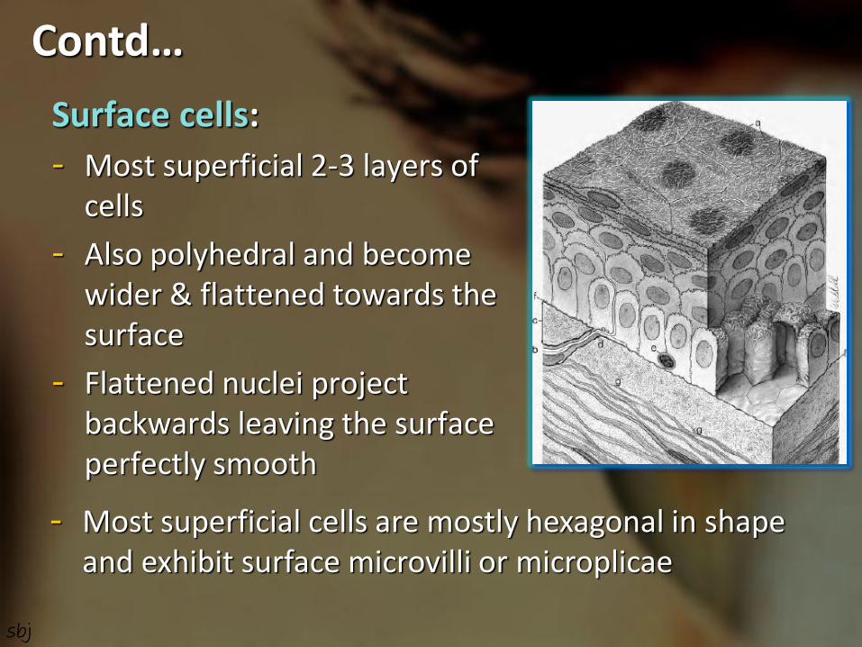
Surface cells:
- Most superficial 2-3 layers of cells
- Also polyhedral and become wider & flattened towards the surface
- Flattened nuclei project backwards leaving the surface perfectly smooth
Contd…
- Most superficial cells are mostly hexagonal in shape and exhibit surface microvilli or microplicae
sbj
Ultrastructural features:
- Epithelial cells shows usual organelles like other actively metabolizing cells
- Moderately abundant mitochondria in wings & middle layer cells but small and scarce in basal cells
- Wing & superficial cells have high glycogen content
Contd…
sbj
- Tonofibrils: cells contain a cytoplasmic meshwork of electrondense intermidiate filaments composed of cytokeratins
- The plasma membrane of contiguous cells interdigitative to each other
- Adhesion is achieved by – • Tight junctions & desmosomes – surface cells
• Desmosomes – wings & superficial cells
• Desmosomes & Hemidesmosomes – in basal cells
Contd…
sbj

Contd…
sbj

- In the basal cells there are anchoring filaments which pass through the hemidesmosomal structure to be inserted into basal lamina
- Langerhans cells (cells of immune recognition system) present near periphery. They are almost absent at central cornea but aggregate in response to infection
Contd…
sbj

Basal lamina:
- The basal lamina is secreted by the basal cells
- 0.5 - 1 µm wide
- Ultrastructurally it is distinguished in to two patrs –
i. Lamina lucida (superficial)
ii. Lamina densa (deep osmiophilc)
Contd…
sbj

- The lamina consist of collagen and glycoprotein constituents which integrated with Bowman’s layer by array of short anchoring filaments
- Lipid solvent, stromal oedema and inflamation may loosened the cohesion between Bowman’s zone and lamina
- With old age, in diabetes and in some corneal disorders it becomes thickened and multilamellar
Contd…
sbj

Epithelial Turnover:
- Early studies suggested that the epithelium replaced approximately weekly by division of basal cells and the oldest shed from the surface
- It is now recognized that the germinative region lies at the limbus, the stem cells, and cells migrate at a very slower rate (123 µm/week) to the center of the cornea which may be as long as a year
Contd…
sbj
- The XYZ hypothesis:
1. Thoft R. and Friend J. (1983) proposed on the basis of experimental evidence that both limbal basal and corneal basal cells are the source for corneal epithelial cells, and there is a balance among division, migration & shedding.
Contd…
sbj

The corneal epithelium is maintained by a balance among sloughing (Z) of cells from the corneal surface, cell division (X) in the basal layer and renewal of basal cells by centripetal migration (Y) of new basal cells originating from the limbal stem cells.
Contd…
sbj

Epithelial Repair:
- Repair of corneal epithelial injury like abration follows a distinctive sequence of events - Injury (abration)
Cells at wound edge retract, thicken and lose attachment
Travel in an amoeboid movement to cover the defect
Cells at wound edge ruffle and send out
filopodia and lamellipodia towards the center of wound
Contd…
sbj

Migration process is halted by contact inhibition
They then anchor and
Mitosis resumes to re-establish epithelial thickness
Surface tight junctions re-establised
Adhesion with Bowman’s layer within 7 days (if basal lamina intact)
• The healing process occurs rapidly, rate of cell migration is 60 – 80 µm/hr
Contd…
sbj

- In case of total epithelial loss including total limbus, cornea is covered with vascularized conjunctival type of epithelium by adjacent conjunctiva
- If a small part of limbus with stem cell is retained then conjunctival type of epithelium is gradually disappear and metabolic behavior of corneal epithelium re-established very slowly
Contd…
sbj
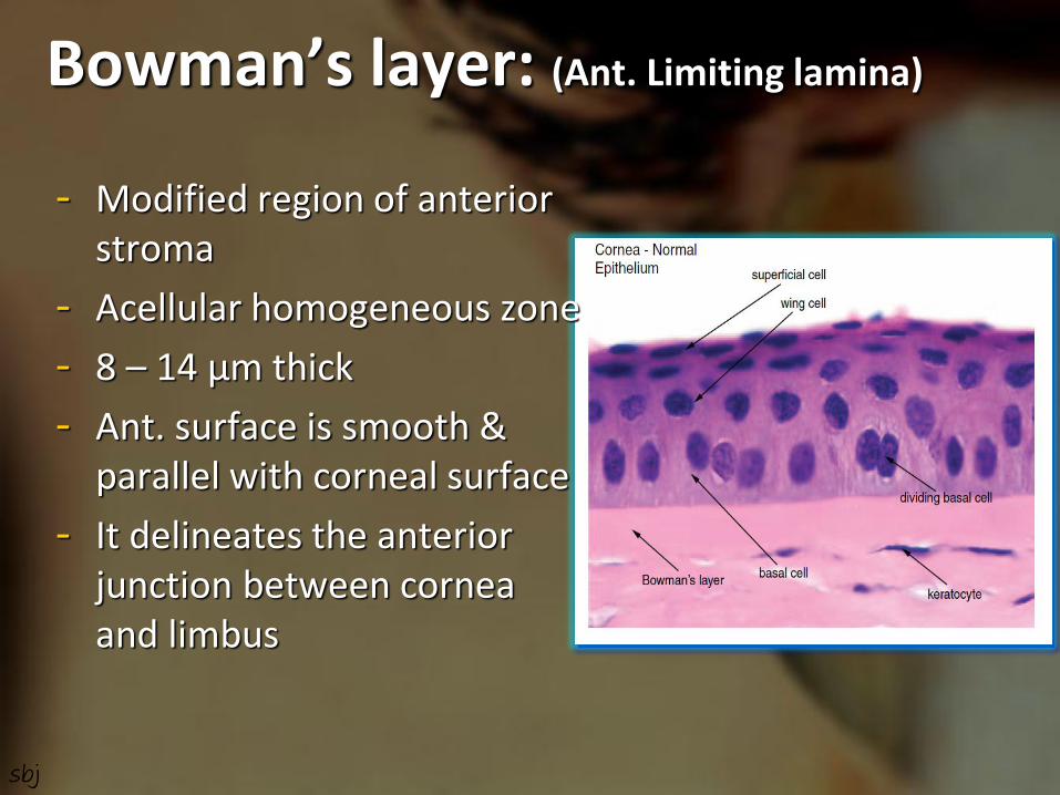
Bowman’s layer: (Ant. Limiting lamina)
- Modified region of anterior stroma
- Acellular homogeneous zone
- 8 – 14 µm thick
- Ant. surface is smooth & parallel with corneal surface
- It delineates the anterior junction between cornea and limbus
sbj

Ultrastructural features:
- Ultrastructurally it is a felted meshwork of fine collagen fibrils of uniform size in a ground substance
- Posteriorly it becomes blended & interweaving with fibrils of ant. stroma
- Compact arrangement of collagen gives it great strength and relatively resistant to trauma both mechanical and infective
Contd…
sbj

- Convex ridges may generate over surface if its tension is relaxed during indentation, hypotony or manipulation causes ant.corneal mosaic, polygonal or chicken-wire pattern over surface
- No regeneration and replaced by coarse scar tissue
- It is perforated many places by nerve to epithelium
Contd…
Fig.: Ant.corneal mosaic
sbj

Stroma: (Substantia propria)
- About 500 µm thick (about 90% of corneal thickness)
- Consists of regularly arranged lamellae of collagen bundles, lie in proteoglycan ground substance with –
- 200 – 300 bundles – centrally
- 500 bundles – peripherally
- Width about 9 – 260 µm
- Height about 1.15 – 2 µm
- Small population of cells – keratocytes present
sbj

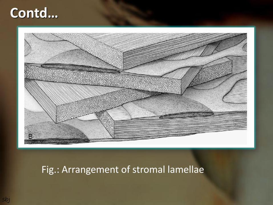
- Arrangement of lamellae –
- Lamellae are arranged in layers, parallel with each other & with corneal surface
- In deeper stroma the lamellae form strap-like ribbons which run approximately at right angles to those in consecutive layers
- At the periphery this right-angular arrangement is slightly changed where it gets scleral fibres
- At the limbus the bundles appeared to take a circular course
Contd…
sbj
Contd…
Fig.: Arrangement of stromal lamellae
sbj

Ultrastructural features:
- Each lamellae comprises of a band of collgen fibrils arranged in parallel with each other
- Fibrils show typical 64 nm periodicity of connective tissue collgens with a microperiod of 6 nm
- There is a unique uniformity of fibril diameter, it is 22 (±1) nm from ant. to post.
- There is remarkable regularity of seperation both within and between lamellae
Contd…
sbj
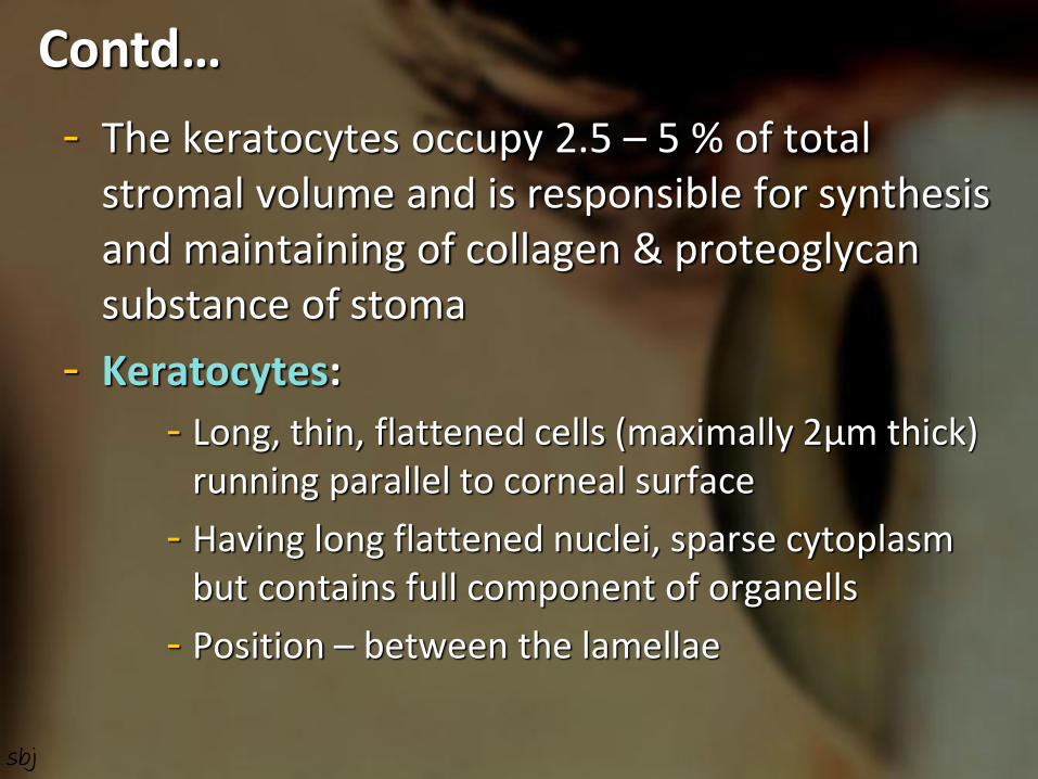
- The keratocytes occupy 2.5 – 5 % of total stromal volume and is responsible for synthesis and maintaining of collagen & proteoglycan substance of stoma
- Keratocytes:
- Long, thin, flattened cells (maximally 2µm thick) running parallel to corneal surface
- Having long flattened nuclei, sparse cytoplasm but contains full component of organells
- Position – between the lamellae
Contd…
sbj
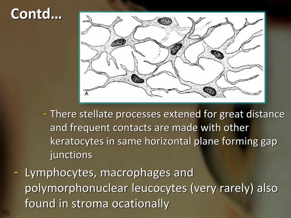
- There stellate processes extened for great distance and frequent contacts are made with other keratocytes in same horizontal plane forming gap junctions
- Lymphocytes, macrophages and polymorphonuclear leucocytes (very rarely) also found in stroma ocationally
Contd…
sbj
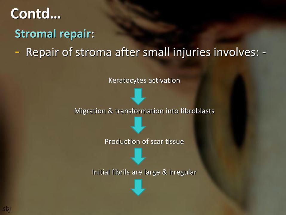
Stromal repair:
- Repair of stroma after small injuries involves: -
Keratocytes activation
Migration & transformation into fibroblasts
Production of scar tissue
Initial fibrils are large & irregular
Contd…
sbj
Remodelling of scar tissue occurs, it ensues –
1. Thinning of fibrils
2. Reformation of lamellae over months
3. Increase in tranperency
- Larger wounds provoke rapid vascular response and leaving vascularised scar along with lymphatic channels
Contd…
sbj

Descemet’s membrane: (Post. Limiting layer)
- It is the basal lamina of corneal endothelium
- First appears at 2nd month of gestation and synthesis continue throughout adult life
Thickness – at birth :- 3 – 4 µm
at childhood :- about 5 µm
at adult :- 10 – 12 µm
- There is a distinct structural difference between fetal & postnatal components
sbj
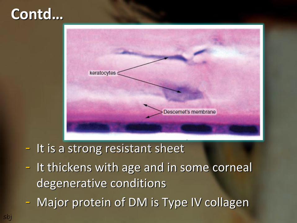
- It is a strong resistant sheet
- It thickens with age and in some corneal degenerative conditions
- Major protein of DM is Type IV collagen
Contd…
sbj

Ultrastructural features:
- In adult human, the anterior 1/3rd of DM corresponds to fetal part, which is like a laminated structure and shows an irregular banded pattern in cross section
- In tangential section it appears to consist of superimposed plates forming a lamellar pattern
Contd…
sbj

- Posterior 2/3rd formed after birth and consist of a homogeneous fibrogranular material
- The zone adjoining the endothelium is the most recently formed
- Modified hemidesmosomes attachment present in between DM & endothelium
Contd…
Descemet’s membrane
sbj

Hassal-Henle warts:
- It is the peripheral excrescence produced by focal overproduction of basal lamina like material in aging cornea
- No clinical abnormality in corneal function
Descemet’s warts of central cornea is called Cornea Guttata, it is associated with increased permiability of endothelium
Contd…
sbj

Contd…
Fig.: Cornea Guttata in Fuchs’ dystrophy
sbj
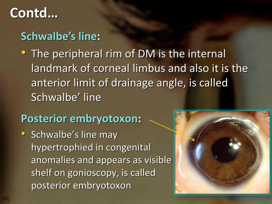
Posterior embryotoxon:
• Schwalbe’s line may hypertrophied in congenital anomalies and appears as visible shelf on gonioscopy, is called posterior embryotoxon
Contd…
Schwalbe’s line:
• The peripheral rim of DM is the internal landmark of corneal limbus and also it is the anterior limit of drainage angle, is called Schwalbe’ line
sbj

Repair of Descemet’s layer:
After traumatic interuption of DM (Path./Mech.)
Endothelium spread its cells to resurface the defect
Synthesis of fresh basal lamina
which is structurally identical to normal descemet’s layer
Contd…
sbj
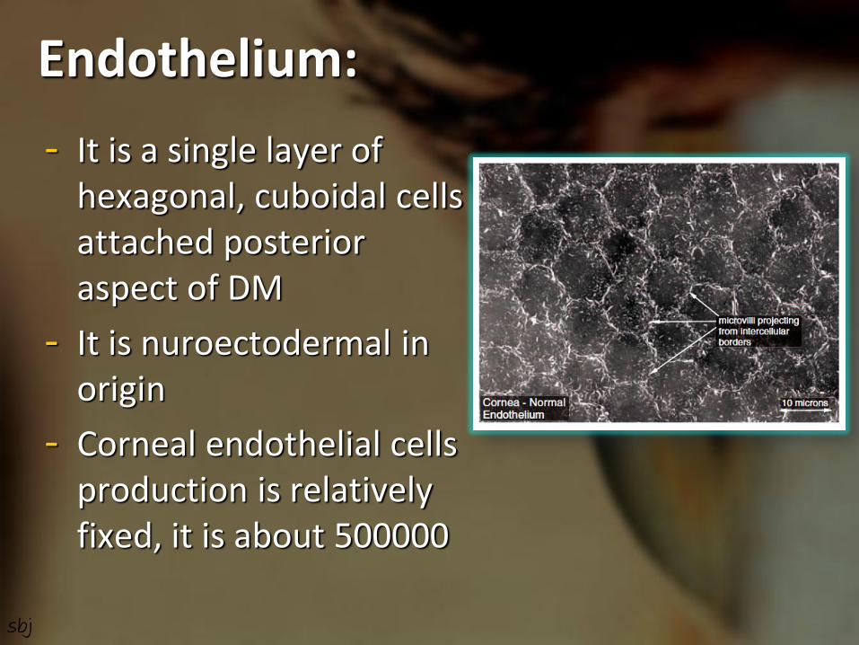
Endothelium:
- It is a single layer of hexagonal, cuboidal cells attached posterior aspect of DM
- It is nuroectodermal in origin
- Corneal endothelial cells production is relatively fixed, it is about 500000
sbj

- Endothelial cells density –
- about 6000 cells/mm² at birth
- 26% lost in 1st year
- Further 26% lost over next 11 years
- Rate of cell loss slows and stabilizes around middle age and then it is about 2500 cells/mm²
- If cells density falls upto 500 cells/mm² corneal oedema devlops and transparency reduced
Contd…
Endothelium of a healthy cornea
Endothelium of a rigid contact lens wearer sbj

- At birth cells are 10 µm in height, with age it becomes flattened to 3-5 µm and 18-20 µm width
- Single oval nucleus located centrally
- Cells shape is hexagonal in youth with age it become polymorphic
Contd…
sbj

Ultrastructural features:
- The anterior cell membrane (Basal) is attached with DM by modified hemidesmosomes
- The posterior cell membrane (Apical) facing Anterior chamber shows 20-30 microvilli
- Lateral borders produce a complex interdigitation with neighboring cells
Contd…
Fig.: 3-D view of deep cornea showing part of endothelium, DM, Stroma
sbj
- Cell junctions with surrounding cells at lateral surfaces –
- Ant. 2/3rd – maculae adharentes
- Post. 1/3rd & apicolateral edges – macculae occludentes
- Endothelium is rich in subcellular organeles – large number of mitochondria, both rough and smooth endoplasmic reticulum, free ribozomes, these reflects that endothelium is extremely active metabolically
Contd…
sbj

Nutrition to endothelium:
- Endothelium gets its nutrition & O₂ from aqueous
- Essential nutrients (such as glucose & amino acids) pass across its surface to supply the cellular needs of all the corneal layers
Contd…
sbj

Fluid regulation:
- The state of relative deturgescence of stroma is maintained by this delicate monolayer of cells by two ways –
- Providing a barrier function to the ingress of salt and metabolites to the stroma
- Actively reducing the osmotic pressure of stroma by metabolically pumping the bicarbonate ions out of the stroma to aqueous
Contd…
sbj
Endothelial Repair:
- Physical & chemical damage to endothelium results in loss of cells
- Neighboring cells move over to fill the gap by sliding process and enlargement of cells occur
- Thus, after injury, the endothelial cell density falls, the cell area increases and the cell height decreases
Contd…
sbj

Blood supply of Cornea: - The cornea is an avascular
structure
- Small loops derived from the anterior ciliary vessels invade its periphery for about 1 mm.
- Actually, these loops are not in cornea but in the subconjunctival tissue which overlaps the cornea.
sbj

Nerve supply of Cornea:
- Cornea is rich in sensory nerve supply derived from ophthalmic division of trigeminal nerve via anterior ciliary nerves and nerves to the surrounding conjunctiva –
Ant.ciliary nerve enter the pericoroidal space a short distance behind the limbus
Connect with each other & conjunctival nerve and form pericorneal plexus
60-80 myelinated branches pass into cornea
sbj

After 1-2 mm lose myelin sheaths and divide into anterior and posterior groups
Anterior nerves (40-50) pass through stroma and form plexus
subjacent to Bowman’s layer
Nerve fibres then penetrate Bowman’s layer and form subepithelial plexus
Fibres then divide dichotomously to form a parallel network
which run for upto 2 mm
Contd…
sbj

And give rise to fine free nerve terminals to superficial epithelial layers
The posterior groups of nerves (40-50) pass posteriorly to
innervate the posterior stroma excluding Descemet’s membrane
Contd…
sbj

Sentinel of the eye:
• The adrenergic fibres from cervical sympathetic, supply the limbus also supply almost whole of the eye and its appendages, giving warning of injury for instance by a foreign body, its called ‘sentinel of eye’
Contd…
sbj
Corneal Nutrition & Metabolism
- Cornea requires energy for normal metabolic activities as well as for maintaining transparency and dehydration
- Energy is generated by the breakdown of glucose in the form of ATP
- Most actively metabolizing layers are epithelium & endothelium
sbj
Sources of Nutrients:
- Oxygen – mainly from atmosphere through tear film, with minor amounts supplied by the aqueous and limbal vasculature
- Normal Po₂ in tears is 155 mm Hg
- In aqueous is about 40 mm Hg
- Minimum 25 mm Hg Po₂ is needed for maintaining deturgescent state and transparency
Contd…
sbj

- Glucose, amino acid, vitamins, and other nutrients supplied to cornea by aqueous humor, a lesser amounts from tears or limbal vessels
- Glucose also derived from glycogen stores in corneal epithelium
- Epithelium consumes O₂ 10 times faster than stroma
Contd…
sbj

Metabolic pathways:
- Three processes or pathways –
1. Penntose shunt (Hexose monophosphate shunt) – occurs both In hypoxic and normoxic condition
- Forms NADPH and Pentose (Ribose 5-P) from gulcose which are used in nucleic acid synthesis
2. Glycolysis (Embden meyerhof pathway) – anaerobic process, glucose/glycogen converted to pyruvate yelding 2 ATPs
3. TCA or Krebs or citric acid cycle – in aerobic conditions pyruvate is oxidized to yield 36 ATP, water, CO₂
Contd…
sbj

Contd…
Fig.: Metabolism of Glucose in cornea sbj

Corneal Transparency The cornea transmits nearly 100% of the light that enters it. Transparency achieved by –
1. Arrangement of stromal lamellae
Two theories –
i) Maurice (1957): The transparency of the stroma is due to the lattice arrangement of collagen fibrils. He explained, because of their small diameter and regularity of separation, back scattered light would be almost completely suppressed by destructive interference
sbj

ii) Goldman et al. (1968): He suggest, a perfect crystalline lattice periodicity is not always necessary for sufficient destructive interference. He explained, if fibril separation and diameter is less than a third of the wavelength of incident light, then almost perfect transparency will ensue. This is the situation which obtains in normal cornea.
Contd… A B
sbj
Other factors of corneal transparency -
2. Corneal epithelium & tear film • Epithelial non-keratinization
• Regular & uniform arrangement of corneal epithelium
• Junctions between cells & its compactness and also tear film maintain a homogenicity of its refractive index
3. Relative deturgescence state of normal cornea
4. Corneal avascularity
5. Non myelenated nerve fibres
Contd…
sbj

Factors affecting corneal Hydration:
i. Stromal swelling pressure exerted by GAGs
ii. Barrier function of epithelium and endothelium
iii. Hydration controled by active pump mechanisms of the corneal endothelium
• The enzyme pump systems are –
• Na⁺/K⁺ ATPase pump system
• Bicarbnate dependent ATPase
• Carbonic anhydrase enzyme
• Na⁺/H⁺ pump
iv. Evaporation of water from corneal surface
v. Intraocular pressure
sbj

Drug permeability across the Cornea:
Factors affecting drug penetration through the cornea are –
1. Lipid and water solubility of the drug
2. Molecular size, weight and concentration of drug
3. Ionic form of the drug
4. pH of the solution
5. Tonicity of the solution
6. Surface active agents
7. Pro-drug form sbj

Corneal wound healing:
• Discussed earlier of this presentation with every layers..
sbj

sbj