anatomy & physiology i bio 1110- lecture and lab regional and directional terms, sections and...
TRANSCRIPT

Anatomy & Physiology I BIO 1110- Lecture and Lab
Regional and Directional Terms, Sections and Cavities
What is Anatomy? “tomy” means “to cut” in Greek Anatomy is the study of the structures of the body:
their composition their location their associated structures Can be Gross or Microscopic
Gross anatomy studies structures large enough to be seen with the unaided eye.
Microscopic anatomy studies structures requiring the use of a microscope (cells and tissues). Cytology= studies cells; Histology= studies tissues
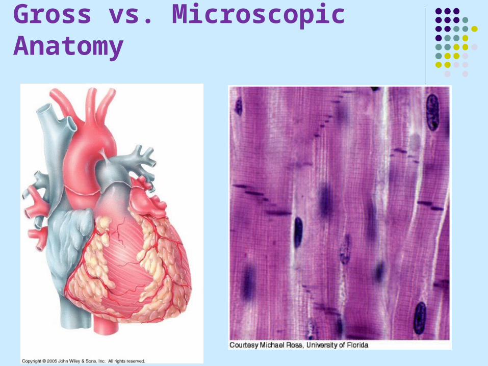
Gross vs. Microscopic Anatomy

What is Physiology? The study of the functions of anatomical
structures. Principle of complementarity says that structure
and function are complementary. 2 examples:
- The left side of heart is more muscular than the right side because it has to work harder to pump blood a further distance.
- Bones are strong and supportive because they store mineral deposits in a ring formation.

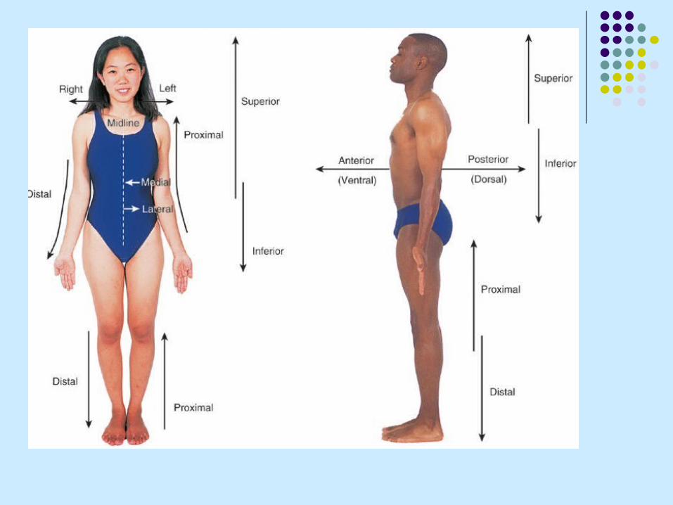
Anatomical Position When using anatomical terminology, one
should assume that the body is in anatomical position. Standing erect, facing the observer, head level,
eyes facing forward, feet flat and forward, arms at the sides with palms facing forward

Anatomical Regional Terms Terms used to identify various regions of the
body. See Figure 1.1 from the lab manual and 1.5 from
the textbook.

Anatomical Regional Terms

Directional Terms Terms used to identify the relative position of
structures associated with the body. Superior- Above Inferior- Below Anterior (ventral)- Closer to the front of
the body Posterior (dorsal)- Closer to the back of
the body Medial- Closer to the midline of the body Lateral- Farther from the midline of the
body

Intermediate- Between 2 structures Ipsilateral- On the same side of the body as another
structure Contralateral- On the opposite side of the body
from another structure Proximal- Closer to the point of attachment of a limb
and the trunk Distal- Farther from the point of attachment of a limb
and the trunk Superficial- Closer to the surface of the body Deep- Farther from the surface of the body
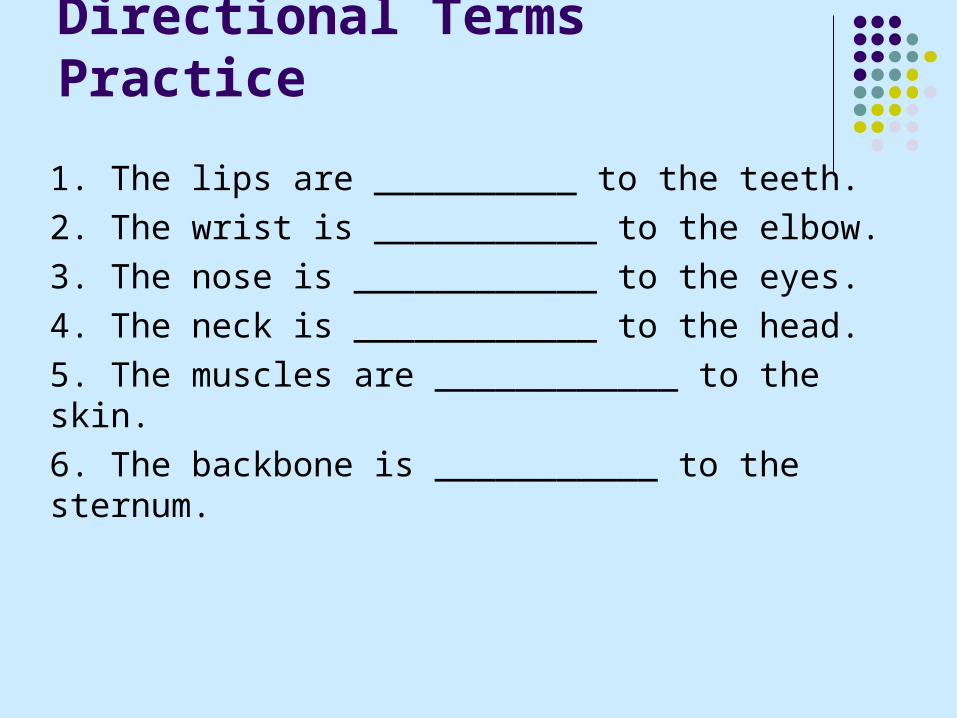
Directional Terms Practice
1. The lips are __________ to the teeth.
2. The wrist is ___________ to the elbow.
3. The nose is ____________ to the eyes.
4. The neck is ____________ to the head.
5. The muscles are ____________ to the skin.
6. The backbone is ___________ to the sternum.

Body Planes
Imaginary flat surfaces that divide the body into smaller sections.
Sometimes to gain a greater understanding of 3D images, anatomists cut the image at different planes
Three planes exists in 3D space
-Two are parallel to the long axis of the body
-One is perpendicular to the long axis.

Body Planes
Figure 1.8

Body Sections- cut along a plane
Sagittal – parallel to the long axis and divides the body into right and left parts Midsagittal – sagittal plane that lies on the midline Parasagittal- not along the midline
Frontal (Coronal) – also parallel to the long axis and divides the body into anterior and posterior parts
Transverse (cross section) – perpendicular to the long axis and divides the body into superior and inferior parts


Body Cavities Confined spaces within the body that contain the
internal organs. Bones, muscles, ligaments and other structures
separate the cavities from one another. Dorsal cavity- The body cavity found near the
posterior surface of the body. The dorsal cavity includes the cranial cavity and the
spinal cavity (aka vertebral canal). The cranial cavity is formed by the cranial bones and
encloses the brain. The spinal cavity is formed by the bones of the
vertebral column and encloses the spinal cord.

Body Cavities
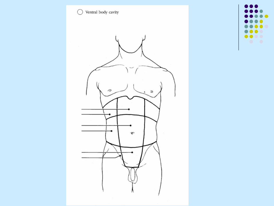
Ventral cavity- The body cavity found near the anterior surface of the body.
The ventral cavity includes the thoracic cavity and the abdominopelvic cavity (divided into the abdominal and pelvic cavities).
The diaphragm is a muscle that divides the thoracic and abdominopelvic cavities.


Thoracic Cavity The thoracic cavity is the region between the neck
and the diaphragm This includes the heart, lungs, esophagus,
trachea, bronchi and thymus. The heart is lined with pericardium
the parietal pericardium lines the cavity while the visceral pericardium covers the heart. A thin layer of serous fluid separates the two layers.
The lungs are lined with pleura the parietal pleura lines the cavity while the
visceral pleura covers the lungs. A thin layer of serous fluid separates the two layers.

Serous Membrane Relationship
Figure 1.10a

Thoracic Pleura

The abdominal cavity contains the stomach, liver, gall bladder, pancreas, spleen, small intestine, appendix and part of the large intestine
The pelvic cavity is formed by the pelvis and contains the bladder, reproductive organs, rectum and the remainder of the large intestine.
The peritoneum lines the abdominopelvic cavity; a parietal peritoneum lines the wall while visceral peritoneum covers the organs.

Abdominopelvic Peritoneum

Dividing the Abdominopelvic Cavity The abdominopelvic cavity is subdivided into nine regions:
Top 3- right hypochondriac region, epigastric region, left hypochondriac region
Middle 3- right lumbar region, umbilical region, left lumbar region
Bottom 3- right iliac (inguinal) region, hypogastric (pubic) region, left iliac (inguinal) region
There is a 2nd method that divides the cavity into quadrants: right upper quadrant (RUQ), left upper quadrant (LUQ), right lower quadrant (RLQ), left lower quadrant (LLQ).