anatomy of the gastrointestinal system - blackboard learn · pdf fileanatomy of the...
TRANSCRIPT

04/02/2016
1
Anatomy of the Gastrointestinal SystemDR CHRIS MOORE
1
System overview
Broadly divided into 2 categories
GI Tract
Oral cavity Oesophagus
StomachSmall
intestine
Large intestine
Rectum and Anus
Accessory organs
Liver Gallbladder
Pancreas Appendix
2

04/02/2016
2
GI System in situ
Stomach
Liver
Gallbladder
Small Intestine
Large Intestine
Rectum & Anus
3
Reveal viscera
Abdominal cavity
Peritoneal cavity contains serous fluid – lies between:
Parietal peritoneum - lines body wall
Visceral peritoneum - lines external surface of most GI organs
4
What kind of
space does the
peritoneal
cavity have?

04/02/2016
3
From the beginning – Oral cavity
Cheeks – cont with lips, reflect onto gums
Lined by mucous membrane
Two palates
Anterior – Hard
Posterior - Soft
5
Teeth - mechanical 6

04/02/2016
4
Tooth surfaces 7
Lingual
Buccal
Distal
MesialOcclusal
Incisal
Labial
Enamel
Dentin
Gingival sulcus
Gingiva (gum)
Pulp in pulp
cavity
Cementum
Root canal
Alveolar bone
Periodontal
ligament
Apical foramen
Nerve
Blood supply
Sagittal section of a mandibular (lower) molar
CROWN
NECK
ROOT
Sagittal
plane

04/02/2016
5
Salivary Glands - chemical
Parotid – watery saliva with enzymes
Parotid ducts open
opposite buccal side of
upper 2nd molars
Submandibular – enzyme and mucous
Sublingual – mucous saliva
Each drained by ducts of
Rivinus in floor of mouth
(10-20 ducts)
9
Parotid
Submandibular
Sublingual
Parotid duct
Opening of parotid duct
(near second maxillary molar)
Second maxillary molar tooth
Tongue (raised in mouth)
Lingual frenulum
Submandibular duct
Mylohyoid muscle
SUBMANDIBULAR GLAND
Zygomatic arch
PAROTID GLAND
Lesser sublingual duct
SUBLINGUAL GLAND
(a) Location of salivary glands

04/02/2016
6
Parotid duct
SUBMANDIBULAR
GLAND
PAROTID
GLAND
(b) Right lateral view
Zygomatic arch
Masseter muscle
Oesophagus
Long, thin, muscular tube
25cm (approx)
Topographically related to trachea
Protects
C-shape
Ant Post
12

04/02/2016
7
Oesophagus
Penetrates
diaphragm at
oesophageal hilus
Two valves -
Oesophageal
sphincters
Upper and Lower
LOS (or LES in U.S) -GERD
13
Inner structure
2 Muscle types
Circular (inner) and
longitudinal (outer)
Muscular layer
differs according
to position
U 1/3 – striated (voluntary)
M 1/3 – striated and
smooth
L 1/3 – smooth
(involuntary)
14
Mucosa
Submucosa
Muscularis
(circular layer)
Muscularis
(longitudinal layer
Vagus nerve

04/02/2016
8
Stomach
Lies beneath liver and diaphragm in epigastric and left hypocondrium regions
Alters size and position in response to eating
A sphincter at each end regulates passage
LES, Pyloric
Three major parts
Pylorus Body
Fundus
15
Identify the
three main
regions of the
stomach
Internal anatomy
Tri-directional muscle layers
Folds – Ruggae
Five layers of tissue (common across GIT)
16

04/02/2016
9
Duodenum – small intestine 1
First section of small intestine (SI)
25cm long (approx)
Four parts – Superior, Descending, Horizontal, Ascending
Receives secretions from pancreatic duct and common bile duct (into descending via major duodenal papilla)
17
Jejunum – small intestine 2
Middle section of SI – 2.5m (approx)
Jejunum and Ileum - no clear division
Villi – site of absorption
Gives velvety appearance
18
04/02/2016
10
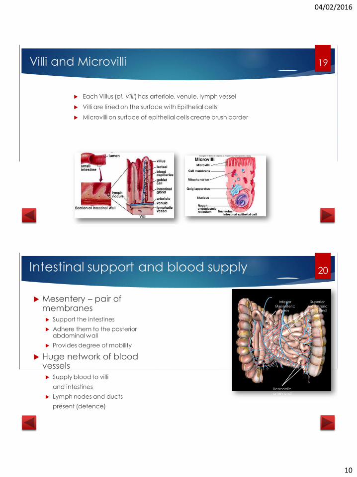
Villi and Microvilli
Each Villus (pl. Villi) has arteriole, venule, lymph vessel
Villi are lined on the surface with Epithelial cells
Microvilli on surface of epithelial cells create brush border
19
Intestinal support and blood supply
Mesentery – pair of membranes
Support the intestines
Adhere them to the posterior abdominal wall
Provides degree of mobility
Huge network of blood vessels
Supply blood to villi
and intestines
Lymph nodes and ducts
present (defence)
Superior
Mesenteric artery and
vein
Ileocoelic
artery and vein
Inferior
Mesenteric vein
20

04/02/2016
11
Ileum – small intestine 3
Not to be confused with Ilium!
Around 2-4m long
Absorbs bile salts and vitamins
Anything missed by Jejunum
really
Toggle in situ
21
Aren’t they the same...
More fat in Mesentery
Ileum has more vascular arcades
Jejunum 2, Ileum 5-6
Plicae circulars more numerous in Jejunum
Absorption greater in Jejunum
Why are there
more circulars?
22

04/02/2016
12
Aren’t they the same...
Ileum has Peyer’s patches
Collections of lymphoid tissue related to immune system
Specialised epithelium containing “M” cells
Sample antigens and present for “analysis”
23
Caecum
Blind pouch – breakdown of plant material
Significant species variation
?
Vermiform appendix
Ileocaecal valve
24

04/02/2016
13
Colon
Ascending
Transverse
Descending
Sigmoid
Note descending and sigmoid are situated more posteriorly
25
Surface features of colon
Teniae Coli
Longitudinal smooth muscle
bands
Slightly shorter than colon
Haustra
Sacculations caused by the
teniae
Teniae Coli
Haustra
26

04/02/2016
14
Rectum and Anus
Separate structures
Rectal folds help regulate flow
Internal anal sphincter
Smooth muscle
External anal sphincter
Striated muscle
Rectum
Anus
27
Physiology of
DigestionGASTROINTESTINAL SYSTEM PART 2
28

04/02/2016
15
A multi-stage process
Ingestion
Propulsion/motility
Mechanical digestion
Chemical digestion
Absorption
Defecation/evacuation/egestion
29
The Enteric nervous system
GI wall has complex network of neurons
Myenteric plexus
Submucosal plexus
Connected to each other, the GI muscles, and the CNS
Together they form the
ENTERIC nervous system
30

04/02/2016
16
Physiology Overview
Digestive activities of the gastrointestinal tract occur in three
overlapping phases:
1. The cephalic phase
2. The gastric phase
3. The intestinal phase
31
Physiology Overview
During the cephalic phase of digestion, the smell, sight, thought, or initial taste of food activates neural centers in the cerebral cortex, hypothalamus, and brain stem to prepare for digestion.
The brain stem activates the facial (CN VII) and glossopharyngeal (CN IX) nerves to stimulate secretion of saliva, while the vagus nerves (CN X) stimulate secretion of gastric juice.
32

04/02/2016
17
Physiology Overview
Once food reaches the stomach, the gastric
phase of digestion begins. Neural and hormonal mechanisms (the hormone gastrin is a key player)
promote secretion of gastric juice and increase gastric motility.
The intestinal phase of digestion begins once food enters the
small intestine. Neural and hormonal responses promote the continued digestion of foods
that have reached the small intestine.
33
The Large Intestine
The gastroileal reflex causes relaxation of the ileocecal valve, intensifies
peristalsis in the ileum, and forces any chyme into the cecum.
The gastrocolic reflex intensifies strong peristaltic waves that begin at
about the middle of the transverse colon and quickly drive the contents
of the colon into the rectum.
This mass peristalsis takes place three or four times a day during or
immediately after a meal, and may lead to defecation.
34

04/02/2016
18
Clinical
Complications
35
Common conditions
Motility and Excretion related
Diarrhoea
Increased fluid caused by increased
gut motility and decreased intestinal
absorption
Constipation
Decreased gut motility leading to
further water absorption in colon,
dryness of faecal matter
Periodontal disease (often confused with peridontitis)
Inflammatory and Blockage
Peptic ulcers
Appendicitis
Pancreatitis and Cholecystitis
Gallstone, pancreatic stones
Inflammatory bowel disease
Two forms: Crohn’s disease and Ulcerative
colitis
Crohn’s – infl of distal ileum/proximal colon
(serosa through mucosa)
UC – infl of mucosa of distal colon/rectum
36

04/02/2016
19
Omphaloceles
Bowel development outside of abdominal cavity
Midgut fails to return back inside the abdominal cavity during
10th week of
embryologic
development
Easily corrected with surgery
following birth
Click here for clinical image. Warning, this is a paediatric image and so may disturb. Please click the image to hide it if you
wish.
37
Please use the back button in
your browser, or close this tab to
return to the main screen. Or click
below to restart and improve your
knowledge
Restart
38