anatomy of retina
TRANSCRIPT

Anatomy Of Retina
MD SALMAN SARKARSchool Of Medical Sciences
Hyderabad Central University

RPE
• The retinal pigment epithelium (RPE), the outermost retinal layer, is a single cell thick and consists of pigmented hexagonal cells.
• These cells are columnar in the area of the posterior pole and are even longer, narrower, and more densely pigmented in the macular area.
• The cells become larger and more cuboidal as the layer nears the ora serrata.

RPE
• the basal aspect of the cell is adjacent to the choroid and the apical surface faces the neural retina.
• The RPE cells contain numerous melanosomes, pigment granules, that extend from the apical area into the middle portion of the cell.
• Pigment density differs in various parts of the retina and in individual cells.

RPE
• In the retina, melanin is densest in the RPE cells located in the macula and at the equator.
• Other pigmented bodies, lipofuscin granules, contain degradation products of phagocytosis, which increase in number with age.
• The cell cytoplasm also contains smooth and rough endoplasmic reticulum, Golgi apparatus, mitochondria, and numerous lysosomes.

RPE
• The apical portion of an RPE cell consists of microvilli that extend into the layer of photoreceptors.
• However, no intercellular junctions connect the RPE and photoreceptor cells.
• A potential space separates the epithelial cell and the photoreceptor, is called subretinalspace.

RPE
• Terminal bars consisting of zonula occludensand zonula adherens join the RPE cells near their apices.
• Desmosomes are present throughout the layer, and gap junctions between the cells allow for electrical coupling, providing a low-resistance pathway for the passage of ions and metabolites.

Photoreceptor Cells
Retinal Pigment Epithelium-Neuroretinal Interface:-• Several factors are involved in maintaining the close
approximation between the photoreceptor cell layer and the RPE layer.
• Passive forces, such as intraocular pressure (IOP), osmotic pressure, fluid transport across the RPE, and presence of the vitreous, help preserve the position of the neural retina.
• Interdigitations between the RPE microvilli and the rod and cone outer segments provide a physical closeness between the two entities.
• The material that occupies the extracellular space between the two layers likely provides adhesive forces.

Photoreceptor Cells
• Composition Of Rods & Cones:-
Outer Segment--The outer segment is made up of a stack of membranous discs (600 to 1000 per rod) and is enclosed by the plasmalemma of the cell.
A connecting stalk, or cilium, extends from the innermost disc, joining the outer segment with the inner segment and acting as a conduit between them.

Photoreceptor Cells Inner Segment:-
The inner segment contains cellular structures and can be divided into two parts. The ellipsoid is nearer the outer segment and contains the numerous mitochondria necessary for the many energy-dependent photoreceptor processes.
The part closer to the cell body, which is sometimes called the myoid, contains other cellular organelles, such as the endoplasmic reticulum and Golgi apparatus; protein synthesis is concentrated in this area. The term “myoid,” however, is derived from a similar area in amphibians that contains a contractile structure. The human myoid does not have contractile properties.

Photoreceptor Cells
Outer Fiber, Cell Body, and Inner Fiber:-
The outer fiber extends from the inner segment to the cell body, the portion containing the nucleus. The inner fiber is an axonal process containing microtubules and runs inward from the cell body, ending in specialized synaptic terminals that contain synaptic vesicles.

Morphology Of Rod
• The plasmalemma, enclosing the rod outer segment, is separate from the disc membrane except for a small region at the base where invaginations of the plasmalemma form discs.
• The rod inner and outer segments are approximately the same width.
• The inner segment is joined to the cell body by the relatively long and narrow outer fiber.

Morphology Of Rod
• The inner fiber extends from the cell body and terminates in a rounded, pear-shaped structure called a spherule.
• The internal surface of the spherule is invaginated forming a synaptic complex that contains bipolar dendrites and horizontal cell processes.
• Rods release the neurotransmitter glutamate.
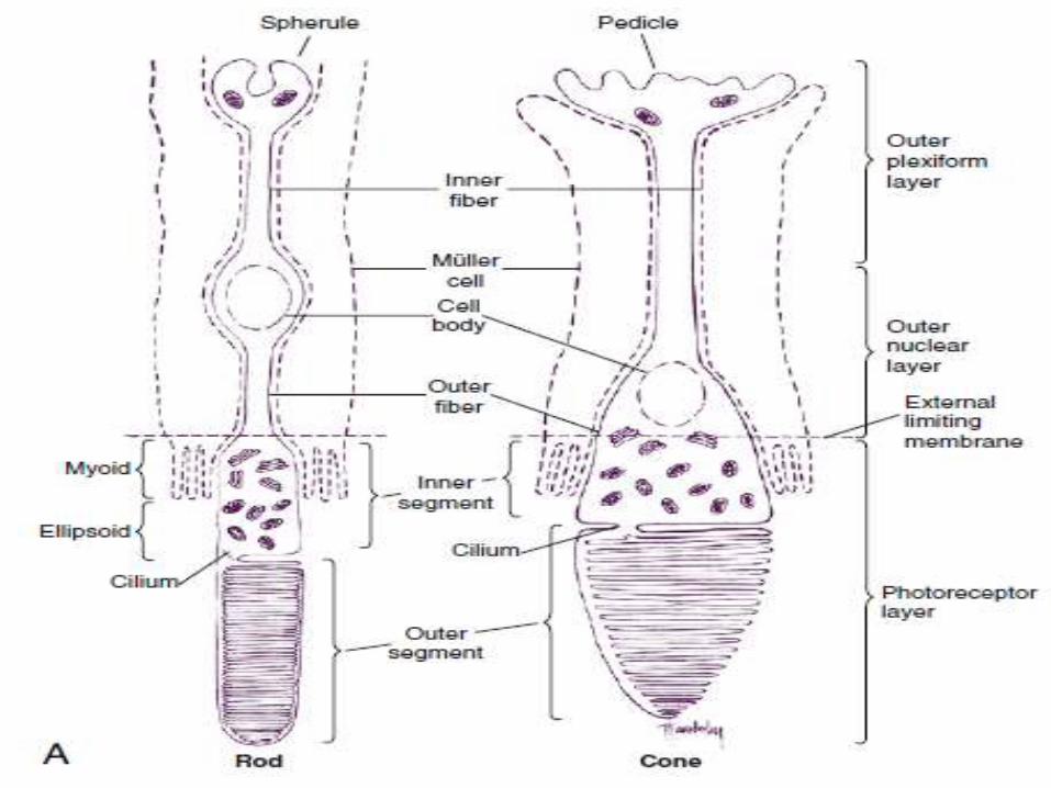

Morphology Of Cone
• As in the rod, the outer segment of the cone is enclosed by a plasmalemma, but in this case the plasma membrane is continuous with the membranes forming most of the discs, and the discs are not separated easily from one another.
• In many cones the discs at the base are wider than those at the tip, giving the characteristic cone shape.
• The cone outer segment is shorter than that of the rod and may not reach the RPE layer.

Morphology Of Cone
• Tubular processes protrude from the apical surface of the epithelial cell to surround the cone outer segment.
• One of three visual pigment molecules is contained within the disc membrane.
• each pigment molecule is activated by the absorption of light in a specific range in the color spectrum.
• The peak absorptions occur at 420 nm (blue), 531 nm (green), and 588 nm (red).

Morphology Of Cone
• The shape of the inner segment also contributes to the cone shape.
• The ellipsoid area of the cone is wider and contains more mitochondria than Rod.
• The outer fiber is short and stout and may even be absent in the Cone.
• The inner fiber terminates in a broad, flattened structure called a pedicle, which has several invaginated areas within its flattened surface.

Morphology Of Cone
• Cone pedicles have three types of synaptic contacts. Triads are found within the invaginations; contacts with bipolar cells occur on the flat surfaces; and gap junctions are located on the lateral expansions (telodendria) of the pedicle and permit electrical communication between adjacent rods or cones.
• As with rods, the neurotransmitter released by cones is glutamate.

BIPOLAR CELLS
• The bipolar cell is the second-order neuron in the visual pathway.
• The nucleus of the bipolar cell is large and contains minimal cell body cytoplasm.
• Its dendrite synapses with photoreceptor and horizontal cells, and its axon synapses with ganglion and amacrine cells.
• Glutamate is its neurotransmitter.

BIPOLAR CELLS
• Bipolar cells relay information from photoreceptors to horizontal, amacrine, and ganglion cells and receive extensive synaptic feedback from amacrine cells.
• Eleven types of bipolar cells have been classified on the basis of morphology, physiology, and dendritic contacts with photoreceptors; all types except the rod bipolar cell are associated with cones.

Types Of Bipolar Cells(Rod Bipolar)
Rod Bipolar:-
Only one type of rod bipolar cell has been identified.
• It has a relatively large body and several spiky dendrites, usually arising from a single, thick process.
• Rod bipolar cells begin to appear 1 mm from the fovea and continue into the periphery.
• These are the only bipolar cells that contact rods.

Types Of Bipolar Cells(Midget Bipolar)
Flat Midget Bipolar Cell:-
• The midget bipolar cell has a relatively small body and can be either flat or invaginating.
• Dendritic terminals of the flat midget bipolar cell end in a flat expansion and make contact only with the flat area of the cone pedicle.

Types Of Bipolar Cells(Midget Bipolar)
Invaginating Midget Bipolar Cell:-
• It is similar to the flat midget bipolar cell, but its dendritic processes are located within the pedicle invaginations, usually in groupings called a triad.
• A triad consists of a single central bipolar dendrite flanked by two horizontal cell processes within an invagination in the cone pedicle.

Types Of Bipolar Cells(Diffuse Cone Bipolar Cells)
Diffuse Cone Bipolar Cells:-
• The two types of diffuse cone bipolar cells are designated type a and type b, called “flat bipolars” and “brush bipolars”.
• In the central retina, the diffuse cone bipolar cell contacts approximately five neighboring cones.
• In the periphery, each contacts 10 to 15 neighboring cones.

Types Of Bipolar Cells(Blue Cone Bipolar Cell )
• The blue cone bipolar cell synapses with up to three cone pedicles.
• It differs from diffuse cone bipolar cells in that it contacts widely spaced rather than neighboring cones.

Types Of Bipolar Cells(Giant Cone Bipolar Cell)
• The giant cone bipolar cell derives its name from the extent of its dendritic tree.

Ganglion Cell
• The next cell in the visual pathway, the third-order neuron, is the ganglion cell.
• Ganglion cells can be bipolar (e.g., a single axon and a single dendrite) or multipolar (a single axon and more than one dendrite).
• Ganglion cells have also been classified on the basis of cell body size, branching characteristics, termination of dendrites, and the expanse of the dendritic tree.

Types Of Ganglion Cell
• Another designation classifies ganglion cells based on the lateral geniculate nucleus layer in which they terminate.
• The P1 ganglion cell, also called the midget ganglion cell, is the most common P cell. P cells terminate in the parvocellular layers.
• The P2 ganglion cell also terminates in the parvocellular layers but has a densely branched, compact dendritic tree that spreads horizontally.
• The M-type ganglion cell projects to the magnocellular layers of the LGN.

HORIZONTAL CELLS
• The horizontal cell transfers information in a horizontal direction parallel to the retinal surface.
• It has one long process, or axon, and several short dendrites with branching terminals; the processes spread out parallel to the retinal surface, and all terminate in the outer plexiform Layer.
• Horizontal cells synapse with photoreceptors, bipolar cells, and other horizontal cells.
• Horizontal cells are joined to each other by an extensive network of gap junctions.
• One type of horizontal cell synapses only within a cone pedicle in the special triad junction.
• Three types of horizontal cells have been differentiated: HI, HII, and HIII.

AMACRINE CELLS
• The amacrine cell has a large cell body, a lobulated nucleus, and a single process with extensive branches that extend into the inner plexiform layer.
• They can also be classified into four groups—narrow field, small field, medium field, and large field.
• Most amacrine cells contain the inhibitory neurotransmitter gamma-aminobutyric acid (GABA) or glycine, and have both presynaptic and postsynaptic endings.
• Amacrine cells are joined to one another via gap junctions.

INTERPLEXIFORM NEURONS
• The interplexiform neuron has a large cell body and is found among the layer of amacrine cells.
• The processes extend into both synaptic layers and convey information between these layers, apparently providing feedback from inner to outer retinal layers.

NEUROGLIAL CELLS
• Müller Cells:-
• Müller cells are large neuroglial cells that extend throughout much of the retina.
• There are 10 million Müller cells in the mammalian retina.
• They play a supportive role, providing structure.
• The apex of the Müller cell is in the photoreceptor layer, whereas the basal aspect is at the inner retinal surface.
NEUROGLIAL CELLS
• Besides providing structure, the Müller cell acts as a buffer by regulating the concentration of potassium ions (K+).
• They help maintain the extracellular pH by absorbing metabolic waste products.
• They recycle GABA and glutamate, removing them from the extracellular space.
• Müller cells metabolize, synthesize, and store Glycogen.

NEUROGLIAL CELLS
• Microglial Cells and Astrocytes:-• Microglial cells are wandering phagocytic cells
and might be found anywhere in the retina.• Their number increases in response to tissue
inflammation and injury.• Astrocytes are star-shaped fibrous cells found in
the inner retina, usually in the nerve fiber and ganglion cell Layers.
• They may contribute to the internal limiting membrane as well as perform some of the same functions as the Müller cells.

10 RETINAL LAYERS
1. Retinal pigment epithelium2. Photoreceptor cell layer3. External limiting membrane4. Outer nuclear layer5. Outer plexiform layer6. Inner nuclear layer7. Inner plexiform layer8. Ganglion cell layer9. Nerve fiber layer10. Internal limiting membrane


RETINAL LAYERS
RETINAL PIGMENT EPITHELIUM:-• There are 4 to 6 million RPE cells, and each cell
interacts with 30 to 40 photoreceptors. PHOTORECEPTOR LAYER:-• The photoreceptor layer contains the outer and inner
segments of rods and cones. EXTERNAL LIMITING MEMBRANE:-• The external limiting membrane (ELM, outer limiting
membrane) is not a true membrane but is actually composed of zonula adherens junctions between photoreceptor cells and between photoreceptors and Müller cells at the level of the inner segments.
RETINAL LAYERS OUTER NUCLEAR LAYER:-
• The outer nuclear layer (ONL) contains the rod and cone cell bodies; the cone cell body and nucleus are larger than those of the rod. Cone outer fibers are very short, and therefore the cone nuclei lie in a single layer close to the external limiting membrane.
OUTER PLEXIFORM LAYER:-
• The outer plexiform layer (OPL; also outer synaptic layer) has a wide external band composed of inner fibers of rods and cones and a narrower inner band consisting of synapses between photoreceptor cells and cells from the inner nuclear layer.
• Rod spherules and cone pedicles synapse with bipolar cell dendrites and horizontal cell processes in the OPL.

RETINAL LAYERS
INNER NUCLEAR LAYER:-
• The inner nuclear layer (INL) consists of the cell bodies of horizontal cells, bipolar cells, amacrinecells, interplexiform neurons, Müller cells, and sometimes displaced ganglion cells
INNER PLEXIFORM LAYER:-
• The inner plexiform layer (IPL; also inner synaptic layer) consists of synaptic connections between the axons of bipolar cells and dendrites of ganglion cells.

RETINAL LAYERS GANGLION CELL LAYER:-
• The ganglion cell layer is generally a single cell thick except near the macula, where it might be 8 to 10 cells thick, and at the temporal side of the optic disc, where it is 2 cells thick.
NERVE FIBER LAYER:-
• The nerve fiber layer (NFL; also stratum opticum) consists of ganglion cell axons. Their course runs parallel to the retinal surface; the fibers proceed to the optic disc, turn at a right angle, and exit the eye through the lamina cribrosa as the optic nerve.
• The fibers generally are unmyelinated within the retina.
• The NFL is thickest at the margins of the optic disc, where all the fibers accumulate.

RETINAL LAYERS
INTERNAL LIMITING MEMBRANE:-
• The internal limiting membrane (inner limiting membrane) forms the innermost boundary of the retina.
• The outer retinal surface of this membrane is uneven and is composed of extensive, expanded terminations of Müller cells (often called footplates) covered by a basement membrane.
• The inner or vitreal surface is smooth.


Clinical Comment Retinal Detachment:-• When a retinal detachment occurs, the separation
usually lies between the RPE cells and the photoreceptors because no intercellular junctions join these cells.
• The RPE cells remain attached to the choroid and cannot be separated from it without difficulty.
• Bruch’s membrane contains fibronectin and laminin, large adhesive glycoproteins with many binding sites that help maintain the adherence of RPE cells to the membrane.
• Fluid can accumulate within the subretinal space, separating the photoreceptors from the nutrients supplied by the choroid.
• If the layers are not repositioned quickly, the affected area of photoreceptor cells will necrose.

Clinical Comment
Retinal Hemorrhages:-
• HEMORRHAGES from retinal vasculature have a characteristic appearance. Because of the arrangement of the nerve fibers, the blood pools in a feathered pattern called a flame-shaped hemorrhage, which is indicative of the NFL location. Hemorrhages in the inner nuclear layer usually appear rounded and often are called dot or blot hemorrhages.

Clinical Comment
Fundus View of the Internal Limiting Membrane:-
• Reflections from the internal limiting membrane produce the retinal sheen seen with the ophthalmoscope.
• In younger persons, this membrane gives off many reflections and appears glistening; the sheen is less evident in older individuals.