anatomy of reflux a growing health problem
TRANSCRIPT

8/10/2019 Anatomy of Reflux a Growing Health Problem
http://slidepdf.com/reader/full/anatomy-of-reflux-a-growing-health-problem 1/10
FEATURE ARTICLE
Anatomy of Reflux: A Growing Health ProblemAffecting Structures of the Head and NeckMICHAEL J. LIPAN, JOY S. REIDENBERG, AND JEFFREY T. LAITMAN*
Gastroesophageal reflux disease (GERD) and laryngopharyngeal reflux (LPR) are sibling diseases that are a modern-
day plague. Millions of Americans suffer from their sequelae, ranging from subtle annoyances to life-threatening
illnesses such as asthma, sleep apnea, and cancer. Indeed, the recognized prevalence of GERD alone has increased
threefold throughout the 1990s. Knowledge of the precise etiologies for GERD and LPR is becoming essential for
proper treatment. This review focuses on the anatomical, physiological, neurobiological, and cellular aspects of these
diseases. By definition, gastroesophageal reflux (GER) is the passage of gastric contents into the esophagus; when
excessive and damaging to the esophageal mucosa, GERD results. Reflux that advances to the laryngopharynx and,
subsequently, to other regions of the head and neck such as the larynx, oral cavity, nasopharynx, nasal cavity,paranasal sinuses, and even middle ear results in LPR. While GERD has long been identified as a source of
esophageal disease, LPR has only recently been implicated in causing head and neck problems. Recent research has
identified four anatomical/physiological “barriers” that serve as guardians to prevent the cranial incursion of reflux:
the gastroesophageal junction, esophageal motor function and acid clearance, the upper esophageal sphincter, and
pharyngeal and laryngeal mucosal resistance. Sequential failure of all four barriers is necessary to produce LPR.
While it has become apparent that GER must precede both GERD and LPR, the head and neck distribution of the latter
clearly separates these diseases as distinct entities warranting specialized focus and treatment. Anat Rec (Part B:
New Anat) 289B:261–270, 2006. © 2006 Wiley-Liss, Inc.
KEY WORDS: laryngopharyngeal reflux; LPR; gastroesophageal reflux disease; GERD; lower esophageal sphincter; LES;
upper esophageal sphincter; UES; esophageal motility; mucosal resistance
INTRODUCTIONPatients presenting with symptoms
such as hoarseness, sensation of alump in the throat (i.e., globus
pharyngeus), and chronic cough havelong been a thorn in the side of mod-
ern medicine, largely due to physi-
cians’ inability to identify the source
of their ailments. The majority of pa-
tients feel better with little more than
patience, but those with persistentproblems have been a medical conun-
drum, often leading to frustration for
both doctor and patient. Slowly, phy-
sicians began to identify acid reflux,
which may travel to regions as far as
the head and neck, as a causative
agent of these various symptoms (Ta-
ble 1). Over the last 15 years, carefully
constructed human clinical trials have
gradually added evidence that reflux
of gastric contents causes a wide
range of symptoms and clinical signs
commonly called laryngopharyngeal
reflux (LPR; Table 2). The symptoms
themselves can cause a significant im-
pact on quality of life, but the more
important point is the implication of
LPR as a risk factor for a number of
life-threatening conditions such ashead and neck cancers, asthma, sleep
apnea, narrowing of the respiratorytract below the vocal folds (i.e., sub-
glottic stenosis), and involuntary
forceful adduction of the vocal folds(i.e., laryngospasm) (Koufman et al.,
2002).
Dr. Lipan has recently graduated with anMD with distinction in research fromMount Sinai School of Medicine (MSSM),New York, and has begun an otolaryngol-ogy residency at Jackson Memorial Hos-pital in Miami, Florida. He has frequently
presented aspects of his research find-ings on the anatomical underpinnings ofgastroesophageal reflux disease and la-ryngopharyngeal reflux at meetings of the
American Association of Anatomists(AAA) and the Association for Research inOtolaryngology.Dr. Reidenberg is associate professor ofanatomy and functional morphology andassociate professor of medical educationat MSSM. She is a recognized expert in thecomparative biology of mammalianthroats, with a particular interest in ceta-ceans. She was the 1999 recipient of theBasmajian-Williams and Wilkins Award ofthe AAA for excellence in teaching andresearch by a gross anatomist.Dr. Laitman is a distinguished professor at
MSSM, professor and director of the Centerfor Anatomy and Functional Morphology,professor of otolaryngology and of medicaleducation. He is a member of the AAA Board of Directors. His research has fo-cused on elucidating the distinctive devel-
opmental and evolutionary features of thehuman aerodigestive tract and how thesemay relate to human disease. In 1997, Drs.Laitman and Reidenberg advanced the the-ory that the particularly low position in theneck of the human larynx was the anatom-ical basis allowing for reflux of gastric ma-terial into portals of the head and neck.*Correspondence to: Jeffrey T. Laitman,Mount Sinai School of Medicine, Center for
Anatomy and Functional Morphology, Box1007, New York, NY 10029. Fax: 212-860-1174; E-mail: [email protected]
DOI 10.1002/ar.b.20120Published online in Wiley InterScience(www.interscience.wiley.com).
THE ANATOMICAL RECORD (PART B: NEW ANAT.) 289B:261–270, 2006
© 2006 Wiley-Liss, Inc.

8/10/2019 Anatomy of Reflux a Growing Health Problem
http://slidepdf.com/reader/full/anatomy-of-reflux-a-growing-health-problem 2/10
Reflux is defined as a backward flow
of fluid (Table 3). The passage of gas-tric contents into the esophagus is re-
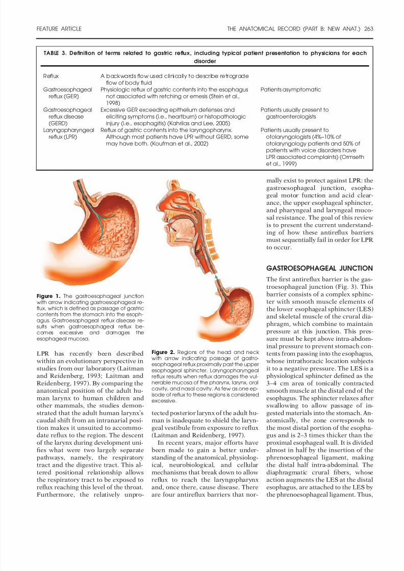
ferred to as gastroesophageal reflux(GER; Fig. 1). GER by itself is consid-
ered physiological and occurs rou-tinely in healthy individuals with no
symptoms or signs of disease. In fact,after-dinner indulgences that contain
ingredients such as chocolate, caf-feine, nicotine, or alcohol may pro-
mote GER and thus relieve discomfortdue to stomach distention following a
large meal. However, excessive GERcan damage the esophageal mucosa
and cause inflammation, a conditioncommonly referred to as gastroesoph-
ageal reflux disease (GERD). Thisbreakdown of the squamous esopha-
geal epithelium is caused by pepsin inan acidic milieu and can lead to heart-
burn, mucosal ulceration, narrowing
of the esophagus, a change to gastricepithelium (i.e., Barrett’s metaplasia),
and, eventually, esophageal cancer.GER can alternatively reflux through
the length of the esophagus to reachthe laryngopharynx and cause LPR,
making GER a common first step forboth diseases (Fig. 2). However, due
to the differences in the subsequentprogression of these diseases, GERD
and LPR are clearly distinct from oneanother.
The impact of reflux is widespread.Abnormal reflux affects millions of
Americans a year. One study demon-strated that 7% of those surveyed ex-
perienced heartburn daily, 14% notedheartburn weekly, with a total of 36%
having heartburn at least monthly(Nebel et al., 1976). LPR may affect a
smaller proportion of the population
than GERD, but it is difficult to diag-nose accurately and thus epidemio-
logical studies are scarce. LPR has
been reported in up to 10% of patients
referred to otolaryngologists for treat-
ment (Koufman, 1991) and 50% of pa-
tients with laryngeal and voice disor-
ders (Koufman et al., 2000). Notably,
President Clinton suffered from LPR,
which led to chronic hoarseness dur-
ing his first presidential campaign in
1992 and intermittently plagued him
throughout his terms in office. Most
importantly, the diagnosis of GERDhas been increasing at an alarming
rate, more than tripling between 1990and 2001 (Altman et al., 2005). This
rise has become a substantial burdenon our population, which is likely to
continue to worsen.Over the last 35 years, evidence for
the association of reflux and ailmentsof the pharynx and larynx has been
mounting, and this cause-and-effectrelationship is gradually being ac-
cepted by physicians. The susceptibil-ity of the human aerodigestive tract to
TABLE 1. Symptoms and conditions associated with laryngopharyngeal reflux grouped by anatomic site
Anatomic Sites Symptoms Conditions
Larynx Hoarseness (Wiener et al., 1989; Smit et al.,
2000)
Voice fatigue
Voice breaksMuscle tension dysphonia
Chronic laryngitis (Hanson et al., 1995)
Subglottic stenosis (Little et al., 1985; Jindal et al.,
1994)
Laryngeal carcinoma (Ward and Hanson, 1988;Qadeer et al., 2005; El-Serag et al., 2001)
Paroxysmal laryngospasm (Loughlin and
Koufman, 1996; Maceri and Zim, 2001)
Contact ulcer (Cherry and Margulies, 1968)
Granuloma (Havas et al., 1999)
Recurrent leukoplakia (Koufman, 1991)
Vocal fold nodule (Kuhn et al., 1998)
Laryngomalacia (Belmont and Grundfast, 1984)
Arytenoids fixation
Renke’s edema
Pachydermia
Oropharynx and
laryngopharynx
Globus (lump in throat sensation) (Smit et
al., 2000)
Dysphagia
Chronic sore throat (pain and irritation)Excessive Throat clearing
Excessive phlegm/saliva
Pharyngeal carcinoma (Qadeer et al., 2005)
Obstructive sleep apnea (Kerr et al., 1992;
Demeter and Pap, 2004)
Lung and
tracheobronchial
tree
Wheezing
Chronic cough (Irwin et al., 1993; Harding
and Richter, 1997)
Asthma exacerbation (Harding and Richter,
1997)
Middle ear Otitis media with effusion (Tasker et al., 2002)
Oral cavity Halitosis Dental erosions (Schroeder et al., 1995)
Sinuses Chronic rhinosinusitis (Ulualp et al., 1999; Parsons,
1996)
Multiple Sites Sudden Infant Death Syndrome (Thatch, 2000;
Nielson et al., 1990)
TABLE 2. Synonyms for laryngopharyngeal reflux
Reflux laryngitis Supraesophageal reflux
Laryngeal reflux Extraesophageal reflux
Pharyngoesophageal reflux Atypical reflux
262 THE ANATOMICAL RECORD (PART B: NEW ANAT.) FEATURE ARTICLE
8/10/2019 Anatomy of Reflux a Growing Health Problem
http://slidepdf.com/reader/full/anatomy-of-reflux-a-growing-health-problem 3/10
LPR has recently been describedwithin an evolutionary perspective in
studies from our laboratory (Laitmanand Reidenberg, 1993; Laitman and
Reidenberg, 1997). By comparing theanatomical position of the adult hu-
man larynx to human children andother mammals, the studies demon-
strated that the adult human larynx’scaudal shift from an intranarial posi-
tion makes it unsuited to accommo-date reflux to the region. The descent
of the larynx during development uni-fies what were two largely separate
pathways, namely, the respiratorytract and the digestive tract. This al-
tered positional relationship allowsthe respiratory tract to be exposed to
reflux reaching this level of the throat.Furthermore, the relatively unpro-
tected posterior larynx of the adult hu-man is inadequate to shield the laryn-
geal vestibule from exposure to reflux(Laitman and Reidenberg, 1997).
In recent years, major efforts havebeen made to gain a better under-
standing of the anatomical, physiolog-ical, neurobiological, and cellular
mechanisms that break down to allowreflux to reach the laryngopharynx
and, once there, cause disease. Thereare four antireflux barriers that nor-
mally exist to protect against LPR: the
gastroesophageal junction, esopha-geal motor function and acid clear-
ance, the upper esophageal sphincter,and pharyngeal and laryngeal muco-
sal resistance. The goal of this reviewis to present the current understand-
ing of how these antireflux barriersmust sequentially fail in order for LPR
to occur.
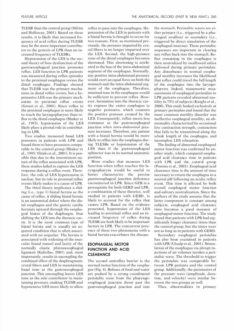
GASTROESOPHAGEAL JUNCTION
The first antireflux barrier is the gas-
troesophageal junction (Fig. 3). This
barrier consists of a complex sphinc-
ter with smooth muscle elements of the lower esophageal sphincter (LES)and skeletal muscle of the crural dia-
phragm, which combine to maintainpressure at this junction. This pres-
sure must be kept above intra-abdom-inal pressure to prevent stomach con-
tents from passing into the esophagus,whose intrathoracic location subjects
it to a negative pressure. The LES is aphysiological sphincter defined as the
3–4 cm area of tonically contractedsmooth muscle at the distal end of the
esophagus. The sphincter relaxes afterswallowing to allow passage of in-
gested materials into the stomach. An-atomically, the zone corresponds to
the most distal portion of the esopha-gus and is 2–3 times thicker than the
proximal esophageal wall. It is dividedalmost in half by the insertion of the
phrenoesophageal ligament, makingthe distal half intra-abdominal. The
diaphragmatic crural fibers, whoseaction augments the LES at the distal
esophagus, are attached to the LES bythe phrenoesophageal ligament. Thus,
TABLE 3. Definition of terms related to gastric reflux, including typical patient presentation to physicians for each
disorder
Reflux A backwards flow used clinically to describe retrograde
flow of body fluid
Gastroesophageal
reflux (GER)
Physiologic reflux of gastric contents into the esophagus
not associated with retching or emesis (Stein et al.,
1998)
Patients asymptomatic
Gastroesophageal
reflux disease
(GERD)
Excessive GER exceeding epithelium defenses and
eliciting symptoms (i.e., heartburn) or histopathologic
injury (i.e., esophagitis) (Kahrilas and Lee, 2005)
Patients usually present to
gastroenterologists
Laryngopharyngeal
reflux (LPR)
Reflux of gastric contents into the laryngopharynx.
Although most patients have LPR without GERD, some
may have both. (Koufman et al., 2002)
Patients usually present to
otolaryngologists (4%–10% of
otolaryngology patients and 50% of
patients with voice disorders have
LPR associated complaints) (Ormseth
et al., 1999)
Figure 1. The gastroesophageal junctionwith arrow indicating gastroesophageal re-flux, which is defined as passage of gastriccontents from the stomach into the esoph-agus. Gastroesophageal reflux disease re-sults when gastroesophageal reflux be-comes excessive and damages theesophageal mucosa.
Figure 2. Regions of the head and neckwith arrow indicating passage of gastro-esophageal reflux proximally past the upperesophageal sphincter. Laryngopharyngealreflux results when reflux damages the vul-nerable mucosa of the pharynx, larynx, oralcavity, and nasal cavity. As few as one ep-isode of reflux to these regions is considered
excessive.
FEATURE ARTICLE THE ANATOMICAL RECORD (PART B: NEW ANAT.) 263

8/10/2019 Anatomy of Reflux a Growing Health Problem
http://slidepdf.com/reader/full/anatomy-of-reflux-a-growing-health-problem 4/10
while diaphragmatic contraction
causes an increase in intra-abdominal
pressure (as occurs with inspiration
or Valsalva maneuver), it simulta-neously prevents the reflux of gastriccontents into the esophagus by super-
imposing its muscular tone onto theesophagus to raise junctional pres-
sure.Three theories have emerged to ex-
plain how reflux crosses the gastro-esophageal junction: transient LES
relaxations (TLESRs), the failure tomaintain substantial pressure at the
LES (i.e., LES hypotension), and ana-tomic disruptions associated with hi-
atal hernia. Investigation of LES func-tion frequently uses manometry, a
clinical test where a transducer mea-sures intraluminal pressures created
by muscular tone. Physiologically, re-flux into the esophagus can occur
multiple times a day without causingdisease; it is an established pattern of
reflux that determines the manifesta-tions of disease. GERD patients usu-
ally have numerous and prolonged pe-riods of reflux exposure, usually in the
recumbent position. LPR patientstend to have more infrequent upright
reflux of short duration that reaches
the laryngopharynx without causingesophageal damage (Koufman et al.,
2002). It is with these patterns inmind that mechanisms of reflux
across the gastroesophageal junctionshould be considered.
The first theory, TLESR, involves a
physiologic phenomenon in whichthere is a sudden drop in pressure at
the LES, which is accompanied bycrural diaphragmatic inhibition and
not preceded by swallowing. TLESRstypically last longer than relaxations
after swallowing and are primarilytriggered by gastric fundus distention
after a meal. The vagus nerve serves asboth the afferent and efferent arc of
the mechanism, resulting in release of nitric oxide and vasoactive intestinal
peptide to mediate muscle relaxation(Hornby and Abrahams, 2000).
Abnormal levels of reflux arethought to be attributed to either an
increased frequency of TLESRs or anincreased frequency of acid reflux
during a TLESR, since not all tran-sient relaxations are accompanied by
reflux. No study has yet shown a rela-tionship between reflux to the laryn-
gopharynx and TLESRs, but conclu-
sions can be drawn from experimentsusing GERD patients. There has not
been consistent evidence that there isan increased frequency of TLESRs in
GERD patients compared to a healthycohort of individuals (i.e., the control
group) (Trudgill and Riley, 2001).However, the frequency of TLESRs
has been found to be position-depen-dent. Patients with GERD had an in-
creased rate of TLESRs compared to
the control group with all subjects ly-ing on their right side, whereas no
difference in frequency was notedwhen subjects sat upright (Sifrim and
Holloway, 2001). Since patients withLPR frequently reflux while upright, it
is unlikely that an increased fre-quency of TLESRs would explain the
abnormal reflux in these patients. In-stead, it is possible that LPR patients
could have an increased frequency of acid reflux during a TLESR. Although
the percentage of TLESRs that resultin reflux in patients with GERD varies
widely in the literature, from 9% to93% (Kahrilas, 1998), most reports
confirm that these patients have agreater rate of reflux with each
Figure 3. The gastroesophageal junction indicating (a) the normal anatomy of this region with the functional tonically contracted regionof the LES indicated with a bracket and (b) anatomic disruption of the junction as occurs with a sliding hiatal hernia. Physiologicphenomena such as hypotension of the lower esophageal sphincter and transient lower esophageal sphincter relaxations result in thepassage of reflux into the esophagus. When they occur in conjunction with a hiatal hernia, this reflux is accentuated.
264 THE ANATOMICAL RECORD (PART B: NEW ANAT.) FEATURE ARTICLE
8/10/2019 Anatomy of Reflux a Growing Health Problem
http://slidepdf.com/reader/full/anatomy-of-reflux-a-growing-health-problem 5/10
TLESR than the control group (Sifrim
and Holloway, 2001). Based on these
results, it is likely that increased fre-
quency of acid reflux during TLESRmay be the more important contribu-
tor to the genesis of LPR than an in-
creased frequency of TLESRs.
Hypotension of the LES is the sec-ond theory of how dysfunction of thegastroesophageal junction promotes
reflux. LES function in LPR patientswas measured during reflux episodes
to the proximal esophagus versus thedistal esophagus. Findings showed
that TLESR was the primary mecha-nism in distal reflux events, but a hy-
potensive LES was the primary mech-anism in proximal reflux events
(Grossi et al., 2001). Since reflux tothe proximal esophagus is more likely
to reach the laryngopharynx than re-flux to the distal esophagus (Shaker et
al., 1995), hypotension of the LESlikely plays a pivotal role in contribut-
ing to LPR.Two studies measured basal LES
pressures in patients with LPR andfound them to have pressures compa-
rable to the control group (Shaker etal., 1995; Ylitalo et al., 2001). It is pos-
sible that due to the intermittent na-ture of the reflux associated with LPR,
these studies failed to capture the LESresponse during a reflux event. There-
fore, the role of LES hypotension isunclear, but its role in proximal reflux
events likely makes it a cause of LPR.The third theory implicates a slid-
ing (i.e., type 1) hiatal hernia as the
cause of reflux. A sliding hiatal herniais an anatomical defect where the dis-
tal esophagus and the gastric cardiaherniate upward through the esopha-
geal hiatus of the diaphragm, thusshifting the LES into the thoracic cav-
ity. It is the most common type of hiatal hernia and is usually an ac-
quired condition that is often associ-ated with no sequelae. The hernia is
associated with widening of the mus-cular hiatal tunnel and laxity of the
normally elastic phrenoesophagealligament (Kahrilas, 2001) and, most
importantly, results in uncoupling thecombined effect of the diaphragmatic
crural fibers and LES in maintainingbasal tone at the gastroesophageal
junction. This uncoupling leaves LEStone as the sole contributor to main-
taining pressure, making TLESR andhypotensive LES more likely to allow
reflux to pass into the esophagus. Hy-potension of the LES in patients with
a hiatal hernia is thought to occur fortwo reasons. First, as mentioned pre-
viously, the pressure imparted by cru-ral fibers is no longer imparted over
the LES. Second, the high-pressure
zone of the distal esophagus becomesshortened. This shortening is attrib-uted to the loss of the intra-abdominal
segment of the esophagus. Normally,any positive intra-abdominal pressure
would exert an equal force on both thestomach and the intra-abdominal seg-
ment of the esophagus. Therefore,minimal tone in the esophagus would
be sufficient to prevent reflux. How-ever, herniation into the thoracic cav-
ity exposes the entire esophagus tonegative pressures, which counters
the positive pressure created by theLES. Consequently, reflux meets less
resistance at the gastroesophageal
junction when intra-abdominal pres-sure increases. Therefore, any patient
with a hiatal hernia would be morelikely to reflux into the esophagus dur-
ing TLESRs or hypotension of theLES than if the gastroesophageal
sphincter was in its normal anatomicposition.
More studies that measure LESfunction when reflux reaches the la-
ryngopharynx would be useful to
better characterize the precisegastroesophageal junction deficiencyencountered in LPR. Since GER is a
prerequisite for both GERD and LPR,a combination of these theories, well
studied in patients with GERD, islikely to account for the reflux that
causes LPR. Based on the evidencepresented, hypotension of the LES
leading to proximal reflux and an in-creased frequency of reflux during
TLESR are both likely to be importantfactors in LPR. The concurrent pres-
ence of these two phenomena with ahiatal hernia exacerbates the disease.
ESOPHAGEAL MOTOR
FUNCTION AND ACID
CLEARANCE
The second antireflux barrier is the
normal motor function of the esopha-gus (Fig. 4). Boluses of food and water
are pushed by a strong coordinatedperistaltic wave from the pharyngo-
esophageal junction down past the
gastroesophageal junction and into
the stomach. Peristaltic waves are ei-
ther primary (i.e., triggered by a pha-
ryngeal swallow) or secondary (i.e.,
triggered by direct simulation of theesophageal mucosa). These peristaltic
sequences are important in clearingany reflux back into the stomach. Re-
flux remaining in the esophagus isthen neutralized by swallowed salivadelivered during primary peristalsis.
Any disturbance in normal esopha-geal motility increases the likelihood
that reflux could travel the full lengthof the esophagus into the laryngo-
pharynx. Indeed, manometric mea-surements of esophageal peristalsis in
LPR patients revealed abnormal mo-tility in 75% of subjects (Knight et al.,
2000). This study looked exclusively atprimary peristalsis and found that the
most common motility disorder wasineffective esophageal motility, an ab-
normality characterized by occasionallow contraction strength, contraction
that fails to be transmitted along thewhole length of the esophagus, and
incomplete LES relaxation.The finding of abnormal esophageal
motor function was confirmed by an-other study, which compared esopha-
geal acid clearance time in patientswith LPR and the control group
(Postma et al., 2001). Esophageal acidclearance time is the amount of time
necessary to return the esophagus to aneutral pH following an acidic reflux
event. This time is affected by bothoverall esophageal motor function
and salivary neutralization. Since theauthors of the study assume that the
latter component is constant among
subjects, esophageal acid clearancetime becomes a good measure of
esophageal motor function. The studyfound that patients with LPR had sig-
nificantly longer clearance times thanthe control group, but the times were
not as long as in patients with GERD.Secondary esophageal peristalsis
has also been examined in patientswith LPR (Ulualp et al., 2001). Stimu-
lation of the esophagus via abrupt in- jections of air volumes invokes a peri-
staltic wave. The threshold to triggerthe peristalsis was comparable be-
tween LPR patients and the controlgroup. Additionally, the parameters of
the pressure wave (amplitude, dura-tion, and velocity) were similar be-
tween the two groups as well.Thus, abnormalities in primary
FEATURE ARTICLE THE ANATOMICAL RECORD (PART B: NEW ANAT.) 265

8/10/2019 Anatomy of Reflux a Growing Health Problem
http://slidepdf.com/reader/full/anatomy-of-reflux-a-growing-health-problem 6/10
peristalsis but not secondary peristal-sis characterize the esophageal dys-
motility found in patients with LPR.
These results are not surprising giventhat the pharyngeal swallow and the
resulting primary peristaltic wave areconsidered to be the predominant
mechanism in returning reflux back tothe stomach (Bremner et al., 1993).
Moreover, the defect in primary
esophageal function associated withLPR is not as severe as that found inGERD (Postma et al., 2001). This con-
clusion reinforces the fact that GERDis characterized by excessive exposure
of reflux to the esophagus, whereas
the sequelae of this excessive contactis usually lacking in LPR patients. Re-
flux in LPR must travel rapidlythrough the esophagus on the way to
the laryngopharynx, where the prob-lems of the disease manifest.
UPPER ESOPHAGEAL SPHINCTER
The third antireflux barrier is the up-
per esophageal sphincter (UES; Fig.5). It is the deficiency in this mecha-
nism that makes LPR unique fromGERD. The UES is defined as a high-
pressure zone that is tonically con-stricted at the pharyngoesophageal
junction. Like the LES, it relaxes toallow the passage of food or liquid
boluses during swallowing. The UESis made up of the most distal fibers of
the inferior pharyngeal constrictor(i.e., the cricopharyngeus muscle) and
the most proximal portion of theesophagus. The cricopharyngeus
Figure 4. a–d: The esophagus exhibiting normal progression of the primary peristaltic wave from the proximal esophagus to the distalesophagus, ending in lower esophageal sphincter relaxation. This mechanism clears reflux back to the stomach and delivers saliva toneutralize any remaining reflux. e–h: The esophagus exhibiting ineffective esophageal motility, the most common abnormality of theprimary peristaltic wave that occurs in laryngopharyngeal reflux. This disorder is characterized by occasional low contraction strength (eand f), loss of contraction as the peristaltic wave is transmitted along the length of the esophagus (e–g), and incomplete LES relaxation(h), which, in combination, increases the chance that reflux reaches laryngopharynx once in the esophagus, as occurs in LPR. Secondaryperistalsis is normal in LPR.
Figure 5. The larynx with the functional ton-ically contracted region of the UES indi-cated with a bracket. Abnormal responseof reflexes that prevent the passage of re-flux to the laryngopharynx is thought to con-tribute to laryngopharyngeal reflux.
266 THE ANATOMICAL RECORD (PART B: NEW ANAT.) FEATURE ARTICLE

8/10/2019 Anatomy of Reflux a Growing Health Problem
http://slidepdf.com/reader/full/anatomy-of-reflux-a-growing-health-problem 7/10
muscle makes the largest contribution
to pressure at the UES in all physio-
logic states (Lang and Shaker, 1997).
The pressure of the UES demonstratesa wide range of variation. For exam-
ple, pressures decrease significantly
during sleep, periods of calmness, and
even expiration (Kahrilas et al., 1987).Pressures have also been shown to belower in the elderly (Fulp et al., 1990).
The main functions of the UES areto prevent air from entering the
esophagus during respiration and toprevent gastric secretions from enter-
ing the pharynx during reflux events.An aberration in accomplishing this
second function is believed to be theprimary defect in LPR, since its man-
ifestations result from reflux abnor-mally breaching this sphincter to
reach the laryngopharynx.Manometric studies have been used
to see if hypotension of the UES hasany role in allowing reflux into the
laryngopharynx. Average pressures of the UES at rest were similar between
patients with LPR and the controlgroup (Shaker et al., 1995; Ulualp et
al., 1998). Electrophysiologic mea-surements of the cricopharyngeus
muscle confirmed these findings andshowed no abnormality in its tonic
activity or activity during swallowingin patients with LPR (Celik et al.,
2005). As with studies of the LES,these studies may be missing mea-
surement of intermittent sphinctericdysfunction around the time of reflux
events. Studies evaluating UES pres-
sures during reflux yielded conflictingresults. Two studies in adults demon-
strated no change in the UES pressureduring esophageal reflux events in pa-
tients with GERD or the control group(Kahrilas et al., 1987; Vakil et al.,
1989), though lack of response couldbe a result of the methodology used.
In both studies, pressures at the UESwere measured during a reflux event,
and the average pressure was com-pared to the average pressure mea-
sured during an equal interval prior tothe reflux event. Therefore, the exper-
iments may have failed to detect ashort lasting change in UES pressures
that may have occurred at the onset of the reflux event.
In contrast to the above studies,Torrico et al. (2000) found that nearly
all reflux events resulted in an in-crease in UES pressure regardless of
whether the reflux occurred in GERD
patients or the control group. This re-
sponse to esophageal stimulation has
been termed the “esophago-UES con-tractile reflex” (Creamer and Schlegel,
1957). The increase in pressure wasnot significantly different between
GERD patients and the control group,but the duration of the pressure in-
crease was nearly double in the con-trol group (25 vs. 15 sec). If a similar
response were to be found in patientswith LPR, it stands to reason that
while the longer contraction may besufficient to restrict normal GER to
the esophagus in healthy individuals,the sphincter’s premature relaxation
may allow reflux to pass into the la-
ryngopharynx in LPR.The opposite of the esophago-UES
contractile reflex is the belch reflex.The venting of gastric gas through the
mouth starts with gastric distentionleading to a relaxation of the LES. The
gas pressure leads to esophageal dis-tention triggering complete UES re-
laxation (Kahrilas et al., 1986). Thus,belching while reflux is in the proxi-
mal esophagus may allow the reflux topass into the laryngopharynx. Patients
with LPR have been shown to havemore episodes of distal reflux reach-
ing the proximal esophagus than pa-tients with GERD or the control
group. Additionally, 30% of refluxevents in the laryngopharynx were as-
sociated with belching (Shaker et al.,1995).
Artificial elicitation of the belch re-flex showed that the rapidity and pat-
tern of esophageal distention deter-mine if the UES relaxes, constricts, or
remains unchanged (Kahrilas et al.,1986). Both air boluses injected into
the esophagus and balloon dilation re-sulted in relaxation, while injected
fluid boluses resulted in constriction
or no change in UES tone. Likewise,abrupt GER could also cause esopha-
geal distention and elicit UES relax-ation as occurs in the belch reflex
(Williams et al., 1999). This abruptand forceful episode of GER accom-
panied by UES relaxation may explainwhy LPR patients exhibit reflux in the
upright position, when it must over-come the effects of gravity, which nor-
mally resists reflux. Evidence that gas-tric reflux triggers UES relaxation was
reported in children (Willing et al.,1993), while the opposite response,
UES contraction, was found in adults
(Torrico et al., 2000). Clearly, a com-
plex interplay exists between the belch
reflex and the esophago-UES contrac-tile reflex, and similarly to the LES,
UES pressures must be measuredduring reflux events that reach the la-
ryngopharynx in order to better un-derstand the precise UES defect asso-
ciated with LPR.Reflexes resulting in UES contrac-
tion have also been described whenregions proximal to this sphincter are
stimulated, namely, the laryngeal andpharyngeal mucosa. An example of
this is the laryngo-UES contractile re-flex, which is elicited by stimulation of
mechanoreceptors of the larynx. Theinternal division of the superior laryn-
geal nerve acts as the afferent arc and
the vagus nerve as the efferent arc of this reflex. A second example is thepharyngo-UES contractile reflex,
which is elicited by stimulation of mechanoreceptors of the posterior
pharyngeal wall. The glossopharyn-geal nerve acts as the afferent arc and
the vagus nerve as the efferent arc of
this reflex. Although speculative, thesereflexes are thought to play a role in
preventing reflux from passing theUES. Reflux contacting the pharyn-
geal or laryngeal mucosa stimulatesthe reflex arc leading to augmentation
of the UES basal resting tone. Thisincrease in sphincter tone would then
prevent further passage of reflux intothe laryngopharynx.
The integrity of the pharyngo-UEScontractile reflex in patients with LPR
was investigated using water stimula-tion (Ulualp et al., 1998). Both pa-
tients and the control group demon-strated an increase in the UES
pressure at a certain threshold volumeof water injection. However, LPR pa-
tients required twice the amount of
water volume to evoke the reflex com-pared to the control group, suggestinga dysfunctional afferent sensory arc,
possibly at the level of the pharyngealreceptors. The laryngeal sensory defi-
ciency in LPR was found to be due tothe damaging effect on the mucosa
from exposure to reflux (Aviv et al.,2000). This loss of mecanosensitivity
was confirmed by infusing acid intothe laryngopharynx of healthy con-
trols to achieve diminished mucosalsensation (Phua et al., 2005). This
causal relationship was supported by
FEATURE ARTICLE THE ANATOMICAL RECORD (PART B: NEW ANAT.) 267
8/10/2019 Anatomy of Reflux a Growing Health Problem
http://slidepdf.com/reader/full/anatomy-of-reflux-a-growing-health-problem 8/10
the ability to reverse the sensory de-fect with aggressive therapy against
LPR (Aviv et al., 2000).Therefore, a defect of the UES may
cause a vicious cycle of events at thelaryngopharynx in patients with LPR.
First, the UES fails to sustain a pro-
longed contraction when the esopha-gus is stimulated by a reflux event.Once an initial reflux event reaches
the laryngopharynx, it causes an in-flammatory reaction that disrupts
normal sensation of the pharyngeal
and laryngeal mucosa. This disrup-tion leads to a diminished pharyngo-
UES contractile reflex that normallywould have prevented further reflux
from reaching the laryngopharynxand perpetuates more damage to the
mucosa.
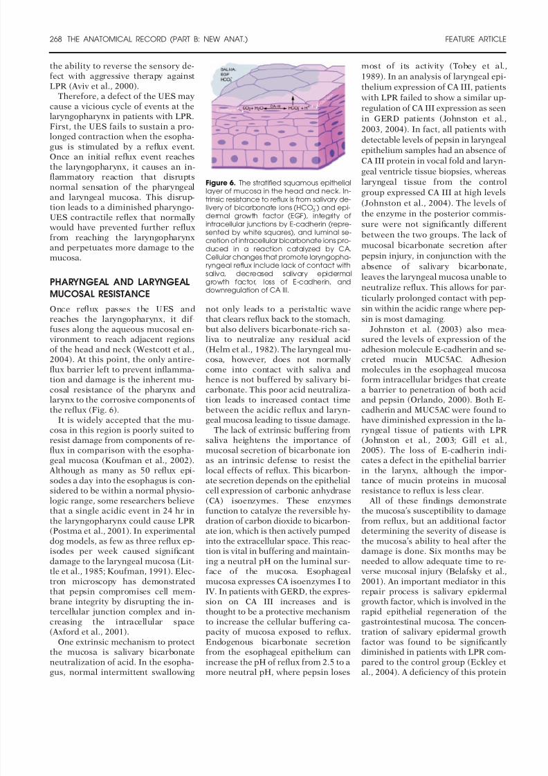
PHARYNGEAL AND LARYNGEAL
MUCOSAL RESISTANCE
Once reflux passes the UES andreaches the laryngopharynx, it dif-
fuses along the aqueous mucosal en- vironment to reach adjacent regions
of the head and neck (Westcott et al.,2004). At this point, the only antire-
flux barrier left to prevent inflamma-tion and damage is the inherent mu-
cosal resistance of the pharynx andlarynx to the corrosive components of
the reflux (Fig. 6).It is widely accepted that the mu-
cosa in this region is poorly suited to
resist damage from components of re-flux in comparison with the esopha-
geal mucosa (Koufman et al., 2002).Although as many as 50 reflux epi-
sodes a day into the esophagus is con-sidered to be within a normal physio-
logic range, some researchers believethat a single acidic event in 24 hr in
the laryngopharynx could cause LPR(Postma et al., 2001). In experimental
dog models, as few as three reflux ep-isodes per week caused significant
damage to the laryngeal mucosa (Lit-tle et al., 1985; Koufman, 1991). Elec-
tron microscopy has demonstratedthat pepsin compromises cell mem-
brane integrity by disrupting the in-tercellular junction complex and in-
creasing the intracellular space(Axford et al., 2001).
One extrinsic mechanism to protectthe mucosa is salivary bicarbonate
neutralization of acid. In the esopha-gus, normal intermittent swallowing
not only leads to a peristaltic wavethat clears reflux back to the stomach,
but also delivers bicarbonate-rich sa-liva to neutralize any residual acid
(Helm et al., 1982). The laryngeal mu-cosa, however, does not normally
come into contact with saliva andhence is not buffered by salivary bi-
carbonate. This poor acid neutraliza-tion leads to increased contact time
between the acidic reflux and laryn-geal mucosa leading to tissue damage.
The lack of extrinsic buffering fromsaliva heightens the importance of
mucosal secretion of bicarbonate ionas an intrinsic defense to resist the
local effects of reflux. This bicarbon-
ate secretion depends on the epithelialcell expression of carbonic anhydrase
(CA) isoenzymes. These enzymesfunction to catalyze the reversible hy-
dration of carbon dioxide to bicarbon-ate ion, which is then actively pumped
into the extracellular space. This reac-tion is vital in buffering and maintain-
ing a neutral pH on the luminal sur-face of the mucosa. Esophageal
mucosa expresses CA isoenzymes I toIV. In patients with GERD, the expres-
sion on CA III increases and isthought to be a protective mechanism
to increase the cellular buffering ca-pacity of mucosa exposed to reflux.
Endogenous bicarbonate secretionfrom the esophageal epithelium can
increase the pH of reflux from 2.5 to amore neutral pH, where pepsin loses
most of its activity (Tobey et al.,
1989). In an analysis of laryngeal epi-
thelium expression of CA III, patients
with LPR failed to show a similar up-
regulation of CA III expression as seen
in GERD patients (Johnston et al.,
2003, 2004). In fact, all patients with
detectable levels of pepsin in laryngeal
epithelium samples had an absence of
CA III protein in vocal fold and laryn-
geal ventricle tissue biopsies, whereas
laryngeal tissue from the control
group expressed CA III at high levels
(Johnston et al., 2004). The levels of
the enzyme in the posterior commis-
sure were not significantly different
between the two groups. The lack of
mucosal bicarbonate secretion after
pepsin injury, in conjunction with the
absence of salivary bicarbonate,
leaves the laryngeal mucosa unable to
neutralize reflux. This allows for par-
ticularly prolonged contact with pep-
sin within the acidic range where pep-
sin is most damaging.
Johnston et al. (2003) also mea-sured the levels of expression of the
adhesion molecule E-cadherin and se-creted mucin MUC5AC. Adhesion
molecules in the esophageal mucosaform intracellular bridges that create
a barrier to penetration of both acidand pepsin (Orlando, 2000). Both E-
cadherin and MUC5AC were found tohave diminished expression in the la-
ryngeal tissue of patients with LPR(Johnston et al., 2003; Gill et al.,
2005). The loss of E-cadherin indi-cates a defect in the epithelial barrier
in the larynx, although the impor-tance of mucin proteins in mucosal
resistance to reflux is less clear.
All of these findings demonstratethe mucosa’s susceptibility to damage
from reflux, but an additional factordetermining the severity of disease is
the mucosa’s ability to heal after thedamage is done. Six months may be
needed to allow adequate time to re- verse mucosal injury (Belafsky et al.,
2001). An important mediator in thisrepair process is salivary epidermal
growth factor, which is involved in therapid epithelial regeneration of the
gastrointestinal mucosa. The concen-tration of salivary epidermal growth
factor was found to be significantlydiminished in patients with LPR com-
pared to the control group (Eckley etal., 2004). A deficiency of this protein
Figure 6. The stratified squamous epitheliallayer of mucosa in the head and neck. In-trinsic resistance to reflux is from salivary de-livery of bicarbonate ions (HCO
3
) and epi-dermal growth factor (EGF), integrity ofintracellular junctions by E-cadherin (repre-sented by white squares), and luminal se-cretion of intracellular bicarbonate ions pro-duced in a reaction catalyzed by CA.Cellular changes that promote laryngopha-ryngeal reflux include lack of contact with
saliva, decreased salivary epidermalgrowth factor, loss of E-cadherin, anddownregulation of CA III.
268 THE ANATOMICAL RECORD (PART B: NEW ANAT.) FEATURE ARTICLE

8/10/2019 Anatomy of Reflux a Growing Health Problem
http://slidepdf.com/reader/full/anatomy-of-reflux-a-growing-health-problem 9/10
could explain the long period of timerequired for the mucosa to heal fully.
Although it is unclear if thesechanges are a consequence of reflux or
a primary defect, these studies inves-tigating the molecular biology of the
laryngeal mucosa in LPR demonstrate
why this region is so sensitive to re-fluxed gastric secretions. The down-regulation of CA, E-cadherin, mucin
MUC5AC, and salivary epidermalgrowth factor combine to cause a dis-
ruption of the basic mucosal barrier
and accounts for the chronic inflam-mation found in LPR. It is this inflam-
mation that leads to symptoms of thedisease (Table 1). If untreated, the in-
flammation continues and may even-tually cause the life-threatening
conditions of subglottic stenosis, la-
ryngospasm, and, most importantly,cancer.
CONCLUSION
The sibling diseases of gastroesopha-
geal reflux disease and laryngopha-ryngeal reflux are a modern-day
plague. Their increasing prevalence inour population has led to an undeni-
able burden on society, from the pa-tients who suffer from them to the
exponentially rising costs of treat-ment. Although GERD and LPR may
often present as only mild distur-bances, their association with severealterations in quality of life, and
even with life-threatening conditions,makes a detailed and precise under-
standing of their respective etiologyimperative for proper treatment.
In order for reflux to travel from thestomach to the laryngopharynx, there
must be a breach in delicate synergis-tic function of four anatomically, neu-
rologically, and physiologically dis-tinct antireflux barriers. This article
has sought to examine the precisemechanisms that fail when gastric re-
flux is allowed to enter such distantand vulnerable regions in the head
and neck. As the impact of LPR be-comes more widely recognized, it is
likely that research will focus on fur-ther characterizing the multifactorial
defects that promote it. Much of ourcurrent understanding is drawn from
the more extensively studied condi-tion GERD. However, it is becoming
dramatically apparent that GERD andLPR are on different ends of a spec-
trum of diseases caused by gastric re-flux. Due to the different anatomy of
these diseases, it stands to reason thatadvances in diagnosis and treatment
will likely follow distinct paths as well.
LITERATURE CITEDAltman KW, Stephens RM, Lyttle CS,
Weiss KB. 2005. Changing impactof gas-troesophageal reflux in medical and oto-laryngology practice. Laryngoscope 115:1145–1153.
Aviv JE, Liu H, Parides M, Kaplan ST,Close LG. 2000. Laryngopharyngeal sen-sory deficits in patients with laryngopha-ryngeal reflux and dysphagia. Ann OtolRhinol Laryngol 109:1000–1006.
Axford SE, Sharp N, Ross PE, et al. 2001.Cell biology of laryngeal epithelial de-fenses in health and disease: preliminarystudies. Ann Otol Rhinol Laryngol 110:1099–1108.
Belafsky PC, Postma GN, Koufman JA.2001. Laryngopharyngeal reflux symp-toms improve before changes in physicalfindings. Laryngoscope 111:979–981.
Belmont JR, Grundfast K. 1984. Congeni-tal laryngeal stridor (laryngomalacia):etiologic factors and associated disor-ders. Ann Otol Rhinol Laryngol 93:430–437.
Bremner RM, Hoeft SF, Costantini M,Crookes PF, Bremner CG, DeMeesterTR. 1993. Pharyngeal swallowing: themajor factor in clearance of esophagealreflux episodes. Ann Surg 218:364–369.
Celik M, Alkan Z, Ercan I, et al. 2005. Cri-copharyngeal muscle electromyography
in laryngopharyngeal reflux. Laryngo-scope 115:138–142.
Cherry J, Margulies SI. Contact ulcer of thelarynx. 1968. Laryngoscope 78:1937–1940.
Creamer B, Schlegel J. 1957. Motor re-sponses of the esophagus to distention.J Appl Physiol 10:498–504.
Demeter P, Pap A. 2004. The relationshipbetween gastroesophageal reflux diseaseand obstructive sleep apnea. J Gastroen-terol 39:815–820.
Eckley CA, Michelsohn N, Rizzo LV,Tadokoro CE, Costa HO. 2004. Salivaryepidermal growth factor concentrationin adults with reflux laryngitis. Otolaryn-
gol Head Neck Surg 131:401–406.El-Serag HB, Hepworth EJ, Lee P, Sonnen-
berg A. 2001. Gastroesophageal refluxdisease is a risk factor for laryngeal andpharyngeal cancer. Am J Gastroenterol96:2013–2018.
Fulp SR, Dalton CB, Castell JA, et al. 1990.Aging-related alterations in human up-per esophageal sphincter function. Am JGastroenterol 85:1569–1572.
Gill GA, Johnston N, Buda A, et al. 2005.Laryngeal epithelial defenses against la-ryngopharyngeal reflux: investigations of E-cadherin, carbonic anhydrase isoen-zyme III, and pepsin. Ann Otol RhinolLaryngol 114:913–921.
Grossi L, Ciccaglione AF, Marzio L. 2001.Transient lower oesophageal sphincterrelaxations play an insignificant role ingastro-oesophageal reflux to the proxi-mal oesophagus. NeurogastroenterolMotil 13:503–509.
Hanson DG, Kamel PL, Kahrilas PJ. 1995.Outcomes of antireflux therapy for thetreatment of chronic laryngitis. Ann OtolRhinol Laryngol 104:550–555.
Harding SM, Richter JE. 1997. The role of gastroesophageal reflux in chroniccough and asthma. Chest 111:1389–1402.
Havas TE, Priestley J, Lowinger DS. 1999.A management strategy for vocal processgranulomas. Laryngoscope 109:301–306.
Helm JF, Dodds WJ, Hogan WJ, SoergelKH, Egide MS, Wood CM. 1982. Acidneutralizing capacity of human saliva.Gastroenterology 83:69–74.
Hornby PJ, Abrahams TP. 2000. Centralcontrol of lower esophageal sphincter re-laxation. Am J Med 108(Suppl 4A):90S–98S.
Irwin RS, French CL, Curley FJ, ZawackiJK, Bennett FM. 1993. Chronic coughdue to gastroesophageal reflux: clinical,diagnostic, and pathogenetic aspects.Chest 104:1511–1517.
Jindal JR, Milbrath MM, Shaker R, HoganWJ, Toohill RJ. 1994. Gastroesophagealreflux disease as a likely cause of “idio-pathic” subglottic stenosis. Ann OtolRhinol Laryngol 103:186–191.
Johnston N, Bulmer D, Gill GA, et al. 2003.Cell biology of laryngeal epithelial de-fenses in health and disease: furtherstudies. Ann Otol Rhinol Laryngol 112:481–491.
Johnston N, Knight J, Dettmar PW, Lively
MO, Koufman J. 2004. Pepsin and car-bonic anhydrase isoenzyme III as diag-nostic markers for laryngopharyngealreflux disease. Laryngoscope 114:2129–2134.
Kahrilas PJ, Dodds WJ, Dent J, Wyman JB,Hogan WJ, Arndorfer RC. 1986. Upperesophageal sphincter function duringbelching. Gastroenterology 91:133–140.
Kahrilas PJ, Dodds WJ, Dent J, Haeberle B,Hogan WJ, Arndorfer RC. 1987. Effect of sleep, spontaneous gastroesophageal re-flux, and a meal on upper esophagealsphincter pressure in normal human vol-unteers. Gastroenterology 92:466 –471.
Kahrilas PJ. 1998. GERD revisited: ad- vances in pathogenesis. Hepatogastroen-terology 45:1301–1307.
Kahrilas PJ. 2001. Supraesophageal com-plications of reflux disease and hiatalhernia. Am J Med 111(Suppl 8A):51S–55S.
Kahrilas PJ, Lee TJ. 2005. Pathophysiologyof gastroesophageal reflux disease. Tho-rac Surg Clin 15:323–333.
Kerr P, Shoenut JP, Millar T, Buckle P,Kryger MH. 1992. Nasal CPAP reducesgastroesophageal reflux in obstructivesleep apnea syndrome. Chest 101:1539–1544.
Knight RE, Wells JR, Parrish RS. 2000.Esophageal dysmotility as an importantco-factor in extraesophageal manifesta-
FEATURE ARTICLE THE ANATOMICAL RECORD (PART B: NEW ANAT.) 269

8/10/2019 Anatomy of Reflux a Growing Health Problem
http://slidepdf.com/reader/full/anatomy-of-reflux-a-growing-health-problem 10/10
tions of gastroesophageal reflux. Laryn-goscope 110:1462–1466.
Koufman JA. 1991. The otolaryngologicmanifestations of gastroesophageal re-flux disease (GERD): a clinical investiga-tion of 225 patients using ambulatory24-hour pH monitoring and an experi-mental investigation of the role of acid
and pepsin in the development of laryn-geal injury. Laryngoscope 101:1–78.
Koufman JA, Amin MR, Panetti M. 2000.Prevalence of reflux in 113 consecutivepatients with laryngeal and voice disor-ders. Otolaryngol Head Neck Surg 123:385–388.
Koufman JA, Aviv JE, Casiano RR, ShawGY. 2002. Laryngopharyngeal reflux: po-sition statement of the committee onspeech, voice, and swallowing disordersof the american academy of otolaryngol-ogy-head and neck surgery. OtolaryngolHead Neck Surg 127:32–35.
Kuhn J, Toohill RJ, Ulualp SO, et al. 1998.Pharyngeal acid reflux events in patients
with vocal cord nodules. Laryngoscope108:1146–1149.
Laitman JT, Reidenberg JS. 1993. Special-izations of the human upper respiratoryand upper digestive systems as seenthrough comparative and developmentalanatomy. Dysphagia 8:318–325.
Laitman JT, Reidenberg JS. 1997. The hu-man aerodigestive tract and gastro-esophageal reflux: an evolutionary per-spective. Am J Med 103:2S–8S.
Lang IM, Shaker R. 1997. Anatomy andphysiology of the upper esophagealsphincter. Am J Med 103:50S–55S.
Little FB, Koufman JA, Kohut RI, MarshallRB. 1985. Effect of gastric acid on the
pathogenesis of subglottic stenosis. AnnOtol Rhinol Laryngol 94:516–519.
Loughlin CJ, Koufman JA. 1996. Paroxys-mal laryngospasm secondary to gastro-esophageal reflux. Laryngoscope 106:1502–1505.
Maceri DR, Zim S. 2001. Laryngospasm:an atypical manifestation of severe gas-troesophageal reflux disease (GERD).Laryngoscope 111:1976–1979.
Nebel OT, Fornes MF, Castell DO. 1976.Symptomatic gastroesophageal reflux:incidence and precipitating factors. Am JDig Dis 21:953–956.
Nielson DW, Heldt GP, Tooley WH. 1990.Stridor and gastroesophageal reflux in
infants. Pediatrics 85:1034 –1039.
Orlando RC. 2000. Mechanisms of reflux-induced epithelial injuries in the esoph-agus. Am J Med 108(Suppl 4A):104S–108S.
Ormseth EJ, Wong RK. 1999. Reflux laryn-gitis: pathophysiology, diagnosis, andmanagement. Am J Gastroenterol 94:2812–2817.
Parsons DS. 1996. Chronic sinusitis: amedical or surgical disease? OtolaryngolClin North Am 29:1–9.
Phua SY, McGarvey LP, Ngu MC, Ing AJ.2005. Patients with gastro-oesophagealreflux disease and cough have impairedlaryngopharyngeal mechanosensitivity.Thorax 60:488 –491.
Postma GN, Tomek MS, Belafsky PC,Koufman JA. 2001. Esophageal motorfunction in laryngopharyngeal reflux issuperior to that in classic gastroesopha-geal reflux disease. Ann Otol Rhinol La-ryngol 110:1114–1116.
Qadeer MA, Colabianchi N, Vaezi MF.2005. Is GERD a risk factor for laryngealcancer? Laryngoscope 115:486–491.
Schroeder PL, Filler SJ, Ramirez B, Laz-archik DA, Vaezi MF, Richter JE. 1995.Dental erosion and acid reflux disease.Ann Intern Med 122:809–815.
Shaker R, Milbrath M, Ren J, et al. 1995.Esophagopharyngeal distribution of re-fluxed gastric acid in patients with refluxlaryngitis. Gastroenterology 109:1575–1582.
Sifrim D, Holloway R. 2001. Transientlower esophageal sphincter relaxations:how many or how harmful? Am J Gas-troenterol 96:2529–2532.
Smit CF, van Leeuwen JA, Mathus-VliegenLM, et al. 2000. Gastropharyngeal andgastroesophageal reflux in globus and
hoarseness. Arch Otolaryngol HeadNeck Surg 126:827–830.Stein JH, et al. 1998. Internal medicine. St.
Louis, MO: C.V. Mosby. chap 329.Tasker A, Dettmar PW, Panetti M, Kouf-
man JA, Birchall J, Pearson JP. 2002. Isgastric reflux a cause of otitis media witheffusion in children? Laryngoscope 112:1930–1934.
Thach BT. 2000. Sudden infant death syn-drome: can gastroesophageal refluxcause sudden infant death? Am J Med108(Suppl 4A):144S–148S.
Tobey NA, Powell DW, Schreiner VJ, Or-lando RC. 1989. Serosal bicarbonateprotects against acid injury to rabbitesophagus. Gastroenterology 96:1466–
1477.
Torrico S, Kern M, Aslam M, et al. 2000.Upper esophageal sphincter functionduring gastroesophageal reflux eventsrevisited. Am J Physiol GastrointestLiver Physiol 279:G262–G267.
Trudgill NJ, Riley SA. 2001. Transientlower esophageal sphincter relaxationsare no more frequent in patients with
gastroesophageal reflux disease than inasymptomatic volunteers. Am J Gastro-enterol. 96:2569–2574.
Ulualp SO, Toohill RJ, Kern M, Shaker R.1998. Pharyngo-UES contractile reflex inpatients with posterior laryngitis. Laryn-goscope 108:1354–1357.
Ulualp SO, Toohill RJ, Hoffmann R,Shaker R. 1999. Pharyngeal pH monitor-ing in patients with posterior laryngitis.Otolaryngol Head Neck Surg 120:672–677.
Ulualp SO, Gu C, Toohill RJ, Shaker R.2001. Loss of secondary esophageal peri-stalsis is not a contributory pathogeneticfactor in posterior laryngitis. Ann OtolRhinol Laryngol 110:152–157.
Vakil NB, Kahrilas PJ, Dodds WJ, Vanagu-nas A. 1989. Absence of an upper esoph-ageal sphincter response to acid reflux.Am J Gastroenterol 84:606 –610.
Ward PH, Hanson DG. 1988. Reflux as anetiological factor of carcinoma of the la-ryngopharynx. Laryngoscope 98:1195–1199.
Westcott CJ, Hopkins MB, Bach K, PostmaGN, Belafsky PC, Koufman JA. 2004.Fundoplication for laryngopharyngealreflux disease. J Am Coll Surg 199:23–30.
Wiener GJ, Koufman JA, Wu WC, CooperJB, Richter JE, Castell DO. 1989.Chronic hoarseness secondary to gastro-esophageal reflux disease: documenta-
tion with 24-h ambulatory pH monitor-ing. Am J Gastroenterol 84:1503–1508.
Williams RB, Ali GN, Wallace KL, WilsonJS, De Carle DJ, Cook IJ. 1999. Esoph-agopharyngeal acid regurgitation: dualpH monitoring criteria for its detectionand insights into mechanisms. Gastroen-terology 117:1051–1061.
Willing J, Davidson GP, Dent J, Cook I.1993. Effect of gastro-oesophageal refluxon upper oesophageal sphincter motilityin children. Gut 34:904 –910.
Ylitalo R, Lindestad PA, Ramel S. 2001.Symptoms, laryngeal findings, and 24-hour pH monitoring in patients with sus-pected gastroesophago-pharyngeal re-
flux. Laryngoscope 111:1735–1741.
270 THE ANATOMICAL RECORD (PART B: NEW ANAT.) FEATURE ARTICLE