anatomy and three-dimensional reconstructions of the brain of a bottlenose dolphin (tursiops...
TRANSCRIPT
Anatomy and Three-DimensionalReconstructions of the Brain of a
Bottlenose Dolphin (Tursiopstruncatus) From Magnetic
Resonance ImagesLORI MARINO,1* KEITH D. SUDHEIMER,2 TIMOTHY L. MURPHY,2
KRISTINA K. DAVIS,2 D. ANN PABST,3 WILLIAM A. MCLELLAN,3
JAMES K. RILLING,4 AND JOHN I. JOHNSON2,5
1Neuroscience and Behavioral Biology Program, Emory University, Atlanta, Georgia2Radiology Department, Michigan State University, East Lansing, Michigan
3Department of Biological Sciences and Center for Marine Science, University ofNorth Carolina at Wilmington, Wilmington, North Carolina
4Department of Psychiatry and Behavioral Sciences, Emory University,Atlanta, Georgia
5Neuroscience Program, Michigan State University, East Lansing, Michigan
ABSTRACTCetacean (dolphin, whale, and porpoise) brains are among the least studied mammalian
brains because of the formidability of collecting and histologically preparing such relativelyrare and large specimens. Magnetic resonance imaging offers a means of observing theinternal structure of the brain when traditional histological procedures are not practical.Furthermore, internal structures can be analyzed in their precise anatomic positions, whichis difficult to accomplish after the spatial distortions often accompanying histological pro-cessing. In this study, images of the brain of an adult bottlenose dolphin, Tursiops truncatus,were scanned in the coronal plane at 148 antero-posterior levels. From these scans acomputer-generated three-dimensional model was constructed using the programs Voxel-View and VoxelMath (Vital Images, Inc.). This model, wherein details of internal andexternal morphology are represented in three-dimensional space, was then resectioned inorthogonal planes to produce corresponding series of virtual sections in the horizontal andsagittal planes. Sections in all three planes display the sizes and positions of major neuro-anatomical features such as the arrangement of cortical lobes and subcortical structures suchas the inferior and superior colliculi, and demonstrate the utility of MRI for neuroanatomicalinvestigations of dolphin brains. Anat Rec 264:397–414, 2001. © 2001 Wiley-Liss, Inc.
Key words: magnetic resonance imaging; dolphin; brain; three-dimensionalmodel
The bottlenose dolphin (Tursiops truncatus) is one of themost extensively studied cetaceans. In the past few de-cades, there has accrued a sizable body of literature on theecological, behavioral, cognitive, physiological, and neuro-biological characteristics of the bottlenose dolphin (e.g.,Herman, 1986; Leatherwood and Reeves, 1990; Morganeet al., 1980; Reiss et al., 1997, Reynolds et al., 2000;Ridgway, 1990 for reviews). Yet, relative to our knowledgeof many other mammals, we know very little about thebottlenose dolphin brain. Those few studies that havebeen done have provided data that have served as thebasis for many intriguing hypotheses about mammalianbrain evolution (e.g., Glezer et al., 1988). These hypothe-ses will possibly never be explored until we understand
how dolphin brain structure relates to function. Althougha number of promising noninvasive functional brain im-
Grant sponsor: Emory University; Grant sponsor: National Sci-ence Foundation; Grant numbers: 9812712; 9814911; 9814912;Grant sponsor: National Institute of Standards and Technology;Grant sponsor: National Marine Fisheries Service Marine Mam-mal Health and Stranding Response Program.
*Correspondence to: Lori Marino, Psychology Building, EmoryUniversity, Atlanta, GA 30322. Fax: (404) 727-0372.E-mail: [email protected]
Received 2 May 2001; Accepted 24 August 2001Published online 00 Month 2001; DOI 10.1002/ar.10018
THE ANATOMICAL RECORD 264:397–414 (2001)
© 2001 WILEY-LISS, INC.
aging methods currently exist (e.g., functional magneticresonance imaging), they rely upon a solid knowledgeabout brain structure and organization and the rangewithin which brain morphometry varies across individu-als. Therefore, neuroanatomical studies, particularlythose based on imaging techniques, of the bottlenose dol-phin form the crucial foundation for future functionalimaging studies.
Compared to many other mammalian brains, the bot-tlenose dolphin brain is unusual in many respects. It hasbeen stated that “…the lobular formations in the dolphinbrain are organized in a pattern fundamentally differentfrom that seen in the brains of primates or carnivores”(Morgane et al., 1980, pp. 105). Because of the 55- to60-million year divergence between cetaceans and othermammals, odontocete brains represent a blend of earlymammalian features and uniquely derived characteristics(Glezer et al., 1988; Manger et al., 1998; Ridgway, 1986,1990). The differences between dolphin and other mam-malian brains of similar size have been noted at the levelof cortical cytoarchitecture and immunohistochemistry(Garey et al., 1985; Garey and Leuba, 1986; Glezer andMorgane, 1990; Glezer et al., 1990, 1992a,b, 1993, 1998;Hof et al., 1992, 1995), cortical surface morphology (Haug,1987; Jacobs et al., 1979; Morgane et al., 1980) and sub-cortical structures (Glezer et al., 1995a,b; Tarpley andRidgway, 1994). These differences are also manifest dur-ing ontogenesis (Buhl and Oelschlager, 1988; Oelschlagerand Buhl, 1985; Oelschlager and Kemp, 1998).
There are a number of published descriptions of bottle-nose dolphin neuroanatomical characteristics [(see Mor-gane, et al., 1986; Ridgway, 1990; for reviews), Morgane etal. (1980), Ridgway and Brownson (1984), Haug (1987),and Tarpley and Ridgway (1994)]. There are, however, fewdetailed systematic anatomical descriptions of whole bot-tlenose dolphin brains and substructures at the qualita-tive or quantitative level. The most complete neuroana-tomical descriptions of the bottlenose dolphin brain arearguably found in a series of monographs describing theanatomy of the rhinencephalon (Jacobs et al., 1979), tel-encephalon (Morgane et al., 1980), and insular formationsof the limbic lobe (Jacobs et al., 1984) in the bottlenosedolphin. There has yet to be a published description of thebottlenose dolphin brain that contains sequential neuro-anatomical images in three orthogonal planes because ofthe practical difficulties associated with the preparation ofsuch large brain specimens. Magnetic resonance imaging(MRI) offers a means of observing the internal structure ofthe brain where traditional procedures of embedding, sec-tioning, staining, mounting, and microscopic examinationof thousands of sections are not practical. Furthermoreinternal structures can be analyzed in their precise ana-tomic positions, which is difficult to accomplish after thespatial distortions often accompanying histological pro-cessing. This study presents an anatomically-labeledthree-dimensional description of the bottlenose dolphinbrain based on a series of MRI images.
MATERIALS AND METHODSSpecimen
The specimen is the postmortem brain of an adult preg-nant female bottlenose dolphin (Tursiops truncatus) thatstranded in February 1999 at Long Beach, North Carolina(Field number WAM 545). The carcass was in exception-ally fresh condition (Smithsonian Condition Code 2;
Geraci and Loundsbury, 1993) with no evidence of dam-age. Total body length was 246 cm and total body weightwas 238 kg. The brain, which was extracted from the skullapproximately 8 hr after the dolphin had died, wasweighed and placed in 10% neutral buffered formalin,with the formalin being changed three times during the 39days before scanning. Fresh brain weight was 1,378 g. Asmall plug of cortical tissue was removed from the rightcortical hemisphere of the fresh brain for biopsy purposes.
Magnetic Resonance ImagingT2-weighted magnetic resonance (MR) images of the
entire brain were acquired in the coronal plane at 148antero-posterior levels with a 1.5 T Philips NT scanner(Philips Medical System, The Netherlands) at Emory Uni-versity School of Medicine. Protocol parameters were: slicethickness 5 2.0 mm, slice interval 5 1.0 mm, Time toRepetition 5 3,000 msec, Time to Echo 5 22 msec, Field ofview 5 200 mm, matrix 5 256 3 256 pixels.
Three-Dimensional Reconstruction andReformatting
Computer-generated three-dimensional reconstructionimages were created using the software programs Voxel-View and VoxelMath programs (Vital Images, Inc.) at theLaser Scanning Microscopy Laboratory at Michigan StateUniversity. The three-dimensional rendered model,wherein details of internal and external morphology arerepresented in three-dimensional space, was then digi-tally resectioned in orthogonal planes to produce corre-sponding virtual section series in the horizontal (122 vir-tual sections) and sagittal (136 virtual sections) planes.
Anatomical Labeling and NomenclatureAll identifiable anatomical structures of the dolphin
brain were labeled in the originally-acquired coronalplane images as well as in the images from the virtualsectioned brain in the sagittal and horizontal planes. Thenomenclature used is from Morgane et al. (1980). As aguide to the identification of structures, the MRI scansand the sections from the three-dimensional reconstruc-tion of the dolphin brain were compared to the publishedphotographs and illustrations of the bottlenose dolphinbrain from Morgane et al. (1980). All scans were alsocompared to a complete alternate series of sectionsstained, respectively, for cell bodies (Nissl method), andfor myelinated fibers in the same three orthogonal planes(coronal or transverse, sagittal, and horizontal). Thesestained sections are from the Yakovlev-Haleem collectionat the National Museum of Health and Medicine and theWelker collection at the University of Wisconsin-Madison.
Volumetric Estimate of Whole Brain WeightThe full antero-posterior extent of the brain in coronal
sections was measured with the image analysis softwareprogram Scion IMAGE for Windows (PC version of NIHIMAGE) using manually-defined areas from successiveslices that are integrated to arrive at a volume estimate.The volume of the biopsied region was estimated andincluded. The entire volumetric estimate was converted toweight units by multiplying the volume by the specificgravity of brain tissue or 1.036 g/cm3 (Stephan et al.,1981).
398 MARINO ET AL.
RESULTS AND DISCUSSIONVolumetric Estimate of Whole Brain Weight
The measured volume of the entire brain was 1363.13cm3. The resulting estimate of whole brain weight was1,412.20 g. This estimate is only 3% more than the freshbrain weight of 1,378 g and is not an atypical brain weightobtained with this method for an adult female bottlenosedolphin (Marino, 1998).
Three-Dimensional Reconstructions
Computer-generated three-dimensional reconstructionsof the bottlenose dolphin whole brain were produced fromthe original scans in the coronal plane. Figure 1a shows adorsal view of the brain. Figure 1b shows the brain at anangle that exposes the left and right hemispheres as wellas the anterior portion of the brain and some of the ventralstructures, including the pons. These three-dimensionalreconstructions clearly display many noted gross morpho-logical differences between the cetacean brain and thebrains of terrestrial mammals (Morgane et al., 1980). Thereason the ventral side of the brain remains visible fromthe angle in Figure 1b is because of the foreshortenedfrontal or “orbital” lobes (Morgane et al., 1980). The fore-shortened orbital lobes, as well as the pronounced bitem-poral width of the brain, are apparent in Figure 1a,b.
Anatomically-Labeled Two-Dimensional MRSections
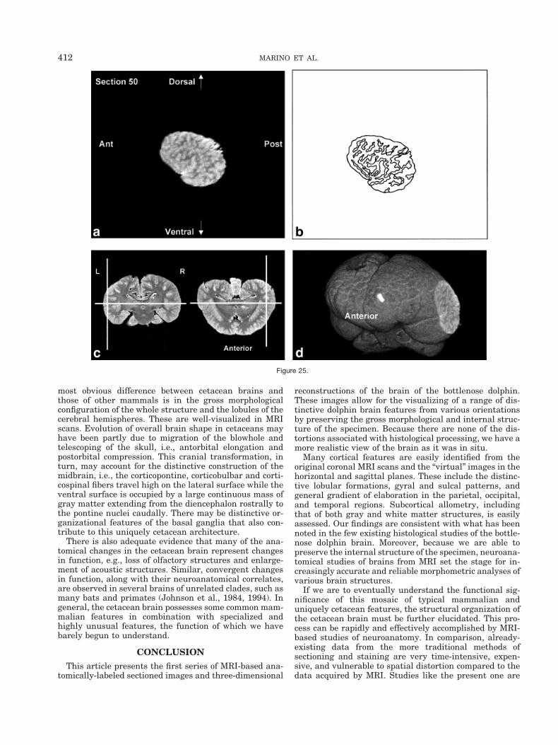
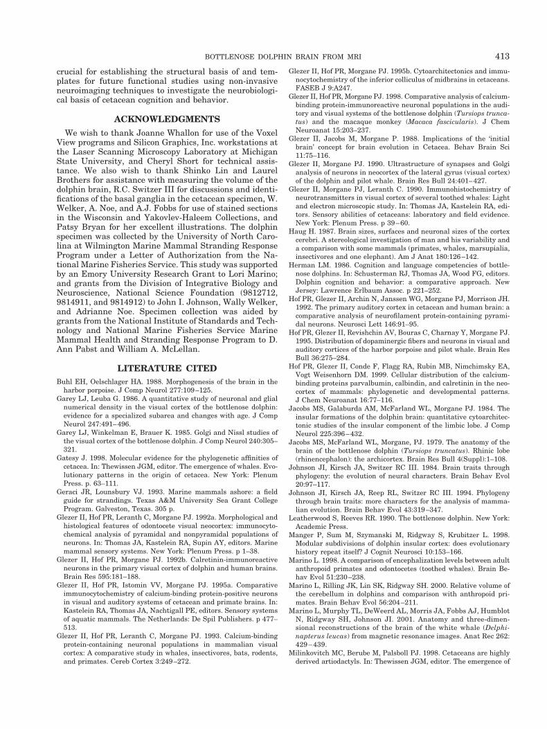
Figures 2–9 display a posterior-to-anterior sequence oforiginally-acquired 2.0 mm-thick coronal MR brain sec-tions at 30 mm intervals, a labeled schematic illustrationof each section, computer-generated cutaways shown attwo orthogonal planes to the section, and a computer-generated three-dimensional reconstruction of the wholebrain showing a cutaway of each section. Figures 10–17display a ventral-to-dorsal sequence of reconstructed “vir-tual” horizontal sections (1.0 mm thick) at 14 mm inter-vals, a labeled schematic illustration of each section, com-puter-generated cutaways at two orthogonal planes to thesection, and a computer-generated three-dimensional re-construction of the whole brain showing a cutaway of eachsection. Figures 18–25 display a midline-to-lateral se-quence of reconstructed “virtual” sagittal sections, also 1.0mm thick, through the right hemisphere at 11 mm inter-vals, a labeled schematic illustration of each section, com-puter-generated cutaways of two orthogonal planes to thesection, and a computer-generated three-dimensional re-construction of the whole brain showing a cutaway of eachsection.
Figures 2–9 demonstrate the excellent level of preser-vation of the spatial relationships among the brain’s struc-tures that has allowed for reconstruction in the horizontaland sagittal planes shown in Figures 10–17 and 18–25,respectively. All of the sagittally-oriented three-dimen-sional reconstructions and, in particular, Figure 18, showthe mesencephalic and pontine flexures reminiscent ofbrainstem flexure patterns in the embryonic state of mostterrestrial mammals. These flexures remain present inadult bottlenose dolphin brains and may represent pedo-morphic features.
The high level of cortical convolution is particularlyclear in Figures 3–9, 13–17, and 18–25. The extremedepth and density of cortical sulci are particularly evidentin Figures 19–23. Figures 18–20 also display an orbital-to-occipital gradient of increased sulcation concordantwith the increased elaboration of the occipital-parietalregion over the orbital region. This occipital-parietal elab-oration is evident in Figure 20 in the three-tiered arrange-ment of limbic, paralimbic, and supralimbic arcuate cor-tical lobules divided by the deep limbic and paralimbicclefts. This specific combination of occipital-parietal orga-nization and elaboration is distinct from other mammalsbut not unique to the bottlenose dolphin among the ceta-ceans (Morgane et al., 1980).
In contrast to the distinctive cortical features, the bot-tlenose dolphin brain generally resembles other mamma-lian brains on a subcortical level (Morgane et al., 1980).Despite the shared subcortical structures between dolphinbrains and other mammal brains, however, the volumetricproportions of various subcortical features of the dolphinbrain are quite different from those of terrestrial mamma-lian brains. These morphometric differences are expres-sions of different ecological histories between dolphinsand terrestrial mammals. For instance, as seen in Figure1b, the olfactory bulbs are absent. In contrast, auditoryprocessing areas, such as the inferior colliculus, seen inFigures 5, 12, and 19, are enlarged, presumably because ofthe substantial amount of acoustic processing conductedby the dolphin. The enlargement of auditory processingstructures is not, notably, accompanied by reduced visual
Fig. 1. Dorsal view (a) and antero-ventral view (b) of computer-generated three-dimensional reconstruction of the bottlenose dolphinbrain.
399BOTTLENOSE DOLPHIN BRAIN FROM MRI
structures, which suggests that vision is an importantsensory-perceptual system in dolphins.
In keeping with the behavioral and electrophysiologicalevidence for a high degree of hemispheric independence(Viamonte et al., 1968; Mukhametov, 1984; Mukhametovet al., 1977), the corpus callosum is small relative to themassive hemispheres, consistent with quantitative find-ings in other odontocete species and qualitative observa-tions of the white whale brain (Marino et al., 2001; Tarp-ley and Ridgway, 1994). This feature is most apparent inthe three planes in Figures 6, 7, 14, and 18–21.
The cerebellum is large relative to the hemispheres(Marino et al., 2000), as is especially evident in Figures 4,5,12, 18, and 19. As shown in Figure 6 the cerebral pe-duncles are high on the lateral surface of the caudal dien-cephalon and through the entire midbrain, rather than onthe basal inferior or ventral surface as in most mammals.The basal surface is instead occupied by a large mass ofgray matter, which appears to be continuous with theventral striatum and the dorsal and ventral pallidum ofthe forebrain reaching from these structures to the pon-tine nuclei caudally.
Evolutionary ConsiderationsCetacean evolution is characterized by distinctive envi-
ronmental pressures associated with a fully aquatic exis-
tence versus a terrestrial lifestyle. These related at-tributes make the comparative study of structure-functionrelationships in cetacean brains, compared to those ofother mammals, especially valuable for improving ourunderstanding of the parameters of mammalian brainevolution.
There is abundant evidence for a phylogenetic link be-tween cetaceans and ungulates (e.g., Milinkovitch et al.,1998) and evidence for a proposed sister-group relation-ship with the hippopotamus (Gatesy, 1998). Given theseaffinities it is worth noting that there are similarities incortical cytoarchitecture and neurochemistry betweencetaceans and ungulates (Hof et al., 1999). It would beinteresting to compare how these similarities (and differ-ences) have been expressed at gross neuroanatomical lev-els in cetaceans and various ungulates. For instance, Hofet al. (1999) have noted the early physical maturity of thebrain in both cetaceans and ungulates and the possibilitythat both brains may be characterized by a number ofpedomorphic features.
The brain of the bottlenose dolphin as revealed in thisstudy is characterized by similar morphological trends asthose found in other cetaceans (Morgane et al., 1980).Although there are differences among cetacean brains,these differences are relatively minor compared to theirstriking dissimilarities to brains of other mammals. The
Figure 2.Figs. 2–9. Posterior-to-anterior sequence of (a) originally-acquired 2.0 mm-thick coronal MR brain sectionsat 30 mm intervals, (b) a labeled schematic illustration of each section, (c) computer-generated cutawaysshown at two orthogonal planes to the section, and (d) a computer-generated three-dimensional reconstruc-tion of the whole brain showing a cutaway of each section.
400 MARINO ET AL.

Figure 3.
Figure 4.
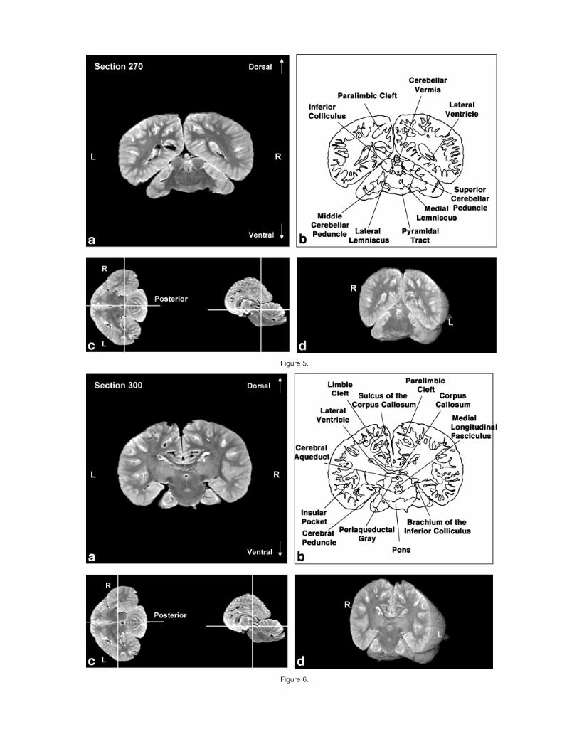
Figure 5.
Figure 6.
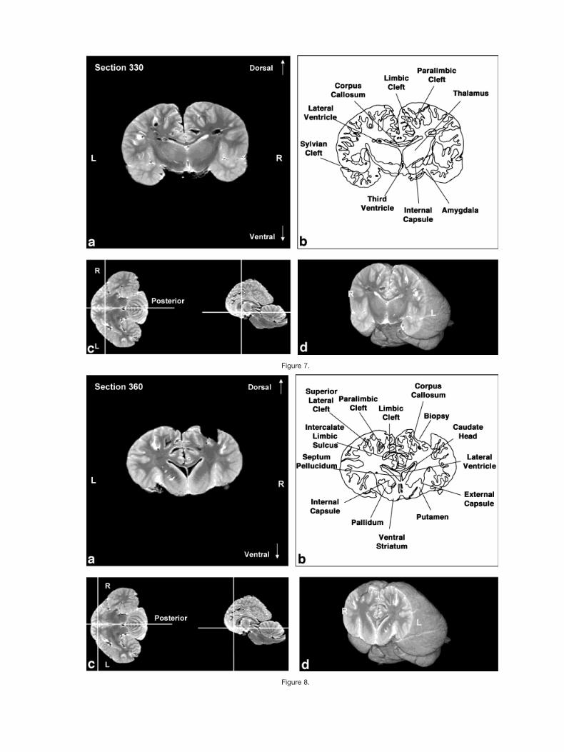
Figure 7.
Figure 8.
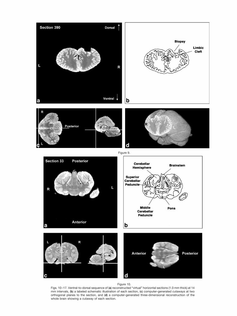
Figure 10.Figs. 10–17. Ventral-to-dorsal sequence of (a) reconstructed “virtual” horizontal sections (1.0 mm thick) at 14mm intervals, (b) a labeled schematic illustration of each section, (c) computer-generated cutaways at twoorthogonal planes to the section, and (d) a computer-generated three-dimensional reconstruction of thewhole brain showing a cutaway of each section.
Figure 9.
Figure 11.
Figure 12.

Figure 13.
Figure 14.
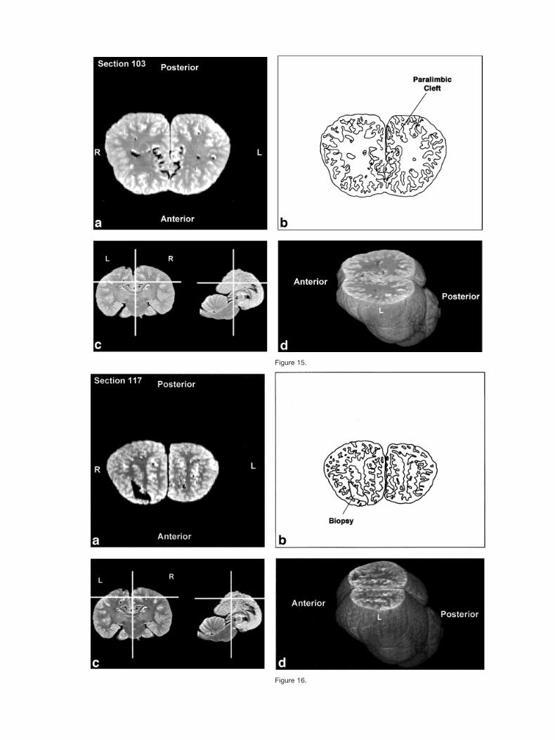
Figure 15.
Figure 16.
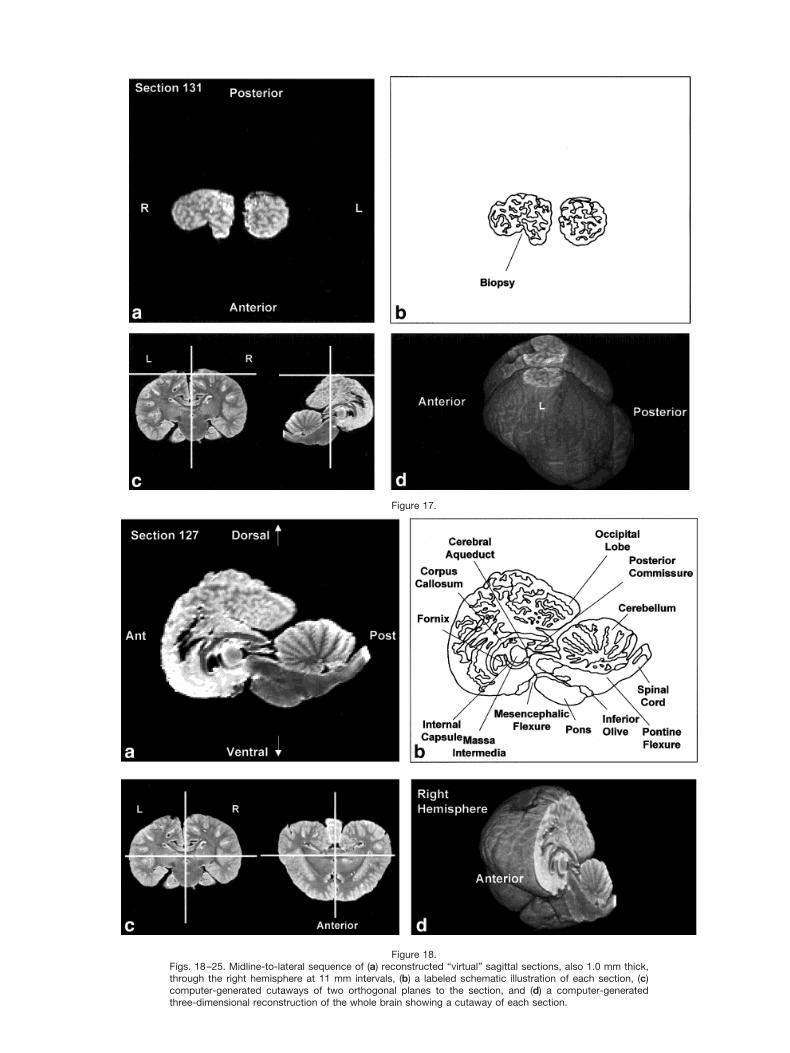
Figure 18.Figs. 18–25. Midline-to-lateral sequence of (a) reconstructed “virtual” sagittal sections, also 1.0 mm thick,through the right hemisphere at 11 mm intervals, (b) a labeled schematic illustration of each section, (c)computer-generated cutaways of two orthogonal planes to the section, and (d) a computer-generatedthree-dimensional reconstruction of the whole brain showing a cutaway of each section.
Figure 17.
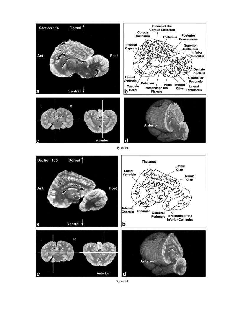
Figure 19.
Figure 20.
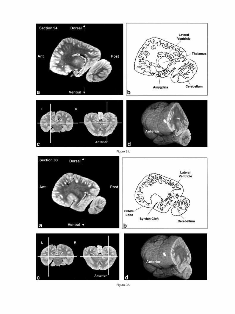
Figure 21.
Figure 22.
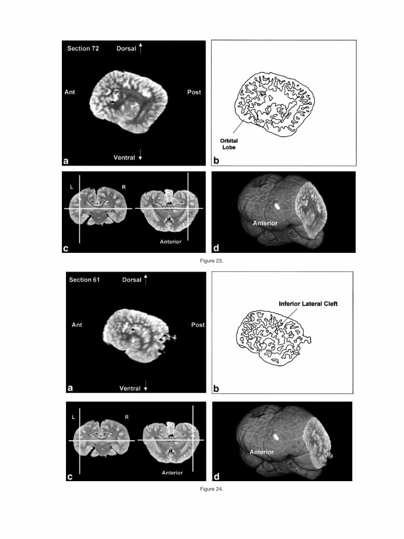
Figure 23.
Figure 24.
most obvious difference between cetacean brains andthose of other mammals is in the gross morphologicalconfiguration of the whole structure and the lobules of thecerebral hemispheres. These are well-visualized in MRIscans. Evolution of overall brain shape in cetaceans mayhave been partly due to migration of the blowhole andtelescoping of the skull, i.e., antorbital elongation andpostorbital compression. This cranial transformation, inturn, may account for the distinctive construction of themidbrain, i.e., the corticopontine, corticobulbar and corti-cospinal fibers travel high on the lateral surface while theventral surface is occupied by a large continuous mass ofgray matter extending from the diencephalon rostrally tothe pontine nuclei caudally. There may be distinctive or-ganizational features of the basal ganglia that also con-tribute to this uniquely cetacean architecture.
There is also adequate evidence that many of the ana-tomical changes in the cetacean brain represent changesin function, e.g., loss of olfactory structures and enlarge-ment of acoustic structures. Similar, convergent changesin function, along with their neuroanatomical correlates,are observed in several brains of unrelated clades, such asmany bats and primates (Johnson et al., 1984, 1994). Ingeneral, the cetacean brain possesses some common mam-malian features in combination with specialized andhighly unusual features, the function of which we havebarely begun to understand.
CONCLUSIONThis article presents the first series of MRI-based ana-
tomically-labeled sectioned images and three-dimensional
reconstructions of the brain of the bottlenose dolphin.These images allow for the visualizing of a range of dis-tinctive dolphin brain features from various orientationsby preserving the gross morphological and internal struc-ture of the specimen. Because there are none of the dis-tortions associated with histological processing, we have amore realistic view of the brain as it was in situ.
Many cortical features are easily identified from theoriginal coronal MRI scans and the “virtual” images in thehorizontal and sagittal planes. These include the distinc-tive lobular formations, gyral and sulcal patterns, andgeneral gradient of elaboration in the parietal, occipital,and temporal regions. Subcortical allometry, includingthat of both gray and white matter structures, is easilyassessed. Our findings are consistent with what has beennoted in the few existing histological studies of the bottle-nose dolphin brain. Moreover, because we are able topreserve the internal structure of the specimen, neuroana-tomical studies of brains from MRI set the stage for in-creasingly accurate and reliable morphometric analyses ofvarious brain structures.
If we are to eventually understand the functional sig-nificance of this mosaic of typical mammalian anduniquely cetacean features, the structural organization ofthe cetacean brain must be further elucidated. This pro-cess can be rapidly and effectively accomplished by MRI-based studies of neuroanatomy. In comparison, already-existing data from the more traditional methods ofsectioning and staining are very time-intensive, expen-sive, and vulnerable to spatial distortion compared to thedata acquired by MRI. Studies like the present one are
Figure 25.
412 MARINO ET AL.
crucial for establishing the structural basis of and tem-plates for future functional studies using non-invasiveneuroimaging techniques to investigate the neurobiologi-cal basis of cetacean cognition and behavior.
ACKNOWLEDGMENTS
We wish to thank Joanne Whallon for use of the VoxelView programs and Silicon Graphics, Inc. workstations atthe Laser Scanning Microscopy Laboratory at MichiganState University, and Cheryl Short for technical assis-tance. We also wish to thank Shinko Lin and LaurelBrothers for assistance with measuring the volume of thedolphin brain, R.C. Switzer III for discussions and identi-fications of the basal ganglia in the cetacean specimen, W.Welker, A. Noe, and A.J. Fobbs for use of stained sectionsin the Wisconsin and Yakovlev-Haleem Collections, andPatsy Bryan for her excellent illustrations. The dolphinspecimen was collected by the University of North Caro-lina at Wilmington Marine Mammal Stranding ResponseProgram under a Letter of Authorization from the Na-tional Marine Fisheries Service. This study was supportedby an Emory University Research Grant to Lori Marino;and grants from the Division of Integrative Biology andNeuroscience, National Science Foundation (9812712,9814911, and 9814912) to John I. Johnson, Wally Welker,and Adrianne Noe. Specimen collection was aided bygrants from the National Institute of Standards and Tech-nology and National Marine Fisheries Service MarineMammal Health and Stranding Response Program to D.Ann Pabst and William A. McLellan.
LITERATURE CITEDBuhl EH, Oelschlager HA. 1988. Morphogenesis of the brain in the
harbor porpoise. J Comp Neurol 277:109–125.Garey LJ, Leuba G. 1986. A quantitative study of neuronal and glial
numerical density in the visual cortex of the bottlenose dolphin:evidence for a specialized subarea and changes with age. J CompNeurol 247:491–496.
Garey LJ, Winkelman E, Brauer K. 1985. Golgi and Nissl studies ofthe visual cortex of the bottlenose dolphin. J Comp Neurol 240:305–321.
Gatesy J. 1998. Molecular evidence for the phylogenetic affinities ofcetacea. In: Thewissen JGM, editor. The emergence of whales. Evo-lutionary patterns in the origin of cetacea. New York: PlenumPress. p. 63–111.
Geraci JR, Lounsbury VJ. 1993. Marine mammals ashore: a fieldguide for strandings. Texas A&M University Sea Grant CollegeProgram. Galveston, Texas. 305 p.
Glezer II, Hof PR, Leranth C, Morgane PJ. 1992a. Morphological andhistological features of odontocete visual neocortex: immunocyto-chemical analysis of pyramidal and nonpyramidal populations ofneurons. In: Thomas JA, Kastelein RA, Supin AY, editors. Marinemammal sensory systems. New York: Plenum Press. p 1–38.
Glezer II, Hof PR, Morgane PJ. 1992b. Calretinin-immunoreactiveneurons in the primary visual cortex of dolphin and human brains.Brain Res 595:181–188.
Glezer II, Hof PR, Istomin VV, Morgane PJ. 1995a. Comparativeimmunocytochemistry of calcium-binding protein-positive neuronsin visual and auditory systems of cetacean and primate brains. In:Kastelein RA, Thomas JA, Nachtigall PE, editors. Sensory systemsof aquatic mammals. The Netherlands: De Spil Publishers. p 477–513.
Glezer II, Hof PR, Leranth C, Morgane PJ. 1993. Calcium-bindingprotein-containing neuronal populations in mammalian visualcortex: A comparative study in whales, insectivores, bats, rodents,and primates. Cereb Cortex 3:249–272.
Glezer II, Hof PR, Morgane PJ. 1995b. Cytoarchitectonics and immu-nocytochemistry of the inferior colliculus of midbrains in cetaceans.FASEB J 9:A247.
Glezer II, Hof PR, Morgane PJ. 1998. Comparative analysis of calcium-binding protein-immunoreactive neuronal populations in the audi-tory and visual systems of the bottlenose dolphin (Tursiops trunca-tus) and the macaque monkey (Macaca fascicularis). J ChemNeuroanat 15:203–237.
Glezer II, Jacobs M, Morgane P. 1988. Implications of the ‘initialbrain’ concept for brain evolution in Cetacea. Behav Brain Sci11:75–116.
Glezer II, Morgane PJ. 1990. Ultrastructure of synapses and Golgianalysis of neurons in neocortex of the lateral gyrus (visual cortex)of the dolphin and pilot whale. Brain Res Bull 24:401–427.
Glezer II, Morgane PJ, Leranth C. 1990. Immunohistochemistry ofneurotransmitters in visual cortex of several toothed whales: Lightand electron microscopic study. In: Thomas JA, Kastelein RA, edi-tors. Sensory abilities of cetaceans: laboratory and field evidence.New York: Plenum Press. p 39–60.
Haug H. 1987. Brain sizes, surfaces and neuronal sizes of the cortexcerebri. A stereological investigation of man and his variability anda comparison with some mammals (primates, whales, marsupialia,insectivores and one elephant). Am J Anat 180:126–142.
Herman LM. 1986. Cognition and language competencies of bottle-nose dolphins. In: Schusterman RJ, Thomas JA, Wood FG, editors.Dolphin cognition and behavior: a comparative approach. NewJersey: Lawrence Erlbaum Assoc. p 221–252.
Hof PR, Glezer II, Archin N, Janssen WG, Morgane PJ, Morrison JH.1992. The primary auditory cortex in cetacean and human brain: acomparative analysis of neurofilament protein-containing pyrami-dal neurons. Neurosci Lett 146:91–95.
Hof PR, Glezer II, Revishchin AV, Bouras C, Charnay Y, Morgane PJ.1995. Distribution of dopaminergic fibers and neurons in visual andauditory cortices of the harbor porpoise and pilot whale. Brain ResBull 36:275–284.
Hof PR, Glezer II, Conde F, Flagg RA, Rubin MB, Nimchimsky EA,Vogt Weisenhorn DM. 1999. Cellular distribution of the calcium-binding proteins parvalbumin, calbindin, and calretinin in the neo-cortex of mammals: phylogenetic and developmental patterns.J Chem Neuroanat 16:77–116.
Jacobs MS, Galaburda AM, McFarland WL, Morgane PJ. 1984. Theinsular formations of the dolphin brain: quantitative cytoarchitec-tonic studies of the insular component of the limbic lobe. J CompNeurol 225:396–432.
Jacobs MS, McFarland WL, Morgane, PJ. 1979. The anatomy of thebrain of the bottlenose dolphin (Tursiops truncatus). Rhinic lobe(rhinencephalon): the archicortex. Brain Res Bull 4(Suppl):1–108.
Johnson JI, Kirsch JA, Switzer RC III. 1984. Brain traits throughphylogeny: the evolution of neural characters. Brain Behav Evol20:97–117.
Johnson JI, Kirsch JA, Reep RL, Switzer RC III. 1994. Phylogenythrough brain traits: more characters for the analysis of mamma-lian evolution. Brain Behav Evol 43:319–347.
Leatherwood S, Reeves RR. 1990. The bottlenose dolphin. New York:Academic Press.
Manger P, Sum M, Szymanski M, Ridgway S, Krubitzer L. 1998.Modular subdivisions of dolphin insular cortex: does evolutionaryhistory repeat itself? J Cognit Neurosci 10:153–166.
Marino L. 1998. A comparison of encephalization levels between adultanthropoid primates and odontocetes (toothed whales). Brain Be-hav Evol 51:230–238.
Marino L, Rilling JK, Lin SK, Ridgway SH. 2000. Relative volume ofthe cerebellum in dolphins and comparison with anthropoid pri-mates. Brain Behav Evol 56:204–211.
Marino L, Murphy TL, DeWeerd AL, Morris JA, Fobbs AJ, HumblotN, Ridgway SH, Johnson JI. 2001. Anatomy and three-dimen-sional reconstructions of the brain of the white whale (Delphi-napterus leucas) from magnetic resonance images. Anat Rec 262:429 – 439.
Milinkovitch MC, Berube M, Palsboll PJ. 1998. Cetaceans are highlyderived artiodactyls. In: Thewissen JGM, editor. The emergence of
413BOTTLENOSE DOLPHIN BRAIN FROM MRI

whales. Evolutionary patterns in the origin of cetacea. New York:Plenum Press. p 113–131.
Morgane P, Jacobs M, Galaburda A. 1986. Evolutionary morphologyof the dolphin brain. In: Schusterman RJ, Thomas JA, Wood FG,editors. Dolphin cognition and behavior: a comparative approach.Hillsdale, New Jersey: Lawrence Erlbaum. p 5–30.
Morgane PJ, Jacobs MS, MacFarland WL. 1980. The anatomy of thebrain of the bottlenose dolphin (Tursiops truncatus). Surface con-figurations of the telencephalon of the bottlenose dolphin with com-parative anatomical observations in four other cetacean species.Brain Res Bull 5(Suppl):1–107.
Mukhametov LM. 1984. Sleep in marine mammals. Exp Brain Res8:227–238.
Mukhametov LM, Supin AY, Polyakova IG. 1977. Interhemisphericasymmetry of the electroencephalographic sleep patterns in dol-phins. Brain Res 134:581–584.
Oelschlager HA, Buhl EH. 1985. Occurrence of an olfactory bulb inthe early development of the harbor porpoise (Phocoena phocoenaL.). In: Duncker HR, Fleischer G, editors. Functional morphology invertebrates. New York: Fischer. p 695–698.
Oelschlager HA, Kemp B. 1998. Ontogenesis of the sperm whalebrain. J Comp Neurol 399:210–228.
Reiss D, McCowan B, Marino L. 1997. Communicative and othercognitive characteristics of bottlenose dolphins. Trends Cognit Sci1:140–145.
Reynolds JE, Wells RS, Eide SD. 2000. The bottlenose dolphin: biol-ogy and conservation. Florida: University Press of Florida.
Ridgway SH. 1986. The central nervous system of the bottlenosedolphin. In: Schusterman RJ, Thomas JA, Wood FG, editors. Dol-phin cognition and behavior: a comparative approach. Hillsdale,New Jersey: Lawrence Erlbaum Assoc. p 31–60.
Ridgway SH. 1990. The central nervous system of the bottlenosedolphin. In: Leatherwood S, Reeves R, editors. The bottlenose dol-phin. San Diego: Academic Press. p 69–97.
Ridgway SH, Brownson RH. 1984. Relative brain sizes and corticalsurface areas in odontocetes. Acta Zool Fennica 172:149–152.
Stephan H, Frahm H, Baron G. 1981. New and revised data onvolumes of brain structures in insectivores and primates. FoliaPrimatol 25:1–29.
Tarpley RL, Ridgway SH. 1994. Corpus callosum size in delphinidcetaceans. Brain Behav Evol 44:156–165.
Viamonte M, Morgane PJ, Galliano RE, Nagel EL, McFarland WL.1968. Angiography in the living dolphin and observations on bloodsupply to the brain. Am J Physiol 214:1225–1249.
414 MARINO ET AL.