analysis of somaclonal variation using molecular markers .... jayanthi, et al.pdf · autoclaved...
TRANSCRIPT

Int.J.Curr.Microbiol.App.Sci (2018) 7(12): 1642-1655
1642
Original Research Article https://doi.org/10.20546/ijcmas.2018.712.192
Analysis of Somaclonal Variation using Molecular Markers in in vitro
Regenerated Chrysanthemum cv. Pusa Centenary
M. Jayanthi1, Rajesh Gupta
1, P.K. Mandal
2* and Kanchan B.M. Singh
1
1Division of Nematology, Indian Agricultural Research Institute, New Delhi - 110012, India
2National Research Centre for Plant Biotechnology, New Delhi-110012, India
*Corresponding author
A B S T R A C T
Introduction
Chrysanthemum is globally the second
economically important floriculture crop
followed by rose and one of the most
important ornamental species (Teixeira da
Silva, 2004). In Chrysanthemum it is reported
that in vitro techniques are useful to obtain
plants that are free of Tomato aspermy virus,
Cucumber mosaic virus and Chrysanthemum
B virus (Ram et al., 2009). Also in vitro
propagation is a useful strategy to get large
number of plants from limited tissues which
can meet the demand in the expected time.
International Journal of Current Microbiology and Applied Sciences ISSN: 2319-7706 Volume 7 Number 12 (2018) Journal homepage: http://www.ijcmas.com
Detection of somaclonal variation in tissue cultured plants is essential for conservation of
elite varieties and also to avoid undesired characters. The genetic fidelity of in vitro
regenerated Chrysanthemum cv. Pusa centenary, a commercially important cultivar was
assessed using RAPD and ISSR markers. Regeneration was obtained via organogenesis
and somatic embryogenesis from leaf and petal explants respectively. Murashige and
Skoog (MS) media with different plant growth regulators (PGR) were used for in vitro
experiments with leaf explants. Among the different PGR combinations used in MS media,
2,4-Dichlorophenoxyacetic acid (2,4-D) (1 mg L-1
) and ἀ-naphthalene acetic acid (NAA)
(1 mg L-1
) along with 0.5 mg L-1
of 6-benzylaminopurine (BAP) or Kinetin (Kin) was
optimal for producing 83-93% callus induction from leaf explants. On transfer to light on
MS media with BAP (0.5 mg L-1
) and reduced auxins, the calli started turning green and
proliferating into plantlets. Petal explants on the same media, produced direct somatic
embryos within 10-14 days. On subculture, profuse production of somatic embryos with
root and shoot poles was obtained. Nineteen primers (9 RAPD and 10 ISSR primers)
produced 825 clear reproducible and scorable bands. While RAPD primers produced
completely monomorphic bands with both leaf and petal regenerated plants in comparison
to their mother plant, it was found that ISSR primers were able to detect variation in leaf
callus derived plantlets. The plants regenerated via organogenesis from leaf explants were
showing a variation of 0.66% with ISSR primers and plants regenerated from distinct
somatic embryos of petal explants showed completely monomorphic bands with both
RAPD and ISSR proving it’s true to type nature. These results indicate that in vitro culture
process induces rearrangements at the DNA level and demonstrates discrepancies between
the pathways involved in regeneration.
K e y w o r d s
Callus induction,
Direct
embryogenesis,
Genetic fidelity,
RAPD and ISSR
Accepted:
12 November 2018
Available Online: 10 December 2018
Article Info

Int.J.Curr.Microbiol.App.Sci (2018) 7(12): 1642-1655
1643
Chrysanthemum Pusa centenary is a newly
released variety which is a gamma ray induced
mutant of Thai Chen Queen. This cultivar is in
demand due to its vigorous growing nature
and it produces yellow flowers which remain
fresh for 20 - 22 days in the field as well as in
vase. To meet the demand we attempted to
develop in vitro protocols for large scale
multiplication of this cultivar from leaf and
petal explants. Though protocols for in vitro
regeneration are reported in Chrysanthemum
(Himstedt et al., 2001; Petty et al., 2003;
Waseem et al., 2009) a varied response of
genotypes to auxins and cytokinins are seen
from these reports and the need to develop
cultivar specific protocols have been
emphasized (Song et al., 2011). They also
accentuate that establishment of propagation
protocols must be optimized for each
commercially important cultivar.
Retaining the genetic integrity of
micropropagated plants is a major concern in
tissue culture. Generally the problem
associated with in vitro culture is the
occurrence of genetic variation resulting from
micropropagation i.e. somaclonal variation
amongst sub-clones of one parental line
arising as a direct consequence of in vitro
culture of cells, tissues and organs (Larkin and
Scowcroft, 1981; Gould, 1986). Hence a
quality checkup for true to type plants at an
early stage is very essential (Zilberman and
Henikoff, 2007). Molecular markers like
Random Amplified Polymorphic DNA
(RAPD) and Inter-Simple Sequence Repeats
(ISSR) have been suggested to be useful for
confirmation of genetic fidelity in
micropropagated plants (Ray et al., 2006).
These two techniques have the advantages of
giving reproducible results, low cost and
primers can be designed without any prior
knowledge of sequences (Pradeep et al.,
2002). RAPD is a simple, dominant, quick and
easy assay marker which requires low
quantities (5-50ng) of template DNA. ISSR
technique is also fast, simple and cost
effective and a highly reliable technique which
uses a single sequence repeat motif as a primer
for amplification of regions between
microsatellites. This paper reports differential
response of leaf and petal explants under in
vitro conditions for plant regeneration and the
use of RAPD and ISSR techniques for
assessing the genetic homogeneity of the
regenerated plants from both the explants.
Materials and Methods
Experiment with leaf explants
Plant material
Leaves were collected from a single mature
plant of Pusa centenary which was maintained
in the glass house. They were washed
thoroughly in running tap water for two hours
followed by washing with tween 20 (2drops
per 100ml of water). Surface sterilization was
done by immersion in 0.1% mercuric chloride
for 5 minutes followed by three washes with
double distilled sterile water. They were cut
into fine pieces of 3-4mm long sections and
placed on the culture media with the adaxial
surface touching the media.
Culture media and growth conditions for
callus induction
The basal media used was MS (Murashige and
Skoog, 1962) basal salts supplemented with
sucrose (3%), and agar (0.8%), (Himedia,
Mumbai, India). For callus induction MS basal
media was supplemented with 2,4-
Dichlorophenoxyacetic acid (2,4-D) (2 mg
L-1
) and ἀ-naphthalene acetic acid (NAA) (2
mg L-1
) singly and in combination as 2,4-D, (1
mg L-1
) and NAA (1mg L-1
) along with
cytokinins like 6-benzylaminopurine(BAP) or
Kinetin at 0.5 mg L-1
. Six different
combinations of plant growth regulators in MS
basal were prepared and coded as M1 to M6

Int.J.Curr.Microbiol.App.Sci (2018) 7(12): 1642-1655
1644
(Table 1). These media was solidified with
agar @ 8 g L-1
(Himedia). For all media
prepared the pH was adjusted to 5.8 with
either 0.1N NaOH or 0.1 N HCl and they were
dispensed in test tubes (Borosil make
25X150mm) and each test tube contained
20ml media. They were sterilized at 1210C at
1.06 kg/cm2 for 15 minutes. Explants were
cultured in test tubes at the rate of one explant
per tube. A total of 27 explants were
inoculated in each treatment and maintained as
three replications of nine explants each.
Explants were cultured in dark at 25 ± 2°C for
callus induction for 1 month. The number of
cultures producing callus was observed at the
end of 45 day.
Culture media and growth conditions for
shoot induction
The callus formation continued for two
months and then they were subcultured to MS
basal media in jam bottles (500ml) with
auxins reduced to 0.1mg/l and cytokinin
concentration unchanged (Table 1) and
transferred to culture room with light provided
by fluorescent bulbs (Philips) with intensity of
2000-3000 lux and maintaining a photoperiod
of 16 h per day with temperature of 25 ± 2°C.
A total of 27 explants with callus were
maintained as three replications of nine
explants each. Calli were cultured in jam
bottles at the rate of one explant with callus
per bottle. The media details are provided in
Table 1.
After four weeks, the number of shoots that
emerged from each combination was counted.
These were again subcultured to MS basal
media for rooting and maintained for 30-40
days. Analysis of variance was performed for
the effect of plant growth regulators on callus
induction and shoot induction separately.
Means were compared using least significant
difference test (LSD: P<0.05).
Experiments with petal explants
M5 media (MS basal with 2,4-D, (1 mg L-1
)
and NAA (1 mg L-1
) along with 6-
benzylaminopurine(BAP) at 0.5 mg L-1
) was
prepared and used for culturing petal explants
collected from the same mother plant. Surface
sterilisation and culture conditions were the
same as used for leaf explants. Thirty explants
of 1-2mm sections were inoculated as three
replications of ten each. These explants were
further sub-cultured onto the MS basal media
with 2,4-D, (0.1 mg L-1
) and NAA (0.1 mg
L-1
) along with BAP at 0.5 mg L-1
and
maintained for 1-2 months. Well-developed
single embryos were transferred to MS basal
media with BAP (0.5 mg L-1
) for plantlet
formation and allowed to remain in the same
media for a period of 30 days. The well-
developed plants were then taken up for
primary hardening. The details on the number
of explants, number of somatic embryos and
the number of plants regenerated are
mentioned in Table 2.
Hardening
The well rooted plantlets were taken out from
the culture media and washed with water
carefully to remove the traces of agar and they
were transplanted in jam bottles containing
autoclaved mixture of Co-copit, Perlite and
Vermicompost (2:1:1) for primary hardening
and incubated in the same culture room in
light at 25 ± 2°C. The plantlets (10-12 cm
height) after sufficient growth were
transferred from the bottles to plastic pots
containing sterile mixture of sand, soil and
vermicompost (1:2:1) and maintained in the
polyhouse where in a temperature of 28±2oC
with relative humidity of 65-75% was
maintained for secondary hardening. The
plantlets were irrigated with tap water twice in
a week for a total period of two weeks to
complete the acclimatization process. The
plantlets obtained from leaf explants and petal
explants were maintained separately.

Int.J.Curr.Microbiol.App.Sci (2018) 7(12): 1642-1655
1645
DNA isolation and quantification
From two different batches of plants
regenerated from leaf and petal explants,
plantlets were selected for DNA extraction.
Young leaves (300-400mg) were collected
from selected plants and total genomic DNA
was extracted using a cetyltrimethyl
ammonium bromide (CTAB) procedure
(Doyle and Doyle, 1990)13
. 300-400mg of
fresh leaves was ground to powder in liquid
nitrogen using a mortar and pestle. The ground
powder was transferred to a 50 ml falcon tube
with 10 ml of CTAB buffer [2% (w/v) CTAB,
1.4 M NaCl, 20 mM Ethylene diamine
tetraacetic acid, 100 mM Tris (tri
(hydroxymethyl) amino methane]-HCl, and
0.2% (v/v) β-mercaptoethanol). The
homogenate was incubated at 60oC for 2 h,
before extraction with an equal volume of
chloroform/isoamyl alcohol (24:1 v/v) by
centrifugation at 10,000 x g for 20 min. DNA
was precipitated from the aqueous phase by
mixing with an equal volume of isopropanol.
After centrifugation at 10,000 x g for 10 min,
the DNA pellet was washed with 70% (v/v)
ethanol, air-dried and resuspended in TE (10
mM Tris-HCl, pH 8.0, and 0.1 mM EDTA)
buffer. The quantification of isolated DNA
was performed by visualizing under UV light,
after electrophoresis on 0.8% (w/v) agarose
gel at 60 V for 45 min and compared to a
known amount of uncut lambda DNA (MBI,
Fermentas). Quantified samples were then
treated with 1μl RNase A per 100 μl DNA
solution at 37 oC for one hour. DNA samples
were finally diluted with sterile TE buffer to
get the final concentration of 25 ng μl-1
. 2 μl
of diluted DNA was subsequently used for
polymerase chain reaction (PCR)
amplification.
Primers
Twenty random primers (10-mer from Sigma-
Aldrich Chemicals, St. Louis, USA) and
twenty Universal ISSR primers (Integrated
device technology, India), were used for initial
DNA amplification and based on consistent
results nine of each were selected for further
analysis.
PCR Amplification conditions
Amplification with both RAPD (Williams et
al., 1990) and ISSR primers (Zietkiewicz et
al., 1994) was carried out in a total volume of
25 μl containing 2 μl (25 ng) of template
DNA, 2.5 μl of 10X PCR buffer containing 15
mM MgCl2, 0.5 μl of dNTPs (10 mM each of
dATP, dGTP, dTTP, dCTP), 1 μl of primer,
0.25 μl of Taq DNA polymerase and 18.75 μl
of sterile MQ water.
Amplification conditions were performed as
initial DNA denaturation at 95ºC for 4 min.
followed by 35 cycles of 1 min denaturation at
95ºC, 1 min annealing at 37ºC and 2 min of
extension at 72ºC with a final extension time
at 72ºC for 10 min. In case of ISSR primers,
optimal annealing temperature was adjusted
according to the base concentrations of the
primers. Hence, annealing temperature was
optimized for each primer using a gradient
PCR.
Electrophoresis of amplified DNA
Amplified DNA fragments were separated and
visualized on a 1.2% (w/v) agarose gel stained
with ethidium bromide 3 μl of 6X loading dye
was added to 25 μl of amplified products and
following homogenization, 5 μl of the
resulting mixture was loaded onto a gel
prepared in 0.5X TAE buffer. DNA ladder (1
Kb marker, #SM0313, Fermentas life science,
Germany) was also loaded flanking the
samples. The sizes of the amplicons were
estimated by comparing with 1kb DNA
ladder. The gel was visualized on a UV
transilluminator and photographed by an
Alphaimager Gel Documentation System
(Alpha Innotech Corporation, USA).

Int.J.Curr.Microbiol.App.Sci (2018) 7(12): 1642-1655
1646
Statistical data analysis
In both marker systems, RAPD and ISSR,
scorable bands were recorded as present (1) or
absent (0) and based on band data amplified.
All the bands (Polymorphic and
monomorphic) were taken into account for
calculation of similarity with a view to avoid
over/under estimation of the distance.
Jaccard’s coefficients of similarity (Jaccard,
1908) was measured and a dendrogram based
on similarity coefficient generated by the Un-
weight Pair Group method using arithmetic
averages (UPGMA) (Sneath and Sokal,
1973), and Sequential Agglomerative
Hierarchial Non-overlapping (SHAN)
clustering. The statistical analysis was done
using the software NTSYS-PC (Numerical
Taxonomy System for Personal Computers,
version 2.02i) (Rohlf, 1992).
Results and Discussion
In vitro responses from leaf explants
In leaf explants the response was seen as
swelling and the initiation of callus was seen
after 15-20 days on the cut edges of the leaf
explant. By the end of 35-40 days the callus
proliferated (Fig. 1A). The initiation of callus
was influenced by the auxin concentration in
the media. Though callus was produced in all
combinations of plant growth regulators the
maximum callus induction of 93% was seen
in MS with 2,4-D, (1 mg L-1
) + NAA (1 mg
L-1
) +Kin (0.5 mg L-1
) (M6) followed by 83%
callus induction in MS with 2,4-D, (1 mg L-1
)
+ NAA (1 mg L-1
) +BAP (0.5 mg L-1
) (M5)
which were not significantly different from
each other. Callus induction percentage
ranged from 26 to 93% in MS basal with six
different combinations of plant growth
regulators. When auxins 2,4-D and NAA
were added at 2 mg L-1
singly in the media
the callus induction was poor and it ranged
from 26 to 52%. Cytokinins viz., kinetin and
BAP did not have any significant effect on
callus induction. Conversely auxins added
singly and in combination had a significant
difference in callus induction (Fig. 2). This
kind of additive effect of auxins has been
reported in other crops like sorghum (Liu et
al., 2013), potato (Kumlay, 2014). In
Chrysanthemum, all the previous studies
(Lema-Rumniska and Niedojadlo, 2014;
Mandal and Datta, 2005; Naing et al., 2013)
have reported that single auxins in
combination with cytokinins are capable of
inducing callus and somatic embryogenesis
but the effect of combination of auxins have
not been reported so far. These calli were
transferred to fresh media with auxins
reduced (Table 1) for shoot multiplication.
Shoot formation was seen within a month
(Fig. 1B) from the calli. The calli derived
from MS with 2,4-D produced more shoots
with BAP than with Kinetin (Fig. 3). Calli
derived from NAA showed less number of
shoots both with BAP and kinetin. Shoot
multiplication from the callus derived from
MS with a combination of auxins was high
and they were significantly different from
other media. A maximum of 24 shoots were
produced with callus derived in M5 media
and this was significantly different from M6
media with Kinetin.
The least number of shoots was produced in
M2 media (Fig. 3). When we compare all the
combinations it is found that MS media with
BAP was found to be better in induction of
shoots than kinetin. This kind of higher
response with BAP has been reported in
Chrysanthemum (Naing et al., 2013) and also
in other crops like Banana (Muhammad et al.,
2007)and pigeon pea (Geetha et al., 1998).
This is attributed to the differences in the
uptake rates reported in different genomes,
varied translocation rates to meristematic
regions, and metabolic processes in which
cytokinin may be degraded or conjugated
with sugars or amino acids to form

Int.J.Curr.Microbiol.App.Sci (2018) 7(12): 1642-1655
1647
biologically inert compounds (Blakesly, 1991;
Kaminek, 2002).
The shoots production continued till the end
of second subculture and resulted in further
shoot induction. At the end of three cycles it
was found that a large number of plants (70-
80 plants) could be regenerated from a single
piece of leaf. The pathway of regeneration
from leaf was indirect i.e. via callus. Single
plantlets of 5-6cm height were transferred for
rooting to MS basal media. 90-95% root
formation was found within 10 -15 days and
after 20-25 days they were removed for
hardening.
In vitro responses from petal explants
In our experiments with petal explants, 10
explants were inoculated as three replications
in M5 media and there was 100% response on
this media for direct embryogenesis. There
was profuse direct embryo formation from the
surface within two weeks of culture (Fig. 1C).
At the end of one month when they were
observed under the stereo microscope the
clear globular embryos with suspensor region
was seen (Fig. 1D). An average of 54 distinct
somatic embryos was observed from a single
explant. These bipolar somatic embryos with
root and shoot pole (Fig. 1E) were separated
singly and on subculture to fresh MS basal
media in light, regenerated into plantlets (Fig
1G). From a single petal explant we were able
to obtain 18-20 plantlets (Table 2).
Hardening of in vitro regenerated plants
After sufficient root growth (Fig. 1H) the
plantlets were removed for hardening The
well rooted plantlets were taken out from the
culture and washed with water carefully to
remove the traces of agar and they were
transplanted in jam bottles containing
autoclaved mixture of Co-copit, Perlite and
Vermicompost (2:1:1) for primary hardening
and incubated in the same culture conditions
for a month (Fig. 1F). After a month the well
grown plantlets (Fig. 1I) from the bottles were
transferred to pots containing sterile mixture
of sand, soil and vermicompost (1:2:1) and
they were transferred to the polyhouse where
in a temperature of 28±2oC with relative
humidity of 65-75% was maintained for
secondary hardening. The plantlets were
irrigated with tap water twice in a week for a
total period of two weeks to complete the
acclimatization process. There was 90-98%
survival of plants. These plants grew well and
flowered (Fig. 1J).
Among the two explants studied, the response
from petals is seen within 10-14 days and it is
able to produce direct embryos and this may
be due to the better meristematic nature of the
petals. This kind of quick response and
meristematic nature of petals of
Chrysanthemum have been reported
previously (Tanaka et al., 2000; Mandal and
Datta, 2005; Song et al., 2011). The effect of
growth plant growth regulators on
adventitious shoot regeneration from leaf,
stem, petiole and petal explants of
Chrysanthemum was studied and it was
reported that petals are the most responsive
for shoot regeneration (Song et al., 2011). A
high concentration of auxin IAA along with
BAP and Kinetin was required for shoot
regeneration from different explants (Song et
al., 2011).
In Chrysanthemum a high genotypic influence
has been reported by many researchers. For
example in a study of somatic embryogenesis
from 10 cultivars of the Lady group it is
reported that the highest response for somatic
embryogenesis was in Lady Salmon and in
Lady yellow (Lema-Rumniska and
Niedojadlo, 2014) when the 2,4-D was at a
concentration of 4mgl-1
. Direct
embryogenesis from ray florets of five
cultivars of Chrysanthemum with a maximum
Int.J.Curr.Microbiol.App.Sci (2018) 7(12): 1642-1655
1648
response was 40% with the production of 7
embryos per explants was reported (Mandal
and Datta, 2005). In our current protocol the
advantage is that we are able to obtain
somatic embryogenesis with low
concentrations of auxins and also obtained
higher percentage embryogenesis and somatic
embryos from the cultivar Pusa centenary
which proves its amenability for large scale
culture.
Genetic fidelity analysis of in vitro
regenerated plants
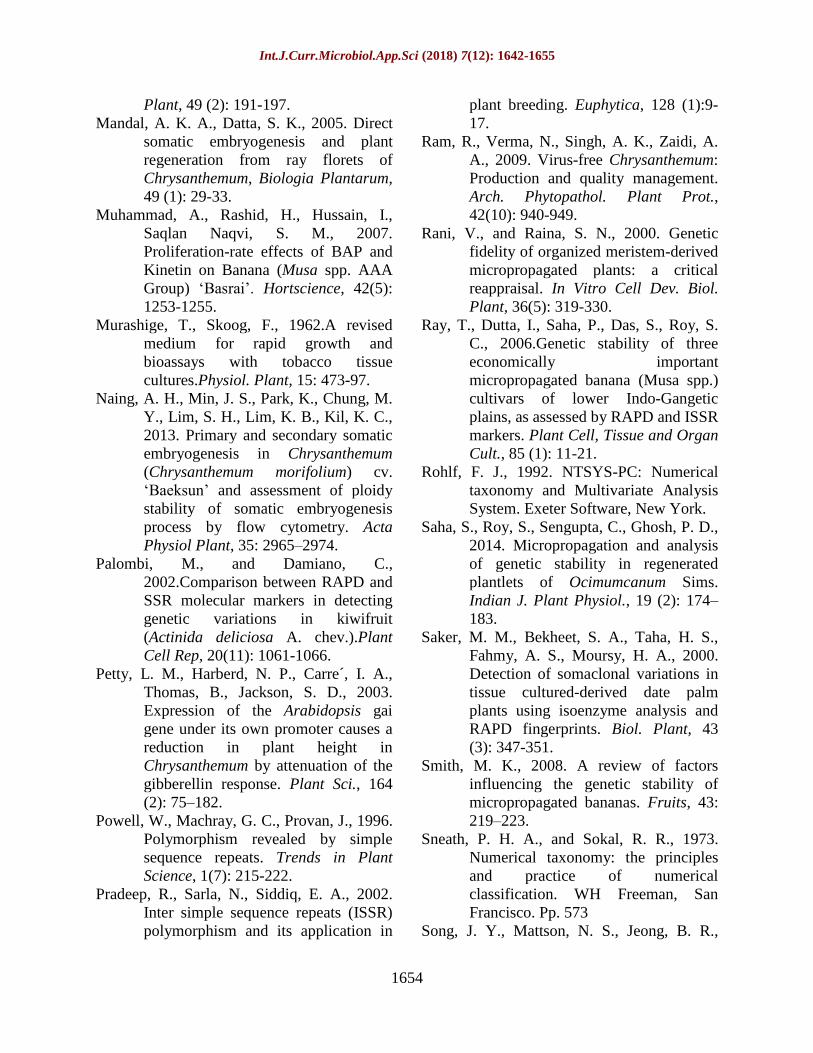
The 9 RAPD primers produced 28 distinct
and scorable bands in the size range of 300 bp
(OPG-08) to 2000 bp (OPD-05). The number
of scorable bands for each primer varied from
1 (OPD-20) to 7 (OPG-08) with an average of
3.11 bands per primer (Table 3). These
primers did not show any polymorphism
between them and the mother plants. PIC
value of all RAPD primers have zero and
morphologically all the clones (L1 to L10
were leaf regenerated and P1 to P20 were
petal regenerated) were similar to the mother
plant, indicating no variations. A
representative pattern of amplification
obtained with primers OPG 7 and OPG 11 are
provided in Figure 4A and 4B. The ISSR
primers used in the present study were mostly
dinucleotide repeats with one tetranucleotide
repeats. Of the 10 primers tested, 9 contained
3(GA)n, 1(CA)n, 3(TC)n and 2(AG)n and one
5’-tetraanchored primers. ISSR primers
produced 47 distinct and scorable bands in the
size range of 300 bp (ISSR-27) to 2000 bp
(ISSR-8).
The number of scorable bands for each primer
varied from 1 (ISSR-21) to 9 (ISSR-12) with
an average of 4.7 bands per primer (Table 4).
Calculated PIC value varied from 0.0 (ISSR-
7) to 0.489 (ISSR-2) and average PIC value
per primer was 0.048. Out of 10 ISSR
primers, 9 primers showed monomorphic
banding pattern in both leaf and petal derived
plants and the mother plant. However, only
one polymorphic band was detected with
ISSR-2 in one of the leaf derived plantlets L1.
A unique band of 1500 bp was present in L1,
which was absent in mother plant as well as in
other in vitro raised plantlets (Fig. 4E). The
polymorphism in tissue cultured raised plants
could result from change in either the
sequence of primer binding site (e.g. point
mutations) or change which alter the size and
prevent successful amplification of target
DNA (e.g. insertion, deletion, inversions)
(e.g. point mutations) or change which alter
the size and prevent successful amplification
of target DNA (e.g. insertion, deletion,
inversions) or alteration in DNA methylation
during in vitro culture (Saker et al., 2000).
The PCR amplification pattern obtained with
primers ISSR 7, ISSR12 and ISSR 2 is
represented in Figure 4C, 4D and 4E. The use
of molecular markers for studying the genetic
stability has been emphasized by many
workers (Jayanthi and Mandal, 2001; Bhatia
et al., 2011; Saha et al., 2014).
The genetic stability of the clones depends on
the genotype (Smith, 2008), cultural
conditions, maintenance of cultures (Haisel et
al., 2001) and number of sub cultural
passages (Chaterjee and Prakash, 1996). In
this study it was found that callus derived
plantlets are showing some amount of
variation while the embryo derived ones are
completely uniform.
The superiority of somatic embryogenesis
derived plantlets has been confirmed by many
studies and this may be due to the fact that the
developmental constraints required by
somatic embryos exert selection against
variant ones. Using two marker systems for
evaluation of somaclonal variation has been
always advantageous (Palombi and Damiano,
2002).
Int.J.Curr.Microbiol.App.Sci (2018) 7(12): 1642-1655
1649
Table.1 Details of Media used for callus induction and shoot multiplication from leaf explants in
chrysanthemum
Media code Callus induction media Shoot multiplication media
(mg L-1
)
2,4-D NAA BAP KIN 2,4-D NAA BAP KIN
M1 2 0 0.5 0 0.1 0 0.5 0
M2 2 0 0 0.5 0.1 0 0 0.5
M3 0 2 0.5 0 0 0.1 0.5 0
M4 0 2 0 0.5 0 0.1 0 0.5
M5 1 1 0.5 0 0.05 0.05 0.5 0
M6 1 1 0 0.5 0.05 0.05 0 0.5
Table.2 Details of experiment with petals as explants in MS media
Total No. of explants %
embryogenesis
No. of somatic
embryos
No of plants
regenerated
No. of plants
hardened
30 (10 explants as
three replications)
100* 54* 20* 18.33*
*Average of three replications
Table.3 Details of RAPD primers, sequences, number and size of amplified fragments obtained
with in vitro regenerated plants of chrysanthemum
S.No Primers 5’–3’ sequence Tm (oC) No. of bands per primer Size range (bp)
1 OPD-05 TGAGCGGACA 37.1 3 700-2000
2 OPD-07 TTGGCACGGG 40.9 3 700-1900
3 OPD-20 ACCCGGTCAC 39.1 1 ~650
4 OPF-10 GGAAGCTTGG 32.3 1 ~500
5 OPF-11 TTGGTACCCC 32.6 4 450-1500
6 OPG-05 CTGAGACGGA 32.8 2 1000-1300
7 OPG-08 TCACGTCCAC 34.5 7 300-1500
8 OPG-11 TGCCCGTCGT 43.2 2 450-750
9 OPH-05 AGTCGTCCCC 38 5 850-1700

Int.J.Curr.Microbiol.App.Sci (2018) 7(12): 1642-1655
1650
Table.4 Details of ISSR primers and amplification products obtained with in vitro regenerated
plants of chrysanthemum
S.No Primers 5’ to 3’
motif
Tm (oC) No. of bands
per primer
Total bands Size range of
bands(bp)
1 ISSR-12 (GA)7A 45.7 9 189 300-1400
2 ISSR-14 (CT)7A 44.7 5 105 500-2000
3 ISSR-16 (CA)7G 51 4 84 550-1500
4 ISSR-2 (TC)7A 47 4 84 500-1500
5 ISSR-21 (TC)7C 48.1 1 21 900
6 ISSR-27 (ATAG)4 39.6 6 126 300-1500
7 ISSR-7 (AG)7T 47 3 63 500-1100
8 ISSR-10 (GA)7T 45.4 5 105 500-1500
9 ISSR-8 (AG)7C 48.8 6 126 300-2000
10 ISSR-11 (GA)7C 46.8 4 84 600-1400
Fig.1 A–E Plant regeneration via embryogenesis from leaf and petals of Chrysanthemum. A.
Embryogenic callus induction from leaf explants after one month of incubation in dark. B.
Plantlet formation from embryogenic calli of leaf explants. C. Direct embryogenesis from petal
explants. D. Distinct globular somatic embryos observed from petal explants. E. Single somatic
embryo with root and shoot pole. F. Rooted plantlets in primary hardening media. G. Plantlet
formation from somatic embryo. H. Rooting of in vitro derived plantlets in MS basal media. I.
Hardened plants ready for secondary hardening in mist chamber. J. Completely hardened plants
flowering in mist chamber

Int.J.Curr.Microbiol.App.Sci (2018) 7(12): 1642-1655
1651
Fig.2 Callus induction obtained with different media (details of media in Table 1) using leaf
explants of Pusa centenary. Values are Mean ± standard error. Means followed by same letter are
not significantly different at p≤0.05
Fig.3 Number of shoots obtained with different media (details of media in Table 1) in Pusa
centenary. Values are Mean ± standard errors. Means followed by same letter are not
significantly different at p≤0.05

Int.J.Curr.Microbiol.App.Sci (2018) 7(12): 1642-1655
1652
Fig.4 A and 4B: RAPD banding pattern in micropropagated plants from leaf and petal explants
compared with mother plant of Pusa centenary using primers OPG 11 and OPD 7. (Mo- mother
plant; P1 to P10 micropropagated plants from petal explants and L1 to L10 micropropagated
plants from leaf explants; M 100 bp marker).
Fig.4C, 4D and 4E: ISSR banding pattern in micropropagated plants from leaf and petal explants
compared with mother plant of Pusa centenary obtained with ISSR 7, ISSR 12 and ISSR 2(Mo-
mother plant; P1 to P10 micropropagated plants from petal explants and L1 to L10
micropropagated plants from leaf explants; M 100 bp marker)
It has been assumed that the use of two
markers, which amplify different regions of
the genome, allows for better chances for the
identification of genetic variations in the
clones. These two marker systems viz., RAPD
and ISSR have been extensively used to
detect somaclonal variation in many other
crops. ISSR markers are able to bring out the
genomic differences than RAPD as is seen in
our studies. ISSR always reveals more
polymorphism than RAPD as is reported in
case of coffee, tea and banana (Rani and
Raina, 2000; Devarumath et al., 2002; Ray et
al., 2006).
The ISSRs are widely distributed throughout
the genomic DNA and make amplification of
genomic DNA possible in much larger
numbers of fragments per primer. This higher
amplification efficiency of ISSRs makes the
genetic inferences much clearer with some
clues for genetic variation at inter- and even
intra-specific levels (Powell et al., 1996). Our
study proves that if true to type plants are
required direct embryogenesis is the preferred
method. Along with several other advantages
of direct somatic embryogenesis, this protocol
opens up the prospect of genetic
transformation and bioreactor based
propagation in this important commercial
cultivar.
Acknowledgement
We wish to thank Director, IARI and Joint
Director (Research), IARI, for providing the
facilities to do the work.

Int.J.Curr.Microbiol.App.Sci (2018) 7(12): 1642-1655
1653
References
Bhatia, R., Singh, K. P., Sharma, T. P., Jhang,
T., 2011.Evaluation of the genetic
fidelity of in vitro-propagated gerbera
(Gerbera jamesonii Bolus) using
DNA-based markers. Plant cell tissue
organ cult., 104(1): 131–135.
Blakesly, D.,1991. Uptake and metabolism of
6-benzyladenine in shoot cultures of
Musa and Rhododendron. Plant Cell,
Tissue Organ Cult., 25 (1): 69-74.
Chaterjee, G. and Prakash, J., 1996. Genetic
stability in commercial tissue culture,
In: Plant Biotechnology: Commercial
Prospects and Problems (Eds.) J
Prakash and RIM Pierik. Oxford IBH
Publishing Co. New Delhi, India, Pp.
111–121.
Devarumath, R. M., Nandy, S., Rani, V.,
Marimuthu, S., Muraleedharan, N.,
2002. RAPD, ISSR and RFLP finger
printing as useful markers to evaluate
genetic integrity of micropropagated
plants of three diploid and triploid
elite tea clones representing Camellia
sinensis (China type) and C.
assamica ssp. assamica (Assam-India
type). Plant Cell Rep, 21(2): 166-173.
Doyle, J. J., Doyle, J. L., 1990. Isolation of
plant DNA from fresh tissue. Focus,
12(1): 13-15.
Geetha, N., Venkatachalam, P., Prakash, V.,
Lakshmi, S. G., 1998. High frequency
induction of multiple shoots and plant
regeneration from seedling explants of
pigeon pea (Cajanus cajan L.). Curr.
Sci., 75 (10): 1036–1041.
Gould, A. R., 1986. Factors controlling
generation of variability in vitro, In:
Cell Culture and Somatic Cell
Genetics in Plants (Eds.) Vasil, I. K.,
Vol 3. Plant Regeneration and Genetic
Variability. Academic Press Inc. New
York. Pp. 549-567.
Haisel, D., Hofman, P., Vagneri, M.,
Lipavska, H., Ticha, L., Schafer, C.,
Capkova, V., 2001. Ex vitro
phenotype stability is affected by in
vitro cultivation. Biol. Plant, 44(3):
321-324.
Himstedt, J. P., Jacobsen, H. J., Fisher-
Kluver, G., 2001. Shoot regeneration
from stem and leaf explants of
Chrysanthemum (Dendranthema x
grandiflorum), Acta Hortic., 560:
421–424.
Jaccard, P., 1908. Nouvellesrecherchessurla
distribution florale, Bull. Soc. Vaud.
Sci. Nat, 44: 223–270.
Jayanthi, M., and Mandal, P. K., 2001. Plant
regeneration through somatic
embryogenesis and rapd analysis of
regenerated plants in Tylophora indica
(Burm. F. Merrill.). In vitro Cellular
and Development Biology, 37 (5):
576-580.
Kaminek, M., 2000. Progress in cytokinin
research. Trends Biotechnol., 10: 159-
162.
Kumlay, A. M., 2014. Combination of the
Auxins NAA, IBA, and IAA with
GA3 Improves the Commercial Seed-
Tuber Production of Potato (Solanum
tuberosum L.) under In vitro
Conditions. BioMed Research
International, 14: 439259.
Larkin, P., and Scowcroft, W. R., 1981.
Somaclonal variation, a novel source
of variability from cell cultures for
plant improvement, Theor. Appl.
Genet. 60 (4): 197–214.
Lema-Rumniska, J., and Niedojadlo, J.,
2014.Somatic embryogenesis in leaf
explants of Chrysanthemum
radiomutants of the ‘lady’ group.
Propagation of ornamental plants, 14
(4): 177-183.
Liu, G., Gilding, E. K., Godwin, I. D., 2013.
Additive effects of three auxins and
copper on sorghum in vitro root
induction. In Vitro Cell. Dev. Biol.-
Int.J.Curr.Microbiol.App.Sci (2018) 7(12): 1642-1655
1654
Plant, 49 (2): 191-197.
Mandal, A. K. A., Datta, S. K., 2005. Direct
somatic embryogenesis and plant
regeneration from ray florets of
Chrysanthemum, Biologia Plantarum,
49 (1): 29-33.
Muhammad, A., Rashid, H., Hussain, I.,
Saqlan Naqvi, S. M., 2007.
Proliferation-rate effects of BAP and
Kinetin on Banana (Musa spp. AAA
Group) ‘Basrai’. Hortscience, 42(5):
1253-1255.
Murashige, T., Skoog, F., 1962.A revised
medium for rapid growth and
bioassays with tobacco tissue
cultures.Physiol. Plant, 15: 473-97.
Naing, A. H., Min, J. S., Park, K., Chung, M.
Y., Lim, S. H., Lim, K. B., Kil, K. C.,
2013. Primary and secondary somatic
embryogenesis in Chrysanthemum
(Chrysanthemum morifolium) cv.
‘Baeksun’ and assessment of ploidy
stability of somatic embryogenesis
process by flow cytometry. Acta
Physiol Plant, 35: 2965–2974.
Palombi, M., and Damiano, C.,
2002.Comparison between RAPD and
SSR molecular markers in detecting
genetic variations in kiwifruit
(Actinida deliciosa A. chev.).Plant
Cell Rep, 20(11): 1061-1066.
Petty, L. M., Harberd, N. P., Carre´, I. A.,
Thomas, B., Jackson, S. D., 2003.
Expression of the Arabidopsis gai
gene under its own promoter causes a
reduction in plant height in
Chrysanthemum by attenuation of the
gibberellin response. Plant Sci., 164
(2): 75–182.
Powell, W., Machray, G. C., Provan, J., 1996.
Polymorphism revealed by simple
sequence repeats. Trends in Plant
Science, 1(7): 215-222.
Pradeep, R., Sarla, N., Siddiq, E. A., 2002.
Inter simple sequence repeats (ISSR)
polymorphism and its application in
plant breeding. Euphytica, 128 (1):9-
17.
Ram, R., Verma, N., Singh, A. K., Zaidi, A.
A., 2009. Virus-free Chrysanthemum:
Production and quality management.
Arch. Phytopathol. Plant Prot.,
42(10): 940-949.
Rani, V., and Raina, S. N., 2000. Genetic
fidelity of organized meristem-derived
micropropagated plants: a critical
reappraisal. In Vitro Cell Dev. Biol.
Plant, 36(5): 319-330.
Ray, T., Dutta, I., Saha, P., Das, S., Roy, S.
C., 2006.Genetic stability of three
economically important
micropropagated banana (Musa spp.)
cultivars of lower Indo-Gangetic
plains, as assessed by RAPD and ISSR
markers. Plant Cell, Tissue and Organ
Cult., 85 (1): 11-21.
Rohlf, F. J., 1992. NTSYS-PC: Numerical
taxonomy and Multivariate Analysis
System. Exeter Software, New York.
Saha, S., Roy, S., Sengupta, C., Ghosh, P. D.,
2014. Micropropagation and analysis
of genetic stability in regenerated
plantlets of Ocimumcanum Sims.
Indian J. Plant Physiol., 19 (2): 174–
183.
Saker, M. M., Bekheet, S. A., Taha, H. S.,
Fahmy, A. S., Moursy, H. A., 2000.
Detection of somaclonal variations in
tissue cultured-derived date palm
plants using isoenzyme analysis and
RAPD fingerprints. Biol. Plant, 43
(3): 347-351.
Smith, M. K., 2008. A review of factors
influencing the genetic stability of
micropropagated bananas. Fruits, 43:
219–223.
Sneath, P. H. A., and Sokal, R. R., 1973.
Numerical taxonomy: the principles
and practice of numerical
classification. WH Freeman, San
Francisco. Pp. 573
Song, J. Y., Mattson, N. S., Jeong, B. R.,

Int.J.Curr.Microbiol.App.Sci (2018) 7(12): 1642-1655
1655
2011. Efficiency of shoot regeneration
from leaf, stem, petiole and petal
explants of six cultivars of
Chrysanthemum morifolium. Plant
Cell, Tissue Organ Cult., 107: 295-
304.
Tanaka, K., Kanno, Y., Kudo, S., Suzuki, M.,
2000. Somatic embryogenesis and
plant regeneration in Chrysanthemum
(Dendranthem agrandiflorum
(Ramat.) Kitamura). Plant Cell
Reports, 19(10): 946-953.
Teixeira da Silva, J. A., 2004. Ornamental
Chrysanthemums: improvement by
biotechnology. Plant Cell, Tissue
Organ Cult., 79(1): 1-18.
Waseem, K., Jilani, M. S., Khan, M. S., 2009.
Rapid plant regeneration of
Chrysanthemum (Chrysanthemum
morifolium L.) through shoot tip
culture. Afr. J. Biotechnol., 8(9):1871–
1877.
Williams, J. G. K., Kubelik, A. R., Livak, K.
J., Rafalski, J. A., Tingey, S. V., 1990.
DNA polymorphisms amplified by
arbitrary primers are useful as genetic
markers. Nucleic Acids Res., 18(22):
6533-6535.
Zietkiewicz, E., Rafalski, A., Labuda, D.,
1994. Genomic fingerprinting by
simple sequence repeat (SSR)-
anchored polymerase chain reaction
amplification, Genomics, 20 (2): 176-
183.
Zilberman, D., and Henikoff, S., 2007.
Genome-wide analysis of DNA
methylation patterns, Development.
134(22): 3959-3965.
How to cite this article:
Jayanthi, M., Rajesh Gupta, P.K. Mandal and Kanchan B.M. Singh. 2018. Analysis of
Somaclonal Variation using Molecular Markers in in vitro Regenerated Chrysanthemum cv.
Pusa Centenary. Int.J.Curr.Microbiol.App.Sci. 7(12): 1642-1655.
doi: https://doi.org/10.20546/ijcmas.2018.712.192