analysis of records of embryo production...
TRANSCRIPT

ANALYSIS OF RECORDS OF EMBRYO PRODUCTION IN
RED BRAHMAN COWS
A Thesis
by
EDGAR HERNANDO RIANO ROCHA
Submitted to the Office of Graduate Studies ofTexas A&M University
in partial fulfillment of the requirements for the degree of
MASTER OF SCIENCE
August 2005
Major Subject: Physiology of Reproduction

ANALYSIS OF RECORDS OF EMBRYO PRODUCTION IN
RED BRAHMAN COWS
A Thesis
by
EDGAR HERNANDO RIANO ROCHA
Submitted to the Office of Graduate Studies ofTexas A&M University
in partial fulfillment of the requirements for the degree of
MASTER OF SCIENCE
Approved by:
Co-Chairs of Committee, David ForrestAndy Herring
Committee Member, Louise AbbottHead of Department, Gary Acuff
August 2005
Major Subject: Physiology of Reproduction
iii
ABSTRACT
Analysis of Records of Embryo Production in
Red Brahman Cows. (August 2005)
Edgar Hernando Riano Rocha, DVM., La Salle University
Co-Chairs of Advisory Committee: Dr. David Forrest Dr. Andy Herring
Records of embryo production in Red Brahman donor cows (n=50) and F1
recipients (n=531) were evaluated from the collection day to the birth of the
embryo produced. The effects of the sire of the donor and the embryo, protocol,
season-protocol, and body condition of the donor on the total number of good,
degenerated, unfertilized, and total embryos were evaluated. The number of
donors collected for protocols 1, 2, and 3 were 50, 39, and 46 respectively. The
production of good transferable embryos, and embryos/collection for protocols 1,
2, and 3 were 171 (4.6), 152 (4.6), and 208 (6.3) respectively. The final status
of each recipient was recorded as non-pregnant, resorption, abortion, and live
calf. The model used to analyze pregnancy state was: protocol, embryo stage,
embryo quality, corpora lutea size, and season. The effects of sire of the embryo,
season-protocol, protocol, embryo stage, embryo quality, body condition score,
and corpora lutea size on gestation length and birth weight were analyzed.
Season-protocol affected (P<0.05) the number of degenerated embryos. Mean
number of degenerated embryos were higher (P<0.05) during winter for
protocols 2 and 3 than during other seasons. The ratio for good embryos differed
(P<0.01) by sire of donor.
The final status of recipients was affected (P<0.01) by protocol. The maximum
percentage of live calves and the minimum percentage of non-pregnant
recipients were achieved for protocol 3.

iv
Gestation length differed (P<0.01) by sire of the embryo, season-protocol,
protocol, and body condition score. Spring-protocol 3 resulted in the shortest
while Fall-protocol 2 resulted in the longest mean gestation length. Calf birth
weight differed (P<0.05) by season-protocol and by embryo quality. The lightest
birth weights resulted from embryo quality grade 2 and from spring-protocol 3.
These results indicate that using protocols that combine 17β-estradiol, FSH
and GnRH (protocol 3) during the spring in conjunction with selection for sire of
donor can increase embryo production by Red Brahman cows. Use of protocol 3
with donors in the spring, selection of embryo sire for short gestation length and
transfer of quality grade 2 embryos can be used to minimize the incidence of
dystocia in recipients.

v
DEDICATION
I would like to dedicate this thesis to my wonderful wife, Adriana, for all her
love, support, sacrifice and patience with me during this period of my life to
accomplish the goals I had for my professional life.

vi
ACKNOWLEDGMENTS
I would like to gratefully thank Dr. David Forrest for the guidance and support
he has given me in the physiology of reproduction area and during the
preparation of this thesis.
I would also like to thank Dr. Andy Herring for orienting me in the area of
beef cattle production and its situation in the southern part of the United States,
for all his advice on the statistical evaluation and interpretation of my research.
I must also thank Dr. Louise Abbott, for what I learned during my class of
mammalian embryology, for her objectivity in the evaluation of this research and
her good disposition to help and support me.
I also need to thank Josephine and Alfredo Muskus, the owners of Santa Elena
Ranch for facilitating all the needed information and making possible the
accomplishment of this study.
I couldn’t forget to thank God, my Father and Savior, for the opportunity to
pursue this degree at Texas A&M University and for giving me the strength and
perseverance to accomplish this goal in my life.
vii
TABLE OF CONTENTS
Page
ABSTRACT ................................................................................. iii
DEDICATION.............................................................................. v
ACKNOWLEDGMENTS ................................................................. vi
TABLE OF CONTENTS ................................................................. vii
LIST OF FIGURES ....................................................................... ix
LIST OF TABLES ......................................................................... xi
INTRODUCTION ......................................................................... 1
Research objectives .......................................................... 1
LITERATURE REVIEW ................................................................. 5
Hormonal regulation of follicles and the corpus luteumduring the bovine estrus cycle............................................ 5 Hormonal changes involved close to the time of estrus andovulation .......................................................................... 9Use of CIDRs to synchronize estrus in donor cows ............... 10Control of follicular recruitment .......................................... 13The use of estradiol in estrus synchronization...................... 15The use of GnRH in donor cows for embryo transferprograms ......................................................................... 16Uses of GnRH in timed AI protocols .................................... 17Cryopreservation............................................................... 20Factors affecting the embryonic survival in the cow ............. 23Conception postpartum ..................................................... 24Early embryonic death associated with short durationof the luteal phase ............................................................ 25Other factors associated with pregnancy losses ................... 27
MATERIALS AND METHODS......................................................... 29
Criteria to select the records of Red Brahman donor cows and the recipients ............................................................. 29

viii
Page
Embryo transfer procedure ................................................ 29Protocols for donors .......................................................... 30Protocols for recipients ...................................................... 32Data analysis .................................................................... 33Statistical analysis ............................................................. 34
RESULTS.................................................................................... 35
Donors............................................................................. 35Recipients ........................................................................ 40
DISCUSSION .............................................................................. 49
Donors............................................................................. 49Recipients ........................................................................ 52
CONCLUSIONS ........................................................................... 57
REFERENCES.............................................................................. 60
VITA ...................................................................................... 75

ix
LIST OF FIGURES
FIGURE Page
1 Example shown for cattle having three follicular wavesduring 21-day estrus cycle................................................ 7
2 The dark irregular ovals represent corpus luteums in development from the ovulated follicle .............................. 8
3 Four different options used to synchronize estrus and/orovulation in beef cows treated with intra-vaginal progesterone-releasing device (CIDR) in combinationwith prostaglandin F2α (PGF 2α) and with or withoutestradiol benzoate (EB) or gonadotropin releasinghormone (GnRH) ............................................................. 12
4 Ovsynch protocol ............................................................. 18
5 Cosynch protocol ............................................................. 19
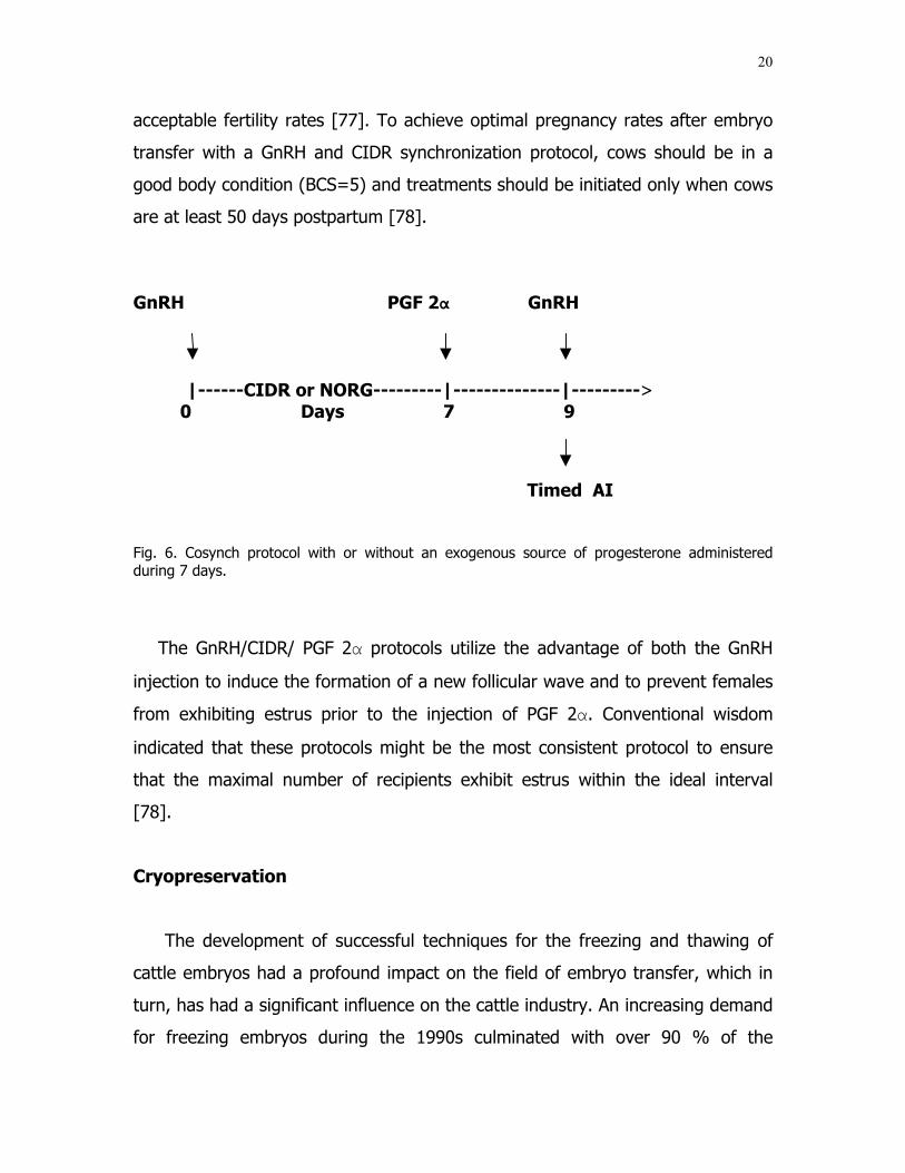
6 Cosynch protocol with or without an exogenous sourceof progesterone administered during 7 days....................... 20
7 Least squares means (±pooled SEM) of degeneratedembryos by season of embryo collection for protocols 1,2, and 3 ....................................................................... 36
8 Least squares means (±pooled SEM) of good embryo-ratio at the collection time for sire of donors ...................... 39
9 Least squares means of degenerated embryo-ratio atthe collection time for season-protocol .............................. 40
10 Percentage of recipients of Brahman embryos that werenon pregnant (NP), underwent resorption (RE), aborted(AB), or delivered a live calf (LC), by protocol. ................... 41
11 Percentage of pregnancies in recipients of fresh Brahmanembryos at the first palpation (39-49 days), second palpation at 60 days, third palpation (>90-120 days),and delivered live calves (LC), by protocol ......................... 42

x
FIGURE Page
12 Percentage of recipients of Brahman embryos that werenon pregnant (NP), underwent resorption (RE), aborted (AB), or delivered a live calf (LC), by embryo stage ............ 43
13 Percentage of recipients of Brahman embryos that werenon pregnant (NP), underwent resorption (RE), aborted(AB), or delivered a live calf (LC), by season ...................... 44

xi
LIST OF TABLES
TABLE Page
1 Comparison of pregnancy rates achieved after transferof bovine embryos that were frozen in glycerol or inethylene glycol ................................................................ 23
2 Least squares means for number of good, degenerated, unfertilized, and total embryos by sire of donors ................ 37
3 Least squares means for number of good, degenerated,unfertilized, and total embryos by protocols ....................... 37
4 Least squares means for number of good, degenerated,unfertilized, and total embryos by season-protocol ............. 38
5 Least squares means (±pooled SEM) for gestation lengthby sire of the embryo....................................................... 45
6 Least squares means (±pooled SEM) for gestation lengthby season-protocol........................................................... 45
7 Least squares means (±pooled SEM) for gestation lengthby protocol...................................................................... 46
8 Least squares means for birth weight by season-protocol.... 47
9 Least squares means for birth weight by embryo quality ..... 47
10 Least squares means for gestation length and EPD’s forbirth weights by sire of the embryo ................................... 55

1
INTRODUCTION
For most embryo transfer (ET) programs in cattle, a major concern is the
ability to synchronize estrus with hormones in donors and recipients to ensure
that they are at the correct stage of the estrus cycle to recover multiple embryos
of good quality from a donor and transfer either a fresh or frozen embryo on a
specific day into a recipient. Numerous factors affect the response of these
donors and recipients to the hormonal treatments used by the industry. Those
factors may include weather patterns, nutrition, age, body condition, number of
days after calving, nursing calf aside and breed composition/genetics. The use of
ET has been increasing within the cattle industry due to improvements achieved
with the ET technique during the last 40 years. The technique has had many
changes in animal preparation (donors and recipients), embryo collection, and
transfer, freezing, and thawing processes. Bovine embryo transfer is the most
available alternative to improve and produce large numbers of offspring from a
donor-female with the highest genetic merit and performance record, in a
shorter time than any other reproductive technology [1]. Currently, the embryo
transfer technique is widely used around the world, with more than 500,000
embryos being transferred each year [2]. Embryo transfer (ET) has had a
significant impact on the beef industry. Registrations of Angus cattle over the
past 15 years give indication of the increased use of ET. In 1987, 3.6% (5,105)
of all calves registered were a result of embryo transfer. In 2002, 25,093 calves
resulting from ET were registered. This was 8.9% of all calves registered [3].
Over half the cattle in the world are found in the tropics (from the Tropic of
Cancer to the Tropic of Capricorn). The subtropics of the United States include
the Gulf Coast region of the southern states and all of Florida. Nearly 30% of
This research follows the style and form of Theriogenology.

2
the U.S. beef herd is maintained in this zone [4]. Compatibility between beef
cattle type (e.g., breed) and the environment is critical in harsh environmental
zones such as the subtropics. In contrast to the abundance of beef cattle
genotypes in the United States that are adapted to temperate climates, germ
plasm resources with adaptation to warm climates, including the subtropics, are
generally limited to the Zebu breeds (Bos indicus) and, within these, primarily to
the American Brahman [4].
Unfortunately, there is not much information available about ET in Bos indicus
cattle since most of the studies and improvements in embryo transfer have been
done in Bos taurus. The majority of the embryo transfer protocols have been
designed and modified in Bos taurus, and applied to Bos indicus with results very
different than expected. Brahman, a Bos indicus breed is the most popular of all
the zebu breeds, has been used widely in commercial beef herds throughout the
warm regions of the United States because of the good performance in extreme
heat stress not cold weather conditions, utilization of low quality of feed, and
high resistance to parasites. Twenty five percent of the cattle in the United
States are estimated to have some percentage of Brahman breeding [4]. The
major niche for Brahman cattle has been in crossbreeding programs that
combine the tropical adaptation of Brahman with the more desirable reproductive
efficiency and carcass characteristics of temperate-adapted Bos taurus breeds
[4]. A cross between Bos indicus and either an English or Continental breed
produces animals with the very high fertility and production that can be used in a
wide variety of environments. Moreover, one of the most cited negative factors
of the Brahman cow is the sub-standard fertility, when compared to the English
breeds of beef cattle [5, 6]. Differences in reproductive physiology between Bos
indicus and Bos taurus, specifically with Brahman cattle are found in the reduced
duration of estrus and a shorter period from onset of estrus to the luteinizing
hormone (LH) surge as well as from the LH surge to ovulation [7]. In addition,
Bos indicus females have a lower preovulatory LH surge than Bos taurus females
[7]. Bos indicus also have higher numbers of ovarian follicles, smaller corpus

3
luteum (CL), lower serum progesterone concentration [7], and higher serum
concentration of insulin growth factor I [8]. Researchers have recently found
differences in timing of ovulation, fertilization or events leading up to cleavage of
early embryos in Brahman compared to Holstein cattle [9]. Another difference
found in Brahman cattle compared with British or Continental breeds is in the
response to the super-ovulation protocols. Brahman cattle are generally thought
to require smaller dosages of follicle stimulating hormone (FSH) than any other
breed. Frozen Brahman embryos obtained with the traditional protocols produce
lower pregnancy rates when compared with English or Continental breeds [10].
The theoretical reason for the decreased pregnancy rate with the frozen
Brahman embryo is caused by the intracellular lipids. However, this has not yet
been demonstrated [11]. It has been found that the different forms of estradiol
stimulate variable responses in the donors. For example using 17ß-estradiol on
Brahman donor cows will result in fewer unfertilized eggs and fewer degenerated
embryos than using estradiol benzoate.
Even though the process of super-ovulation has demonstrated large
differences in response [11], the use of lower dosages/frequencies of FSH in
Brahman donor cows other than in Bos taurus or Continental breeds may
produce more viable embryos per collection than the use of FSH suggested in
standard protocols. The use of gonadotropin releasing hormone (GnRH) to
induce ovulation or luteinization of the largest follicle present at the time of
treatment is common [12]. One protocol for synchronization of ovulation involves
administration of GnRH on the first day and a second GnRH injection is repeated
32 hours after the injection of prostaglandin (PGF 2α) for fixed-time AI in beef
and dairy cattle [13,14,15]. It has been suggested to use a single injection of
(GnRH) in Brahman donors in estrus 6 hours before AI to synchronize ovulation
of the new dominant follicle because the FSH will produce ovulation in the
following 24 hours after treatment which may increase the conception rate. The
objectives of this study were to analyze records of a commercial ET program to
determine if the use of 17ß-estradiol, the individualized dosage of FSH, and the

4
application of GnRH 6 hours before AI in Brahman donors would result in an
increase in the number and quality of embryos per collection, which can be
reflected with a higher conception rate in recipients, compared to the
conventional existing protocols to produce Brahman embryos.
Research objectives
The overall aim of this study was to quantify the relationship of traits
associated with Brahman donors and with recipient females based on the
efficiency of embryo transfer technology.
The specific objectives were to:
1. Quantify the effects of superstimulation protocol, sire of donor, sire of
embryo, season within protocol and donor body condition on embryo
production (good, degenerate, unfertilized, and total embryos).
2. Determine the relationships of superstimulation protocol, embryo stage,
embryo quality, corpus luteum size and season of collection with the
outcomes of transfer of embryos into recipients (live calf, embryo resorption,
abortion, and non-pregnant recipient).
3. Quantify the effects of sire of embryo, season within protocol, embryo stage,
embryo quality, recipient body condition and corpus luteum size on gestation
traits (gestation length and birth weight) in recipients of embryo transfer.
These results should provide information to accelerate the use of embryo
transfer technology in Brahman females.

5
LITERATURE REVIEW
Hormonal regulation of follicles and the corpus luteum during the
bovine estrus cycle
It is important to review the bovine estrus cycle and the role of the four main
hormones involved in this process that produce changes in the pre-ovulatory
follicle and the corpus luteum. Bovine estrus has been described as a short
period (approximately 15 to 18 hours) of sexual receptivity that is manifested
every 18 to 24 days, with ovulation occurring 10 to 14 hours after the cessation
of behavioral signs of estrus [16,17]. 17ß-estradiol from the preovulatory follicle
causes the cow to manifest estrus behavior and have a LH surge [18]. The LH
surge causes ovulation of the preovulatory follicle about 28 hours later. The cells
that remain from the preovulatory follicle develop into the corpus luteum. The
corpus luteum grows in size during the first part of the estrus cycle and then
reaches a plateau phase in which it maintains a large size (20-25 mm diameter)
[19]. The major hormone coming from the corpus luteum is progesterone and
the increase in size of the corpus luteum is reflected in increased concentrations
of progesterone in the blood. If the cow becomes pregnant the corpus luteum is
maintained, and progesterone concentration remains elevated. The high
progesterone concentration prevents the cow from coming into estrus or having
a subsequent ovulation. If the cow does not become pregnant, then the corpus
luteum will degenerate at about day 17-20 of the estrus cycle (day of estrus =
0). The reason the corpus luteum regresses is because of secretion of
prostaglandin (PGF 2α) from the non-pregnant uterus. After exposure to PGF 2αthere is a decrease in circulating progesterone concentrations as well as a
subsequent decrease in size of the corpus luteum [20]. The temporal profiles
circulating concentrations of FSH, LH and estrogen during the estrus cycle are
depicted in Figure 1. The changes in circulating progesterone concentration

6
during the estrus cycle and associated with initiation of pregnancy are depicted
in Figure 2.
Starting around the time of ovulation, a group of small follicles (cohort) begins
to grow on the ovaries. This growth is known as the follicular wave. From this
group of follicles a single dominant follicle is selected to continue growth,
whereas, other follicles of the follicular wave undergo atresia or regression [21].
In cattle, follicular waves occur throughout the cycle, but under natural
conditions only the dominant follicle of the wave present at the time of luteal
regression subsequently ovulates [22, 23]. Due to the presence of a functional
corpus luteum and high progesterone concentrations, this first dominant follicle
does not cause estrus behavior and does not continue to ovulation. The first
dominant follicle will become non- functional and a second follicular wave begins
at about mid-cycle. A dominant follicle is again selected from this second
follicular wave, and this follicle continues to ovulation because its growth
corresponds to the time of regression of the corpus luteum [24]. Some cows also
show three waves of follicular growth such that the second dominant follicle
regresses, a third follicular wave is initiated, and the third dominant follicle
becomes the ovulatory follicle. Both the first and the second follicular waves are
preceded by an increase in FSH concentrations [21]. These increases in FSH are
essential for the initiation of a follicular wave. The subsequent decrease in FSH is
essential for selecting a single dominant follicle. There is also a FSH surge in
association with the LH surge that causes ovulation. This FSH surge occurs near
the onset of estrus and is of shorter duration than the subsequent FSH surge
[25]. Near the time of estrus there are two surges in FSH that are difficult to
discriminate because they are temporally adjacent. The first surge corresponds
to the GnRH/LH surge that induces ovulation and a second occurs near the time
of ovulation and is associated with emergence of the first follicular wave [25].
Emergence of the follicular wave has generally been retrospectively determined
7
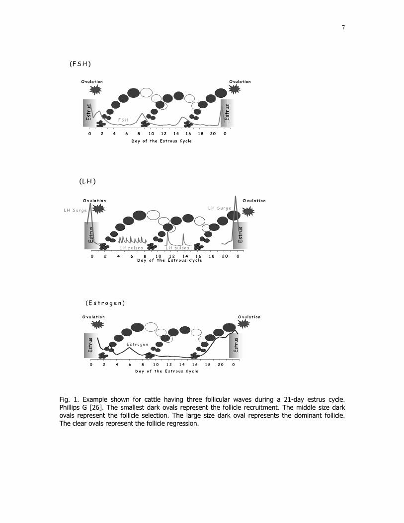
0 2 4 6 8 10 12 14 16 18 20 0
Estr
us
Estr
us
Day of the Estrous Cycle
O vu lationO vu lation
(FS H )
FSH
0 2 4 6 8 1 0 1 2 1 4 1 6 1 8 2 0 0
Estr
us
Estr
us
D a y o f th e E st r o u s C y c le
L H S u rg e
O vu la t io nO v u la t io n
L H S u rg e
L H p u ls e s L H p u ls e s
(L H )
0 2 4 6 8 1 0 1 2 1 4 1 6 1 8 2 0 0
Estr
us
Estr
us
D a y o f t h e E s t r o u s C y c le
O v u la t i o nO v u la t i o n
E s t r o g e n
( E s t r o g e n )
Fig. 1. Example shown for cattle having three follicular waves during a 21-day estrus cycle.Phillips G [26]. The smallest dark ovals represent the follicle recruitment. The middle size darkovals represent the follicle selection. The large size dark oval represents the dominant follicle.The clear ovals represent the follicle regression.

8
0 2 4 6 8 1 0 1 2 1 4 1 6 1 8 2 0 0
Estr
us
Estr
us
D a y o f t h e E s t r o u s C y c l e
C o r p u s L u t e u m ( C L )P r o g e s t e r o n e
G r o w t h R e g r e s s i o n
P r o g e s t e r o n e
0 2 4 6 8 1 0 1 2 1 4 1 6 1 8 2 0 0
Estr
us
Estr
us
C o r p u s L u t e u m ( C L ) R e g r e s s io nP r o s t a g la n d in F 2 α ( P G )
D a y o f t h e E s t r o u s C y c le
G r o w t h R e g r e s s io n
P G
Corpus luteum (CL) MaintenanceWhen cow becomes pregnant
Fig. 2. The dark irregular ovals represent corpus luteums in development from the ovulatedfollicle. They take approximately 10 days to reach mature size. Corpus luteum producesprogesterone. - Late in the estrus cycle, uterus produces PG, which causes regression of corpusluteum. - Presence of embryo blocks uterus to produce PG late in the estrus cycle which causesmaintenance of corpus luteum and production of progesterone for maintenance of pregnancy.Phillips G [26].
E s t r u s
D a y o f t h e E s t r o u s C y c l e
P G
M a i n t e n a n c e
0 2 4 6 8 1 0 1 2 1 4 1 6 1 8 2 0 0
E m b r y o
G r o w t h

9
as the time when the first follicles of the follicular wave reached ≥ 4 mm [25].
Following emergence, follicles continue growth and circulating FSH begins to
decline up until the time of follicular deviation. Follicular deviation has been
defined as the beginning of the greatest difference in growth rates between the
largest follicle and the second largest follicle at or before the examination when
the second largest follicle reached its maximum diameter [24].
Hormonal changes involved close to the time of estrus and ovulation
Standing estrus and the luteinizing hormone (LH) surge, which is requisite to
induce ovulation are two events initiated by the high circulating estradiol
concentration. The elevated estradiol is due to growth of a large preovulatory
follicle on the ovary [25]. After regression of the corpus luteum the dominant
follicle grows and produces increasing amounts of estradiol. The cow becomes
sexually active prior to the onset of standing estrus due to the increasing
amounts of estradiol in the absence of circulating progesterone [25].
Progesterone is low due to regression of the corpus luteum. If the corpus luteum
does not regress and progesterone remains elevated some or all of the
subsequent events such as the LH surge, estrus, and ovulation do not occur even
when estradiol is elevated. After estradiol attains a threshold concentration for a
certain time period, there is a change in the brain that causes the cow to begin
to stand solidly during mounting or more exactly during the onset of estrus [25].
There is also secretion of gonadotropin-releasing hormone (GnRH) in large
amounts from a region of the brain called hypothalamus. The secretion of GnRH
causes the LH surge. Induction of a LH surge and ovulation can be accelerated
with a GnRH injection prior to the normal time of ovulation [27]. The onset of
estrus is due to the high circulating estradiol concentration. Estrus behavior ends
prior to ovulation in cattle. The end of estrus may be due to a decrease in
circulating estradiol because estradiol production in the follicle is dramatically
10
reduced following the LH surge. The time from the onset of estrus until ovulation
is between 25 and 34 hours [28].
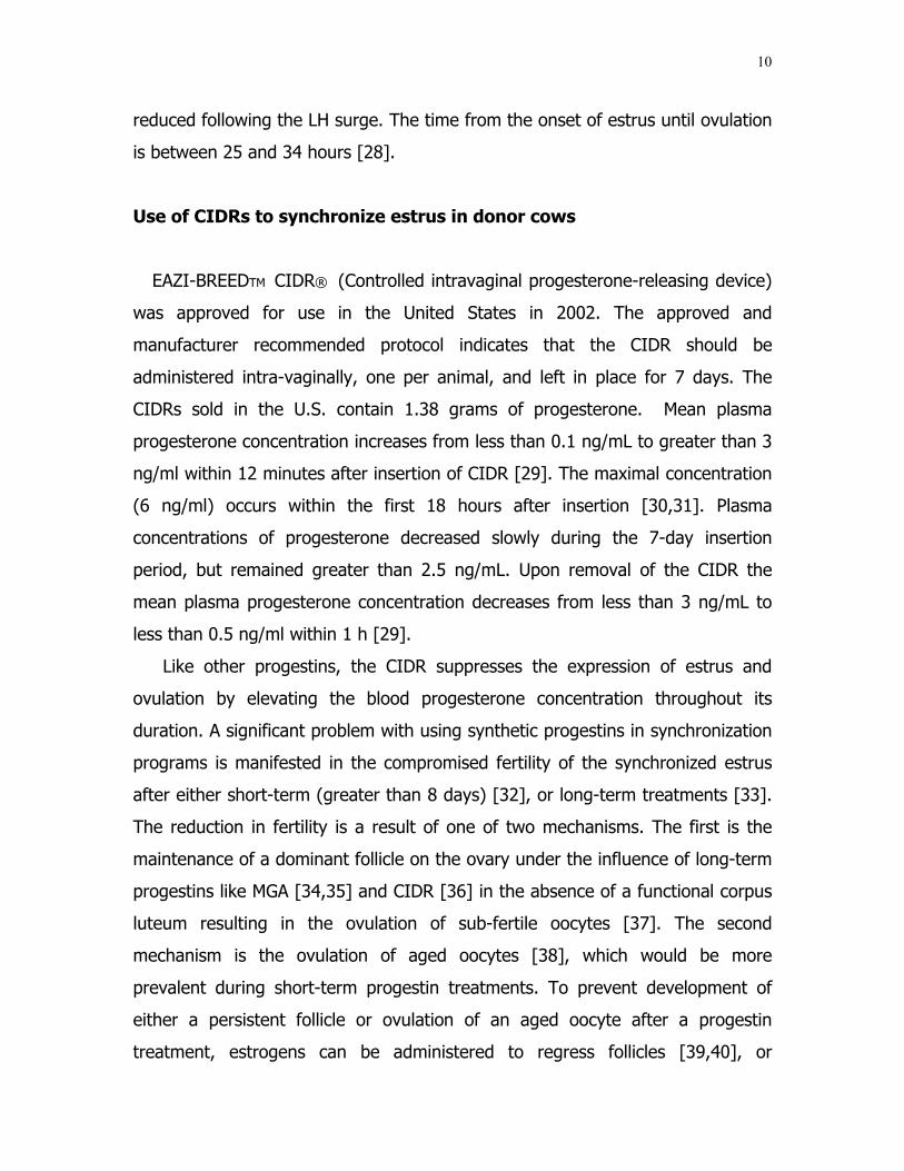
Use of CIDRs to synchronize estrus in donor cows
EAZI-BREEDTM CIDR® (Controlled intravaginal progesterone-releasing device)
was approved for use in the United States in 2002. The approved and
manufacturer recommended protocol indicates that the CIDR should be
administered intra-vaginally, one per animal, and left in place for 7 days. The
CIDRs sold in the U.S. contain 1.38 grams of progesterone. Mean plasma
progesterone concentration increases from less than 0.1 ng/mL to greater than 3
ng/ml within 12 minutes after insertion of CIDR [29]. The maximal concentration
(6 ng/ml) occurs within the first 18 hours after insertion [30,31]. Plasma
concentrations of progesterone decreased slowly during the 7-day insertion
period, but remained greater than 2.5 ng/mL. Upon removal of the CIDR the
mean plasma progesterone concentration decreases from less than 3 ng/mL to
less than 0.5 ng/ml within 1 h [29].
Like other progestins, the CIDR suppresses the expression of estrus and
ovulation by elevating the blood progesterone concentration throughout its
duration. A significant problem with using synthetic progestins in synchronization
programs is manifested in the compromised fertility of the synchronized estrus
after either short-term (greater than 8 days) [32], or long-term treatments [33].
The reduction in fertility is a result of one of two mechanisms. The first is the
maintenance of a dominant follicle on the ovary under the influence of long-term
progestins like MGA [34,35] and CIDR [36] in the absence of a functional corpus
luteum resulting in the ovulation of sub-fertile oocytes [37]. The second
mechanism is the ovulation of aged oocytes [38], which would be more
prevalent during short-term progestin treatments. To prevent development of
either a persistent follicle or ovulation of an aged oocyte after a progestin
treatment, estrogens can be administered to regress follicles [39,40], or

11
gonadotropin-releasing hormone (GnRH) can be administered to ovulate the
dominant follicle [41,42] at the initiation of a progestin treatment, resulting in
emergence of a new cohort of follicles 2 to 5 days later [39,40,43]. Estrus
synchronization programs using CIDRs, are combined with prostaglandin PGF 2αone day before removing the CIDR or the same day of the insert removal. The
timing of estrus following recommended administration of the CIDR and PGF 2αhas been very synchronous [31]. More than 60% of the animals consistently
exhibit estrus within a 24-hour period. Pregnancy rates of cattle bred 12 h after
estrus detection have been satisfactory [31]. CIDRs are also combined with
estradiol and gonadotropin releasing hormone (GnRH) at the time of the
insertion to more effectively control follicular growth and to induce a timed
ovulation following CIDR removal [44,45]. The timed ovulation programs have
been useful to eliminate estrus detection of embryo transfer recipients [46].
Transfer of embryos to cows with a palpable corpus luteum 7 days after
administration of GnRH or 8 days after administration of estradiol to induce
estrus has been successful. Several basic CIDR synchronization protocols are
shown in Fig. 3. All programs consist of a CIDR treatment for 7 days, with PGF
2α administered at CIDR removal. Either estradiol benzoate or GnRH can be
administered at CIDR insertion as a means of initiating follicle turnover.
Following CIDR removal, multiple AI programs can be used [47]. In option 1,
cattle can be inseminated by observed estrus for approximately 5 days after
CIDR removal. In option 2 and 3, estradiol benzoate can be injected 24 hours
after CIDR removal and cattle can be inseminated either by observed estrus
(option 2) or timed AI approximately 24 to 36 hours after estradiol benzoate
(option 3). In option 4, cattle can be timed AI and injected with GnRH 48 hours
after CIDR removal. With option 3 and 4, 48-hour calf removal can also be
implemented in the timed AI programs [47].
Treatment of postpartum beef cows with a 7-day CIDR results in an increased
percentage of cattle detected in estrus and conceiving early in the breeding

12
season in both cycling and anestrus postpartum beef cows. The use of estradiol
benzoate or GnRH in combination with CIDRs can enhance pregnancy rates [47].
Option 1 Inject
PGF 2α & CIDR In CIDR Out Estrus Detection and AI-------------------------------------------------l----------------------------l 0 1 2 3 4 5 6 7 8 9 10 11 12
Option 2
Inject EB Inject Inject Or GnRH PGF 2α EB & & CIDR In CIDR Out Estrus Detection and AI-------------------------------------------------l----------------------------l 0 1 2 3 4 5 6 7 8 9 10 11 12
Option 3
Inject EB Inject Inject Or GnRH PGF 2α EB & & TAI CIDR In CIDR Out -------------------------------------------------l---------------------------- 0 1 2 3 4 5 6 7 8 9 10 11 12
Option 4
Inject EB Inject Inject GnRH Or GnRH PGF 2α & TAI & & CIDR In CIDR Out ------------------------------------------------------------------------------ 0 1 2 3 4 5 6 7 8 9 10 11 12
Fig. 3. Four different options used to synchronize estrus and/or ovulation in beef cows treatedwith intravaginal progesterone-releasing device (CIDR) in combination with prostaglandin F2α(PGF 2α) and with or without estradiol benzoate (EB) or gonadotropin-releasing hormone(GnRH).

13
Control of follicular recruitment
It is known that 8 to 12 days after estrus (equivalent to days 7 to 11 after
ovulation) would be the approximate time of emergence of the second follicular
wave in two or three wave cycles) [48] and a cohort of growing follicles would
be present around that time. Emergence of each follicular wave in the bovine
estrus cycle is preceded by a transient increase in endogenous FSH [49], all 2 to
3 mm follicles that emerge in a wave have FSH receptors and for the first 2 days
after emergence follicular development is supported by FSH. As the small follicles
develop they secrete inhibin and estradiol, both of which inhibit FSH secretion
[50]. By the second or third day after follicular wave emergence FSH reaches
basal levels and continued follicular growth is dependent on LH secretion. A
dominant follicle is selected approximately 2.5 days after emergence because it
develops LH receptors while the subordinate follicles do not [24]. After selection
of the dominant follicle, LH secretion is able to support the development of the
single dominant follicle, but the subordinate follicles become atretic and regress.
The necessity of waiting until mid-cycle to initiate super-stimulatory treatments
implies monitoring estrus and an obligatory delay. To obviate these problems, an
alternative approach is to initiate super-stimulation treatments subsequent to the
exogenous control of follicular wave emergence [51]. There are several possible
ways to control follicular development in cattle. Follicular wave emergence has
been altered experimentally by mechanical follicle ablation or by hormone
treatments. Ultrasound-guided follicle aspiration (used for oocyte retrieval in IVF
programs) has been used as a method of follicle ablation [52,53], the ablation of
ovarian follicles (≥ 5mm), altering the endogenous release of LH and FSH or
administration of exogenous steroids or gonadotropins can cause regression of a
dominant follicle and emergence of a new follicular wave [54]. Estradiol-
progesterone treatments (to hormonally suppress follicular development) have
been extensively investigated [39,55,56]. All of these treatments have resulted in

14
comparable super-ovulatory response in synchronization of follicular wave
emergence [57].
Predicting the functional activity of a dominant follicle and corpus luteum
might be important before starting a super-ovulation regimen or a
synchronization program [58]. Successful super-ovulation depends upon the
ability to control the development of follicles with exogenous FSH. The timing of
FSH administration to optimize super-ovulation must begin at a time when a new
wave of follicles has emerged, but before a dominant follicle has been selected.
After emergence, but before dominant follicle selection, all follicles have FSH
receptors and injections of FSH can act to rescue follicles otherwise destined to
become atretic [59]. The number of follicles that respond to FSH treatment and
develop into ovulatory follicles is dependent on the number of small, antral
follicles that emerge in the follicular wave. Control of the process whereby
primordial follicles develop into antral follicles is poorly understood, however, the
development of primordial follicles eligible to emerge in a follicular wave is not
controlled by FSH and LH [60]. Instead, development of primordial follicles is
dependent on several growth factors produced locally and systemically [59].
Major factors that influence pre-antral follicular growth include: growth-
differentiating factor 9 (GDF-9), bone morphogenic proteins (BMPs) activins,
inhibins, basic fibroblast growth factor (bFGF), epidermal growth factor (EGF),
insulin and insulin-like growth factors (IGF-I and IGF-II) [59].
Variability in response continues to be one of the most frustrating problems
associated with estrus synchronization and super-ovulation programs in cattle.
The techniques designed to control follicular wave dynamics reduce the
variability caused by treating cows at different stages of the estrus cycle [51].
Protocols involving synchronization of follicular wave emergence do offer the
convenience of being able to initiate super-stimulatory treatments quickly and at
a self-appointed time, without the necessity of estrus detection and without
sacrificing results [51].
15
The use of estradiol in estrus synchronization
Estrogens have been shown to induce follicle regression, followed by
synchronous follicular wave emergence, if administered in the face of high levels
of progesterone, and induce LH release and ovulation of a dominant follicle if
administered in the face of low levels of progesterone. It was found that
estradiol valerate treatment, in some cases, caused prolonged suppression of the
pre-wave FSH surge and delayed emergence of the next follicular wave [61].
This was attributed to the prolonged action of the valerate ester. Therefore, a
series of experiments were designed to investigate the effects of a shorter acting
estrogen, 17β-estradiol on follicle wave dynamics. When 17β-estradiol was
injected in the presence of progesterone, FSH suppression occurred and a FSH
surge preceded the new follicle wave 4 to 5 days later [61]. Therefore, it is
recommended that if donors could not be injected with progesterone, they
should be implanted with progesterone before treatment with estrogen. Estradiol
benzoate (2 mg) and progesterone (50 mg) given at the time of device insertion
resulted in more synchronous follicular wave emergence
than treatment with 2 mg estradiol benzoate alone [62].
Estradiol and progesterone treatments have been increasingly used over the
past years in estrus synchronization programs in beef and dairy cattle [63].
Nowadays, treatments consist of insertion of a progesterone (CIDR) device and
the administration of estradiol with progesterone on day 0 (to synchronize
follicular wave emergence). Prostaglandin at the time of device removal on days
7 or 8 (to ensure luteolysis), and the subsequent application of a lower dose of
estradiol 24 hours later or GnRH/LH 48 to 54 hours later to synchronize ovulation
[63].

16
The use of GnRH in donor cows for embryo transfer programs
Administration of GnRH during the bovine estrus cycle causes regression or
ovulation of the dominant follicle and initiates the emergence of a new wave of
follicular growth an average of 2.5 days following treatment [27]. Atresia or
ovulation of the dominant follicle depends on the status (growing, static or
regressing) of the dominant follicle at the time of GnRH injection [64,65]. The
scientific literature of the 1970s and 1980s demonstrated that injections of
gonadotropin-releasing hormone (GnRH) could induce ovulation of ovarian
follicles in milked [66] and suckled cows [67]. Injections of GnRH induced the
release of luteinizing hormone (LH) [66] and follicle-stimulating hormone (FSH)
[68] from the anterior pituitary gland. Resulting elevated blood concentrations of
LH and FSH either caused follicular rupture (ovulation), if a follicle(s) was
present, or may have induced some new follicular development.
Gonadotropin releasing hormone has been used widely since it first became
available commercially in the 1970’s as a treatment for follicular cysts [69]. The
implementation of GnRH in donors has been used to select the ovulatory follicle,
which should cause premature ovulation of that follicle. Using these concepts
researchers have made tremendous strides in developing numerous systems to
synchronize the estrus cycle for an artificial insemination and embryo transfer
[30]. Martinez et al. [44] reported the efficacy of the GnRH products available in
the United States. Cystorelin® induced a greater LH surge than Fertagyl® and
Factrel®. Cystorelin® also induced a greater ovulation rate, but all products
synchronized follicular wave emergence. GnRH is a decapeptide, a linear chain of
ten amino acids. The base for Cystorelin®, Fertagyl® and Ovacyst® is
diacetate, tetrahydrate. Therefore, these three products are chemically identical.
Factrel® has a HCL base, which should not alter bioactivity. Even though the
products are chemically similar, Martinez et al. [44] found differences among
them, which can be related to the active compound in the product. The dose
depended on the type of the ovaric cyst, the clinical claim for GnRH product.

17
Perhaps this explains the variability in response when doses less than the
recommended dose are used. In a retrospective analysis between Cystorelin®
and Factrel®, Stevenson et al. [70] did not detect an effect of GnRH product on
AI pregnancy rates in cows treated with two different GnRH estrus
synchronization protocols.
Twagiramungu et al. [71] proposed the use of a second GnRH treatment after
prostaglandin treatment to ensure ovulation of the existent dominant follicle.
They found that the second GnRH treatment improved the precision of ovulation
and permitted fixed-time insemination without adversely affecting pregnancy
rates. The method consists of an injection of GnRH followed by prostaglandin 7
days later, a second GnRH injection 48 hours after prostaglandin treatment, and
fixed time insemination 24 hours later. The rationale for the treatment is that the
first injection of GnRH is intended to induce an LH release and ovulation or
luteinization of the dominant follicle present at the time, thus resulting in the
emergence of a new follicular wave within 2 days. The administration of
prostaglandin 7 days after treatment is intended to induce the regression of the
original or induced CL, and the second GnRH injection is intended to induce
synchronous ovulation of the new GnRH-induced dominant follicle. The
importance of the second GnRH injection was demonstrated by a higher rate of
ovulation than in cows that were not given a second injection (97% vs. 77%
respectively) [55].
Uses of GnRH in timed AI protocols
One of the newest breeding programs in cattle is known as Ovsynch, which
has been developed for dairy cows (Fig. 4). This protocol uses the initial GnRH
injection of 100 ųg to induce ovulation of a dominant follicle that develops into a
new corpus luteum in the cycling dairy cow. In cycling dairy cows, about 60
percent of the cows given the initial GnRH injection ovulate a follicle in response
to the LH released by the GnRH injection, depending on the stage of their estrus

18
cycle [72]. Following this induced ovulation, a new wave of follicles emerges
from both ovaries within 48 hours, from which a new dominant follicle develops.
Seven days after the initial GnRH injection, PGF 2α is injected to lyse or regress
the original corpus luteum (if one was present at the time of the initial GnRH
injection) and the new corpus luteum induced by the GnRH. During the next 48
hours the new dominant follicle rapidly matures, and at 48 hours after PGF 2α a
second GnRH injection (100 ug) is administered, and the cow is inseminated in
the next 8 to 24 hours. The highest pregnancy rate occurs when the timed AI
occurred at 16 hours [73]. In practice, on dairy farms, cows are inseminated any
time they are detected in estrus during this protocol and further hormonal
injections are discontinued.
GnRH PGF 2α GnRH Timed AI
|--------------------------------|--------------|--------|------> 0 Days 7 9 10 3-5 PM 3-5 PM 3-5 PM 7-9 AM
Fig. 4. Ovsynch Protocol.
Based on the distributions of estrus after the select Synch protocol and the
early success with Ovsynch in beef cows, the Cosynch protocol was developed
and tested [74,75]. This protocol was designed to reduce the number of trips
through the working facility to three (Fig.5). Comparisons of Cosynch to Ovsynch
were made and pregnancy rates were identical at 48% [74]. A further study,
Geary et al. [76] also incorporated 48-hour calf removal (between the injections

19
of PGF 2α and the second GnRH injection) after both the Ovsynch and Cosynch
protocols. Calf removal produced pregnancy rates that were 9 percentage points
greater than rates after each protocol without calf removal.
GnRH PGF 2α GnRH
|--------------------------------|--------------|-----------> 0 Days 7 9
Timed AI
Fig.5. Cosynch protocol.
The combination of Cosynch with a progestin (CIDR inserted from day -7 to
day 0) (Fig. 6) indicates that addition of a CIDR for progesterone
supplementation improved pregnancy rates after a fixed-time AI [77]. But
progesterone did not seem to improve pregnancy rates in suckled beef cows
cycling at the initiation of treatments. Progesterone seemed to be more effective
in a enhancing pregnancy rates in cows that were cycling but in the latter stages
of the estrus cycle at first injection of GnRH and subsequently had no luteal
structure at the PGF 2α injection or in non-cycling cows. Along with parity, days
post-partum, calf removal, and cow condition, previous reports [77] also indicate
that location variables (which could include differences in pasture and diet, breed
composition, body condition, post-partum interval, and geographic location) may
affect the success of fixed-time AI protocols. Therefore, a sound strategy for
utilizing a GnRH protocol, in the absence of progesterone, may be to select cows
that calved earlier in the calving season that tended to be in a good body
condition. A high percentage of these cows should be cycling and result in
20
acceptable fertility rates [77]. To achieve optimal pregnancy rates after embryo
transfer with a GnRH and CIDR synchronization protocol, cows should be in a
good body condition (BCS=5) and treatments should be initiated only when cows
are at least 50 days postpartum [78].
GnRH PGF 2α GnRH
|------CIDR or NORG---------|--------------|---------> 0 Days 7 9
Timed AI
Fig. 6. Cosynch protocol with or without an exogenous source of progesterone administeredduring 7 days.
The GnRH/CIDR/ PGF 2α protocols utilize the advantage of both the GnRH
injection to induce the formation of a new follicular wave and to prevent females
from exhibiting estrus prior to the injection of PGF 2α. Conventional wisdom
indicated that these protocols might be the most consistent protocol to ensure
that the maximal number of recipients exhibit estrus within the ideal interval
[78].
Cryopreservation
The development of successful techniques for the freezing and thawing of
cattle embryos had a profound impact on the field of embryo transfer, which in
turn, has had a significant influence on the cattle industry. An increasing demand
for freezing embryos during the 1990s culminated with over 90 % of the

21
embryos having been frozen in 1999 [79]. Although not all cattle breed
associations keep records of the animals produced by embryo transfer, the
records of some associations indicate that embryo transfer is being widely used
by breeders of genetically superior cattle. This is evidenced by the fact that 86 %
of the top 100 Holstein bulls (as evaluated by Holstein Type-production Index in
2000) were produced by embryo transfer methodology [79].
The first published report of success in freezing and thawing mammalian
embryos for the production of live-born young involved the use of dimethyl
sulfoxide (DMSO) or glycerol as cryoprotectants for laboratory mouse embryos
[80]. Shortly thereafter, cattle [81] embryos were successfully frozen and
thawed with production of offspring. Embryos should be recovered between 6.5
and 7.5 days (normally range from late morula to mid-blastocysts) for optimum
viability after freezing and thawing. The embryos to be frozen should be graded
1 (excellent or good) or 2 (fair) according to the International Embryo Transfer
Society [82]. Improved protocols utilized 10% (1.4 M) glycerol as the
cryoprotectant. A general freezing protocol cited by Hastler [79], utilizes glycerol
as follows:
1. Equilibrate embryos in 10% glycerol for 10 to 20 minutes.
2. Load embryos in 0.25 cc straws; seal and label straws; (embryos can be
loaded while they are equilibrating).
3. Place straws in freezer that is maintaining a holding temperature of -6°C
4. Seed straws and maintain at -6ºC for 15 minutes.
5. Decrease temperature at 0.5ºC per minute to -32ºC.
6. Plunge straws into liquid nitrogen.
Another method for freezing embryos is through the use of ethylene glycol,
which is another cryoprotectant. Voelkel and Hu [83], reported acceptable
pregnancy rates following the transfer of bovine embryos in ethylene glycol and
transferred after thawing directly from the straws in which the embryos had
22
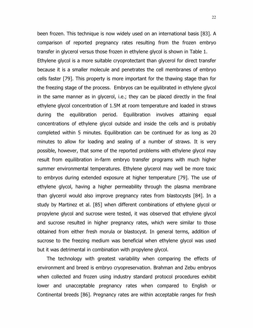
been frozen. This technique is now widely used on an international basis [83]. A
comparison of reported pregnancy rates resulting from the frozen embryo
transfer in glycerol versus those frozen in ethylene glycol is shown in Table 1.
Ethylene glycol is a more suitable cryoprotectant than glycerol for direct transfer
because it is a smaller molecule and penetrates the cell membranes of embryo
cells faster [79]. This property is more important for the thawing stage than for
the freezing stage of the process. Embryos can be equilibrated in ethylene glycol
in the same manner as in glycerol, i.e.; they can be placed directly in the final
ethylene glycol concentration of 1.5M at room temperature and loaded in straws
during the equilibration period. Equilibration involves attaining equal
concentrations of ethylene glycol outside and inside the cells and is probably
completed within 5 minutes. Equilibration can be continued for as long as 20
minutes to allow for loading and sealing of a number of straws. It is very
possible, however, that some of the reported problems with ethylene glycol may
result from equilibration in-farm embryo transfer programs with much higher
summer environmental temperatures. Ethylene glycerol may well be more toxic
to embryos during extended exposure at higher temperature [79]. The use of
ethylene glycol, having a higher permeability through the plasma membrane
than glycerol would also improve pregnancy rates from blastocysts [84]. In a
study by Martinez et al. [85] when different combinations of ethylene glycol or
propylene glycol and sucrose were tested, it was observed that ethylene glycol
and sucrose resulted in higher pregnancy rates, which were similar to those
obtained from either fresh morula or blastocyst. In general terms, addition of
sucrose to the freezing medium was beneficial when ethylene glycol was used
but it was detrimental in combination with propylene glycol.
The technology with greatest variability when comparing the effects of
environment and breed is embryo cryopreservation. Brahman and Zebu embryos
when collected and frozen using industry standard protocol procedures exhibit
lower and unacceptable pregnancy rates when compared to English or
Continental breeds [86]. Pregnancy rates are within acceptable ranges for fresh

23
embryos, but are lower than expected when frozen and thawed. Brahman
embryos frozen in glycerol have a lower pregnancy rate (30%) compared with
48% for direct transfer. The theoretical reasoning for the decreased pregnancy
rate in frozen Brahman embryo is intracellular lipids [11].
Table 1Comparison of pregnancy rates achieved after transfer of bovine embryos that were frozen inglycerol or in ethylene glycol. (X²= P>0.05)Glycerol Ethylene Glycol
No. % Preg. No. % Preg. References
92 48.9 189 58.2 [87]185 56.8 780 59.6 [88]56 69.6 56 50.0 [89]423 45.9 97 47.4 [90]838 55.3 228 51.8 *1609 58 11,376 59.2 [91]225 53.8 218 51.8 **Total3428 55.4 12,944 58.8
* Holland Genetics, Unpublished data** Em Tram, Inc., Unpublished data
Factors affecting the embryonic survival in the cow
Embryonic mortality in cattle is the main source of economic loss for livestock
producers. [92]. In beef herds, pregnancy losses represent an even more
important economic factor because the number of calves sold determines most
of the income.
In order to properly standardize bovine reproductive terms, the Committee on
Bovine Reproductive Nomenclature [93], established that the embryonic period
of gestation extends from conception to the end of the differentiation stage, at
approximately 42 days of gestation, and that the fetal period extends from
gestation day 42 to the delivery of the calf. Secretion of progesterone by the

24
corpus luteum is essential throughout gestation to achieve 100% success in the
maintenance of uterine quiescence and survival of the embryo/fetus, and for
normal parturition, in the cow [94]. Prior to and immediately after estrus,
progesterone regulates the establishment and timing of mechanisms necessary
for luteal regression in the non-pregnant cow and for maternal recognition of
pregnancy. Utilizing this knowledge, researchers and breeders can transfer
embryos into ovariectomized cows and pregnancy can be completed successfully
by providing exogenous progesterone on a continuous daily basis [95].
Much of the pregnancy loss in cattle is due to early embryonic death [96] after
either natural or artificial insemination or embryo transfer. According to the
review by Thatcher et al. [42], approximately 30 % of repeat breeder cows
experience embryonic loss by day 7 of pregnancy. Additional embryonic losses
occur gradually from days 8 to 17 (approximately 40 % of total losses) and
between days 17 and 24 (approximately 24 % of total losses). Losses between
days 17 and 24 were estimated at 6 to 12 % of pregnancies in two studies in
which dairy heifers were bred at synchronized estrus after two treatments with
PGF 2α 11 days apart [97]. Published estimates of late embryonic death rate
(days 27 to 42) average 10 to 12% [42,98,99]. These factors must be
considered in conjunction with estimates of fertilization failure (12%) [100], and
fetal losses that may range as high as 8% of animals pregnant at 42 days [99].
Thus, wastage occurs throughout pregnancy, but is concentrated in the
embryonic period, or the first 40 days after breeding.
Conception postpartum
During the postpartum period, the cow undergoes a transition from an
anovulatory, anestrus, infertile animal into a fertile animal with normal ovulatory
estrus cycle and a higher conception rate. Holness et al [101] reported that only
12% of first postpartum ovulations were fertile and particularly low in the early

25
postpartum period in Bos indicus cattle. Ovulation failure was a significant factor
limiting pregnancy rate in these early postpartum cows.
Early embryonic death associated with short duration of the luteal
phase
A short luteal phase may occur following first ovulation, or first estrus
[102,103]. Short-lived corpus luteum occurred in the pubertal heifer and the
postpartum cow returning to ovulatory activity [104,105]. Obviously, a corpus
luteum that had regressed before day 14 could not support pregnancy; maternal
recognition of pregnancy in the cow occurs on days 14 to 17. Variables such as
follicular development, pre-ovulatory and post-ovulatory concentrations of
gonadotropins, and luteal receptors for LH were shown to be responsible for
some variations in level of luteal function [106], but not for luteal duration. It has
been observed that pretreatment with progesterone usually resulted in formation
of a corpus luteum with a normal functional lifespan, in response to weaning or
injection of gonadotropins [107,108]. Secretion of PGF 2α rose during treatment
of anestrus cows with progesterone, in the same manner as during a short luteal
phase after injection of hCG in control cows [109]. Thus, if the uterus had not
been exposed previously to progesterone, secretion of PGF 2α increased
prematurely when the first corpus luteum began to secrete progesterone.
Several studies have reported that beef cows, from which calves were weaned
at about 35 days postpartum, would consistently exhibit estrus in 4 to 5 days
and form a corpus luteum [110,111,112]. The studies provide evidence that
ovulation and fertilization occurred at the expected time after an estrus
preceding a short luteal phase in early weaned cows. Logically, fertility should be
improved by pretreatment of the postpartum cow with progesterone because of
the prevention of the shortened luteal phase [113]. Progesterone therapy was
tested by providing a daily supplement of melengestrol acetate (MGA) in feed

26
beginning on day 4 after breeding [114]. No pregnancies were maintained in
cows with short luteal phases. In contrast, 41% of all norgestomet pre-treated
cows and 50% of those cows that had normal luteal phases maintained
pregnancy regardless of whether or not they received MGA. Supplemental
injections of 200 mg progesterone daily gave the same results. No pregnancies
occurred in cows with short-lived corpus luteum. Surprisingly, 12 of the 13
control cows that were deleted from the experiment for having a short luteal
phase before breeding conceived at the post-weaning estrus, even though they
were bred at an average of only 33 days postpartum. Combining all of the above
results, it was concluded that about half of the difference in ability to maintain
pregnancy between cows with short and normal luteal phases (when
supplemental progesterone was provided) could be attributed to effects on the
oocyte or embryo before day 7 after estrus and the other half to a hostile uterine
environment on or after day 7. The apparent timing of embryo loss was strikingly
similar to the timing of increased uterine secretion of PGF 2α on days 4 through
9 after estrus in cows with a short luteal phase [109]. Moreover concentrations
of PGF 2α in flushing from the uterine lumen of cows with short luteal phases
were more than double the concentration from cows with normal luteal phases
[115]. Embryo quality tended to be correlated negatively with concentrations of
PGF 2α in flushing from the uterine lumen. Because embryo quality was lower on
day 6 [115] than on day 3 [114]. It was proposed that the specific problem in
short luteal phase cows was likely to have occurred after the embryo entered the
uterus. A direct embryo-toxic effect of PGF 2α seemed possible because that had
been suggested for mouse [116], and shown for rabbit [117] and rat [118]
embryos. Effects of PGF 2α on embryo survival have been examined in several
studies in cows in which daily supplemental progesterone was provided to
replace the regressed corpus luteum. It was shown that PGF 2α was detrimental
to embryos when given to normally cycling beef cows during days 4 to 7 after
estrus and insemination, an interval similar to that during which high embryo

27
mortality had been observed in cows with short luteal phases. The majority of
embryonic mortality in sub-fertile dairy cows occurred 6 to 7 days after estrus
[119], when the morula was developing into the blastocyst. Maurer and Chenault
[120] observed that 67% of embryonic mortality had occurred or was occurring
by day 8 of gestation in beef cows. Seals et al. [121] showed that premature
luteal regression by PGF 2α on days 5 through 8 caused embryonic death in
cows supplemented with progesterone (confirming the results of Buford et al.
[122), but treatment on either days 10 through 13 or 15 through 18 of
pregnancy was not effective. Many products of luteolysis or partial luteolysis
could play a role in embryonic loss [122]. Involvement of luteal PGF 2α is worthy
of further evaluation, because it was observed that the short-lived corpus luteum
produced more PGF 2α than did a corpus luteum with a normal life span.
Other factors that increase secretion of PGF 2α, such as heat stress [123],
uterine infection [124], or mastitis [125,126] could cause embryonic death
through this mechanism.
Other factors associated with pregnancy losses
In cattle sub-luteal concentrations of progesterone during the estrus cycle
preceding insemination induce increased frequency of LH pulses resulting in a
persistent dominant follicle [127,128]. In beef cows, larger pre-ovulatory follicles,
that maintained dominance for an extended period before the LH surge, reduced
conception rate compared to smaller pre-ovulatory follicles (36% versus 91%)
[114]. Exposure of the oocyte to high peak frequency of LH induces the
premature resumption of meiosis [37,129], and the biochemical and
morphological changes in the oocyte in persistent follicles reduce fertility in cattle
[127,130] due to embryo mortality before the 16-cell stage [131]. Therefore,
extending the period of follicle dominance either by exogenous progestins [131],
or when cows have cycles with two waves of follicle growth instead of three-

28
follicle growth waves [132], compromises fertility. Fortunately, the presence of a
persistent follicle does not alter the developmental potential of oocytes from
smaller follicles; that is, if the persistent follicle regresses, normal fertility is
resumed [99].

29
MATERIALS AND METHODS
Criteria to select the records of Red Brahman donor cows and the
recipients
Records of Red Brahman donor cows (n=50) were selected from 200 potential
cow records available to develop this study. The selection was based on previous
reproductive records and performance, and only the cows with a minimum of
two births were included. The donor cows were cycling normally at the time of
the reproductive evaluation by transrectal palpation with normal conformation of
the uterus, including cervix, and ovaries. None of the cows had evidence of
reproductive disease. Other additional selection criteria included, body condition
(only the cows with a score between 5 to 7 on a scale 1 to 9), and cows had to
be at least two months post partum. Some of the donors were collected two or
three times per year, depending on their prior records, and some of them could
not be collected at some point during the study because they were pregnant.
Records of recipients F1 Brahman–Holstein cows comprised 87% of the
recipients for this study (n=531). During this study approximately 30% of the
recipients were rejected before transferring them with an embryo. The selection
criteria for recipients included a complete reproductive exam by transrectal
palpation, analysis of performance records lactating cows with more than two
births, a body condition score between 5 to 6 on the scale 1 to 9, good milking
ability, feminine phenotype, and docile temperament.
Embryo transfer procedure
The donors and the recipients involved in this study belong to the Santa Elena
Ranch, a pure Red Brahman cattle, located in Madisonville, Texas. The Brahman
embryos were collected and transferred under the direction of Ultimate Genetics-
30
Embryo transfer division. The same technician collected, evaluated, and
transferred the embryos produced by the three protocols from 2001-2004.
The process to produce bovine embryos involved the preparation of two
groups of cows, the embryo donor cows and the embryo recipients. Both groups
were synchronized with the use of hormones. Donors had been selected by
genetics, production and reproductive performance. The synchronization started
with CIDR’s (1.38 mg of Progesterone intra-vaginal device) 2.5 mg of 17β-
estradiol + 50 mg of progesterone on day 0. To improve the efficiency of the
embryo production, it was important to increase the number of released oocytes
at the time of ovulation and maximize the collected number of high quality
transferable embryos using FSH from day 5 to 8. The total dosage of FSH
depended on several factors such as age, weight, and previous records. The
dosage used for GnRH was 0.0042 mg/mL (5mL im as a total dosage) to the
donors, 6 hours before the AI (artificial insemination). Eight days after breeding
the donor, the embryos were collected using non-surgical procedures. The
embryos were classified according to the stage and quality by IETS [82], and
were transferred to the recipients previously prepared with the same protocol
without the application of FSH, and GnRH. The recipients that did not conceive to
embryo transfer, after a new physical and reproductive evaluation, were
synchronized again from 60 days after the last embryo collection for the next
embryo transfer protocol.
Protocols for donors
Results from the new protocol used for the donors in 2004 were compared
with results of the three previous years. The production, quality, quantity, and
stage of the embryos, pregnancies, resorptions, abortions, non-pregnant cows
and births of the calves through the conventional embryo transfer technique
were evaluated. The quality of the embryo and the stage of development of the
embryo are described according to the IETS. Quality is a number based on
31
morphological integrity of the embryo which ranges from “1” to “4” as follows:
Code 1: Excellent or Good. Symmetrical and spherical embryo mass with
individual blastomeres (cells) that are uniform in size, color, and density. This
embryo is consistent with its expected stage of development. This judgement
should be based on the percentage of embryonic cells represented by the
extruded material in the perivitelline space. The zona pellucida should be smooth
and have no concave or flat surfaces that might cause the embryo to adhere to a
petri dish or a straw. Code 2: Fair. Moderate irregularities in overall shape of the
embryonic mass or in size, color and density of individual cells. At least 50% of
the cellular material should be an intact, viable embryonic mass. Code 3: Poor.
Major irregularities in shape of the embryonic mass or in size, color and density
of individual cells. At least 25% of the cellular material should be an intact, viable
embryonic mass. Code 4: Dead or degenerating. Degenerating embryos, oocytes
or 1-cell embryos: non-viable [82]. Only embryos with quality code 1 and 2 were
transferred and evaluated. The code for stage of development is also numeric,
ranging from "1“, an unfertilized oocyte or a 1-cell embryo to "9“, expanding
hatched blastocyst [82]. Only embryos number 4 (Morula, day 6), number 5
(Early blastocyst, day 7), and number 6 (Blastocyst, day 7-8), were transferred
and evaluated.
Pregnancies (the act of carrying a developing embryo than fetus within the
uterus) [133], resorptions (the organic process in which the substance of some
differentiated structure that has been produced by the body undergoes lysis and
assimilation) [133], abortions (expulsion of the products of conception before the
embryo or fetus is viable) [133], non-pregnant, and live calves were also
evaluated.
The new protocol was based on the previous response of the donors to the FSH
for the super-ovulation process, the substitution of estradiol benzoate for 17β-
estradiol, and the addition of a single dose of GnRH 6 hours before the artificial
insemination of the donor. The schedule for this protocol was:
32
Day 0: Application of CDIR (Intravaginal 1.38 mg progesterone), 50 mg
progesterone and 2.5 mg of 17β-estradiol (2mL im) in the morning.
Day 5 to 8: Application of FSH am/pm. The dosage depended on the individual
response of each donor to the treatment in previous flushes. The
age and weight was also considered. Based on the above criteria,
the total dosage ranged from 11mL to 14mL of FSH (Folltropin).
Day 7: Application of 25 mg of prostaglandin F2-α (Lutalyse, 5mL, im) in
the morning and the CIDR removal in the afternoon,
Day 8-9: 25 hours after the CIDR removal, 0.0042 mg /mL (5mL im) of
GnRH were injected. 6 hours later, the donors were bred through
artificial insemination for the first time and then two more
inseminations with a 10-hr interval, for a total of three
inseminations.
Protocols for recipients
The new protocol for the recipients involved the elimination of the application
of estradiol benzoate on day 0, and the addition of an injection of 50 mg
progesterone and 2.5 mg of 17β-estradiol (2mL im), at the moment of the
application of the CIDR.
The protocol used in this study was:
Day 0: a.m. Application of CIDR plus a mixture of 50 mg of progesterone
and 2.5 mg of 17β- estradiol (2mL im)
Day 8: a.m. Application of 25 mg of prostaglandin F2-α (Lutalyse, 5mL im)
Day 9: a.m. CIDR removal
Day 10: Begin detection of estrus.

33
Estrus is typically observed in 87% of the recipients synchronized [11]. Before
the embryo transfer all donors and recipients were again evaluated through
transrectal palpation. Donors were evaluated to check the potential embryos
than can be collected on day 8 after the insemination based upon the number of
corpora lutea. Recipients were evaluated prior to embryo transfer to check the
response and CL stage. Only recipients with a CL greater than 10 mm in
diameter received a fresh embryo. The corpora lutea were graded from 1 to 3,
with 1= excellent, 2= good (close to 10 mm), and 3 = small corpora lutea that
are not good to receive an embryo. The embryo flush was non-surgical, using
the standard media and protocols for this technology. The uterine horns of the
donors were flushed to collect the embryos. Pregnancy in the recipients was
determined by transrectal palpation three times at 39-49 days, at 60 days, and
at 90-120 days after the embryo transfer to confirm their pregnancies.
Data analysis
Independent variables that were evaluated for donor cow traits included body
condition on the day of the synchronization for the embryo transfer process, the
type of protocols used, the season of the year, the sire used to fertilize the
mature oocytes, and the sire of the donor collected. At embryo collection, the
total number of embryos collected per donor was recorded, as well as the
number of good embryos to be transferred to the recipients, degenerate ova,
and unfertilized embryos.
For the recipient traits, the independent variables included body condition, CL
size, and stage and quality code score of the embryo that was transferred.
Dependent variables studied included, palpation results (positive pregnancy, non-
pregnant, resorption, abortions), and characteristic traits of the calves born
(births, birth weight).

34
Statistical analysis
Analysis of Variance (ANOVA) was used to determine the effects of the
independent variables on the response in donor and recipient females. This
model included the effects of the new protocol for Red Brahman donor cows,
used in 2004, compared to the results obtained from the protocols used from
2001 to 2003 to produce Brahman embryos. The General Linear Model Procedure
of the SAS (2002-2003) statistical package was used to perform the analysis of
the response data for donors and recipients.
For donor traits the model included sire of the donor, sire of the embryo, type
of protocol, season within protocol, and body condition. Least squares means of
significant effects were separated by two tailed t-tests. Response traits analyzed
included total, good, degenerated, and unfertilized embryos collected. Ratios of
good, degenerated, and unfertilized embryos, based on the total embryos
collected were also evaluated with the same statistical model.
For recipient traits, the three protocols used in this study, the embryo stage,
embryo quality, corpora lutea size, and season within protocol were analyzed by
the final status of the transferred embryos (non-pregnant, resorptions, abortions,
and live calves) using Chi-square analysis Freq- Procedure in SAS (2002-2003)
statistical package. The General Linear Model (GLM) procedure of SAS was also
used to analyze birth weight and gestation length. The model included the sire of
the embryo, season-protocol, protocol, embryo stage, embryo quality, body
condition, and size of the corpora lutea. Least square means of significant effects
were separated by two tailed t-tests. Differences were considered significant at
(P<0.05).
35
RESULTS
A total of 135 collections were carried out on 50 Red Brahman donor cows
during 2001 to 2004, using three different protocols. The number of good
transferable embryos collected with protocol 1 (2001-2002) was 171, with
protocol 2 (2003) 152 good transferable embryos were collected, and with
protocol 3 (2004) 208 good transferable embryos were collected. The total
number of embryos produced during this time was 531 good transferable
embryos.
Donors
The traits analyzed in the donors (number of good, degenerated, unfertilized,
and total embryos) were not affected by sire of donor, sire of embryo, protocol,
and body condition score. The number of degenerated embryos differed by
season-protocol (P<0.0157). Fig. 7 shows the least squares means for winter
from protocol 2 and winter from protocol 3 as the two most affected seasons in
the number of degenerated embryos. These two seasons are different from the
other seasons involved in this study = (P <0.05), between 2001-2004. The traits
of good, degenerated, unfertilized, and total embryos were not affected by sire
of the donors (P>0.05), but there appear to be some underlying biological
differences in the least squares means (±SEM) between the 13 sires of donors
presented in this study (Table 2). The sires of the donors with the seemingly
most consistent and highest embryo production were 70/7, 4/95, 3/42, 56/3, and
24/8 with mean of 7.3 good embryos per collection. The sires of the donors that
were in the middle were 4/115, 80/3, and “x” with a mean of 5.0 good embryos
per collection. Sires of donors with the lowest embryo production were 3/180,
2/40, 0/170, and 72/7 with a mean of 3.3 good embryos per collection.

36
Table 2 also shows the sire of the donors organ
the means of total and good embryo production, a
of degenerated, and unfertilized embryos for th
donors.
The traits evaluated (good, degenerated, unfe
not differ statistically by the three types of pro
However, Table 3 shows in protocol 3 a higher ran
embryos, and a slight higher mean of degene
protocol 1 and protocol 2, but protocol 3 had t
embryos of all protocols.
Fig. 7. Least squares means (+/- pooled SEM) of degeof embryo collection for protocols 1,2 and 3. Winters (the highest in number for degenerated embryos (P <0
0123456789
Fall
Sprin
g
Summer
Wint
er Fall
Sprin
g
Summer
Win
1 2
Protocol
Deg
ener
ated
em
bryo
s
*
ized from high to low bas
nd from the low to high m
e majority of this sires o
rtilized, and total embryo
tocol in the donors (P>
k for mean production of
rated embryos compared
he lowest mean of unfer
nerated embryos by season *) from protocol 2 and 3 are .05).
ter Fall
Sprin
g
Summer
Wint
3
*
ed on
eans
f the
s) did
0.05).
good
with
tilized
er

37
Table 2Least squares means for number of good, degenerated, unfertilized, and total embryos by sire ofdonors (P>0.05).Sire of Donor N Good Degenerated UFO Total Embryo70/7 3 11.6 4.7 0.0 16.24/95 11 6.7 2.2 1.0 9.83/42 5 6.6 3.5 0.3 10.456/3 20 6.5 1.4 1.9 10.04/115 5 6.0 5.3 2.3 13.724/8 9 5.0 1.4 -0.4 6.080/3 50 4.7 2.6 3.5 10.8“x” 4 4.5 2.6 0.4 7.42/40 3 4.3 1.5 3.7 9.63/180 3 4.2 1.6 1.2 7.02/28 5 4.1 -1.7 0.2 2.60/170 13 2.7 1.8 4.0 8.572/7 4 0.1 2.7 5.5 8.3
Pooled SEM 0.47 0.41 0.42 0.58“x” Represents all the sires of donors that were collected only once.
The embryos were collected using three different protocols and in different
years. Embryos collected by protocol 1 included 2001-2002, embryos collected
by protocol 2 included 2003, and embryos collected by protocol 3 included 2004.
Table 3Least squares means for number of good, degenerated, unfertilized, and total embryos byprotocols (P>0.05).Protocol N Good Degenerated UFO Total Embryo1 50 4.6 1.3 2.0 7.92 39 4.6 2.3 2.1 9.03 46 6.3 3.3 1.4 10.9
Pooled SEM 0.16 0.14 0.14 0.20
The analysis of the traits (good, degenerated, unfertilized, and total embryos)
by season-protocol was only different for degenerated embryos (P<0.0157). The
other types of embryo production (good, unfertilized, and total) did not differ by
season-protocol combination in the donors evaluated, however, there are some

38
differences in the means of the embryos for the 3 protocols and the 12 seasons
evaluated. Table 4 shows that the highest production of good embryos was in
the protocol 3 for spring, winter, and summer. The highest number of
degenerated embryos was in protocol 3 for winter and fall, and in protocol 2 for
winter, the seasons that showed the least production of unfertilized embryos
were summer and spring for protocol 3, and summer and spring for protocol 1.
Table 4Least squares means for number of good, degenerated, unfertilized, and total embryos byseason-protocol. Degenerated embryos differed by season-protocol (P<0.05). Good, unfertilized,and total embryos were not significant by season-protocol (P>0.05).Season Protocol n Good Degenerated UFO Total
EmbryoSpring 3 11 8.3 1.3 0.9 10.4Winter 3 7 6.1 1.6 1.3 15.3Summer 3 22 5.7 1.0 0.3 7.3Winter 1 9 5.7 1.1 3.4 10.3Fall 3 6 5.4 1.6 3.0 10.6Winter 2 9 5.3 1.5 1.6 11.6Summer 2 16 5.2 1.1 1.3 8.0Fall 1 11 4.7 1.5 3.6 9.8Summer 1 25 4.2 2.1 0.3 6.7Spring 2 10 4.1 1.3 1.0 5.9Spring 1 5 3.8 0.4 0.5 4.8Fall 2 4 3.8 2.1 4.4 10.4
Pooled SEM 0.41 0.36 0.37 0.53
The ratios for good, degenerated, and unfertilized embryos were evaluated,
based on the total embryos produced by donors with the same model selected
for the donors (Sire of the donor, sire of the embryo, type of protocol, season-
protocol, and body condition).
The ratio for good embryos differed by sire of the donor (P<0.0089). Fig. 8
shows the 13 least squares means (±Pooled SEM) of good-ratio embryos at the
collection time by the sire of the donors involved in this study. The least squares
means for sire and respective ratio were: 2/28: 0.90, 24/8: 0.87, 3/180: 0.70,
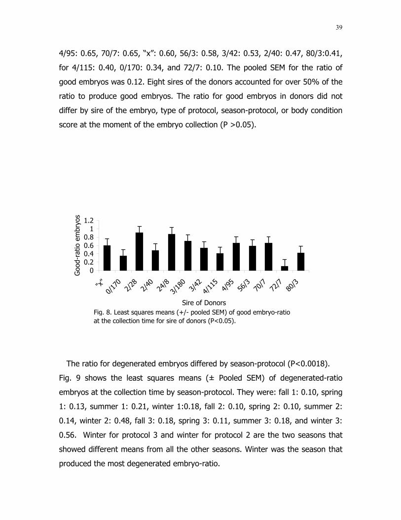
39
4/95: 0.65, 70/7: 0.65, “x”: 0.60, 56/3: 0.58, 3/42: 0.53, 2/40: 0.47, 80/3:0.41,
for 4/115: 0.40, 0/170: 0.34, and 72/7: 0.10. The pooled SEM for the ratio of
good embryos was 0.12. Eight sires of the donors accounted for over 50% of the
ratio to produce good embryos. The ratio for good embryos in donors did not
differ by sire of the embryo, type of protocol, season-protocol, or body condition
score at the moment of the embryo collection (P >0.05).
The ratio for degenerated embryos differed by season-protocol (P<0.0018).
Fig. 9 shows the least squares means (± Pooled SEM) of degenerated-ratio
embryos at the collection time by season-protocol. They were: fall 1: 0.10, spring
1: 0.13, summer 1: 0.21, winter 1:0.18, fall 2: 0.10, spring 2: 0.10, summer 2:
0.14, winter 2: 0.48, fall 3: 0.18, spring 3: 0.11, summer 3: 0.18, and winter 3:
0.56. Winter for protocol 3 and winter for protocol 2 are the two seasons that
showed different means from all the other seasons. Winter was the season that
produced the most degenerated embryo-ratio.
Fig. 8. Least squares means (+/- pooled SEM) of good embryo-ratio at the collection time for sire of donors (P<0.05).
00.20.40.60.8
11.2
"x"
0/17
02/
282/
4024
/83/
180
3/42
4/11
54/
9556
/370
/772
/780
/3
Sire of Donors
Goo
d-ra
tio e
mbr
yos

40
The ratio for unfertilized embryos in donors did not differ by sire of the
embryo, type of protocol, season-protocol, and body condition score at the
moment of the embryo collection (P >0.05).
Recipients
The recipients were analyzed using two sets o
included the three protocols, the three types o
embryo quality, the three sizes of the corpora lu
the embryo transfer, and the four seasons inv
was evaluated by the final status of the em
abortions, and live calves). The distribution o
(Non- pregnant, resorptions, abortions, and liv
Fig. 9. Least squares means of degencollection time for season-protocol (Pprotocol 2 and 3 are the highest to prembryos.
0
0.1
0.2
0.3
0.4
0.5
0.6
0.7
Fall
Sprin
g
Summer
Wint
er Fall
Sprin
g
Summer
Win
1 2
Protocol
Deg
ener
ated
-rat
io e
mbr
yos
*
f criteria. The fir
f embryo stage, t
tea of the recipie
olved in the proc
bryo (Non-pregna
f the final status
e calves) at the e
erated embryo-ratio a<0.05). Winters (*) froduce degenerated ra
ter Fall
Sprin
g
Summer
Wint
3
*
st set of criteria,
he two types of
nt at the time of
ess. This model
nt, resorptions,
of the embryos
nd of the study
t the om tio
er
41
was different across protocols (P<0.0008). Fig.10 shows the percentages of the
final status for the three protocols used.
Protocol
Fig. 10. Percentage of recipients of Brahman embryos that were non pregnant (NP),underwent resorption (RE), aborted (AB), or delivered a live calf (LC), by protocol (P<0.05).
Pregnancy rates were recorded three times after good embryos were
transferred in the recipients. Transrectal palpations in the recipients were
performed for the first time at 39-49 days, for the second time at 60 days, and
for the third time at 90-120 days after the transfer. Fig. 11 shows the pregnancy
rates of recipients with transferred embryos for the three protocols as follows:
For protocol 1: 57.3% were pregnant at the first transrectal palpation, 45.6%
were pregnant at the second transrectal palpation, and 40.90% were pregnant
at the third transrectal palpation. For protocol 2: 64.50 % were pregnant at the
0.00%
10.00%
20.00%
30.00%
40.00%
50.00%
60.00%
1 2 3
NP
RE
AB
LC

42
first transrectal palpation, 58.60% were pregnant at the second transrectal
palpation, and 55.90% were pregnant at the third transrectal palpation. For
protocol 3: 70.70% were pregnant at the first transrectal palpation. 66.30%
were pregnant at the second transrectal palpation, and 62.50% were pregnant
at the third transrectal palpation. These results show that with protocol 1 the
percentage difference between the first palpation and the live calves is very large
(17.5%) compared with the difference seen in protocols 2 (8.6%) and 3 (9.2%)
where a significant decrease is noticeable.
Protocol
Fig. 11. Percentage of pregnancies in recipients of fresh Brahman embryos atthe first palpation (39 - 49 days), second palpation at 60 days, third palpation(>90 - 120 days), and delivered live calves (LC), by protocol.
57.3
0%
45.6
0%
40.9
0%
39.8
0%
64.5
0%
58.6
0%55
.90%
55.9
0%
70.7
0%
66.3
0%62
.50%
61.5
0%
0.00%
10.00%
20.00%
30.00%
40.00%
50.00%
60.00%
70.00%
1 2 3
P. 39 to 49 d
P. 60 d
P.>90 to 120d
LC

43
The distribution of the final status for other variables (embryo quality
transferred, the size of the corpora lutea at the time of the embryo transfer, and
the seasons) did not differ. However, Fig. 12 shows a trend (P<0.1332) in the
percentages of live calves with more calves born from stage 6 than stage 5 or
stage 4 embryos.
Embryo Stage
Fig. 12. Percentage of recipients of Brahman embryos that were non pregnant (NP), underwent resorption (RE), aborted (AB), or delivered a live calf (LC), by embryo stage.
Even though season did not affect the final status of the embryo (P<0.1026).
Fig . 13 shows what may be biological differences in the percentages of the final
status of the embryos. This study found that winter was the best season to get
live calves from embryos transferred in recipients in the previous spring, and the
seasons with less number of resorptions were summer and winter. The season
with least number of abortions was spring.
0.00%
10.00%
20.00%
30.00%
40.00%
50.00%
60.00%
4 5 6
NP
RE
AB
LC

44
The second aspect to the analysis of the recipient data was evaluation of
gestation length and birth weight of the born calves, which were evaluated by
sire of the embryo, season-protocol, protocol, embryo stage, embryo quality,
body condition and corpora lutea size in the recipients at the time of the embryo
transfer. Gestation length differed by sire of the embryo (P<0.0007), by season-
protocol (P<0.0001), by protocol (P<0.0067), and by body condition score
(P<0.0042). Table 5 shows the least squares means (±Pooled SEM) with the
differences found in gestation length by the 14 sires of the embryos in this study.
Season
Fig. 13. Percentage of recipients of Brahman embryos that were non pregnant (NP), underwent resorption (RE), aborted (AB), or delivered a live calf (LC), by season.
0.00%
10.00%
20.00%
30.00%
40.00%
50.00%
60.00%
Fall Spring Summer Winter
NP
RE
AB
LC

45
The sires of the embryos with low least squares means (± Pooled SEM) for
gestation length were: 2/400 with 279.9 days, and 56/3 and 2/28 with 280.2
days. The sires of the embryos that were in the middle for gestation length were:
3/180 with 283.5 days, 80/3 and 92/1 with 283.6, 4/95 with 284.0, 4/45 with
284.4, and 70/1 with 284.9, and the sires of the embryos that had the highest
gestation length were: 6/125 with 285.5, 10/9 with 286.9, 4/115 with 288, 4/128
with 290.0, and 80/2 with 292.6.
Table 5Least squares means (± pooled SEM) for gestation length by sire of the embryo (P<0.05).Sire of embryo n Gestation length (d)
LS Means80/2 5 292.64/128 5 290.04/115 14 288.010/9 12 286.96/125 33 285.570/1 4 284.94/45 5 284.44/95 34 284.080/3 12 283.692/1 1 283.63/180 35 283.52/28 16 280.256/3 3 280.22/400 6 279.9
Pooled SEM 0.38
Table 6Least squares means (± pooled SEM) for gestation length by season-protocol (P<0.05).Season Protocol N LS meansSpring 3 16 276.4Spring 2 14 283.8Summer 1 30 283.9Winter 2 28 284.2Fall 1 15 285.3Winter 1 20 286.5Winter 3 16 287.4Summer 2 37 288.2Spring 1 3 288.9Fall 2 6 289.0
Pooled SEM 0.3

46
The gestation length in recipients differed by season-protocol (P<0.0001). In
Table 6, spring-protocol 3 had the lowest least squares means (± Pooled SEM)
for gestation length (276.4), summer-protocol 2, spring-protocol 1, and fall-
protocol 2 had the highest least squares means (± Pooled SEM) for gestation
length (288.2, 288.9, and 289.0), respectively.
The analysis for gestation length differed by protocol (P<0.0067) in the
recipients. Table 7 shows protocol 3 as the least square mean (± Pooled SEM
281.89 (± 0.13) with the lowest gestation length of the 3 protocols. The least
square means (± Pooled SEM) for protocol 1 and 2 were 286.14, and 286.31,
respectively.
Table 7Least squares means (± pooled SEM) for gestation length by protocol. (P<0.05).Protocol n LS means1 68 286.142 85 286.313 32 281.89
Pooled SEM 0.13
The regression of gestation length on body condition score (on the scale of 1
to 9) of the recipients (P<0.0042) showed that gestation length was affected by
1.8 more days per each point more in the body condition of the recipient.
The last trait analyzed in this study was birth weight of the calves born of the
embryo transfer program with the same model used for gestation length (sire of
the embryo, season-protocol, protocol, embryo stage, embryo quality, body
condition score, and corpora lutea size).
Birth weight differed by season-protocol combination (P<0.0109). Table 8
shows that the least squares means with the lowest birth weights were for
spring-3 with 74.7, followed by fall-2 with 77.2, and summer-2 with 79.6. The
birth weights found with middle least squares means were for summer-1 with

47
80.1, spring-2 with 80.6, fall-1 with 81.9, winter-2 with 84.0, and winter-1 with
84.7. The birth weights found with the highest least squares means were for
spring 1-with 87.3, and winter-3 with 89.4.
Table 8Least squares means for birth weight by season-protocol. (P<0.05).Season Protocol N LS meansFall 1 15 81.9Spring 1 3 87.3Summer 1 30 80.1Winter 1 20 84.7Fall 2 6 77.2Spring 2 14 80.6Summer 2 37 79.6Winter 2 28 84.0Spring 3 16 74.7Winter 3 16 89.4
Pooled SEM 0.4
Birth weight of the calves born through the embryo transfer technology
differed by embryo quality at the moment of the transfer (P<0.031). The highest
weight was for calves from embryos classified as quality 1 at the moment of the
embryo collection that were later transferred to the recipients (Table 9).
Table 9Least squares means for birth weight by embryo quality (P<0.05).Embryo Quality N LS means1 75 84.02 110 80.2
Pooled SEM 0.1
Birth weight of the calves born through the embryo transfer technology
differed when regressed on corpora lutea size of the recipients (P<0.0018). The

48
calves born from recipients that had CL1 were 2.05 (kg) heavier than calves from
recipients that had CL2 at the day of the embryo transfer.
Other models were evaluated in the traits for donors such as number of AI,
sire of the donors within protocols, sire of the embryos within AI, age of the
donor, weight of the donor, FSH mL, protocols within FSH mL, protocols within
age, protocols within weight, and protocols within body condition score, but the
results were not significant, and were excluded for the final analysis.
For recipients age was evaluated with the models used in SAS with no
significant values. The breed of recipients was not evaluated because most of
them (87%) were crossed F1 Brahman-Holstein, and the remaining 13% were
represented by few recipients of many different crosses.

49
DISCUSSION
Donors
The number of degenerated embryos, as well as the ratio for the same trait
evaluated in Red Brahman cows, was affected by season-protocol. The highest
number of degenerated embryos was produced in winter protocol 3 and winter
protocol 2. If we compare the degenerated embryos for winter for the three
protocols, winter protocol 3 produced the highest number. For the ratio of
degenerated embryos, winter protocol 3 and winter protocol 2 were the seasons
that produced the most degenerated embryo-ratio. The highest embryo
productions are compared in bovine embryo transfer technology, the production
of gametes has been affected by seasons in both genders [134]. Indicating that
embryo production is affected by the season of the year. Bos indicus cattle have
reduced reproductive performance in winter [134]. In a study of reproduction of
Bos indicus breeds and crosses by Bastidas and Randell [135], the number of
transferable embryos produced per Brahman donor cow was lower in winter
[135]. The analysis of good, unfertilized, and total embryos did not differ by
season-protocol, even though there were differences on their least squares
means for the embryo production. Spring, winter, and summer, for protocol 3,
were the seasons with the highest number of good embryos produced, and also
the seasons with the lowest number of unfertilized embryos found, not including
winter for the same protocol 3. In Summary, the better combination to produce
good embryos were the seasons that used protocol 3, not including winter.
These results could be due to the improvements of protocol 3 using the FSH for
superovulation under different criteria from the one used for protocol 1 and 2.
Also the addition of only one dosage of GnRH to synchronize ovulation in the
donors, 24 hours after the CIDR removal, or 36 hours after the application of
PFG 2α (induction the regression of the corpora lutea) to allow the follicle to
ovulate and the female to demonstrate behavioral estrus. GnRH has been used

50
with CIDRs and estradiol to control follicular growth, and a second dosage of
either GnRH or estradiol to induce timed ovulation following CIDR removal [44].
Season could also have some effect on the production of embryos due to the
variability of embryo production through the length of the 4 seasons and even
more through the 12 periods evaluated from 2001-2004. A study on Brahman
cows found that Brahman donors produced the greatest number of good
embryos in the spring season [136]. A possible explanation for some of the
seasonal influences found in Bos indicus cattle may be the function of the
pituitary which is altered during the winter, suggesting that day length affects
the estral cycle, especially in the Brahman breed. Brahman cows have a lower
pre-ovulatory luteinizing hormone surge during the winter compared with the
spring or summer periods [137].
The number of good, degenerated, unfertilized, and total embryos was not
affected by sire of the donors. However the ratio for good embryos differed by
sire of the donor. The differences in the least squares means for embryos
produced by the 13 sires of the donors evaluated are shown in Table 2, and Fig.
8. The differences found could be related to genetic factors related to their
fertility. A number of genetic based variations are known to have direct effects
on fertility and reproductive outcome in cattle [138]. Currently, the cattle
industry is requesting the use of genetic markers to predict fertility in males as
well as in females. Genetic markers can be used to identify inheritance of specific
genes of animals from their parents [139]. An example of fertility tests that
identify genetic markers is Fertility Associated Antigen (FAA) to select bulls.
Differences in production of good, degenerated, unfertilized, and total embryos
were not significant by the three types of protocols used in the donors studied.
However, the three protocols showed slight differences using the least squares
mean analysis of embryo production (Table 3). Protocol 3 had the highest least
squares mean for good embryos compared with protocol 2 and 1. Protocol 3 also
had the lowest least squares means for unfertilized embryos compared with
protocol 2 and 1, but at the same time protocol 3 had the highest least squares

51
means for degenerated embryos compared with protocol 2 and 1. The better
response in protocol 3 to produce more good embryos per collection can be
related to the changes made to the super ovulation protocol. The dosage and
criteria to use FSH in the donors was based per each individual, according to
their previous records, age, and weight. However the dosage used in protocol 3
was lower than that used in protocols 2 and 1. Massive ovarian responses are
often associated with a number of problems, and the final goal of super
ovulation is to obtain good quality embryos. Large ovarian responses often result
in the reduced embryo quality, massive ovarian responses may be associated
with a reduction in the proportion of follicles that ovulate [140]. Follicular
maturation is also strongly altered when super ovulation is induced [141]. The
alterations are in the differentiation of granullosa/thecal cells induced by PMSG
or FHS [142,143]. The other factor involved in the highest number of good
embryos produced by protocol 3 over protocols 2 and 1 was the use of 17β-
estradiol. 17β-estradiol intramuscularly induces regression of the dominant
follicle and resets the development of follicles on the ovaries of a donor. This
allows the use of FSH to be started 4.5 days later when a new wave of follicles is
developed [144]. The number of good embryos produced by protocol 3 in this
study could be compared with a study made in 58 cows superovulated following
the application of 17β-estradiol to reset follicular development, producing a total
of 394 embryos, with an average of 6.8 grade 1 or 2 embryos per donor [144].
This same author reported that when a control group of 56 donors not treated
with 17β, resulted in 353 embryos and a rate of 6.3 grade 1 or 2 embryos per
donor [144]. There are other aspects relevant to the embryo transfer industry,
called variability between females and within each female. The first can be
detected as soon as super ovulation is attempted, the second requires successive
attempts at super ovulation in the same female [141]. Variation among donors
is still one of the most frustrating aspects of embryo transfer.

52
Recipients
The results showed significant differences in the final status (non-pregnant,
resorptions, abortions, and live calves) for the three protocols used (Fig. 10).
Transrectal palpations in the pregnant recipients revealed loss of early and late
pregnancies through the full length of gestation (Fig. 11). Analysis of the results
for recipients for protocol 1, showed resorptions of 11.7% between the first
and the second transrectal palpation, abortions of 4.7% between the second and
the third transrectal palpations, and more abortions of 1.1% between the third
transrectal palpation and the birth time of the calves. Recipients with transferred
embryos using protocol 2 showed resorptions of 5.9% between the first and the
second transrectal palpation, abortions of 2.7% between the second and the
third transrectal palpations, and 0% abortions between the third transrectal
palpation and the birth time of the calves. Finally recipients with transferred
embryos using protocol 3 showed resorptions of 4.4% between the first and the
second transrectal palpation, abortions of 3.8% between the second and the
third transrectal palpations, and 1% of abortions between the third transrectal
palpation and the birth time of the calves. Most of the lost pregnancies occurred
between the first and the second palpation, and the protocols that showed the
least lost pregnancies were 2 and 3. Between protocol 1 and 3 there is a big
difference in the percentages of total lost embryos. Losses of pregnancy are
characterized by early embryonic death, which occurs prior to the period of
corpus luteum maintenance in the cow at days 15-17 of the cycle, and late
embryonic death, which occurs from corpus luteum maintenance to the end of
the differentiation stage, at approximately 42 days of gestation [91, 92]. After 50
days of gestation, pregnancy losses are less frequent and characterize fetal
death [91, 92].
It is interesting to compare the fetal loss rates in this study with the fetal loss
rates reported by [145,146] under field conditions. This evaluation showed fetal
losses of 5.8% for protocol 1, 2.7% for protocol 2, and 4.8% for protocol 3 while

53
fetal loss rates of 5.6% between the time of pregnancy diagnosis and parturition
are thought to be common in cattle [145,146]. The fetal loss rates reported by
Dunne and Baxter et al [145,146] include the losses by reproductive diseases, in
this study Red Brahman cows donors and recipients were tested for reproductive
diseases, even though the herds of origin of the donors and recipients were free
of the main reproductive diseases. This also explains the lower fetal loss rates in
embryo transfer with the three protocols used compared with the results
reported by Dunne and Baxter et al [145,146]. However, there are other many
factors involved in the pregnancy losses and fetal death such genetic, nutritional,
and management aspect that are not discussed in this study.
The analysis of the recipients by the stage of the embryo on the day of
transfer approached significance but was not significant of (P<0.05). The slight
difference shown in the percentages of live calves between embryo stage 4 and
embryo stage 6 may suppose that embryos have better chances of survival
through the whole gestation when transferred as morula or blastocyst, but not
with early blastocyst. Spell et al [147| determined effect of type and
developmental stage of embryos on pregnancy rates after embryo transfer in
Angus cows for both groups, donors and recipients. The embryos collected were
assigned a developmental stage and quality grade according to standards set
forth by the International Embryo Transfer Society. Developmental stage codes
were: 3 for early morula, 4 for morula, 5 for early blastocyst, and 6 for
blastocyst. Pregnancy diagnosis was performed 30 days after the transfer (37
total days of gestation). The pregnancy rates for embryos staged 3, 4, 5, and 6
were: 50%, 73.4%, 73.1%, and 63.2% respectively [147]. These results can not
be compared because the cited study evaluated pregnancy rates only at 37 days
of gestation, while in this study (Red Brahmans) the recipients were evaluated
through the length of the whole pregnancy, until the birth of the calves. A major
problem for the cattle breeding industry is the high rate of early embryo loss
[148]. The evaluation of any kind of effect of an embryo at 37 days of pregnancy
rate is considered too premature to make any kind of definitive statement.

54
The last part of this study was the gestation length and birth weight of the
embryo calves born. Gestation length differed in the analysis by the sire of the
embryos, season-protocol, and corpora lutea size in the recipient on the day of
the transfer. The differences found among the 14 sires of the embryos for the
least squares means for gestation length were: The sires of the embryos that
had the lowest least squares means were: 2/400, 56/3, and 2/28. The sires of
the embryos that had the highest least squares means were: 80/2, 4/128, 4/115,
and 10/9. The rest of sires of the embryos are in between the lowest and the
highest least squares means for gestation length. However the 14 Red Brahman
bulls used to produce embryos showed that the gestation length for the
recipients (dam of these embryos), was under normal parameters. Randel [134]
reported the gestation length by other sources: Bos taurus with 282 days
[134,149], Brangus 286 days [134,150], Brahman 293 days [134, 151], Nelore
291 days [134, 152]. In another study by Amen et al [153] that evaluated birth
weight and gestation length (n =511), Bos indicus- Bos taurus reciprocal
backcrossed embryo transfer calves were studied. The average for gestation
length in males and females was 291.2 and 288.5, respectively [153]. Results for
gestation length were affected by the breed composition of the embryo transfer
calves themselves, as well as the cross that produced the calves when Bos
indicus and Bos taurus combinations are involved [153].
The use of EPDs (Expected Progeny Difference) from the American Brahman
Breeders Association [154], which is an estimate of an animal’s genetic value as
a potential parent as compared to another animal of the same breed [155]. The
birth weights can be predicted by using the EPDs as an alternative to predict the
gestation length that can be influenced by the sire of the donor. The birth weight
EPDs for some of the lowest least squares means for the red Brahman bulls used
in this study coincide with a shorter gestation length as shown in Table 10 for
bulls 2/400, 56/3 and 2/28. In this case it was more accurate to predict short
gestation length based on the EPDs of the sires because these sires had a high
accuracy to predict low birth weights furthermore short gestation length.

55
Accuracy value, expressed on a 0 to 1 scale gives an indication of how much
confidence one can place in the EPD, and is based on how thoroughly the animal
has been evaluated genetically [155]. For the highest least squares means for
gestation length, the bull: 80/2 was not possible to evaluate because its EPDs
are not avaliable yet. The sires 4/128, 4/115 and 10/9 were the ones with the
highest gestation length and high EPDs for birth weight, but the accuracy of
those EPDs are not high compared to the sires with low EPDs. For that reason in
this study we can not predict high gestation length for the sires of the donors.
However, cows of different breeds have different gestation lengths. The breeds
derived from India have gestation lengths about 10 days longer than Bos taurus
breeds [134].
Table 10Least squares means for gestation length and EPD’s for birth weights by sire of the embryo. Sire of embryo N Birth Weight Gestation length (d) Birth weight (lb)
EPD ACC LS Means LS Means80/2 5 - - 292.6 82.04/128 5 1.2 0.64 290.0 81.64/115 14 1.0 0.73 288.0 83.010/9 12 1.5 0.46 286.9 78.96/125 33 3.1 0.62 285.5 81.570/1 4 -0.1 0.65 284.9 81.54/45 5 2.9 0.63 284.4 81.64/95 34 2.6 0.70 284.0 85.480/3 12 5.4 0.88 283.6 82.492/1 1 1.6 0.65 283.6 81.03/180 35 3.9 0.71 283.5 84.32/28 16 -0.2 0.83 280.2 81.156/3 3 0.2 0.86 280.2 81.92/400 6 0.65 0.71 279.9 78.2
The gestation length in recipients differed by season-protocol. Spring-protocol
3 and 2 had the lowest least squares means for gestation length, in contrast
Summer-protocol 2, Spring-protocol 1 and Fall-protocol 2 had the highest least
squares means for gestation length. Contrary to this study Ritchie et al. [156]
reported that calves born in the fall season are in general lighter in weight and

56
experience less dystocia that those born in the spring. Then this leads us to think
that the protocol has an important influence on the gestation length. The
analysis for the gestation length differs by protocol, and Protocol 3 was the one
with lowest least squares means for gestation length.
For the analysis of gestation length by body condition, no literature was found
to compare with the results obtained in this study. However there are many
other factors that affect gestation length through the body condition of the
recipient such as genetic and nutritional factors, age, breed of the recipient and
breed of the embryo as well as EPDs of the sires of donors and embryos.
Birth weight differed based on embryo quality. This result could be used to
match embryos quality grade 1 with recipients with big frame score and mature,
embryos with quality grade 2 with recipients with small frame score and young.
This would prevent potential dystocias at the birth time with some embryos from
sires known as carriers of high birth EPDs.
The last trait is the corpora lutea. Birth weight is influenced by corpora lutea,
which means that calves from recipients that had CL1 were 2.05 kg heavier than
calves from recipients with CL2. Since recipients with CL1 produce heavier calves
it would be desirable to transfer grade 2 embryos to them, and recipients with
CL2 transferred with embryos grade 1.

57
CONCLUSIONS
Registered Red Brahman donor cows, and recipients F1 Brahman-Holstein,
from Santa Elena Ranch, located in Madisonville, Texas were evaluated. The
study involved the whole process of embryo transfer, through the analysis of
records of fresh embryo production from the day of the collection to the birth of
the embryos produced. Fifty donors were selected based on reproductive and
production records. The study included records produced during years 2001 to
2004 with three different protocols. For 2001 and 2002 with protocol 1, 2003
with protocol 2, and 2004 with protocol 3. A total of 135 collections were made.
For donors the traits evaluated were: sire of the donor, sire of the embryo,
protocol, season-protocol, and body condition by the production of the donor at
the collection time as follows: good, degenerated, unfertilized, and total embryos
produced. The total number of good embryos collected was 531 categorized as
follows: 171 embryos for protocol 1, 152 embryos for protocol 2, and 208 for
protocol 3. The embryos were transferred immediately to 531 recipients
previously selected by reproductive and production records. Records of
transrectal palpation were recorded at 39 to 49 days, at 60 days, and between
90 to 120 days after the embryo transfer. These results were evaluated as non-
pregnant, resorptions, abortions, and number of live calves born through this
reproductive technology. The model used for these traits was: protocol, embryo
stage, embryo quality, corpora lutea size, and season. The recipients were
evaluated according to the gestation length and birth weight of the calves by the
traits: sire of the embryo, season-protocol, protocol, embryo stage, embryo
quality, body condition score, and corpora lutea size.
Spring and summer for protocol 3 were the best seasons to produce good
numbers of embryos, and the least number of unfertilized embryos for the
donors.
Protocol 3 with the use of 17ß-estradiol in combination with FSH in individual
treatments, dosages, and criteria, plus the use of GnRH in Red Brahman donors

58
previously synchronized with (CIDRs), was the best alternative to produce
quality, and quantity number of embryos per collection for the majority of the
seasons except winter. Winter was identified as the season that produced the
highest number of degenerated embryos. Further studies are needed to
determine the cause resulting in produce unfertilized, and degenerated embryos
at the collection time in order to maximize the production.
There are genetic variations present in the sires of the donors that affect the
embryo production independently of the protocols used and season of the
collection. The sires of the donors with the most regular production of embryos
were: 70/7, 2/28, 4/95, 3/42, 56/3, and 24/8. The sires of the donor with
average production of good embryos were: 4/115, 80/3, and “X“. The sires of
the embryos with the lowest embryo production were: 3/180, 2/40, 0/170, and
72/7.
There are differences between the protocols with respect to the final result of
the transferred embryos. Protocol 3 had the highest percentage of live calves
(61.54%) compared with protocol 2 and 1, respectively (55.92%, and 39.77%).
Protocol 3 also had the lowest percentage of resorptions of the three protocols
evaluated. Protocol 2 had the lowest percentage of abortions (2.63%) compared
with protocol 3 (4.81%), and protocol 1 (5.85%). Based on the results on the
donors collection and the embryo transfer to the recipients in this study,
protocol 1 was found different from protocol 3.
The majority of the lost pregnancies for the embryos were between the first
and the second palpation after the transfer. The protocol 3 showed the lowest
number of lost pregnancies.
It is believed that embryos in stage 4 and in stage 6 have better chances of
survival once they are transferred in the recipient. However, it requires further
study with a larger numbers of embryos and recipients to validate this potential
hypothesis.
The results showed that the sires of the embryos with the lowest gestation
lengths were: 2/400, 56/3, and 2/28. The sires of embryos with the highest
59
gestations lengths were: 80/2, 4/128, 4/115, and 10/9. However the 14 Red
Brahman bulls used in this study showed normal gestation lengths for the
recipients.
The use of EPDs to predict birth weights in calves from the sire of the donor
could be used to predict gestation length. According to the results of this study,
the donor’s sires: 2/400, 56/3, and 2/28 with high ACC should be selected for
this trait.
The results showed that gestation length in recipients differed by season-
protocol, with the protocol more influential on the gestation length. For further
studies, it is important to analyze the effect of gestation length by body condition
of the recipient on the day of the embryo transfer because this study found that
it was significant. However there may be other factors involved that were not
evaluated in this analysis.
Birth weight by embryo quality, and birth weight by corpora lutea size of the
recipient can be used together as a parameter to choose the right embryo from
the collection for the right recipient at the moment of the embryo transfer, to
decrease the risk of loosing the pregnancy and reduce dystocias.
This study should be very beneficial to the Brahman breeders in that it is a
model designed specially for the breed. It should help them to have a better
understanding of the unique attributes of the breed with respect to reproductive
and production performance. However, there are still many unanswered
questions to solve in this field, and further research is needed relative to embryo
production in Brahman cattle.

60
REFERENCES
[1] Lohuis MM. Potential benefits of bovine embryo-manipulation technologiesto genetic improvement programs. Theriogenology 1995;43:51-60.
[2] Tribulo H, Moreno D, Cutaia L, Gatti G, Tribulo R, Caccia M, Bo GA.Pregnancy rates in embryo recipients treated with progesterone vaginaldevices and eCG and transferred without estrus detection. Theriogenology2002;57:563 (abstract).
[3] Grimes JF. Utilization of embryo transfer in beef cattle. Ohio StateUniversity Cooperative Extension Fact sheet No. 0017.
[4] Chase CC, Jr. Embryo transfer in tropically adapted cattle in thesemitropics. In: Fields MJ, Sand RS, Yelish JV. (Eds.). Factors AffectingCalf Crop: Biotechnology of Reproduction 2002. CRC. Press, Boca Raton,FL, p.145-55.
[5] Reynolds WL, DeRoven TM, High JW. The age and weight at puberty ofAngus, Brahman and zebu crosses heifers. J Anim Sci 1963;22:243-44.
[6] Warnick AC, Burns WC, Koger M, Hazen MW. Puberty in English, Brahmanand crossbreeds of beef heifers. Proc of Southern Agric Workers 1956;46-53.
[7] Randel RD. Seasonal effects on female reproductive functions in thebovine (Indian Breeds). Theriogenology 1984;21:170-85.
[8] Alvarez P, Spicer LJ, Chase CC, Payton ME, Hamilton TD, Stewart RE,Hammond AC, Olson TA, Wettermann RP. Ovarian and endocrinecharacteristics during an estrus cycle in Angus, Brahman, and Senepolcows in a subtropical environment. J Anim Sci 2000;78:1291-1302.
[9] Krininger CE, Block, J AL- Katanani YM, Rivera RM, Chase CC, Hansen PJ.Differences between Brahman and Holstein cows in response to estrussynchronization, superovulation, and resistance of embryos to heat shock.Anim Reprod Sci 2003;78:13-24.
[10] Voelkel SA, Hu XY. Direct transfer of frozen-thawed bovine embryos.Theriogenology 1992;37:23-37.
[11] Looney CR, Schneider HJ, Ruiz F, Moore GD, Kristynik AM, Romo S.Environmental and breed factors influence reproductive technologies inthe bovine. In: Joint Conv Proc. Tampa, Florida, 2004. p. 31-39.

61
[12] Macmillan Kl, Thatcher WW. Effects of an agonist of gonadotropin-releasing hormone on ovarian follicles in cattle. Biol Reprod 1991;45:883-89.
[13] Pursley JR, Kosorok MW, Wiltbank MC, Reproductive management oflactating dairy cows using synchronization of ovulation. J Dairy Sci 1997;80:301-06.
[14] Roy GL, Twagiramungu H. A fixed time AI program using the GnRH –PGF-GnRH method for beef females. J Anim Sci 1996;74 (suppl 1): 462.
[15] Thatcher WW, Moreira F, Santos JEP, Mattos RC, Lopez FL, Pancarci SM,Risco CA. Effects of hormonal treatments on reproductive performanceand embryo production. Theriogenology 2001;55:75-90.
[16] Smith RD. Estrus detection. In: Morrow DA, (ed). Current Therapy inTheriogenology. W.B. Saunders, Philadelphia, PA, 1986. p.153-7.
[17] Allrich RD. Estrus behavior and detection in cattle. Vet Clin North AmerFood Anim Pract 1993;9:249-62.
[18] Sartori R, Haughian J, Rosa GJM, Shaver RD, Wiltbank MC. Differencesbetween lactating cows and nulliparous heifers in follicular dynamics,luteal growth, and serum steroid concentrations. J Dairy Sci 2000;83(suppl 1):212 (abstract).
[19] Vasconcelos JLM, Sartori R, Oliveira HN, Guenther JG, Wiltbank MC.Reduction in size of the ovulatory follicle reduces subsequent luteal sizeand pregnancy rate. Theriogenology 2001;56:307-14.
[20] Anderson LE, Wu Y-L, Tsai S-J, Wiltbank MC. Prostaglandin F-2α receptorin the corpus Luteum: Recent information on the gene, messengerribonucleic acid, and protein. Biol Reprod 2001;64:1041-7.
[21] Adams GP, Matteri RL, Ginther OJ. The effect of progesterone on growth
of ovarian follicles, emergence of follicular waves and circulating FSH inheifers. J Reprod Fertil 1992;95:627-40.
[22] Fortune JE. Ovarian follicular growth and development in mammals. Biol.Reprod 1994;50:225-32.
[23] Ginther OJ, Beg MA, Bergfelt DR, Donadeu FX, Kot K. Follicle selection inmonovular species. Biol Reprod 2001;65:638-47.

62
[24] Ginther OJ, Wiltbank MC, Fricke PM, Gibbons JR, Kot K. Selection of thedominant follicle in cattle. Biol Reprod 1996;55:1187-94.
[25] Wiltbank MC. Hormonal regulation of follicles and the corpus luteumduring the bovine estrus cycle. In: Proceedings of the Annual ConventionAmerican Embryo Transfer Association. Albuquerque, New Mexico, 2002.p. 14-26.
[26] Phillips G. The estrus cycle in cattle. March 18, 2005.www.outreach.missouri.edu/adair/livestock/presentations.htm
[27] Pursley JR, Mee MO, Wiltbank MC. Synchronization of ovulation in dairycows using PGF-2α and GnRH. Theriogenology 1995;44: 915-23.
[28] Walker WL, Nebel RL, McGilliard ML. Time of ovulation relative tomounting activity in dairy cattle. J Dairy Sci 1996;79:1555-61.
[29] Rathbone MJ, Bunt CR, Burggrafs, Macmillan KL, Burke CR, Pickering KL.Reegineering of a commercially available bovine intravaginal insert (CIDRinsert) containing progesterone. J Control Release 2002;85:105-15.
[30] Cliff L. Methods to increase reproductive efficiency in cattle. Minutes:Annual Meeting NC-1006. St Louis, Mo. September 23-24, 2004.
[31] Lucy MC, Billings HJ, Butler WR, Ehnis LR, Fields MJ, Kesler DJ, Kinder JE,Mattos RC, Short RE, Thatcher WW, Wetteman RP, Yelich JV, Hafs HD.Efficacy of an intravaginal progesterone insert and an injection of PGF 2alpha for synchronizing estrus and shortening the interval to pregnancy inpostpartum beef cows, peripubertal beef heifers, and dairy heifers. J AnimSci 2001;79:982-95.
[32] Beal WE, Chenault JR, Day ML, Corah LR. Variation in conception ratesfollowing synchronization of estrus with melengestrol acetate andprostaglandin F2α. J Anim Sci 1988;66:599-602.
[33] DeBois CHW, Bierschwal CJ. Estrus cycle synchronization in dairy cattlegiven a 14-day treatment of melengestrol acetate. Am J Vet Res1970;31:1545-48.
[34] Anderson LH, Day ML. Acute progesterone administration regressespersistent dominant follicles and improves fertility of cattle in which estruswas synchronized with melengestrol acetate. J Anim Sci 1994;72:2955-61.

63
[35] Fike KE, Wehrman ME, Lindsey BR, Bergfield EG, Melvin EJ, Quintal JA,Zanella EL, Kojima FM, Kinder JE. Estrus synchronization of beef cattlewith a combination of melengestrol acetate and an injection ofprogesterone and 17β-estradiol. J Anim Sci 1999;77:715-23.
[36] Sirois J, Fortune JE. Lengthening the bovine estrus cycle with lowconcentrations of exogenous progesterone: a model for studying ovarianfollicular dominance. Endocrinology 1990;127:916-25.
[37] Revah I, Butler WR. Prolonged dominance of follicles and reduced viabilityof bovine oocytes. J Reprod Fertil 1996;106:39-47.
[38] Mihm M, Curran N, Hyttel P, Boland MP, Roche JF. Resumption of meiosisin cattle oocytes from preovulatory follicles with a short and a longduration of dominance. J Reprod. Fertil 1994;13:14 (abstract series).
[39] Bo GA, Adams GP, Pierson RA, Mapletoff RJ. Exogenous control offollicular wave emergence in cattle. Theriogenology 1995;43:31-40.
[40] Burke CR, Day ML, Bunt CR, Macmillan KL. Use of a small dose ofestradiol benzoate during diestrus to synchronize development of theovulatory follicle in cattle. J Anim Sci 2000;78:145-51.
[41] Macmillan KL, Day AM, Taufa VK, Peterson AJ, Pearce MG. Effects of anagonist of gonadotropin-releasing hormone in cattle. II. Interactions withprostaglandin F 2α and unilateral ovariectomy. Anim Reprod Sci 1985;8:213-23.
[42] Thatcher WW, Macmillian KL, Hansen PJ, Drost M. Concepts for regulationof corpus luteum function by the conceptus and ovarian follicles toimprove fertility. Theriogenology 1989;31:149-64.
[43] Thatcher WW, De la Sota DL, Schmitt EJP, Diaz TC, Badinga L, SimmenFA, Staples CR, Drost M. Control and management of ovarian follicles incattle to optimize fertility. Reprod Fertil Dev 1996;8:203-17.
[44] Martinez MF, Kastelic JP, Adams GP, Mapletoft RJ. The use of aprogesterone releasing device (CIDR-B) or melengestrol acetate withGnRH, LH. or estradiol benzoate for fixed-time AI in beef heifers. J AnimSci 2002;80:1746-51.
[45] Stevenson JS, Lamb GC, Johnson SK, Medina-Britos MA, Grieger DM,Harmoney KR, Cartmill JA, EL-Zarkouny SZ, Dahlen CR, Marple TJ.Supplemental norgestomet, progesterone, or melengestrol acetate

64
increases pregnancy rates in suckled beef cows after timed inseminations.J Anim Sci 2003;81:1681-92.
[46] Utt MD, Jounsan FD, Beal WE. The effects of varying the interval fromfollicular wave emergence to progestin withdrawal on follicular dynamicsand the synchrony of estrus in beef cattle. J Anim Sci 2003;81:1562-67.
[47] Yellich JV. A vaginal insert (CIDR) to synchronize estrus and timed AI. In:Fields MJ, Sand RS, Yelish JV. (Eds.). Factors Affecting Calf Crop:Biotechnology of Reproduction 2002. CRC. Press, Boca Raton, FL, p. 87-100.
[48] Ginther OJ, Knopf L, Kastelic JP. Temporal associations among ovarianevents in cattle during estrus cycles with two and three follicular waves. JReprod Fert 1989;87:223-30.
[49] Adams GP. Comparative patterns of follicle development and selection inruminants. J Reprod Fertil Suppl 1999;54:17-32.
[50] Roche JF, Mihm M, Diskin MG, Ireland JJ. A review of regulation of folliclegrowth in cattle. J Anim Sci 1998; 78:2670-89.
[51] Mapletoft RJ, Colazo MG, Martinez MF, Kastelic JP. Estrogen esters tosynchronize follicular wave emergency and ovulation in CIDR-treatedcattle. In: Proceedings of the Annual Convention American EmbryoTransfer Association. Albuquerque, New Mexico, 2002. p.27-38.
[52] Baracaldo MI, Martinez M, Adams GP, Mapletoft RJ. Superovulatoryresponse following transvaginal follicle ablation in cattle. Theriogenology2000;53:1239-50.
[53] Bergfelt DR, Lightfott KC, Adams GP. Ovarian dynamics followingultrasound-guided transvaginal follicle ablation in heifers. Theriogenology1994; 42:895-907.
[54] Roche JF, Austin E, Ryan M, O’Rouke M, Mihm M, Diskin M. Hormonalregulation of the estrus cycle of cattle. Reprod Dom Anim 1998;33:227-31.
[55] Bergfelt DR, Bo GA, Mapletoft RJ, Adams GP. Superovulatory responsefollowing ablation induced follicular wave emergency at random stages ofthe estrus cycle in cattle. Anim Repro Sci 1997;49;1-12.

65
[56] Bo GA, Cassia M, Martinez M, Mapletoft RJ. Follicular wave emergenceafter treatment with estradiol benzoate and CIDR-B vaginal devices inbeef cattle. 13th International Congress on Anim Reprod. Sidney, Australia,1996;7:22 (abstract).
[57] Bo GA, Barucelli PS, Moreno D, Cutaia L, Cassia M, Tribulo R, Tribulo H,Mapletoft RJ. The control of follicular wave development self- appointedembryo transfer programs in catlle. Theriogenology 2002;57:53-72.
[58] Ali A, Lange A, Gilles M, Glatzel PS. Morphological and functionalcharacteristics of the dominant follicle and corpus luteum in cattle andtheir influence on ovarian function. Therigenology 2001;56:569-56.
[59] Beal WE, Jousan FD, Utt MD, Hinshaw RH, Whitman SS. Revisiting donorand embryo factors affecting bovine embryo transfer pregnancy rates. In:Joint Convention Proceedings of the Canadian Embryo TransferAssociation and American Embryo Transfer Association. Calgary, Alberta,2003. p.14-23.
[60] Webb R, Garnsworthy PC, Gong JG, Armstrong DG. Control of folliculargrowth: local interactions and nutritional influences. J Anim Sci 2003;81(Suppl 1):4 (abstract).
[61] Bo GA, Adams GP, Pierson RA, Tribulo HE, Caccia M, Mapletoft RJ.Follicular wave dynamics after estradiol-17β treatment of heifers with orwithout a progesterone implant. Theriogenology 1994;41:1555-69.
[62] Moreno D, Cutaia L, Villata ML, Ortisi F, Bo GA. Follicle wave emergence inbeef cows treated with progesterone releasing devices, estradiol benzoateand progesterone. Theriogenology 2001;55:408 (abstract).
[63] Bo GA, Cutaia L, Tribulo R. Tratamientos hormonales para inseminacionartificial a tiempo fijo en bovinos para carne: Algunas experienciasrealizadas en Argentina. Segunda Parte. Taurus 2002;15:17-32.
[64] Sircox RW, Powell KL, Kiser TE. Ability of dominant follicles to respond toexogenous GnRH administration is dependent on their stage ofdevelopment. J Anim Sci 1993;71:(Suppl 1) 513 (abstract).
[65] Twagiramungu H, Guilbault LA, Proulx JG. Influence of corpus luteum and
induced ovulation on ovarian follicular dynamics in postpartum cyclic cowstreated with buserelin and cloprostenol. J Anim Sci 1994;72:1796-1805.

66
[66] Britt JH, Kitto RJ, Harrison DS. Ovulation, estrus and endocrine responseafter GnRH in early post partum calves. J Anim Sci 1974;39:915-19.
[67] Schams D, Hofer F, Hoffman B, Ender M, Karc H. Effects of synthetic LH-RH treatment on bovine ovarian function during estrtus cycle and post-partum period. Acta Endocrinologica 1973;177 (Suppl 1):273.
[68] Foster JP, Lamming GE, Peters AR. Short term relationships betweenplasma LH, FSH and progesterone concentrations in post-partum dairycows and the effect of GnRH injection. J Reprod Fertil 1980;59:321-7.
[69] Drost M, Thatcher WW. Applications of gonadotropin releasing hormoneas a therapeutic agent in animal reproduction. Anim Reprod Sci1992;28:11-19.
[70] Stevenson JS, Thompson KE, Forbes WL, Lamb GC, Grieger DM, Corah LR.Synchronizing estrus and (or) ovulation in beef cows after combinations ofGnRH, norgestomet, and prostaglandin with or without timedinsemination. J Anim Sci 2000;78:1747-58.
[71] Twagiramungu, H, Guilbault LA, Dufour JJ. Synchronization of ovarianfollicular waves with a gonadotropin-releasing hormone agonist toincrease the precision of estrus in cattle: A review. J Anim Sci 2002;73:3141-51.
[72] Stevenson JS. Use of GnRH to syncrhonize estrus and/or ovulation in beefcows with or without timed insemination. In: Fields MJ, Sand RS, YelishJV. (Eds.). Factors Affecting Calf Crop: Biotechnology of Reproduction2002. CRC.Press, Boca Raton, p. 43-56.
[73] Pursley JR, Silcox RW, Wiltbank MC. Effect of time of artificial inseminationon pregnancy rates, calving rates, pregnancy loss, and gender ratio aftersynchronization of ovulation in lactating dairy cows. J Dairy Sci 1998;81:2139-44.
[74] Geary TM, Whittier JC. Modifications of the Ovsynch estrussynchronization protocol for use in beef cows. J Anim Sci 1997;75(suppl.1):236.
[75] Stevenson JS, Thompson KE, Forbes WL, Lamb GC, Grieger DM, Corah LR.Syncrhonizing estrus and/or ovulation in beef cows after combinations ofGnRH, Norgestomet, and prostaglandin F2α with or without timedinsemination. J Anim Sci 2000;78:1747-58.

67
[76] Geary TM, Whittier JC, Hallford DM, MacNeil MD. Calf removal improvesconception to the Ovsynch and Co-Synch protocols. J Anim Sci 2001;79:1-4.
[77] Lamb GC, Stevenson JS, Kesler DJ, Garverick HA, Brown DR, Salfen BE.Inclusion of an intravaginal progesterone insert plus GnRH andprostaglandin F 2α for ovulation control in postpartum suckled beef cows.J Anim Sci 2001;79:2253-59.
[78] Lamb GC. Synchronization of estrus in recipients using CIDRs and GnRH.American Embryo Transfer Association, Canadian Embryo TransferAssociation. In: Joint Conv Proc. Tampa, Florida, 2004. p.40-49.
[79] Hasler JF. The freezing, thawing, and transfer of cattle embryos. In: FieldsMJ, Sand RS, Yelish JV. (Eds.). Factors Affecting Calf Crop: Biotechnologyof Reproduction 2002. CRC. Press, Boca Raton, p.119-29.
[80] Whittingham DG, Leibo SP, Mazur P. Survival of mouse embryos frozen to-196 ̊ C and –269 ̊ C. Science 1972;178:411-14.
[81] Wilmunt I, Rowson LEA. Experiments on the low-temperature preservationof cow embryos. Vet Rec 1973;92:686-90.
[82] Robertson I, Nelson RE. Certification and identification of embryos. 3rd.Edition. In: David A. Stringfellow and Sarah M. Seidel. (Ed) Manual of theInternational Embryo Transfer Society. 1998. Savoy, Illinois p. 103-16.
[83] Voelkel SA, Hu XY. Use of ethylene glycol as a cryoprotectant for bovineembryos allowing direct transfer of frozen-thawed embryos to recipientfemales. Theriogenology 1992;37:687-97.
[84] Szell A, Shelton JN, Szell K. Osmotic characteristics of sheep and cattleembryos. Cryobiology 1989;26:297-301.
[85] Martinez AG, Brogliatti GM, Valcarcel A, De las Heras MA. Pregnancy ratesafter transfer of frozen bovine embryos: a field trial. Therigenology2002;58:963-72.
[86] Steele R, Hasler JF. Pregnancy rates resulting from transfer of fresh andfrozen Holstein and Jersey embryos. Reprod Fert 2004; 16: 182(abstract).

68
[87] Lange H. Cryopreservation of bovine embryos and demi-embryos usingethylene glycol for direct transfer after thawing. Theriogenology1995;43:258 (abstract).
[88] Hinshaw RH, Beal WE, Whitman SS, Roberts III S. Evaluating embryofreezing methods and electronic estrus detection in a contract recipientbovine embryo transfer program. In: Proc Fifteenth Ann Convention AETA.Portland, OR, 1996. p. 11-18.
[89] Looney CR, Broek DM, Gue CS, Funk DJ, Faber DC. Field experiences withbovine embryos frozen-thawed in ethylene glycol. Theriogenology1996;45:170 (abstract).
[90] Arreseigor CJ, Sisul A, Arreseigor AE, Stahringer RC. Effect ofcryoprotectant, thawing method, embryo grade and breed on pregnancyrates of cryopreserved bovine embryos. Theriogenology 1998;49:160(abstract).
[91] Leibo SP, Mapletoft RJ. Direct transfer of cryopreserved cattle embryos inNorth America. In: Proc Seventeenth Ann Convention AETA. San Antonio,TX, 1998. p. 91-98.
[92] Zavy MT. Embryonic mortality in cattle. In: Zavy MT, Geisert RD.Embryonic mortality in domestic species. CRC Press, Boca Raton, Florida,1994. p. 99-140.
[93] Committee on bovine reproductive nomenclature. Recommendations forstandardizing bovine reproductive terms. Cornell Vet 1972;62:216-37.
[94] McDonald LM, Nicholls RE, McNutt SH. Study of corpus luteum ablationand progesterone replacement therapy in the cow. Am J Vet Res1952;13:446-51.
[95] Inskeep EK, Baker RD. Successful transfer of bovine embryos intoovariectomized recipients. J Anim Sci 1985;61 (suppl.1):409.
[96] Sreenan JM, Diskin MG. The extent and timing of embryonic mortality incattle. In: Martinus Nijhoff (eds). Embryonic Mortality in Farm Animals.Dordrecht, Holland, 1986, p.1-12.
[97] Van Cleefft J, Drost M, Thatcher WW. Effects of post inseminationprogesterone supplementation on fertility and subsequent estrusresponses of dairy heifers. Theriogenology 1991;36:795-807.

69
[98] Smith MW, Stevenson JS. Fate of the dominant follicle, embryonalsurvival, and pregnancy rates in dairy cattle treated with prostaglandin F2α and progestins in the absence or presence of a functional corpusluteum. J Anim Sci 1995;73:3743-51.
[99] Vasconcelos JL, Silcox RL, Lacerda JA, Pursley JR, Wiltbang MC.Pregnancy rate, pregnancy loss and response to heat stress after AI attwo different times from ovulation in dairy cows. Biol Reprod 1997;56(Suppl 1):140.
[100] Kidder HE, Blanc WG, Wiltbang JN, Ulberg LC, Casida LE. Fertilizationrates and embryonic death rates in cows bred to bulls of different levels offertility. J Dairy Sci 1954;37:691-97.
[101] Holness DH, Hale DH, Hopley JDH. Ovarian activity and conception duringthe post-partum period in Africakaner and Maschona cows. Zimbabwe JAgric Res 1980;18:3-10.
[102] Menge AC, Mares SE, Tyler WJ, Casida LE. Variation and associationamong post-partum reproduction and production characteristics inHolstein-Friesian cattle. J Dairy Sci 1962;45:233-41.
[103] Morrow DA, Roberts SJ, McEntee K, Grey HG. Post-partum ovarian activityand uterine involution in dairy cattle. J Am Vet Med Assoc 1966;149:1596-1609.
[104] Garverick HA, Smith MF. Mechanisms associated with subnormal lutealfunction. J Anim Sci 1986;62 (Suppl 2):2.
[105] Lauderdale JW. A review of patterns of change in luteal function. J AnimSci 1986;62 (Suppl 2):79-91.
[106] Lishman AW, Inskeep EK. Deficiencies in luteal function during re-initiationof breeding activity in beef cows and in ewes. South African J Anim Sci1991;21:59-75.
[107] Ramirez-Godinez JA, Kiracofe GH, Schalles RR, Niswender GD. Endocrinepatterns in the post-partum beef cow associated with weaning: acomparison of the short and subsequent normal cycles. J Anim Sci1982;55:153-158.
[108] Sheffel CE, Pratt BR, Ferrel WL, Inskeep EK. Induced corpora lutea in thepost-partum beef cow. II. Effects of treatment with progesterone andgonadotropins. J Anim Sci 1982;54:830-6.

70
[109] Cooper DA, Carver DA, Villeneuve P, Silvia WJ, Inskeep EK. Effects ofprogesterone treatment on concentrations of prostaglandins and oxytocinin plasma from the posterior vena cava of post-partum beef cows. JReprod Fertil 1991;91:411-21.
[110] Bellows RA, Short RE, Urick JJ, Pahnish OF. Effects of early weaning onpost-partum reproduction of the dam and growth of calves born asmultiples or singles. J Anim Sci 1974;39:589-600.
[111] Casida LE, Graves WE, Hauser ER, Lauderdale JW, Riesen JW, SaiduddinS,Tyler WJ. Studies on the post-partum cow. Univ Wisconsin Madison.Res. Bull.270.
[112] Ramirez-Godinez JA, Kiracofe GH, Carnahan DL, Spire MF, Beeman KB,Stevenson JS, Shalles RR. Evidence for ovulation and fertilization in beefcows with short estrus cycles. Theriogenology 1982;17:409-14.
[113] Ramirez-Godinez JA, Kiracofe GH, McKee RM, Shalles RR, Kittok RJ.Reducing the incidence of short estrus cycles in beef cows withnorgestomet. Theriogenology 1981;15:613-23.
[114] Breuel KF, Fukuda A, Schrick FN. Effects of prostaglandin F 2α ondevelopment of a-cell rat embryos in vitro. Biol Reprod 1993;48 (Suppl1):173.
[115] Schrick FN, Inskeep EK, Butcher RL. Pregnancy rates for embryostransferred from early post-partum beef cows into recipients with normalestrus cycles. Biol Reprod 1993;49:617-21.
[116] Harper MJK, Skarnes RC. Inhibition of abortion and fetal death producedby endo-toxin or prostaglandin F2α. Prostaglandins 1972;2:295-99.
[117] Maurer RR, Beier HM. Uterine proteins and development in-vitro of rabbitpre-implantation embryos. J Reprod Fertil 1976;48:33-41.
[118] Breuel KF, Lewis PE, Schrick FN, Lishman AW, Inskeep EK, Butcher RL.Factors affecting fertility in the post-partum cow: role of the oocyte andfollicle in conception rate. Biol Reprod 1993;48:655-61.
[119] Ayalon N. A review of embryonic mortality in cattle. J Reprod Fertil1978;54:483-93.
[120] Maurer RR, Chenault JR. Fertilization failure and embryonic mortality inparous and non-parous beef cattle. J Anim Sci 1983;56:1186-89.

71
[121] Seals RC, Lemaster JW, Hopkins FM, Schrick FN. Effects of elevatedconcentrations of prostaglandin F2α on pregnancy rates in progesterone-supplemented cattle. Prostaglandins 1998;56:377-89.
[122] Bufford WI, Ahmad N, Schrick FN, Butcher RL, Lewis PE, Inskeep EK.Embryo-toxicity of a regressing corpus luteum in beef cows supplementedwith progesterone. Biol Reprod 1996;54:531-37.
[123] Malayer JR, Hansen PJ, Gross TS, Thatcher WW. Regulation of heatshock-induced alterations in the release of prostaglandins by the uterineendometrium of cows. Theriogenology 1990;34:219-30.
[124] Manns JG, Nkuuhe JR, Bristol F. Prostaglandin concentrations in uterinefluid of cows with pyometra. Can J Comp Med 1985;49:436-8.
[125] Cullor JS. Mastitis and its influence upon reproductive performance indairy cattle. International Symp Bov Mastitis 1990, Indianapolis IN,p.176-84.
[126] Barker AR, Schrick FN, Lewis MJ, Dowlen HH, Oliver SP. Influence ofclinical mastitis during early lactation on reproductive performance ofjersey cows. J Dairy Sci 1998; 81:1285-90.
[127] Inskeep EK. Factors that affect embryo survival in the cow: application of technology to improve calf crop. In: Fields MJ, Sand RS, Yelish JV. (Eds.). Factors Affecting Calf Crop: Biotechnology of Reproduction 2002. CRC. Press, Boca Raton, FL, p. 255-79.
[128] Bridges PJ, Fortune JE, Characteristics of developing prolonged dominantfollicles in cattle. Dom Anim Endocrinol 2003;25:199-214.
[129] Mihm M,Curran N, Hyttel P, Boland MP, Roche JF. Effect of dominantfollicle persistence on follicular fluid estradiol and inhibin and on oocytematuration in heifers. J Reprod Fertil 1999;116:293-304.
[130] Minhm M, Baguisi A, Boland MP, Roche JF. Association between theduration of dominant ovulatory follicle and pregnancy rate in beef heifers.J Reprod Fertil 1994;102:123-30.
[131] Ahmad N, Schrick FN, Butcher RL, Inskeep EK. Effect of persistent follicleson early embryonic losses in fertility to ovarian follicular waves beforebreeding in dairy cows. J Anim Sci 2002;80:1053-58.

72
[132] Townson D, Tsang PC, Butler WR, Frajblat M, Griel Jr, Johnson LC, MilvaeRA, Niksic GM, Pate JL. Relationship of fertility to ovarian follicular wavesbefore breeding in dairy cows. J Anim Sci 2002;80:1053-58.
[133] The American Heritage® Dictionary of the English Language, FourthEdition Copyright © 2004, 2000 by Houghton Mifflin Company. June 5,2005. www.answers.com
[134] Randel RD. Reproduction of Bos indicus breeds and crosses. In:Proceedings of Symposium on Tropically Adapted Breeds. Regional projectS/1013. American Society of Animal Science. Southern section meeting.Little Rock, Arkansas, 2005. p.24-37.
[135] Bastidas P, Randel RD. Seasonal effects on embryo transfer results inBrahman cows. Theriogenology 1987;28:531-40.
[136] Massey JM, Oden AJ. No seasonal effect on embryo donor performance inthe southwest region of the USA. Theriogenology 1984;21:196-217.
[137] Harrison LM, Hansen TR, Randel RD. Evidence for seasonal and nutritionalmodifications of ovarian and pituitary function in crossbred heifers andBrahman cows. J Anim Sci 1982;55:649-56.
[138] Dawson GR, Zhang HM, McCaully TC, and Ax RL. Genetic markers topredict fertility. In: Proceedings of the Annual convention. AmericanEmbryo Transfer Association. Albuquerque, New Mexico, 2002. p.54-61.
[139] DeNise SK, Medrano JF. Genetic technologies in cow-calf operations. In:Fields MJ, Sand RS, Yelisch JV. (Eds.). Factors Affecting Calf Crop:Biotechnology of Reproduction 2002. CRC. Press, Boca Raton, Fl, p.177-93.
[140] Saumande J, Chupin D. Induction of superovulation in cyclic heifers. Theinhibitory effect of large dosages of PMSG. Theriogenology 1986;25:233-47.
[141] Driancourt MA. Regulation of ovarian follicular dynamics in farm animals.Implications for manipulation of reproduction. Theriogenology2001;55:1211-39.
[142] Soumano K, Lussier JG, Price CA. Levels of messenger RNA encodingovarian receptors for FSH and LH in cattle during superovulation withequine chorionic gonadotropin versus FSH. J Endocrinol 1998;156:373-8.

73
[143] Soumano K, Price CA. Ovarian follicular steroidogenic acute regulatoryprotein, low density lipoprotein receptor, cytochrome P450SCC messengerribonucleic acids in cattle undergoing superovulation. Biol Reprod1997;56:516-22.
[144] Beal WE. Streamlining embryo transfer. Department of Animal and PoultryScience. Virginia Tech. Blacksburg, VA. April 29, 2005.www.apsc.vt.edu/Faculty/Beal/publications/streamlining AETA 1999.pdf.
[145] Dunne LD, Diskin MG, Sreenan JM. Embryo and fetal loss in beef heifers
between day 14 of gestation and full term. Anim Reprod Sci 2000;58:39-44.
[146] Baxter SJ, Ward WR. Incidence of fetal loss in dairy cattle after pregnancydiagnosis using an ultrasound scanner. Vet Rec 1997;140:287-8.
[147] Spell AR, Beal WE, Corah LR, Lamb GC. Evaluating recipient and embryofactors that affect pregnancy rates of embryo transfer in beef cattle.Theriogenology 2001;56:287-97.
[148] Sreenan JM, Diskin MG, Morris TG. Embryo survival in cattle: a majorlimitation to the achievement of high fertility. In: Fertility in the HighProducing Dairy Cow, BSAS Occasional publication 2001;1:93-105.
[149] Lush JL. Animal breeding plans. 1945. 7th Ed. Iowa State Univ Press Ames.
[150] Reynolds WL. Breeds and reproduction. In: Cunha TJ, Warnick AC, KogerM (Ed.). Factors Affecting Calf Crop 1967. Univ Florida Press. Gainesville.p.244-49.
[151] Plasse D, Koger M, Warnick AC. Reproductive behavior of Bos indicusfemales in a subtropical environment. II Gestation length in Brahmancattle. J Anim Sci 1968;27:101-04.
[152] Veiga JS, Chieff A, Paiva OM. Duracao do periodo de gestacio em femeasde raca Nelore e eidade na epoca da primeira cria. Rep Fac Med Vet. S.Paulo. 1946;3:55-6.
[153] Amen TS, Herring AD, Gill CA, Sanders JO. Investigation of birth weightand gestation length in Bos Indicus-Bos Taurus reciprocal cross calvesproduced through embryo transfer. J Anim Sci 2004;82 (suppl 1):449(abstract 882).
[154] Performance Summary. The Brahman Journal. June 2005;1-62.

74
[155] Herring AD. Understanding and utilizing EPDs. In: Beef cattle shortcourse. Texas A&M University. College Station, TX, 2004. p.B1-B14.
[156] Ritchie HD, Anderson PT. Calving difficulty in beef cattle. Michigan StateUniversity. East Lansing MSU extension beef bulletin-E 1611921.

75
VITA
Name: Edgar Hernando Riano Rocha
Place of Birth: Bogotá, Colombia
Address: 612 San Saba
College Station, Texas 77845
Education: La Salle University, 1986-1993. Bogotá Colombia
Doctor of Veterinary Medicine, April 1994
Texas A & M University, 2002-2005
M.S., Physiology of Reproduction, August 2005
Work experience: 1997-2000. Private consultant in sanitary programs,
selection, production, and reproduction for registered
Brahman, commercial Brahman and crossed cattle.
1994-1999. Colombian Brahman Breeders Association.
ASOCEBU. Bogotá Colombia.
National Coordinator, and assistant for the Technical
department.