analysis of mesenchymal stem cells (mscs) secretome from … · 2018-02-01 · 7 1.1.1.1...
TRANSCRIPT

UNIVERSITÀ DEGLI STUDI DI MILANO
SCUOLA DI DOTTORATO Scienze Biochimiche
DIPARTIMENTO DI MEDICINA VETERINARIA
CORSO DI DOTTORATO DI RICERCA
IN SCIENZE BIOCHIMICHE Ciclo XXX
TESI DI DOTTORATO DI RICERCA
Analysis of mesenchymal stem cells (MSCs) secretome from mouse models and human patients to characterize
their immunomodulatory properties: a proteomic approach.
Dott.ssa Fabiana SANTAGATA MATRICOLA: R10884
TUTOR: prof.ssa Gabriella TEDESCHI COORDINATORE DEL DOTTORATO: prof. Sandro SONNINO
Anno Accademico 2016 - 2017
1
AIM OF THE WORK/SUMMARY ........................................................................................ 3
INTRODUCTION ................................................................................................................. 5
1.1 Stem cells ..................................................................................................................... 6
1.1.1 Definition and origin ................................................................................................. 6
1.1.1.1 Self-renewal .................................................................................................. 7
1.1.1.2 Potency ......................................................................................................... 7
1.1.2 Types of stem cells .................................................................................................. 8
1.1.2.1 Embryonic stem cells ........................................................................................ 8
1.1.2.2 Adult stem cells ................................................................................................. 9
1.1.2.3 Other types: fetal and amniotic stem cells ....................................................... 10
1.1.3 Clinical application/Therapeutic use ...................................................................... 10
1.1.4 Potential risks of the use of stem cells ................................................................... 11
1.2 Mesenchymal stem cells ........................................................................................... 12
1.2.1 Definition ................................................................................................................ 12
1.2.1.1 MSC classification ........................................................................................... 12
1.2.2 Studying MSCs in vivo ........................................................................................... 13
1.2.2.1 Perivascular localization in vivo ....................................................................... 13
1.2.3 MSCs: potential for differentiation .......................................................................... 14
1.2.4 MSCs: immune privilege of MSCs ......................................................................... 14
1.2.4.1 MSC as “sensor of inflammation” .................................................................... 14
1.2.5 Clinical application/ Therapeutic use ................................................................. 17
1.2.5.1 Regenerative medicine: stem cell transplantation ........................................... 17
1.2.5.2 Immune Intervention ........................................................................................ 17
1.2.6 Potential risks in using MSCs ................................................................................ 18
1.2.7 Animal Models ....................................................................................................... 18
1.3 Study of MSC secretome ........................................................................................... 19
1.3.1 Proteomic characterization of MSC secretome ...................................................... 20
MATERIALS AND METHODS……………………………………………………………….. 24
2.1 Isolation of murine MSC ............................................................................................... 25
2.2 Isolation of human MSC ............................................................................................... 25
2.3 Collection of conditioned medium (CM) ....................................................................... 26
2.3.1 Collection of conditioned medium (CM) of murine ................................................. 26
2.3.2 Collection of conditioned medium (CM) of human MSC ........................................ 26

2
2.4 Proteomic analysis .................................................................................................... 26
2.4.1 Sample preparation ............................................................................................... 26
2.4.2 LC-ESI-MS/MS ...................................................................................................... 27
2.4.3 Data processing ..................................................................................................... 28
2.4.4 Statistical analysis ................................................................................................. 28
2.4.5 Bioinformatic analysis ............................................................................................ 28
RESULTS………………………………………………………………………………………….30
3.1 Proteomic characterization of murine MSC (mMSC) secretome ........................... 31
3.1.1 Proteins up-regulated in stimulated murine MSC-CM........................................ 32
3.2 Proteomic characterization of human MSC (hMSC) secretome ............................ 45
3.2.1 Proteins up-regulated in stimulated human MSC-CM ............................................ 46
3.2.3 Comparison between different stimulation conditions ....................................... 56
3.3 Proteomic based comparison between mouse and human MSC-CM ................... 62
3.3.1 Functional evidence of human and mouse MSC secretome similarities or
differences ...................................................................................................................... 64
3.3.1.1 Macrophage colony-stimulating factor (M-CSF) .............................................. 64
3.3.1.2 TIMP-1............................................................................................................. 65
CONCLUSION ................................................................................................................... 71
SUPPLEMENTARY MATERIALS ..................................................................................... 75
REFERENCES .................................................................................................................. 94

3
AIM of WORK / SUMMARY
Mesenchymal stem cells (MSC) are multipotent progenitor cells with self-renewable
capacity and the potential to differentiate into various cell types, especially of the
mesodermal lineages. They have immunomodulatory properties and, in particular when
exposed to pro-inflammatory cytokines, they acquire immunosuppressive and anti-
inflammatory properties due in part to an array of soluble mediators. The characterization
of the totality of soluble mediators, also indicated as “secretome”, could be useful to clarify
the mechanism of MSC activity and, eventually, to design strategies to modulate their
properties for the design of rational therapy design or improvement of existing therapies.
However, until now, a thorough characterization of pro-inflammatory primed MSC
secretome is still lacking, being its characterization in vivo very difficult, so a commonly
used approach is the analysis of media conditioned by cells in culture. The aim of this
investigation is the proteomic characterization of bone marrow derived cultured MSC
secretome following stimulation with pro-inflammatory cytokines. We performed the study
using a protocol set up in our laboratory and published in Nonnis et al, 2016, using two
different models: murine and human. The chapter 3 of this dissertation, that concerns
results obtained from the application of the previously mentioned protocol, is divided into
three main parts: the first one describes results obtained from the proteomic
characterization of murine MSC secretome; the second one those from human MSC
secretome, and the last one is the comparison between the results from the two models, in
order to define a unique molecular mechanism for MSC activity. Despite important
differences among human and mouse, secreted proteins in both models are associated
with inflammation and angiogenesis. In particular, the attention was focused on two
proteins: CSF1 and TIMP1, which are present in conditioned medium of stimulated MSC
(st MSC-CM) of both species and play a key role in immunity/inflammation and
angiogenesis, respectively. This work allows to confirm the potential therapeutic role of
MSC secretome and to design pre-clinical experiments and clinical trials. Results reported
in this phD thesis have been in part published in:
Mouse mesenchymal stem cells inhibit high endothelial cell activation and
lymphocyte homing to lymph nodes by releasing TIMP-1 L.Zanotti, R.Angioni, B.Cali,
C.Soldani, C.Ploia, F.Moalli, M.Gargesha, G.D’Amico, S.Elliman, G.tedeschi, E.Maffioli,
A.Negri, S. Zacchigna, A.Sarukhan, JV Stein and A. Viola, Leukemia (2016) 30, 1143–
1154 and in Proteomic analysis of the secretome of human bone marrow-derived

4
Mesenchymal Stem Cells primed by pro-inflammatory cytokines E.Maffioli, S.Nonnis,
R. Angioni, F.Santagata, B.Calì, L. Zanotti, A.Negri, A.Viola, G.Tedeschi, Journal of
proteomics (2017) 166, 115-126.

INTRODUCTION

6
1.1 Stem cells
1.1.1 Definition and origin The human body comprises over 200 different cell types that are organized into tissues
and organs to provide all the functions required for viability and reproduction. Historically,
biologists have been interested primarily in the events that occur prior to birth. The second
half of the twentieth century was a golden era for the developmental biology, since the key
regulatory pathways that control specification and morphogenesis of tissues were defined
at the molecular level [1]. The origins of stem cell research lie in a desire to understand
how tissues are maintained in adult life, rather than how different cell types arise in the
embryo. Stem cells are unspecialized cells found in multicellular organisms, characterized
by the ability to self-renew by mitosis while in undifferentiated state, and to give rise to
various differentiated cell types by cell differentiation [Fig.1], [2]. For definition, a stem cell
possesses two important properties:
self-renewal: the ability to go through numerous cycles of cell division while
maintaining the undifferentiated state [3];
potency: the capacity to differentiate into specialized cell types [3].
Fig.1 The two fundamental properties of a stem cell: self-renewal and potency. Self-renewal is the
cell’s ability to replicate itself, and potency is the capacity to differentiate into many cell types [3].

7
1.1.1.1 Self-renewal
To ensure self-renewal, stem cells undergo two types of cell division: symmetric division,
which gives rise to two identical daughter cells both endowed with stem cell properties;
and asymmetric division, which produces only one stem cell and a progenitor cell with
limited self-renewal potential. Progenitors can go through several rounds of cell division
before terminally differentiating into a mature cell [Fig. 2], [4].
Fig.2 A stem cell division and differentiation. A: stem cell; B: progenitor cell; C: differentiated cell; 1:
symmetric stem cell division; 2: asymmetric stem cell division; 3: progenitor division; 4: terminal
differentiation [4].
1.1.1.2 Potency
In terms of the capacity to differentiate into different cell types, stem cells can be divided in
[Fig.3]:
totipotent stem cells: produced from the fusion of an egg and sperm cell, they can
differentiate into embryonic and extraembryonic cell types. Such cells can construct
a complete, viable organism [5];
pluripotent stem cells: descendants of totipotent cells, these cells can differentiate
into nearly all cells [5], for example, cells derived from any of the three germ layers
[7];
multipotent stem cells: these cells can differentiate into a number of cell types, but
only those of a closely related family of cells [5];
oligopotent stem cells: these cells can differentiate into only a few cell types (such
as lymphoid or myeloid stem cells) [5];

8
unipotent cells: they can produce only one cell type, their own, but have the
property of self-renewal, which distinguishes them from non-stem cells (e.g.
progenitor cells, which cannot self-renew) [5].
Fig. 3 Classification of stem cells about their potency. Totipotent, pluripotent, multipotent, oligopotent
and unipotent stem cells [8].
1.1.2 Types of stem cells
In mammals, there are two broad types of stem cells: embryonic stem cells [9], which are
isolated from the inner cell mass of blastocysts, and adult stem cells [11], which are found
in various tissues [Fig.4]. In a developing embryo, stem cells can differentiate into all the
specialized cells (ectoderm, endoderm and mesoderm) but can also maintain the normal
turnover of regenerative organs, such as blood, skin, or intestinal tissues. In adult
organisms, stem cells and progenitor cells act as a repair system for the body,
replenishing adult tissues.
1.1.2.1 Embryonic stem cells
Embryonic stem cells (ES) are the cells of the inner cell mass of a blastocyst, an early-
stage embryo [9]. Human embryos reach the blastocyst stage 4–5 days post fertilization,
at which time they consist of 50–150 cells. ES cells are pluripotent and give rise during
development to all derivatives of the three primary germ layers: ectoderm, endoderm and
mesoderm. In other words, they can develop into each of the more than 200 cell types of
the adult body when given sufficient and necessary stimulation for a specific cell type.
They do not contribute to the extra-embryonic membranes or the placenta. A human
embryonic stem cell is defined by the expression of several transcription factors and cell
surface proteins. Among the transcription factors, Oct-4, Nanog, and Sox2 form the core
regulatory network that ensures the suppression of genes leading to differentiation and
maintenance of pluripotency [10]; while the cell surface antigens most commonly used to

9
identify human ES cells are the glycolipids stage specific embryonic antigen 3 and 4 and
the keratan sulfate antigens Tra-1-60 and Tra-1-81[10].
1.1.2.2 Adult stem cells
Adult stem cells, also called somatic stem cells, are stem cells which maintain and repair
the tissue in which they are found [11]. They are present in children, as well as adults [12].
Pluripotent adult stem cells are rare and generally small in number, but they can be found
in umbilical cord blood and other tissues [13]. Bone marrow is a rich source of adult stem
cells [14], which have been used in treating several conditions including liver cirrhosis [15],
chronic limb ischemia [16] and end stage heart failure [17]. The quantity of bone marrow
stem cells declines with age and is greater in males than females during reproductive
years [18]. DNA damage accumulates with age in both stem cells and in the cells that
comprise the stem cell environment. This accumulation is considered to be responsible, at
least in part, for increasing stem cell dysfunction with aging [19]. Most adult stem cells are
lineage-restricted (multipotent) and are generally referred to by their tissue origin
(mesenchymal stem cell, adipose-derived stem cell, endothelial stem cell, dental pulp stem
cell, etc.) [10].
Embryonic stem cells Adult stem cells
Fig.4 Types of stem cells. A. Embryonic stem cells are obtained from the inner mass of blastocyst and can
be removed and cultivated for different uses. B. Adult stem cells are stem cells which maintain and repair the
tissue in which they are found. They can be extracted, selected, grown and under stimulation, also
differentiated into a specific cell type [21].

10
1.1.2.3 Other types: fetal and amniotic stem cells
The primitive stem cells located in the organs of fetuses are referred to as fetal stem cells.
They can be fetal proper stem cells and extraembryonic fetal stem cells [22] The fetal
proper stem cells come from the tissue of the fetus, and are generally obtained after an
abortion, they are not immortal but have a high level of division and are multipotent [22];
the extraembryonic fetal stem cells, from extraembryonic membranes, are not
distinguished from adult stem cells [22]. These stem cells are acquired after birth; they are
not immortal but have a high level of cell division and are pluripotent [23]. Multipotent stem
cells are also found in amniotic fluid, and are known also as amniotic stem cells. These
stem cells are very active, expand extensively without feeders and are not tumorigenic;
they can differentiate in cells of adipogenic, osteogenic, myogenic, endothelial, hepatic
and neuronal lines [24] and represent a topic of active research.
1.1.3 Clinical application/Therapeutic use
Stem cell therapy is the use of stem cells to treat or prevent a disease or pathological
condition exploiting their capacity to differentiate in various cells type. Stem cells are used
in neurodegenerative diseases, diabetes, heart disease, and other conditions [25], but also
for research purpose to further understand human development, organogenesis, and
diseases [26]. For example, by using human embryonic stem cells to produce specialized
cells in the lab, scientists can gain access to adult human cells without taking tissue from
patients. They can then study these specialized adult cells in detail to catch complications
of diseases, or to study cells reactions to potentially new drugs. Adult stem cells have
limitations in their potency since they are not able to differentiate into cells from all three
germ layers. However, a genetic reprogramming allows for the creation of pluripotent cells,
called induced pluripotent stem cells (iPSCs), [Fig.5]. It is important to note that these are
not adult stem cells, but adult cells (e.g. epithelial cells) reprogrammed to give rise to cells
with pluripotent capabilities [27, 28, 29] and they are also different from embryonic stem
cells (ESs). Importantly, the chromatin of iPSCs appears to be more "closed" or
methylated than that of ESs [30, 31] and similarly, the gene expression pattern is different
from ESs or even among iPSCs sourced from different origins [30].

11
Fig.5 Induced pluripotent stem (iPS) cells. They are created artificially in the lab by "reprogramming" a
patient's own cells. These cells can be made from readily available cells including fat, skin, and fibroblasts
(cells that produce connective tissue) [32].
Adult stem cell treatments have been successfully used for many years to treat leukemia
and related bone/blood cancers through bone marrow transplants [33]. They are also used
in veterinary medicine to treat tendon and ligament injuries in horses [34].
1.1.4 Potential risks of the use of stem cells
The use of stem cells presents also some disadvantages:
1. it is difficult to obtain the exact cell type needed, because not all cells in a
population differentiate uniformly and undifferentiated cells can create tissues other
than the desired one [23];
2. some stem cells form tumors after transplantation [81];
3. stem cell treatments may require immunosuppression by radiation before the
transplant to remove the person's previous cells and to avoid the targeting of stem
cells by patient's immune system. One approach to avoid the second possibility is to
use stem cells from the same patient who is being treated [23].

12
1.2 Mesenchymal stem cells
1.2.1 Definition
The term mesenchymal stem cells (MSCs) [Fig.6] is referred to stem cells present in the
hematopoietic microenvironment of bone marrow that can differentiate into different
tissues developing from the mesoderm. It was first used to refer to a hypothetical
postnatal, multipotent and selfrenewing precursor derived from an original embryonic
MSC, the function of which was to maintain the turnover of skeletal tissues in homeostasis
or tissue repair during adulthood.
Fig.6 Mesenchymal Stem Cells [36].
MSCs refer to cultivated cells that are used in research and in the clinic. When cultivated,
these cells are a mix of cells ranging from stem cells to mature stromal cells; in this case,
MSCs refer to multipotent mesenchymal stromal cells [37]. Although MSCs were first
described in bone marrow, they have been found in all tissues and are present within the
pericyte population in the vasculature wall [38]. Many studies have further reported
mesenchymal stromal cell differentiation into multiple other cell types of mesodermal and
nonmesodermal origin, including endothelial cells [39], cardiomyocytes [40], hepato cytes
[41] and neural cells [42].
1.2.1.1 MSC classification
The International Society of Cellular Therapy defines MSCs or multipotent stromal cells by
three main characteristics [37]:
1. their adhesion to plastic;
2. their expression of a specific set of membrane molecules (CD73, CD90, CD105),
together with lack of expression of the hematopoietic markers CD14, CD34 and
CD45 and human leucocyte antigen-DR (HLA-DR);
3. their ability to differentiate within three main pathways -- osteoblastic, chondrogenic
and adipocytic.
Although these main characteristics can be applied to all cultivated MSCs, some
differences might depend on the tissue origin [43]. An additional important consideration is
13
that mesenchymal stromal cells derived from various postnatal or embryonic tissues using
identical culture conditions display significant differences in colony morphology,
differentiation potential and gene expression [44, 45]. Data suggest that mesenchymal
stromal cell cultures may originate from an array of tissuespecific multipotent precursor
cells that are present in native tissues and have diverse degrees of plasticity and
selfrenewal.
1.2.2 Studying MSCs in vivo
After years of investigating MSCs out of their native context, little has been learned
regarding the identity and function of their precursors in vivo. It is important to note that the
fundamental biological properties of mesenchymal stromal cells are likely to be altered by
culture conditions and thus should not be directly ascribed to their presumed in vivo
counterpart. Progress in our understanding of bona fide MSCs largely relies on having the
capacity to recognize progenitor cells in situ, prospectively isolate them and finally assay
their multi potency and selfrenewal capacity in vivo [170].
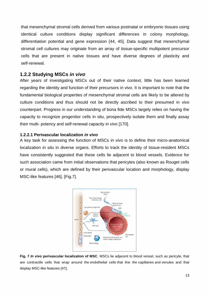
1.2.2.1 Perivascular localization in vivo
A key task for assessing the function of MSCs in vivo is to define their micro-anatomical
localization in situ in diverse organs. Efforts to track the identity of tissueresident MSCs
have consistently suggested that these cells lie adjacent to blood vessels. Evidence for
such association came from initial observations that pericytes (also known as Rouget cells
or mural cells), which are defined by their perivascular location and morphology, display
MSClike features [46], [Fig.7].
Fig. 7 In vivo perivascular localization of MSC. MSCs lie adjacent to blood vessel, such as pericyte, that
are contractile cells that wrap around the endothelial cells that line the capillaries and venules and that
display MSC-like features [47].

14
Pericytederived cultures are similar to mesenchymal stromal cell cultures in terms of
morphology and cellsurface antigen expression, and can be induced to differentiate into
not only osteoblasts, chondrocytes and adipocytes but also smooth muscle cells and
myocytes under appropriate conditions [48, 49].
1.2.3 MSCs: potential for differentiation
MSCs can differentiate in vitro to osteoblasts or chondrocytes and should be able to
differentiate in vivo as well. The transplantation and differentiation of MSCs to functional
osteoblasts was demonstrated in vivo in an animal model and in humans, which led to
treatment for osteogenesis imperfecta and bone loss [43]. MSCs participate in stem cell
niches such as the hematopoietic niche, and recently, bone marrow sub-endothelial cells
expressing CD146, a population of MSC, were found to reconstitute the hematopoietic
microenvironment [44]. The multipotent character of MSCs and their plasticity within
mesodermic lineages is supported by lineage priming [45]. However, the differentiation
towards other lineages originating from the mesoderm, such as skeletal and myocardial
muscle cells, remains questionable and could depend on the origin of MSCs; for example,
adipose-tissue-derived MSCs are able to differentiate to cardiomyocytes [46].
1.2.4 MSCs: immune privilege of MSCs
In addition to their stem/progenitor properties, MSCs have also been shown to possess
broad immunoregulatory abilities and are capable of influencing both adaptive and innate
immune responses. Recent findings have demonstrated that MSCs actively interact with
components of the innate immune system and that, through these interactions; they
display both anti-inflammatory and pro-inflammatory effects [50].
1.2.4.1 MSC as “sensor of inflammation”
Inflammation serves as a localized or systemic protective response elicited by infection,
injury or tissue destruction to eliminate pathogens and preserve host integrity. Within hours
after the onset of an inflammatory response, molecules expressed by pathogens or
associated with tissue injury are recognized by Toll-like receptors (TLRs) present on innate
effector cells. TLR ligation triggers phagocytosis and the release of inflammatory
mediators, which may initiate innate immune responses that provide a first line of
nonspecific defence, mainly through the activation of phagocytic cells, including
macrophages and neutrophils [51]. TLR ligation may not only activate phagocytic cells but
also stromal cells, including MSCs, thus creating an inflammatory environment [52, 53].
Human and mouse MSCs dynamically express a number of distinct and overlapping TLRs

15
in culture. Moreover, in vitro stimulation of specific TLRs affects the subsequent immune
modulating responses of MSCs [54, 55, 56]. Under hypoxic culture conditions, stimulation
of MSCs with the pro-inflammatory cytokines IFN-γ, TNF, IFN-α, and IL-1β upregulates
expression of a subset of TLRs, thus increasing the sensitivity of MSCs to the
inflammatory milieu [57]. However, prolonged stimulation with TLR ligands causes
downregulation of TLR2 and TLR4 [58], most likely as a self-regulatory mechanism to
prevent overactive skewing of the immune response. To direct appropriate immune
responses to a diversity of pathogenic insults, the different TLRs are activated by specific
endogenous or pathogen-associated molecules, including lipopolysaccharide (LPS) from
Gram-negative bacteria (TLR4) and double strand RNA (dsRNA) carried by some viruses
(TLR3) [53]. This has suggested that MSCs may polarize into two distinctly acting
phenotypes following specific TLR stimulation, resulting in different immune modulatory
effects and distinct secretomes. The TLR4-primed MSC population exhibits a pro-
inflammatory profile (MSC1) and the TLR3-primed MSC population delivers anti-
inflammatory signals (MSC2), [Fig.8].
Fig. 8 The polarization of MSCs into a Pro-inflammatory and Anti- Inflammatory Phenotype. (A) In the
presence of an inflammatory environment, MSCs become activated and adopt an immune-suppressive
phenotype (MSC2) by secreting high levels of soluble factors that suppress T cell proliferation. (B) In the
absence of an inflammatory environment, MSCs may adopt a pro-inflammatory phenotype (MSC1) and
enhance T cell responses by secreting chemokines that recruit lymphocytes to sites of inflammation [59].
In particular, besides their potential for differentiation, MSCs can exert an
immunosuppressive effect in vitro and in vivo [60, 61] by acting on all immune effectors
[62]. In fact, MSCs are unable to induce considerable alloreactivity because of a number of

16
unique characteristics that protect MSCs from alloreactive natural killer (NK)-cell-mediated
lysis [63]. However, some controversial data have been obtained with animal-derived
MSCs. For example, in vitro and in vivo, murine allogeneic MSCs can elicit an immune
response in immunocompetent mice [64, 65].
1.2.4.1.1 MSC interaction with immune cells (T cells, B cells, NK cells and dendritic
cells)
As “sensor of inflammation”, MSCs interact with cells involved in the process [Fig.9].
About T-cells, MSC may inhibit their proliferation induced by several stimuli both in vitro
and in vivo [66, 67, 68], arresting activated T-cells in the G0/G1 phase of the cell cycle [69]
without inducing apoptosis [66, 68] and the same is for B-cells [70]. Moreover, MSCs can
also modulate B-cell migration and production of IgM, IgA and IgG, without inducing
apoptosis [70, 71]. MSCs may also inhibit both IL-2 and IL-15 induced NK proliferation
[72, 73] and may interfere also with dendritic cells (DC) function and support the
development of tolerogenic antigen-presenting cells (APCs). In vivo results indicate that
MSCs actively interact with cells of the innate immune system and modulate their function
to establish a fine balance between pathogen elimination and repair processes, aiming at
controlling inflammation, preventing organ failure, and preserving tissue homeostasis.
Therefore, the further elucidation of mechanisms that trigger a functional switch between
MSC phenotypes remains an important research goal for future studies [74].
Fig. 9 MSC interactions with immune cells. MSCs are immune privileged cells that inhibit both innate
(neutrophils, dendritic cells and natural killer cells) and adaptive (T cells and B cells) immune cells (INF-
indicates interferon; TNF-indicates tumor necrosis factor) [74].

17
1.2.5 Clinical application/ Therapeutic use
The wide range of in vivo effects of MSCs, from cell replacement and immunosuppression
to trophic effects, drives their increasing use in regenerative medicine and immune
intervention. Whatever the use, MSCs must be produced according to good manufacturing
practices (GMPs), with relevant controls, to obtain efficient and safe cell therapy [75]. The
ability of MSCs to adopt a different phenotype in response to sensing an inflammatory
environment is crucial for understanding their therapeutic potential in immune-mediated
disorders. The available evidence suggests that responses to MSC treatment may be
independent of the MSC donor or dose of the immune-suppressive treatment employed.
This heterogeneity in response might be related to the presence or absence of the
appropriate environment in the patient capable of activating MSCs.
1.2.5.1 Regenerative medicine: stem cell transplantation
Stem cell transplantation is a procedure that replaces unhealthy blood-forming cells with
healthy cells arising from stem cells. There are two different types of stem cell
transplantation: autologous and allogenic. In the first one stem cells come from patient
own body, instead in the allogenic one cells are from a healthy person (the donor). In this
last case, an important medical complication is the graft versus host disease (GvHD) that
consist in the fact that immune cells in the donated tissue (the graft) recognize the
recipient (the host) as foreign (nonself); so the transplanted immune cells then attack the
host's body cells. It has been observed [76] that, because of the profound
immunomodulatory effect of MSCs in vivo and in vitro, use of ex vivo-expanded MSCs for
treatment or prophylaxis of steroid-resistant GvHD was recommended. Another important
potential application of MSCs in allogeneic stem cell transplantation is for enhancement of
engraftment. In fact, since MSCs secrete many growth factors stimulating hematopoiesis,
provide a scaffold for hematopoiesis and support primitive progenitor cells in vivo, they
might enhance engraftment after stem cell transplantation [77].
1.2.5.2 Immune Intervention
Under the effect of inflammatory cytokines, MSCs are capable to migrate to inflamed
tissues and modulate the local inflammatory reactions thanks to the effects on both innate
and adaptive immunity [67, 78]. In addition, MSCs may recruit and support local
autologous stem cells inside the injured tissues, thus promoting cell survival and tissue
repair [75]. Therefore, MSC-based treatments may represent a novel strategy against
systemic autoimmunity and inflammation, in diseases such as rheumatoid arthritis, multiple
sclerosis and type-I diabetes. In vitro and in vivo studies in animal model [79, 80] suggest

18
the use of MSCs for managing autoimmune and inflammatory diseases. In addition, no
evidence exists of systemic immunosuppression and increased risk of infections as side-
effects of MSC infusion in immunocompetent individuals, which suggests that the immune-
regulatory effects of MSCs are restricted to inflamed tissues. For these reasons, MSC-
based immunotherapy may become suitable in the future for many severe inflammatory
diseases [171]:
1. Bone and cartilage repair
2. Heart and vessels
3. Epithelium
4. Central nervous system
1.2.6 Potential risks in using MSCs
The use of MSCs could imply different risks, because of the differentiation potentials, their
immunosuppressive properties and some possible immortalisation/transformation during
long-term culture. Recent concerns have been expressed about the potential
transformation of MSCs during the culture process, as shown recently in describing the
transformation of human MSCs in cells cultured for a long time. In fact, although they can
be managed safely during the standard ex vivo expansion period (6-8 weeks), human
mesenchymal stem cells can undergo spontaneous transformation following long-term in
vitro culture (4-5 months) [81]. Human cells have two control points that regulate their life
span in vitro, the senescence and crisis phases. Senescence is associated with moderate
telomere shortening and is characterized by cell cycle arrest and positive β-galactosidase
staining at pH 6 [82]. If cells bypass this stage, they continue to grow until telomeres
become critically short and cells enter crisis phase, characterized by generalized
chromosome instability that provokes mass apoptosis [83]. Human cells immortalize at low
frequency seem resistant to spontaneous transformation, instead MSC in long-term
cultures immortalize at high frequency and undergo spontaneous transformation [81]
supporting recent cautionary speculation that “mutant stem cells may seed cancer” [84].
1.2.7 Animal Models – different biological and functional properties of
MSCs in mouse and man
Animal models are of critical importance for translating in vitro immune regulatory
properties of MSCs into therapeutic application and dissecting mechanisms of efficacy.
However, it’s important to note that murine MSCs are intrinsically different from human
cells. Although murine and human MSCs share properties such as multi lineage

19
differentiation capacity, they are also distinct with respect to other properties. Ex vivo
expansion with murine cells is slower than with human cells, and murine MSCs require
weeks before entering a linear growth rate [85]. At this stage, murine MSCs undergo
transformation and immortalization in culture. Several reports have indicated that
transformed murine MSCs have an increased proliferation rate, display an altered
morphology, carry cytogenetic abnormalities, and form tumors following injection into
syngeneic mice. Murine BM-derived MSCs in long-term culture gradually exhibit increased
telomerase activity and proceed to a malignant state, resulting in sarcoma formation in
vivo [86, 87]. This susceptibility to malignant transformation may be attributed to the high
degree of chromosomal instability in genetically unstable inbred mice, characterized by the
development of both structural and numerical aberrations even at early culture passages.
Therefore, culture-expanded murine MSCs should be regarded as transformed cells, even
in the absence of a malignant phenotype. These differences should be taken into
consideration when interpreting data. The dissimilarities between MSCs isolated from
murine and human species require a careful evaluation when choosing animal models to
test MSCs in preclinical studies and so, when interpreting in vivo effects of murine MSCs,
especially in light of efforts to look at clinical application of MSCs.
1.3 Study of MSC secretome
Mesenchymal stem cells represent a promising therapeutic approach and they action is
due mainly to secreted mediators that often acts through chemotactic signaling [88], that
are referred as “secretome”. This term was first used by Tjalsma et al. [89] to include all
proteins that are synthesized and processed by the secretary pathway and proteins
located in the secretion machinery, although, the term was recently limited to include only
the set of secreted or extracellular proteins in a species. For the use of MSC, it’s very
important to know as much as possible the identity of molecules responsible for their
activities. Since the secretome plays a direct role in the biological activities of MSCs, the
qualitative and quantitative analysis of the protein component of MSC secretome is a
fundamental step in order to identify key players in the control and regulation of the many
biological processes influenced by these cells [90]. Previously, studies used to compare
plasma/serum from cancer patients with those from normal controls because, as
suggested by Liotta, "the blood contains a treasure trove of previously unstudied
biomarkers that could reflect the ongoing physiologic state of all tissues", and the latter,

20
therefore, appears to be more attractive [91]. However, the prospects of blood proteomics
are challenged by the fact that blood is a very complex body fluid, comprising an
enormous diversity of proteins and protein isoforms with a large dynamic range of at least
9–10 orders of magnitude. The abundant blood proteins, such as albumin,
immunoglobulin, fibrinogen, transferrin, haptoglobin and lipoproteins, may mask the less
abundant proteins, which are usually potential markers [92]. So, studies redirected to the
“secretome” although, only few studies have characterized the cellular secretome in vivo,
assuming the idea that cells grown in vitro and stimulated using specific factors to which
cells are exposed under certain conditions in vivo, present in vitro a secretion phenotype
similar to one in vivo [93,94]. In particular, for this purpose an optimized protocol set up in
our laboratory was used to allow the collection of the secretome of human bone marrow
mesenchymal stem cells (BM-MSC) in order to detect the differential expression of
secreted protein induced by exposing cells to specific stimulation conditions.
1.3.1 Proteomic characterization of MSC secretome
The proteomic approach for the characterization of MSC secretome, described in this
thesis, is a shotgun label free approach to allow direct identification and quantification of
«all detectable» proteins in the secretome of stimulated (MIX) vs unstimulated (CTR)
MSC, both in mouse and human. The shot gun approach is a “bottom-up” protein analysis
which allow the characterization of all the proteins present in a sample without any
previous purification before digestion and nano LC-MS/MS analysis. For many years 2D-
PAGE/MS, which is an overall, comparative, quantitative proteomic technique, was the
gold standard for analysis of protein expression and biomarker discovery. However, there
are several disadvantages associated with gel-based proteomic techniques. For example,
any 2D approach is subjected to the restrictions imposed by the gel method, which include
limited dynamic range, difficulty in handling hydrophobic proteins, and difficulty in detecting
proteins with extreme molecular weights and pI values [95]. Another negative aspect is
that spots on a 2D gel often contain more than one protein, making quantification
ambiguous; throughput is low and gel-to-gel reproducibility can be a challenge [95].
Furthermore, co-migration of proteins can cause problems during the excision and
identification steps as there may be more than one protein present in the gel spot excised.
Moreover, low-abundance proteins may be masked in the gel by high-abundance
housekeeping proteins. Therefore, in more recent years, there has been a move towards
gel-free MS methods for proteome analysis. The gel-free methods are based on the high-
throughput ‘‘shotgun” analysis of peptides from a digested complex protein sample using
21
an on-line high performance liquid chromatography (HPLC) method, prior to identification
using MS [96]. Label-free approaches have been developed for quantitative shotgun
proteomic and are methods that don’t use chemical tags for quantification. In this method,
each sample is processed separately and individually analysed through LC-MS/MS [95].
The basic steps of all the label-free techniques are:
- sample preparation including protein extraction, reduction, alkylation, and digestion;
- sample separation by liquid chromatography (LC or LC/LC) and analysis by MS/MS;
- data analysis including peptide/protein identification, quantification, and statistical
analysis.
Shotgun proteomic has provided powerful tools for studying large scale protein expression
and characterization in complex biological systems [99, 100]. This proteomic strategy
converts a complex protein mixture to an even more complicated peptide mixture. For this
reason, to resolve complex peptide mixtures, high-resolution HPLC separations are
necessary to maximize peptide separation for acquisition of tandem mass spectra. The
liquid chromatography step separates the peptides and elutes them directly into the ESI
ionizer of the mass spectrometer.
There are two different kind of measurement used for the quantification (Fig.10): the first
one calculates ion intensity changes as peptide peak areas or heights in chromatography,
while the second is based on the spectral counting of identified proteins after tandem mass
analysis. The two measures are calculated for each LC-MS/MS or LC/LC-MS/MS runs, so
it is possible to point out differences in protein abundance comparing the results of
different analyses. The relative quantification by peak intensity is based on extracting the
ion chromatogram of the peptides and comparing them across range (Fig.10 A). In LC-MS,
an ion with a specific m/z and intensity is detected and recorded at a certain time and it
has been observed that its signal intensity from electrospray ionization (ESI) correlates
with the concentration [101]. As reported in [101], it was experimentally observed that the
chromatographic peaks area of each peptide, increases with the increase of the ion’s
concentration and the sum of the peak’s area of all the peptides identified correlates
linearly with the protein concentration. The strong correlation between chromatographic
peak areas and the peptide/protein concentration was still observed when the when the
protein was spiked into a complex mixture and its digests were detected by LC-MS/MS
[102, 103]. Although these early studies showed that the relative quantification of the
peptides could be achieved via direct comparison of peak intensity of each peptide ion in

22
multiple LC-MS datasets, applying this method for the analysis of changes in protein
abundances in complex biological samples had some practical constraints. First, even the
same sample can result in differences in the peak intensities of the peptides from run to
run. These differences are caused by experimental variations such as differences in
sample preparation and sample injection. Normalization is required to account for this kind
of variation. Second, any experimental drifts in retention time and m/z will significantly
complicate the direct, accurate comparison of multiple LC-MS datasets. Chromatographic
shifts may occur as a result of multiple sample injections onto the same reverse-phase LC
column. Unaligned peak comparison will result in large variability and inaccuracy in
quantification. Thus, highly reproducible LC-MS and careful chromatographic peak
alignment are required and critical in this comparative approach [104 – 109]. In the
spectral counting approach, relative protein quantification is achieved by comparing the
number of identified MS/MS spectra from the same protein in each of the multiple LC-
MS/MS or LC/LC-MS/MS datasets (Fig.10 B). This is possible because an increase in
protein abundance typically results in an increase in the number of its proteolytic peptides,
and vice versa. This increased number of (tryptic) digests then usually results in an
increase in protein sequence coverage, the number of identified unique peptides, and the
number of identified total MS/MS spectra (spectral count) for each protein [110]. Liu et al.
[111] studied the correlation between relative protein abundance and sequence coverage,
peptide number, and spectral count. It was demonstrated that among all the factors of
identification, only spectral count showed strong linear correlation with relative protein
abundance with a dynamic range over 2 orders of magnitude [111]. Therefore, spectral
count can be used as a simple but reliable index for relative protein quantification. Spectral
counting-based quantification is proved more reproducible and has a larger dynamic range
than the peptide ion chromatogram-based quantification [112]. In contrast to the
chromatographic peak intensity approach, which requires delicate computer algorithms for
automatic LC-MS peak alignment and comparison, no specific tools or algorithms have
been developed specially for spectral counting due to its ease of implementation.
However, normalization and statistical analysis of spectral counting datasets are
necessary for accurate and reliable detection of protein changes in complex mixtures.
Relative quantification by spectral count has been widely applied in different biological
complex [113, 114, 115] and it is the type of measurement used in this work.

23
Fig.10 Label-free quantitative proteomics. Control and sample are subject to individual LC-MS/MS
analysis. Quantification is based on the comparison of peak intensity of the same peptide (A) or the spectral
count of the same protein (B) [95].

MATERIAL AND METHODS

25
2.1 Isolation of murine MSC
C57BL/6J mice were purchased from Charles River Laboratories (Calco, Italy). All mice
used as primary cell donors or recipients were between 8 and 12 weeks of age.
Procedures involving animals and their care conformed to institutional guidelines in
compliance with national (4D.L. N.116, G.U., suppl. 40, 18-2-1992) and international (EEC
Council Directive 2010/63/UE; National Institutes of Health Guide for the Care and Use of
Laboratory Animals) law and policies. The protocol was approved by the Italian Ministry of
Health on 18 June 2007 and modified by Protocol 162/2011-B. All efforts were made to
minimize the number of animals used and their suffering. In all the experiment, the mice
were sex and age matched, no further randomization was applied. MSC were isolated as
described [116] by flushing the femurs and tibias from 8 week-old, C57Bl/6 female mice
and cultured in 25 cm2 tissue culture flasks at a concentration of 2X10⁶ cells/cm2 using
complete Dulbecco modified Eagle medium low glucose (DMEM, Lonza, Braine-L’Alleud,
Belgium) supplemented with 20% heat inactivated fetal bovine serum (Biosera, Ringmer,
United Kingdom), 2mM glutamine (Lonza), 100 U/ml penicillin/streptomycin (Lonza). Cells
were incubated at 37°C in 5% CO2. After 48 hours, the non-adherent cells were removed.
After reaching 70–80% confluence, the adherent cells were trypsinized (0.05% trypsin at
37°C for 3 minutes), harvested and expanded in larger flasks. MSC at passage 10 were
screened by flow cytometry for the expression of CD106, CD45, CD117, CD73, CD105,
MHC-I, SCA-1 and CD11b and used to perform experiments (BD Pharmingen, Oxford,
UK).
2.2 Isolation of human MSC
MSC were provided by Orbsen Therapeutics Ltd (Galway, Ireland). Ethical approvals are
granted from the NUIG Research Ethics Committee and the Galway University Hospitals
Clinical Research Ethics Committee (CREC). Briefly, bone marrow was harvested from
volunteers, and the cell culture was set up as previously described [117]. MSC were
characterized according to international guidelines [118]. All samples were obtained with
informed consent. Procurement of the sample conformed to European Parliament and
Council directives (2001/20/EC; 2004/23/EC).

26
2.3 Collection of conditioned medium (CM)
2.3.1 Collection of conditioned medium (CM) of murine
MSCMSC were plated and let grow until confluence in ventilated cap flask. Growth
medium was substituted with DMEM low glucose supplemented with 10% FBS, 2mM
glutamine, 100 U/ml penicillin/streptomycin, with or without 25 ng/ml mIL1b, 20 ng/ml
mIL6, 25 ng/ml mTNFa for 24 hours. Then this medium was changed with DMEM low
glucose supplemented with 2mM glutamine, 100 U/ml penicillin/streptomycin for the
following 18 hours. Conditioned medium was harvested and centrifuged at 4000 rpm for 10
min.
2.3.2 Collection of conditioned medium (CM) of human MSC
MSC were plated in with MEM Alpha with Glutamax supplemented with 10% FBS, 2 mM
glutamine, 100 U/mL penicillin/streptomycin and let grow until confluence in in a humidified
incubator with 5% CO2 and 37°C. At the moment of the confluence, medium was
substituted with MEM Alpha with Glutamax supplemented with 2% FBS, 2 mM glutamine,
100 U/mL penicillin/streptomycin, with (st hMSC) or without (unst hMSC) 25 ng/mL hIL1b,
20 ng/mL hIL6, 25 ng/mL hTNFa. 24 hours later, after three washes in MEM Alpha with
Glutamax, the medium was changed with MEM Alpha with Glutamax supplemented with 2
mM glutamine, 100 U/mL penicillin/streptomycin for the following 18 hours. Conditioned
medium was harvested and centrifuged at 4000 rpm for 10 min. Both isolation and
collection, were performed in collaboration with the lab of prof.ssa Antonella Viola from
University of Padua.
2.4 Proteomic analysis
Proteomic methods used are the same for both mouse and human samples.
2.4.1 Sample preparation
The protein concentration of MSC secretome was determined using the Bradford assay
and then, proteins (150 ug for mouse samples, in the range 135-200 ug for human
samples) were precipitated with 10 % tricholoracetic acid (TCA) for 2 hours on ice.
Prior to proteolysis, proteins were subjected to reduction with dithiothreitol (10 mM DTT in
50mM NH4HCO3), for 30 minutes at 56°C; and to alkylation with iodoacetamide (200mM
IAA in 1M NH4HCO3), for 30 minutes at room temperature in the dark. Then protein were

27
digested with sequence-grade trypsin (Roche) for 16 hours at 37°C using a protein:trypsin
ratio of 50:1. The reaction was stopped by acidification with 98% formic acid and the pellet
was then desalted using Zip-Tip C18 (Millipore) before mass spectrometric (MS) analysis.
The following protocol was applied:
Equilibrate the ZipTip for Sample Binding:
1) pre-wet the tips with 50% CH3CN 3 times (3 x 100 µl);
2) wash the tips with TFA 0.1% 3 times (3 x 100 µl).
Bind and Wash the Peptides:
1) bind the sample to ZipTip pipette tip. Aspirate and dispense the material 10-13 cycles
for maximum binding of complex mixtures;
2) wash the tips with 5% CH3CN/0.1% TFA at least once.
Elute the Peptides:
1) elute the sample with 50% CH3CN in HCOOH 1%, 3 times (3 x 100 µl), into a clean vial,
for mass spectrometry analysis.
2.4.2 LC-ESI-MS/MS
Analysis was performed on a DionexUltiMate 3000 HPLC System with a
PicoFritProteoPrep C18 column (200 mm, internal diameter of 75 μm) (New Objective,
USA). Gradient: 1% ACN in 0.1 % formic acid for 10 min, 1-4 % ACN in 0.1% formic acid
for 6 min, 4-30% ACN in 0.1% formic acid for 147 min and 30-50 % ACN in 0.1% formic for
3 min at a flow rate of 0.3 μl/min. The eluate was electrosprayed into an LTQ
OrbitrapVelos (Thermo Fisher Scientific, Bremen, Germany) through a Proxeon
nanoelectrospray ion source (Thermo Fisher Scientific). The LTQ-Orbitrap was operated in
positive mode in data-dependent acquisition mode to automatically alternate between a full
scan (m/z 350-2000) in the Orbitrap (at resolution 60000, AGC target 1000000) and
subsequent CID MS/MS in the linear ion trap of the 20 most intense peaks from full scan
(normalized collision energy of 35%, 10 ms activation). Isolation window: 3 Da, unassigned
charge states: rejected, charge state 1: rejected, charge states 2+, 3+, 4+: not rejected;
dynamic exclusion enabled (60 s, exclusion list size: 200). Five technical replicate
analyses of each biological sample were performed. Data acquisition was controlled by
Xcalibur 2.0 and Tune 2.4 software (Thermo Fisher Scientific).

28
2.4.3 Data processing
Mass spectra were analyzed using MaxQuant software (version 1.3.0.5) [119]. The initial
maximum allowed mass deviation was set to 6 ppm for monoisotopic precursor ions and
0.5 Da for MS/MS peaks. Enzyme specificity was set to trypsin, defined as C-terminal to
arginine and lysine excluding proline, and a maximum of two missed cleavages were
allowed. Carbamidomethylcysteine was set as a fixed modification, N-terminal acetylation
and methionine oxidation as variable modifications. The spectra were searched by the
Andromeda search engine against the mouse Uniprot sequence database (release
29.05.2013 for mouse, release 2014_01 for human). Protein identification required at least
one unique or razor peptide per protein group. Quantification in MaxQuant was performed
using the built in XIC-based label free quantification (LFQ) algorithm [125] using fast LFQ.
The required false positive rate was set to 1% at the peptide and 1% at the protein level,
and the minimum required peptide length was set to 6 amino acids.
2.4.4 Statistical analysis
Statistical analyses were performed using the Perseus software (version 1.4.0.6,
www.biochem.mpg.de/mann/tools/ [119]). Only proteins present and quantified in at least 3
out of 5 technical repeats were considered as positively identified in a sample and used for
statistical analyses. Proteins were considered differentially expressed if they were present
only in unstimulated (unst-) or stimulated (st-) MSC-CM (both in human and mouse) or
showed significant t-test difference (cut-off at 1% permutation-based False Discovery
Rate) in both biological replicates.
2.4.5 Bioinformatic analysis
Proteins were considered secreted or involved in inflammation/angiogenesis according to
the following databases/datasets: Geno Ontology [121], NextProt [122], UniProt [123],
Gene Cards [124], datasets [125, 126] and manual literature mining. Bioinformatic
analyses were carried out by DAVID software (release 6.7) in order to cluster enriched
annotation groups of Biological Function within the set of identified secretome proteins
[124]. GOBP and groups were filtered for significant terms (modified Fisher exact EASE
score p value <0.05 and at least five counts). Networks of up-regulated proteins in st
hMSC-CM involved in inflammation or angiogenesis was performed using String [128]
(active interactions: text mining, experiments, databases).

29
The mass spectrometry proteomics data, for human samples, have been deposited to the
ProteomeXchange Consortium via the PRIDE [129] partner repository with the dataset
identifier PXD005746.

RESULTS AND DISCUSSION

31
3.1 Proteomic characterization of murine MSC (mMSC)
secretome
The secretome of MSC stimulated (st-MSC-CM) or not (unst-MSC-CM) with inflammatory
cytokines was analyzed by a shotgun label free proteomic approach. The analysis allowed
the identification of 1613 and 1630 proteins in the secretome of control (CTR) and
stimulated (MIX) MSC, respectively. Proteins were considered differentially expressed if:
(a) a protein was present only in MSC-CM or in control; (b) the LFQ (label free
quantification) intensity resulted statistically significant as calculated by Perseus (t-test cut
off at 1% permutation-based false discovery rate).
According to this analysis, 7.6% or 8.3% of the proteins detected in the secretome of
control or stimulated MSC, respectively, were differentially expressed, either upregulated
or downregulated, as shown in the Venn diagram reported in Fig.11.
Fig.11 Venn diagram. Description of the comparison between unstimulated MSC in conditioned medium
and the stimulated one. 1613 and 1630 proteins were identified in the secretome of control
(CTR/unstimulated, unst-) and treated (MIX/stimulated, st-) MSC respectively. Among these, 1610 proteins
were present in both samples st- and unst- MSC-CM. Applying a t-test with p<0.05,in total 89 resulted
significantly UP regulated or present only in stimulated MSC-CM, and 52 significantly DOWN regulated in
stimulated or present only in unstimulated MSC-CM.

32
These proteins were clustered according to their functions using the DAVID platform [130]
filtered for significant Gene Ontology Biological Process (GOBP) terms using a P-value of
≤ 0.05 and at least five gene counts for each category.
3.1.1 Proteins up-regulated in stimulated murine MSC-CM
The differential expression analysis identified 141 proteins that were either up (Tab.1) or
down (Tab.2) regulated in stimulated MSC.
We focused our attention mainly on the proteins up-regulated or exclusively expressed in
MSC. These proteins are listed in Tab.1. A GO enrichment analysis was performed as
shown in Fig.12. In detail, in Fig.12A the bar chart shows the fold enrichment of the top 26
most enriched GO terms in MSC versus unstimulated MSC-CM, while in Fig.12B, the
histograms report the GOBP categories related to angiogenesis or inflammation.
Interestingly 18% and 30% of the proteins, listed in Tab.1, belong to these two categories
and are shown in bold in Tab.1, while concerning the 52 proteins that were significantly
down-regulated or present only in the secretome of unstimulated MSC [Tab.2], the GO
analysis revealed that most terms are related to metabolic processes [Fig.13]. The
presence of an ‘angiogenesis-related’ signature among up-regulated proteins was also
confirmed by the analyses of human MSC secretome, reported in the following chapter,
which reveals that all the 16 up-regulated proteins in stimulated MSC secretome common
to human and mouse are modulators of angiogenesis [Tab.3].
Angiogenesis is the physiological process through which new blood vessels form from pre-
existing vessels [131]. It is a normal and vital process in growth and development,
although, it is also involved in the process of transition of tumors from a benign state to
a malignant one [132]. The process involves a complex and dynamic interaction between
endothelial cells and the corresponding extracellular environment. Its chemical stimulation
is performed by various angiogenic proteins, including several growth factors [133] that are
also involved in inflammation processes [134]. Critical, in all phases of the process, are
proteolysis and remodeling of the extracellular matrix (ECM) that affect endothelial cells
migration, invasion into the perivascular tissue and morphologic formation of luminal
structures. In these two events very important is the role taken on matrix
metalloproteinase (MMP), a family of proteases that degrade ECM proteins and are critical
in vascular remodeling, cellular migration, and sprout formation [135]. The activities of
these metalloproteinases are precisely regulated under physiological conditions at the
levels of transcription, zymogen activation and inhibition by endogenous inhibitors.
Disruption of the balance between the production of active enzymes and their inhibition

33
may result in diseases associated with uncontrolled ECM turnover, inflammation, cell
growth and migration, such as arthritis, cardiovascular disease, cancer, pulmonary
disease, nephritis, neurological disorders and tissue ulceration [136]. Tissue inhibitors of
metalloproteinases (TIMPs) are endogenous inhibitors of these metalloproteinases and
are consequently important regulators of ECM turnover, tissue remodelling and cellular
behaviour, so the balance between MMPs and their natural inhibitors is very critical.
Interestingly, the proteomic analysis of the murine MSC secretome indicated that the cells
activated by inflammatory cytokines, upregulate the expression of several proteins
potentially involved in angiogenesis and inflammation through multiple pathways and, in
particular, among these proteins, the attention was focused on the tissue inhibitors of
metalloproteinases TIMP-1, because of its well-known anti-angiogenic properties [137].
We thus used the tube formation assay to analyze the effect of MSC-derived TIMP-1 on
angiogenesis. Although the blocking anti-TIMP-1 antibody had no effect on the ability of
endothelial cells to form tubes when cultured in the supernatants of unstimulated MSC, it
totally reverted the anti-angiogenic properties of the supernatant from stimulated MSC
[Fig. 14a], indicating that, at least in this in vitro setting, TIMP-1 is one of the key MSC-
secreted molecules targeting the endothelium. In an in vivo setting, in the lab of our
collaborator in Padua, the injection of neutralizing anti-TIMP-1 antibody [138] 1 day after
MSC transplantation reverted the MSC-induced reduction of endothelial cell numbers and
high endothelial venules (HEV) in lymph nodes (dLNs) [Fig. 14b-d], suggesting that TIMP-
1 may be directly responsible for the anti-inflammatory effects of MSC on LNs. To confirm
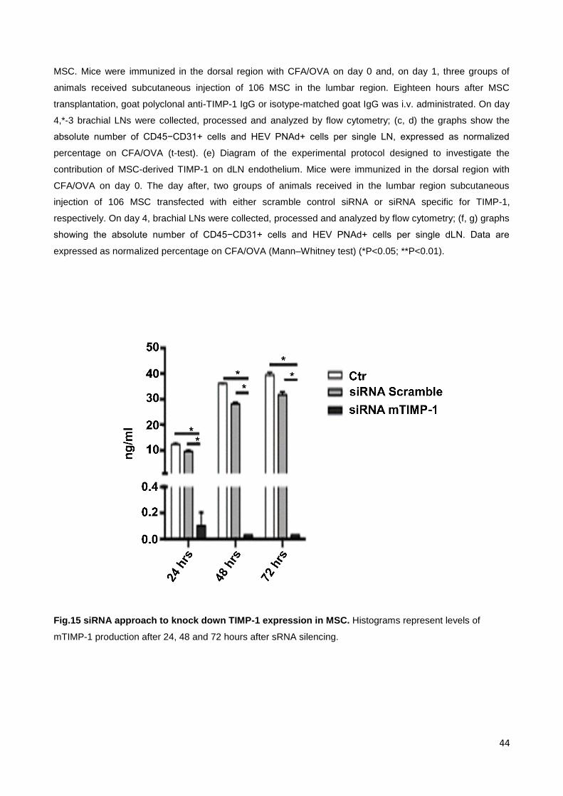
this hypothesis, they used a siRNA approach to knock down TIMP-1 expression in MSC
[Fig. 15]. Again, the absolute cell numbers of endothelial cells and HEV in dLN were
reduced by MSC transfected with the scramble siRNA control but not by MSC with TIMP-1
siRNA [Fig. 14 e-g]. On the basis of these results, we speculated that overexpression of
TIMP-1 might be sufficient to mimic the effects of MSC transplantation, in terms of
inhibition of angiogenesis in the inflamed lymph nodes. TIMP-1 overexpression by AAV9-
mediated gene transfer [139] in mice immunized with CFA/OVA [Fig.16a] inhibited the
inflammatory reaction in the draining LNs, as indicated by the reduced total cellularity
[Fig.16b], which was due to a decreased number of both CD45+ cells [Fig.16c] and
endothelial and HEV cells [Fig.16d, e].

34
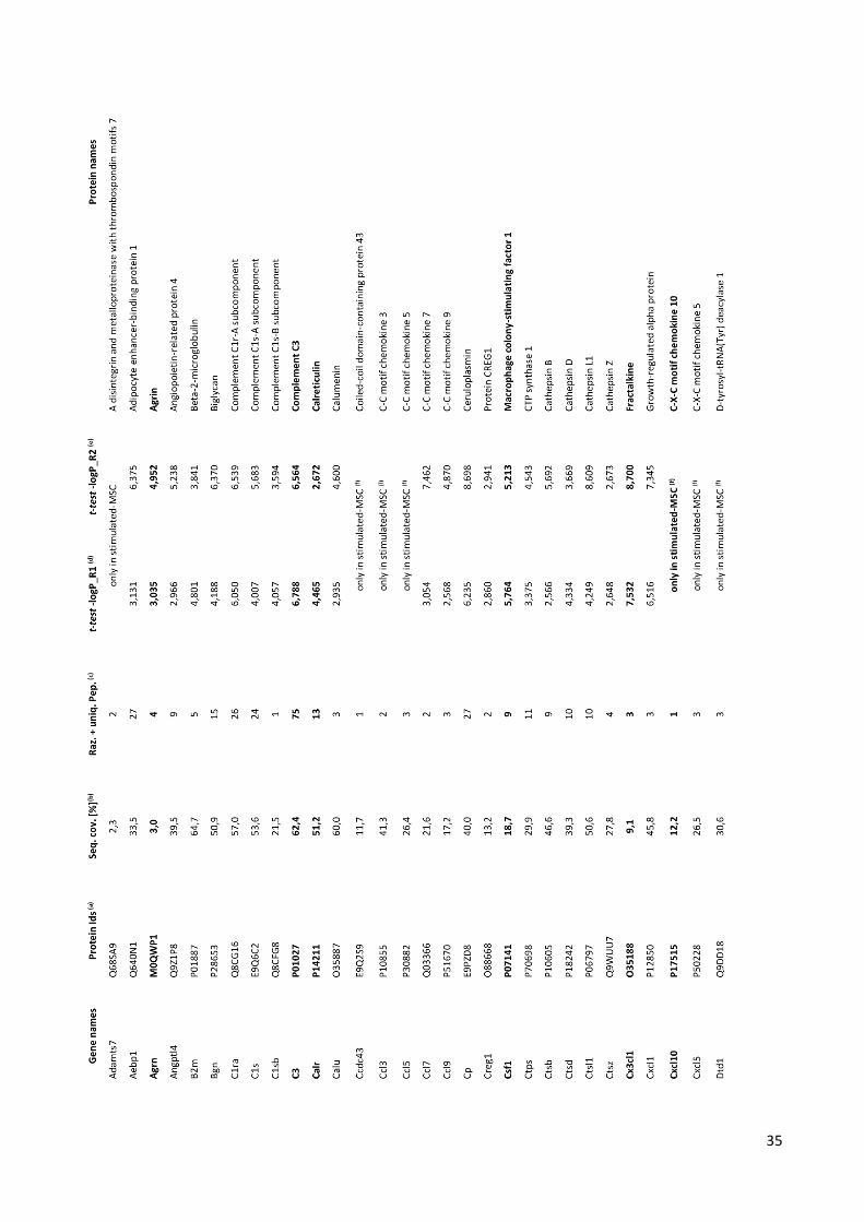
Tab.1 Proteins up-regulated in stimulated-MSC vs unstimulated-MSC, or present only in stimulated-
MSC secretome in both biological replicates. Proteins categorized as modulators of angiogenesis
according to GOBP analysis (see Fig.12a and b) are shown in bold. Legend: (a) Uniprot accession ID, (b)
percentage of the protein sequence that is coverage by the identified peptides, (c) number of unique and
razor peptides associated with the protein, (d) –log p-value t-test for biological replicate 1, (e) –log p-value t-
test for biological replicate 2, (f) not detected in un-stimulated MSC secretome.
35

36

37

38
Tab.2 Proteins down-regulated in stimulated-MSC vs unstimulated-MSC, or present only in
unstimulated-MSC secretome in both biological replicates. Legend: (a) Uniprot accession ID, (b)
percentage of the protein sequence that is coverage by the identified peptides, (c) number of unique and
razor peptides associated with the protein, (d) –log p-value t-test for biological replicate 1, (e) –log p-value t-
test for biological replicate 2, (f) not detected in un-stimulated MSC secretome.

39

40

41
Fig.12 Distribution into biological processes of the proteins upregulated in MSC-CM. The proteins that
were significantly upregulated or present only in MSC-CM were classified into different biological processes
according to the GO classification system. (a) The bar chart shows the count of the top 26 most-enriched GO
terms in MSC-CM versus unstimulated MSC-CM. Color coding indicates the fold enrichment. (b) Proteins
were categorized as modulators involved in inflammation processes and/or angiogenesis. The histograms
report the GOBP groups related to these categories.

42
Fig.13 Distribution into biological processes of the proteins downregulated in MSC-CM. The proteins
that were significantly downregulated or present only in unstimulated secretome MSC-CM were classified
into different biological processes according to the GO classification system. Analysis reveal that most term
are related to metabolic processes.
Tab.3 Common proteins up regulated in stimulated- vs- unstimulated- mouse and human secretome.
Proteins involved in angiogenesis are indicated in detail.

43
Fig.14 TIMP-1 mediates the anti-angiogenic effect of MSC-CM in vitro and the anti-inflammatory effect
of MSC in vivo. SVEC4-10 network formation in matrigel in the presence of MSC-CM or unst MSC-CM and
anti-TIMP-1 blocking antibody.
(a) anti-mTIMP1 blocking antibody restores SVEC4-10 network formation in matrigel in the presence of
MSC-CM. Representative images at 6 h (left) and segment length quantification as percentage of variation
(right) are shown. Data are expressed as mean±s.e.m. (*P<0.05, **P<0.01; one-way ANOVA). (b) Diagram
of the experimental protocol designed to block the TIMP-1 activity during the anti-inflammatory effects of
44
MSC. Mice were immunized in the dorsal region with CFA/OVA on day 0 and, on day 1, three groups of
animals received subcutaneous injection of 106 MSC in the lumbar region. Eighteen hours after MSC
transplantation, goat polyclonal anti-TIMP-1 IgG or isotype-matched goat IgG was i.v. administrated. On day
4,*-3 brachial LNs were collected, processed and analyzed by flow cytometry; (c, d) the graphs show the
absolute number of CD45−CD31+ cells and HEV PNAd+ cells per single LN, expressed as normalized
percentage on CFA/OVA (t-test). (e) Diagram of the experimental protocol designed to investigate the
contribution of MSC-derived TIMP-1 on dLN endothelium. Mice were immunized in the dorsal region with
CFA/OVA on day 0. The day after, two groups of animals received in the lumbar region subcutaneous
injection of 106 MSC transfected with either scramble control siRNA or siRNA specific for TIMP-1,
respectively. On day 4, brachial LNs were collected, processed and analyzed by flow cytometry; (f, g) graphs
showing the absolute number of CD45−CD31+ cells and HEV PNAd+ cells per single dLN. Data are
expressed as normalized percentage on CFA/OVA (Mann–Whitney test) (*P<0.05; **P<0.01).
Fig.15 siRNA approach to knock down TIMP-1 expression in MSC. Histograms represent levels of
mTIMP-1 production after 24, 48 and 72 hours after sRNA silencing.

45
Fig. 16. TIMP-1 overexpression in vivo mimics MSC transplantation. (a) Diagram of the experimental
protocol designed to overexpress TIMP-1 in immunized mice. One day after AAV9-TIMP-1 or AAV9-LacZ
administration (day 0), mice were immunized with CFA/OVA. Brachial dLNs were collected 4 days after
immunization and processed for flow cytometry. The graphs show the absolute number of total cells (b),
CD45+ cells (c), CD45−CD31+ (d) and HEV PNAd+ (e) cells per single LN, expressed as normalized
percentage on CFA/OVA. Error bars represent standard error (*Po0.05; **Po0.01; Mann–Whitney test).
3.2 Proteomic characterization of human MSC (hMSC)
secretome
To the best of our knowledge, the present study reports for the first time a quantitative
proteomic characterization of the secretome of human bone marrow-derived MSC primed
with pro-inflammatory cytokines. Proteomic analyses were conducted under exactly the
same conditions used in the previous investigation on mMSC in order to avoid variations
with methodology, allowing direct comparative analysis between the results obtained with
the two organisms. The proteomic characterization of human MSC secretome was
performed on samples from two different patients, indicated as “donors H30” and “donors
H34”, before (CTR condition/ unstimulated human MSC conditioned medium) and after
stimulation (MIX condition/ stimulated human MSC conditioned medium). Fig.17
summarizes the results of the proteomic characterization of secretome of hMSC before
(unst-) and after (st-) stimulation with inflammatory cytokines. Only the 497 proteins
present in at least 3 out of 5 technical repeats in both biological replicates (donors H30
and H34) were considered for further analysis: these proteins are listed in Tab. Suppl.

46
together with their main identification parameters. Fig.17 highlights the number of proteins
detected in stimulated human MSC conditioned medium (465 in st hMSC-CM) and
unstimulated human MSC conditioned medium (457 in unst hMSC-CM). As shown in
Fig.17, 32 proteins and 39 proteins were exclusively identified in unst- and st- cells
respectively. Amongst the 465 proteins identified in st hMSC-CM (proteins in groups 1, 2,
4 and 5 of Tab.S1.), 133 are listed as cytokine or chemokine or functionally related to
these classes of compounds according to the NextProt database [122].
Fig.17 Summary of the results obtained in the proteomic characterization of hMSC-CM. Venn diagram
showing proteins detected in at least 3 out of 5 technical replicas in both patients in stimulated hMSC-MC
and unstimulated hMSC-CM
3.2.1 Proteins up-regulated in stimulated human MSC-CM
Since MSC enhance their therapeutic efficacy following priming by cytokines [59, 140],
analyses were focused on proteins over-expressed or present only in stimulated human
MSC compared to unstimulated human MSC secretome. In particular 39 proteins are
present only in stimulated hMSC-CM, while 426 are common to stimulated and
unstimulated hMSC-CM [Fig.17]. The statistical analysis of the common proteins indicates
that 57 proteins are up-regulated in stimulated hMSC-CM (t-test difference, cut-off at 1%
permutation-based False Discovery Rate). A Pearson correlation coefficient R=0.73 was
calculated by comparing the log(2) t-test difference value in the two biological replicates
[Fig.18]. Overall, 96 proteins are up-regulated or present only in stimulated hMSC-CM.
These proteins, listed in Tab. 5, are predicted to be potentially secreted, included in
exosomes according to annotations in Gene Ontology [141], NextProt [122], UniProt [142],
Gene Cards [124], in datasets [125, 143] or from manual literature mining. A subsequent
bioinformatics analysis, performed with the aim of evaluating enriched proteins, showed

47
that 70% and 64% are involved in inflammation or angiogenesis, respectively [Tab. 4, Tab.
7] and to evaluate the extended network of interaction, amongst inflammation or
angiogenesis related proteins up-regulated in stimulated hMSC-CM, a STRING (Search
Tool for the Retrieval of Interacting Genes/Proteins) [128] analysis was used. Fig.19,
represents the graphical expression of the network considered, showing the proteins
involved in the processes of inflammation (A) and angiogenesis (B), respectively. Red
symbols are proteins present only in stimulated hMSC secretome, yellow symbols indicate
proteins with protease or protease inhibitor activity. Moreover, a number of proteases
(BMP1, C1R, C1S, CFB, CTSB, MMP1, MMP2, MMP3, MMP10 MMP13, PSMA5, PSME2,
QPCT) and protease inhibitors (C3, COL7A1, FBLN1, FN1INHBA, ITIH2, SERPINB2,
SERPINE1, TIMP1) are up-regulated in stimulated hMSC secretome, strengthening our
suggestion, based on the results obtained in mouse MSC secretome, that a fine but
complex tuning of proteolytic activity is a key mechanism regulating MSC effects on
angiogenesis and tissue remodeling [144]. MMPs are presently considered not only
effectors but also regulators of a number of biological processes since they can activate,
inactivate or antagonize the biological functions of growth factors, cytokines and
chemokines by proteolytic processing and thus either promote or suppress inflammation
and angiogenesis [145, 146]. Notably several protease/protease inhibitors listed above are
amongst the proteins showing large quantitative differences in stimulated vs unstimulated
hMSC-CM [Tab.4, Fig.18, Fig.19]. In conclusion, the proteomic analysis of human model
reveal that pro-inflammatory cytokines have a strong impact on the secretome of human
bone marrow-derived MSC and that the large majority of cytokine-induced proteins are
involved in inflammation and, or angiogenesis.

48
Fig. 18 Pearson correlation from 2 databases: H30 and H34. T-test difference (difference of log(2) mean
intensity of a protein in stimulated and unstimulated hMSC-CM replicas, [120]) observed in the two patients
for the 57 proteins present in stimulated and unstimulated hMSC-MC and significantly overrepresented in
stimulated hMSC-MC according to t-test p-value (cut-off at 1% permutation-based False Discovery Rate).
Pearson correlation coefficient R = 0.73. Complete protein identities and detailed values are reported in
Tab.4.

49
Tab.4 Proteins overrepresented or present only in st hMSC-CM. Legend: (a) t-test diff: difference of
log(2) mean intensity of a protein in technical replicas of st- versus unst hMSC-CM from t-test analysis using
Perseus [120] as detailed in the text; (b) proteins related to angiogenesis or inflammation according to
criteria detailed in “Materials and methods”.

50
51

52

53

54
Fig. 19 Network interactions of overrepresented proteins in stimulated hMSC-CM involved in
inflammation or in angiogenesis. Overrepresented proteins in stimulated hMSC-CM involved in
inflammation (A) or angiogenesis (B), respectively, according to targeted accurate literature mining as
reported in Tab.4, have been searched for possible interactions using String [128]. Active interactions: text
mining, experiments, databases; edges thickness indicates “confidence”. Red symbols: proteins present only
in stimulated hMSC secretome or showing high t-test difference according to Fig.18. Yellow edges indicate
proteins with proteases/protease inhibitors activity.
A

55
B

56
3.2.3 Comparison between different stimulation conditions
Since it has been established that tissue origin, growth and stimulation conditions may
influence the type and quantity of proteic components of MSC secretome [147], we
compared the list of up-regulated proteins in st hMSC-CM with those reported in recent
studies performed using a similar mass spectrometry based quantitative proteomic
approach on human MSC. Tab. 5 confirms that different stimulation conditions lead to up-
regulation of largely different sets of proteins. Notably, 24 proteins (25%) detected as up-
regulated in our study were up-regulated also in TNF-α stimulated MSC deriving from a
different adult tissue (adipose tissue) [148]. This finding gives experimental evidences at
molecular level, supporting the idea that the type of stimulus influences the MSC
secretome.

57
Tab5. Comparison of up-regulated proteins in st hMSC-CM according to the present study with those
reported in previous mass spectrometry based proteomic studies on stimulated human MSC-CM.
Stimulation condition: (a) Dexamethasone 10 -3
M, retinoic acid 10-7
M, ascorbic acid 10 uL/mL, transferrin
10 uL/mL, TGF-beta310 ng/mL, FBS 15%; (b) 20%FBS, 10 mM beta-glycerophosphate, 50 uM ascorbate-2-
phosphate and 100 nM dexamethasone; (c) hTNF-alpha 10 ng/mL; (d) 10 mMbeta-glycerophosphate,
50ug/mL 2-phosphate ascorbate, 10 nM dexamethasone, 10 nM 1.25 dihydroxyvitamin D3; (e) 100 ng/mL
recombinant human GF-II; (f) proteins related to angiogenesis or inflammation, accordind to criteria detailed
in materials and methods.

58

59
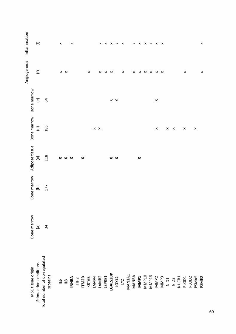
60

61

62
3.3 Proteomic based comparison between mouse and human
MSC-CM
Murine models allowed to elucidate the molecular pathway involved in the effects of
mMSC on the complex crosstalk between inflammation and angiogenesis [156], but,
because it is widely accepted that significant differences exist between mouse and human
MSC [59, 149], and because of the tremendous relevance of inflammation-induced
angiogenesis in human diseases, we focused our attention on comparing mouse and
human MSC secretome.
Tab.S1 lists the 286 proteins (out of the 465 proteins identified in stimulated hMSC-CM,
(62%) present in stimulated hMSC-CM that have been detected also in stimulated mMSC-
CM. The number of proteins significantly up-regulated or present only in the secretome of
stimulated MSC is similar in the two species: 89 in mouse (Tab.1 and 96 in human,
respectively [Tab.4]).
A comparative analysis of GO_BP category enrichment of over-represented proteins in
human and/or mouse [Fig.20] suggests that:
a) proteins up-regulated in the secretome of stimulated MSC from both organisms are, for
the most part, involved in similar biological processes, mainly related to defense, immune
and inflammatory response, chemotaxis and extracellular matrix remodeling;
b) there are clear important differences among human and mouse. Thus, only stmMSC-
CM is enriched in proteins involved in chromatin structure assembly, cell proliferation
regulation and related processes. On the contrary, complement activation, leukocyte
migration, bone development and metabolic processes specifically related to collagen are
amongst the statistically enriched GO functional categories in human but not in mouse.
Such differences are confirmed by the observation that only 23 proteins are up-regulated
or present only in stimulated MSC-CM both in mouse and human [Tab.6]; this again points
to a fine species-related tuning of the overall effects of secretome from the two organisms;
interestingly, our analysis indicates that 74 % and 83% of the common up-regulated
proteins are associated with angiogenesis or inflammation, respectively.

63
Fig.20 Distribution into biological processes of the proteins overrepresented in stimulated hMSC-CM
in human and mouse. The proteins that were significantly up-regulated or present only in stimulated MSC-
CM (Table 1 and [156]) were classified into different biological processes according to the Gene Ontology
classification system (GOBP) using DAVID software [150]; confidence level: medium; only categories
showing modified Fisher exact EASE score p value<0.05 and at least 5 counts in hMSC are represented.
The bars represent the percentage of proteins involved in a category out of the total number of
overrepresented proteins in human (96) or mouse (89) secretome. Asterisks indicate Fold Enrichment range
for each category: * 1-5, ** 6-10, ***>10.

64
Tab.6 Proteins overrepresented or present only in st MSC-CM common to mouse and human
3.3.1 Functional evidence of human and mouse MSC secretome
similarities or differences
Our proteomic results indicate that the majority of secreted proteins from both human and
mouse MSC are associated with inflammation and angiogenesis (Tab.4 and [156]). To
identify specific functional analogies or differences of human and mouse MSC in the
regulation of these two important processes, we focused on two proteins, M-SCF/CSF1
and TIMP1, which are present in st MSC-CM of both species and play a key role in
immunity/inflammation and angiogenesis, respectively [151, 152].
3.3.1.1 Macrophage colony-stimulating factor (M-CSF)
M-CSF is a growth factor secreted by a large variety of cells including macrophages,
endothelial cells, fibroblast and lymphocytes. Locally produced M-CSF in the vessel wall
contributes to the development and progression of atherosclerosis [153]. By interacting
with its membrane receptor (CSF1R or M-CSF-R), it stimulates the survival, proliferation,
and differentiation of monocytes and macrophages [154, 155].

65
Our proteomic data indicated that M-CSF (CSF1) is up-regulated in the secretome of both
human and mouse MSC upon stimulation by inflammatory cytokines [Tab.S1, Tab.4,
Fig.19]. As reported in Tab.S1 and Fig.20, M-CSF is amongst the proteins showing the
highest increase in stimulated human secretome according to mass spectrometric
analysis.
3.3.1.2 TIMP-1
Concerning angiogenesis, it has been analysed the effect of mMSC-CM on in vitro
angiogenesis exploiting the tube formation assay [156]. Interestingly, in the case of human
cells, MSC-CM was able to inhibit tube formation even when MSC had not been primed by
cytokines. However, pre-activation with pro-inflammatory cytokines strengthened the anti-
angiogenic effects of hMSC-CM, thus supporting our hypothesis that, during an
inflammatory response, MSC target angiogenesis and thus dampen the inflammatory
response [156].
The anti-angiogenic effect of MSC is mediated by TIMP-1 [156], according to many studies
based on in vitro and in vivo approaches.
Because the proteomic analyses indicate that TIMP-1 is one of the proteins up-regulated
in both human and mouse stMSC-CM [Tab.4], we compared the results obtained by
blocking TIMP-1 in SVEC4-10 cells incubated in the presence of mMSC-CM [Fig.12a] with
those generated using HUVEC cells and hMSC-CM [Fig.12b]. By inhibiting TIMP-1 activity
with a specific blocking antibody, we observed the complete recovery of HUVEC cell ability
to form tubes even in the presence of st hMSC-CM, indicating that TIMP1 is one of the key
secreted molecules targeting endothelial cells in both mouse and human MSC. TIMP-1
concentration was measured by ELISA in st and unst, human and mouse MSC-CM (Fig.
21a for mouse and b for human). In accordance with our data of tubulogenesis showing
that unst mMSC-CM has no effect on angiogenesis (Figs. 22 and Fig.23a), the
concentration of TIMP-1 in mMSC-CM was about 5 times higher when cells had been
primed by pro-inflammatory cytokines. Thus, in mouse MSC, the anti-angiogenic
phenotype is acquired only after licensing with pro-inflammatory cytokines. In human MSC,
however, the basal high level of secreted TIMP1 may explain the partial anti-angiogenic
effect of the unst hMSC-CM (Figs. 22, 23b). In fact, in support of this hypothesis, TIMP-1
blockade restored the formation of the endothelial network in the presence of unst or st
hMSC-CM. Again, proteomic data fully agree with functional assays and ELISA results for
human TIMP1. As reported in Fig. 2 and Table 1, this protein is listed amongst those over-
represented in st hMSC but showing relative lower level increase following stimulation.

66
Additional bioinformatics analyses of proteomic data further support the observation that
even relatively small changes in the level of TIMP1 can result in very significant
modulation of secretome properties. First of all, TIMP-1 concentration will greatly influence
the proteolytic potential of the secretome and, consequently, the overall activity of a
number of secretome components, including proteins which level is not increased following
stimulation and proteins not directly involved in inflammation and angiogenesis; secondly,
but not less importantly, TIMP1 is functionally related to a number of overrepresented
proteins in stimulated secretome besides proteases [Fig. 19], like cytokines and structural
proteins (such as IL6, IL8, CCL2 CXCL12, COL3A1). The complete list of the 54 proteins
of stimulated hMSC-CM functionally correlated to TIMP1 according to String [171] is
reported Tab. 8.
Fig.21 Mouse and human MSC-derived TIMP-1 quantification. MSC-derived TIMP-1 concentration in A)
mouse and B) human unstimulated or stimulated MSC conditioned medium was measured with ELISA. Data
are expressed as mean ± SEM (*p b 0.05, parametric t-test), 2 independent experiments.

67
Fig.22 Effect of human or mouse MSC conditioned medium on tube formation assay. The effect of
unstimulated or stimulated MSC media on endothelial cells was determined by a tube formation assay. Cells
were seeded on the top of a matri-gel phase in the presence of unstimulated or stimulated A)mouse, B)
human MSC-CM. 6 h later, images were acquired with a phase contrast inverted microscope at 4× objective
magnification. Analysis was performed with ImageJ Angiogenesis Analyzer. A) SVEC4-10 network formation;
quantification of the tube segment length (expressed in pixel number) and representative images at 4 h. B)
Huvec network formation; quantification of the tube segment length and representative images (expressed in
pixel number) at 4 h. 3 independent experiments, data are expressed as mean ± SEM (*p b 0.05, **p b 0.01,
One way ANOVA

68
Fig. 23: Timp-1 blocking reverts the anti-angiogenic effect of mouse and human MSC conditioned
media. In order to investigate the role of MSC-derived TIMP-1 on angiogenesis, the tube formation assay
was performed in the presence of A) mouse or B) human TIMP-1 blocking antibody. Representative images
of A) SVEC4-10 cell line or B) Huvec cells are taken with a phase contrast inverted microscope at 4×
objective magnifications. Graphs show the quantification of the tube segment length measured with ImageJ
Angiogenesis Analyzer. Data are expressed as mean ± SEM (*p<0.05, **p<0.01; One way Anova), 3
independent experiments.

69
Tab.7: TIMP1 interactions with other proteins in st hMSC-CM according to String

70

CONCLUSION

72
MSC have been studied across a range of clinical indications and represent a promising
therapeutic approach in many diseases in view of their potent immunomodulatory
properties. To design better therapeutic protocols and define the clinical endpoints, it is
important to identify the specific targets of MSC anti-inflammatory action in vivo. Recently,
it was demonstrated that MSC have a potent stabilizing effect on the vascular
endothelium, having the capacity of inhibiting endothelial permeability after traumatic brain
injury [156] and in hemorrhagic shock [157]. The data obtained from mouse MSCs suggest
that the effects of MSC are all mediated by soluble factors released by MSC [158]. This is
in agreement with another study showing an antiangiogenic activity for soluble factors
present in media derived from MSC/glioma co-cultures. Moreover, the proteomic analysis
of hMSC-CM and mMSC-CM confirms that exposure to pro-inflammatory cytokines results
in significantly higher secretion of a number of immunomodulatory and angiogenesis-
related proteins by MSC from both species. Notably, 62% of the proteins identified in st
hMSC-CM were also identified in st mMSC-CM, clearly highlighting the existence of a
common signature in the secretome of human and mouse MSC. However, although
human and mouse MSC show a similar proteomic signature in response to stimulation by
pro-inflammatory cytokines, our data indicate that they may induce different biological
responses. Thus, even if M-CSF is up-regulated in both human and mouse MSC-CM, only
hMSC-CM induce macrophage differentiation efficiently because of its high concentration
of M-CSF. In both species, several up-regulated proteins are associated with
angiogenesis. This process requires degradation of the vascular basement membrane and
remodelling of the extracellular matrix to allow endothelial cells migration and invasion into
the surrounding tissue [159]. The extended network of interactions amongst inflammation
and angiogenesis-related proteins in stimulated hMSC-CM makes it extremely difficult to
assess the in vivo physiological importance of each factor. The angiogenesis requires the
action of matrix metalloproteinases (MMPs) that degrade both matrix and non-matrix
proteins and have central roles in morphogenesis, wound healing, tissue repair and in
progression of chronic diseases [159] and, in particular, the presence of a number of
protease and protease inhibitors implies the possibility of additional self-modulation of the
properties of the various components of the secretome [146]. The balance between MMPs
and their natural inhibitors, the TIMPs, is critical for extracellular matrix remodelling and
angiogenesis. The TIMP family comprises four protease inhibitors: TIMP-1, TIMP-2, TIMP-
3 and TIMP-4. With the exception of TIMP-4 [160], all three TIMPs inhibit angiogenesis in

73
vivo [137], although through diverse mechanisms. MSC secrete both MMPs and their
inhibitors, and thus contribute to the regulation and protection of the perivascular niche
[161]. Using both in vitro and in vivo assays, we identified the metalloproteinase inhibitor
TIMP-1 as the molecule responsible for the anti-angiogenic effects of MSC. TIMP-1 is
known to inhibit endothelial cells migration by MMP-dependent and MMP independent
mechanisms [162, 163]. The latter involve regulation of various biological processes such
as cell growth, apoptosis and differentiation through the CD63 receptor [164, 165]. In
addition, TIMP-1 was shown to induce secretion of soluble VEGFR-1 by human
endothelial cells, leading to a decrease of bioavailable VEGF and of blood vessel growth
[166]. TIMP-3 has also been identified as a soluble factor produced by MSC with beneficial
effects on endothelial cell function in a mouse model of traumatic brain injury [167];
however, we did not find evidence for TIMP-3 upregulation in the mouse or human MSC
secretomes. It is likely that, in vivo, other soluble factors in addition to TIMP-1 contribute to
MSC-mediated immune regulation: MSC are also known to produce prostaglandin E2 and
thus inhibit the activation of macrophages [168], which are a source of multiple growth
factors that enhance endothelial cell proliferation and survival [169]. Therefore, by
identifying TIMP-1 as a critical effector of the anti-inflammatory properties of MSC, and by
observing its anti-angiogenic role, both in mouse and in human, we can confirm the
important role of TIMP-1 as key secreted molecule targeting endothelial cells. The
identification of TIMP-1 as potential effector molecule responsible for the anti-angiogenic
properties of MSC, both in mouse and in human, allow to confirm that MSC exert specific
effect by secretion of a broad range of bioactive molecules and allow to design pre-clinical
experiments and clinical trials, with the aim of exploit their potentially therapeutic role.
Moreover, these data could be also a starting point for further analysis aimed to
understand important differences between human and mouse also in a pharmacological
field. In fact, although mouse and rats are still considered the animal models of choice for
applied pharmacological research and drug test, up to now, many important biochemistry
pathways involved in life processes, in both species, have not been yet clearly understood,
often precluding the possibility to translate results in humans without leading to errors.
Indeed, there are few studies in literature focused on this particular aspect, therefore the
data reported in this thesis represent a source for new studies evaluating similarities and
differences between human and mouse models in pharmacological field.

74
The results reported in this phD thesis have been partially published in the papers listed
below:
Mouse mesenchymal stem cells inhibit high endothelial cell activation and
lymphocyte homing to lymph nodes by releasing TIMP-1 Leukemia (2016) 30,
1143-1154.
Proteomic analysis of the secretome of human bone marrow-derived
Mesenchymal Stem Cells primed by pro-inflammatory cytokines Journal of
proteomics (2017) 166, 115-126.
And in following poster presentation:
S. Nonnis, E. Maffioli, F. Santagata, F. Grassi Scalvini, S. Morelli, A. Negri, A. Viola,
G. Tedeschi. Proteomic analysis of bone marrow derived human MSC secretome
stimulated with pro- inflammatory cytokines. SIB 2017, 20-22 settembre 2017,
Caserta.
G. Tedeschi, L. Zanotti, F. Santagata, E. maffioli, S. Nonnis, A. Negri, A. Viola. Human
mesenchymal stem cells secretome before and after treatment with pro
inflammatory cytokines investigated by a label free proteomic approach. XI ItPA
national Congress, 16 – 19 maggio 2016, Perugia.
S. Nonnis, L. Zanotti, F. Santagata, E. Maffioli, A. Negri, A. Viola, G. Tedeschi.
Proteome characterization of human stromal mesenchymal stem cells secretome
before and after treatement with pro inflammatory cytokines. Proteine 2016, 30
marzo-1aprile 2016, Bologna.
S. Nonnis, L. Zanotti, E. Maffioli, F. Santagata, A. Negri, L. Chiesa, A. Viola, G.
Tedeschi. Human mesenchymal stem cells secretome investigated by using a label
free proteomic approach. 58th National Meeting of the Italian society of biochemistry
and molecular biology (SIB), 14-16 settembre 2015, Urbino.
S. Nonnis, L. Zanotti, E. Maffioli, F. Santagata, , A. Negri, A. Viola, G.
Tedeschi. Proteomic analysis of the mesenchymal stem cells secretome using a
label free approach. EupA IX Annual Congress, 23-28 giugno 2015, Milano.

SUPPLEMENTARY MATERIALS

76
Tab.S1 Proteins identified and quantified in hMSC secretome in at least 3 out of 5 technical replicas
in at least one stimulation condition in both patients. Notes (a) cytokine or chemokine or proteins
functionally related to these classes of compounds according to the nextProt database [122]; (b) proteins
present also in mouse stimulated MSC-CM; (c) number of razor+unique peptides used to identified the
protein in patients H30 or H34; (d) posterior error probability score calculated for the identification of protein;
(e) percentage of the protein sequence that is covered by the identified peptides.

77
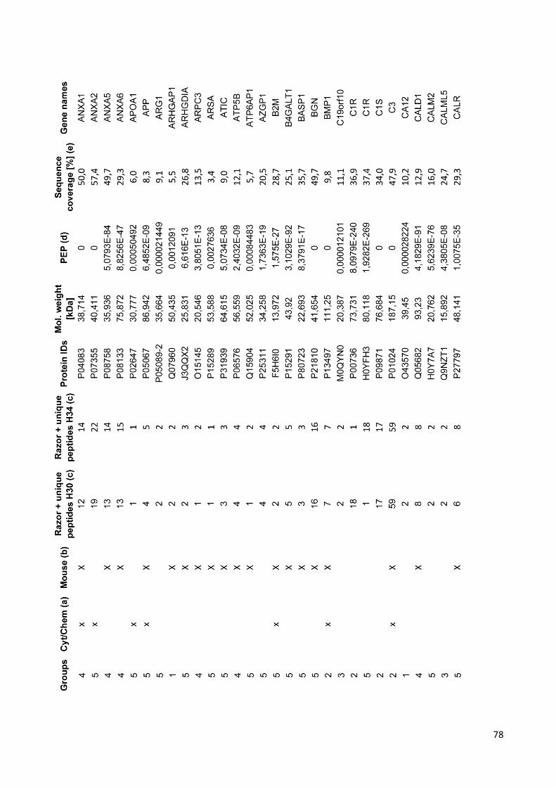
78

79

80

81

82

83

84

85

86

87

88

89

90
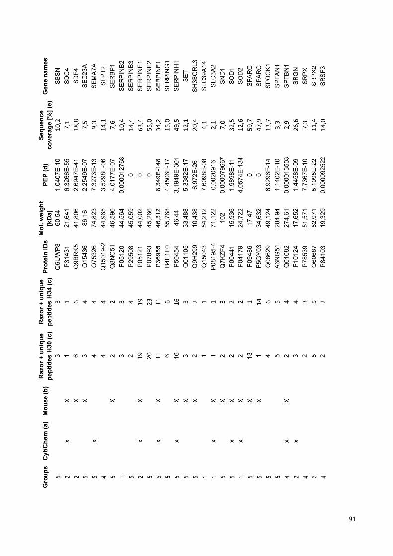
91

92

93

REFERENCES

95
1. AM. Arias, Drosophila melanogaster and the development of biology in the 20th century,
Methods in Molecular Biology, 2008, 420, 1–25.
2. http://www.biology-online.org/dictionary/Stem_cell
3. http://nas-sites.org, Understanding stem cells: an over view of the science and issues
from the National academies.
4. https://en.wikipedia.org/wiki/Stem_cell
5. HR. Schöler, The Potential of Stem Cells: An Inventory, In: N. Knoepffler, D. Schipanski,
SL. Sorgner, editors, Human biotechnology as Social Challenge, 2007, p. 28.
7. F. Ulloa-Montoya, CM. Verfaillie, WS. Hu, Culture systems for pluripotent stem cells,
Journal of Bioscience and Bioengeneering, 2005, 100, 12–27.
8. www.bioinformant.com
9. JA. Thomson, J. Itskovitz-Eldor, SS. Shapiro, MA. Waknitz, JJ. Swiergiel, VS. Marshall,
JM. Jones, Blastocysts Embryonic Stem Cell Lines Derived from Human, Science, 1998,
282, 1145–47.
10. LA. Boyer, TI. Lee, MF. Cole, SE. Johnstone, SS. Levine, JP. Zucker, MG. Guenther,
RM. Kumar, HL. Murray, RG. Jenner, DK. Gifford, DA. Melton, R. Jaenisch, RA. Young,
Core transcriptional regulatory circuitry in human embryonic stem cells, Cell, 2005, 122,
947–56.
11. Mayo foundation for medical education and research “Stem cells” Mayo Clinic, Web.
March 23, 2013.
12. Y. Jiang et al., Pluripotency of mesenchymal stem cells derived from adult marrow,
Nature, 2002, 418, 41-9.
13. MZ. Ratajczak, B. Machalinski, W. Wojakowski, J. Ratajczak, M. Kucia, A hypothesis
for an embryonic origin of pluripotent Oct-4(+) stem cells in adult bone marrow and other
tissues, Leukemia, 2007, 21, 860–7.
14. SD. Narasipura, JC. Wojciechowski, N. Charles, JL. Liesveld, MR. King, P-Selectin
coated microtube for enrichment of CD34+ hematopoietic stem and progenitor cells from
human bone marrow, Clinical Chemistry, 2008, 54, 77–85.

96
15. S. Terai, T. Ishikawa, K. Omori, K. Aoyama, Y. Marumoto, Y. Urata, Y. Yokoyama, K.
Uchida, T. Yamasaki, Y. Fujii, K. Okita, I. Sakaida, Improved liver function in patients with
liver cirrhosis after autologous bone marrow cell infusion therapy, Stem Cells, 2006, 24,
2292–8.
16. R. Subrammaniyan, J. Amalorpavanathan, R. Shankar et al., Application of autologous
bone marrow mononuclear cells in six patients with advanced chronic critical limb ischemia
as a result of diabetes: our experience, Cytotherapy, 2011, 13, 993–9.
17. N. Madhu Sankar, K.Vaidyanathan, Use of Bone Marrow derived Stem Cells in
Patients with Cardiovascular Disorders, Journal of Stem Cells and Regenerative Medicine,
2007, 3, 28-29.
18. VD. Dedeepiya, YY. Rao, GA. Jayakrishnan, JK. Parthiban, S. Baskar, SR. Manjunath,
R. Senthilkumar, SJ. Abraham, Index of CD34+ Cells and Mononuclear Cells in the Bone
Marrow of Spinal Cord Injury Patients of Different Age Groups: A Comparative Analysis,
Bone Marrow Research, 2012, 1-8.
19. B. Barrilleaux, DG. Phinney, DJ. Prockop, KC. O' Connor, Review: ex vivo engineering
of living tissues with adult stem cells, Tissue Engineering, 2006, 12, 3007–19.
20. JM. Gimble, AJ. Katz, BA. Bunnell, Adipose-derived stem cells for regenerative
medicine, Circulation Research, 2007, 100, 1249–60.
21. http://slideplayer.com/slide/9129448
22. A. Bongso, L. EngHin, Stem cells: their definition, classification and sources, Stem
Cells: From Benchtop to Bedside. World Scientific, 2005, p. 5. ISBN 981-256-126-9.
23. K.L. Moore, T.V.N. Persaud, A.G. Torchia, Before We Are Born: Essentials of
Embryology and Birth Defects. Philadelphia, PA: Saunders, Elsevier, 2013.
24. P. De Coppi, G. Bartsch, MM. Siddiqui, T. Xu, CC. Santos, L. Perin, G. Mostoslavsky,
AC. Serre, EY. Snyder, JJ. Yoo, ME. Furth, S. Soker, A. Atala, Isolation of amniotic stem
cell lines with potential for therapy, Nature Biotechnology, 2007, 25, 100–106.
25. T. Bubela, MD. Li, M. Hafez, M. Bieber, H. Atkins, Is belief larger than fact:
Expectations, optimism and reality for translational stem cell research, BMC Medicine,
2012, 10, 133.

97
26. M. Ader, EM. Tanaka, Modeling human development in 3D culture. Current Opinion in
Cell Biology, 2014, 31, 23-28.
27. Making human embryonic stem cells, The Economist, 2007-11-22.
28. M. Brand, J. Palca, A. Cohen, Skin Cells Can Become Embryonic Stem Cells, 2007,
National Public Radio.
29. Breakthrough Set to Radically Change Stem Cell Debate, News Hour with Jim Lehrer,
2007.
30. EA. Kimbrel, R. Lanza, Pluripotent stem cells: The last 10 years, Regenerative
Medicine, 2016, 11, 831–847.
31. K. Takahashi, S. Yamanaka, Induction of pluripotent stem cells from mouse embryonic
and adult fibroblast cultures by defined factors, Cell, 2006, 126, 663–76.
32. http://learn.genetics.utah.edu/content/stemcells/quickref
33. "Bone Marrow Transplant".ucsfchildrenshospital.org
34. E. Kane, Stem-cell therapy shows promise for horse soft-tissue injury, DVM
Newsmagazine, 2008.
35. K.L. Moore, T.V.N. Persaud, A.G. Torchia, Before We Are Born: Essentials of
Embryology and Birth Defects. Philadelphia, PA: Saunders, Elsevier, 2013
36. http://www.sigmaaldrich.com
37. EM. Horwitz, K. Le Blanc, M. Dominici, I. Mueller, I. Slaper-Cortenbach, FC. Marini,
RJ. Deans, DS. Krause, A. Keating, International Society for Cellular Therapy: Clarification
of the nomenclature for MSC: the International Society for Cellular Therapy position
statement, Cytotherapy, 2005, 7, 393–399.
38. M. Crisan, S. Yap, L. Casteilla, CW. Chen, M. Corselli, TS. Park, G. Andriolo, Sun B,
Zheng B, Zhang L, Norotte C, Teng PN, Traas J, Schugar R, Deasy BM, Badylak S,
Bühring HJ, Giacobino JP, Lazzari L, Huard J, Peault B: A perivascular origin for
mesenchymal stem cells in multiple human organs, Cell Stem Cell 2008, 3, 301–313.
39. J. Oswald, et al., Mesenchymal stem cells can be differentiated into endothelial cells in
vitro, Stem Cells, 2004, 22, 377–384.

98
40. S. Makino, et al., Cardiomyocytes can be generated from marrow stromal cells in vitro,
Journal of clinic investigations, 1999, 103, 697-705.
41. S. Snykers, et al., In vitro differentiation of embryonic and adult stem cells into
hepatocytes: state of the art, Stem Cells, 2009, 27, 577-605.
42. A. Arthur, et al., Adult human dental pulp stem cells differentiate toward functionally
active neurons under appropriate environmental cues, Stem Cells, 2008, 26, 1787-95.
43. DA. De Ugarte, K. Morizono, A. Elbarbary, Z. Alfonso, PA. Zuk, M. Zhu, JL. Dragoo, P.
Ashijan, B. Thomas, P. Benhaim, I. Chen, J. Frazer, MH. Hedrick, Comparison of
multilineage cells from human adipose tissue and bone marrow, Cells Tissues Organs,
2003, 174:101–109.
44. S. Meirelles, L. Chagastelles, NB. Nardi, Mesenchymal stem cells reside in virtually all
postnatal organs and tissues, Journal of cells science, 2006, 119, 2204–2213.
45. N. Kaltz, et al. Novel markers of mesenchymal stem cells defined by genome-wide
gene expression analysis of stromal cells from different sources, Experimental Cell
Research, 2010, 316, 2609–2617.
46. M. Corselli, CW. Chen, M. Crisan, L. Lazzari, B. Peault, Perivascular ancestors of adult
multipotent stem cells, Arteriosclerosis, Thrombosis and vascular biology, 2010, 30,
1104–1109.
47. http://endonovo.com/cytotronics-platform.
48. C. Farrington-Rock, et al., Chondrogenic and adipogenic potential of microvascular
pericytes, Circulation, 2004, 110, 2226–2232.
49. MJ. Doherty, et al., Vascular pericytes express osteogenic potential in vitro and in vivo,
Journal of Bone and Mineral Research, 1998, 13, 828–838.
50. A. Keating, Mesenchymal stromal cells: new directions, Cell stem cell, 2012, 10, 709-
16.
51. S. Gordon, A. Mantovani, Diversity and plasticity of mononuclearphagocytes,
European Journal of immunology, 2011, 41, 2470–2472.

99
52. A. Mantovani et al., Macrophage plasticity and polarization in tissue repair and
remodelling, The Journal of pathology, 2013, 229, 176-85.
53. Waterman et al., A new mesenchymal stem cells (MSC) paradigm: polarization into a
pro-inflammatory MSC1 or an immunosuppressive MSC2 phenotype, PlOsOne, 2010, 5,
4.
54. K. Nemeth et al., Bone marrow stromal cells use TGF-β to suppress allergic responses
in a mouse model of ragweed-induced asthma, PNAS, 2010, 107, 5652–5657.
55. SL. Tomchuck et al., Toll-like receptors on human mesenchymal stem cells drive their
migration and immunomodulating responses, Stem cells, 2008, 26, 99-107.
56. Delarosa et al., Toll-like receptors as modulators of mesenchymal stem cells, Frontiers
in Immunology, 2012, 3, 182.
57. Raicevic et al., Inflammation modifies the pattern and the function of Toll-like receptors
expressed by human mesenchymal stromal cells, Human Immunology, 2010, 71-235.
58. IF. Mo et al., Prolonged exposure to bacterial toxins downregulated expression of toll-
like receptors in mesenchymal stromal cell-derived osteoprogenitors, BMC Cell Biology,
2008, 9, 52.
59. ME. Bernardo, EW. Fibbe, Mesenchymal Stromal Cells: Sensors and Switchers of
Inflammation, Cell Stem Cell, 2013, 13.
60. AJ. Nauta, WE. Fibbe, Immunomodulatory properties of mesenchymal stromal cells,
Blood, 2007, 110, 3499–3506.
61. A. Bartholomew, C. Sturgeon, M. Siatskas, K. Ferrer, K. McIntosh, S. Patil, W. Hardy,
S. Devine, D. Ucker, R. Deans, A. Moseley, R. Hoffman, Mesenchymal stem cells
suppress lymphocyte proliferation in vitro and prolong skin graft survival in vivo,
Experimental Hematology, 2002, 30, 42–48.
62. A. Tyndall, UA. Walker, A. Cope et al, Immunomodulatory properties of mesenchymal
stem cells: a review based on an interdisciplinary meeting held at the Kennedy Institute of
Rheumatology Division, London, UK, 31 October 2005, Arthritis Research and Therapy,
2007, 9:301.

100
63. I. Rasmusson, O. Ringden, B. Sundberg, K. Le Blanc, Mesenchymal stem cells inhibit
the formation of cytotoxic T lymphocytes, but not activated cytotoxic T lymphocytes or
natural killer cells, Transplantation, 2003, 76, 1208–1213.
64. N. Eliopoulos, J. Stagg, L. Lejeune, S. Pommey, J. Galipeau, Allogeneic marrow
stromal cells are immune rejected by MHC class I and II mismatched recipient mice,
Blood, 2005, 106, 4057–4065.
65. AJ. Nauta, G. Westerhuis, AB. Kruisselbrink, EG. Lurvink, R. Willemze, WE. Fibbe,
Donor-derived mesenchymal stem cells are immunogenic in an allogeneic host and
stimulate donor graft rejection in a non myeloablative setting, Blood, 2006, 108, 2114–
2120.
66. WT. Tse, JD. Pendleton, WM. Beyer, MC. Egalka, EC. Guinan, Suppression of
allogeneic T-cell proliferation by human marrow stromal cells: implications in
transplantation, Transplantation, 2003, 75, 389–397.
67. S. Aggarwal, MF. Pittenger, Human mesenchymal stem cells modulate allogeneic
immune cell responses, Blood, 2005, 105, 1815–1822.
68. M. Di Nicola, C. Carlo-Stella, M. Magni, M. Milanesi, PD. Longoni, P. Matteucci, S.
Grisanti, AM. Gianni, Human bone marrow stromal cells suppress T-lymphocyte
proliferation induced by cellular or nonspecific mitogenic stimuli, Blood, 2002, 99, 3838–
3843.
69. S. Glennie, I. Soeiro, PJ. Dyson, EW. Lam, F. Dazzi, Bone marrow mesenchymal
stem cells induce division arrest anergy of activated T cells, Blood, 2005, 105, 2821–2827.
70. A. Corcione, F. Benvenuto, E. Ferretti, D. Giunti, V. Cappiello, F. Cazzanti, M. Risso,
F. Gualandi, GL. Mancardi, V. Pistoia, A. Uccelli, Human mesenchymal stem cells
modulate B-cell functions, Blood, 2006, 107, 367–372.
71. I. Rasmusson, K. Le Blanc, B. Sundberg, O. Ringden: Mesenchymal stem cells
stimulate antibody secretion in human B cells, Scandinavian Journal of Immunology, 2007,
65, 336–343.
72. M. Krampera, L. Cosmi, R. Angeli, A. Pasini, F. Liotta, A. Andreini, V. Santarlasci, B.
Mazzinghi, G. Pizzolo, F. Vinante, P. Romagnani, E. Maggi, S. Romagnani, F. Annunziato,

101
Role of the IFN-γ in the immunomodulatory activity of human mesenchymal stem cells,
Stem cells, 2005, 24, 386–398.
73. PA. Sotiropoulou, SA. Perez, AD. Gritzapis, CN. Baxevanis, M. Papamichail,
Interactions between human mesenchymal stem cells and natural killer cells, Stem Cells,
2006, 24, 74–85.
74. AR. Williams, JM. Hare, S. Dimmeler, D. Losordo, Mesenchymal Stem Cells: Biology,
Pathophysiology, Translational Findings, and Therapeutic Implications for Cardiac
Disease, Circulation Research, 2011, 109, 923-40.
75. DG. Phinney, DJ. Prockop, Concise review: mesenchymal stem/multipotent stromal
cells: the state of trans differentiation and modes of tissue repair, Stem cells, 2007, 25,
2896-2902.
76. K. Le Blanc, I. Rasmusson, B. Sundberg, C. Gotherstrom, M. Hassan, M. Uzunel, O.
Ringden, Treatment of severe acute graftversus- host disease with third party
haploidentical mesenchymal stem cells, Lancet, 2004, 363, 1439–1441.
77. G. Behre, S. Theurich, T. Weber, M. Christopeit, Reply to ‘The correlation between
cotransplantation of mesenchymal stem cells and higher recurrence rates in hematologic
malignancy patients: outcome of a pilot clinical study’ by Ning et al., Leukemia, 2009,
23,178–180.
78. SM. Devine, C. Cobbs, M. Jennings, A. Bartholomew, R. Hoffman, Mesenchymal stem
cells distribute to a wide range of tissues following systemic infusion into nonhuman
primates, Blood, 2003, 101, 2999–3001.
79. F. Djouad, V. Fritz, F. Apparailly, P. Louis-Plence, C. Bony, J. Sany, C. Jorgensen, D.
Noël, Reversal of the immunosuppressive properties of mesenchymal stem cells by tumor
necrosis factor alpha in collagen-induced arthritis, Arthritis Rheum, 2005, 52, 1595–1603.
80. VS. Urban, J. Kiss, J. Kovacs, E. Gócza, V. Vas, E. Monostori, F. Uher, Mesenchymal
stem cells cooperate with bone marrow cells in therapy of diabetes, Stem Cells, 2008, 26,
244–253.
81. D. Rubio, J. Garcia-Castro, MC. Martin, R. De La Fuente, JC. Cigudosa, AC. Lloyd, A.
Bernard, Spontaneous human adult stem cell transformation, Cancer Research, 2005, 65,
3035–3039.

102
82. AC. Lloyd, Limits to lifespan, Nature Cell Biology, 2002, 4.
83. L. Chin, SE. Artandi, Q. Shen, p53 deficiency rescues the adverse effects of telomere
loss and cooperates with telomere dysfunction to accelerate carcinogenesis, Cell, 1999,
97, 527–38.
84. J. Marx, Cancer research, Mutant stem cells may seed cancer, Science, 2003, 301,
1308–10.
85. Phinney et al., Plastic adherent stromal cells from the bone marrow of commonly used
strains of inbred mice: variations in yield, growth, and differentiation, Biochemical journal,
1999, 72, 570–585.
86. Miura et al., Accumulated chromosomal instability in murine bone marrow
mesenchymal stem cells leads to malignant transformation, Stem Cells, 2006, 24, 1095–
1103.
87. Tolar et al., Sarcoma derived from cultured mesenchymal stem cells, Stem Cells,
2007, 25: 371–379.
88. P.R. Amable, M.V. Teixeria, R.B. Carias, J.M. Granjeiro, R. Borojevic, Protein
synthesis and secretion in human mesenchymal cells derived from bone marrow, adipose
tissue and Wharton’s jelly, Stem cells Research and Therapy, 2014, 53 – 66.
89. H. Tjalsma, A. Bolhuis, J.D., Jongbloed et al., Signal peptide-dependent protein
transport in Bacillus subtilis: a genome-based survey of the secretome, Microbiology and
Molecular Biology Review, 2000, 64 , 515 – 547.
90. S. Nonnis, E. Maffioli, L. Zanotti, F. Santagata, A. Negri, A. Viola, S. Elliman, G.
Tedeschi, Effect of fetal bovine serum in culture media on MS analysis of mesenchymal
stromal cells secretome, EuPA Open Proteomics, 2016, 28-30.
91. LA. Liotta, M. Ferrari, E. Petricoin, Clinical proteomics: written in blood, Nature, 2003,
425, 905-10.
92. GS. Omenn, DJ. States, M. Adamski, TW. Blackwell, R. Menon, H. Hermjakob, R.
Apweiler, BB. Haab, RJ. Simpson, JS. Eddes, Overview of the HUPO Plasma Proteome
Project: results from the pilot phase with 35 collaborating laboratories and multiple

103
analytical groups, generating a core dataset of 3020 proteins and a publicly-available
database, Proteomics, 2005, 5, 3226-45.
93. J.E. Celis, P. Gromov, T. Cabezon, J.M. Moreira, N. Ambartsumian, K. Sandelin,
F.Rank, I. Gromova, Proteomic characterization of the interstitial fluid perfusing the breast
tumor microenvironment: a novel resource of for biomarkers and therapeutic target
discovery, Molecular and Cellular Proteomics, 2004, 3, 327-344.
94. Y. Shi, CA. Elmets, JW. Smith, YT. Liu, YR. Chen, CP. Huang, W. Zhu, HN.
Ananthaswamy, RL. Gallo, CM. Huang, Quantitative proteomes and in vivo secretomes of
progressive and regressive UV-induced fibrosarcoma tumor cells: mimicking tumor
microenvironment using a dermis-based cell-trapped system linked to tissue chamber,
Proteomics, 2007, 7, 4589–4600.
95. W. Zhu, JW. Smith, CM. Huang, Mass Spectrometry-Based Label-Free Quantitative
Proteomics, Journal of Biomedicine and Biotechnology, 2010.
96. CC. Wu, MJ. Mac Coss, Shotgun proteomics: tools for the analysis of complex
biological systems, Current Opinion in Molecular Therapeutics, 2002, 4, 242– 250.
97. EI. Chen, JR. Yates, Cancer proteomics by quantitative shotgun proteomics, Molecular
Oncology, 2007, 1,144–159.
98. SP. Gygi, B. Rist, SA. Gerber, F. Turecek, MH. Gelb, R. Aebersold, Quantitative
analysis of complex protein mixtures using isotope coded affinity tags, Nature
Biotechnology, 1999, 17, 994–999.
99. A. Motoyama, JR. Yates, Multidimensional LC separations in shotgun proteomics,
Analytical Chemistry, 2008, 7187-7193.
100. B. Domon, R. Aebersold, Mass spectrometry and protein analysis, Science, 2006,
212-217.
101. RD. Voyksner, H. Lee, Investigating the use of an octupole ion guide for ion storage
and high-pass mass filtering to improve the quantitative performance of electrospray ion
trap mass spectrometry, Rapid Communications in Mass Spectrometry, 1999, 1427–1437.

104
102. D. Chelius, PV. Bondarenko, Quantitative profiling of proteins in complex mixtures
using liquid chromatography and mass spectrometry, Journal of Proteome Research,
2002, 4, 317–323.
103. PV. Bondarenko, D. Chelius, TA. Shaler, Identification and relative quantitation of
protein mixtures by enzymatic digestion followed by capillary reversed-phase liquid
chromatography-tandem mass spectrometry, Analytical Chemistry, 2002, 74, 4741–4749.
104. J. Lengqvist, J. Andrade, Y. Yang, G. Alvelius, R. Lewensohn, and J. Lehtiö,
Robustness and accuracy of high speed LC-MS separations for global peptide quantitation
and biomarker discovery, Journal of Chromatography B, 2009, 13, 1306–1316.
105. N. Fatima, D. Chelius, B. T. Luke et al., Label-free global serum proteomic profiling
reveals novel celecoxib-modulated proteins in familial adenomatous polyposis patients,
Cancer Genomics and Proteomics, 2009, 6, 41–49.
106. S. K. Huang, MM. Darfler, MB. Nicholl et al., LC/MS-based quantitative proteomic
analysis of paraffin-embedded archival melanomas reveals potential proteomic biomarkers
associated with metastasis, PLoS ONE, 2009.
107. Y. Levin, E. Schwarz, L. Wang, F. M. Leweke, and S. Bahn, Label-free LC-MS/MS
quantitative proteomics for large-scale biomarker discovery in complex samples, Journal of
Separation Science, 2007, 30, 2198–2203.
108. JPC. Vissers, JI. Langridge, and JMFG. Aerts, Analysis and quantification of
diagnostic serum markers and protein signaturs for Gaucher disease, Molecular and
Cellular Proteomics, 2007, 6, 755–766.
109. JTJ. Huang, T. McKenna, C. Hughes, F. M. Leweke, E. Schwarz, and S. Bahn, CSF
biomarker discovery using label-free nano-LC-MS based proteomic profiling: technical
aspects, Journal of Separation Science, 2007, 30, 214–225.
110. MP. Washburn, D. Wolters, JR. Yates, Large-scale analysis of the yeast proteome by
multidimensional protein identification technology, Nature Biotechnology, 2001, 19, 242–
247.
111. H. Liu, R. G. Sadygov, JR. Yates, A model for random sampling and estimation of
relative protein abundance in shotgun proteomics, Analytical Chemistry, 2004, 76, 4193–
4201.

105
112. B. Zybailov, MK. Coleman, L. Florens, MP. Washburn, Correlation of relative
abundance ratios derived from peptide ion chromatograms and spectrum counting for
quantitative proteomic analysis using stable isotope labeling, Analytical Chemistry, 2005,
77, 6218–6224.
113. JX. Pang, N. Ginanni, AR. Dongre, SA. Hefta, GJ. Opiteck, Biomarker discovery in
urine by proteomics, Journal of Proteome Research, 2002, 1, 161–169.
114. PV. Rao, AP. Reddy, X. Lu et al., Proteomic identification of salivary biomarkers of
type-2 diabetes, Journal of Proteome Research, 2009, 8, 239–245.
115. M. Asara, HR. Christofk, LM. Freimark, LC. Cantley, A label-free quantification
method by MS/MS TIC compared to SILAC and spectral counting in a proteomics screen,
Proteomics, 2008, 8, 994–999.
116. L. Zanotti, A. Sarukhan, E. Dander, M. Castor, J. Cibella, C. Soldani et al.,
Encapsulated mesenchymal stem cells for in vivo immunomodulation, Leukemia, 2013, 27,
500-3.
117. DF. Mc Auley, GF. Curley, UI. Hamid, JG. Laffey, J. Abbott, DH. McKenna, X. Fang,
MA. Matthay, JW. Lee, Clinical grade allogeneic human mesenchymal stem cells restore
alveolar fluid clearance in human lungs rejected for transplantation, American Journal of
Physiology, Lung Cellular and Molecular Physiology, 2014, 306, 809-15.
118. M. Dominici, K. Le Blanc, I. Mueller, I. Slaper-Cortenbach , F. Marini, D. Krause, R.
Deans, A. Keating, E. Horwitz, Minimal criteria for defining multipotent mesenchymal
stromal cells, The International Society for Cellular Therapy position statement,
Cytotherapy, 2006, 8, 315-7.
119. J. Cox, M. Mann, Max Quant enables high peptide identification rates, individualized
p.p.b.-range mass accuracies and proteome-wide protein quantification, Nature
Biotechnology, 2008, 26, 1367–72.
120. S. Tyanova, T. Temu, P. Sinitcyn, A. Carlson, MY. Hein, T. Geiger, M. Mann, J. Cox,
The Perseus computational platform for comprehensive analysis of (prote) omics data,
Nature Methods, 2016, 13, 731-40.
121. The Gene Ontology Consortium, Gene Ontology Consortium: going forward, Nucleic
Acids Research, 2015, 43.

106
122. P. Gaudet, PA. Michel, M. Zahn-Zabal, I. Cusin, PD. Duek, O. Evalet, A. Gateau, A.
Gleizes, M. Pereira, D. Teixeira, Y. Zhang, L. Lane, A. Bairoch, The neXtProt
knowledgebase on human proteins: current status, Nucleic Acids Res, 2015, 43, 764-70.
123. The UniProt Consortium, UniProt: a hub for protein information, Nucleic Acids
Research, 2015.
124. G. Stelzer, R. Rosen, I. Plaschkes, S. Zimmerman, M. Twik, S. Fishilevich, T. Iny
Stein, R. Nudel, I. Lieder, Y. Mazor, S. Kaplan, D. Dahary, D. Warshawsky, The
GeneCards Suite: From Gene Data Mining to Disease Genome Sequence Analysis,
Current Protocol in Bioinformatics, 2016, 54,1.
125. B. Rocha, Secretome analysis of human mesenchymal stem cells undergoing
chondrogenic differentiation, Journal of Proteome Research, 2014, 13, 1045-54.
126. SP. Kristensen, L. Chen, M. Overbeck Nielsen, DW. Qanie, I. Kratchmarova, Kassem
M, Andersen JS, Temporal Profiling and Pulsed SILAC Labeling Identify Novel Secreted
Proteins During ex vivo Osteoblast Differentiation of Human Stromal Stem Cells, Molecular
and Cellular Proteomics, 2012, 11, 989-1007.
127. DW. Huang, BT. Sherman, RA. Lempicki, Bioinformatics enrichment tools: paths
toward the comprehensive functional analysis of large gene lists, Nucleic Acids Research,
2009, 37,1-13.
128. D. Szklarczyk, A. Franceschini, S. Wyder, K. Forslund, D. Heller, J. Huerta-Cepas,
M. Simonovic, A. Roth, A. Santos, KP Tsafou, M. Kuhn, P. Bork, LJ Jensen, C. von
Mering, STRING v10: protein-protein interaction networks, integrated over the tree of life,
Nucleic Acids Research, 2015, 43.
129. JA., Vizcaíno, A. Csordas, N. del-Toro, JA. Dianes, J. Griss, I. Lavidas, G. Mayer, Y.
Perez-Riverol, F. Reisinger, T. Ternent, QW. Xu, R. Wang, H. Hermjakob, 2016 update of
the PRIDE database and related tools, Nucleic Acids Research, 2016, 44.
130. W. Huang, BT. Sherman, RA Lempicki, Systematic and integrative analysis of large
gene lists using DAVID bioinformatics resources, Nature Protocols, 2009, 4,44-57.
131. Angiogenesis insights from a systematic overview, New York: Nova Science, 2013.

107
132. N. Naoyo, Y.Hirohisa et al., Angiogenesis in Cancer, Vascular Health and Risk
Management, 2006, 2, 213–219.
133. AA. Ucuzian, AA. Gassman, AT. East, Molecular Mediators of Angiogenesis, Journal
of Burn Care and Research, 2010, 31, 58-75.
134. SM. Wahl, H. Wong, NJ. McCartney-Francis, Role of growth factors in inflammation
and repair, Cell Biochemistry, 1989, 40, 193-9.
135. R. Visse, H. Nagase, Matrix metalloproteinases and tissue inhibitors of
metalloproteinases: structure, function, and biochemistry, Circulation Research, 2003, 92,
827-39.
136. K. Brew, H. Nagase, The tissue inhibitors of metalloproteinases (TIMPs): An ancient
family with structural and functional diversity, Biochimica et Biophysica Acta, 2010, 1803,
55–71.
137. E. Lambert, E. Dasse, B. Haye, E. Petitfrere, TIMPs as multifacial proteins, Critical
Reviews in Oncology/Hematology, 2004, 49,187–198.
138. SJ. Crocker, RF. Frausto, JK. Whitmire, N. Benning, R. Milner, JL. Whitton
Amelioration of coxsackievirus B3-mediated myocarditis by inhibition of tissue inhibitors of
matrix metalloproteinase-1, American Journal Pathology, 2007, 171, 1762–1773.
139. S. Zacchigna, L. Pattarini, L. Zentilin, S. Moimas, A. Carrer, M. Sinigaglia et al., Bone
marrow cells recruited through the neuropilin-1 receptor promote arterial formation at the
sites of adult neo-angiogenesis in mice, Journal of Clinical Investigation , 2008, 118,
2062–2075.
140. ME. Groh, B. Maitra, E. Szekely, ON. Koç, Human mesenchymal stem cells require
monocyte-mediated activation to suppress alloreactive T cells, Experimental Hematology,
2005, 33:928-34.
141. Gene Ontology Consortium, Gene ontology consortium: going forward, Nucleic Acids
Research, 2015, 43, D1049–56.
142. UniProt Consortium, UniProt: a hub for protein information, Nucleic Acids Research,
43, 2015, D204–12.

108
143. S.P. Kristensen, L. Chen, M. Overbeck Nielsen, D.W. Qanie, I. Kratchmarova, M.
Kassem, J.S. Andersen, Temporal profiling and pulsed SILAC labeling identify novel
secreted proteins during ex vivo osteoblast differentiation of human stromal stem cells,
Molecular and Cellular Proteomics, 2012, 989–1007.
144. L. Zanotti, R. Angioni, B. Calì, C. Soldani, C. Ploia, F. Moalli, M. Gargesha, G.
D’amico, S. Elliman, G. Tedeschi, E. Maffioli, A. Negri, S. Zacchigna, A. Sarukhan, JV
Stein, A. Viola, Mouse mesenchymal stem cells inhibit high endothelial cell activation and
lymphocyte homing to lymph nodes by releasing TIMP-1, Leukemia, 2016, 30, 1143-54.
145. L. Nissinen, VM. Kahari, Matrix metalloproteinases in inflammation, Biochimica and
Biophysica Acta, 2014, 2571-80.
146. D. Rodríguez, CJ. Morrison, CM. Overal, Matrix metalloproteinases: What do they not
do? New substrates and biological roles identified by murine models and proteomics,
Biochimica and Biophysica Acta, 2010,1803, 39-54.
147. A. Clabaut, C. Grare, T. Léger, P. Hardouin, O. Broux, Variations of secretome
profiles according to conditioned medium preparation: The example of human
mesenchymal stem cell-derived adipocytes, Electrophoresis, 2015, 36,2587-93.
148. MJ. Lee, J. Kim, MY. Kim, YS. Bae, SH. Ryu, TG. Lee, JH. Kim, Proteomic analysis
of tumor necrosis factor-alpha-induced secretome of human adipose tissue-derived
mesenchymal stem cells, Journal of Proteome Research, 2010, 9,1754-62.
149. R. Romieu-Mourez, DL. Coutu, J. Galipeau, The immune plasticity of mesenchymal
stromal cells from mice and men: concordances and discrepancies, Frontiers in
Bioscience, 2012, 4, 824-37.
150. D.W. Huang, B.T. Sherman, R.A. Lempicki, Bioinformatics enrichment tools: paths
toward the comprehensive functional analysis of large gene lists, Nucleic Acids Research,
2009, 37, 1-13.
151. JA. Hamilton, Colony-stimulating factors in inflammation and autoimmunity, Nature
Reviews Immunology, 2008, 8, 533-44.
152. Q. Xiang, A. Sang, Complex role of matrix metalloproteinases in angiogenesis, Cell
Research, 1998, 8,171-7.

109
153. T. Rajavashisth , JH Qiao, S. Tripathi, J. Tripathi, N. Mishra, M. Hua, XP Wang, A.
Loussararian, S. Clinton, P. Libby, A. Lusis , Heterozygous osteopetrotic (op) mutation
reduces atherosclerosis in LDL receptor- deficient mice, The Journal of Clinical
Investigation, 1998, 101, 2702–10.
154. N. Tojo, E. Asakura, M. Koyama, T. Tanabe, N. Nakamura, Effects of macrophage
colony-stimulating factor (M-CSF) on protease production from monocyte, macrophage
and foam cell in vitro: a possible mechanism for anti-atherosclerotic effect of M-CSF,
Biochimica and Biophysica Acta, 1999, 1452, 275-84.
155. P. Fixe, V. Praloran, Macrophage colony-stimulating-factor (M-CSF or CSF-1) and its
receptor: structure-function relationships, European Cytokine Network, 1997, 125-36.
156. S. Pati, AY. Khakoo, J. Zhao, F. Jimenez, MH. Gerber, M. Harting et al., Human
mesenchymal stem cells inhibit vascular permeability by modulating vascular endothelial
cadherin/beta-catenin signalling, Stem Cells and Development, 2011, 20, 89–101.
157. S. Pati, MH. Gerber, TD. Menge, KA. Wataha, Y. Zhao, JA. Baumgartner et al., Bone
marrow derived mesenchymal stem cells inhibit inflammation and preserve vascular
endothelial integrity in the lungs after hemorrhagic shock, PLoS One, 2011.
158. IA. Ho, HC. Toh, WH. Ng, YL. Teo, CM. Guo, KM. Hui et al., Human bone marrow
derived mesenchymal stem cells suppress human glioma growth through inhibition of
angiogenesis, Stem Cells, 2013, 31, 146–155.
159. P. Lu, K. Takai, VM. Weaver and Z.Werb, Cold Extracellular Matrix Degradation and
Remodeling in Development and Disease, Spring Harbor Perspect Biology, 2011, 3, 12.
160. CA. Fernandez, MA. Moses, Modulation of angiogenesis by tissue inhibitor
ofmetalloproteinase-4, Biochemical and Biophysical Research Communication, 2006, 345,
523–529.
161. TP. Lozito, RS. Tuan, Mesenchymal stem cells inhibit both endogenous and
exogenous MMPs via secreted TIMPs, Journal of Cellular Physiology, 2011, 226, 385–
396.
162. MJ. Reed, T. Koike, E. Sadoun, EH. Sage, P. Puolakkainen, Inhibition of TIMP1
enhances angiogenesis in vivo and cell migration in vitro, Microvascular Research, 2003,
65, 9–17.

110
163. T. Akahane, M. Akahane, A. Shah, CM. Connor, UP. Thorgeirsson, TIMP-1 inhibits
microvascular endothelial cell migration by MMP-dependent and MMP independent
mechanisms, Experimental Cell Research, 2004, 301,158–167.
164. KK. Jung, Liu XW, R. Chirco, R. Fridman, HR. Kim, Identification of CD63 as a tissue
inhibitor of metalloproteinase-1 interacting cell surface protein, EMBO J, 2006, 25, 3934–
3942.
165. WG. Stetler-Stevenson, Tissue inhibitors of metalloproteinases in cell signaling:
metallo proteinase-independent biological activities, Science Signal, 2008, 8, 1, 27.
166. E. Bruegmann, R. Gruemmer, J. Neulen, K. Motejlek, Regulation of soluble vascular
endothelial growth factor receptor 1 secretion from human endothelial cells by tissue
inhibitor of metalloproteinase 1, Molecular Human Reproduction, 2009, 15, 749–756.
167. T. Menge, M. Gerber, K. Wataha, W. Reid, S. Guha, CS. Cox et al., Human
mesenchymal stem cells inhibit endothelial proliferation and angiogenesis via cell-cell
contact through modulation of the VE-Cadherin/beta-catenin signaling pathway, Stem
Cells and Development, 2013, 22, 148–157.
168. K. Nemeth, A. Leelahavanichkul, PS. Yuen, B. Mayer, A. Parmelee, K. Doi et al.,
Bone marrow stromal cells attenuate sepsis via prostaglandin E(2)-dependent re-
programming of host macrophages to increase their interleukin-10 production, Nature
Medicine, 2009, 15, 42–49.
169. C. Baer, ML. Squadrito, ML. Iruela-Arispe, M. De Palma, Reciprocal interactions
between endothelial cells and macrophages in angiogenic vascular niches, Experimental
Cell Research, 2013, 319, 1626–1634.
170. C. Arrieta, J. Ritz, L. Silbersten, The elusive nature and function of MSC, Nature
reviews and molecular cell biology, 2011, 12, 126-131.
171. N. Kim, SG. Cho, Clinical applications of mesenchymal stem cells, Korean Joournal
of Intern Medicine, 2013, 28, 387-402.