analyses on molecular mechanisms of activation of
TRANSCRIPT
Aus dem Institut für Klinische Chemie der
Ludwig-Maximilians-Universität München
Komm. Direktor: Prof. Dr.med. Dr.h.c. D. Seidel
Analyses on molecular mechanisms of activation of
intravascular Tissue Factor
Dissertation
zum Erwerb des Doktorgrades der Humanbiologie
an der Medizinischen Fakultät der
Ludwig-Maximilians-Universität zu München
Vorgelegt von
Christoph Reinhardt
aus
Reutlingen, Baden-Württemberg
2007

Mit Genehmigung der Medizinischen Fakultät
der Universität Mūnchen
Berichterstatter: Prof. Dr. med. Bernd Engelmann
Mitberichterstatter: Prof. Dr. med. Dr. h.c. Wolfgang Schramm
Prof. Dr. med. Ursula Gresser
Dekan: Prof. Dr. med. D. Reinhardt
Tag der mūndlichen Prūfung: 12.11.2007
Contents ____________________________________________________________________________________________________________________________________________________________________________________________________________________________
i. Table of Contents
I. Introduction I.1 Tissue Factor – the principal initiator of coagulation…………………….…1 I.2 Regulation of blood coagulation………………………………………….…3 I.3 The structural biology of TF………….……………………………………..4 I.4 Tissue Factor Pathway Inhibitor-1 – the physiologic inhibitor of the coagulation start……………………………………………………………..5 I.5 Proteolytic cleavage of TFPI……………………………..………….……....7 I.6 The procoagulant platelet-neutrophil microenvironment……………………8 I.7 Cellular microparticles……………………….…………………………..…10 1.8 Tissue specific expression pattern of TF………….………………………..10 I.9 Intravascular TF………….…………………………………………………11 I.10 The encrypted or latent state of TF………….………………………….......14 I.11 Potential role of disulfide switching in human TF………….…….………..16 I.12 Aims of the investigation…………………………………………………...17
II. Materials and Methods II.1 Materials……………………………………………………………….…..19 II.1.1 Instruments……………………………………………………………...….19 II.1.2 Reagents, pharmaceuticals and general material…………………………...19 II.1.3 Cell culture materials……………………………………………………….21 II.1.4 Enzymes and proteins………………………………………………………21 II.1.5 Antibodies…………………………………………………………………..22 II.1.6 Kits……………………………………………………………………….…23
Contents ____________________________________________________________________________________________________________________________________________________________________________________________________________________________
II.1.7 Phagmid……………………………………………………….………........23 II.1.8 PCR-primers…………………………………………………………......…23 II.1.8.1 Cloning primers………………………………………………………....….24 II.1.8.2 Site-directed mutagenesis primers……………………………………….....24 II.1.9 Bacterial strains and cell lines……………………………….…………......24 II.1.10 Bacterial and cell culture media…………………………………………....25 II.1.11 Buffers and solutions…………………………………………………….....25 II.2 Methods…………………………………………………………………....28 II.2.1 Cell isolation techniques………………………………………………..….28 II.2.1.1 Blood recovery……………………………………………………………..28 II.2.1.2 Isolation of platelets………………………………………………...…...…28 II.2.1.3 Preparation of platelet supernatant………………………………………....29 II.2.1.4 Isolation of peripheral blood monocytes (PBM)………………………..….29 II.2.1.5 Isolation of polymorphonuclear neutrophils (PMN)…………………..…...30 II.2.1.6 Stimulation of isolated blood cells………………………………………....31 II.2.1.7 Isolation of microparticles derived from stimulated blood cells…….….….31 II.2.2 Cell culture techniques………………………………………………...…...32 II.2.2.1 Bacterial cell cultures…………………………….……………………...…32 II.2.2.2 Preparation of competent DH5α-cells………………………………….….32 II.2.2.3 Transformation of competent bacteria……………….………….………....32 II.2.2.4 Cultivation of Chinese Hamster Ovary cells………………………..…..…33 II.2.2.5 Transfection of Chinese Hamster Ovary cells………….….………......…..33 II.2.3 DNA techniques…………………………………………………………...34 II.2.3.1 Electrophoresis of DNA on agarose gels…………………………….....…34 II.2.3.2 Isolation of DNA from agarose gels (Qiagen gel extraction kit)….……....34
Contents ____________________________________________________________________________________________________________________________________________________________________________________________________________________________
II.2.3.3 Purification of plasmid DNA (QIAquick PCR purification kit)………....35 II.2.3.4 Maxi-preparation of plasmid DNA (Qiagen plasmid maxi kit)……….…35 II.2.3.5 Measurement of DNA concentration………………………………....….36 II.2.3.6 DNA sequencing…………………………………………………….…...36 II.2.3.7 Polymerase Chain Reaction (PCR)………………………………....……36 II.2.3.8 Restriction digests of DNA fragments……………………………….…..37 II.2.3.9 Ligation of DNA fragments……………………………….……………..38 II.2.3.10 Construction of the protein expression phagmid pBK-CMV-TF….…….38 II.2.3.11 Site-directed mutagenesis of TF C49S, TF C57S, TF C186S,
TF C209S, TF C49S/C57S and TF C186S/C209S in pBK-CMV-TF…..38 II.2.4 Protein analyses………………………………………………….……....39 II.2.4.1 Determination of protein concentrations………………………………...39 II.2.4.2 UV-Spectroscopy……………………………………………….…….…40 . II.2.4.3 Circular Dichroism-Spectroscopy………………………………….....…40 II.2.4.4 Sodiumdodecylsulfate polyacrylamide gel electrophoresis
(SDS-PAGE)……………………………………………………………..41 II.2.4.5 Immunoblot………………………………………………………………42 II.2.4.6 Nα-(3-maleimidylpropionyl)biocytin-labelling of the reduced cysteine
residues in recombinant sTF1-219 and the extracellular protein domains of monocytes……………………………………………………………..43
II.2.4.7 Biochemical detection of protein S-glutathionylation in
membrane proteins……………………………………………….………44 II.2.4.8 Ellman’s assay…………………………………………….……………...44 II.2.5 Functional assays……………………………………………………..…..45 II.2.5.1 Factor Xa formation assay………………………………………….…….45 II.2.5.2 Two-stage factor Xa formation assay……………………………...….….46 II.2.5.3 Thrombelastography (TEG)………………………………...……………46 II.2.5.4 Statistics……………………………………………………………...…..47

Contents ____________________________________________________________________________________________________________________________________________________________________________________________________________________________
III. Results III.A The procoagulant activity of platelet TF…………..…………………….48 III.A.1 Collagen-stimulated platelets expose TF procoagulant activity…………....48 III.A.2 The TF procoagulant activity in collagen-activated platelets is largely encrypted.…………………………………………………………..52 III.B Decomposition of platelet TFPI by neutrophil serine proteases……….55 III.B.1 Human polymorphonuclear neutrophils and isolated human neutrophil elastase (NE) evoke TF procoagulant activity…………….………………..55 III.B.2 NE is surface associated on myeloid blood cells and their microparticles…56 III.B.3 Cell surface association of serine proteases results from polar interactions with glycosaminoglycans and with nucleic acids………………..………...57 III.B.4 Platelet TFPI is degraded by NE in platelet-neutrophil conjugates…..…….59 III.C A disulfide switch in the TF molecule regulates its procoagulant activity…………………………………………………………………….62 III.C.1 TF contains a labile disulfide that is essential for its procoagulant function…………………………………………………………………....62 III.C.2 Protein Disulfide Isomerase oxidizes the C186/C209 pair………………..64 III.C.3 Glutathionylation of C186/C209 vicinal thols of TF………….…………..67 III.C.4 In vitro protein S-glutathionylation of TF is reversible…………………...69
IV. Discussion IV.1 TF procoagulant activity of activated platelets……………………………70 IV.2 Encryption of platelet TF activity…………………………………………71 IV.3 Neutrophil surface proteases trigger the TF procoagulant activity in platelet-neutrophil conjugates………………………………………….….72
Contents ____________________________________________________________________________________________________________________________________________________________________________________________________________________________
IV.4 Characterization of the procoagulant microenvironment formed between activated platelets and polymorphonuclear neutrophils…………..74
IV.5 Disulfide switch of TF regulates initiation of intravascular coagulation on monocytes – potential role for TF encryption………………………..….76 IV.6 Protein S-glutathionylation of TF – a potential safety device.……….….....78 IV.7 Model for the redox regulation of intravascular TF activity.………………80
V.1 Summary………………………………………………………………......82 V.2 Zusammenfassung………………………….………………………….….83 VI. References…………………………………………………….....………...85 VII. Appendix……………………………………………………………..…....98 VII.A UV-spectrum and Circular Dichroism-spectra of the recombinant TF
extracellular domain.....................................................................................98 VIII. Acknowledgements…………………………………………………….….99
Curriculum Vitae……………….………………………………...…...…101
Introduction ____________________________________________________________________________________________________________________________________________________________________________________________________________________________
1
I. Introduction
I.1 Tissue Factor – the principal initiator of coagulation
The type I membrane protein tissue factor (TF) is the major cellular initiator of the clotting
process and its primary role is to maintain hemostasis. In contrast to the TF initiated
extrinsic pathway of coagulation the factor XII-mediated intrinsic pathway was not
believed to play an important role for coagulation. However, factor XII-mediated fibrin
formation was recently found to be essential for the formation and stabilization of platelet-
rich occlusive thrombi in vivo (Renne T, 2005).
In TF initiated coagulation (extrinsic pathway of coagulation) the zymogen plasma factor
VII (VII) binds to its cofactor TF, which is expressed on the cell surface. Factor VII
subsequently undergoes proteolytic activation by VIIa, IXa, Xa and thrombin, which are
present in trace amounts in the circulation. The TF/VIIa complex is formed. This initiator
complex of coagulation cleaves and thereby activates the zymogens plasma factors X and
IX (belonging to the intrinsic pathway of coagulation) by limited proteolysis. The serine
proteases Xa and IXa participate in a series of membrane dependent proteolytic reactions
leading to thrombin generation, fibrin deposition, and clot formation (Fig.1). IXa binds to
its cofactor VIIIa on the negatively charged cell surface of the activated platelets and
activates X. This membrane bound complex is called Xase complex. Xa assembles with its
cofactor Va to form a complex together with negatively charged phospholipids, such as
phosphatidylserine (PS) on the membrane of activated platelets that converts prothrombin
to thrombin. Thrombin is the central serine protease of the coagulation network, which in
turn cleaves soluble fibrinogen, forming an insoluble fibrin polymer or clot. It also impairs
coagulation by activating factors V, VIII and XI, and moreover is a strong platelet agonist.

Introduction ____________________________________________________________________________________________________________________________________________________________________________________________________________________________
2
Fig.1: Schematic view of the coagulation network subdivided in an initiation phase,
a propagation phase and a termination phase.
The transaminase factor XIII, which is crucial for the stabilization of fibrin polymers, also
undergoes proteolytical activation by thrombin. Although association of VIIa and TF is
greatly enhanced in the presence of calcium ions and negatively charged phospholipids
forming a complex with the γ-carboxyglutamic acid residues of the protease domain of
VIIa, neither factor is absolutely essential for the interaction (Sabharwal AK, 1995; Ruf W,
1991). Although the low amidolytic activity of VIIa is enhanced up to 100-fold in the
presence of TF (Higashi S, 1992), membrane anchoring is not essential for this to occur
(Ruf W and Kalnik MW, 1991). In contrast, the activation of X and IX is highly dependent
on membrane anchoring (Neuenschwander PF, 1993), and is supported by negatively
charged phospholipids (Edgington TS, 1991; Krishnaswamy S, 1992; Fiore MM, 1994).
TF / VIIa
IX
IXa
X
Xa
XIa
XI
V
Va
VIII
VIIIa
Prothrombin Thrombin
Fibrinogen Fibrin
feedbackaugmentation
extrinsic pathway intrinsic pathwayinitiation
termination
propagation
= cofactor
XIIa
XIIKallikrein

Introduction ____________________________________________________________________________________________________________________________________________________________________________________________________________________________
3
I.2 Regulation of blood coagulation
Blood coagulation is tightly regulated to generate a local fibrin clot at the site of vascular
injury without compromising blood flow inside the vasculature. To achieve this, a complex
network of positive and negative feedback reactions have evolved that result in controlled
fibrin deposition and platelet activation only at the site of vascular injury (Gomez K, 2006).
To fulfil this role TF is expressed constitutively in subendothelial tissues (vascular smooth
muscle cells and fibroblasts), thereby protecting the vertebrate organism from infection and
lethal blood loss in case of injury. During evolution several mechanisms have evolved
regulating the initiation, propagation and termination phases of coagulation inside the
vasculature and restricting coagulation to the site of injury.
The initiation phase of coagulation is regulated by the trivalent Kunitz-type inhibitor Tissue
Factor Pathway Inhibitor-1 (TFPI) (Fig.3). TFPI is forming a quarternary high affinity
complex with TF, VII / VIIa and X / Xa (Dickinson CD, 1997). This inhibitory complex
prevents the diffusion of Xa into the prothrombinase complex and at the same time inhibits
VIIa. Thus thrombin, the central protease of the coagulation network, cannot be generated.
The propagation phase of the coagulation cascade is controlled by serpins, such as
antithrombin III, heparin cofactor II and by the anticoagulant protein C pathway. Serpins
inhibit the activated plasmatic coagulation factors (such as Xa, IXa and thrombin)
irreversibly by covalent binding to their active site serine. Activated Protein C cleaves and
thereby inactivates the coagulation cofactors VIIIa and Va (cofactors in the activation of X
and prothrombin) resulting in the down regulation of the activity of the coagulation system
(Dahlbäck B, 2005).
The termination phase is regulated by the plasmin-dependent fibrinolysis pathway and its
inhibitors, the serpin antiplasmin and the thrombin activatable fibrinolysis inhibitor (TAFI),
which protects the fibrin clot against lysis (Mosnier LO, 2006).
Introduction ____________________________________________________________________________________________________________________________________________________________________________________________________________________________
4
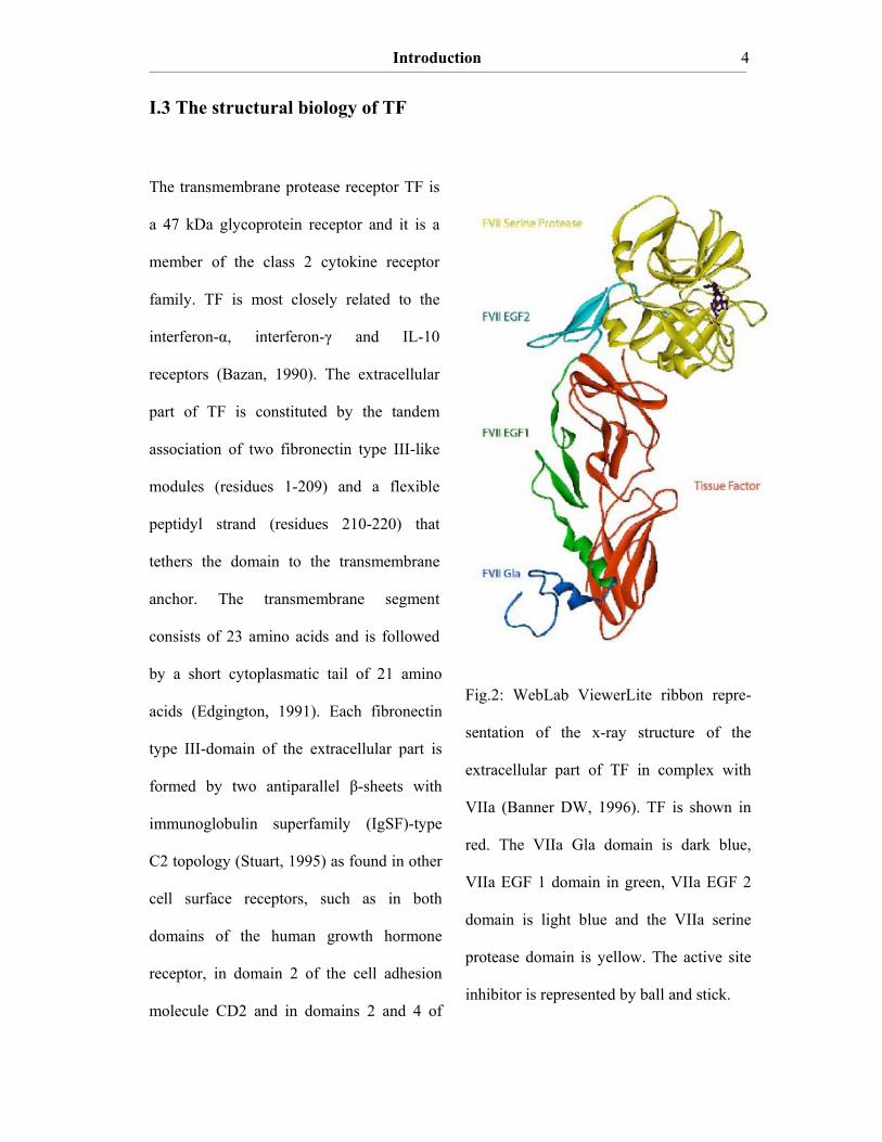
Fig.2: WebLab ViewerLite ribbon repre-
sentation of the x-ray structure of the
extracellular part of TF in complex with
VIIa (Banner DW, 1996). TF is shown in
red. The VIIa Gla domain is dark blue,
VIIa EGF 1 domain in green, VIIa EGF 2
domain is light blue and the VIIa serine
protease domain is yellow. The active site
inhibitor is represented by ball and stick.
I.3 The structural biology of TF
The transmembrane protease receptor TF is
a 47 kDa glycoprotein receptor and it is a
member of the class 2 cytokine receptor
family. TF is most closely related to the
interferon-α, interferon-γ and IL-10
receptors (Bazan, 1990). The extracellular
part of TF is constituted by the tandem
association of two fibronectin type III-like
modules (residues 1-209) and a flexible
peptidyl strand (residues 210-220) that
tethers the domain to the transmembrane
anchor. The transmembrane segment
consists of 23 amino acids and is followed
by a short cytoplasmatic tail of 21 amino
acids (Edgington, 1991). Each fibronectin
type III-domain of the extracellular part is
formed by two antiparallel β-sheets with
immunoglobulin superfamily (IgSF)-type
C2 topology (Stuart, 1995) as found in other
cell surface receptors, such as in both
domains of the human growth hormone
receptor, in domain 2 of the cell adhesion
molecule CD2 and in domains 2 and 4 of
TF
Introduction ____________________________________________________________________________________________________________________________________________________________________________________________________________________________
5
CD4. TF contains two disulfide bridges at positions 49-57 and 186-209 and one
cytoplasmatic half-cysteine at position 245 that is acylated by palmitic acid or stearic acid
(Bach RR, 1988). Human TF contains N-linked glycosylation sites at Asn 11, Asn 124 and
Asn 137. In contrast to the four helix bundle ligands of the interferon and IL-10 receptor,
TF binds the multidomain serine protease factor VII / VIIa with subnanomolar affinity and
acts as a cofactor (Fig.2). Cell surface protease cascades are triggered by the regulation of
protease receptors, such as the urokinase receptor of the fibrinolytic system (Ellis V, 1992)
and TF (Ruf W, 1994).
1.4 Tissue Factor Pathway Inhibitor-1 – the physiologic inhibitor of
the coagulation start
The 45 kDa glycoprotein TFPI is an important coagulation inhibitor, since it prevents the
initiation phase by forming a quarternary high affinity complex with TF / VIIa and Xa.
TFPI consists of a negatively charged N-terminus followed by three modules of Kunitz
domains and a positively charged C-terminus (Fig.3). Mechanistically, TFPI first binds
trace amounts of Xa (Ki = 4.4 nM) (Hackeng TM, 2006) and TFPI / Xa subsequently binds
to the initiator complex TF / VIIa. Xa is bound by Kunitz domain 2 of TFPI and VIIa binds
to Kunitz domain 1. Kunitz domain 3 is essential for the binding to cell surface
proteoglycans and to lipoproteins.
TFPI was found to be expressed by endothelial cells of the microvasculature,
megakaryocytes, platelets, monocytes and macrophages (Werling RW, 1993; Van der Logt,
1994). Inside the vasculature there are three different pools of TFPI that differ significantly
in their structure and in their inhibitory activity (Broze, 1994). About 85% of the total TFPI
amount is tightly bound to heparan sulfate-containing proteoglycans at the surface of
Introduction ____________________________________________________________________________________________________________________________________________________________________________________________________________________________
6
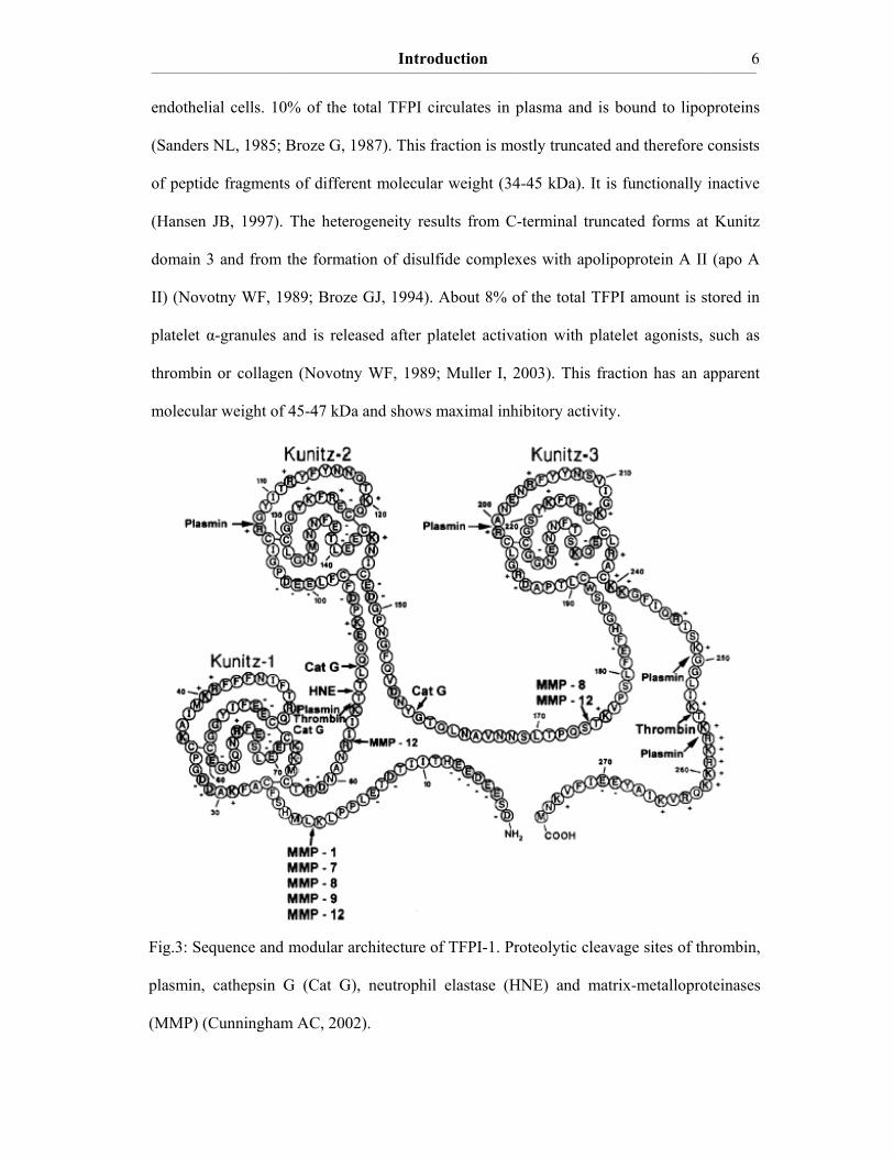
Fig.3: Sequence and modular architecture of TFPI-1. Proteolytic cleavage sites of thrombin,
plasmin, cathepsin G (Cat G), neutrophil elastase (HNE) and matrix-metalloproteinases
(MMP) (Cunningham AC, 2002).
endothelial cells. 10% of the total TFPI circulates in plasma and is bound to lipoproteins
(Sanders NL, 1985; Broze G, 1987). This fraction is mostly truncated and therefore consists
of peptide fragments of different molecular weight (34-45 kDa). It is functionally inactive
(Hansen JB, 1997). The heterogeneity results from C-terminal truncated forms at Kunitz
domain 3 and from the formation of disulfide complexes with apolipoprotein A II (apo A
II) (Novotny WF, 1989; Broze GJ, 1994). About 8% of the total TFPI amount is stored in
platelet α-granules and is released after platelet activation with platelet agonists, such as
thrombin or collagen (Novotny WF, 1989; Muller I, 2003). This fraction has an apparent
molecular weight of 45-47 kDa and shows maximal inhibitory activity.

Introduction ____________________________________________________________________________________________________________________________________________________________________________________________________________________________
7
I.5 Proteolytic cleavage of TFPI
In inflammation, wound healing and during infection stimulated human polymorphonuclear
neutrophils (PMN) and, to a lower extent also human peripheral blood monocytes, release
cationic serine proteases (neutrophil elastase (NE), proteinase 3 and cathepsin G (Cat G))
and matrix-metalloproteinases (MMP-1, MMP-2, MMP-3, MMP-8, MMP-9, MMP-10,
MMP-11) from their azurophilic granules that are capable of binding to the cell membrane
of neutrophils (Campbell EJ, 1989; Owen CA, 1995; 1997; 1999). It was found that cell
surface-bound NE is catalytically active and is resistant to inhibition by naturally occurring
protease inhibitors, such as the serpin α1-proteinase inhibitor (Owen CA, 1995).
In vitro studies have shown that the connecting regions between the Kunitz domains as well
as the acidic N-terminal and basic C-terminal regions of recombinant human TFPI are very
susceptible to limited proteolytic decomposition by NE, Cat G (Petersen LC, 1992; Higuchi
DA, 1992) and MMPs (Belaaouaj AA, 2000; Cunningham AC, 2002). The degradation of
TFPI by Cat G was found to be significantly slower than cleavage by NE (Higuchi DA,
1992). Serine proteases that are part of the coagulation cascade, such as thrombin (Ohkura
N, 1997), factor Xa (Salemink I, 1998) and plasmin (Li A, 1998) also cause limited
proteolysis of TFPI (cleavage sites Fig. 3). It was also found that the anticoagulant activity
of TFPI was greatly reduced by limited proteolysis. Therefore, proteolytic inactivation of
TFPI could be a mechanism capable of generating local procoagulant environments. These
findings may represent a regulatory link between innate immunity and the coagulation start,
since induction of coagulation accompanies the inflammatory response to a multitude of
stimuli (Esmon CT, 2004; Opal SM, 2003). It is not yet established which proteases are
responsible for TFPI decomposition in blood and if this degradation also occurs in the
cellular context. It is also unclear whether this mechanism is relevant for the initiation of
coagulation in vivo.
Introduction ____________________________________________________________________________________________________________________________________________________________________________________________________________________________
8
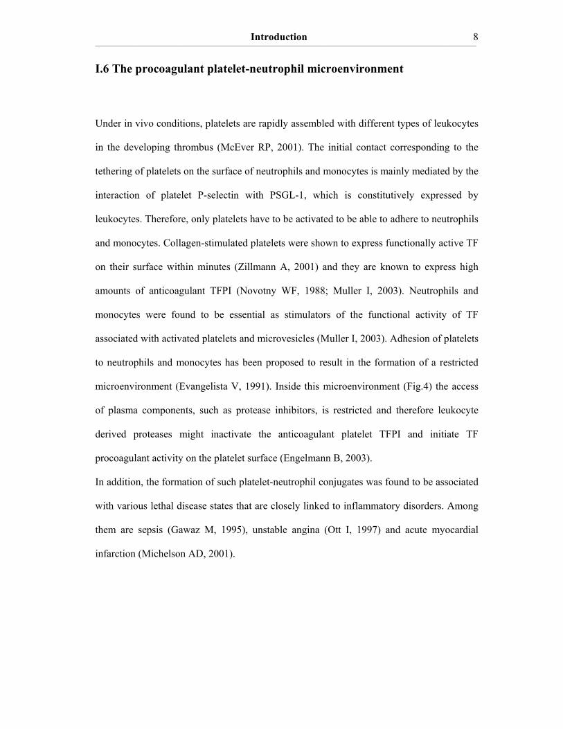
I.6 The procoagulant platelet-neutrophil microenvironment
Under in vivo conditions, platelets are rapidly assembled with different types of leukocytes
in the developing thrombus (McEver RP, 2001). The initial contact corresponding to the
tethering of platelets on the surface of neutrophils and monocytes is mainly mediated by the
interaction of platelet P-selectin with PSGL-1, which is constitutively expressed by
leukocytes. Therefore, only platelets have to be activated to be able to adhere to neutrophils
and monocytes. Collagen-stimulated platelets were shown to express functionally active TF
on their surface within minutes (Zillmann A, 2001) and they are known to express high
amounts of anticoagulant TFPI (Novotny WF, 1988; Muller I, 2003). Neutrophils and
monocytes were found to be essential as stimulators of the functional activity of TF
associated with activated platelets and microvesicles (Muller I, 2003). Adhesion of platelets
to neutrophils and monocytes has been proposed to result in the formation of a restricted
microenvironment (Evangelista V, 1991). Inside this microenvironment (Fig.4) the access
of plasma components, such as protease inhibitors, is restricted and therefore leukocyte
derived proteases might inactivate the anticoagulant platelet TFPI and initiate TF
procoagulant activity on the platelet surface (Engelmann B, 2003).
In addition, the formation of such platelet-neutrophil conjugates was found to be associated
with various lethal disease states that are closely linked to inflammatory disorders. Among
them are sepsis (Gawaz M, 1995), unstable angina (Ott I, 1997) and acute myocardial
infarction (Michelson AD, 2001).

Introduction ____________________________________________________________________________________________________________________________________________________________________________________________________________________________
9
Neutrophil
Platelet
P-selectinPSGL-1
proteases
activeinactive
Tissue FactorPathway Inhibitor
MVMV
MV
MV
COAGULATION
Fig.4: Cellular model for the intravascular tissue factor pathway modified according to
Engelmann et al., 2003. Platelet activation leads to the exposure of TF on the cell
surface, whereby the formation of the initiator complex of coagulation is enabled.
Concomitantly, TFPI is released from the platelet α-granules and inhibits the initiator
complex. Due to the concomitant presentation / activation of platelet adhesion molecules
(P-selectin) platelets are enabled to interact with neutrophils via P-selectin / PSGL-1-
interactions. Circulating and acutely shedded microvesicles (MV) are recruited to the
platelet-neutrophil conjugates. Secreted neutrophil proteases could inactivate TFPI.
Thereby, the functional activity of the TF associated with platelets and microvesicles
might be enhanced.
Introduction ____________________________________________________________________________________________________________________________________________________________________________________________________________________________
10
I.7 Cellular microparticles
Microparticles (microvesicles) are small membrane vesicles (< 1μm in diameter) that are
released from the plasma membrane of cells upon activation (Wiedmer T, 1991), during
apoptosis (Aupeix K, 1997) and by shear stress (Reininger AJ, 2006). They constitute a
heterogeneous population, differing in cellular origin, numbers, size, antigenic composition
and functional properties. Microparticles are described to play a role in intercellular
communication, immunity and coagulation (Hugel B, 2005). Microparticles support
coagulation by the exposure of negatively charged phospholipids (PS) that are essential for
thrombin generation and in the case of monocyte- and platelet-derived microparticles also
by the exposure of TF (Muller I, 2003). Under physiologic conditions, about 80% of the
plasma microparticles are derived from platelets (Berckmans RJ, 2001). The presence of
microparticles has also been documented at sites of inflammation, such as the acellular lipid
core of the atherosclerotic plaque (Mallat Z, 1999). Furthermore, increased numbers of
circulating microparticles have been reported in patients with acute coronary syndromes
(Mallat Z, 2000).
I.8 Tissue specific expression pattern of TF
TF is expressed in many tissues and it exhibits a distinct, nonuniform tissue specific pattern
of expression. High levels of TF are detected in highly vascularized organs, such as the
lung, brain and placenta (Fleck RA, 1990). Intermediate levels are found in the heart,
kidney, intestine, testes and uterus. In contrast, low levels of TF are observed in the liver,
spleen, skeletal muscle, and thymus. The cell types that express TF in these organs include

Introduction ____________________________________________________________________________________________________________________________________________________________________________________________________________________________
11
cardiomyocytes in the heart, bronchiolar and alveolar epithelial cells in the lung, astrocytes
in the brain, and trophoblasts in the placenta (Eddleston M, 1993; Erlich J, 1999; Pawlinski
R, 2002). The constitutive expression of TF in various tissues, such as the vasculature of
the heart, may reflect a need for additional hemostatic protection in these tissues. In
contrast, nonvital tissues that express low levels of TF, such as skeletal muscle, do not
require additional hemostatic protection. These tissues appear to rely more on the intrinsic
pathway of coagulation to maintain hemostasis. TF is constitutively expressed in the
vascular wall, such as by fibroblasts of the adventitia and by smooth muscle cells of the
media of arteries and veins. Endothelial cells probably do not express TF under physiologic
conditions (Østerud B, 2006). This findings led Drake and coworkers to propose the
popular concept of TF acting as a hemostatic “envelope” encapsulating the vascular bed.
Rupture of the integrity of the envelope would trigger the clotting process instantly (Drake
TA, 1989).
1.9 Intravascular TF
In recent years the envelope paradigm of TF expression and function has been challenged
by the demonstration of intravascular TF (blood-borne or circulating TF) (Giesen PLA,
1999; Zillmann A, 2001; Muller I, 2003; Engelmann B, 2003). Induced expression of TF in
cells within the vasculature is implicated in the pathogenesis of thrombosis in
atherosclerosis, disseminated intravascular coagulation, malignancy and hyperacute
rejection of xenografts (Wilcox JN, 1989; Levi M, 1999; Rickles FR, 2001; Robson SC,
1999) and it has been proposed that intravascular TF contributes to the propagation of the
growing thrombus (Giesen PLA, 1999).
Introduction ____________________________________________________________________________________________________________________________________________________________________________________________________________________________
12
TF de novo-synthesis in monocytes was first reported in 1975 by Rivers and coworkers
observing procoagulant activity in endotoxin-stimulated leukocyte suspensions (Rivers RP,
1975). TF expression on monocytes can be achieved by specific inflammatory stimuli, such
as endotoxin (e.g. lipopolysaccharide (LPS)) (Gregory SA, 1989), phorbol esters (Lyberg
T, 1981), C-reactive protein (Cermak J, 1993) and proinflammatory mediators, like tumor
necrosis factor-α (TNF-α) (Conkling PR, 1988) and interleukin 1-β (IL-1β) (Herbert JM,
1992). Interestingly, platelets were found to regulate monocyte TF activity. In 1974,
Niemetz and Marcus (Niemetz J, 1974) proposed that platelets enhance the procoagulant
activity of white blood cells. This was also confirmed in monocyte cell cultures, in which
isolated platelets added to monocytes enhanced LPS-induced TF activity (Lorenzet
R,1986). Increased expression levels of monocyte TF might play a role in sepsis (Drake
TA, 1993; Lupu C, 2005) and it was found that patients with unstable and stable coronary
syndromes exhibit elevated levels of TF expression on circulating monocytes (Leatham
EW, 1995).
In rapidly processed blood (to avoid the activation of TF gene transcription), TF was barely
noticeable in neutrophils by TF-specific ELISA measurements and no TF procoagulant
activity could be detected (Muller I, 2003). This observation is in accordance with the
findings of Østerud and coworkers, who failed to detect TF antigen on neutrophils in
stimulated whole blood (Østerud B, 2000). However, there is emerging evidence that
neutrophils might be able to express TF under certain inflammatory conditions (Maugeri N,
2006; Ritis K, 2006).
Blood eosinophils were found to store TF, which is mainly embodied in their specific
granules and exposed on their cell membrane after cell activation (Moosbauer C, 2006).
Eosinophils are the cells with the highest TF content in blood under resting conditions.
They contain approximately one forth of the TF molecules compared to fully activated

Introduction ____________________________________________________________________________________________________________________________________________________________________________________________________________________________
13
monocytes. The observations indicate TF as one of the critical mediators of the initial
eosinophil migration across the activated endothelium (Moosbauer C, 2006).
There is strong evidence that platelets contain preformed TF, which is released within 5
minutes after collagen type I stimulation. Platelet TF contributes to the collagen-triggered
activation of blood coagulation (Zillmann A, 2001). Immunoelectron microscopy showed
TF antigen localized in the α-granules and the open canalicular system of resting platelets.
The ability of activated platelets to trigger the initiation of coagulation was low. This
suggests that platelet TF is cryptic (Maynard JR, 1975). One reason for the low TF
procoagulant activity on activated platelets could be the concomitant release of TFPI, the
physiologic inhibitor of the initiator complex of coagulation (Novotny WF, 1988). The
presence of TF in platelets was confirmed by several authors (Camera M, 2003; Engelmann
B, 2006). It is still a matter of debate whether TF is transported to platelets by leukocyte-
derived microparticles (Del Conde I, 2005) and / or if the spliceosome of proplatelets that
extend from megakaryocytes might potentially be capable of translating TF from pre-
mRNAs (Denis MM, 2005; Schwertz H, 2006).
Microparticles support coagulation by exposure of negatively charged phospholipids that
are essential for thrombin generation. In the case of monocyte- and platelet-derived
microparticles their main, and probably central procoagulant function is the exposure of TF
(Muller I, 2003). Under physiologic conditions, 80% of the plasma microparticles are
derived from platelets (Berckmans RJ, 2001). TF was detected on platelet-derived
microparticles and in vitro generated platelet microparticles (Muller I, 2003). Apparently,
the filopodia of activated platelets are the preferential sites for the formation of TF-positive
microparticles (Leon C, 2004). In vitro generated monocyte-derived microparticles (Satta
N, 1994) and circulating monocyte-derived microparticles (Falati S, 2003) were shown to
expose TF on their membrane. Circulating monocyte microparticles are able to adhere to
activated endothelial cells and to activated platelets by P-selectin / PSGL-1 interactions and
Introduction ____________________________________________________________________________________________________________________________________________________________________________________________________________________________
14
interestingly they were found to play a significant role in fibrin stabilization of the nascent
thrombus by the delivery of procoagulant TF.
A substantial part of total soluble TF in plasma has been suggested to be constituted by an
alternatively spliced human TF (Bogdanov VY, 2003). However, the procoagulant activity
of soluble TF is rather low compared to full-length TF.
I.10 The encrypted or latent state of TF
TF encryption has been suggested as the post-translational suppression of TF procoagulant
activity on the cell surface (Bach RR, 2006). The discrepancy between TF antigen and the
expression of TF procoagulant activity has previously been observed in a variety of cell
types (Maynard JR, 1977; Walsh JD, 1991; Drake TA, 1989). A stimulus is required to
uncover the latent proteolytic activity of the encrypted TF-VIIa complex (Bach RR, 1990).
Until now several mechanisms were proposed to activate the encrypted TF: freezing and
thawing, sonication, protease treatment, phospholipase treatment, non-ionic detergents,
apoptosis, complement, and Ca2+-ionophores (Bach RR, 1996; 2006). There is a significant
variation among these methods with respect to the level of TF procoagulant activity evoked
as well as to secondary effects on cell structure.
The nature of TF de-encryption is unclear. One mechanism leading to de-encryption of
latent TF is the treatment of cells with Ca2+-ionophores. This leads to an increase in
cytosolic Ca2+ which in turn causes a disruption of PS asymmetry. PS is no longer
sequestered on the inner leaflet of the plasma membrane. This does not necessarily mean
that TF de-encryption is coupled to PS exposure, but it has been known for a long time that
PS accelerates coagulation reactions on membrane surfaces (Lentz BR, 2003). However,
Wolberg and coworkers discovered that Ca2+-ionophore treatment of cells induces changes
Introduction ____________________________________________________________________________________________________________________________________________________________________________________________________________________________
15
in TF procoagulant activity that could not fully be reduced to the basal level by saturating
concentrations of the PS-binding protein annexin V. This indicates that the increase in TF
activity after ionophore treatment does not solely result from increased PS exposure
(Wolberg AS, 1999).
It has been reported that after treatment of human pericytes with Ca2+-ionophore the TF
procoagulant activity increased but the prothrombinase complex assembly and function
were not affected. Therefore it is reasonable to assume that Ca2+-ionophore treatment does
not only result in membrane alterations, but may actuate intracellular processes that lead to
covalent modifications, dimerization, and/or conformational changes in the TF molecule to
increase its cofactor activity (Bouchard BA, 1997). Bach and Moldow suggested a
mechanism for the Ca2+-ionophore-induced TF de-encryption resulting in a change in TF
quarternary structure (Bach RR and Moldow CF, 1997). They propose that during de-
encryption of TF by Ca2+-ionophore inactive TF dimers are converted to procoagulant TF
monomers. This model runs counter to a well established dogma. Self-association usually
results in the activation of cell surface receptors. It also was demonstrated that TF
dimerization does not inhibit TF procoagulant activity, which contradicts the model
proposed by Bach and Moldow (Donate F, 2000).
Another model of TF de-encryption is based on the association of TF with distinct lateral
membrane domains. It was recently demonstrated that palmitoylation of cytoplasmic
cysteines can target integral membrane proteins to lipid rafts (Zacharias DA, 2002). TF is
such a palmitoylated integral membrane protein. Disruption of lipid rafts by methyl-β-
cyclodextrin extraction results in an increase in the basal expression of TF procoagulant
activity (Dietzen DJ, 2004). However, the meaning of this finding is unclear because there
is increasing evidence that cholesterol extraction impairs a cells’ ability to expose PS
(Kunzelmann-Marche C, 2002).
_________________
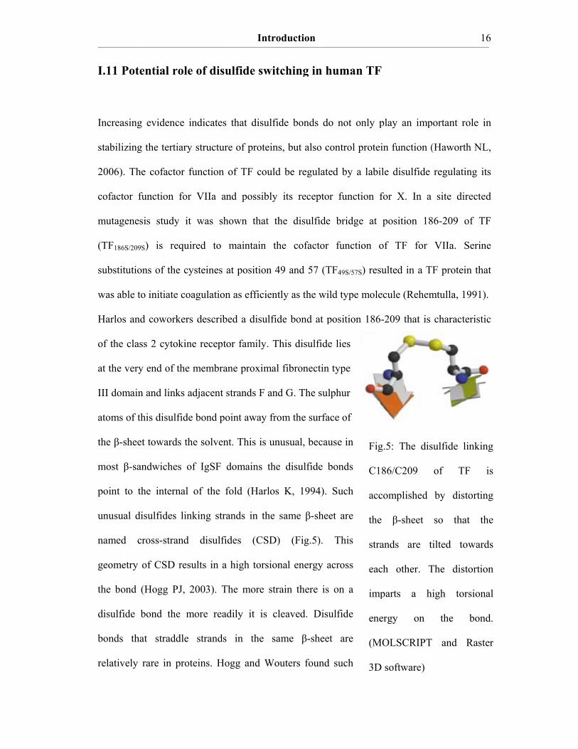
I.11 P
Increas
stabiliz
2006).
cofacto
mutage
(TF186S
substitu
was ab
Harlos
of the
at the v
III dom
atoms o
the β-s
most β
point t
unusua
named
geomet
the bon
disulfid
bonds
relative
_____________________________
Potential r
sing evidenc
zing the terti
The cofacto
or function
enesis study
S/209S) is re
utions of the
ble to initiate
and cowork
class 2 cyto
very end of t
main and link
of this disulf
sheet toward
β-sandwiche
to the intern
al disulfides
cross-stra
try of CSD
nd (Hogg P
de bond the
that stradd
ely rare in p
______________________________
role of disu
ce indicates
iary structur
or function
for VIIa an
y it was sh
equired to
e cysteines a
e coagulation
kers describe
okine recepto
the membran
ks adjacent s
fide bond po
ds the solven
es of IgSF d
nal of the f
linking stra
and disulfid
results in a
PJ, 2003). Th
e more read
dle strands
proteins. Ho
Intro_____________________________
ulfide swit
that disulfi
e of proteins
of TF could
nd possibly
hown that th
maintain th
at position 4
n as efficient
ed a disulfid
or family. T
ne proximal
strands F and
oint away fro
nt. This is un
domains the
fold (Harlos
ands in the
des (CSD)
high torsion
he more str
dily it is cl
in the sa
ogg and Wo
oduction ______________________________
tching in h
de bonds do
s, but also c
d be regulate
its receptor
he disulfide
he cofactor
49 and 57 (T
tly as the wil
de bond at p
his disulfide
l fibronectin
d G. The sul
om the surfa
nusual, becau
e disulfide b
K, 1994).
same β-shee
) (Fig.5).
nal energy a
rain there is
leaved. Disu
ame β-sheet
outers found
______________________________
Fig
C1
acc
the
stra
eac
imp
ene
(M
3D
human TF
o not only p
ontrol protei
ed by a labi
r function f
e bridge at
function o
TF49S/57S) res
ld type mole
position 186
e lies
type
lphur
ace of
use in
bonds
Such
et are
This
across
on a
ulfide
t are
such
______________________________
g.5: The dis
86/C209
complished
e β-sheet
ands are t
ch other. T
parts a h
ergy on
MOLSCRIPT
D software)
F
play an imp
in function (
ile disulfide
for X. In a
position 18
of TF for
sulted in a T
ecule (Rehem
-209 that is
______________________________1
sulfide linki
of TF
by distorti
so that t
tilted towar
The distorti
high torsion
the bon
T and Rast
portant role
(Haworth NL
regulating i
site directe
86-209 of T
VIIa. Serin
TF protein th
mtulla, 1991
characterist
____ 16
ing
is
ing
the
rds
ion
nal
nd.
ter
in
L,
its
ed
TF
ne
hat
).
tic
Introduction ____________________________________________________________________________________________________________________________________________________________________________________________________________________________
17
CSD in mammalian cell surface receptors (e.g. TF, thrombomodulin, growth hormone
receptor, erythropoietin receptor, interferon-γ receptor and interleukin receptors). These
authors propose that the function of some of these proteins might be controlled by cleavage
of their cross-strand disulfide bond. They suggest that CSD can be reduced or oxidized
reversibly by cellular oxidoreductases (e.g. protein disulfide isomerase, thioredoxin,
glutaredoxin). Mechanistically the action of these enzymes is characterized by a thiol-
disulfide exchange reaction. This was successfully established for the CD4 receptor on
CEM-T4 cells (a thymocyte-derived cell line) (Matthias LJ, 2003). However, regulation of
the TF procoagulant activity by a reversible, oxidoreductase-mediated cleavage of the CSD
at position 186-209 has not yet been thoroughly investigated. Regulation of TF by such a
disulfide switch might be of major interest in nearly all branches of clinical medicine, since
many pathologies are related to coagulation disorders and venous and arterial thrombosis
are the leading causes of mortality in industrialized countries.
I.12 Aims of the investigation
Increasing evidence indicates an important role of intravascular TF in the pathogenesis of
lethal diseases, such as disseminated intravascular coagulation (DIC), arterial and venous
thrombosis, acute myocardial infarction (AMI) and stroke. Blood-borne TF was detected on
stimulated monocytes, activated platelets and their microparticles. Most of the TF
molecules present on blood cells however, are not functionally active (encrypted or latent
state of TF). Therefore it is of major interest to characterize on a molecular level why TF is
cryptic and to reveal the underlying mechanisms that activate TF.
Introduction ____________________________________________________________________________________________________________________________________________________________________________________________________________________________
18
In the present study the following central questions were addressed:
1. Do isolated platelets exhibit TF procoagulant activity after activation?
2. Is the initiation of intravascular coagulation triggered by the proteolytic
decomposition of platelet TFPI by neutrophil surface proteases?
3. Is there an intramolecular disulfide switch in the TF molecule triggering its
procoagulant activity?
4. Which oxidoreductases are capable of regulating this thiol-disulfide exchange?
Materials and Methods ____________________________________________________________________________________________________________________________________________________________________________________________________________________________
19
II. Materials and Methods
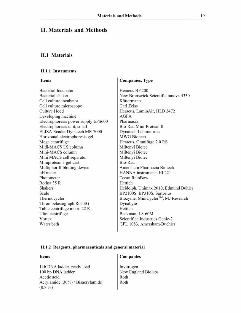
II.1 Materials II.1.1 Instruments
Items Bacterial Incubator Bacterial shaker Cell culture incubator Cell culture microscope Culture Hood Developing machine Electrophoresis power supply EPS600 Electrophoresis unit, small ELISA Reader Dynatech MR 7000 Horizontal electrophoresis gel Mega centrifuge Midi-MACS LS column Mini-MACS column Mini MACS cell separator Miniprotean 3 gel cast Multiphor II blotting device pH meter Photometer Rotina 35 R Shakers Scale Thermocycler Thrombelastograph RoTEG Table centrifuge mikro 22 R Ultra centrifuge Vortex Water bath
Companies, Type Heraeus B 6200 New Brunswick Scientific innova 4330 Köttermann Carl Zeiss Heraeus, LaminAir, HLB 2472 AGFA Pharmacia Bio-Rad Mini-Protean II Dynatech Laboratories MWG Biotech Heraeus, Omnifuge 2.0 RS Miltenyi Biotec Miltenyi Biotec Miltenyi Biotec Bio-Rad Amersham Pharmacia Biotech HANNA instruments HI 221 Tecan RainBow Hettich Heidolph, Unimax 2010, Edmund Bühler BP2100S, BP310S, Sartorius Biozyme, MiniCyclerTM, MJ Research Dynabyte Hettich Beckman, L8-60M Scientificc Industries Genie-2 GFL 1083, Amersham-Buchler
II.1.2 Reagents, pharmaceuticals and general material
Items 1kb DNA ladder, ready load 100 bp DNA ladder Acetic acid Acrylamide (30%) / Bisacrylamide (0.8 %)
Companies Invitrogen New England Biolabs Roth Roth
Materials and Methods ____________________________________________________________________________________________________________________________________________________________________________________________________________________________
20
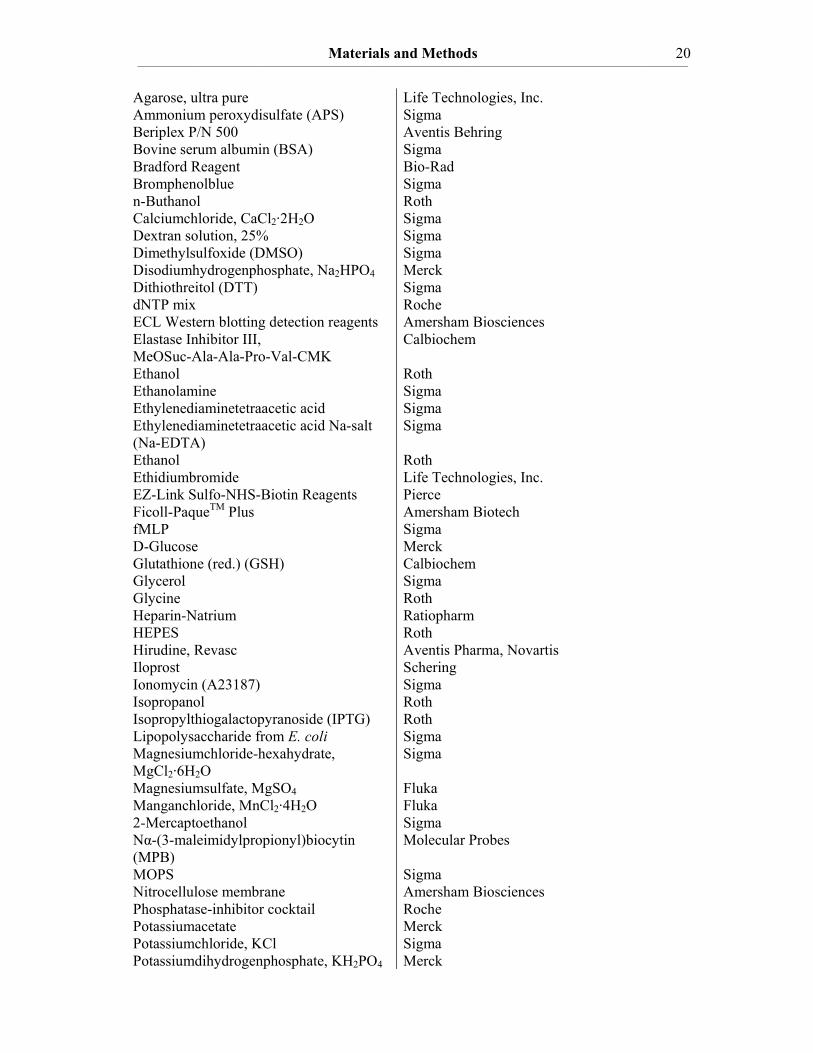
Agarose, ultra pure Ammonium peroxydisulfate (APS) Beriplex P/N 500 Bovine serum albumin (BSA) Bradford Reagent Bromphenolblue n-Buthanol Calciumchloride, CaCl2·2H2O Dextran solution, 25% Dimethylsulfoxide (DMSO) Disodiumhydrogenphosphate, Na2HPO4 Dithiothreitol (DTT) dNTP mix ECL Western blotting detection reagents Elastase Inhibitor III, MeOSuc-Ala-Ala-Pro-Val-CMK Ethanol Ethanolamine Ethylenediaminetetraacetic acid Ethylenediaminetetraacetic acid Na-salt (Na-EDTA) Ethanol Ethidiumbromide EZ-Link Sulfo-NHS-Biotin Reagents Ficoll-PaqueTM Plus fMLP D-Glucose Glutathione (red.) (GSH) Glycerol Glycine Heparin-Natrium HEPES Hirudine, Revasc Iloprost Ionomycin (A23187) Isopropanol Isopropylthiogalactopyranoside (IPTG) Lipopolysaccharide from E. coli Magnesiumchloride-hexahydrate, MgCl2·6H2O Magnesiumsulfate, MgSO4 Manganchloride, MnCl2·4H2O 2-Mercaptoethanol Nα-(3-maleimidylpropionyl)biocytin (MPB) MOPS Nitrocellulose membrane Phosphatase-inhibitor cocktail Potassiumacetate Potassiumchloride, KCl Potassiumdihydrogenphosphate, KH2PO4
Life Technologies, Inc. Sigma Aventis Behring Sigma Bio-Rad Sigma Roth Sigma Sigma Sigma Merck Sigma Roche Amersham Biosciences Calbiochem Roth Sigma Sigma Sigma Roth Life Technologies, Inc. Pierce Amersham Biotech Sigma Merck Calbiochem Sigma Roth Ratiopharm Roth Aventis Pharma, Novartis Schering Sigma Roth Roth Sigma Sigma Fluka Fluka Sigma Molecular Probes Sigma Amersham Biosciences Roche Merck Sigma Merck

Materials and Methods ____________________________________________________________________________________________________________________________________________________________________________________________________________________________
21
Protease-inhibitor cocktail Protein marker, Page Ruler Rubidiumchloride Sodiumacetate hexahydrate Tri-Sodiumcitrate SDS ultra pure S2222, chromogenic substrate Sodiumhydrogencarbonate, NaHCO3 Sodiumdihydrogenphosphate, NaH2PO4 Sodiumhydroxide, NaOH Streptavidin-agarose beads TEG caps TEMED Thromborel S Tris-(hydroxymethyl)-aminomethane (Tris-base) Triton-X100 Tween 20 Whatman 3MM Papier X-ray film Zeba Desalt Spin Columns
Roche Fermentas Fluka Merck Roth Roth Haemochrom Diagnostica Sigma Merck Sigma Sigma ROTEM Roth Dade Behring Roth Sigma Sigma Schleicher & Schuell Fuji Pierce
II.1.3 Cell culture materials
Items Ampicillin Bacto-Agar Culture flasks FuGene 6 Transfection Reagent Fetal bovine serum Kanamycin MEM-alpha medium Pemicillin/Streptomycin (100x) Trypanblue Trypsin/EDTA Yeast extract
Companies Sigma Roth Falcon Roche Invitrogen-Gibco Sigma Invitrogen-Gibco Invitrogen-Gibco Sigma Invitrogen-Gibco Life Technologies Inc.
II.1.4 Enzymes and Proteins
Items Annexin V, recombinant Apyrase grade VII (from potato) Cathepsin G from human leukocytes Chondroitinase ABC, Proteus vulgaris Collagen, Type I Corn Trypsin Inhibitor DNase I
Companies BD Biosciences Pharmingen Sigma Sigma Calbiochem Horm, Nycomed Calbiochem Sigma

Materials and Methods ____________________________________________________________________________________________________________________________________________________________________________________________________________________________
22
Factor X, human Glutaredoxin, recombinant, E. coli Human Neutrophil Elastase Pfu Taq Polymerase Protein-Disulfide Isomerase (PDI) Ribonuclease I, E. coli Sal I restriction endonuclease Soluble Tissue Factor 1-219 Streptavidin, horseradish peroxidase conjutated Streptavidin-Agarose, Streptomyces avidinii T4 DNA Ligase Thioredoxin, recombinant, E. coli α-Thrombin Xba I restriction endonuclease
Haemochrom Diagnostica Calbiochem Calbiochem Stratagene Sigma Fermentas New England Biolabs Kindly provided by PD Dr. Victor Magdolen, Klinikum rechts der Isar, Technische Universität München Pierce Sigma New England Biolabs Calbiochem Sigma Fermentas
II.1.5 Antibodies Items Anti-human CD14 magnetic MicroBeads Anti-human CD15 magnetic MicroBeads Goat anti-human TFPI (C-20) polyclonal antibody Goat anti-mouse IgG, horseradish peroxidase conjugated Mouse Anti-Glutathione monoclonal antibody Mouse anti-goat IgG, horseradish peroxidase conjugated Mouse anti-human Tissue Factor-VIC7 monoclonal antibody
Antigen CD14 CD15 TFPI Mouse IgG, carboxyterm. Glutathione Goat IgG, carboxyterm. TF
References Miltenyi Biotec Miltenyi Biotec Santa Cruz (sc-18713) Calbiochem (#401253) Virogen (101-A) Santa Cruz (sc-2354) Dr. rer. nat. Sybille Albrecht, Pathologisches Institut, Technische Universität Dresden
Materials and Methods ____________________________________________________________________________________________________________________________________________________________________________________________________________________________
23
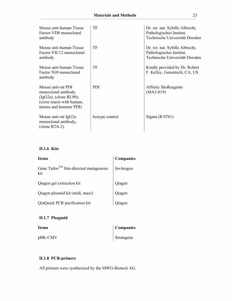
Mouse anti-human Tissue Factor-VD8 monoclonal antibody Mouse anti-human Tissue Factor-VIC12 monoclonal antibody Mouse anti-human Tissue Factor 5G9 monoclonal antibody Mouse anti-rat PDI monoclonal antibody (IgG2a), (clone RL90), (cross reacts with human, mouse and hamster PDI) Mouse anti-rat IgG2a monoclonal antibody, (clone R2A-2)
TF TF TF PDI Isotype control
Dr. rer. nat. Sybille Albrecht, Pathologisches Institut, Technische Universität Dresden Dr. rer. nat. Sybille Albrecht, Pathologisches Institut, Technische Universität Dresden Kindly provided by Dr. Robert F. Kelley, Genentech, CA, US Affinity BioReagents (MA3-019) Sigma (R 0761)
II.1.6 Kits
Items Gene TailorTM Site-directed mutagenesis kit Qiagen gel extraction kit Qiagen plasmid kit (midi, maxi) QiaQuick PCR purification kit
Companies Invitrogen Qiagen Qiagen Qiagen
II.1.7 Phagmid
Items pBK-CMV
Companies Stratagene
II.1.8 PCR-primers All primers were synthesized by the MWG-Biotech AG.
Materials and Methods ____________________________________________________________________________________________________________________________________________________________________________________________________________________________
24
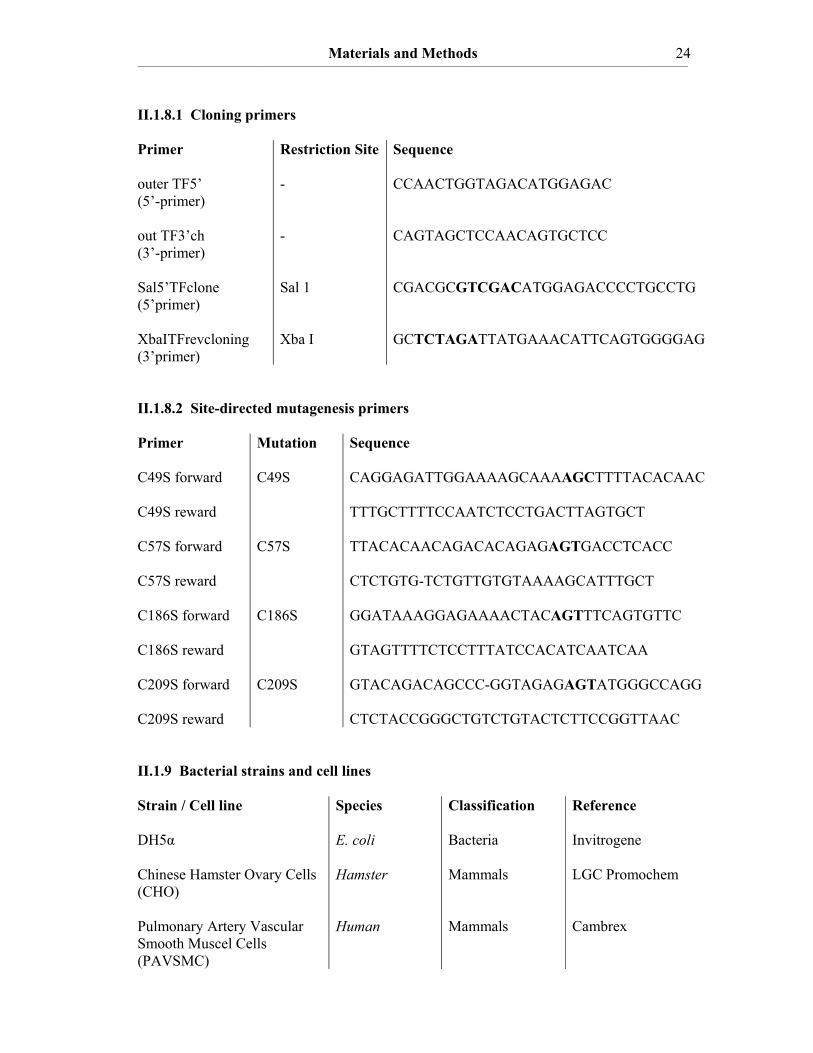
II.1.8.1 Cloning primers Primer outer TF5’ (5’-primer) out TF3’ch (3’-primer) Sal5’TFclone (5’primer) XbaITFrevcloning (3’primer)
Restriction Site - - Sal 1 Xba I
Sequence CCAACTGGTAGACATGGAGAC CAGTAGCTCCAACAGTGCTCC CGACGCGTCGACATGGAGACCCCTGCCTG GCTCTAGATTATGAAACATTCAGTGGGGAG
II.1.8.2 Site-directed mutagenesis primers Primer C49S forward C49S reward C57S forward C57S reward C186S forward C186S reward C209S forward C209S reward
Mutation C49S C57S C186S C209S
Sequence CAGGAGATTGGAAAAGCAAAAGCTTTTACACAAC TTTGCTTTTCCAATCTCCTGACTTAGTGCT TTACACAACAGACACAGAGAGTGACCTCACC CTCTGTG-TCTGTTGTGTAAAAGCATTTGCT GGATAAAGGAGAAAACTACAGTTTCAGTGTTC GTAGTTTTCTCCTTTATCCACATCAATCAA GTACAGACAGCCC-GGTAGAGAGTATGGGCCAGG CTCTACCGGGCTGTCTGTACTCTTCCGGTTAAC
II.1.9 Bacterial strains and cell lines Strain / Cell line DH5α Chinese Hamster Ovary Cells (CHO) Pulmonary Artery Vascular Smooth Muscel Cells (PAVSMC)
Species E. coli Hamster Human
Classification Bacteria Mammals Mammals
Reference Invitrogene LGC Promochem Cambrex

Materials and Methods ____________________________________________________________________________________________________________________________________________________________________________________________________________________________
25
II.1.10 Bacterial and cell culture media Freeze Medium for CHO cells 40% MEM-α 50% FBS 10% DMSO Luria-Bertani (LB) Medium 10 g/l Trypton 5 g/l Yeast extract 10 g/l NaCl pH 7.5 For LB-Agar plates add 1.5% (w/v) of Bacto-Agar. Minimum Essential Medium (MEM) alpha (for CHO cells) For formulation see Gibco-Invitrogen Psi broth medium 20 g/l Trypton 5 g/l Yeast extract 5 g/l Magnesiumsulfate pH 7.5 RPMI-1640 Medium (for monocytes and monocytic cell lines) See Gibco-Invitrogen II.1.11 Buffers and solutions Antibody buffer 0.13% Na-EDTA 0.15% BSA Dissolved in PBS Blocking buffer 5% BSA in TBS/T Blotting buffer 150 mM Glycine 20 mM Tris 0.1% SDS 20% Methanol Buffer P1 50 mM Tris-HCl, pH 8.0 10 mM EDTA 10 mg/ml RNase A Buffer P2 10% SDS 200 mM NaOH

Materials and Methods ____________________________________________________________________________________________________________________________________________________________________________________________________________________________
26
Buffer P3 3 M Potassium acetate, pH 5.5 Buffer QBT 15% Ethanol 0.15% Triton X-100 Buffer QC 2.0 M NaCl 50 mM MOPS, pH 7.0 15% Ethanol Buffer QF 1.25 mM NaCl 50 mM Tris-HCl, pH 8.5 15 % Ethanol Ca2+/Hepes 10 mM Hepes, pH = 7.4 100 mM CaCl2 Cell lysis buffer 50 mM Tris, pH 8.0 150 mM NaCl 5 mM EDTA 1% Triton X-100 10 x DNA-Gel Loading Buffer 40% (w/v) saccharose 0.25% bromphenolblue 0.25% xylencyanol, use as 1x solution EDTA buffer 50 mM Tris-HCl 20 mM EDTA 1 mg/ml BSA Gel buffer (500 ml) 3 M Tris-HCl, pH 8.45 0.3 % SDS (dissolve in 300 ml and adjust to pH 8.45 with HCl) Hank’s Balanced Salt Solution (HBSS) 0.4 mM KH2PO4 0.6 mM MgSO4 5.4 mM KCl 1.3 mM CaCl2·2H2O 0.5 mM MgCl2·6H2O 5.6 mM α-D-Glucose 0.3 mM Na2HPO4 137 mM NaCl

Materials and Methods ____________________________________________________________________________________________________________________________________________________________________________________________________________________________
27
4.2 mM NaHCO3 pH 7.4 Phosphate-Buffered Saline (PBS) 136 mM NaCl 2,6 mM KCl 10 mM NaH2PO4 1.5 mM KH2PO4, pH 7.4 PBS / EDTA 100 ml PBS (10x), pH 7.4 1 ml Na-EDTA (0.5 M stock, pH 8.0) 900 ml ddH2O Resuspension buffer 138 mM NaCl 2.7 mM KCl 12 mM NaHCO3 0.4 mM NaH2PO4 1 mM MgCl2·6H2O 5 mM D-Glucose 5 mM Hepes, pH 7.35 Running buffer (for SDS-PAGE) 25 mM Tris 250 mM Glycine 0.1% SDS 4x SDS-loading buffer (for SDS-PAGE) (Laemmli buffer) 10 ml 1M Tris-HCl, pH 6.8 23 ml 10% Glycerol (87%) 10 ml 10% (w/v) SDS 2 ml 2-Mercaptoethanol 4 ml 0.5 % Bromphenolblue Separating buffer (4x) 75.0 ml 2 M Tris-HCl, pH 8.8 4.0 ml 10 % (w/v) SDS 21.0 ml ddH2O Transformation buffer I (Tfb I) 0.588 g 30 mM Potassiumacetate, pH 5.8, adjusted with acetic acid 2.42 g 100 mM Rubidiumchloride 0.294 g 10 mM Calciumchloride 2.0 g 50 mM MnCl2·4H2O 30 ml 15% (v/v) Glycerol 200 ml total volume
Materials and Methods ____________________________________________________________________________________________________________________________________________________________________________________________________________________________
28
Transformation buffer II (Tfb II) 0.21 g 10 mM MOPS, pH 6.5, adjusted with NaOH 1.1 g 75 mM Calciumchloride 0.121 g 10 mM Rubidiumchloride 15 ml 15% (v/v) Glycerol 100 ml total volume 1x Tris-Acetate-EDTA (TAE) 40 mM Tris-HCl 40 mM Acetic acid 2 mM EDTA, pH 7.8 10x Tris-Buffered Saline (TBS) 400 mM Tris-HCl 1.37 M NaCl TBS/T 1x TBS + 0.1% Tween 20 II.2 Methods
II.2.1 Cell isolation techniques
II.2.1.1 Blood recovery
Venous blood was obtained from healthy donors (age 18-35 years), who did not take drugs
acting on the coagulation system for at least 14 days. The blood was anticoagulated with
tri-sodiumcitrate, hirudine or heparine-sodium, respectively. All experiments performed
with human blood were approved by the local ethic commission.
II.2.1.2 Isolation of platelets
Blood obtained from healthy donors (anticoagulated by tri-sodiumcitrate, 12.5 mM) was
centrifuged in 10 ml centrifuge tubes at 1300 rpm for 15 minutes at 24°C. 2 ml platelet-rich
plasma (PRP) were aspirated from each centrifuge tube and filled into a separate centrifuge
tube. Apyrase grade VII (0.475 U/ml) and iloprost (10 ng/ml) were added to each 2 ml
Materials and Methods ____________________________________________________________________________________________________________________________________________________________________________________________________________________________
29
sample of PRP and the tubes containing PRP were centrifuged at 1300 rpm for 10 minutes
at 24°C. The supernatant was discarded and the platelet pellet was resuspended in
resuspension buffer.
II.2.1.3 Preparation of platelet supernatant
1 x 10¹⁰ isolated platelets per ml resuspension buffer were stimulated with 0.1 U/ml
thrombin and 8 µg/ml type I collagen for 30 minutes at 37°C. The stimulated platelets were
centrifuged at 12600 rpm for 30 min at 24°C and the pellet was separated from the
supernatant.
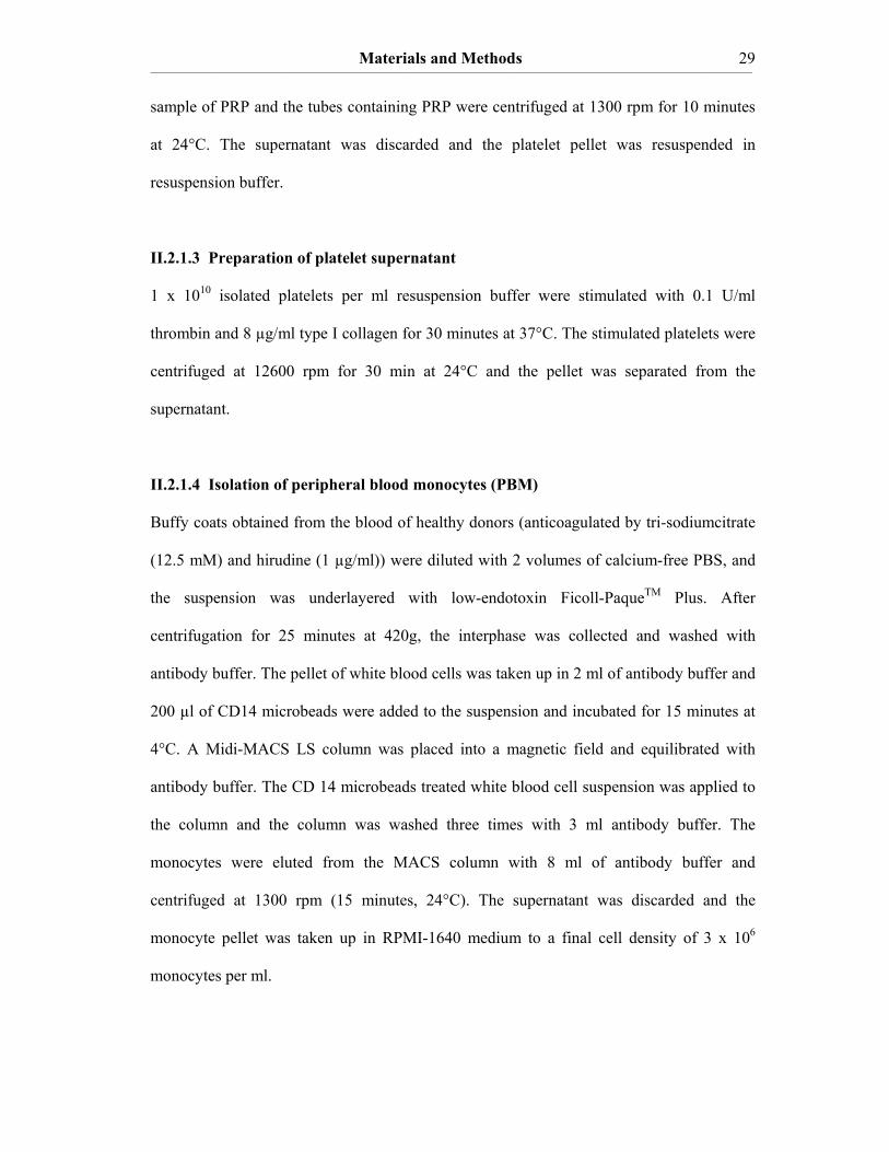
II.2.1.4 Isolation of peripheral blood monocytes (PBM)
Buffy coats obtained from the blood of healthy donors (anticoagulated by tri-sodiumcitrate
(12.5 mM) and hirudine (1 µg/ml)) were diluted with 2 volumes of calcium-free PBS, and
the suspension was underlayered with low-endotoxin Ficoll-PaqueTM Plus. After
centrifugation for 25 minutes at 420g, the interphase was collected and washed with
antibody buffer. The pellet of white blood cells was taken up in 2 ml of antibody buffer and
200 µl of CD14 microbeads were added to the suspension and incubated for 15 minutes at
4°C. A Midi-MACS LS column was placed into a magnetic field and equilibrated with
antibody buffer. The CD 14 microbeads treated white blood cell suspension was applied to
the column and the column was washed three times with 3 ml antibody buffer. The
monocytes were eluted from the MACS column with 8 ml of antibody buffer and
centrifuged at 1300 rpm (15 minutes, 24°C). The supernatant was discarded and the
monocyte pellet was taken up in RPMI-1640 medium to a final cell density of 3 x 10⁶
monocytes per ml.

Materials and Methods ____________________________________________________________________________________________________________________________________________________________________________________________________________________________
30
II.2.1.5 Isolation of polymorphonuclear neutrophils (PMN)
Blood was anticoagulated with Na-heparin (10 µl/ml) or tri-sodiumcitrate (12.5 mM) and
inverted twice. Whole blood was added to 3% dextrane (in HBSS) in a ratio of 2 to 1. The
cups were inverted twice and the separation of red blood cells and serum, containing other
blood cells occurred in about 30 minutes at room temperature (RT). After separation the
supernatant was removed with a sterile plastic Pasteur pipette and it was layered on top of 7
ml low-endotoxin Ficoll-PaqueTM Plus in a 15 ml Falcon. The Falcons were spun for 30
minutes at 1200 rpm and RT. The supernatant was removed and the pellet was resuspended
in 1 ml of RPMI-1640 medium. All suspensions were pooled into a new 15 ml Falcon,
which was filled up to 10 ml RPMI-1640 medium. Then it was centrifuged for 10 minutes
at 1000 rpm and RT. The supernatant was removed and the pellet was resuspended in 1 ml
pyrogen-free water for hypertonic lysis of residual red blood cells. After 20 to 40 s of gentle
resuspension, 10 ml HBSS were added and subsequently the cells were centrifuged for 10
minutes at 1000 rpm at RT. If residual red blood cells were present, the same step was
repeated several times.
Alternatively another PMN isolation method was applied. Blood was anticoagulated with
tri-sodiumcitrate (12.5 mM) and centrifuged for 15 minutes at 1300 rpm and RT. The PRP
was removed and the buffy coat (interphase of white blood cells) was transferred to a new
centrifuge tube. After centrifugation at 1300 rpm for 10 minutes at RT the layer of white
blood cells was transferred to a Falcon tube and filled up with antibody buffer to 2 ml of
total volume. Per 2 ml of white blood cells 200 µl of anti-human CD15 magnetic
MicroBeads were added and incubated for 15 min at 4°C. The Mini-MACS columns were
equilibrated with at least 2 ml of antibody buffer and the samples of white blood cells were
diluted by adding 600 µl of antibody buffer. After applying the samples onto the columns
the columns were washed fourfold with 500 µl of antibody buffer to remove all other blood
cells. Subsequently the columns were removed from the magnetic field and the PMN were
Materials and Methods ____________________________________________________________________________________________________________________________________________________________________________________________________________________________
31
eluted by applying 3 x 2 ml of antibody buffer. The PMN were pelleted by spinning the
Falcon tube at 1000 rpm for 10 minutes at RT and resuspended in 1 ml of resuspension
buffer.
II.2.1.6 Stimulation of isolated blood cells
For inducing TF expression of isolated monocytes 3 x 10⁶ monocytes per ml RPMI-1640
medium were stimulated with 10 ng/ml LPS for 5 hours at 37°C. For monocyte
microparticle formation 3 x 10⁶ isolated monocytes per ml resuspension buffer were
stimulated with 10 µg/ml LPS for 16 hours at 37°C.
For the formation of neutrophil microparticles 3 x 10⁶ PMN per ml resuspension buffer
were stimulated with 100 nM fMLP for 2 hours at 37°C.
For immunoblots 3 x 10⁸ platelets were stimulated with 0.1 units/ml thrombin and 12 µg/ml
collagen type 1. For functional assays platelets were stimulated with 12 µg/ml collagen or 3
µM A23187 Ca2+-ionophore in 2 mM Ca2+-containing resuspension buffer.
TF expression in PAVSMC was induced by treatment of the cells with 1 unit/ml thrombin
for 4 hours at 37°C.
II.2.1.7 Isolation of microparticles derived from stimulated blood cells
After stimulation of isolated blood cells (platelets, neutrophils, monocytes), the samples
were centrifuged for 15 minutes at 4500 rpm and RT. 250 µl of the obtained supernatant
was filled in each Eppendorf-tube and it was centrifuged for 30 min at 12600 rpm and
24°C. Afterwards, 225 µl of the supernatant were removed and 25 µl were left on the
bottom of the tubes. Subsequently, the microparticles of each tube were washed with 225 µl
PBS, vortexed and again centrifuged for 30 min at 12600 rpm and 24°C. After the
centrifugation, all the supernatant was removed and the microparticle pellet of each tube
was resuspended in 25 µl resuspension buffer.

Materials and Methods ____________________________________________________________________________________________________________________________________________________________________________________________________________________________
32
II.2.2 Cell culture techniques
II.2.2.1 Bacterial cell cultures
Transformed DH5α-bacteria were selected on LB plates with ampicillin (100 µg/ml) or
kanamycin (50 µg/ml) for 24 hours. For the preparation of overnight mini-cultures one
colony was picked and inoculated in LB medium with the appropriate antibiotic and shaken
overnight at 37°C. The overnight mini-culture was then used to prepare glycerol stocks or
to isolate and purify plasmid DNA for the transfection of eukaryotic cells. For the storage
of transformed bacteria a glycerol stock was prepared by growing the bacteria to an OD of
0.8 at 600 nm. Then 500 µl of the bacterial culture was added to 500 µl of 80% glycerol
and mixed thoroughly. The stocks were immediately frozen at -80°C.
II.2.2.2 Preparation of competent DH5α-cells
A 20 ml pre-culture was grown in LB medium overnight at 37°C and 180 rpm. The next
day 1 ml from the pre-culture was inoculated in 100 ml of psi broth medium and cultivated
at 37°C and 180 rpm to an OD of 0.5 at 600 nm. The culture was put on ice for 15 minutes.
The cells were pellet at 5000 x g for 5 minutes and the supernatant was removed. The pellet
was resuspended in 40 ml of Tfb I per 100 ml of bacterial cell culture and again kept on ice
for 15 minutes, followed by centrifugation at 5000 g for 5 minutes. The supernatant was
removed the bacterial cell pellet was resuspended in 4 ml Tfb II per 100 ml of culture and
put on ice for 15 minutes. Subsequently 50 µl aliquots were prepared in 1.5 ml tubes and
immediately frozen in liquid nitrogen and kept in –80°C.
II.2.2.3 Transformation of competent bacteria
The competent bacteria were thawed on ice. About 40 ng of ligated DNA or purified
phagmid DNA were added to 50 µl of competent cells, mixed carefully and kept on ice for
Materials and Methods ____________________________________________________________________________________________________________________________________________________________________________________________________________________________
33
additional 20 minutes. The bacteria were heat shocked at 42°C for 90 seconds, then 1 ml of
LB medium was added and the transformation samples were shaken at 37°C for 30
minutes. A selection of transformed bacteria was achieved by plating the bacterial
suspensions onto antibiotic containing agar plates. Only these bacteria which had taken up
the plasmid, containing an antibiotic resistance cassette, were able to grow on the plate.
II.2.2.4 Cultivation of Chinese Hamster Ovary cells
The Chinese Hamster Ovary (CHO) cells were cultured in 75 cm2 cell culture flasks
containing MEM-α medium supplemented with 100 µl penicillin/streptomycin (100x)
mingled with 10% fetal bovine serum (FBS) at 5% CO2 (37°C). The cells were cultured
until a confluence of approximately 90% was achieved. The cells were washed with 5 ml of
PBS and released from their culture flask by adding 2 ml of PBS/EDTA and subsequent
incubation for 20 minutes at 37°C. Freezing cultures were prepared by spinning the cells at
1000 rpm for 15 minutes and resuspending them in 1 ml of freeze medium for CHO cells
per flask of confluent cells. Aliquots were set into an ethanol-filled container and frozen
gradually at –80°C.
II.2.2.5 Transfection of Chinese Hamster Ovary cells
One day before transfection, the cells were treated with trypsin/EDTA and split into a new
culture flask to become 80% confluent. For each culture flask to be transfected, 36 µl of
FuGene 6 transfection reagent was diluted into serum free MEM-α medium. Subsequently
15 µg of DNA were added. The transfection mixture was prepared in such a way that the
total volume was 800 µl and it was incubated for 45 minutes. Then the transfection mixture
was added dropwise into each of the cell cultures and they were mixed thoroughly. Finally
the transfected cell cultures were incubated for 16 hours at 37°C. During that time the
protein expression from the transfected plasmid should proceed.

Materials and Methods ____________________________________________________________________________________________________________________________________________________________________________________________________________________________
34
II.2.3 DNA techniques
II.2.3.1 Electrophoresis of DNA on agarose gels
Double stranded DNA fragments can be separated according to their length on agarose gels.
Agarose was added to 1 x TAE to a final concentration of 0.7-2.0%. The mixture was
boiled in the microwave until the agarose was completely molten. The agarose was cooled
down to about 50°C before ethidium bromide was added to a concentration of 5 µg/ml and
poured into the gel tray. DNA gel loading buffer was added to the samples and they were
applied to the gel. Electrophoresis was performed in 1 x TAE at 3-8 V/cm. The DNA
fragments were visualized in the gel by UV-light.
II.2.3.2 Isolation of DNA from agarose gels (Qiagen gel extraction kit)
This protocol was designed for the extraction of DNA fragments from 0.7-2.0% standard
agarose gels in TAE or TBE buffer. DNA molecules were adsorbed to Qiagen silica
columns. All non-nucleic acid impurities, such as agarose, proteins, salts and ethidium
bromide were removed during the washing steps. The desired DNA band was excised from
the gel under the UV-light. The gel slice was weighed and 5 volumes of buffer QG were
added to one volume of gel for DNA fragments from 100 bp to 4 kb, for DNA fragments >
4 kb, 2 volumes of buffer QG plus 2 volumes of ddH2O were added, and then incubated for
10 minutes at 50 °C to solubilize the agarose. The solubilized agarose was resuspended by
vortexing and the sample was applied to the Qiagen silica columns to bind DNA. The
sample was centrifuged for 30 seconds, then the column was washed with 500 µl of buffer
QG and subsequently twice with buffer PE. Thereafter it was centrifuged for additional 30
seconds to remove residual alcohol from the column. The column was span for 1 min to
elute the DNA in 30-50 µl of Tris-HCl or ddH2O.

Materials and Methods ____________________________________________________________________________________________________________________________________________________________________________________________________________________________
35
II.2.3.3 Purification of plasmid DNA (QIAquick PCR purification kit)
This protocol was designed to purify single- or double-stranded PCR products or DNA
plasmids ranging from 100 bp to 10 kb. DNA adsorbs to the silica matrix in the presence of
high salt concentrations while contaminants pass through the coloumn. The impurities were
removed by washing steps and the DNA was eluted with Tris-HCl or ddH2O. Five volumes
of buffer PB was added to one volume of the contaminants and mixed. A QIAquick spin
column was placed in a collection tube, the mixed sample was added to the column and
centrifuged for 30-60 seconds. The flow-through was discarded and the column was placed
back into the same collection tube. 0.75 ml buffer PE was added to the column and it was
centrifuged for 30-60 seconds. The flow-through was discarded and the column was placed
back into the same collection tube. The column was spun for one additional minute at
maximum speed and placed in a clean 1.5 ml microfuge tube. 30-50 µl of elution buffer
(EB) or ddH2O were added to the centre of the column and it was centrifuged for one
minute. The purified DNA was stored at –20°C.
II.2.3.4 Maxi-preparation of plasmid DNA (Qiagen plasmid maxi kit)
Bacterial cultures containing plasmids or recombinant plasmids were grown in 50 ml LB
medium overnight in a 37°C-incubator shaking at 180 rpm. The bacteria were harvested
and the DNA plasmids were isolated by using the Qiagen plasmid maxi kit. The extraction
method applied is based on Birnboim’s alkali lysis principle. The bacterial pellet was
resuspended in 10 ml of buffer P1. 10 ml of buffer P2 were added and mixed gently. Then
the lysate was incubated at RT for 5 minutes, 10 ml of chilled buffer P3 were added, mixed
immediately and incubated on ice for further 20 minutes. The suspension was centrifuged
for 30 min at 4000 rpm and 4°C and the supernatant was filtered over a folded filter. The
supernatant was applied to an equilibrated QIAGEN-tip 500 and it was allowed to enter the
resin by gravity flow. The QIAGEN-tip was washed twice with buffer QC. The DNA was
Materials and Methods ____________________________________________________________________________________________________________________________________________________________________________________________________________________________
36
eluted with 15 ml of buffer QF. This procedure resulted in the isolation of a DNA-salt
pellet, which was precipitated by 0.7 volumes of isopropanol (10.5 ml), and centrifuged at
4000 rpm for 30 minutes. The obtained pellet was washed twice with 70% ethanol and air-
dried at RT. The pellet was then carefully solved in ddH2O and quantified.
II.2.3.5 Measurement of DNA concentration
DNA concentrations were determined with a UV spectrophotometer measuring the
absorbance (A) at a wavelength of 260 nm. The absorption of 1.0 at a wavelength of 260
nm corresponds to a double stranded DNA concentration of 50 µg/ml. The ratio of A260 nm /
A280 nm, which is a measure of the DNA purity, was over 1.8. This means, that the DNA
preparations were pure from proteins.
II.2.3.6 DNA sequencing
All sequencing reactions were performed by SeqLab (Göttingen, Germany). The evaluation
of all sequencing results were done with the program Chromas.
II.2.3.7 Polymerase Chain Reaction (PCR)
All oligonucleotid primers used in the PCRs were synthesized by MWG Biotech
(Germany) and delivered in lyophilized form. The oligonucleotides were dissolved in
sterile water to obtain a 100 pM solution. From the primer solutions the experimental
mixtures for conventional PCR, sequencing and site directed mutagenesis were prepared.
The coding regions of full length human TF were amplified from a HL-60 cDNA. For the
amplification of the TF cDNA with outer primers the reaction was performed in the
presence of 5 pM oligonucleotide primers (5’-primer: CCAACTGGTAGACATGGAGAC;
3’-primer: CAGTAGCTCCAACAGTGCTCC), 20 mM of each of the four deoxy-
nucleoside triphosphates (dNTPs), and 2.8 units of Pfu Turbo DNA polymerase in 20 µl
Materials and Methods ____________________________________________________________________________________________________________________________________________________________________________________________________________________________
37
buffer supplied by the manufacturer at an annealing temperature of 59.5°C. For the
amplification of TF cDNA containing a Sal I / Xba I-restriction site (bold sequence) inner
primers (5’-primer: CGACGCGTCGACATGGAGACCCCTGCCTG; 3’-primer: GC-
TCTAGATTATGAAACATTCAGTGGGGAG) were used with the outer primers’
amplification product as a template and an annealing temperature of 65.0°C. All other
reaction conditions were the same as in the previous reaction. The PCRs were performed
according to the following protocol:
Step 1: initial denaturation 94.0°C for 2 min
Step 2: denaturation 94.0°C for 20 seconds
Step 3: annealing appropriate annealing temperature for 1 min
Step 4: elongation 72.0°C for 1 min
Step 5: closing the cycle and back to step 2
Step 6: final elongation 72.0°C for 5 minutes
Step 7: End keep at 4°C
II.2.3.8 Restriction digests of DNA fragments
Digestion of DNA with restriction endonuclases was performed according to the
manufacturer’s instructions using recommended buffer systems and the appropriate reaction
temperatures. Generally, 1 unit of enzyme was used to digest 1 µg of DNA. Plasmid DNA
was usually digested for 1-2 hours. Completion of the reactions was monitored by agarose
gel electrophoresis.
Materials and Methods ____________________________________________________________________________________________________________________________________________________________________________________________________________________________
38
II.2.3.9 Ligation of DNA fragments
The plasmid DNA and the DNA fragments were prepared by cutting with suitable restrict-
ion enzymes and subsequent purification. A 1:3 molar ratio of vector to insert DNA
fragments were incubated with 1 unit of T4 DNA-ligase in 1x ligation buffer in a total
volume of 20 µl overnight at 16°C. The next day the mixture was heated for 10 minutes to
inactivate the T4 DNA-ligase.
II.2.3.10 Construction of the protein expression phagmid pBK-CMV-TF
cDNA obtained from HL-60 cells was amplified by polymerase chain reaction (PCR) as
described before. A DNA fragment of 0.9 kb was obtained as PCR product by 31 cycles of
40 seconds at 94°C, 60 seconds at 65°C, and 60 seconds at 72°C with a thermal cycler.
After the fragment was purified using the QIAquick PCR purification kit and the Qiagen
gel extraction kit, it was ligated to the Sal I / Xba I-digested pBK-CMV phagmid with T4
DNA-ligase and transformed into calcium-competent E. coli DH5-α cells. A white
transformed colony formed on LB medium containing 50 μg/mL ampicillin (LBamp) and
IPTG was inoculated and grown overnight in LBamp (3 mL). The picked clone was
sequenced to verify, that no random mutation had occurred during the PCR reactions. The
cloned construct, named pBK-CMV-TF, was subsequently transformed into calcium-
competent E. coli DH5-α-cells and selected by its ampicillin resistance on LBamp agar
plates and a 1 l culture was grown in LBamp to amplify and isolate the cloned product with
the Plasmid Maxi Kit.
II.2.3.11 Site-directed mutagenesis of TF C49S, TF C57S, TF C186S, TF C209S, TF
C49S/C57S and TF C186S/C209S in pBK-CMV-TF
Site-directed mutagenesis was performed using the Gene TailorTM Site-Directed
Mutagenesis Kit. The method is used to exchange one single amino acid within a
Materials and Methods ____________________________________________________________________________________________________________________________________________________________________________________________________________________________
39
polypeptide chain or to remove or insert up to five amino acids in one single reaction. The
pBK-CMV-TF phagmid was methylated to insure that the parental plasmid that is not
containing the desired mutation will be degraded by the transformed bacterial strain. This
will greatly increase the efficiency of the mutagenesis reaction. The primers were designed
as described in the manufacturer’s booklet containing the desired mutations (see material
section). Thereby it is important the primers to be overlapping and containing protruding
ends to achieve a recircularization of the vector. For the mutagenesis PCR of the
methylated template, Platinum® Pfx polymerase was used, which has a very reliable proof
reading activity for long elongation reactions. After mutating and amplifying the construct,
the transformation was carried out according to the protocol supplied by the manufacturer.
Subsequently the mutated constructs were sequenced and correctness of the inserted
mutations was verified.
II.2.4 Protein analyses
II.2.4.1 Determination of protein concentrations
All protein concentrations were determined employing the Bio-Rad Assay, which is based
on the Lowry method. 5µl of the protein sample was pipetted into a 96 well microtiter plate
then 25 µl of reagent A and 200 µl of reagent B were added. It was incubated for 12 min to
allow the colour reaction to reach its end point. Afterwards, the absorbance of the samples
at 700 nm was measured with a photometer. The obtained absorbance values were
transformed into protein concentrations by a BSA standard curve, recorded with BSA
solutions of known protein concentration.
Materials and Methods ____________________________________________________________________________________________________________________________________________________________________________________________________________________________
40
II.2.4.2 UV-Spectroscopy
Protein concentrations can be determined by UV-Spectroscopy, since proteins show a
maximum of absorbance at about 280 nm due to their aromatic constituents (F, Y, W). If
the sequence of a certain protein is known, its extinction coefficient at 280 nm (ε280) can be
calculated by applying the software protparam that is available at www.expasy.ch.
Therefore, the protein concentration c can be calculated using the Lambert-Beer equation
A = ε c d, d representing the path length of the cuvette and A the measured absorption of
the protein solution. For all measurements a cuvette with a path length of 1 cm and a Perkin
Elmer UV-Spectrophotometer was used. The recorded UV spectra gives information on the
purity of a protein solution and the possible occurrence of protein aggregates.
II.2.4.3 Circular Dichroism-Spectroscopy
Circular Dichroism-Spectroscopy (CD) is a biophysical method that measures the angle of
which the plane of polarized light is changed after such light is passed through a solution
containing a chiral substance. Amino acids are containing chiral centers and therefore CD-
Spectroscopy can be applied to proteins. The CD-Spectra of proteins are solely composed
of the CD-Spectra of their single amino acid constituents and of their secondary structure
elements. Therefore it is possible to calculate the composition of a proteins’ secondary
structure by spectra deconvolution. The deconvolution of protein spectra is achieved by the
comparison of characterized protein spectra with a well known secondary structure
compsition to the CD-spectrum of a sample protein solution. This can be calculated with
the commercially available software CDN. CD-Spectra were recorded with a JasCo
photometer at 25 °C and the cuvette that was used for all measurements had a path length
of 0.1 cm.
Materials and Methods ____________________________________________________________________________________________________________________________________________________________________________________________________________________________
41
II.2.4.4 Sodiumdodecylsulfate polyacrylamide gel electrophoresis (SDS-PAGE)
30 µl of cell lysates of a defined total protein concentration were mixed with 10 µl of 4 x
Laemmli buffer (optionally containing 2-mercaptoethanol as a reductant) and boiled for at
least 3 min to denature all proteins. The SDS complexes were electrophoretically separated
on a polyacrylamid gel of appropriate acrylamid percentage. Vertical gels were set in
between two glass plates with a thickness of 1.5 mm. Gels (7.5%, 12%) were commercially
available as ready gels from Bio-Rad or Pierce or they were poured in the appropriate
percentage according to the following pipet scheme in between the fixed glass plates and
polymerized for at least 30 min at RT:
compounds 7.5% 12% 16.5% stacking gel
ddH2O
Separating buffer (4x)
Gel buffer
Acryl-Bis
10% APS
TEMED
32% (v/v) Glycerol
2.43 ml
1.25 ml
-
1.25 ml
25 µl
3 µl
-
3.45 ml
2.5 ml
-
4.0 ml
50 µl
5 µl
-
-
-
3.5 ml
3.5 ml
32,5 µl
3.25µl
3.5 ml
3.05 ml
1.25 ml
-
0.66 ml
25 µl
5 µl
-
The gels are composed of a low percentage stacking gel, which is focussing all the proteins
of the sample to enter the separating gel simultaneously and a separating gel that separates
the proteins of the applied sample according to their apparent molecular weight. First the
separating gel was poured in between the glass plates and n-buthanol was stacked on it
during the polymerization. After half an hour the n-butanol was poured from the
polymerized gel and the stacking gel was added to polymerize. A comb was inserted in
between the glass plates to form the slots in which the samples were applied. After the gel
was ready it was fixed in the gel chamber and the running buffer was filled into the

Materials and Methods ____________________________________________________________________________________________________________________________________________________________________________________________________________________________
42
chamber. The protein samples were applied into the slots and the gel was run at about 100
V. The negatively charged SDS-protein complexes run into the direction of the anode at the
bottom of the vertical gel. The proteins were separated according to their electrophoretic
mobility (µ = v/E), which largely depends on their molecular weight.
II.2.4.5 Immunoblot
Proteins separated by SDS-PAGE were transferred to a nitrocellulose membrane using a
wet transfer system. All components were soaked beforehand in transfer buffer and the
nitrocellulose membrane was activated by incubation in the methanol containing transfer
buffer for several minutes. The gel was placed on top of a sponge and a 3 mm Whatman
filter paper, subsequently the nitrocellulose membrane was pressed onto the gel. The
blotting sandwich was completed after the addition of a second filter paper and a sponge.
Transfer in the cassette assembly was carried out at 270 mA for 90 minutes. The remaining
protein binding capacity of the membrane was neutralized by incubation in blocking buffer
for > 1 hour on a shaker. After the blocking procedure, the membrane was incubated for 90
minutes with a primary antibody which was diluted to a final concentration of 2.5 - 10
µg/ml in blocking buffer. The membrane was washed three times with washing buffer
removing excess primary antibody unspecifically bound to the membrane. A peroxidase-
conjugated, secondary antibody was used, binding to the Fc region of the primary antibody,
which was diluted 1:1000 – 1:10000. Excess antibody was removed by washing three times
with washing buffer. Protein-antibody complexes were detected on a x-ray film using the
chemoluminescence reaction catalysed by the antibody-conjugated peroxidase. ECL
solutions were used as a substrate for this reaction according to the manufacturer’s
protocol.
Materials and Methods ____________________________________________________________________________________________________________________________________________________________________________________________________________________________
43
II.2.4.6 Nα-(3-maleimidylpropionyl)biocytin-labelling of the reduced cysteine residues
in recombinant sTF1-219 and the extracellular protein domains of monocytes
40 ng recombinant sTF1-219 (TF extracellular domain) or 3 x 10⁶ monocytes were
resuspended in 170 µl PBS and incubated in 125 µM MPB for 30 minutes at room
temperature. MPB is not able to pass the plasma membrane of intact cells. It binds to the
free thiol groups of cysteines in the cells’ extracellular protein domains, resulting in a
covalently linked MBP-label. On one occasion, sTF1-219 was preincubated with thioredoxin,
dithiothreitol, glutaredoxin or protein disulfide isomerase for 30 minutes at 37°C before
labelling with MPB. Excess MPB was quenched with GSH (200 µM) for 30 minutes at RT,
to exclude further MPB labelling of proteins after the cells were lysed. The labeled cells
were washed three times with PBS and sonicated in 200 µl of ice-cold cell lysis buffer.
Before the cells were sonicated, 25 µl protease-inhibitor cocktail and 25 µl phosphatase-
inhibitor cocktail were added to each sample. Streptavidin-agarose beads were incubated
with the cell lysates overnight on a rotating wheel to isolate the biotin-labelled proteins.
The streptavidin-agarose beads were washed three times with cell lysis buffer, and the
biotin-labelled proteins were released from the beads by boiling them in 30 µl of SDS-
Laemmli buffer for 3 minutes under non-reducing conditions. Samples were resolved on
SDS-PAGE under non-reducing conditions and transferred to a nitrocellulose membrane.
The TF antigen was detected by immunoblot with primary antibody concentrations of 3
µg/ml mouse-anti-human VIC12-TF-antibody and a 1:2000 dilution of horseradish
peroxidase conjugated goat-anti-mouse IgG. Alternatively, the MPB-labelled, reduced form
of recombinant sTF1-219 could be detected with streptavidin peroxidase, which binds
specifically to the biotin label.
Materials and Methods ____________________________________________________________________________________________________________________________________________________________________________________________________________________________
44
II.2.4.7 Biochemical detection of protein S-glutathionylation in membrane proteins
Reduced glutathione (GSH) was labelled with biotin by the reaction of the primary amine
of GSH with N-hydroxysulfosuccinimide-biotin (NHS-biotin). The reaction was performed
by adding stoichiometric amounts (10 mM) of NHS-biotin and GSH to PBS for 1 hour at
RT. Unreacted NHS-biotin was quenched by the addition of 50 mM ethanolamine for 1
hour. The glutathionylation of extracellular domains of membrane proteins was performed
by incubating the cells with 100 µM diamide and 125 µM biotin-GSH for 10 min at RT.
Subsequently, the cells were centrifuged for 5 min at 1300 rpm, the supernatant was
removed and 25 µl protease inhibitor cocktail and 212.8 µl cell lysis buffer were added to
each sample. Then protein S-glutathionylation in sTF, in the TF of TF-overexpressing CHO
cells and in stimulated VSMCs was detected (II.2.4.4).
II.2.4.8 Ellman’s assay
Ellman’s assay is used to determine the concentration of reduced thiol groups in a pure
protein solution. First, a 3 mg/ml DTNB solution was prepared (pH > 7). The appropriate
protein concentration should be at least 10 μM. 970 μl of the thiol-containing protein in
Phosphate buffered solution (pH = 7-7.5) was mixed with 30 μl DTNB and incubated for
10 min at RT. The absorbance of the protein solution at 412 nm was measured and the
absorbance of the blank (buffer plus DTNB) was subtracted. The extinction coefficient ε
for the measurement is 13700 M-1 cm-1 (in Guanidinium, pH 7.4) or 14150 M-1 cm-1 (in
H2O, pH 7.4). Concentrations could be determined according to the Lambert-Beer equation.
Materials and Methods ____________________________________________________________________________________________________________________________________________________________________________________________________________________________
45
II.2.5 Functional assays
II.2.5.1 Factor Xa formation assay
The formation of factor Xa is a measure for the functional activity of TF, since factor Xa is
formed in the ternary initiator complex of blood coagulation, consisting of TF, factor VIIa
and factor X. Factor VIIa either cleaves the zymogen factor X directly, thereby
transforming it to the active protease factor Xa, or it cleaves the zymogen factor IX
resulting in the formation of the active factor IXa, which in turn activates factor X by
proteolytic cleavage. The formed factor Xa cleaves the chromogenic substrate S2222 (N-
Benzyl-L-Ile-L-Glu-L-Gly-L-Arg-pNA·HCl) and thereby the cleavage product para-
nitroaniline (pNA) is formed, which can be quantified by its absorption at 405 nm. The
amount of pNA formed is directly proportional to the concentration of factor Xa. Therefore,
the concentration of factor Xa can be determined photometrical. The coagulation factors
that are necessary for the formation of Xa were added in form of the commercially
available Beriplex, which besides the factors VII, X, IX and II also contains the
anticoagulant zymogen protein C and the serpin inhibitor antithrombin. Thus, similar to the
conditions in whole blood, not only procoagulant, but also anticoagulant components are
included in this photometric assay. For the measurement of TF-derived procoagulant
activity, isolated blood cells (platelets, neutrophils, monocytes) were incubated for 15
minutes together with their activators (collagen type I, A23187) at 37°C. During the
incubation time a 96 well plate was prepared by adding 50 µl of an 8 mM CaCl2 solution to
each well. In each measurement a standard curve was prepared from recombinant TF. The
chromogenic substrate together with the coagulation factors was prepared as a stock
solution, mixing 3370 ml resuspension buffer, 400 µl of S2222 and 225 µl of Beriplex. 100
µl of the stock solution was added to each cell sample in every well and the absorption
Materials and Methods ____________________________________________________________________________________________________________________________________________________________________________________________________________________________
46
values were measured photometrically during 30 minutes in 6 intervals in an ELISA reader
and expressed as a concentration of factor Xa in units/ml.
II.2.5.2 Two-stage factor Xa formation assay
To further characterize TF procoagulant activity, this assay was used to show that factor Xa
formation proceeded via the extrinsic pathway. Therefore, 20 µl CaCl2 (50 mM) were
added into each well of a microtiterplate and incubated for 3 minutes at 37°C. The standard
of recombinant TF (Recombiplastin®) was prepared as follows: 20 µl of recombinant TF
were diluted in 180 µl of resuspension buffer. 100 µl from this stock were diluted in 100 µl
of resuspension buffer and vortexed. Then 100 µl were taken from the previous dilution and
again it was diluted in 100 µl of resuspension buffer. Thus, seven 1:2 dilutions were
prepared, the last dilution being the first point in a standard curve of six points. After
incubation, 20 µl of recombinant TF standard solution and of the samples were pipetted
into the appropriate wells. Subsequently, 2.8 µl of recombinant human factor VIIa (1µM)
was added in each well. The microtiterplate was shaken for 15 minutes on a shaker.
Afterwards, 15.84 µl human factor X was added to each well and it was shaken for 30
minutes at 37°C. 10 µl of the chromogenic substrate S2222 was added to each well and the
colour reaction was incubated for 30 minutes at 37°C. After 30 minutes the reaction was
stopped by the addition of 20 µl of EDTA buffer to each well. The absorption values of the
samples were determined as described above.
II.2.5.3 Thrombelastography (TEG)
Thrombelastography (TEG) is a state of the art method to measure the fibrin formation rate
in whole blood and therefore allows functional examination of the blood coagulation
system under conditions that are close to the in vivo situation. The principle of TEG
depends on a stamp, which is turning slowly 4.75° forwards and backwards in a small tube
Materials and Methods ____________________________________________________________________________________________________________________________________________________________________________________________________________________________
47
containing blood. If a stimulus induces the formation of fibrin polymers the blood
resistance increases, which restricts movement of the stamp. This increasing resistance is
time dependent and is recorded by a detection system and visualized in graphic form. The
method measures the rate of fibrin formation (coagulation time; CT) and the rate of
thrombus growth (clot formation time; CFT). For TEG experiments blood was collected in
a syringe containing tri-sodiumcitrate (12.5 mM) as anticoagulant and corn trypsin inhibitor
(32 µg/ml) to exclude the initiation of blood coagulation via the intrinsic pathway. 269.1 µl
whole blood pre-stimulated for 5 minutes at 37°C with collagen (10 µg/ml) were pipetted
into a TEG cap, 5.2 µl anti-PDI antibody (400 µg/ml), 30 µl microparticles and 60 µl
Ca2+/Hepes were added.
II.2.5.4 Statistics
Statistical significance (marked with *) was tested by applying the Mann-Whitney-rank-
sum-test comparing the mean values of n independent experiments. p values that were <
0.05 were considered to be significant. Values were expressed as mean +/- standard
deviation.
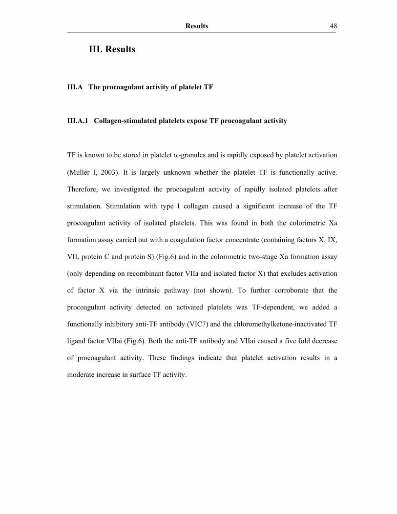
Results ____________________________________________________________________________________________________________________________________________________________________________________________________________________________
48
III. Results
III.A The procoagulant activity of platelet TF
III.A.1 Collagen-stimulated platelets expose TF procoagulant activity
TF is known to be stored in platelet α-granules and is rapidly exposed by platelet activation
(Muller I, 2003). It is largely unknown whether the platelet TF is functionally active.
Therefore, we investigated the procoagulant activity of rapidly isolated platelets after
stimulation. Stimulation with type I collagen caused a significant increase of the TF
procoagulant activity of isolated platelets. This was found in both the colorimetric Xa
formation assay carried out with a coagulation factor concentrate (containing factors X, IX,
VII, protein C and protein S) (Fig.6) and in the colorimetric two-stage Xa formation assay
(only depending on recombinant factor VIIa and isolated factor X) that excludes activation
of factor X via the intrinsic pathway (not shown). To further corroborate that the
procoagulant activity detected on activated platelets was TF-dependent, we added a
functionally inhibitory anti-TF antibody (VIC7) and the chloromethylketone-inactivated TF
ligand factor VIIai (Fig.6). Both the anti-TF antibody and VIIai caused a five fold decrease
of procoagulant activity. These findings indicate that platelet activation results in a
moderate increase in surface TF activity.
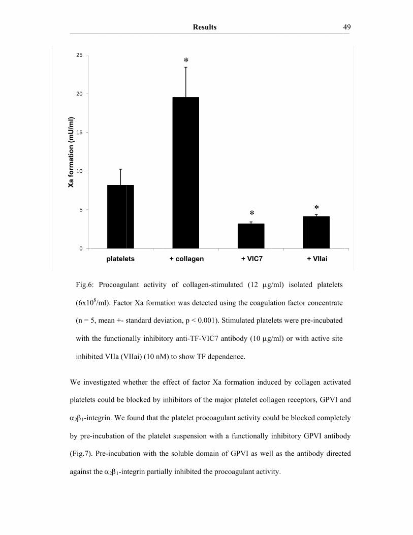
_________________
0
5
10
15
20
25
We inv
platelet
α2β1-in
by pre-
(Fig.7)
against
Fig.6
(6x10
(n =
with
inhib
Xa fo
rmat
ion
(mU
/ml)
_____________________________
platele
vestigated w
ts could be b
ntegrin. We
-incubation
). Pre-incuba
t the α2β1-in
6: Procoagu
08/ml). Facto
5, mean +- s
the function
bited VIIa (V
______________________________
ets
whether the e
blocked by
found that th
of the plate
ation with th
ntegrin partia
ulant activity
or Xa forma
standard dev
nally inhibit
VIIai) (10 nM
Re_____________________________
+ collage
effect of fac
inhibitors of
he platelet p
let suspensi
he soluble d
ally inhibited
*
y of collag
ation was det
viation, p < 0
tory anti-TF-
M) to show T
esults ______________________________
en
ctor Xa form
f the major p
procoagulant
on with a fu
domain of G
d the procoag
gen-stimulat
tected using
0.001). Stim
-VIC7 antib
TF dependen
______________________________
+ VIC7
mation induc
platelet colla
t activity cou
unctionally i
GPVI as well
gulant activi
ted (12 μg/
g the coagula
mulated platel
body (10 μg/
nce.
*
______________________________
+
ced by colla
agen recepto
uld be blocke
inhibitory G
l as the anti
ity.
/ml) isolate
ation factor c
lets were pre
/ml) or with
______________________________4
+ VIIai
agen activate
ors, GPVI an
ed complete
GPVI antibod
body directe
ed platelets
concentrate
e-incubated
h active site
*
____ 49
ed
nd
ely
dy
ed

_________________
Activat
membr
by pre
negativ
0
1
2
3
4
5
6
7
8
9
10
fold
ele
vatio
n of
TF
proc
oagu
lant
act
ivity
Fig.7
(6x10
and a
devia
_____________________________
ted platelets
rane leaflet.
eincubation o
vely charged
collagealone
: Procoagu
08/ml). The c
anti-α2β1-in
ation, p < 0.0
______________________________
s are known
The TF acti
of the activ
d phospholip
ene
ulant activity
concentratio
tegrin antibo
05 vs collage
Re_____________________________
n to expose
ivity measur
vated platele
pids in a Ca2+
anti-GPVI
*
y of collag
ons of anti-G
ody were in
en alone, coa
esults ______________________________
negatively c
red on activ
ets with ann
+-dependent
GP
gen-stimulat
GPVI inhibito
all cases 10
agulation fac
______________________________
charged pho
vated platelet
nexin V, wh
manner (Fig
PVI solubledomain
*
ted (12 μg
ory antibody
0 μg/ml (n =
ctor assay).
______________________________
ospholipids o
ts was stron
hich specific
g.8).
anti-α2β
g/ml) isolate
y, GPVI solu
3–9, means
______________________________5
on their out
ngly decrease
cally binds
1-integrin
*
ed platelets
uble domain
+- standard
____ 50
ter
ed
to
s
n
d
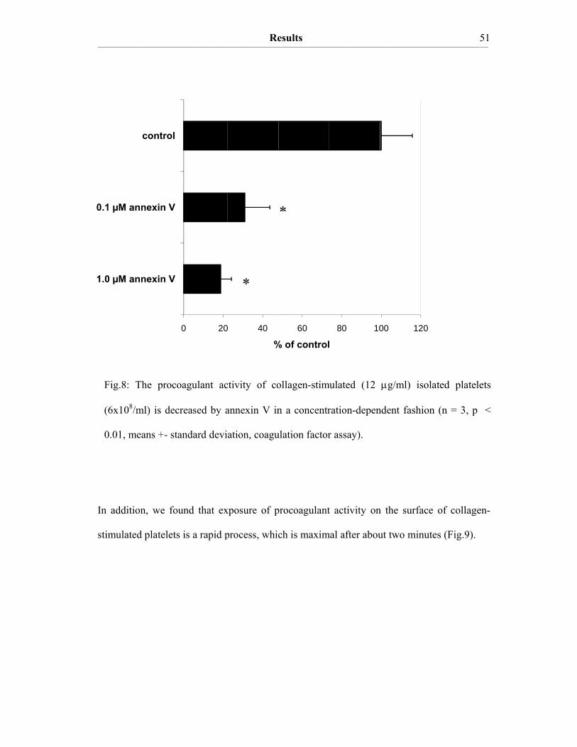
_________________
1.0 µM
0.1 µM
In add
stimula
Fig.8
(6x10
0.01,
_____________________________
annexin V
annexin V
control
ition, we fo
ated platelets
: The proco
08/ml) is dec
means +- st
______________________________
0 20
ound that ex
s is a rapid p
oagulant ac
creased by a
tandard devi
Re_____________________________
0 40
%
xposure of p
process, whic
ctivity of co
annexin V in
ation, coagu
*
esults ______________________________
60
% of control
procoagulant
ch is maxim
ollagen-stim
n a concentr
ulation factor
*
______________________________
80 10
l
t activity on
mal after abou
mulated (12
ration-depen
r assay).
______________________________
00 120
n the surface
ut two minut
μg/ml) isol
ndent fashion
______________________________5
e of collagen
tes (Fig.9).
lated platele
n (n = 3, p
____ 51
n-
ets
<

Results ____________________________________________________________________________________________________________________________________________________________________________________________________________________________
52
III.A.2 The TF procoagulant activity in collagen-activated platelets is largely
encrypted
From our experiments on the functional activity of TF we hypothesize that TF procoagulant
activity in platelets is largely encrypted. Indeed the total procoagulant activity of resting
and collagen-activated platelets is lower than the procoagulant activity of Ca2+-ionophore-
treated (A23187) platelets (Fig.10). Ca2+-ionophore (Kauffman RF, 1980) is known to de-
encrypt TF procoagulant activity in various cell types (monocytes, HL-60 cells, fibroblasts)
Fig.9: Time dependence of the procoagulant activity on the surface of collagen-stimulated (12
μg/ml) isolated platelets (6x10⁸/ml) (coagulation factor assay). Representative experiment for
a total of 8 experiments.
Xa fo
rmat
ion
(mU
/ml)
0
10
20
30
40
50
60
0 100 200 300 400 500 600
time of collagen stimulation (sec.)

Results ____________________________________________________________________________________________________________________________________________________________________________________________________________________________
53
Xa fo
rmat
ion
(mU
/ml)
(Bach RR, 1990; Drake TA, 1989). De-encryption of TF procoagulant activity in isolated
platelets, however, is novel. After platelet activation with A23187 most of the TF
procoagulant activity was found in the supernatant (Fig.10), whereas after collagen
stimulation most of the TF procoagulant activity could be detected in the platelet pellet
(Fig.10). Most likely A23187 strongly induces microparticle formation and therefore TF
procoagulant activity is mostly recovered in the platelet supernatant.
After activation with A23187 platelet procoagulant activity was rapidly released (Fig.11).
In the supernatant, a high activity was already observed after 15 seconds, which strongly
increased thereafter. In the pellet, the highest activity was already detected after 15 seconds.
Fig.10: Activation of TF procoagulant activity in isolated platelets (6x10⁸/ml). Xa
formation was measured 120 seconds after platelet activation with Ca2+-ionophore
A23187 (10 μM) or collagen (12 μg/ml) with the coagulation factor assay (n = 4).
0
2
4
6
8
10
12
14
16
18
20
A23187 pellet A23187 supernatant
collagen pellet collagen supernatant
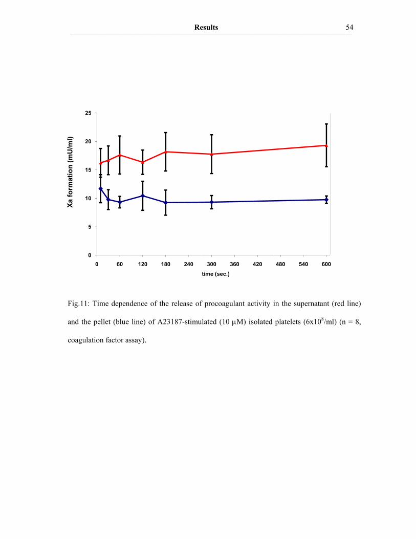
_________________
0
5
10
15
20
25
Fig.11:
and the
coagula
Xa fo
rmat
ion
(mU
/ml)
_____________________________
0
5
0
5
0
5
0 60
Time depen
pellet (blue
ation factor a
______________________________
120 1
ndence of th
e line) of A2
assay).
Re_____________________________
80 240
he release of
23187-stimu
esults ______________________________
300 360
time (sec.)
f procoagula
ulated (10 μM
______________________________
0 420
ant activity i
M) isolated
______________________________
480 540
in the supern
platelets (6x
______________________________5
600
natant (red l
x108/ml) (n
____ 54
line)
= 8,
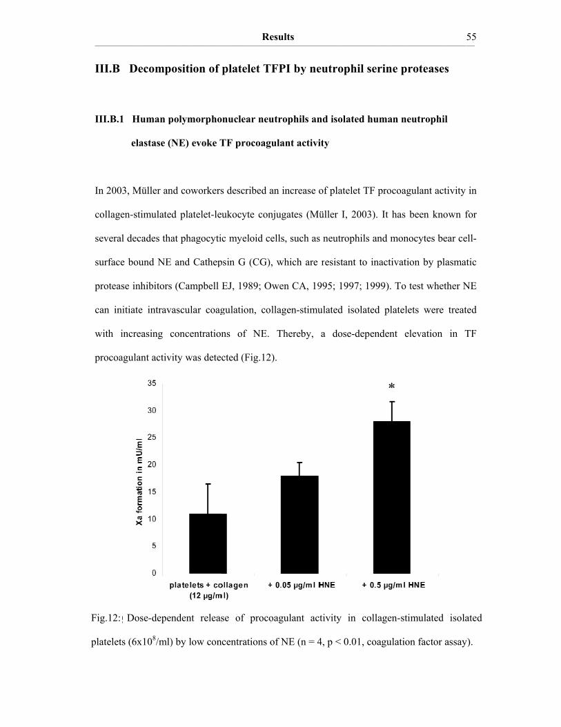
_________________
III.B
III.B.1
In 2003
collage
several
surface
proteas
can ini
with i
procoa
III.B.2
Fig.12:
platelets
_____________________________
Decompo
1 Human p
elastase (N
3, Müller an
en-stimulated
l decades tha
e bound NE
se inhibitors
itiate intrava
increasing c
agulant activi
2 NE is surDose-depen
s (6x10⁸/ml)
______________________________
osition of
polymorpho
NE) evoke T
nd coworkers
d platelet-le
at phagocyti
and Cathep
(Campbell
ascular coag
concentration
ity was dete
rface associandent releas
) by low con
Re_____________________________
platelet T
onuclear neu
TF procoag
s described a
eukocyte con
ic myeloid ce
psin G (CG)
EJ, 1989; O
gulation, co
ns of NE.
cted (Fig.12
ated on myese of proco
ncentrations o
esults ______________________________
FPI by ne
utrophils an
gulant activi
an increase o
njugates (Mü
ells, such as
, which are
Owen CA, 19
llagen-stimu
Thereby,
2).
eloid blood coagulant act
of NE (n = 4
______________________________
eutrophil s
nd isolated h
ity
of platelet T
üller I, 2003
neutrophils
resistant to
995; 1997; 1
ulated isolat
a dose-dep
cells and thtivity in co
4, p < 0.01, c
______________________________
serine pro
human neut
F procoagul
3). It has be
and monocy
inactivation
999). To tes
ted platelets
endent elev
eir micropa
*
ollagen-stimu
coagulation f
______________________________5
teases
trophil
lant activity
een known f
ytes bear cel
n by plasmat
st whether N
were treate
vation in T
articles ulated isola
factor assay)
____ 55
in
for
ll-
tic
NE
ed
TF
ated
).
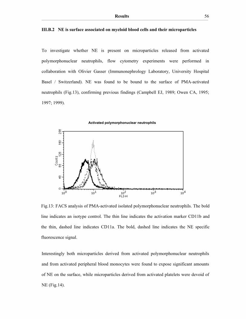
Results ____________________________________________________________________________________________________________________________________________________________________________________________________________________________
56
III.B.2 NE is surface associated on myeloid blood cells and their microparticles
To investigate whether NE is present on microparticles released from activated
polymorphonuclear neutrophils, flow cytometry experiments were performed in
collaboration with Olivier Gasser (Immunonephrology Laboratory, University Hospital
Basel / Switzerland). NE was found to be bound to the surface of PMA-activated
neutrophils (Fig.13), confirming previous findings (Campbell EJ, 1989; Owen CA, 1995;
1997; 1999).
Interestingly both microparticles derived from activated polymorphonuclear neutrophils
and from activated peripheral blood monocytes were found to expose significant amounts
of NE on the surface, while microparticles derived from activated platelets were devoid of
NE (Fig.14).
isotype ---
-- anti-CD11b
--anti-NE anti-CD11a --
Activated polymorphonuclear neutrophils
Fig.13: FACS analysis of PMA-activated isolated polymorphonuclear neutrophils. The bold
line indicates an isotype control. The thin line indicates the activation marker CD11b and
the thin, dashed line indicates CD11a. The bold, dashed line indicates the NE specific
fluorescence signal.
100 101 102 103 104FL2-H

Results ____________________________________________________________________________________________________________________________________________________________________________________________________________________________
57
III.B.3 Cell surface association of serine proteases results from polar interactions
with glycosaminoglycans and with nucleic acids
The surface association of NE may result from polar interactions of positively charged
patches on the enzyme surface with the negatively charged glycosaminoglycans (GAGs) on
the neutrophil cell membrane (Kostoulas G, 1997). To test this, isolated neutrophils were
pre-treated with chondroitin-4,5-sulfate which is a major component of neutrophil GAGs
and with chondroitinase ABC, an enzyme degrading GAGs. Additionally, they could also
result from the negatively charged backbone of nucleic acids (DNA, RNA), that are
released by activated neutrophils within neutrophil extracellular traps (NETs) (Brinkmann
V, 2004). Therefore, activated neutrophils were also pre-treated with nucleases (DNase I,
RNase A) to degrade the NETs. Subsequently, platelet-neutrophil conjugates were
investigated for their procoagulant activity to test whether surface association of NE is
required for proteolytic inactivation of platelet TFPI. Procoagulant activity was found to be
isotype antibody anti-NE
Platelet-derived microparticles Monocyte-derived microparticles Neutrophil-derived microparticles
Fig.14: FACS analysis of microparticles derived from activated platelets, neutrophils and
monocytes. The thin line indicates the fluorescence signal of the biotinylated isotype
control antibody (detection with streptavidin-Cy5) and the bold line indicates the
fluorescence signal of the NE-specific antibody.
100 101 102 103 104FL3-H
100 101 102 103 104FL3-H
100 101 102 103 104FL3-H
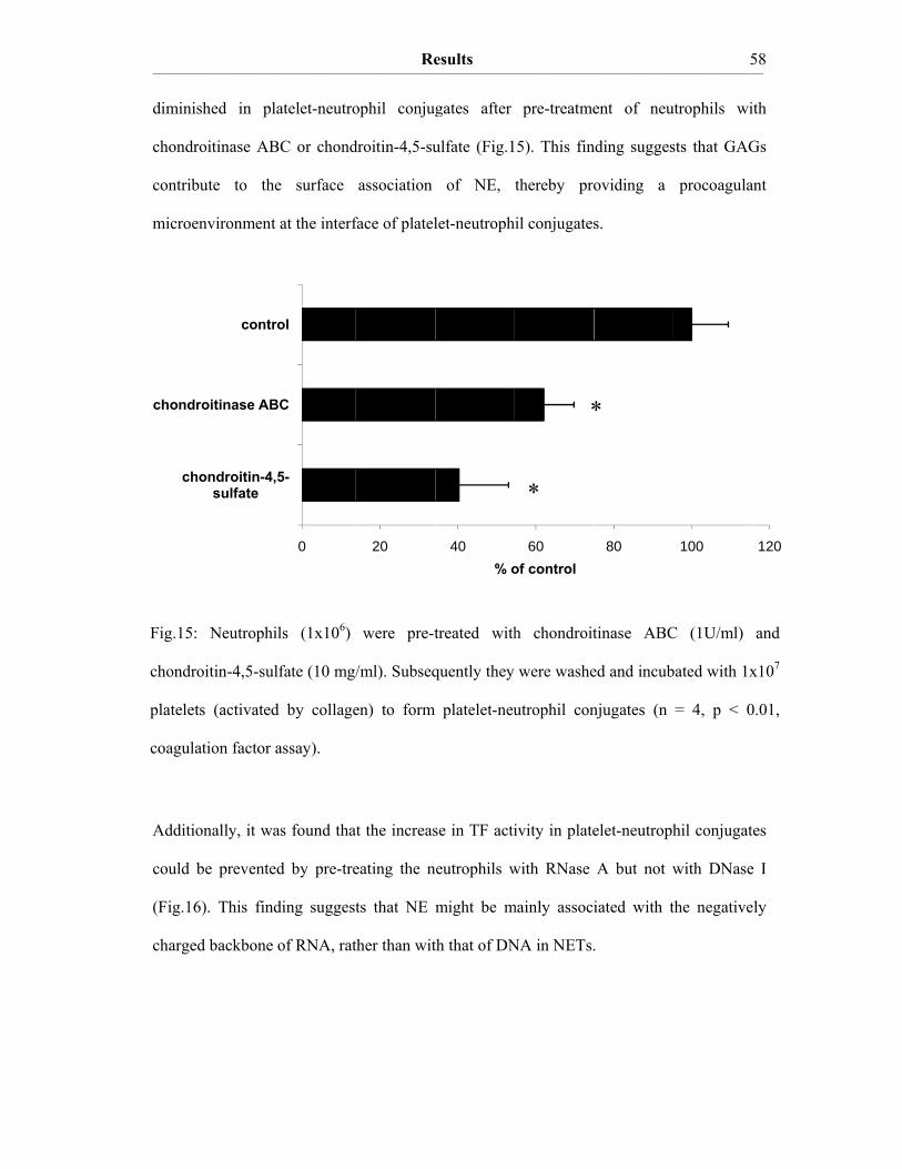
_________________
diminis
chondr
contrib
microe
Additio
could b
(Fig.16
charge
ch
chondr
Fig.15:
chondro
platelet
coagula
_____________________________
shed in pl
roitinase AB
bute to the
environment
onally, it wa
be prevente
6). This find
d backbone
hondroitin-4,sulfate
roitinase AB
contr
Neutrophi
oitin-4,5-sul
ts (activated
ation factor a
______________________________
atelet-neutro
BC or chond
e surface
at the interf
as found tha
d by pre-tre
ding suggest
of RNA, rat
0
5-
BC
rol
ils (1x10⁶)
lfate (10 mg/
d by collage
assay).
Re_____________________________
ophil conjug
droitin-4,5-su
association
face of platel
t the increas
eating the n
ts that NE
ther than wit
20
were pre-t
/ml). Subseq
en) to form
esults ______________________________
gates after
ulfate (Fig.1
of NE,
let-neutroph
se in TF acti
neutrophils w
might be m
th that of DN
40% o
treated with
quently they
platelet-neu
______________________________
pre-treatme
5). This fin
thereby pro
il conjugates
ivity in plate
with RNase
mainly associ
NA in NETs.
60of control
*
*
h chondroit
were washe
utrophil con
______________________________
ent of neu
nding sugges
oviding a
s.
elet-neutroph
A but not w
iated with t
.
80
*
tinase ABC
ed and incub
njugates (n
______________________________5
utrophils wi
sts that GAG
procoagula
hil conjugat
with DNase
the negative
100
C (1U/ml)
bated with 1x
= 4, p < 0
____ 58
ith
Gs
ant
es
e I
ely
120
and
x107
0.01,
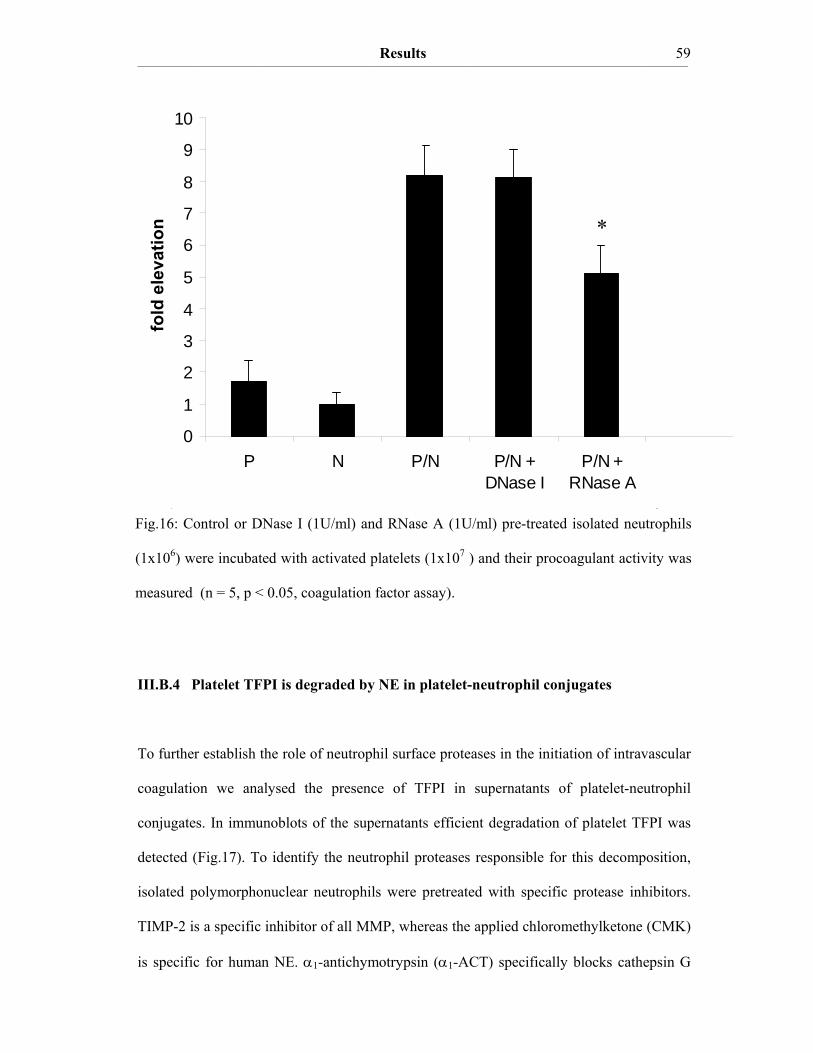
Results ____________________________________________________________________________________________________________________________________________________________________________________________________________________________
59
III.B.4 Platelet TFPI is degraded by NE in platelet-neutrophil conjugates
To further establish the role of neutrophil surface proteases in the initiation of intravascular
coagulation we analysed the presence of TFPI in supernatants of platelet-neutrophil
conjugates. In immunoblots of the supernatants efficient degradation of platelet TFPI was
detected (Fig.17). To identify the neutrophil proteases responsible for this decomposition,
isolated polymorphonuclear neutrophils were pretreated with specific protease inhibitors.
TIMP-2 is a specific inhibitor of all MMP, whereas the applied chloromethylketone (CMK)
is specific for human NE. α1-antichymotrypsin (α1-ACT) specifically blocks cathepsin G
0
1
2
3
4
5
6
7
8
9
10
P N P/N P/N +DNase I
P/N +RNase A
*
Fig.16: Control or DNase I (1U/ml) and RNase A (1U/ml) pre-treated isolated neutrophils
(1x10⁶) were incubated with activated platelets (1x10⁷ ) and their procoagulant activity was
measured (n = 5, p < 0.05, coagulation factor assay).
fold
ele
vatio
n

Results ____________________________________________________________________________________________________________________________________________________________________________________________________________________________
60
(CG). TIMP-2 did not show any effect on the degradation of platelet TFPI in the platelet-
neutrophil conjugates. The NE-specific CMK however blocked the degradation of platelet
TFPI almost completely and pre-treatment of isolated neutrophils with α1-ACT partially
inhibited TFPI degradation (Fig.17). This indicates that NE and, to a lower extent CG,
degrade TFPI and thereby might initiate intravascular coagulation on the surface of
activated platelets.
The TFPI degradation products detected in immunoblots from suspensions of activated
platelets and neutrophils and the degradation products achieved by limited proteolysis of
the supernatant of stimulated platelets with isolated NE were of the same size and intensity,
indicating that NE is the major protease that degrades TFPI in the cellular system (Fig.18).
P P / NN
TFPI
P / N TIMP-2 CMK α1-ACT
TFPI
47 kD
47 kD
Fig.17: Degradation of platelet secreted TFPI in platelet-neutrophil conjugates (P/N)
detected by Western blot analysis. Isolated neutrophils (N) were pretreated with 1 mM
TIMP-2, a NE-specific CMK or the CG-specific protease inhibitor α1-ACT.

Results ____________________________________________________________________________________________________________________________________________________________________________________________________________________________
61
In addition, isolated microparticles derived from fMLP-activated polymorphonuclear
neutrophils (N-MP) were also capable of degrading platelet TFPI in a NE-specific manner
when added to stimulated platelets. Degradation of platelet TFPI by N-MP could be
prevented by pre-treatment of the N-MP with an NE-specific inhibitor (not shown).
TFPI
P / N P + NE
47 kD
36 kD
33 kD
P
Fig.18: Degradation products obtained in platelet-neutrophil conjugates and those obtained
by limited proteolysis of platelet supernatants with isolated human NE (15 μg/ml) detected
by Western blot analysis.

Results ____________________________________________________________________________________________________________________________________________________________________________________________________________________________
62
III.C A disulfide switch in the TF molecule regulates its procoagulant
activity
III.C.1 TF contains a labile disulfide that is essential for its procoagulant function
To assess the contribution of extracellular cysteines for TF activity, we substituted the
cysteines C49/C57 and C186/C209 by serine. In C186S/C209S TF procoagulant activity
was completely lost (Fig.19). Substitutions of C49 and C57 by serine had no effect on the
procoagulant activity (Fig.19). Treatment of the C49S/C57S mutant with thioredoxin
(TRX) or dithiotreitol (DTT) decreased the procoagulant activity. Phenylarsine oxide
(PAO), a compound that binds to vicinal thiols thereby mimicking a disulfide, increased the
procoagulant activity of the C49S/C57S mutant (Fig.19). This suggests that the oxidized
state of the C186/C209 disulfide pair represents the active state of TF (in accordance with
Ahamed, 2006).
To register the thiol states of TF we performed MPB-immunoblot experiments. MPB
selectively recognizes free thiol groups. The oxidoreductase TRX reduced the labile
disulfide bond in the extracellular TF domain (sTF), whereas addition of PAO oxidized the
disulfide (Fig.20). This finding implies a physiological role for TRX in the inactivation
(encryption) of TF procoagulant activity.

_________________
Fig.1
TRX,
Fig.20:
domain
were re
agarose
control
density
(right p
_____________________________
9: TF cyste
, 1 mM DTT
MPB-Imm
n of sTF aft
eacting with
e beads was
immunoblo
y of the sTF b
panel) and st
______________________________
eine mutants
T or 100 μM
munoblot cha
ter treatment
h the thiol
s performed
ot MPB inc
bands of n =
atistically an
Re_____________________________
s (C49S/C5
M PAO respec
aracterizing
t with TRX
reactive co
d, selecting
orporation d
= 3 experime
nalyzed by th
esults ______________________________
7S, C186S/
ctively (coag
the dithiol
X and PAO (
mpound MP
for reduced
did not affe
ents was eva
he Mann-Wh
______________________________
/C209S) wer
gulation fact
oxidation
(left panel).
PB. A pull-
d dithiols in
ct the bindi
luated by us
hitney-rank-
______________________________
re treated w
tor assay).
state in the
Reduced di
-down with
n sTF. Acco
ing of VIC1
sing Scion Im
-sum-test (p
______________________________6
with 10 μM
e extracellul
ithiols in sT
h streptavidin
ording to th
12. The mea
mage softwa
< 0.05).
____ 63
M
lar
TF
n-
he
an
are

Results ____________________________________________________________________________________________________________________________________________________________________________________________________________________________
64
III.C.2 Protein Disulfide Isomerase oxidizes the C186/C209 pair
Activated platelets, which are known to decrypt TF, secrete protein disulfide isomerase
(PDI). PDI is located to the external surface of the platelet plasma membrane (Chen K,
1995; Essex DW, 1995). This oxidoreductase could be involved in the regulation of the
thiol state of TF. PDI was indeed found to oxidize the dithiol species of the C186/C209 pair
of the sTF to its functionally active disulfide state (Fig.21). About 70 % of the active site
cysteines of PDI were determined to be in the oxidized state (quantification of free PDI
thiols by the colorimetric Ellman’s assay). The supernatant of collagen-activated platelets
(P-SN) is also capable of oxidizing sTF free dithiols (Fig.21). The oxidation of TF free
dithiols to the disulfide could also be demonstrated on LPS-stimulated isolated monocytes,
which are known to express encrypted TF on their cell surface (data not shown), indicating
that also cell TF is present in the reduced state.
kDa
Fig.21: MPB-immunoblot characterizing the dithiol oxidation state in the sTF after
treatment with the supernatant of activated platelets (P-SN) and recombinant PDI.
Treatment with P-SN or PDI did not influence binding of VIC12 (control Western blot).
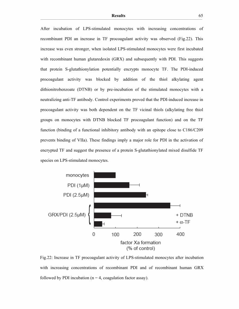
Results ____________________________________________________________________________________________________________________________________________________________________________________________________________________________
65
After incubation of LPS-stimulated monocytes with increasing concentrations of
recombinant PDI an increase in TF procoagulant activity was observed (Fig.22). This
increase was even stronger, when isolated LPS-stimulated monocytes were first incubated
with recombinant human glutaredoxin (GRX) and subsequently with PDI. This suggests
that protein S-glutathionylation potentially encrypts monocyte TF. The PDI-induced
procoagulant activity was blocked by addition of the thiol alkylating agent
dithionitrobenzoate (DTNB) or by pre-incubation of the stimulated monocytes with a
neutralizing anti-TF antibody. Control experiments proved that the PDI-induced increase in
procoagulant activity was both dependent on the TF vicinal thiols (alkylating free thiol
groups on monocytes with DTNB blocked TF procoagulant function) and on the TF
function (binding of a functional inhibitory antibody with an epitope close to C186/C209
prevents binding of VIIa). These findings imply a major role for PDI in the activation of
encrypted TF and suggest the presence of a protein S-glutathionylated mixed disulfide TF
species on LPS-stimulated monocytes.
Fig.22: Increase in TF procoagulant activity of LPS-stimulated monocytes after incubation
with increasing concentrations of recombinant PDI and of recombinant human GRX
followed by PDI incubation (n = 4, coagulation factor assay).

Results ____________________________________________________________________________________________________________________________________________________________________________________________________________________________
66
In whole blood TEG experiments the clotting time of collagen-activated blood was delayed
by an anti-PDI antibody when compared to the appropriate buffer control and the IgG2a
isotype control (Fig.23). This indicates a significant role for the PDI-triggered activation
mechanism of TF in the whole blood system.
Treatment of sTF with GRX increased the reduced fraction of the labile disulfide pair,
whereas pre-incubation with GRX and subsequent treatment with PDI increased the
formation of disulfides (Fig.24). This suggests the presence of constitutive protein S-
glutathionylation of sTF.
Fig.23: TEG experiment of collagen-activated whole blood (collagen activates intravascular
TF) incubated with equal volumes of resuspension buffer, IgG2a isotype antibody or
functional inhibitory anti-PDI antibody (n = 3). The clotting time of one representative
experiment is indicated in seconds.
buffer-control
95 s
isotype-control
(IgG2a)
70 s
anti-PDI
123 s
buffer-control
95 s
isotype-control
(IgG2a)
70 s
anti-PDI
123 s

Results ____________________________________________________________________________________________________________________________________________________________________________________________________________________________
67
III.C.3 Glutathionylation of C186/C209 vicinal thiols of TF
Cysteine residues in proteins can be glutathionylated if they are surface exposed and if the
binding of gluthathione is facilitated by an appropriate electrostatic protein surface. We
tested, whether TF might undergo this common cysteine modification. In a CHO cell
system transfected with full-length TF we found that wild type TF (wt) could be
glutathionylated by reaction with biotin-labeled glutathione (biotin-GSH), whereas the
C186S/C209S mutant could not (Fig.25). The C186/C209 disulfide pair is solvent
accessible and has all the structural requirements to undergo protein S-glutathionylation.
However, the C49/C57 disulfide pair does not undergo a reaction with biotin-GSH, since
no signal was detected in the C186S/C209S TF mutant (Fig.25, right panel).
kDa
Fig.24: MPB-immunoblot characterizing the dithiol oxidation state of sTF. sTF was pre-
treated with GRX, PDI or GRX plus PDI. The addition of GRX or PDI did not affect
MPB incorporation of TF (control Western blot).

Results ____________________________________________________________________________________________________________________________________________________________________________________________________________________________
68
In Pulmonary Artery Vascular Smooth Muscle Cells (PAVSMC), which are known to
express high levels of TF antigen, the labile cysteine pair of the TF molecule was also
found to be susceptible to in vitro protein S-glutathionylation (Fig.26). This protein S-
glutathionylation could be reversed by subsequent treatment of the cells with low
concentrations of GRX or DTT, indicating that biotin-GSH is covalently bound to TF via a
disulfide linkage.
wt C186S / C209SkDa
kDa
Fig.25: GSH-immunoblot detecting in vitro protein S-glutathionylated TF on transfected
CHO cells. Cells were incubated with 125 µM biotin-GSH and 100 µM diamide that is
acting as an oxidant. After cell lysis a biotin-selective pull-down with streptavidin-agarose
was performed. TF antigen was detected with the VIC12 anti-TF antibody. According to the
control Western blot, binding of VIC12 was not affected by treatment of sTF with biotin-
GSH or the serine mutation.
Fig.26: GSH-immunoblot detecting in vitro protein S-glutathionylated TF on PAVSMC. A
biotin-selective pull-down with streptavidin-agarose was performed and TF was detected
with the anti-TF antibody (VIC12).

Results ____________________________________________________________________________________________________________________________________________________________________________________________________________________________
69
III.C.4 In vitro protein S-glutathionylation of TF is reversible
To test whether cell lysate containing the oxidoreductases GRX and PDI might regulate the
protein S-glutathionylation of TF we examined its incorporation of biotin-GSH. Thereby,
we could distinguish whether GRX and PDI act as a reductant or as an oxidant. We
observed that pre-treatment of sTF with CHO cell lysate prevented the incorporation of
biotin-GSH into TF (Fig.27). In addition, the reductants DTT and GRX also prevented
biotin-GSH incorporation. Moreover, protein S-glutathionylation of sTF was slightly
reversed by PDI.
Fig.27: GSH-immunoblot detecting in vitro protein S-glutathionylation of TF. A biotin-
selective pull-down with streptavidin-agarose was performed and TF was detected with the
VIC12 anti-TF antibody. According to the control Western blot, binding of VIC12 was not
affected by treatment of sTF with biotin-GSH, CHO cell lysate, DTT, GRX and PDI.

Discussion ____________________________________________________________________________________________________________________________________________________________________________________________________________________________
70
IV. Discussion
IV.1 TF procoagulant activity of activated platelets
Recent studies indicate that there is a pool of TF in the blood circulation, so-called
intravascular or blood borne or circulating TF (Giesen PL, 1999; Engelmann et al., 2003).
Giesen and co-workers reported blood borne TF in thrombi formed on collagen-coated
glass slides that were exposed to flowing human blood. The collagen-coated glass slides
were devoid of TF, implying that TF procoagulant activity is derived from human blood.
TF in the formed thrombi was found to be active, since abundant fibrin was detected.
Independently, our research group identified TF in peripheral human blood from healthy
donors. TF antigen appeared on the surface of collagen-activated platelets adhering to
leukocytes (Zillmann A, 2001). This is a rapid process, since TF antigen was already
detectable after 5 minutes of stimulation. Therefore, de novo synthesis of TF is not
possible. It is still a matter of debate how platelets acquire TF. According to one hypothesis
TF is transported to platelets by leukocyte-derived microparticles (Del Conde I, 2005).
Alternatively, the spliceosome of proplatelets that extend from megakaryocytes is capable
of translating TF from pre-mRNAs (Denis MM, 2005; Schwertz H, 2006). It was shown
that intravascular TF was competent to start coagulation and TF antigen was detected in
isolated platelets, whereas no TF antigen was detectable in isolated resting neutrophils and
rapidly isolated monocytes (Zillmann A, 2001). However, this study did not contain direct
evidence for the presence of TF procoagulant activity on the surface of activated platelets.
We detected a rapid increase in TF procoagulant activity of isolated human platelets after a
10 minute stimulation with collagen type I (in agreement with Camera, 2003). When the
isolated platelets were pre-incubated with a functional inhibitory anti-TF antibody, the
procoagulant activity was completely blocked. This shows that the formation of factor Xa

Discussion ____________________________________________________________________________________________________________________________________________________________________________________________________________________________
71
by activated platelets is TF dependent. TF procoagulant activity was also substantially
blocked by pre-incubation of isolated platelets with a functional inhibitory anti-GPVI
antibody, but only slightly blocked by pre-incubation with an antibody directed against
α2β1-integrin. This implicates a pivotal role for GPVI-dependent kinase signalling in the
surface exposure of TF on the platelet membrane.
IV.2 Encryption of platelet Tissue Factor activity
TF encryption indicates the post-translational suppression of TF procoagulant activity on
the cell surface (Bach RR, 2006). A discrepancy between TF antigen and the expression of
TF procoagulant activity has previously been observed in a variety of cell types (Maynard
JR, 1977; Walsh JD, 1991; Drake TA, 1989). Treatment of cells with the Ca2+-ionophore
A23187 is known to de-encrypt TF procoagulant activity (Bach RR, 1996; 2006).
Treatment of platelets with Ca2+-ionophore resulted in a three times higher procoagulant
activity compared to collagen-treated platelets, indicating that about two thirds of the total
platelet TF procoagulant activity might be encrypted in collagen activated platelets. While
most of the procoagulant activity was found in the pellet after collagen treatment, nearly no
activity was observed in the supernatant. However in Ca2+-ionophore treated samples most
of the TF activity was found in the supernatant, which might be due to the formation of
procoagulant platelet microparticles (Henriksson CE, 2006).
The underlying mechanism leading to the increase of TF activity after treatment with Ca2+-
ionophore is not understood (Bach RR, 2006). It was suggested that the exposure of the
cofactor phosphatidylserine (PS) contributes only partially to the increase of TF
procoagulant activity in LPS-stimulated monocytes and non-transformed human dermal
fibroblasts (Wolberg AS, 1999). In our experiments, the increase in platelet procoagulant
Discussion ____________________________________________________________________________________________________________________________________________________________________________________________________________________________
72
activity evoked by the treatment with Ca2+-ionophore could not be fully suppressed in the
presence of saturating concentrations of the PS binding protein annexin V (Fig.8),
indicating that the increase in TF procoagulant activity did not result solely from increased
exposure of PS. Therefore, other yet unknown posttranslational mechanisms have to be
involved in encrypting TF procoagulant activity on the cell surface of platelets.
IV.3 Neutrophil surface proteases trigger the TF procoagulant activity in platelet-
neutrophil conjugates
TFPI is the physiologic inhibitor of the initiator complex of coagulation. Recently, a
glycosyl phosphatidylinositol (GPI) anchored variant of TFPI was characterized on the
surface of cultured endothelial cells (Zhang J, 2003). A GPI anchor was predicted and
identified for the TFPI splice variant β lacking Kunitz domain 3 but not for the TFPI splice
variant α. TFPI-α was found to be the most abundant form expressed by endothelial cells.
It was described recently that TFPI-β is responsible for the bulk of the cellular VIIa/TF
inhibitory activity (Piro O, 2005). However, in our study we focussed on TFPI-α. This
variant was found to be by far the predominant splice form in platelets (Bidzhekov K, TFPI
mRNA analysis, personal communication).
Recombinant human TFPI-α is known to be highly susceptible to limited proteolysis by
neutrophil serine proteases, such as NE and CG (Petersen LC, 1992; Higuchi DA, 1992)
and further to coagulation proteases like thrombin (Ohkura N, 1997) and plasmin
(Salemink I et al, 1998) as well as to MMP (Belaaouaj AA, 2000; Cunningham AC, 2002).
In previous studies performed in our laboratory an increased TF procoagulant activity was
detected in platelet-neutrophil conjugates compared to isolated neutrophils or collagen-
activated platelets (Müller I, 2003). Based on these data a model for the initiation of

Discussion ____________________________________________________________________________________________________________________________________________________________________________________________________________________________
73
intravascular coagulation was suggested (Engelmann B, 2003). In this model, neutrophil
proteases were proposed to inactivate platelet TFPI in a microenvironment formed by
platelet-neutrophil conjugates (Fig.4). The platelet-neutrophil microenvironment is mainly
dependent on P-selectin / PSGL-1 interactions formed between activated platelets and
polymorphonuclear neutrophils (Müller I, PhD thesis; McEver RP, 1997; Palabrica T,
1992). Platelet-neutrophil adhesion (firm adhesion) is further stabilized by fibrinogen
bridges formed by the activated platelet receptor GPIIb/IIIa and the leukocyte receptor
Mac-1 (CD11b/CD18) (Spangenberg P, 1993).
To experimentally address this hypothesis, collagen activated platelets were treated with
increasing concentrations of isolated human NE. A concentration dependent increase in
platelet TF procoagulant activity was noted. This finding implies that NE is able to trigger
the release of TF procoagulant activity on activated platelets. Since NE is the major serine
protease stored in the azurophilic granules of neutrophils it appears likely that this enzyme
is also responsible for the increased TF procoagulant activity in platelet-neutrophil
conjugates. In immunoblot experiments of the supernatants obtained from platelet-
neutrophil conjugates, rapid degradation of platelet TFPI was noted (Fig.17). This was
markedly reduced by pre-treatment of neutrophils with a NE-specific CMK inhibitor. This
suggests that in platelet-neutrophil conjugates TFPI is most efficiently degraded by NE. In
confirmation of this conclusion, the degradation products obtained in platelet-neutrophil
conjugates migrated at the same size as those obtained by treatment of activated platelets
with isolated human NE (Fig.18).
In addition, we observed degradation of platelet TFPI by neutrophil-derived microparticles
in the supernatant of collagen-activated platelets (data not shown). This finding could
suggest a novel role for these plasma components in the initiation of intravascular
coagulation.
Discussion ____________________________________________________________________________________________________________________________________________________________________________________________________________________________
74
Since TF and its physiologic inhibitor TFPI are both stored in the platelet α-granules and
are exposed on the membrane after platelet activation, it appears likely that TF in activated
platelets is rapidly inactivated by the formation of a quarternary complex consisting of
TF/VIIa and Xa/TFPI. If the concentrations of functionally active TFPI are substantially
lowered by NE, the whole coagulation cascade may in principle proceed on the membrane
surface of activated platelets. This would enable coagulation to take place exactly at the site
of the growing thrombus. Hence the thrombus could be efficiently stabilized by the fibrin
polymers.
IV.4 Characterization of the procoagulant microenvironment formed between
activated platelets and polymorphonuclear neutrophils
Since a procoagulant microenvironment was found to be formed mainly by P-selectin /
PSGL-1-interactions between activated platelets and polymorphonuclear neutrophils
(Müller I, PhD thesis) we decided to further investigate the conditions leading to a local
intravascular coagulation start.
In this context, it was of major interest to test whether NE is bound to the surface of
activated neutrophils and their microparticles or if it is simply secreted into the surrounding
media. We detected NE on the surface of activated neutrophils and on the surface of
microparticles derived from activated neutrophils and monocytes by FACS analysis. NE is
able to bind negatively charged cell surface structures once released from the azurophilic
granules of myeloid cells. Proteoglycans, glycosaminoglycans and neutrophil extracellular
traps (NETs) are negatively charged structures present on the surface of activated
neutrophils. Proteoglycans and glycosaminoglycans contain negatively charged sulfate
groups (Hornebeck W, 1994) and the backbones of nucleic acids, which are contained in

Discussion ____________________________________________________________________________________________________________________________________________________________________________________________________________________________
75
the NETs of activated neutrophils (Brinkmann V, 2004) consist of negatively charged
phosphate esters. NE possesses a highly asymmetric distribution of positively charged
residues that are solvent exposed (Navia MA, 1989), which enables them to bind to
polyanionic surfaces. Cell surface bound NE could be removed by pre-treatment of
neutrophils with chondroitinase ABC and by chondroitin-4,5-sulfate. NE could also be
removed from the surface of PMA-activated neutrophils by treatment with RNase A, but
not with DNase I. As a consequence of the NE removal, the proteolytic activity of the
neutrophils that maintains TF activity was decreased. These findings suggest a major role
for different polyanionic cell surface molecules for the inactivation of TFPI.
TFPI decomposition may play a significant role in pathologies associated with acute and
chronic inflammation. In atherosclerosis chronic inflammation of the vessel wall is induced
by transmigration of monocytes into the vascular wall and their differentiation to
macrophages and foam cells. An atherosclerotic plaque is formed. Rupture of a specific
type of plaque leads to arterial thrombus formation, acute myocardial infarction and stroke
(Fuster V, 2005). Sepsis is another inflammatory disorder resulting in activation of
coagulation. It is associated with widespread blood clotting in various organs (disseminated
intravascular coagulation) (Creasey AA, 1993). Both, arterial thrombosis and septic shock
are leading causes of mortality in many countries.
Disseminated intravascular coagulation is a frequent complication that can finally lead to
multiple organ failure. It is becoming increasingly clear that coagulation and innate
immunity have coevolved early in eukaryotic development, and that these systems continue
to function as a highly integrated unit following tissue injury (Opal SM, 2003). In
disseminated intravascular coagulation during sepsis leukocytes are activated (Grisham
MB, 1988) and the surface expression of platelet adhesion molecules is increased (Gawaz
M, 1995). The occurrence of platelet-leukocyte aggregates was also found to be increased
in this study. We therefore assume that the neutrophil-dependent proteolytic mechanism of
Discussion ____________________________________________________________________________________________________________________________________________________________________________________________________________________________
76
coagulation activation might play a role in the development of disseminated intravascular
coagulation and multiple organ failure.
IV.5 Disulfide switch of TF regulates initiation of intravascular coagulation on
monocytes – potential role for TF encryption
Thiol-disulfide exchange is an emerging mechanism of cell surface protein regulation
(Hogg PJ, 2003). From its x-ray structure (Harlos K, 1994) TF was found to contain a labile
disulfide bond that could potentially be subject to rapid disulfide switching (Hogg PJ, 2003;
Schmidt B, 2006). TF is a member of the class 2 cytokine receptor family (Bazan JF, 1990)
and its x-ray structure revealed an unusual disulfide bridging cysteine 186 with cysteine
209 inside an antiparallel β-sheet. This disulfide bridge is characteristic of the cytokine type
2 receptor family. It lies at the very end of domain 2 and links adjacent strands F and G.
The sulphur atoms of this S-S bond point away from the surface of the β-sheet towards the
solvent (Harlos K, 1994). It has been established by site directed mutagenesis experiments
that the integrity of this energetically unfavourable disulfide in TF is required for the
binding of factor VII (Rehemtulla A, 1991), which defines its role as the cofactor
responsible for the coagulation start (Ruf W, 1998). Residues close to the allosteric
disulfide have been identified to interact with factors IX and X (Kirchhofer D, 2000).
To assess the role of extracellular cysteines for TF function, we generated different mutants
with cysteine to serine substitutions. Experiments performed with the C49S/C57S and
C186S/C209S mutants showed that the C186S/C209S double mutant was devoid of TF
procoagulant activity. The C49S/C57S double mutant of TF, in contrast, was functionally
active. The procoagulant activity of the C49S/C57S mutant could be partially decreased by
treatment with the oxidoreductase TRX, which is known to reduce disulfide bonds to

Discussion ____________________________________________________________________________________________________________________________________________________________________________________________________________________________
77
vicinal thiols. Indeed TRX has been shown to reduce factor VIII and other coagulation
factors (Savidge G, 1979). The TF procoagulant activity of C49S/C57S was profoundly
decreased by the chemical disulfide reductant DTT. Treatment of the C49S/C57S double
mutant with PAO that is known to coordinate with vicinal thiols increased TF activity.
Coordination of vicinal thiols with PAO mimics a disulfide linkage, which is likely
functional, suggesting that oxidation of the C186/C209 pair de-encrypts TF.
These functional data could be confirmed in thiol-selective immunoblot experiments in
which the amount of reduced dithiols in sTF was analysed in a semiquantitative manner.
sTF exhibited proper folding (Andersson D, 2001). Indeed nearly the same content of
secondary structures was observed as to be deduced from the protein’s x-ray structure. The
fold of sTF was tested by recording a circular dichroism (CD) spectrum and the content of
secondary structure was calculated by deconvolution of the spectrum. With this method it
could be excluded that sTF is denaturated by reduction of the C186/C209 disulfide with 1
mM DTT (Appendix A2).
In contrast to TRX, the addition of PDI turned out to increase the TF procoagulant activity
of isolated LPS-stimulated monocytes. Inhibition by the anti-TF antibody and by treatment
of the cells with DTNB unambiguously demonstrated activation of TF by thiol-disulfide
exchange. Moreover, thiol-selective immunoblots analysing the amount of free thiol groups
of PDI-treated sTF and cell TF are in agreement with the conclusion that PDI oxidizes TF.
Recently, TF was found to co-immunoprecipitate PDI, which confirms that TF is a
substrate for PDI (Ahamed J, 2006). Our findings thus imply a major role for PDI in the de-
encryption of latent TF procoagulant activity on the cell surface of monocytes and other
cell types that express TF constitutively (Fig.22, 23, 24). Collagen-activated platelets were
also found to have an oxidative effect on the allosteric C186/C209 dithiol of sTF. Activated
platelets are known to release thiol isomerases that are capable of regulating disulfide
modifications (Essex DW, 2001). In addition, activated platelets are known to de-encrypt
Discussion ____________________________________________________________________________________________________________________________________________________________________________________________________________________________
78
the TF procoagulant activity of monocytes (Østerud B, 2001). According to our results this
finding can now be explained by the release of platelet PDI that catalyses thiol-disulfide
interchange of monocyte TF.
IV.6 Protein S-glutathionylation of TF – a potential safety device
Protein S-glutathionylation is currently emerging as a common posttranslational cysteine
modification of various proteins and it is a main form of protein S-thiolation (Ghezzi P,
2005). The term protein S-glutathionylation indicates the formation of mixed disulfides
between proteins and glutathione (GSH). The thiol oxidoreductase glutaredoxin (GRX)
reverses protein S-glutathionylation by a thiol / disulfide exchange mechanism (Ghezzi P
and Bonetto V, 2005). Proteins of the plasma membrane of cells exposed to blood, such as
TF are at the interface between an oxidizing (plasma) and a reducing environment
(cytoplasm) (Fahey RC, 1977). In particular, protein S-glutathionylation is associated with
the stabilization of extracellular proteins and the protection of proteins against irreversible
oxidation of critical cysteine residues (Biswas S, 2005).
After incubation with GRX and subsequent PDI addition, the TF activity of monocytes was
significantly higher than after treatment with PDI alone. Based on these results, we
hypothesize that monocyte TF might be protein S-glutathionylated and that
glutathionylation could be involved in TF encryption. We were able to confirm S-
glutathionylation of TF by different methods (thiol-selective immunoblots of sTF, in vitro
glutathionylation of vascular smooth muscle cells and of TF cysteine mutants in a CHO cell
system).
Cell lysis is known to de-encrypt the TF activity in various cell types (Drake TA, 1989).
Hence we were interested if CHO cell lysates that are completely devoid of TF are able to
Discussion ____________________________________________________________________________________________________________________________________________________________________________________________________________________________
79
reverse the protein S-glutathionylation of glutathionylated sTF. In fact, the lysate of CHO
cells strongly diminished protein S-glutathionylation of sTF. This suggests that the release
of thiol oxidoreductases during cell rupture, such as GRX and PDI, could activate latent
TF. In the case of vascular injury cell lysis of several vascular wall cells occurs, such as
fibroblasts, vascular smooth muscle cells and endothelial cells. Thereby, thiol
oxidoreductases could act as a response to injury preventing lethal blood loss of the
organism by activation of encrypted TF. Damage of vascular wall cells also occurs during
atherogenesis (Schecter AD, 1997). This could indicate a significant role for thiol
oxidoreductases in the initiation of intravascular coagulation during the rupture of
atherosclerotic plaques.
Recently, platelet deposition, TF accumulation and fibrin generation were observed in a
mouse cremaster muscle arteriole after laser-induced endothelial injury (Falati S, 2002).
The observed fibrin formation could result from activation of intravascular TF and / or of
vascular wall TF. Falati and co-workers favour a major proportion of TF carried by
microparticles. The observed increase of TF antigen at the site of injury could result from
TF-bearing platelet microparticles (Muller I, 2003) and from monocyte microparticles
(Falati S, 2003) that are recruited to the site of injury via GPVI-mediated adhesion to
collagen (Penz S, 2005; Suzuki H, 2003) or by P-selectin / PSGL-1 interactions. The TF-
bearing microparticles subsequently adhere to the activated platelets in the growing
thrombus that expose P-selectin on their surface. Thereby the integrity of the growing
thrombus could be additionally stabilized by fibrin polymerisation (Falati S, 2003). The
procoagulant activity of TF-bearing microparticles, when present alone, was found to be
limited (Müller I, 2003; Engelmann B, 2006). In the murine thrombosis model the latent TF
of platelet and monocyte microparticles might be rapidly activated by the thiol
oxidoreductases locally released by damaged cells from the injured vessel wall and by
activated platelets, which are the major component of the growing thrombus.
Discussion ____________________________________________________________________________________________________________________________________________________________________________________________________________________________
80
PDI and GRX are not the only proteins that are known to reside in the ER and may regulate
the procoagulant activity of TF. Recently the ER chaperone 78-kDa glucose regulated
protein (Grp78) was found to inhibit TF procoagulant activity by physically interacting
with the extracellular domain of TF (Watson LM, 2003; Bhattacharjee G, 2005). Both, VIIa
generation and Xa formation were blocked in Grp78 overexpressing cells. It was shown
that Grp78 is present on the surface of endothelial cells and monocyte / macrophage-like
cells in atherosclerotic lesions. Moreover, the secretion of factor VII and a mutant form of
tissue plasminogen activator is increased in response to Grp78 overexpression, whereas von
Willebrand factor and factor VIII secretion is decreased (Dorner AJ, 1988; 1990;1992).
These findings might suggest that both PDI and Grp78 are secreted from the ER after cell
activation (Chen K, 1995; Bhattacharjee, 2005) and could play a significant role in the
regulation of intravascular TF procoagulant activity.
IV.6 Model for the redox regulation of intravascular TF activity
Based on our findings on the regulation of TF activity by a disulfide switch of cysteines
186 and 209 we suggest a model (Fig.28) how TF procoagulant activity might be de-
encrypted on the surface of microparticles and activated platelets inside the growing
thrombus. Our mechanistic model would give an explanation for the observed rapid fibrin
formation in the murine laser-injury thrombosis model (Falati S, 2002, 2003).
We hypothesize that both damaged cells and activated platelets release intracellular
oxidoreductases (GRX, PDI) leading to a change of the redox state in the nascent thrombus.
Subsequently mixed TF disulfides, such as glutathionylated TF are reduced by GRX
leading to a reactive TF intermediate which contains a reduced C186/C209 disulfide pair.
The conformation of this reduced TF state is well ordered (CD spectra, Appendix A2) and

Discussion ____________________________________________________________________________________________________________________________________________________________________________________________________________________________
81
therefore the dithiol in position C186/C209 is readily oxidized by PDI to the appropriate
disulfide. From our experiments it appears that the oxidized disulfide species of TF
represents the only TF species capable of initiating coagulation.
TF (S-SG)2Mixed Disulfide
Start of thecoagulationcascade
TF (SH)2
TRX (red)
Monomeric TF
(S-S)
PDI (ox) GRX(red)
(GSH)
Dithiol Disulfide
Exchange
Monothiol Mechanism
Activated GSH
(GSSG, GSNO, GSOH)
Platelet
Plasma
2 e-
2 e-
de-encryptedstate
encryptedstate
Fig.28: Model of redox regulation of TF. The oxidized C186/C209 disulfide pair (S-S) is
suggested to represent the functionally active state of TF, whereas all other TF species are
unable of initiating coagulation.
Zusammenfassung ____________________________________________________________________________________________________________________________________________________________________________________________________________________________
82
V.1 Summary
Both activated platelets and circulating microparticles were described to express tissue
factor (TF), the principal initiator of coagulation, on their cell surface (intravascular TF). It
is still not clear whether TF is functionally active on activated platelets. TF expressed on
activated monocytes and various other cell types has been described to be functionally
inactive (encrypted or latent TF). In the present study, cellular mechanisms are analyzed
that could release the TF procoagulant activity of blood components.
Tissue factor pathway inhibitor-1 (TFPI) represents the main physiologic inhibitor of the
coagulation start. It inhibits the ternary initiator complex of the extrinsic coagulation
pathway by first binding the circulating factors X / Xa and subsequently interacting with
VII / VIIa. We found that after stimulation with thrombin and collagen type I, TFPI was
recovered in the platelet releasate and it was degraded by neutrophil elastase (NE) released
from activated neutrophils. TFPI degradation was also induced by NE on neutrophil
microparticles. We found that NE is bound to negatively charged macromolecules
(proteoglycans, RNA) on the surface of activated neutrophils by polar interactions. Overall,
we could provide substantial experimental evidence that upon interaction of activated
platelets with PMN a microenvironment is formed, which allows the efficient degradation
of TFPI by the PMN-associated serine protease NE. This cross talk between the innate
immune system and the coagulation system might be of general importance in pathologies,
such as sepsis, arterial and venous thrombosis and myocardial infarction.
TF-encryption has represented an unsolved problem for several decades. We reveal that
thiol-disulfide exchange in the extracellular C186/C209 disulfide pair of TF triggers the TF
procoagulant activity. Formation of the intramolecular C186/C209 disulfide activates TF
procoagulant function, whereas reduction of the disulfide to the appropriate sulfhydryls and
the formation of mixed disulfides (protein S-glutathionylation of TF) were found to
Zusammenfassung ____________________________________________________________________________________________________________________________________________________________________________________________________________________________
83
suppress its procoagulant function. TF activation is supported by the thiol isomerase protein
disulfide isomerase (PDI) and it is facilitated by the reactive oxygen species (ROS)
detoxifying enzyme glutaredoxin (GRX). Protein S-glutathionylation of TF was uncovered
as one reversible mechanism preventing the intravascular coagulation start.
We suggest that thiol isomerases are injury-responsive signals driving coagulation through
posttranslational cysteine modifications of TF. This mechanism could help to explain the
augmented occurrence of vasoocclusive pathologies during ageing, where increasing
concentrations of ROS might favour TF oxidation.
V.2 Zusammenfassung
Das Membranprotein Tissue Factor (TF) ist das zentrale Startermolekül der Blutgerinnung
und wird schnell auf aktivierten Thrombozyten und auf Monozyten exponiert (intravasaler
TF). Es ist noch unklar, ob TF auf aktivierten Plättchen funktionell aktiv ist. Der von
stimulierten Monozyten und von verschiedenen anderen Zellen exprimierte TF wurde als
funktionell inaktiv (encrypted oder latent) beschrieben. In der vorliegenden Studie wurden
molekulare Mechanismen der TF-Aktivierung untersucht.
Tissue Factor Pathway Inhibitor-1 (TFPI) ist der wichtigste physiologische Inhibitor des
Gerinnungsstarts. Er inhibiert den ternären Starterkomplex des extrinsischen Blutgerin-
nungssystems, indem er zunächst an den Faktor X / Xa und anschliessend an den TF
gebundenen Faktor VII / VIIa bindet. Der im Überstand aktivierter Plättchen vorhandene
TFPI wurde nachweisbar durch die neutrophile Serinprotease Neutrophile Elastase (NE)
abgebaut. Auch mit Mikropartikeln aus Neutrophilen wurde ein Abbau beobachtet.
Unsere Ergebnisse bestätigen die Hypothese, dass durch die Interaktion von aktivierten
Plättchen mit Neutrophilen ein Mikroenvironment gebildet wird, das den effektiven Abbau

Zusammenfassung ____________________________________________________________________________________________________________________________________________________________________________________________________________________________
84
von TFPI durch Neutrophilen-assoziierte NE ermöglicht und auf diese Weise den
intravasalen Gerinnungsstart steuert.
Auf der Zelloberfläche exprimierter TF ist kryptisch (inaktiv). Die Ursachen für die TF
Enkryption sind seit mehreren Jahrzehnten Gegenstand der Forschung und sind bislang
ungeklärt. Wir fanden, dass ein Oxidoreductase-abhängiger Thiol-Disulfid-Austausch des
extrazellulären C186/C209-Disulfidpaars von TF den Gerinnungsstart durch Monozyten
induziert. Die Bildung der C186/C209 Disulfidbrücke aktiviert die prokoagulatorische
Funktion im TF-Molekül, während Reduktion zu den entsprechenden Sulfhydrylen und die
Bildung gemischter Disulfide (Protein S-Glutathionylierung von TF) dessen prokoagul-
atorische Funktion unterdrücken. Die Aktivierung von TF wird durch die Thiolisomerase
Protein Disulfid Isomerase (PDI) gefördert, ein Prozess, der durch die Wirkung des reaktive
Sauerstoffspezies (ROS) entgiftenden Enzyms Glutaredoxin (GRX) erleichtert wird. Die
Protein S-Glutathionylierung von TF wurde als einer der reversiblen Mechanismen
identifiziert, der den intravasalen Gerinnungsstart verhindert und dadurch zur Enkryption
beiträgt. Unsere Ergebnisse lassen vermuten, dass Thiolisomerasen bei Gefässverletzungen
als Signale wirken, welche die Gerinnung mittels posttranslationaler Cystein-Modifikat-
ionen des TF-Moleküls propagieren. Dieser Mechanismus ist möglicherweise am
verstärkten Auftreten vasookklusiver Pathologien im Alter beteiligt, bei denen ROS die
Oxidation von TF fördern könnte.
References ____________________________________________________________________________________________________________________________________________________________________________________________________________________________
85
VI. References
A Ahamed J, Versteeg HH, Kerver M, Chen VM, Mueller BM, Hogg PJ, Ruf W. Disulfide isomerization switches tissue factor from coagulation to cell signalling. Proc Natl Acad Sci USA. 2006;103:13932-7. Andersson D, Carlsson U, Freskgard PO. Contribution of tryptophan residues to the CD spectrum of the extracellular domain of human tissue factor. Eur J Biochem 2001;268:1118-1128. Aupeix K, Hugel B, Martin T, Bischoff P, Lill H, Pasquali JL, Freyssinet JM. The significance of shed membrane particles during programmed cell death in vitro, and in vivo, in HIV-1 infection. J Clin Invest 1997;99:1546-54. B Bach R, Konigsberg WH and Nemerson Y. Human tissue factor contains thioester-linked palmitate and stearate on the cytoplasmic half-cysteine. Biochemistry 1988;27:4227-4231. Bach R, Rifkin DB. Expression of tissue factor procoagulant activity: regulation by cytosolic calcium. Proc Natl Acad Sci USA 1990;87:6995-6999. Bach RR, Moldow CF. Mechanism of tissue factor activation on HL-60 cells. Blood 1997;89:3270-3276. Bach RR. Tissue factor encryption. Arterioscler Thromb Vasc Biol 2006;26:456-461. Banner DW, D’Arcy A, Chene C, Winkler FK, Guha A, Konigsberg WH, Nemerson Y, and Kirchhofer D. The crystal structure of the complex of blood coagulation factor VIIa with soluble tissue factor. Nature 1996;380:41-46. Bazan JF. Structural design and molecular evolution of a cytokine receptor superfamily. Proc Natl Acad Sci USA 1990;87:6934-6938. Belaaouaj AA, Li A, Wun TC, Welgus HG, Shapiro SD. Matrix metalloproteinases cleave tissue factor pathway inhibitor. Effects on coagulation. J Biol Chem 2000;275:27123-27128. Berckmans RJ, Nieuwland R, Boing AN, Romijn FP, Hack CE, Sturk A. Cell-derived microparticles circulate in healthy humans and support low grade thrombin generation. Thromb Haemost 2001;85:639-646. Biswas S, Chida AS, Rahman I. Redox modifications of protein-thiols: emerging roles in cell signalling. Biochem Pharmacol 2006;71:551-564.

References ____________________________________________________________________________________________________________________________________________________________________________________________________________________________
86
Bogdanov VY, Balasubramanian V, Hathcock J, Vele O, Lieb M, Nemerson Y. Alternatively spliced human tissue factor: a circulating, soluble, thrombogenic protein. Nat Med 2003;9:458-462. Bouchard BA, Shatos MA, Tracy PB. Human brain pericytes differentially regulate expression of procoagulant enzyme complexes comprising the extrinsic pathway of blood coagulation. Arterio Thromb Vasc Biol 1997;17:1-9. Breimo ES, Osterud B. Generation of TF-rich microparticles in an ex-vivo whole blood model. Blood Coagul Fibrinolysis 2005;16:399-405. Brinkmann V, Reichard U, Goosmann C, Fauler B, Uhlemann Y, Weiss DS, Weinrauch Y, Zychlinsky A. Neutrophil extracellular traps kill bacteria. Science 2004;303:1532-1535. Broze GJ, Miletich JP. Characterization of the inhibition of tissue factor in serum. Blood 1987;69:150-155. Broze GJ, Lange GW, Duffin KL, MacPhail L. Heterogeneity of plasma tissue factor pathway inhibitor. Blood Coagul Fibrinolysis 1994;5:551-559. Butenas S, Bouchard BA, Brummel-Ziedins KE, Perhami-Seren B, Mann KG. Tissue factor activity in whole blood. Blood 2005;105:2764-2770. C Camera M, Frigerio M, Toschi V, et al. Platelet activation induces cell-surface immunoreactive tissue factor expression, which is modulated differently by antiplatelet drugs. Arterioscler Thromb Vasc Biol 2003;23:1690-1696. Campbell EJ, Silverman EK, Campbell MA. Elastase and cathepsin G of human monocytes. J Immun 1989;143:2961-2968. Cermak J, Key NS, Bach RR, Balla J, Jacob HS, Vercellotti GM. C-reactive protein induces human peripheral blood monocytes to synthesize tissue factor. Blood 1993;82:513-520. Chen K, Detwiler TC, Essex DW. Characterization of protein disulphide isomerase released from activated platelets. Br J Haematol 1995;90:425-431. Conkling PR, Greenberg CS, Weinberg JB. Tumor necrosis factor induces tissue factor-like activity in human leukaemia cell line U937 and peripheral blood monocytes. Blood 1988;72:128-133. Creasey AA, Chang AC, Feigen L, Wun TC, Taylor FB Jr, Hinshaw LB. Tissue factor pathway inhibitor reduces the mortality from Escherichia coli septic shock. J Clin Invest 1993;91:2850-2860. Cunningham AC, Hasty KA, Enghild JJ, Mast AE. Structural and functional characterization of tissue factor pathway inhibitor following degradation by matrix metalloproteinase-8. Biochem J 2002;367:451-458.

References ____________________________________________________________________________________________________________________________________________________________________________________________________________________________
87
D Dahlbäck B, Villoutreix BO. The anticoagulant protein C pathway. FEBS Letters 2005;579:3310-3316. Del Conde I, Shrimpton CN, Thiagarajan P, Lopez JA. Tissue factor-bearing microvesicles arise from lipid rafts and fuse with activated platelets to initiate coagulation. Blood 2005;106:1604-1611. Denis MM, et al. Escaping the Nuclear Confines: Signal-Dependent Pre-mRNA Splicing in Anucleate Platelets. Cell 2005;122:379-391. Dickinson CD, Ruf W. Active site modification of factor VIIa affects interactions of the protease domain with tissue factor. J Biol Chem 1997;272:19875-19879. Dietzen DJ, Page KL, Tetzloff TA. Lipid rafts are necessary for tonic inhibition of cellular tissue factor procoagulant activity. Blood 2004;103:3038-3044. Donate F, Kelly CR, Ruf W, Edgington TS. Dimerization of tissue factor supports solution-phase autoactivation of factor VII without influencing proteolytic activation of factor X. Biochemistry 2000;39:11467-11476. Dorner AJ, Krane MG, Kaufman RJ. Reduction of endogenous GRP78 levels improves secretion of a heterologous protein in CHO cells. Mol Cell Biol 1988;8:4063-4070. Dorner AJ, Wasley LC, Kaufman RJ. Protein dissociation from GRP78 and secretion are blocked by depletion of cellular ATP levels. Proc Natl Acad Sci USA 1990;87:7429-7432. Dorner AJ, Wasley LC, Kaufman RJ. Overexpression of GRP78 mitigates stress induction of glucose regulated proteins and blocks secretion of selective proteins in Chinese hamster ovary cells. EMBO J 1992;11:1563-1571. Drake TA, Morrissey JH, Edgington TS. Selective cellular expression of tissue factor in human tissues. Implications for disorders in hemostasis and thrombosis. Am J Pathol 1989;134:1087-1097. Drake TA, Ruf W, Morrissey JH, Edgington TS. Functional tissue factor is entirely cell surface expressed on lipopolysaccharide-stimulated human blood monocytes and a constitutively tissue factor-producing neoplastic cell line. The Journal of Cell Biology 1989;109:389-395. Drake TA, Cheng J, Chang A and Taylor, Jr. FB: Expression of tissue factor, thrombomodulin, and E-selectin in baboons with lethal Escherichia coli sepsis. Am J Pathol 1993;142:1458-1470.

References ____________________________________________________________________________________________________________________________________________________________________________________________________________________________
88
E Eddleston M, de la Torre JC, Oldstone MB, Loskutoff DJ, Edgington TS, Mackman N. Astrocytes are the primary source of tissue factor in the murine central nervous system. A role for astrocytes in cerebral hemostasis. J Clin Invest 1993;92:349-358. Edgington TS, Mackman N, Brand K, Ruf W. The structural biology of expression and function of tissue factor. Thromb Haemost 1991;66:67-79. Egorina EM, Sovershaev MA, Bjorkoy G, et al. Intracellular and surface distribution of monocyte tissue factor. Application to intersubject variability. Arterioscler Thromb Vasc Biol 2005;25:1493-1498. Ellis V, Pyke C, Erikson J, Solberg H, and Dano K: The urokinase receptor: involvement in cell surface proteolysis and cancer invasion. Ann. NY Acad. Sci USA 1992;667:13-31. Engelmann B, Luther T, Muller I. Intravascular tissue factor pathway – a model for rapid initiation of coagulation within the blood vessel. Thromb Haemost 1993;89:3-8. Engelmann B. Initiation of coagulation by tissue factor carriers in blood. Blood Cells Mol Dis 2006;36:188-190. Erlich J, Parry GC, Fearns C, et al. Tissue Factor is required for uterine hemostasis and maintenance of the placental labyrinth during gestation. Proc Natl Acad Sci USA 1999;96:8138-8143. Esmon CT. Interactions between the innate immune and blood coagulation systems. TRENDS in Immunology 2004;25:536-542. Essex DW, Chen K, Swiatkowska M. Localization of protein disulfide isomerase to the external surface of the platelet plasma membrane. Blood 1995;86:2168-2173. Essex DW, Li M, Miller A, Feinman RD. Protein disulfide isomerase and sulfhydryl-dependent pathways in platelet activation. Biochemistry 2001;40:6070-6075. Evangelista V, Rajtar G, de Gaetano G, White JG, Cerletti C. Platelet activation by fMLP-stimulated polymorphonuclear leukocytes: the activity of cathepsin G is not prevented by antiproteinases. Blood 1991;77:2379-2388. F Fahey RC, Hunt JS, Windham GC. On the cysteine and cystine content of proteins. Differences between intracellular and extracellular proteins. J Mol Evol 1977;10:155-160. Falati S, Gross P, Merrill-Skoloff G, Furie BC, Furie B. Real-time in vivo imaging of platelets, tissue factor and fibrin during arterial thrombus formation in the mouse. Nat Med 2002;8:1175-1180. Falati S, Liu Q, Gross P, Merrill-Skoloff G, Chou J, Vandendries E, Celi A, Croce K, Furie BC, Furie B. J Exp Med 2003;197:1585-1598.
References ____________________________________________________________________________________________________________________________________________________________________________________________________________________________
89
Fiore MM, Neuenschwander PF, Morrissey JH. The biochemical basis for the apparent defect of soluble mutant tissue factor in enhancing the proteolytic activities of factor VIIa. J Biol Chem 1994;269:143-149. Fleck RA, Rao LV, Rapaport SI, Varki N. Localization of human tissue factor antigen by immunostaining with monospecific, polyclonal anti-human tissue factor antibody. Thromb Res 1990;59:421-437. Fuster V, Moreno PR, Fayad ZA, Corti R, Badimon JJ. Atherothrombosis and high-risk plaque: part I: evolving concepts. J Am Coll Cardiol 2005;46:937-954. G Gawaz M, Fateh-Moghadam S, Pilz G, Gurland HJ, Werdan K. Platelet activation and interaction with leukocytes in patients with sepsis or multiple organ failure. Eur J Clin Invest 1995;25:843-851. Ghezzi P. Oxidoreduction of protein thiols in redox regulation. Biochem Soc Trans 2005;33:1378-1381. Ghezzi P, Bonetto V, Fratelli M. Thiol-disulfide balance: from the concept of oxidative stress to that of redox regulation. Antioxid Redox Signal 2005;7:964-972. Giesen PL, Nemerson Y. Tissue Factor on the loose. Semin Thromb Hemost 2000;26:379-384. Giesen PLA, Rauch U, Bohrmann B, Kling M, Roque M, Fallon JT, Badimon JJ, Himber J, Riederer MA and Nemerson Y: Blood-borne tissue factor: Another view of thrombosis. Proc Natl Acad Sci USA 1999;96:2311-2315. Gomez K and McVey JH. Tissue Factor Initiated Blood Coagulation. Frontiers in Bioscience 2006;11:1349-1359. Greeno EW, Bach RR, Moldow CF. Apoptosis is associated with increased cell surface tissue factor activity. Laboratory Investigation 1996;75:281-289. Gregory SA, Morrissey JH, Edgington TS. Regulation of tissue factor gene expression in the monocyte procoagulant response to endotoxin. Mol Cell Biol 1989;9:2752-5. Grisham MB, Everse J, Janssen HF. Endotoxemia and neutrophil activation in vivo. Am J Physiol 1988;254:H1017-22. H Hackeng TM, Sere KM, Tans G, Rosing J. Protein S stimulates inhibition of the tissue factor pathway by tissue factor pathway inhibitor. Proc Natl Acad Sci USA 2006;103:3106-3111.

References ____________________________________________________________________________________________________________________________________________________________________________________________________________________________
90
Hansen JB, Huseby KR, Huseby NE, Ezban M, Nordoy A. Tissue factor pathway inhibitor in complex with low density lipoprotein isolated from human plasma does not possess anticoagulant function in tissue factor induced coagulation in vitro. Thromb Res 1997;85:413-425. Harlos K, Martin DMA, O’Brien DP, Jones EY, Stuart DI, Polikarpov I, Miller A, Tuddenham EGD, and Boys CWG. Crystal structure of the extracellular region of human tissue factor. Nature 1994;370:662-666. Haworth NL, Feng LL, Wouters MA. High torsional energy disulfides: relationship between cross-strand disulfides and right-handed staples. Journal of Bioinformatics and Computational Biology 2006;4:155-168. Henriksson CE, Klingenberg O, Hellum M, Landsverk KS, Joo GB, Westvik AB, Kierulf P. Calcium ionophore-induced de-encryption of tissue factor in monocytes is associated with extensive cell death. Thromb Res 2006 (Jul 14). Herbert JM, Savi P, Laplace MC, Lale A. IL-4 inhibits LPS-, IL-1 beta- and TNF alpha-induced expression of tissue factor in endothelial cells and monocytes. FEBS Lett 1992;310:31-33. Higashi S, Nishimura H, Fujii S, Takada K, Iwanaga S. Tissue factor potentiates the factor VIIa-catalyzed hydrolysis of an ester substrate. J Biol Chem 1992;267:17990-17996. Higuchi DA, Wun TC, Likert KM, Broze GJ. The effect of leukocyte elastase on tissue factor pathway inhibitor. Blood 1992;79:1712-1719. Hogg PJ. Disulfide bonds as switches for protein function. Trends Biochem Sci 2003;28:210-214. Hornebeck W, Lafuma C, Robert L, Moczar M, Moczar E. Heparin and its derivatives modulate serine proteases (SERPS) serine proteinase inhibitors (SERPINS) balance. Physiopathological relevance. Pathol Res Pract 1994;190:895-902. Hugel B, Martinez MC, Kunzelmann C, Freyssinet JM. Membrane microparticles: two sides of a coin. Physiology 2005;20:22-27. J Jin M, Drwal G, Bourgeois T, Saltz J, Wu HM. Distinct proteome features of plasma microparticles. Proteomics 2005;5:1940-1952. K Kauffman RF, Taylor RW, Pfeiffer DR. Cation transport and specificity of ionomycin. J Biol Chem 1980;255:2735-2739. Kirchhofer D, Lipari MT, Moran P, Eigenbrot C, Kelley RF. The tissue factor region that interacts with substrates factor IX and factor X. Biochemistry 2000;39:7380-7387.
References ____________________________________________________________________________________________________________________________________________________________________________________________________________________________
91
Kostoulas G, Horler D, Niggi A, Casu B, Baici A. Electrostatic interactions between human leukocyte elastase and sulphated glycosaminoglycans: physiological implications. J Biol Chem 1997;378:1481-1489. Krishnaswamy S, Field KA, Edgington TS, Morrissey JH, Mann KG. Role of the membrane surface in the activation of human coagulation factor X. J Biol Chem 1992;267:26110-26120. Kunzelmann-Marche C, Freyssinet JM, Martinez MC. Loss of plasma membrane phospholipid asymmetry requires raft integrity. Role of transient receptor potential channels and ERK pathway. J Biol Chem 2002;277:19876-19881. L Leatham EW, Bath PM, Tooze JA, Camm AJ. Increased monocyte tissue factor expression in coronary disease. Br Heart J 1995;73:10-13. Lentz BR. Exposure of platelet membrane phosphatidylserine regulates blood coagulation. Prog in Lipid Res 2003;42:423-438. Leon C, Alex M, Klocke A, Morgenstern E, Moosbauer C, Eckly A, Spannagl M, Gachet C, Engelmann B. Platelet ADP receptors contribute to the initiation of intravascular coagulation. Blood 2004;103:594-600. Levi M and Ten Cate H: Disseminated intravascular coagulation. N Engl J Med 1999; 341:586-592. Li A, Wun TC. Proteolysis of tissue factor pathway inhibitor (TFPI) by plasmin: effect on TFPI activity. Thromb Haemost 1998;80:423-427. Lorenzet R, Niemetz J, Marcus AJ, Broekman MJ. Enhancement of mononuclear procoagulant activity by platelet 12-hydroxyeicosatetraenoic acid. J Clin Invest 1986;78:418-423. Lupu C, Westmuckett AD, Peer G, Ivanicu L, Zhu H, Taylor FB, Lupu F. Tissue factor-dependent coagulation is preferentially up-regulated within arterial branching areas in a baboon model of Escherichia coli sepsis. Am J Pathol 2005;167:1161-1172. Lyberg T, Prydz H. Phorbol esters induce synthesis of thromboplastin activity in human monocytes. Biochem J 1981;194:699-706. M Mallat Z, Hugel B, Ohan J, Leseche G, Freyssinet JM, Tedgui A. Shed membrane microparticles with procoagulant potential in human atherosclerotic plaques: a role for apoptosis in plaque thrombogenicity. Circulation 1999;99:348-53.
References ____________________________________________________________________________________________________________________________________________________________________________________________________________________________
92
Mallat Z, Benamer H, Hugel B, Benessiano J, Steg PG, Freyssinet JM, et al. Elevated levels of shed membrane microparticles with procoagulant potential in the peripheral circulating blood of patients with acute coronary syndromes. Circulation 2000;101:841-843. Matthias LJ, Yam PTW, Jiang XM, and Hogg PJ. Disulfide exchange in CD4. BioFactors 2003;17:241-248. Maugeri N, Brambilla M, Camera M, Carbone A, Tremoli E, Donati MB, de Gaetano G, Cerletti C. Human polymorphonuclear leukocytes produce and express functional tissue factor upon stimulation. J Thromb Haemost 2006;4:1323-1330. Maynard JR, Heckman CA, Pitlick FA, Nemerson Y. Association of tissue factor activity with the surface of cultured cells. J Clin Invest 1975;55:814. Maynard JR, Dreyer BE, Stemerman MB, Pitlick FA. Tissue factor coagulant activity of cultured human endothelial and smooth muscle cells and fibroblasts. Blood 1977;50:387-396. McEver RP, Cummings RD. Perspectives series: cell adhesion in vascular biology. Role of PSGL-1 binding to selectins in leukocyte recruitment. J Clin Invest 1997;100:485-491. McEver RP. Adhesive interactions of leukocytes, platelets, and the vessel wall during hemostasis and inflammation. Thromb Haemost 2001;86:746-756. Michelson AD, Bernard MR, Krueger LA, Valeri CR, Furman MI.Circulating monocyte-platelet aggregates are a more sensitive marker of in vivo platelet activation than platelet surface P-selectin: studies in baboons, human coronary intervention, and human acute myocardial infarction. Circulation 2001;104:1533-1537. Moosbauer C, Morgenstern E, Cuvelier SL, Manukyan D, Bidzhekov K, Albrecht S, Lohse P, Patel KD, Engelmann B. Eosinophils are a major intravascular location for tissue factor storage and exposure. Blood 2006;[Epub ahead of print]. Mosnier LO, Bouma BN. Regulation of fibrinolysis by thrombin activatable fibrinolysis inhibitor, an unstable carboxypeptidase B that unites pathways of coagulation and fibrinolysis. Arterioscler Thromb Vasc Biol. 2006;[Epub ahead of print]. Müller I. Funktion und Aktivierungsmechanismus eines schnell aktivierbaren intravaskulären Tissue Factor-Systems. Dissertation thesis. Fakultät für Chemie und Pharmazie, Ludwig-Maximilians-Universität München, 2003. Müller I, Klocke A, Alex M, Kotzsch M, Luther T, Morgenstern E, Zieseniss S, Zahler S, Preissner K, Engelmann B. Intravascular tissue factor initiates coagulation via circulating microvesicles and platelets. FASEB J 2003;17:476-478. Muller YA, Ultsch MH, Kelley RF, and De Vos AM. Structure of the extracellular domain of human tissue factor: location of the factor VIIa binding site. Biochemistry 1994;33:10864-10870.
References ____________________________________________________________________________________________________________________________________________________________________________________________________________________________
93
N Navia MA, McKeever BM, Springer JP, Lin TY, Williams HR, Fluder EM, Dorn CP, Hoogsteen K. Proc Natl Acad Sci USA 1989;86:7-11. Nemerson Y. Tissue factor and hemostasis. Blood 1988;71:1-8. Neuenschwander PF, Fiore MM, Morrissey JH. Factor VII autoactivation proceeds via interaction of distinct protease-cofactor and zymogen-cofactor complexes. Implications of a two-dimensional enzyme kinetic mechanism. J Biol Chem 1993;268:21489-21492. Niemetz J, Marcus AJ. The stimulatory effect of platelets and platelet membranes on the procoagulant activity of leukocytes. J Clin Invest 1974;54:1437-1443. Novotny WF, Girard T, Miletich JP, Broze GJ. Purification and characterization of the lipoprotein-associated coagulation inhibitor from human plasma. J Biol Chem 1989;264:18832-18837. O Ohkura N, Enjyoji KI, Kamikubo YI, Kato H. A novel degradation pathway of tissue factor pathway inhibitor: Incorporation into fibrin clot and degradation by thrombin. Blood 1997;90:1883-1892. Opal SM, Esmon CT. Bench-to-bedside review: Functional relationships between coagulation and the innate immune response and their respective roles in the pathogenesis of sepsis. Critical Care 2003;7:23-38. Østerud B, Rapaport SI. Activation of factor IX by the reaction product of tissue factor and factor VIIa : additional pathway for initiating blood coagulation. Proc Natl Acad Sci USA 1977;74:5260-5264. Østerud B. Tissue factor expression by monocytes: regulation and pathophysiological roles. Blood Coagul Fibrinolysis 1998;9(Suppl 1):S9-14. Østerud B, Rao LV, Olsen JO. Induction of tissue factor expression in whole blood: lack of evidence for the presence of tissue factor expression in granulocytes. Thromb Haemost 2000;83:861-867. Østerud B. The role of platelets in decrypting monocyte tissue factor. Semin Hematol 2001;38(Suppl 12):2-5. Østerud B and Bjorklid E. Sources of Tissue Factor. Semin Thromb Hemost 2006;32:11-23. Ott I, Neumann FJ, Gawaz M, Schmitt M, Schömig A. Increased neutrophil-platelet adhesion in patients with unstable angina. Circulation 1997;94:1239-1246.
References ____________________________________________________________________________________________________________________________________________________________________________________________________________________________
94
Owen CA, Campbell MA, Sannes PL, Boukedes SS, Campbell EJ. Cell surface-bound elastase and cathepsin G on human neutrophils: a novel, non-oxidative mechanism by which neutrophils focus and preserve catalytic activity of serine proteases. J Cell Biol 1995;131:775-789. Owen CA, Campbell MA, Boukedes SS, Campbell EJ. Cytokines regulate membrane-bound leukocyte elastase on neutrophils: a novel mechanism for effector activity. Am J Physiol 1997;272:L385-93. Owen CA, Campbell EJ. The cell biology of leukocyte-mediated proteolysis. J Leuk Biol 1999;65:137-150. P Palabrica T, Lobb R, Furie BC, Aronovitz M, Benjamin C, Hsu YM, Sajer SA, Furie B. Leukocyte accumulation promoting fibrin deposition is mediated in vivo by P-selectin on adherent platelets. Nature 1992;359:848-51. Pawlinski R, Fernandes A, Kehrle B, et al. Tissue Factor deficiency causes cardiac fibrosis and left ventricular dysfunction. Proc Natl Acad Sci USA 2002;99:15333-15338. Penz S, Reininger AJ, Brandl R, Goyal P, Rabie T, Bernlocher I, Rother E, Goetz C, Engelmann B, Smethurst PA, Ouwehand WH, Farndale R, Nieswandt B, Siess W. Human atheromatous plaques stimulate thrombus formation by activating platelet glycoprotein VI. FASEB J 2005;19:898-909. Petersen LC, Bjorn SE, Nordfang O. Effect of leukocyte proteinases on tissue factor pathway inhibitor. Thromb Haemost 1992;67:537-541. Petersen LC, Freskgard P-O, Ezban M. Tissue Factor-dependent Factor VIIa signalling. Trends in Cardiovascular Medicine 2000;10:47-52. Pinder PB, Hunt JA, Zacharski LR. In vitro stimulation of monocyte tissue factor activity by autologous platelets. Am J Hematol 1985;19:317-325. Piro O, Broze GJ. Role for the Kunitz-3 domain of tissue factor pathway inhibitor-alpha in cell surface binding. Circulation 2004;110:3567-3572. Piro O, Broze GJ. Comparison of cell-surface TFPI alpha and beta. J Thromb Haemost 2005;3:2677-2683. R Rehemtulla A, Ruf W, and Edgington TS. The integrity of the cysteine 186-cysteine 209 bond of the second disulfide loop of tissue factor is required for binding of factor VII. J Biol Chem 1991;266:10294-10299.
References ____________________________________________________________________________________________________________________________________________________________________________________________________________________________
95
Reininger AJ, Heijnen AF, Schumann H, Specht HM, Schramm W, Ruggeri ZM. Mechanism of platelet adhesion to von Willebrand factor and microparticle formation under high shear stress. Blood 2006;107:3537-3545. Renne T, Pozgajova M, Grüner S, Schuh K, Pauer HU,Burfeind P, Gailani D, Nieswandt B. Defective thrombus formation in mice lacking coagulation factor XII. J Exp Med 2005;202:271-281. Rickles FR. Relationship of blood clotting and tumor angiogenesis. Thromb Haemost 2001;31 (Suppl1):16-20. Ritis K, Doumas M, Mastellos D, Micheli A, Giaglis S, et al. A novel c5a receptor-tissue factor cross-talk in neutrophils links innate immunity to coagulation pathways. J Immunol 2006;177:4794-4802. Rivers RP, Hathaway WE, Weston WL. The endotoxin-induced coagulant activity of human monocytes. Br J Haematol 1975;30:311-316. Robson SC, Schulte AE and Bach FH. Factors in xenograft rejection. Ann NY Acad Sci 1999;875:261-276. Ruf W, Kalnik MW, Lund-Hansen T, Edgington TS. Characterization of factor VII association with tissue factor in solution. High and low affinity calcium binding sites in factor VII contribute to functionally distinct interactions. J Biol Chem 1991;266:15719-15725. Ruf W, Rehemtulla A, Morrissey JH, Edgington TS. Phospholipid-independent and –dependent interactions required for tissue factor receptor and cofactor function. J Biol Chem 1991;266:2158-2166. Ruf W, and Edgington TS: Structural Biology of tissue factor, the initiator of thrombogenesis in vovo. FASEB 1994;8:385-390. Ruf W, Dickinson CD. Allosteric Regulation of the cofactor-dependent serine protease coagulation factor VIIa. TCM 1998;8:350-356. S Sabharwal AK, Birktoft JJ, Gorka J, Wildgoose P, Petersen LC, Bajaj SP. High affinity Ca(2+)-binding site in the serine protease domain of human factor VIIa and its role in tissue factor binding and development of catalytic activity. J Biol Chem 1995;270:15523-15530. Salemink I, Franssen J, Willems GM, Hemker HC, Li A, Wun TC, Lindhout T. Factor Xa cleavage of tissue factor pathway inhibitor is associated with loss of anticoagulant activity. Thromb Haemost 1998;80:273-280. Sanders NL, Bajaj SP, Zivelin A, Rapaport SI. Inhibition of TF /factor VIIa activity in plasma requires factor X and an additional plasma factor. Blood 1985;66:204-212.
References ____________________________________________________________________________________________________________________________________________________________________________________________________________________________
96
Satta N, Toti F, Bohbot A, Dachary-Prigent J, Eschwege V, Hedman H, Freyssinet JM. Monocyte vesiculation is a possible mechanism for dissemination of membrane-associated procoagulant activities and adhesion molecules after stimulation by lipopolysaccharide. J Immunol 1994;153:3245-55. Savidge G, Carlebjörk G, Thorell L, Hessel B, Holmgren A, Blombäck B. Reduction of factor VIII and other coagulation factors by the thioredoxin system. Thromb Res 1979;16:587-599. Schecter AD, Giesen PL, Taby O, Rosenfield CL, Rossikhina M, Fyfe BS, Kohtz DS, Fallon JT, Nemerson Y, Taubman MB. Tissue Factor expression in human arterial smooth muscle cells. TF is present in three cellular pools after growth factor stimulation. J Clin Invest 1997;100:2276-2285. Schmidt B, Ho L, Hogg PJ. Allosteric disulfide bonds. Biochemistry 2006;45:7429-7433. Schwertz H, Tolley ND, Foulks JM et al. Signal-dependent splicing of tissue factor pre-mRNA modulates the thrombogenecity of human platelets. J Exp Med 2006; in press. Spangenberg P, Redlich H, Bergmann I, Lösche W, Götzrath M, Kehrel B. The platelet glycoprotein IIb/IIIa complex is involved in the adhesion of activated platelets to leukocytes. Thromb Haemost 1993;70:514-521. Stuart DI, Jones EY. Recognition at the cell surface: recent structural insights. Curr Opin Struct Biol 1995;5:735-743. Suzuki H, Murasaki K, Kodama K, Takayama H. Intracellular localization of glycoprotein VI in human platelets and its surface expression upon activation. Br J Haematol 2003;121:904-912. T Tilley R, Mackman N. Tissue factor in hemostasis and thrombosis. Semin Thromb Hemost. 2006;32:5-10. U Uehara T, Nakamura T, Yao D, Shi Z-Q, Gu Z, Ma Y, Masliah E, Nomura Y, Lipton SA. S-nitrosylated protein-disulphide isomerase links protein misfolding to neurodegeneration. Nature 2006;441:513-517. V Van der Logt CPE, Dirven RJ, Reitsma PH, Bertina RM. Expression of tissue factor and tissue factor pathway inhibitor in monocytes in response to bacterial lipopolysaccharide and phorbolester. Blood Coagul Fibrinolysis 1994;5:211-220.
References ____________________________________________________________________________________________________________________________________________________________________________________________________________________________
97
W Walsh JD, Geczy CL. Discordant expression of tissue factor antigen and procoagulant activity on human monocytes activated with LPS and low dose cycloheximide. Thromb Haemost 1991;66:552-558. Watson LM, Chan AK, Berry LR, Li J, Sood SK, Dickhout JG, Xu L, Werstuck GH, Bajzar L, Klamut HJ, Austin RC. Overexpression of the 78-kDa glucose-regulated protein/immunoglobulin-binding protein (GRP78/BiP) inhibits tissue factor procoagulant activity. J Biol Chem 2003;278:17438-17447. Werling RW, Zacharski LR, Kisiel W, Bajaj SP, Memoli VA, Rousseau SM. Distribution of tissue factor pathway inhibitor in normal and malignant human tissues. Thromb Haemost 1993;69:366-369. Wiedmer T, Sims PJ. Participation of protein kinases in complement C5b-9-induced shedding of platelet plasma membrane vesicles. Blood 1991;78:2880-6. Wilcox JN, Smith KM, Schwartz SM and Gordon D: Localization of tissue factor in the normal vessel wall and in the atherosclerotic plaque. Proc Natl Acad Sci USA 1989; 86:2839-2843. Wolberg AS, Monroe DM, Roberts HR and Hoffman MR. Tissue factor de-encryption: ionophore treatment induces changes in tissue factor activity by phosphatidylserine-dependent and –independent mechanisms. Blood Coagul Fibrinolysis 1999;10:201-210. Z Zacharias DA, Violin JD, Newton AC, Tsien RY. Partitioning of lipid-modified monomeric GFPs into membrane microdomains of liver cells. Science 2002;296:913-916. Zhang J, Piro O, Lu L, Broze GJ. Glycosyl Phosphatidylinositol Anchorage of Tissue Factor Pathway Inhibitor. Circulation 2003;108:623-627. Zillmann A, Luther T, Muller I, Kotzsch M, Spannagl M, Kauke T, Oelschlagel U, Zahler S, Engelmann B. Platelet-associated tissue factor contributes to the collagen-triggered activation of blood-coagulation. Biochem Biophys Res Commun 2001;281:603-609.
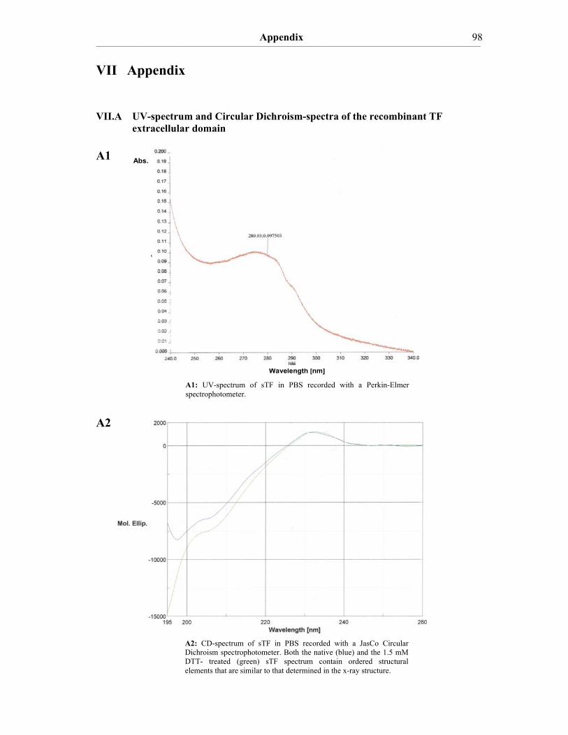
Appendix ____________________________________________________________________________________________________________________________________________________________________________________________________________________________
98
VII Appendix VII.A UV-spectrum and Circular Dichroism-spectra of the recombinant TF
extracellular domain A1 A2
A1: UV-spectrum of sTF in PBS recorded with a Perkin-Elmer spectrophotometer.
A2: CD-spectrum of sTF in PBS recorded with a JasCo Circular Dichroism spectrophotometer. Both the native (blue) and the 1.5 mM DTT- treated (green) sTF spectrum contain ordered structural elements that are similar to that determined in the x-ray structure.
Abs.
Wavelength [nm]

Acknowledgements ____________________________________________________________________________________________________________________________________________________________________________________________________________________________
99
VIII Acknowledgements
I would like to express my appreciation and thanks to my supervisor Prof. Dr. med. Bernd
Engelmann for giving me the opportunity to join his research group and to work on this
fascinating field of blood coagulation, for his support and his critical remarks on my work,
and guiding me to complete this dissertation thesis.
I am very grateful to Prof. Dr. med. Wolfgang Siess and the graduate school 438 “Vascular
Biology in Medicine” for three years of support and helpful discussions during the lecture
series and the yearly symposium in Herrsching.
I would like to thank Prof. Dr. med. Dr. h.c. Dietrich Seidel for giving me the opportunity
to perform my work in the Institute of Clinical Chemistry of the Ludwig-Maximilinas-
Universität München.
I want to thank our cooperation partners that were either providing us with various
materials used to perform this study or helped us with their expertise to conduct certain
experiments. In particular, I want to thank PD Dr. rer. nat. Viktor Magdolen (Technische
Universität München, Klinikum Rechts der Isar), Prof. Dr. rer. nat. Johannes Buchner
(Technische Universität München, Lehrstuhl für Biotechnologie), PD Dr. med. Steffen
Massberg (Technische Universität München, Deutsches Herzzentrum), Dr. rer. nat. Manuel
Than (Max Planck Institut für Biochemie, Martinsried), Dr. rer. nat. Stefan Uebel (Max
Planck Institut für Biochemie, Martinsried), Dr. rer. nat. Josef Kellermann (Max Planck
Institut für Biochemie, Martinsried), Dr. rer. nat. Sonja Hess (NIDDK, National Institute of
Health, Bethesda, US) and Dr. rer. nat. Jeannette Winter (University of Michigan, Ann
Arbor, US).
I am grateful to Pia Lohse and Prof. Dr. med. Peter Lohse for their kind help and support
concerning Molecular Biology and technical difficulties.

Acknowledgements ____________________________________________________________________________________________________________________________________________________________________________________________________________________________
100
Thanks to Dr. sc. nat. Christian Stirnimann (Institute of Biochemistry at the University of
Zürich, Switzerland) for the helpful discussions.
I am grateful to the Mathematisch-Naturwissenschaftliche Fakultät der Universität Zürich
(Switzerland), that gave me the opportunity to get educated as a biochemist.
I like to thank my colleagues and friends in the laboratory: Kiril, Davit, Tini, Berid, Lenka,
Nina, Verena, Geraldine, Sonja, Silke and Katja. We had an exciting and fruitful research
period together at the university hospital in Großhadern and I am grateful for the pleasant
work environment and our Biergarten and pub visiting evenings.
I want to thank Gabriella Anderson, which was the tandem partner of Daniela at the
University of Manchester and helped me to avoid English language mistakes in the present
work during her stay in München this summer.
Finally, I want to thank my parents, my brother Philip and my whole family in Reutlingen
for their moral support throughout the past years and in particular Daniela, who shared all
doubts and hopes with me. Without their support, understanding and love the present work
would never have succeeded.
101
CURRICULUM VITAE
Personal data
born December, 25th 1978 in Reutlingen, single, German
Educational background
● March 2007 Position in the research group of Prof. Dr. Fredrik Bäckhed at the Wallenberg Laboratory at the Sahlgrenska University Hospital, University of Gothenburg (Sweden) ● November 2006 Position at the Institute of Vascular Biology and
Thrombosis Research, Medical University of Vienna (Austria) in the research group of Prof. Erhard Hofer
● November 2005 Passed intermediate examination for the Dr. rer. Biol. Hum. (PhD in Human Biology) at the Medical
Faculty of the Ludwig-Maximilians-University of Munich
● April 2003-June 2006 PhD-student in Human Biology at the Institute for Clinical Chemistry in Prof. Dr. med. Bernd Engelmann’s Division of Haemostasis and Vascular Biology at the Ludwig-Maximilians-University of München (Germany) ● since April 2003 Member of graduate school 438 “Vascular Biology in Medicine” (speaker: Prof. Dr. med. Wolfgang Siess) supported with a scholarship ● Sept. 1998-March 2003 Diploma-student in Biochemistry with minor subject Immunology at the University of Zürich (Switzerland) ● Febr. 2001-Oct. 2002 Diploma thesis in the Structural Biology group of Prof. Dr. phil. nat. Markus Grütter at the University of Zürich on the “Biochemical and Structural Characterization of Helicobacter pylori Cysteine-rich Proteins C and E” ● ST 2001 Visiting student at the Institute for Physiologic Chemistry at the Eberhard-Karls-University of Tübingen (Germany) ● August 2001 Examination in Physical Biochemistry at the chair of Prof. Dr. rer. nat. Dr. h.c. Wolfgang Voelter at the Eberhard-Karls-University of Tübingen
102
● Aug.‐Sept. 1998 Additional Examinations to achieve the “Kantonale Maturität des Kantons Zürich” to get access to the Mathematisch-Naturwissenschaftliche Fakultät at the University of Zürich and to inscribe in Biochemistry ● June 1998 German Abitur at the Bildungszentrum Reutlingen Nord (Baden-Württemberg) Publication List Mittl P.R.E., Luthy L., Reinhardt C. and Joller H. 2003. Detection of High Titers of Antibody against Helicobacter pylori Cysteine-Rich Proteins A, B, C, and E in Helicobacter pylori-Infected Individuals. Clinical and Diagnostic Laboratory Immunology 2003;10(4):542-545. Reinhardt C, Brühl M-L, Grahl L, Manukyan D, Grahl L, Lorenz M, Altmann B, Dlugai S, Hess S, Konrad I, Mackman N, Ruddock L, Massberg S and Engelmann B. Protein disulfide isomerase signals vessel injury for initiation of fibrin generation. (in revision) Massberg S, Grahl L, Bidzhekov K, Konrad I, Kennerknecht E, Reinhardt C, Roes J, and Engelmann B.The neutrophil anti-microbial arsenal critically stabilizes nascent thrombi. (in preparation) Reinhardt C. New locations of intravascular tissue factor: indications. Hämostaseologie 2007;27(1):55-58. (review article)
Contributions to international Congresses Reinhardt C, Morgenstern E, and Engelmann B. October 13-15th, 2005. Neutrophil surface proteases – Activators of the intravascular Tissue Factor pathway. XXth European Platelet Meeting, Ede, The Netherlands (oral presentation) Reinhardt C, Grahl L, Bidzhekov K, Morgenstern E and Engelmann B. March 26-29th, 2006. Neutrophil surface proteases – activators of the intravascular Tissue Factor, 85th annual meeting of the German Physiological Society and Joint meeting of the Federation of European Physiological Societies, Munich, Germany (poster presentation). Abstract was short listed for the FEPS Young Investigators Award poster session. Grahl L, von Bruehl M, Reinhardt C, Manukyan D, Altmann B, Dlugai S, Konrad I, Lorenz M, Mackman N, Massberg S, Engelmann B. July 6th-12th, 2007. Protein Disulfide Isomerase (PDI) uncovers Tissue Factor (TF)-dependent fibrin formation after vessel injury. Congress of the International Society on Thrombosis and Haemostasis, Geneva, Switzerland. Abstract was awarded with an ISTH Young Investigators Award to Davit Manukyan.

103
Hiermit erkläre ich, die vorliegende Arbeit eigenständig und nur mit den angegebenen
Quellen und Hilfsmitteln angefertigt zu haben.
München im Februar 2007,
Christoph Reinhardt