an unusual new fossil shark (pisces: chondrichthyes> from...
TRANSCRIPT
Records of the Western AustralulIl Museulll Supplement No. 57: 151-156 (1999).
An unusual new fossil shark (Pisces: Chondrichthyes> from the LateDevonian of South Africa
M. Eric Andersonl, John A. Long2
, Robert W. Gess3 and Norton Hiller'
I J.L.B. Smith Institute of Ichthyology, Private Bag 1015, Grahamstown 6140, South Africa;email: [email protected]
2 Western Australian Museum, Francis St, Perth, WA 6000, Australia
3 Department of Geology, Rhodes University, P.G. Box 94, Grahamstown 6140, South Africa
• Canterbury Museum, Rolleston Ave, Christchurch, New Zealand
Abstract - A new stem-group chondrichthyan fish, PlesioselacJllIs macracanthllsgen. et sp. nov., is described from the Late Devonian Witpoort Formation,representing an estuarine lagoon site, near Grahamstown, South Africa. Basedon a single, fairly complete specimen, it is distinctive in its a single dorsal finbraced by a large, stout spine with numerous ribs and posterior denticles,apparently no second dorsal or anal fin, an amphistylic jaw suspension, and adistinctive triangular palatoquadrate. It is suggested that the species mayrepresent a high-latitude, Late Devonian relict taxon.
INTRODUCTION
In 1985 a shale bed was exposed by a road cuttingthrough the Witpoort Formation in the upperWitteberg Group, near Grahamstown, South Africa.It was soon found to be fossiliferous, and thedeposits were interpreted as representing astagnant coastal lagoon sheltered by a barrier islandcomplex in the Famennian stage of the UpperDevonian (Hiller and Taylor 1992; Long et al. 1997).The age of the site is based on brachiopodsoccurring in layers just above the fossiliferous shalebeds, and the overall nature of the fish fauna.Fossils of terrestrial and aquatic plants, aquaticinvertebrates and fishes have been reported (Taylorand Hiller 1993; Anderson et al. 1994; Cess andHiller 1995a, 1995b; Hiller and Cess 1996; Long etal. 1997). We consider that the site represents theearliest, relatively complete, aquatic community sofar discovered in Africa.
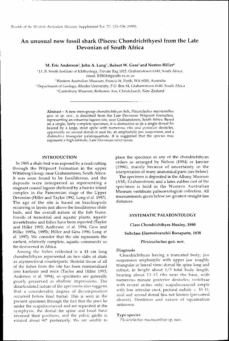
Among the fishes collected is a 44 cm longchondrichthyan represented on two slabs of shaleas asymmetrical counterparts. Skeletal tissue of allof the fishes from the site has been remineralizedinto kaolinite and mica (Taylor and Hiller 1993;Anderson et al. 1994), so specimens are generallypoorly preserved as shallow impressions. Thedisarticulated nature of the specimens also suggeststhat a considerable degree of decompositionoccurred before final burial. This is seen in thepresent specimen through the fact that the jaws lieunder the scapulocoracoid and are separated at thesymphysis, the dorsal fin spine and basal havereversed their positions, and the pelvic girdle isrotated about 90 0 posteriorly. We are unable to
place the specimen in any of the chondrichthyanorders as arranged by Nelson (1994) or Janvier(1996), mainly because of uncertainty in theinterpretation of many anatomical parts (see below).
The specimen is deposited in the Albany Museum(AM), Crahamstown; and a latex rubber cast of thespecimen is held in the Western AustralianMuseum vertebrate palaeontological collection. Allmeasurements given below are greatest straight-linedistances.
SYSTEMATIC PALAEONTOLOGY
Class Chondrichthyes Huxley, 1880
Subclass Elasmobranchii Bonaparte, 1838
Plesioselachus gen. novo
DiagnosisChondrichthyan having a truncated body; jaw
suspension amphistylic with upper jaw roughlytriangular in lateral view; dorsal fin spine long androbust, in height about 1/3 total body length,bearing about 13-15 ribs near the base, withnumerous minute posterior denticles; vertebraewith neural arches only; scapulocoracoid simplewith low articular crest; pectoral radials c. 10-11;anal and second dorsal fins not known (presumedabsent). Dentition and nature of squamationunknown.
Type speciesPlesioselachlls macracallthlls sp. nov.
152
EtymologyFrom the Greek 'plesios' (common, used to denote
primitive in zoological nomenclature), and'selachus' a shark, referring to its plesiomorphicanatomy.
Plesioselachus macracanthus sp. novoFigures 1-5
Chondrichthyan: Anderson et al. 1994: 401.
Chondrichthyan: Gess and Hiller 1995a: 290-293,figures 57, 58.
Material Examined
HolotypeAM 4817, entire fish (Figures I, 2), slightly
damaged, 44 cm in length, asymmetricallypreserved as two counterparts.
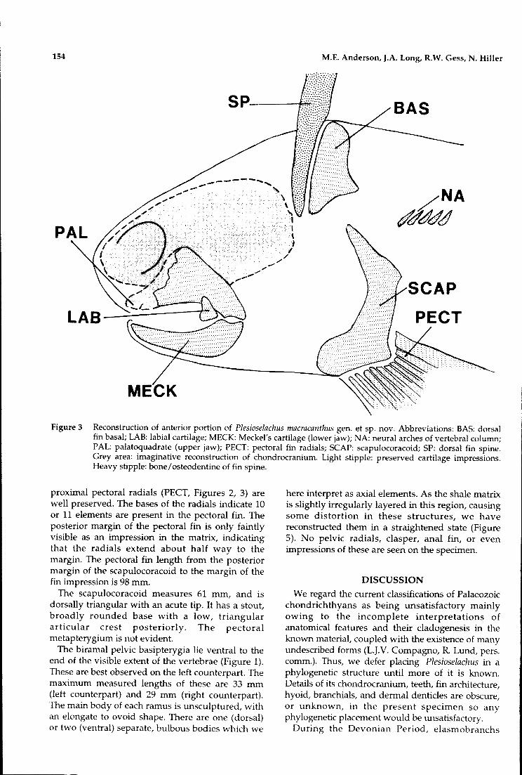
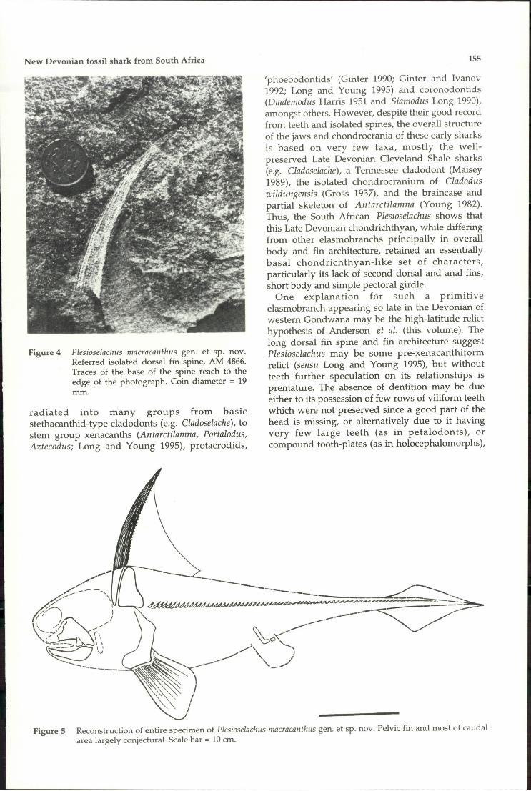
Referred MaterialAM 4866 an isolated dorsal fin spine (Figure 4).
Type Locality and HorizonN-2 highway bypass shale bed, c. 3 km south of
central Grahamstown, Eastern Cape Province,South Africa. Collected by R.W. Gess and M.Stonestreet, June 1990. Middle part of the WitpoortFormation, Witteberg Group, Cape Supergroup.
M.E. Anderson, J.A. Long, R.W. Gess, N. Hiller
Middle Famennian stage of the Late Devonian.
DiagnosisAs for genus.
EtymologyFrom the Greek 'makros', long, and 'akantha',
thorn, in reference to the dorsal fin spine.
DescriptionThe skeletal elements of the head (Figures I, 2)
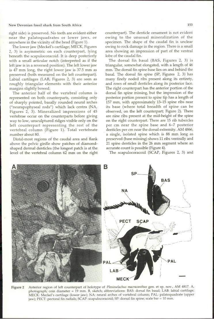
are preserved slightly dissociated, deeplyembedded in the right counterpart. The head, aspreserved, measures 41 mm in length, and about 20mm of the snout is estimated to be missing (givinga total estimated length of at least 61 mm). Theorbits are not seen, nor is there any trace of theneurocranium or visceral skeleton, these structuresare added to the reconstruction in Figure 3 purelyfor relative placement of the jaw and shouldergirdle structures.
The palatoquadrates (PAL, Figures 2, 3) arefragmented. One side (possibly the left) is about 3/4intact, missing only the anterior region of thesuborbital lamina. The cleaver-shaped dorsallamina is highly arched and the ventral surface isgently bowed with a shallow articular notchposteriorly situated. Only the broken posterior endof the other palatoquadrate (presumed to be the
Figure 1 Left counterpart of holotype of Plesioselachus macracanthus gen. et. cp. nov., AM 4817.
New Devonian fossil shark from South Africa
right side) is preserved. No teeth are evident eithernear the palatoquadrates or lower jaws, oranywhere in the vicinity of the head (Figure 1).
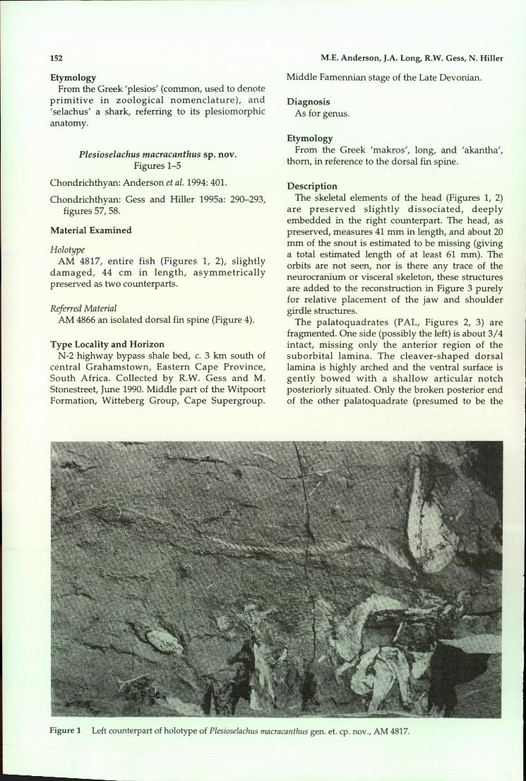
The lower jaw (Meckel's cartilage; MECK, Figures2, 3) is asymmetric on each counterpart, lyingbeneath the scapulocoracoid. It is deep posteriorlywith a small articular notch (interpreted as if theleft jaw is in a reversed position). The left lower jawis 47 mm long, the right lower jaw is 37.5 mm aspreserved (both measured on the left counterpart).Labial cartilages (LAB, Figures 2, 3) are seen asroughly triangular elements with their anteriormargins slightly bowed.
The anterior half of the vertebral column isrepresented on both counterparts, consisting onlyof sharply pointed, basally rounded neural arches("neurapophyseal rods") which lack centra (NA,Figures 2, 3). Mineralized impressions of 45vertebrae occur on the counterparts before givingway to low, unsculptured ridges visible only on theleft counterpart representing the rest of thevertebral column (Figure 1). Total vertebratenumber about 80.
Distal-most regions of the caudal area and flankabove the pelvic girdle show patches of diamondshaped dennal denticles (the longest patch is at thelevel of the vertebral column 62 mm on the right
153
counterpart). The denticle ornament is not evidentowing to the unusual mineralization of thespecimen. The shape of the caudal fin is unclearowing to rock damage in the region. There is a smallarea showing an impression of part of the ventrallobe of the caudal fin.
The dorsal fin basal (BAS, Figures 2, 3) istriangular, somewhat elongated, with a length of 46mm. The dorsal fin spine base lies on and behind thebasal. The dorsal fin spine (SP, Figures 2, 3) hasmany finely noded ribs present along its entirety,and rows of small denticles along its posterior face.The right counterpart has the anterior portion of thedorsal fin spine missing, but the impression of theposterior portion present to spine tip has a length of157 mm, with approximately 13-15 spine ribs nearits base (where total breadth of spine can beobserved, on the left counterpart; Figure 2). Thereare nine ribs present at the mid-height of the spineon the right counterpart. There are 15 rib tuberclesper cm near the spine base and 6-7 posteriordenticles per cm near the dorsal extremity. AM 4866,a single, isolated spine which is 88 mm long aspreserved (base missing) shows 11 ribs ventrally and21 spine denticles in the 26 mm segment where anaccurate count is possible (Figure 4).
The scapulocoracoid (SCAP, Figures 2, 3) and
BAS
BSP
\\
\\
)I\
""-
- - - __J
~\\
'. {-s. \-; \-J'~ I
..J/-?c? _J~iiII.....~.., \ r --..::-::---,,,.:.. .,-- I
I(- ---'
Figure 2 Anterior region of left counterpart of holotype of Plesioselachus macracanthus gen. et sp. nov., AM 4817. A,photograph; coin diameter = 19 mm. B, sketch; abbreviations: BAS: dorsal fin basal; LAB: labial cartilage;MECK: Meckel's cartilage (lower jaw); NA: neural arches of vertebral column; PAL: palatoquadrate (upperjaw); PECT: pectoral fin radials; SCAP: scapulocoracoid; SP: dorsal fin spine; scale bar = 10 mm.
154
MECK
SP--~
M.E. Anderson, J.A. Long, R.W. Gess, N. HilIer
SCAP
PECT
Figure 3 Reconstruction of anterior portion of Plesioselachus macracanthus gen. et sp. novo Abbreviations: BAS: dorsalfin basal; LAB: labial cartilage; MECK: Meckel's cartilage (lower jaw); NA: neural arches of vertebral column;PAL: palatoquadrate (upper jaw); PECT: pectoral fin radials; SCAP: scapulocoracoid; SP: dorsal fin spine.Grey area: imaginative reconstruction of chondrocranium. Light stipple: preserved cartilage impressions.Heavy stipple: bone/osteodentine of fin spine.
proximal pectoral radials (PECT, Figures 2, 3) arewell preserved. The bases of the radials indicate 10or 11 elements are present in the pectoral fin. Theposterior margin of the pectoral fin is only faintlyvisible as an impression in the matrix, indicatingthat the radials extend about half way to themargin. The pectoral fin length from the posteriormargin of the scapulocoracoid to the margin of thefin impression is 98 mm.
The scapulocoracoid measures 61 mm, and isdorsally triangular with an acute tip. It has a stout,broadly rounded base with a low, triangulararticular crest posteriorly. The pectoralmetapterygium is not evident.
The biramal pelvic basipterygia lie ventral to theend of the visible extent of the vertebrae (Figure 1).These are best observed on the left counterpart. Themaximum measured lengths of these are 33 mm(left counterpart) and 29 mm (right counterpart).The main body of each ramus is unsculptured, withan elongate to ovoid shape. There are one (dorsal)or two (ventral) separate, bulbous bodies which we
here interpret as axial elements. As the shale matrixis slightly irregularly layered in this region, causingsome distortion in these structures, we havereconstructed them in a straightened state (Figure5). No pelvic radials, clasper, anal fin, or evenimpressions of these are seen on the specimen.
DISCUSSION
We regard the current classifications of Palaeozoicchondrichthyans as being unsatisfactory mainlyowing to the incomplete interpretations ofanatomical features and their cladogenesis in theknown material, coupled with the existence of manyundescribed forms (L.J.V. Compagno, R Lund, pers.comm.). Thus, we defer placing Plesioselachus in aphylogenetic structure until more of it is known.Details of its chondrocranium, teeth, fin architecture,hyoid, branchials, and dermal denticles are obscure,or unknown, in the present specimen so anyphylogenetic placement would be unsatisfactory.
During the Devonian Period, elasmobranchs
New Devonian fossil shark from South Africa
Figure 4 Plesioselachus macracanthus gen. et sp. novoReferred isolated dorsal fin spine, AM 4866.Traces of the base of the spine reach to theedge of the photograph. Coin diameter = 19mm.
radiated into many groups from basicstethacanthid-type cladodonts (e.g. Cladoselache), tostem group xenacanths (Antarctilamna, Portalodus,Aztecodus; Long and Young 1995), protacrodids,
155
'phoebodontids' (Ginter 1990; Ginter and Ivanov1992; Long and Young 1995) and coronodontids(Diademodus Harris 1951 and Siamodus Long 1990),amongst others. However, despite their good recordfrom teeth and isolated spines, the overall structureof the jaws and chondrocrania of these early sharksis based on very few taxa, mostly the wellpreserved Late Devonian Cleveland Shale sharks(e.g. Cladoselache), a Tennessee cladodont (Maisey1989), the isolated chondrocranium of Cladoduswildungensis (Gross 1937), and the braincase andpartial skeleton of Antarctilamna (Young 1982).Thus, the South African Plesioselachus shows thatthis Late Devonian chondrichthyan, while differingfrom other elasmobranchs principally in overallbody and fin architecture, retained an essentiallybasal chondrichthyan-like set of characters,particularly its lack of second dorsal and anal fins,short body and simple pectoral girdle.
One explanation for such a primitiveelasmobranch appearing so late in the Devonian ofwestern Gondwana may be the high-latitude relicthypothesis of Anderson et al. (this volume). Thelong dorsal fin spine and fin architecture suggestPlesioselachus may be some pre-xenacanthiformrelict (sensu Long and Young 1995), but withoutteeth further speculation on its relationships ispremature. The absence of dentition may be dueeither to its possession of few rows of viliforrn teethwhich were not preserved since a good part of thehead is missing, or alternatively due to it havingvery few large teeth (as in petalodonts), orcompound tooth-plates (as in holocephalomorphs),
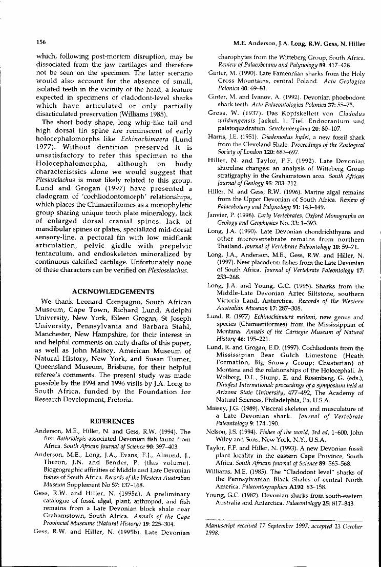
Figure 5 Reconstruction of entire specimen of Plesioselachus macracanthus gen. et sp. novo Pelvic fin and most of caudalarea largely conjectural. Scale bar = 10 cm.
156
which, following post-mortem disruption, may bedissociated from the jaw cartilages and thereforenot be seen on the specimen. The latter scenariowould also account for the absence of small,isolated teeth in the vicinity of the head, a featureexpected in specimens of cladodont-Ievel sharkswhich have articulated or only partiallydisarticulated preservation (Williams 1985).
The short body shape, long whip-like tail andhigh dorsal fin spine are reminscent of earlyholocephalomorphs like Echinochimaera (Lund1977). Without dentition preserved it isunsatisfactory to refer this specimen to theHolocephalomorpha, although on bodycharacteristsics alone we would suggest thatPlesioselachus is most likely related to this group.Lund and Grogan (1997) have presented acladogram of 'cochliodontomorph' relationships,which places the Chimaeriformes as a monophyleticgroup sharing unique tooth plate mineralogy, lackof enlarged dorsal cranial spines, lack ofmandibular spines or plates, specialized mid-dorsalsensory-line, a pectoral fin with low midflankarticulation, pelvic girdle with prepelvictentaculum, and endoskeleton mineralized bycontinuous calcified cartilage. Unfortunately noneof these characters can be verified on Plesioselachus.
ACKNOWLEDGEMENTS
We thank Leonard Compagno, South AfricanMuseum, Cape Town, Richard Lund, AdelphiUniversity, New York, Eileen Grogan, St JosephUniversity, Pennsylvania and Barbara Stahl,Manchester, New Hampshire, for their interest inand helpful comments on early drafts of this paper,as well as John Maisey, American Museum ofNatural History, New York, and Susan Turner,Queensland Museum, Brisbane, for their helpfulreferee's comments. The present study was madepossible by the 1994 and 1996 visits by J.A. Long toSouth Africa, funded by the Foundation forResearch Development, Pretoria.
REFERENCES
Anderson, M.E., Hiller, N. and Gess, RW. (1994). Thefirst Bothriolepis-associated Devonian fish fauna fromAfrica. South African Journal of Science 90: 397-403.
Anderson, M.E., Long, J.A., Evans, F.J., Almond, J.,Theron, J.N. and Bender, P. (this volume).Biogeographic affinities of Middle and Late Devonianfishes of South Africa. Records of the Western AustralianMuseum Supplement No 57: 137-168.
Gess, RW. and Hiller, N. (1995a). A preliminarycatalogue of fossil algal, plant, arthropod, and fishremains from a Late Devonian block shale nearGrahamstown, South Africa. Annals of the CapeProvincial Museums (Natural History) 19: 225-304.
Gess, R.W. and Hiller, N. (1995b). Late Devonian
M.E. Anderson, J.A. Long, R.W. Gess, N. Hiller
charophytes from the Witteberg Group, South Africa.Review of Palaeobotany and Palynology 89: 417-428.
Ginter, M. (1990). Late Famennian sharks from the HolyCross Mountains, central Poland. Acta GeologicaPolonica 40: 69-81.
Ginter, M. and Ivanov, A. (1992). Devonian phoebodontshark teeth. Acta Palaeontologica Polonica 37: 55-75.
Gross, W. (1937). Das Kopfskellett von Cladoduswildungensis Jaekel. 1. Tiel. Endocranium undpalatoquadratum. Senckenbergiana 20: 80-107.
Harris, J.E. (1951). Diademodus hydei, a new fossil sharkfrom the Cleveland Shale. Proceedings of the ZoologicalSociety of London 120: 683-697.
Hiller, N. and Taylor, F.F. (1992). Late Devonianshoreline changes: an analysis of Witteberg Groupstratigraphy in the Grahamstown area. South AfricanJournal ofGeology 95: 203-212.
Hiller, N. and Gess, RW. (1996). Marine algal remainsfrom the Upper Devonian of South Africa. Review ofPalaeobotany and Palynology 91: 143-149.
Janvier, P. (1996). Early Vertebrates. Oxford Monographs onGeology and Geophysics No. 33: 1-393.
Long, J.A. (1990). Late Devonian chondrichthyans andother microvertebrate remains from northernThailand. Journal of Vertebrate Paleontology 10: 59-71.
Long, J.A., Anderson, M.E., Gess, RW. and Hiller, N.(1997). New placoderm fishes from the Late Devonianof South Africa. Journal of Vertebrate Paleontology 17:253-268.
Long, J.A. and Young, G.c. (1995). Sharks from theMiddle-Late Devonian Aztec Siltstone, southernVictoria Land, Antarctica. Records of the WesternAustralian Museum 17: 287-308.
Lund, R (1977) Echinochimaera meltoni, new genus andspecies (Chimaeriformes) from the Mississippian ofMontana. Annals of the Carnegie Museum of NaturalHistory 46: 195-221.
Lund, Rand Grogan, E.D. (1997). Cochliodonts from theMississipian Bear Gulch Limestone (HeathFormation, Big Snowy Group; Chesterian) ofMontana and the relationships of the Holocephali. InWolberg, D.L., Stump, E. and Rosenberg, G. (eds.),Dinofest International: proceedings ofa symposium held atArizona State University, 477-492, The Academy ofNatural Sciences, Philadelphia, Pa, USA.
Maisey, J.G. (1989). Visceral skeleton and musculature ofa Late Devonian shark. Journal of VertebratePaleontology 9: 174-190.
Nelson, J.5. (1994). Fishes of the world, 3rd ed, 1-600, JohnWiley and Sons, New York, N.Y., USA.
Taylor, F.F. and Hiller, N. (1993). A new Devonian fossilplant locality in the eastern Cape Province, SouthAfrica. South African Journal of Science 89: 565-568.
Williams, M.E. (1985). The "Cladodont level" sharks ofthe Pennsylvanian Black Shales of central NorthAmerica. Palaeontographica A190: 83-158.
Young, G.c. (1982). Devonian sharks from south-easternAustralia and Antarctica. Palaeontology 25: 817-843.
Manuscript received 17 September 1997; accepted 13 October1998.