an unusual form lung injury - ipus.org.il · an unusual form lung injury meir krupsky md tel...
TRANSCRIPT

An Unusual form Lung Injury
Meir Krupsky MD
Tel –Aviv Sourasky Medical CTR

Case description
•
a 88 y.o. male•
No smoking history
•
Dancing instructor!!•
Past medical history:–
HTN
–
No TB contacts

1st admission
•
SOB -
Inspiratory, productive (whithish) cough, NO fever -
3 months duration
•
Weight loss 10Kg/ year•
Physical examination –rales
bilateral bases (more Rt)
•
FEV1=88% FEV1/FCV=76% O2 Sat=98%•
Labs:
101Glucose1.1Creat415KPlt12.3Hb
176LDH46Urea3.1Globulin13700WBC
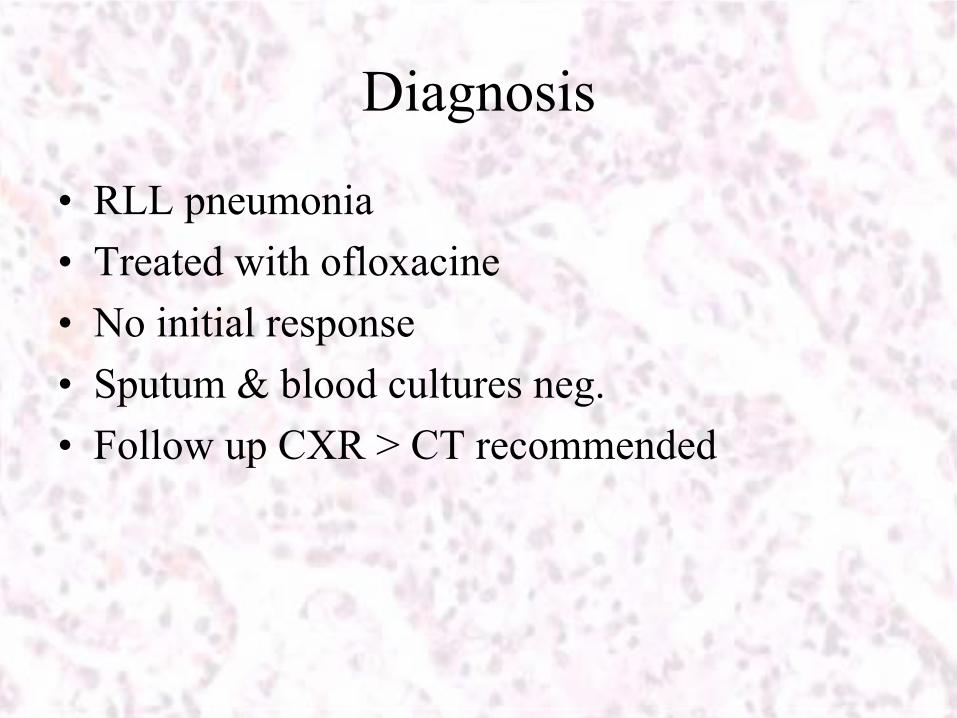
Diagnosis
•
RLL pneumonia•
Treated with ofloxacine
•
No initial response•
Sputum & blood cultures neg.
•
Follow up CXR > CT recommended
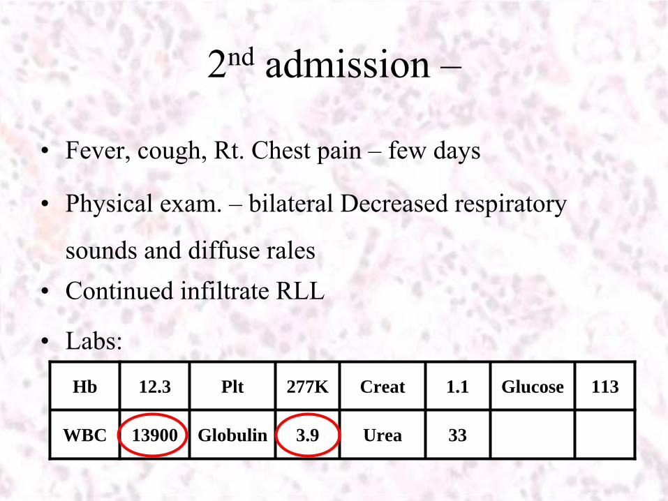
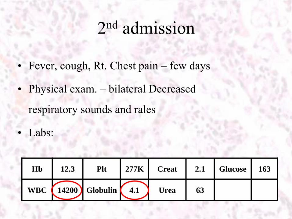
2nd admission –
•
Fever, cough, Rt. Chest pain –
few days
•
Physical exam. –
bilateral Decreased respiratory
sounds and diffuse rales•
Continued infiltrate RLL
•
Labs:
113Glucose1.1Creat277KPlt12.3Hb
33Urea3.9Globulin13900WBC
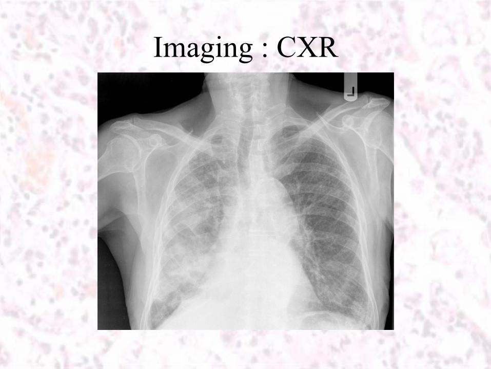
Imaging : CXR
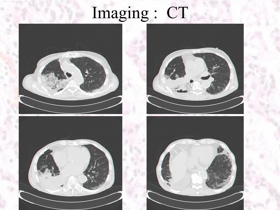
Imaging : CT
Imaging : CT
•
Rt. Lung base consolidation-homogeous, GGO, mix alveolar & interstitial opacities
•
Interlobular septal
thickening•
Crazy-
Paving Pattern

Additional testing
•
Bronchoscopy
–
Rt. thickened bronchial congested & hyperemic mucosa, easy bleed, No obsrtruction
No Mass/Infiltrative tumor
•
Biopsies -
RLL & RUL •
BAL-
RLL

Case description
•
a 77 y.o. male•
Past medical history:–
D.M. –
Insulin Rx.
–
CRF-
diabetic nephropathy–
HTN
–
Permanent pacemaker d/t
C-AVB–
CVA –
10 months ago
Medications
•
Insulin•
Furosamide
•
PPI’s•
???

1st admission
•
Fever without localizing complaints, 3 days duration•
Recent dental treatment
•
Physical examination –
rales
Rt. Lung base
•
Labs:
256Glucose1.8Creat415KPlt12.3Hb
176LDH56Urea3.5Globulin17800WBC
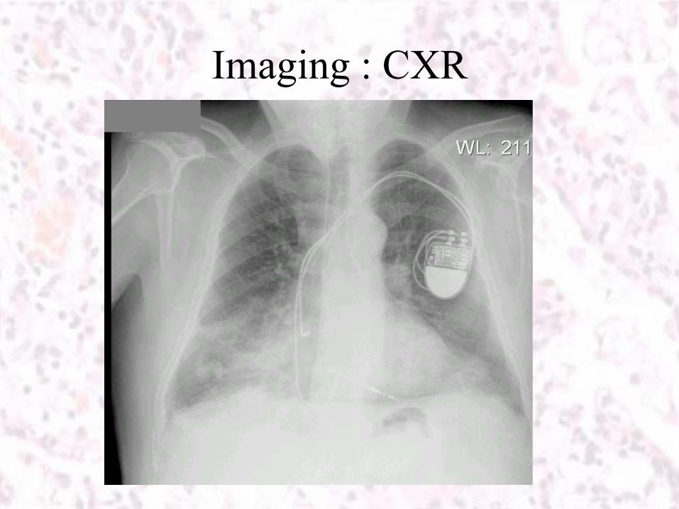
Imaging : CXR
Diagnosis
•
RLL pneumonia•
Treated with amoxycillin/clavulanate
&
ofloxacine•
No initial response, blood cultures neg.
•
TTE, oral surgeon consultation –
no abnormality
•
Slow decrease in temp. –
discharged•
Follow up CXR > CT recommended

•
CXR -
Continued infiltrate RLL•
ESR-
88
•
No further evaluation done
2nd admission
•
Fever, cough, Rt. Chest pain –
few days
•
Physical exam. –
bilateral Decreased
respiratory sounds and rales
•
Labs:
163Glucose2.1Creat277KPlt12.3Hb
63Urea4.1Globulin14200WBC
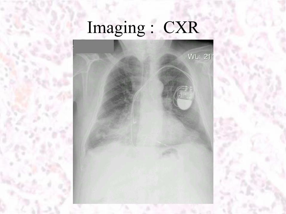
Imaging : CXR
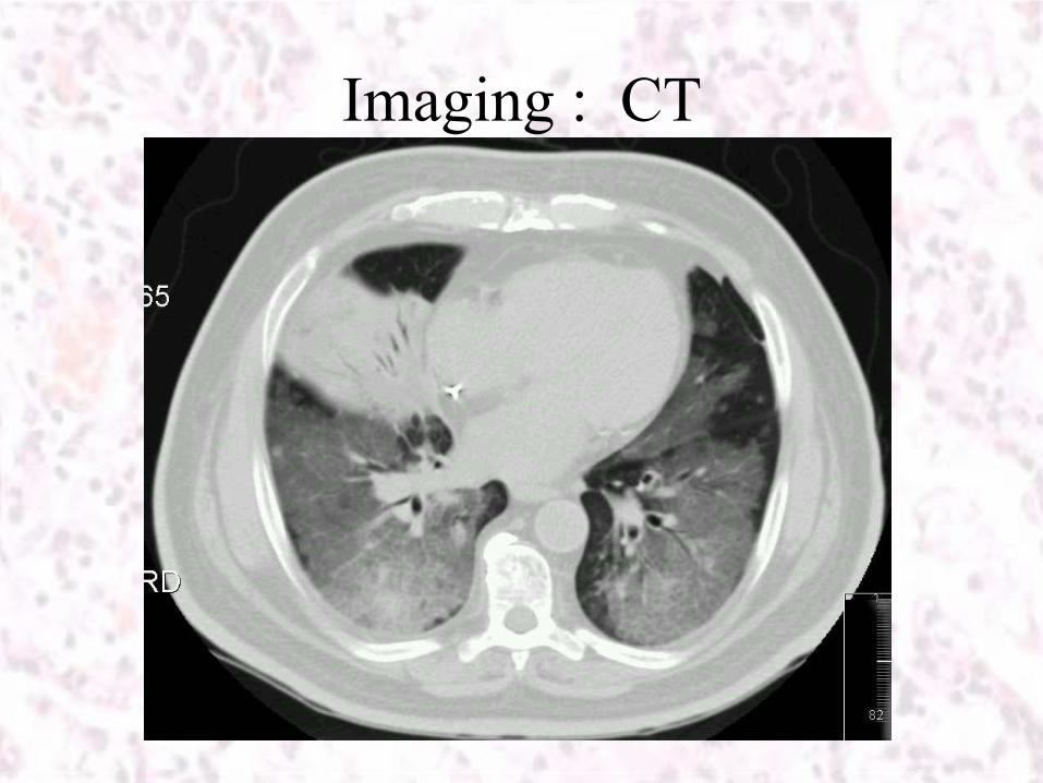
Imaging : CT
Additional testing
•
Pulmonary Function tests –
low DLCO, mild combined restriction & obstruction
•
Sputum for culture and Sudan Black staining –
negative•
Gallium scan –
a Ga. avid pulmonary lesion –
bilateral (most
active -
RML)•
Bronchoscopy
–
thickened bronchial mucosa,
No obstruction No tumor•
Biopsies and BAL

Imaging : CT

Histology
•
Fragments of lung parenchyma showing:–
Numerous clear droplets
–
Intra-alveolar and intersitial
macrophages with vacuolated cytoplasm
–
Reactive hyperplasia and septal
thickening
–
No granulomata
–
No malignancy

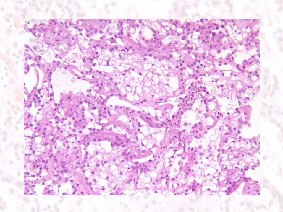
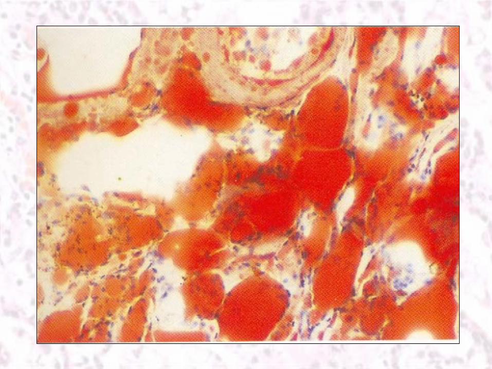
Diagnosis: Lipoid pneumonia
Lipoid Pneumonia: an Unusual form of Drug Induced Lung Injury
Lipoid pneumonia (LP)
•
the result of foreign body type reaction to the presence of lipid material within the lung parenchyma.
•
LP can be caused by the deposition of:
–
Endogenous lipid material
–
aspiration or inhalation of Exogenous lipids


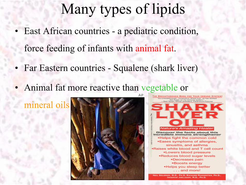
Many types of lipids•
East African countries -
a pediatric condition,
force feeding of infants with animal fat.
•
Far Eastern countries -
Squalene
(shark liver)
•
Animal fat more reactive than vegetable
or
mineral oils
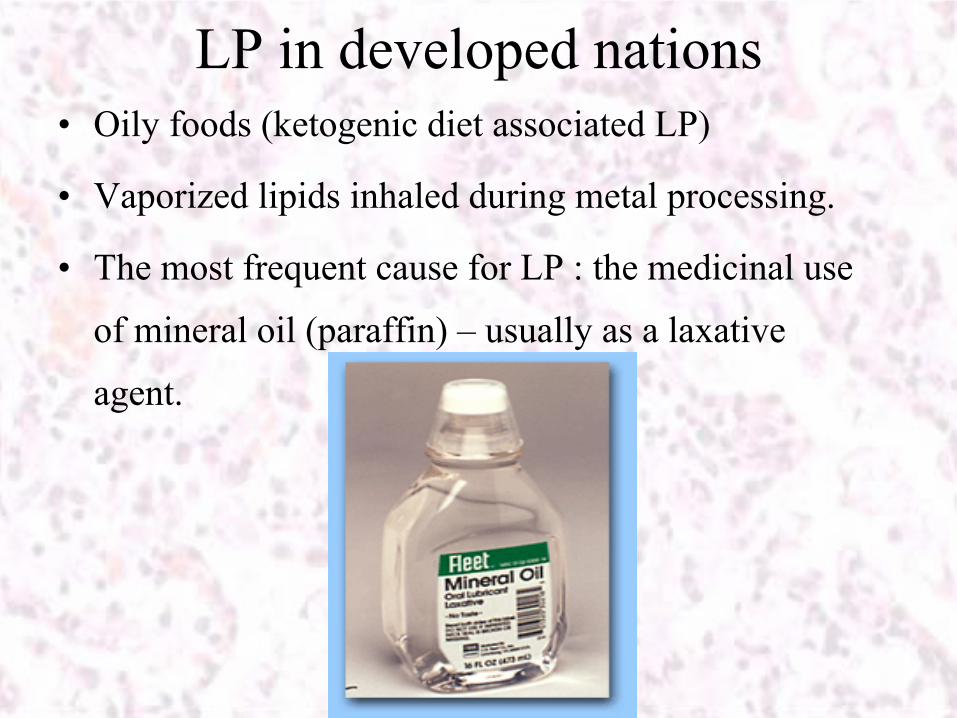
LP in developed nations•
Oily foods (ketogenic
diet associated LP)
•
Vaporized lipids inhaled during metal processing.
•
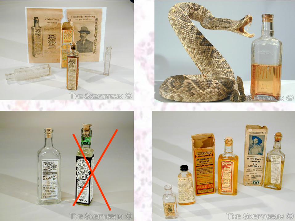
The most frequent cause for LP : the medicinal use
of mineral oil (paraffin) –
usually as a laxative
agent.
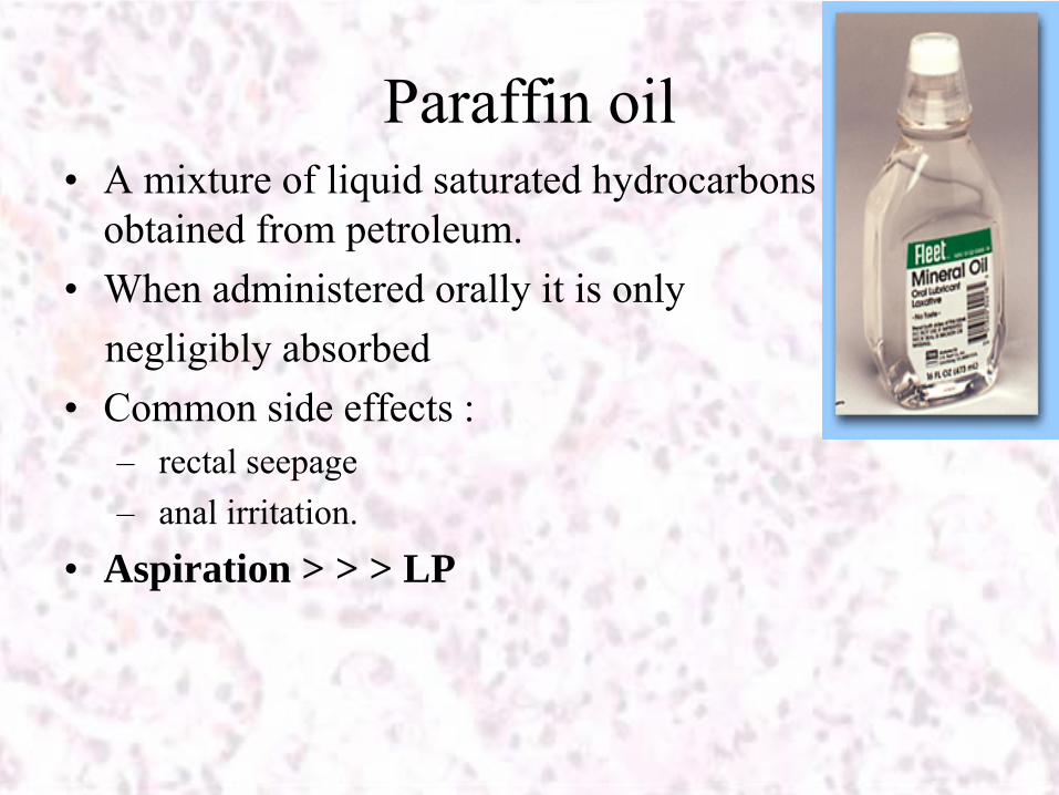
Paraffin oil•
A mixture of liquid saturated hydrocarbons obtained from petroleum.
•
When administered orally it is only negligibly absorbed
•
Common side effects :–
rectal seepage
–
anal irritation.
•
Aspiration > > > LP
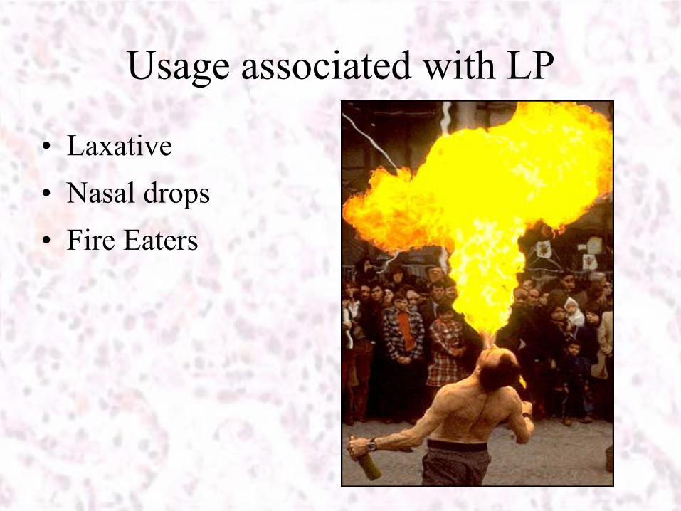
Usage associated with LP
•
Laxative•
Nasal drops
•
Fire Eaters
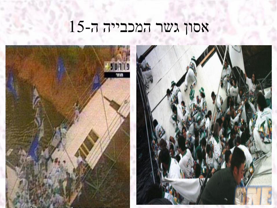
15-אסון ג שר המכ ביי ה ה
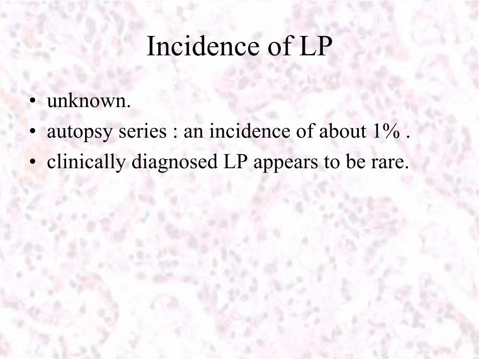
Incidence of LP
•
unknown. •
autopsy series : an incidence of about 1% .
•
clinically diagnosed LP appears to be rare.
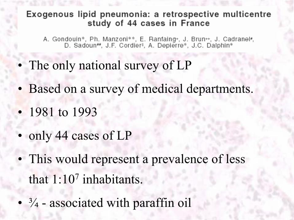
•
The only national survey of LP
•
Based on a survey of medical departments.
•
1981 to 1993
•
only 44 cases of LP
•
This would represent a prevalence of less that 1:107
inhabitants.
•
¾
-
associated with paraffin oil
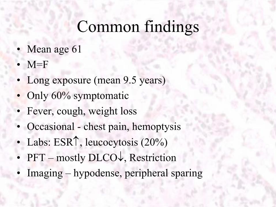
Common findings•
Mean age 61
•
M=F•
Long exposure (mean 9.5 years)
•
Only 60% symptomatic•
Fever, cough, weight loss
•
Occasional -
chest pain, hemoptysis•
Labs: ESR, leucocytosis
(20%)
•
PFT –
mostly DLCO, Restriction•
Imaging –
hypodense, peripheral sparing
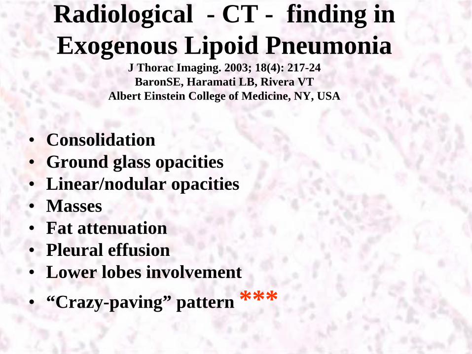
Radiological - CT - finding in Exogenous Lipoid Pneumonia
J Thorac Imaging. 2003; 18(4): 217-24 BaronSE, Haramati LB, Rivera VT
Albert Einstein College of Medicine, NY, USA
•
Consolidation•
Ground glass opacities
•
Linear/nodular opacities•
Masses
•
Fat attenuation•
Pleural effusion
•
Lower lobes involvement•
“Crazy-paving” pattern ***
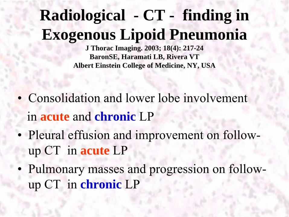
Radiological - CT - finding in Exogenous Lipoid Pneumonia
J Thorac Imaging. 2003; 18(4): 217-24 BaronSE, Haramati LB, Rivera VT
Albert Einstein College of Medicine, NY, USA
•
Consolidation and lower lobe involvement in acute and chronic LP
•
Pleural effusion and improvement on follow- up CT in acute LP
•
Pulmonary masses and progression on follow- up CT in chronic LP
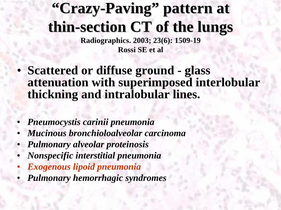
““CrazyCrazy--PavingPaving”” pattern at pattern at thinthin--section CT of the lungssection CT of the lungs
Radiographics. 2003; 23(6): 1509-19 Rossi SE et al
•
Scattered or diffuse ground - glass attenuation with superimposed interlobular thickning and intralobular lines.
•
Pneumocystis carinii pneumonia•
Mucinous bronchioloalveolar carcinoma
•
Pulmonary alveolar proteinosis•
Nonspecific interstitial pneumonia
•
Exogenous lipoid pneumonia•
Pulmonary hemorrhagic syndromes
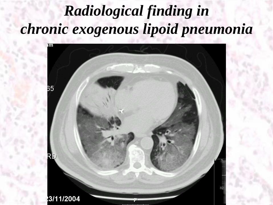
Radiological finding in chronic exogenous lipoid pneumonia

Treatment of LP
•
???–
Severe anecdotal cases:
•
Whole lung lavage•
Corticosteroids
–
Milder cases:•
Corticosteroids
•
Avoidance of further exposure without specific therapy
Natural history of LP - ????
•
two deaths unrelated to the lipid pneumonia.
•
In the 32 cases in which the oil was discontinued:
–
5 patients deteriorated (despite corticosteroid therapy in
one case)
–
27 patients remained stable/ improved regardless of
concomitant treatment

Imaging follow-up
•
3 complete cures (14%)•
6 improvements (29%)
•
10 stable courses (48%)•
2 deteriorations(10%)
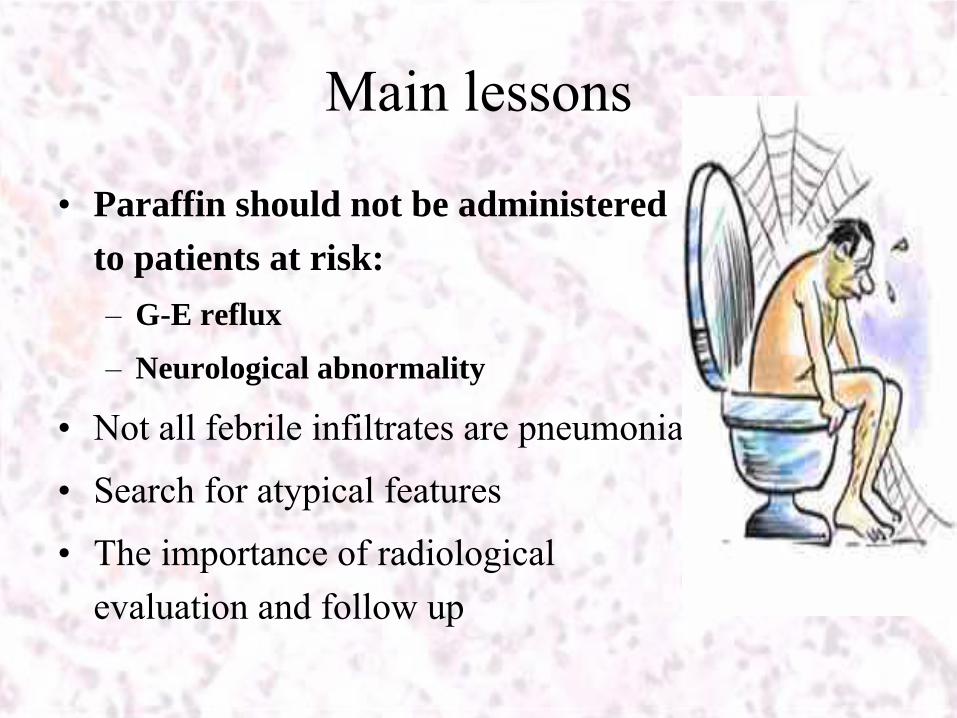
Main lessons
•
Paraffin should not be administered to patients at risk:–
G-E reflux
–
Neurological abnormality
•
Not all febrile infiltrates are pneumonia
•
Search for atypical features
•
The importance of radiological evaluation and follow up

ExLP-
Exogenous Lipoid Pneumonia•
Most Elderly –late 7-8th decade
•
Infants and mentally retarded
•
Impaired swallowing mechanism: neurological and esophageal disorders.
•
Consistent use of Oils :mineral, animal, vegetable oils in laxatives, nasal drops, mout spray, oral lubricants, insecticides or traditional folk remedies, occupational fire hazar of fire eaters (maccabia
1997)
•
The irritation causing agent enters the lung: Aspiration, inhalation or ionized vegetable -
radiopaque medium for lymphangiography, bronchography
etc.
•
Vegetable oils, mostly expectorated, residual oils leads to ExLP
•
Mineral oil -
Liquid petroleum or paraffin, mixture of long chain saturated hydrocarbons.
ExLP- Exogenous Lipoid Pneumonia•
Lung irritation -
Diffuse parenchymal
reaction
-
localized masses / parafinnomas
•
Animal fat is the most harmful to lung tissue –
hydrolized
by (pulmonary) lipase into FFA > > severe inflammatory reaction & tissue necrosis. Observed in infants with ExLP, cultural practice of forced feedings of animal fat (Ghee).
•
ExLP
3 stages: 1. Toxic agitation of capillary endothelium -
alveolar exudative
damage
2. Macrophages (alveolar & interstitial) activation -
oil phagocytosis
& degradation
3. Fibrointerstitial
and granulomatous
reactions
•
High lipid content (animal) repress phagocytosis leaving the lymphocytes as the main cells responsible for fat removal.
•
Histology -
Fat-laden macrophages and prominant
pleural lymphocytes, easily mistaken as lymphocytic
carcinomatosis.
Questions? (and maybe some answers…)Paraffin: Yes or No?
EnLP-
Endogenous Lipoid Pneumonia (Golden pneumonitis / Cholesterol pneumonitis)
•
Collection of intrinsic lipids in the lungs
•
Chronic bronchial obstruction/Obstructive pneumonitis: foreign bodies, tumorsBronchiolitis
obliterans
( chemotherapy/ radiotherapy -
release of
lipids in alveoli )
•
Pulmonary alveolar proteinosis, repetitive fungal pneumonia, Fat embolism, Lipid storage diseases: Gaucher’s, Niemann-Pick and Disseminated lipogranulomatosis
•
Normal lung chemically analyzed-
fat content 8.63/100g of dry tissue 19% is cholesterol ( percentage marked increase in smokers)
EnLP-
Endogenous Lipoid Pneumonia (Golden pneumonitis
/ Cholesterol pneumonitis
)
•
EnLP-
Link to Lung Cancer (in resected
lungs of 33/147 patients with lung cancer) 18% in Adeno-ca, 31% in Squamous
cell ca.
•
Lung parenchyma distal
to obstructive tumor.Transbronchial
dissemination of breakdown products of
NSCLC cells, including mucin, could contribute to the spread of non-
obstructive component of EnLP.
•
Histology of coexisting NSCLC & EnLP (lipids) similar to coexisting Pulmonary alveolar proteinosis (surfactant=lipids & protein) & NSCLC (Squamous
cell ca
& Large cell ca)
EnLP - NSCLC
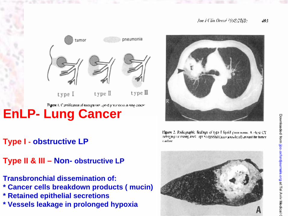
EnLP- Lung Cancer
Type I - obstructive LP
Type II & III – Non- obstructive LP
Transbronchial dissemination of:* Cancer cells breakdown products ( mucin)* Retained epithelial secretions* Vessels leakage in prolonged hypoxia

LP & PET

Take Home message Lipoid pneumonia (LP)
•
Old man & Lung infiltrate & Fever & ESR & WBC is not Sine qua non – Infective Pneumonia
•
ExLP
and EnLP
–
two different entities.
•
In contradistinction to ExLP-
External Oils :mineral, animal, vegetable oils , EnLP-
Obstructive pneumonitis
•
Unlike ExLP, the accumulation of lipid-rich cellular debris in EnLP does not manifest radiologically as lipid- containing opacities
•
Gallium scan – ExLP avid pulmonary lesion
•
Several entities (Infections, lipid storage, PAP) are considered
within the spectrum of EnLP.
•
EnLP
confirmed diagnosis is histopathologic -
imaging vary
•
EnLP Link to NSCLC (Type I , II & III)
•
PET-FDG scan - ExLP avid pulmonary lesion