an sem study of acanthogyrus (acanthosentis) tilapiae tilapiae.pdfmalawiensis amin and hendrix, 1999...
TRANSCRIPT

Sci Parasitol 13(2):57-63, June 2012 ISSN 1582-1366
ORIGINAL RESEARCH ARTICLE
57
An SEM study of Acanthogyrus (Acanthosentis) tilapiae
(Acanthocephala: Quadrigyridae) from Africa documenting
previously unreported features and host parasite interface
Omar M. Amin1�, Richard A. Heckmann2
1 – Institute of Parasitic Diseases (IPD), 11445 E. Via Linda, #2-419, Scottsdale, Arizona, 85259, U.S.A.
2 – Brigham Young University, Department of Biology, Provo, Utah 84602, U.S.A.
Correspondence: Tel. 480-767-2522, Fax 480-767-5855, E-mail [email protected]
Abstract. Acanthogyrus (Acanthopsentis) tilapiae (Baylis, 1948) is the most widely distributed species of
Acanthogyrus in many countries associated with the Nile River in Africa. It has been described by various authors
but much of its external morphological features remained unknown until recently explored by SEM in our
present study from specimens collected from cichlid fishes in Lake Malawi, Africa. Newly observed features
include the proboscis armature and sensory pores, epidermal micropores, and trunk spines. Attachment and
interface between worm and host intestinal lining are also reported for the first time.
Keywords: Acanthogyrus (Acanthopsentis) tilapiae; Acanthocephala; Cichlid fish; Lake Malawi; Africa;
Morphology; SEM.
Received 12/04/2012. Accepted 12/05/2012.
Introduction
Only 5 of the 38 known species of the subgenus
Acanthosentis are found in Africa. Thirty-one of
the other 33 species are Asian, mostly in the
Indian subcontinent and China. In Africa,
Acanthogyrus (Acanthopsentis) tilapiae (Baylis,
1948) is a widely distributed endemic species
mostly in cichlids from countries associated
with the Nile River (Amin and Hendrix, 1999).
The 4 other African species appear to be of
restricted distribution: Acanthogyrus
(Acanthosentis) maroccanus (Dollfus, 1951) in
Morocco, Acanthogyrus (Acanthosentis)
nigeriensis Dollfus and Golvan, 1956 in the
Niger River, Acanthogyrus (Acanthosentis)
papilo Troncy and Vassiliadis, 1974 in West
Africa, and Acanthogyrus (Acanthosentis)
malawiensis Amin and Hendrix, 1999 in Lake
Malawi.
Until 1984, A. (A.) tilapiae was reported from at
least 10 species of the cichlid genus Tilapia in
Tanzania (Baylis, 1948), Congo (Prudhoe,
1951; Golvan, 1957), Madagascar (Golvan,
1965), Uganda (Khalil and Thurston, 1973),
Chad (Troncy, 1974), Nigeria (Shotter, 1974),
Egypt (Amin, 1978), and unidentified locations
(Marchand and Mattei, 1976; Marchand, 1984).
Amin and Hendrix (1999) added 19 new host
records from 18 cichlid and 1 bagrid fish
species from southeastern Lake Malawi. Khalil

Sci Parasitol 13(2):57-63, June 2012 ISSN 1582-1366
ORIGINAL RESEARCH ARTICLE
58
and Thurston (1973) also reported A. (A.)
tilapiae from unidentified cichlids in Lake
Malawi, Lake Tanganyika, and other lakes in
the Congo. In their ecological study of A. (A.)
tilapiae in Lake Malawi, Amin et al. (2008)
examined 9 species of fish from 7 different
sites and added 5 new host records.
Since its imperfect description by Baylis
(1948), descriptive accounts with various
degrees of coverage and detail were reported
only by Troncy (1970), Amin (1978), and Amin
and Hendrix (1999). The presented SEM study
is based on extensive collections of A. (A.)
tilapiae from cichlid fishes from Lake Malawi,
Africa (Amin et al., 2008). The new
observations fill an important gap of missing
information and enhance our understanding of
the morphology of A. (A.) tilapiae especially
related to the proboscis armature and sensory
pores, epidermal micropores, and trunk spines.
Attachment and interface between worm and
host intestinal lining are also reported.
Materials and methods
About 2,000 specimens of A. (A.) tilapiae
(Baylis, 1948) were collected from 9 species of
cichlid fish hosts (Cichlidae: Perciformes) in
Lake Malawi, Africa, during September 2005.
Cichlids were captured by gill nets while scuba
diving in 7 sites: (1) Chirwa Islands
(10°27’48.94”S; 34°16’35.77”E), Chilumba (19
September); (2) Domwe Island (13°58’05.43”S;
34°49’04.01”E) (6 September, 16 September);
(3) Luwino Reef (10°26’17.41”S; 34°17’0.16”E)
(19 September); (4) Mpanga Rocks
(10°25’49.65”S; 34°16’44.64”E) (19
September); (5) Otter Point (14°02’21.33”S;
34°49’23.85”E) (2 September); (6) Thumbi
West Island (14°01’22.83”S; 34°49’16.63E) (3
September); and (7) Zimbawee Rock
(13°57’40.73”S; 34°48’08.88”E) (10
September). The species of fish included
Labeotropheus trewavasae (Fryer 1956)
(scrapemouth mbuna), Pseudotropheus
emmiltos (Stauffer et al. 1997) (red top),
Pseudotropheus zebra (Boulenger 1899) (zebra
mbuna), Melanochromis vermivorus (Trewavas
1935) (purple mbuna), Nimbochromis
polystigma (Regan 1922), Protomelas
taeniolatus (Trewavas 1935) (spindle hap),
Pseudotropheus elongates (Fryer 1956)
(elongate mbuna), Tropheops microstoma
(Trewavas 1935), and Rhamphochromis sp.
(Regan 1922) (see Amin et al., 2008).
Fish were kept alive for up to 1 to 3 days before
dissection. After the fish were killed with an
overdose of MS222, they were weighed,
measured, and sexed, and the intestines were
removed and transferred to petri dishes
containing 0.6% saline. Collected worms were
fixed directly in 10% formalin, then later,
washed in water and transferred into 70%
ethanol to transport to our Arizona facility for
processing. Worms were punctured with a fine
needle and subsequently stained in Mayer’s
acid carmine, destained in 4% hydrochloric
acid in 70% ethanol, dehydrated in ascending
concentrations of ethanol (24 hr each), and
cleared in graduated concentrations of
terpineol in 100% ethanol to 100% terpineol,
then 50% terpineol in 50% Canada balsam (24
hours each). Whole worms were then mounted
in Canada balsam.
For SEM studies, 23 specimens from P. zebra as
well as those affixed to host tissue previously
fixed in 70% ethanol were placed in critical-
point drying baskets and dehydrated using
ethanol series of 95% and 3 N 100% for at least
10 min per soak followed by critical point
drying (Lee, 1992). Samples were mounted on
SEM sample mounts, gold coated and observed
with a scanning electron microscope (XL30
ESEMFEG; FEI, Hillsboro, Oregon). Digital
images of the structures were obtained using
digital imaging software attached to a
computer.
Voucher specimens of A. tilapiae from Lake
Malawi were deposited at the U.S. National
Parasite Collection (USNPC) at Beltsville,
Maryland # 99961.
Results and discussion
Morphologically, our specimens were similar
to those of the first Lake Malawi survey by
Amin and Hendrix (1999), as well as those
reported from Egypt by Amin (1978). The Lake
Malawi specimens from cichlids from both
collections were, however, smaller than those
from the Nile River in Egypt from Tilapia
niloticus (Linnaeus) (up to 60.0 cm SL) and

Sci Parasitol 13(2):57-63, June 2012 ISSN 1582-1366
ORIGINAL RESEARCH ARTICLE
59
Tilapia zillii (Gervais) (up to 40.0 cm SL)
(Amin, 1978). The Egyptian specimens were, in
turn, somewhat smaller than those reported by
Baylis (1948) from Tilapia lidole (Trewavas)
(up to 38.0 cm SL) in Lake Nyasa (Lake
Malawi) but of similar size to those reported by
Troncy (1970) from the amphibious giant otter
shrew Potamogale velox du Chaillu (the only
known, but accidental, mammalian host of A.
tilapiae) in Yaoundé (Cameroon). Host factors
appear to enhance greater growth in the larger
Tilapia compared with the smaller cichlids
examined (up to 11.1 cm SL) (Amin et al.,
2008). Available measurements of other
structures such as the proboscis and proboscis
hooks were comparable in specimens reported
by Baylis (1948), Troncy (1970), Amin (1978)
and Amin and Hendrix (1999). There are,
however discrepancies in other characters
such as the number of sub-cuticular giant
nuclei and the number of transverse rings of
trunk spines. Baylis (1948) counted 2 dorsal
and 3 ventral giant nuclei but we found usually
2 dorsal and 6 ventral nuclei that, however,
varied between 2-4 dorsal and 4-6 ventral
nuclei in various combinations. Baylis (1948)
also counted 14 closely spaced rings of trunk
spines in the “forbody” and 18-20 more widely
spaced rings in the “body proper”. We found no
such division of trunk into 2 regions which
appeared to be a result of contraction in his
specimens, and counted a total of 28-38 and
28-42 rings of spines in males and females,
respectively, that appeared to merge closer
more posteriorly (figure 8).
New SEM observations
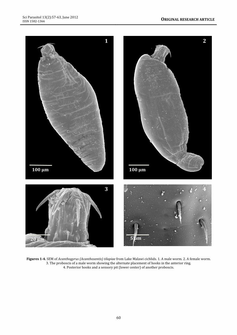
These observations include (1) the ovoid shape
of males (figure 1) compared to the elongate
shape of females with parallel sides (figure 2).
(2) The arrangement of anterior proboscis
hooks at alternating levels (figure 3) and not in
a straight circle as pictured in Baylis (1948,
figure 2) and Golvan (1957, figure 7). (3) The
presence of paired sensory pits at the base of
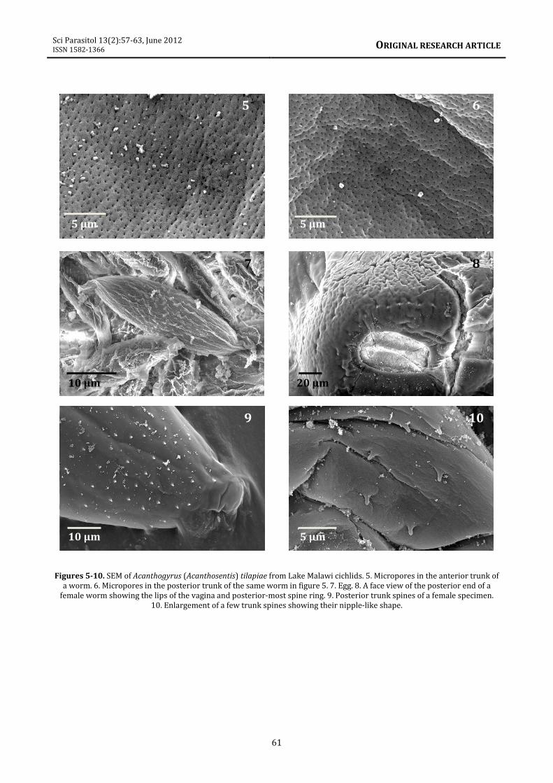
the proboscis (figure 4). (4) Dermal
micropores with different pore size and
spacing distributed in the epidermis of the
anterior trunk (figure 5) and posterior trunk
(figure 6); see Amin et al. (2009) for
implications to differential absorption. (5) The
ovoid eggs have corrugated surface (figure 7).
(6) The oblong lips of the vulva and the
extension of the posterior-most ring of trunk
spines to the genital orifice (figure 8). (7) The
distribution of trunk spines in the posterior
end of the trunk (figure 9). (8) The blunt-ended
nipple-like shape of trunk spines was observed
to be consistent throughout the body (figure
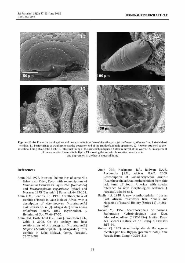
10). (9) The circular arrangement of posterior
trunk spines at the recessed posterior end of a
female (figure 11).
Host-parasite interface
This interface is depicted by an attached worm
(figure 12), site of attachment within the host
intestinal folds after removal of the parasite in
figure 12 (figure 13), and the actual site of
attachment enlarged to show the holes where
proboscis hooks were inserted in a depression
caused by the insertion of the proboscis (figure
14).
The description of A. (A.) tilapiae is greatly
enhanced by the new information provided
and documented for the first time with SEM
images. An interesting perspective on the host-
parasite interface is also demonstrated
showing evidence of the deep penetration of
proboscis hooks in host intestinal lining for the
first time.
Acknowledgments
We are grateful to Dr. Atif Naggar of Ain Shams
University, Cairo, Egypt, currently at Brigham
Young University, Provo, Utah, for his artful
preparation of the plates (figures 1-14). This
project was supported by an institutional grant
from the Institute of Parasitic Diseases to OMA.
1
Sci Parasitol 13(2):57-63, June 2012 ISSN 1582-1366
ORIGINAL RESEARCH ARTICLE
60
Figures 1-4. SEM of Acanthogyrus (Acanthosentis) tilapiae from Lake Malawi cichlids. 1. A male worm. 2. A female worm.
3. The proboscis of a male worm showing the alternate placement of hooks in the anterior ring.
4. Posterior hooks and a sensory pit (lower center) of another proboscis.
3 4
100 µm 100 µm
20 µm 5 µm
1 2
Sci Parasitol 13(2):57-63, June 2012 ISSN 1582-1366
ORIGINAL RESEARCH ARTICLE
61
Figures 5-10. SEM of Acanthogyrus (Acanthosentis) tilapiae from Lake Malawi cichlids. 5. Micropores in the anterior trunk of
a worm. 6. Micropores in the posterior trunk of the same worm in figure 5. 7. Egg. 8. A face view of the posterior end of a
female worm showing the lips of the vagina and posterior-most spine ring. 9. Posterior trunk spines of a female specimen.
10. Enlargement of a few trunk spines showing their nipple-like shape.
5 6
7 8
9 10
5 µm 5 µm
20 µm 10 µm
10 µm 5 µm
Sci Parasitol 13(2):57-63, June 2012 ISSN 1582-1366
ORIGINAL RESEARCH ARTICLE
62
Figures 11-14. Posterior trunk spines and host-parasite interface of Acanthogyrus (Acanthosentis) tilapiae from Lake Malawi
cichlids. 11. Perfect rings of trunk spines at the posterior end of the trunk of a female specimen. 12. A worm attached to the
intestinal lining of a cichlid host. 13. Intestinal lining of the same fish in figure 12 after removal of the worm. 14. Enlargement
of the same attachment site in figure 13 showing the anterior hook attachment marks
and depression in the host’s mucosal lining
References
Amin O.M. 1978. Intestinal helminthes of some Nile
fishes near Cairo, Egypt with redescriptions of
Camallanus kirandensis Baylis 1928 (Nematoda)
and Bothriocephalus aegyptiacus Ryšavý and
Moravec 1975 (Cestoda). J. Parasitol. 64:93-101.
Amin O.M., Hendrix S.S. 1999. Acanthocephala of
cichlids (Pisces) in Lake Malawi, Africa, with a
description of Acanthogyrus (Acanthosentis)
malawiensis sp. n. (Quadrigyridae) from Labeo
cylindricus Peters, 1852 (Cyprinidae). J.
Helminthol. Soc. W. 66:47-55.
Amin O.M., Oosterhout C.V., Blais J., Robinson J.R.L.,
Cable J. 2008. On the ecology and host
relationships of Acanthogyrus (Acanthosentis)
tilapiae (Acanthocephala: Quadrigyridae) from
cichlids in Lake Malawi. Comp. Parasitol.
75:278-282.
Amin O.M., Heckmann R.A., Radwan N.A.E.,
Anchundia J.S.M., Alcivar M.A.Z. 2009.
Redescription of Rhadinorhynchus ornatus
(Acanthocephala:Rhadinorhynchidae) from skip
jack tuna off South America, with special
reference to new morphological features. J.
Parasitol. 95:656-664.
Baylis H.A. 1948. A new acanthocephalan from an
East African freshwater fish. Annals and
Magazine of Natural History (Series 11) 14:861-
868.
Golvan Y.J. 1957. Acanthocephala de poisons.
Exploration Hydrobiologique Lacs Kivu,
Edouard et Albert (1952-1954). Institut Royal
des Sciences Naturelles de Belgique, Brussels
3:55-64.
Golvan Y.J. 1965. Acanthocéphales de Madagascar
récoltés par E.R. Brygoo (première note). Ann.
Parasit. Hum. Comp. 40:303-316.
11 12
13 14
50 µm 100 µm
50 µm 50 µm
Sci Parasitol 13(2):57-63, June 2012 ISSN 1582-1366
ORIGINAL RESEARCH ARTICLE
63
Khalil L.F., Thurston J.P. 1973. Studies on the
helminth parasites of freshwater fishes of
Uganda including the descriptions of two new
species of digeneans. Revue de Zoologie et de
Botanique Africaines 87:209-248.
Lee R.E. 1992. Scanning electron microscopy and X-
ray microanalysis. Prentice Hall. Englewood
Cliffs, New Jersey, 458 pp.
Marchand B. 1984. A comparative ultrastructural
study of the shell surrounding the mature
acanthor larvae of 13 acanthocephalan species.
J. Parasitol. 70:886-901.
Marchand B., Mattei X. 1976. La spermatogenèse
des Acanthocéphales. II. Variations du nombre
de fibres centrales dans le flagella spermatique
d’Acanthosentis tilapiae Baylis, 1947
(Eocanthocephala: Quadrigyridae). J.
Ultrastruct. Res. 55:391-399.
Prudhoe S. 1951. Trematoda, Cestoda and
Acanthocephala. Resultats Scientifiques.
Exploration Hydrobiologique (1946-1947).
Mission Hydrobiologique Belge au Lac
Tanganika. Brussels 3:1-10
Shotter R.A. 1974. Seasonal variation in the
occurrence of the acanthocephalan
Acanthogyrus (Acanthosentis) tilapiae (Baylis)
in the intestine of the cichlid fish Tilapia zilli
(Gervais) from a river and lake at Zaria in North
Nigeria. Proceedings of the 3rd International
Congress of Parasitology, Munich III (Sec.
B3):399.
Troncy P.M. 1970. Contribution a l’étude des
helminthes d’Afrique, Principalement du Tchad.
B. Mus. Natl. Hist. Nat. (2e Série) 41:1487-1511.
Troncy P.M. 1974. Acanthocéphales parasites de
poissons du Chad – Note de synthèse.
Proceedings of the 3rd International Congress of
Parasitology, Munich III (Sect. G2):1662.