an overview of liquid chromatography-mass … as_0.pdf · an overview of liquid chromatography-mass...
TRANSCRIPT
Pharmaceutical Methods Vol 5 ● Issue 2 ● Jul-Dec 2014 47
An Overview of Liquid Chromatography-Mass Spectroscopy Instrumentation
Subramani Parasuraman1*, Anish R2, Subramani Balamurugan3, Selvadurai Muralidharan4, Kalaimani Jayaraj Kumar5 and Venugopal Vijayan5
1Unit of Pharmacology, Faculty of Pharmacy, AIMST University, Bedong 08100, Kedah, Malaysia. 2Gulf Pharmaceutical Industries (Julphar), United Arab Emirates.
3Department of Pharmacology, College of Pharmacy, Madras Medical College, Chennai, India.4Unit of Pharmaceutical Chemistry, Faculty of Pharmacy, AIMST University, Bedong 08100, Kedah, Malaysia.
5Unit of Pharmaceutical Technology, Faculty of Pharmacy, AIMST University, Bedong 08100, Kedah, Malaysia.
ABSTRACT
Chromatography is a separation technique used to separate the individual compound from a mixture using a stationary and mobile phase. Discovery of chromatography is a millstone event in biomedical research. Chromatographic separation is based on the principles of adsorption, partition, ion exchange, molecular exclusion, affinity and Chirality. There are many types of chromatography available for quantitative and qualitative analysis of pharmaceutical agents, which includes Liquid Chromatography-Mass Spectrometry (LC-MS). Combination of chromatography with spectrometry is first reported in 1967 and first LC-MS system was introduced in 1980s. LC-MS is an analytical chemistry technique that combines the physical separation capabilities of liquid chromatography with the mass analysis and mass spectrometry. Mainly the LC-MS contains liquid chromatography assembly, ion generation unit/ ionization source, mass analyzer and mass spectrometric data acquisition. LC-MS is most commonly used in biomedical sciences for pharmacokinetic analysis, genetic analysis, structural elucidation, etc. The main objective of this review is to overview the principle, instrumentation and application of LC-MS.
Key words: Analysis, Chromatography, HPLC-MS, LC-MS.
*Corresponding address: Dr. S Parasuraman Faculty of Pharmacy, AIMST University, Bedong 08100, Kedah, Malaysia. E-mail: [email protected]
DOI: 10.5530/phm.2014.2.2
Chromatography
Chromatography is a separation technique to separate the individual compound from a mixture using a stationary and mobile phase. The chromatography word obtained from Greek, ‘chroma’ means colour and ‘graphein’ mean writing, hence the word chromatography means ‘colour writing’.1,2
Chromatography was first developed by the Russian botanist ‘Mikhail Tswett’ in 1903; he separated coloured plant pigments through calcium carbonate column.3,4
Types of chromatography
1. Based upon the nature of stationary and mobile phase5
• Gas-solid chromatography
• Gas-liquid chromatography
• Solid-liquid chromatography (column chromatography, Thin Layer Chromatography [TLC], High-Performance Liquid Chromatography [HPLC], Liquid Chromatography-Mass Spectrometry [LC-MS])
• Liquid-liquid chromatography (paper partition chromatography, column chromatography)
2. Based on the principles of separation and type of chromatographic method
• Adsorption chromatography: The mobile (liquid
Review Ar t ic le
Parasuraman, et al.: An Overview of LC-MS Instrumentation
48 Pharmaceutical Methods Vol 5 ● Issue 2 ● Jul-Dec 2014
or gaseous) phase is adsorbed into the surface of a stationary solid phase. The separation of the compound is based on affinity towards stationary phase. The compounds which have more affinity with stationary phase will be eluted slowly and compounds with less affinity with stationary phase will be eluted fast. Eg. Column chromatography, TLC, HPLC and LC-MS.6
• Partition chromatography: Separation of compounds is based on partition of a solute between two solvents.7 In this form of chromatography a liquid stationary phase, which is immiscible with the mobile phase, is adsorbed to the surface of the solid adsorbent. Chromatography is carried out as described for adsorption column chromatography. Partition of component of sample between the mobile phase (sample) and stationary phase (liquid on solid support) retards the elution of some components of sample which gives the basis for separation.
• Ion exchange chromatography: The principle involved for this method is reversible exchange of ions, between the ions present in the solution (mobile phase) and ion exchange resin (stationary solid phase). This method can be further classified as cationic exchange chromatography and anionic exchange chromatography.8
• Molecular exclusion chromatography: It’s otherwise called as gel permeation or gel filtration. This method is used to separate the proteins, peptides and oligonucleotides on the basis of size. The column is packed with inertporous spheres (column media). When a mixture of different sized molecules passed through the column, the smaller molecules will enter the pores of the spheres (column media) and will take more time for elution. On the other hand, the larger molecules could not enter the pores of the spheres and will be eluted faster.9
• Affinity chromatography: This method is most selective and used to separate the antibodies, proteins and enzymes from the biological matrix. It is based on biological interactions between two molecules, like enzyme and substrate, receptor and ligand, or antibody and antigen. When a mobile phase containing mixture of proteins/antibodies/enzymes are passed through the stationary phase, only the specific protein binds to its respective ligand in the stationary phase. This protein later can be extracted by changing the ionic strength or pH.10
• Chiral chromatography: This kind of chromatography used to separate optical isomers (levo and dextro forms) of the molecules.11
3. Based on different type of analytical method
• Capillary electrophoresis
• Chromatography with conventional detectors
• Gas chromatography (GC)
• Liquid chromatography
• Super critical fluid chromatography
• Hyphenated techniques (Mass-spectrometry)
• GC-MS and GC-MS/MS
• LC-MS and LC-MS/MS
• Supercritical Fluid Chromatography-MS (SFC-MS), Capillary Zone Electrophoresis-MS (CZE-MS)
Liquid Chromatography-Mass Spectrometry
Liquid Chromatography-Mass Spectrometry (LC-MS) or High Pressure Liquid Chromatography-Mass Spectrometry (HPLC-MS) is an analytical technique that coupled high resolution chromatographic separation with sensitive and specific mass spectrum detection. This includes High
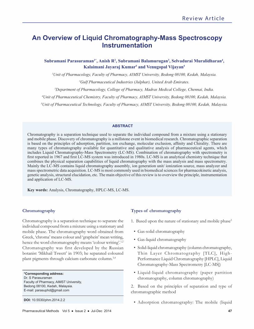
Figure 1: Instrumentation of HPLC
Mobile phase
Gradient controller
Pump
Injector
Column
Detector
Parasuraman, et al.: An Overview of LC-MS Instrumentation
Pharmaceutical Methods Vol 5 ● Issue 2 ● Jul-Dec 2014 49
Performance Liquid Chromatography (HPLC)-MS, Capillary Electrophoresis (CE)-MS and Capillary Electrochromatography (CEC)-MS. The combination of Gas Chromatography and Mass Spectrometry (MS) was first reported in 1958 and made available commercially in 1967.12 Combination of LC with MS is an important development in the history of chromatography (1980s). Mass spectrometry in LC-MS helps to determine the elemental composition and structural elucidation of a sample.13
Principle of LC-MS
Typical LC-MS system is combination of HPLC with MS using interface (ionization source) (Figure 1). The sample is separated by LC, and the separated sample species are sprayed into atmospheric pressure ion source, where they are converted into ions in the gas phase. The mass analyzer is then used to sort ions according to their mass to charge ratio and detector counts the ions emerging from the mass analyzer and may also amplify the signal generated from each ion. As a result, mass spectrum (a plot of the ion signal as a function of the mass-to-charge ratio) is created, which is used to determine the elemental orisotopic nature of a sample, the masses of particles and of molecules, and to elucidate the chemical structures of molecules.14,15
Requirement of LC
Usually LC used in LC-MS is HPLC. The principle of separation in HPLC, is normal phase mode or reverse
phase mode of adsorption. Normal phase constricts with polar stationary phase with non-polar solvent/mobile phase and reverse phase constricts with non-polar stationary phase with polar solvent/mobile phase. Normal phase mode not widely used in biomedical research and not advisable for pharmaceutical applications since most of the drug are polar in nature and takes longer time to be elute and detected. Reverse phase mode have wide range of pharmaceutical application. Examples for reverse phase columns are octadecylsilane (ODS)/ C18,C8,C4, etc.
HPLC Instrumentation
Mainly HPLC instrument contains pump, mixing unit (solvent degassing system), injector (manual/auto), guard column, analytical columns, detectors, recorder and integrators (Figure 1).
Detector used in HPLC
1. UV detector
• Single wavelength (filter)
• Variable wavelength (monochromator)
• Multiple wavelengths (Photodiode array detector [PDA])
2. Florescence
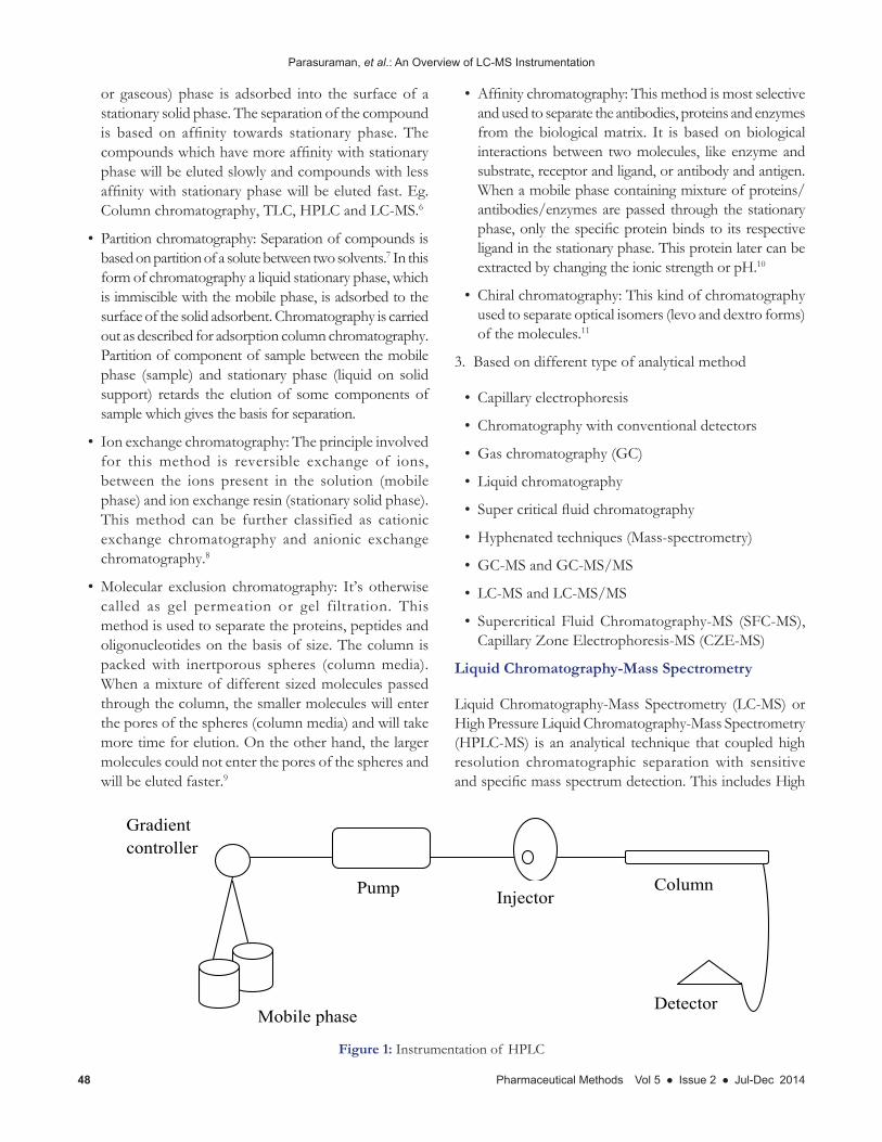
Figure 2: Instrumentation of LC-MS
Parasuraman, et al.: An Overview of LC-MS Instrumentation
50 Pharmaceutical Methods Vol 5 ● Issue 2 ● Jul-Dec 2014
3. Electrochemical detector
4. Mass spectrometric
Requirements of LC-MS instrumentation
Mainly the LC-MS contains liquid chromatography assembly, ion generation unit/ ionization source, mass analyzer and mass spectrometric data acquisition (Figure 2).
The effluent mobile phase with separated compound from the liquid chromatography is interfaced with the ionization source of the Mass spectrometer.
Ionization source
Most common ionization sources are Electrospray ionization (ESI), Atmospheric pressure chemical ionization (APCI) and Matrix-assisted laser desorption/ionization (MALDI). A part from this Electron impact (EI) and Chemical ionization (CI) or negative chemical ionization are also used as ionization source in MS.18
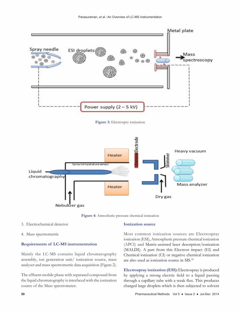
Electrospray ionization (ESI): Electrospray is produced by applying a strong electric field to a liquid passing through a capillary tube with a weak flux. This produces charged large droplets which is then subjected to solvent
Figure 3: Electrospry ionization
Figure 4: Atmosheric pressure chemical ionization
Parasuraman, et al.: An Overview of LC-MS Instrumentation
Pharmaceutical Methods Vol 5 ● Issue 2 ● Jul-Dec 2014 51
evaporation. Increase in charge density resulting from solvent evaporation causes coulombic repulsion to overcome the liquid surface tension, resulting in release of ions from droplets. This is the principle of ion formation using this technique.
Detection sensitivity in this technique is limited to 10-8 µl, and needs large volume to increase the sensitivity of the detection. By using ESI high mass sample, non-volatile molecules, liquids can be ionized and disadvantage of this source of ionization is poor sensitivity, low fragmentation and source is instable (Figure 3).19,20
Atmospheric pressure chemical ionization (APCI): Principle of this technique involves nebulization of the mobile phase with nitrogen gas and vaporization by heating it to relatively high temperature (above 400°C). The resulting vapor is then subjected to a corona discharge electrode to create ions.
APCI (Figure 4) is most commonly used ionization source used in LC-MS. APCI are used for analysis of pharmaceutical, environmental, toxicological, clinical and chemical industrial/ laboratory samples.21,22
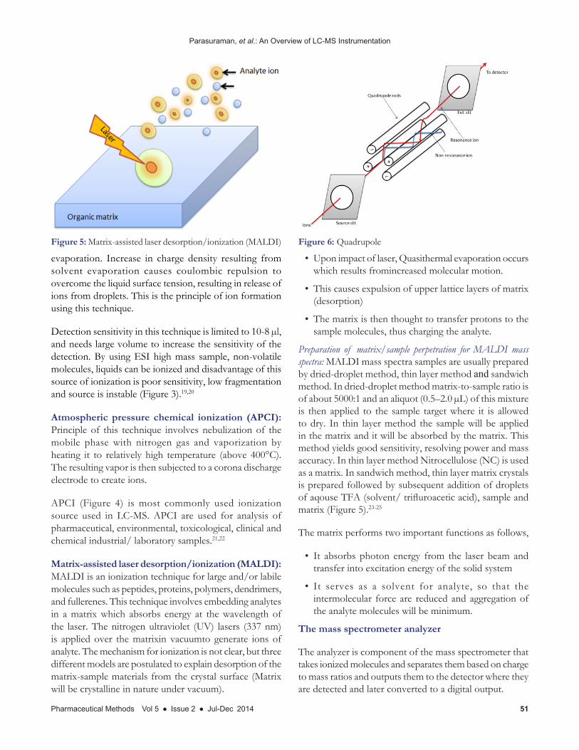
Matrix-assisted laser desorption/ionization (MALDI): MALDI is an ionization technique for large and/or labile molecules such as peptides, proteins, polymers, dendrimers, and fullerenes. This technique involves embedding analytes in a matrix which absorbs energy at the wavelength of the laser. The nitrogen ultraviolet (UV) lasers (337 nm) is applied over the matrixin vacuumto generate ions of analyte. The mechanism for ionization is not clear, but three different models are postulated to explain desorption of the matrix-sample materials from the crystal surface (Matrix will be crystalline in nature under vacuum).
• Upon impact of laser, Quasithermal evaporation occurs which results fromincreased molecular motion.
• This causes expulsion of upper lattice layers of matrix (desorption)
• The matrix is then thought to transfer protons to the sample molecules, thus charging the analyte.
Preparation of matrix/sample perpetration for MALDI mass spectra: MALDI mass spectra samples are usually prepared by dried-droplet method, thin layer method and sandwich method. In dried-droplet method matrix-to-sample ratio is of about 5000:1 and an aliquot (0.5–2.0 µL) of this mixture is then applied to the sample target where it is allowed to dry. In thin layer method the sample will be applied in the matrix and it will be absorbed by the matrix. This method yields good sensitivity, resolving power and mass accuracy. In thin layer method Nitrocellulose (NC) is used as a matrix. In sandwich method, thin layer matrix crystals is prepared followed by subsequent addition of droplets of aqouse TFA (solvent/ trifluroacetic acid), sample and matrix (Figure 5).23-25
The matrix performs two important functions as follows,
• It absorbs photon energy from the laser beam and transfer into excitation energy of the solid system
• It serves as a solvent for analyte, so that the intermolecular force are reduced and aggregation of the analyte molecules will be minimum.
The mass spectrometer analyzer
The analyzer is component of the mass spectrometer that takes ionized molecules and separates them based on charge to mass ratios and outputs them to the detector where they are detected and later converted to a digital output.
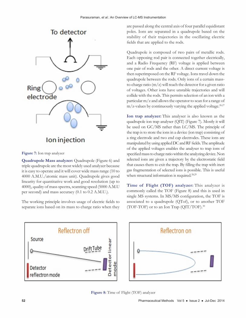
Figure 5: Matrix-assisted laser desorption/ionization (MALDI) Figure 6: Quadrupole
Parasuraman, et al.: An Overview of LC-MS Instrumentation
52 Pharmaceutical Methods Vol 5 ● Issue 2 ● Jul-Dec 2014
Quadrupole Mass analyzer: Quadrupole (Figure 6) and triple quadrupole are the most widely used analyzer because it is easy to operate and it will cover wide mass range (10 to 4000 A.M.U./atomic mass unit). Quadrupole gives good linearity for quantitative work and good resolution (up to 4000), quality of mass spectra, scanning speed (5000 A.M.U per second) and mass accuracy (0.1 to 0.2 A.M.U.).
The working principle involves usage of electric fields to separate ions based on its mass to charge ratio when they
are passed along the central axis of four parallel equidistant poles. Ions are separated in a quadrupole based on the stability of their trajectories in the oscillating electric fields that are applied to the rods.
Quadrupole is composed of two pairs of metallic rods. Each opposing rod pair is connected together electrically, and a Radio Frequency (RF) voltage is applied between one pair of rods and the other. A direct current voltage is then superimposed on the RF voltage. Ions travel down the quadrupole between the rods. Only ions of a certain mass-to-charge ratio (m/z) will reach the detector for a given ratio of voltages. Other ions have unstable trajectories and will collide with the rods. This permits selection of an ion with a particular m/z and allows the operator to scan for a range of m/z-values by continuously varying the applied voltage.26,27
Ion trap analyzer: This analyser is also known as the quadrupole ion trap analyser (QIT) (Figure 7). Mostly it will be used on GC/MS rather than LC/MS. The principle of the trap is to store the ions in a device (ion trap) consisting of a ring electrode and two end cap electrodes. These ions are manipulated by using applied DC and RF fields. The amplitude of the applied voltages enables the analyser to trap ions of specified mass to charge ratio within the analyzing device. Non selected ions are given a trajectory by the electrostatic field that causes them to exit the trap. By filling the trap with inert gas fragmentation of selected ions is possible. This is useful when structural information is required.28,29
Time of Flight (TOF) analyzer: This analyzer is commonly called the TOF (Figure 8) and this is used in single MS systems. In MS/MS configuration, the TOF is associated to a quadrupole (QTof), or to another TOF (TOF-TOF) or to an Ion Trap (QIT/TOF).30
Figure 7: Ion trap analyzer
Figure 8: Time of Flight (TOF) analyzer

Parasuraman, et al.: An Overview of LC-MS Instrumentation
Pharmaceutical Methods Vol 5 ● Issue 2 ● Jul-Dec 2014 53
Principle of the time of flight analyzer is formation of ions from ion source and it will be accelerated to high velocity by an electric field present in the ‘drift tube’ of the instrument. The accelerated ions will be detected by detector by linear mode or reflection mode.
Magnetic Sector Mass Analyser: In Magnetic sector analyzers, ions are accelerated through a flight tube by magnetic field, where the ions are separated by charge to mass ratios. As moving charges enter a magnetic field, the
charge is deflected to a circular motion of a unique radius in a direction perpendicular to the applied magnetic field. Ions in the magnetic field experience two equal forces; force due to the magnetic field and centripetal force. When similar ions pass through the magnetic field, they all will be deflected to the same degree and will all follow the same trajectory path. Other will collide with either side of the flight tube wall or will not pass through the slit to the detector.
Fourier transform-ioncyclotron resonance (FT-ICR): Ionsentering a chamber are trapped in circular orbits by powerful electrical and magnetic fields. When excited by a Radio-frequency (RF) electrical field, the ions generate a time dependent current. This current is converted by Fourier transform into orbital frequencies of the ions which corresponds to their mass-to-chargeratios. FT-ICR mass analyzers can perform multiple stages of mass spectrometry without additional mass analyzers. They also have a wide mass range and excellent mass resolution (Figure 9).31,32
Detectors
Three deferent kinds of detectors are used in Mass Spectrometry, i.e. Electron multipliers (Figure 10a), Dynolyte photomultiplier (Figure 10b) and Micro channel
Figure 9: Fourier transform-ion cyclotron resonance (FT-ICR)
Figure 10: Detectors used in LC-MS
Parasuraman, et al.: An Overview of LC-MS Instrumentation
54 Pharmaceutical Methods Vol 5 ● Issue 2 ● Jul-Dec 2014
plates (Figure 10 c).33 Electron multipliers dynode is used to convert either –ve, +ve ions into electrons, that will be amplified and detected. This will be widely used in quadrupole and ion trap instruments.
The dynode of Dynolyte photomultipliers converts the charged ions into electrons. These electrons sticks to a phosphor and emits photons, and that photons are made to strike the photomultiplier to achieve multiplied signals for recording.
Microchannel Plate (MCP) are commonly employed in ToF spectrometers. This will have very low time response and high degree of sensitivity (<1 ns and single ion signal >50 mV respectively).
Mass spectrometric data acquisition system
The data acquisition system is designed to digitalize electrical signals from the detector and transfer them to the data system in a compatible format.
• Full mass spectra (library to solve the analytical problem)
• Raw data of a small range (determine of isotope pattern/molecular weight)
• Mass chromatograms of selected ions (quantification)
• MS/MS experiments with Collisionally Activated Decompositions (CAD), such as daughter ion, parent ion and neutral loss scans structural information.
Applications of LC-MS
LC-MS/LC-MSMS are most widely used in food industries, pharmaceutical and chemical industries for quantitative
and qualitative analysis.34-36 Applications of LS-MSMS are as follows.
• Molecular weight determination: Able to determine the molecule weight of chemical substance, pharmaceutical substances, proteins, etc.
• Structural determination/elucidation: Tandem mass spectrometry used to determine structural information using mass spectral fragmentations.
• Pharmaceutical applications: It’s used to determine the pharmacokinetic profile of the pharmaceuticals like drug, drug metabolites/degradation product, impurities and chiral impurities. The separation and detection of chiral impurities in pharmaceuticals are of great importance because the D-isomer of a drug can have different pharmacological, metabolic and toxicological activity from the L-isomer.
• Clinical and biochemical applications: MALDI-TOF MS is used in SNP genotyping, quantification of DNA, gene expression analysis, DNA and RNA sequencing.
• Food and Environmental applications: use to identify aflatoxins (toxic metabolic product in certain fungi), determine the vitamin D3 in poultry fed supplements, etc.
• Capillary electrophoresis/MS applications: Used for analysis of peptides.
CONCLUSION
The advancement of hyphenated techniques, high resolution mass analyzers as well as high throughput separation approaches, quantitative and quantitative analysis of pharmaceutical drugs and metabolites can be achieved with good sensitivity.
REFERENCES
1. Oona McPolin. An introduction to HPLC for Pharmaceutical analysis. Mourne Training Service, Northern Ireland (UK); 2009. P. 1-9.
2. Idzaid Bin Idros. Development of a Method for the Analysis of Neonicotinoid Insecticides using Thin-Layer Chromatography. Bachelor thesis (Bs-3/10B). The University of Applied Sciences Offenburg, 2011. Available in: opus.hs-offenburg.de/files/96/Thesis_IDROS.pdf [Last accessed on 08/06/2015].
3. Ettre LS, Sakodynskii KI. MS Tswett and the discovery of chromatography I: Early work (1899–1903). Chromatographia 1993; 35(3-4): 223-31.
4. Ettre LS, Sakodynskii KI. MS Tswett and the discovery of chromatography II: Completion of the development of chromatography (1903–1910). Chromatographia 1993; 35(5-6): 329-38.
5. Types of chromatography. Available in: http://www.rpi.edu/dept/chem-eng/Biotech-Environ/CHROMO/chromtypes.html [Last accessed on 08/06/2015].
6. Adsorption chromatography. Available in: http://medical-dictionary.thefreedictionary.com/adsorption+chromatography [Last accessed on 08/06/2015].
7. Lawrence JF. Organic Trace Analysis by Liquid Chromatography. Academic Press Inc, New York; 1981. p. 112-3.
8. Chromatographic method for protein purification. Available in http://www.biotech.kth.se/courses/gru/courselist/BB2040_ENG/ChromMethods.pdf [Last accessed on 08/06/2015].
9. Determann H. Gel Chromatography. 2nd ed, Springer-Verlag, Berlin; 2012. 10. Cuatrecasas P, Wilcher M, Anfinsen CB. Selective enzyme purification by
affinity chromatography. Proceedings of the national academy of science 1968; 61(2): 636-43.
11. Beesley TE, Scott RPW. Chiral chromatography. Jony Wiley and Sons Ltd. England. Available in http://sci.uokufa.edu.iq/ar/teaching/zynaba/Chiral%20Chromatography%20-%20Thomas%20E.%20Beesley.pdf [Last accessed on 08/06/2015].
12. Ardrey RE. Liquid Chromatography - Mass Spectrometry: An Introduction. Jony Wiley and Sons Ltd. England; 2003.
13. Pitt JJ. Principles and Applications of Liquid Chromatography-Mass
Parasuraman, et al.: An Overview of LC-MS Instrumentation
Pharmaceutical Methods Vol 5 ● Issue 2 ● Jul-Dec 2014 55
Spectrometry in Clinical Biochemistry. Clin Biochem Rev. 2009; 30(1): 19-34.14. Korfmacher WA. Foundation review: Principles and applications of LC-MS
in new drug discovery. Drug Discov Today 2005; 10(20): 1357-67.15. Lim CK, Lord G. Current development in LC-MS for pharmaceutical analysis.
Bio Pharm Bull. 2002; 25(5): 547-57.16. Fong GW, Lam SK. HPLC in the pharmaceutical Industry. Volume 47. Marcel
Dekker Inc, New York; 1991.17. Mcmaster MC. HPLC a practical user’s guide. 2nd edition, A john wiley and
sons, Inc. publication, New Jersey; 2007. p. 181-93.18. Sparkman OD. Mass Spectrometry Pitt Con. J. Am. Soc. Mass Spectrom
2006; 17(6): 873-84.19. Ho CS, Lam CW, Chan MH, Cheung RC, Law LK, Lit LC, et al. Electrospray
ionisation mass spectrometry: principles and clinical applications. Clin Biochem Rev. 2003; 24(1): 3-12.
20. Fenn JB, Mann M, Meng CK, Wong SF, Whitehouse CM. Electrospray ionization for mass spectrometry of large biomolecules. Science 1989; 246(4926): 64-71.
21. Byrdwell WC. Atmospheric pressure chemical ionization mass spectrometry for analysis of lipids. Lipids 2001; 36(4): 327-46.
22. Van Breemen RB, Dong L, Pajkovic ND. Atmospheric Pressure Chemical Ionization Tandem Mass Spectrometry of Carotenoids. Int J Mass Spectrom. 2012 Feb 15; 312: 163-72.
23. Calderaro A, Arcangeletti MC, Rodighiero I, Buttrini M, Gorrini C, Motta F, et al. Matrix-assisted laser desorption/ionization time-of-flight (MALDI-TOF) mass spectrometry applied to virus identification. Sci Rep. 2014 Oct 30; 4: 6803.
24. Marvin LF, Roberts MA, Fay LB. Matrix-assisted laser desorption/ionization time-of-flight mass spectrometry in clinical chemistry. Clin Chim Acta. 2003; 337(1-2): 11-21.
25. Ragoussis J, Elvidge GP, Kaur K, Colella S. Matrix-Assisted Laser
Desorption/Ionisation, Time-of-Flight Mass Spectrometry in enomics Research. Plos Genetics 2006; 2(7): e100.
26. LC/MS booklet. Available in http://ccc.chem.pitt.edu/wipf/Waters%20LC-MS%20primer.pdf [Last accessed on 03/06/2015]
27. Mass Spectrometry (MS) Primer. Available in http://www.chemweb.ucc.ie/Mass%20Spec/MS%20summary.pdf [Last accessed on 03/06/2015]
28. Stafford G. Ion trap mass spectrometry: a personal perspective. J Am Soc Mass Spectrom 2002 Jun; 13(6): 589-96.
29. Jonscher KR, Yates JR. The Whys and Wherefores of Quadrupole Ion Trap Mass Spectrometry. Ion Trap Mass Spectrometry Available in https://www.abrf.org/ABRFNews/1996/September1996/sep96iontrap.html [Last accessed on 09/06/2015].
30. Chernushevich IV, Loboda AV, Thomson BA. An introduction to quadrupole-time-of-flight mass spectrometry. J Mass Spectrom. 2001; 36(8): 849-65.
31. Marshall AG, Hendrickson CL, Jackson GS. Fourier transform ion cyclotron resonance mass spectrometry: a primer. Mass Spectrom Rev. 1998; 17(1): 1-35.
32. Marshall AG, Hendrickson CL. Fourier transform ion cyclotron resonance detection: principles and experimental configurations. Int. J. Mass Spectrom 2005; 215(1-3): 59-75.
33. Neetu SK, Ankit G, Ruchi T, Ajay B, Prashant B. A review on mass spectrometry detector. Int. Res. J. Pharm. 2012; 3(10): 33-42.
34. Kaufmann B, Souverain S, Cherkaoui S, Christen P, Veuthey JL. Rapid liquid chromatography-Mass spectrometric analysis of withanolides in crude plant extracts by use of a monolithic column. Chromatographia 2002; 56(3-4): 137-41.
35. Lee MS, Kerns EH. LC/MS applications in drug development. Mass Spectrom Rev. 1999; 18(3-4): 187-279.
36. Prakash C, Shaffer CL, Nedderman A. Analytical strategies for identifying drug metabolites. Mass Spectrom Rev. 2007; 26(3): 340-69.