an official american thoracic society/european ... - · pdf filediseases, lung function,...
TRANSCRIPT

American Thoracic Society Documents
An Official American Thoracic Society/EuropeanRespiratory Society Statement: Update of theInternational Multidisciplinary Classification of theIdiopathic Interstitial Pneumonias
William D. Travis, Ulrich Costabel, David M. Hansell, Talmadge E. King, Jr., David A. Lynch, Andrew G. Nicholson,Christopher J. Ryerson, Jay H. Ryu, Moises Selman, Athol U. Wells, Jurgen Behr, Demosthenes Bouros,Kevin K. Brown, Thomas V. Colby, Harold R. Collard, Carlos Robalo Cordeiro, Vincent Cottin, Bruno Crestani,Marjolein Drent, Rosalind F. Dudden, Jim Egan, Kevin Flaherty, Cory Hogaboam, Yoshikazu Inoue, Takeshi Johkoh,Dong Soon Kim, Masanori Kitaichi, James Loyd, Fernando J. Martinez, Jeffrey Myers, Shandra Protzko,Ganesh Raghu, Luca Richeldi, Nicola Sverzellati, Jeffrey Swigris, and Dominique Valeyre; on behalf of the ATS/ERSCommittee on Idiopathic Interstitial Pneumonias
THIS OFFICIAL STATEMENT OF THE AMERICAN THORACIC SOCIETY (ATS) AND THE EUROPEAN RESPIRATORY SOCIETY (ERS) WAS
APPROVED BY THE ATS BOARD OF DIRECTORS, JUNE 2013, AND BY THE ERS STEERING COMMITTEE, MARCH 2013
CONTENTS
Executive SummaryIntroductionMethodsSummary of Major Revisions of the IIP ClassificationGeneral Progress in IIPs since 2002
Multidisciplinary ApproachObserver Agreement in Diagnosis of IIP
Important Differential Diagnostic ConsiderationsHypersensitivity PneumonitisCollagen Vascular DiseaseFamilial Interstitial PneumoniaCoexisting Patterns
Progress in Specific IIPs since 2002Chronic Fibrosing IIPsSmoking-related IIPsAcute or Subacute IIPs
Rare IIPsIdiopathic Lymphoid Interstitial PneumoniaIdiopathic Pleuroparenchymal Fibroelastosis
Rare Histologic PatternsAcute Fibrinous and Organizing PneumoniaBronchiolocentric Patterns of Interstitial Pneumonia
Unclassifiable IIPClinical Classification of Disease BehaviorBiomarkers
Background: In 2002 the American Thoracic Society/European Res-piratory Society (ATS/ERS) classification of idiopathic interstitialpneumonias (IIPs)definedseven specificentities, andprovided stan-dardizedterminologyanddiagnosticcriteria. Inaddition, thehistorical“gold standard” of histologic diagnosis was replaced by amultidiscipli-nary approach. Since2002manypublicationshaveprovidednew infor-mation about IIPs.
Purpose: The objective of this statement is to update the 2002 ATS/ERS classification of IIPs.Methods: An international multidisciplinary panel was formed anddeveloped key questions that were addressed through a review ofthe literature published between 2000 and 2011.Results: Substantial progress has beenmade in IIPs since thepreviousclassification. Nonspecific interstitial pneumonia is now better de-fined. Respiratory bronchiolitis–interstitial lung disease is now com-monly diagnosed without surgical biopsy. The clinical course of idi-opathic pulmonary fibrosis and nonspecific interstitial pneumonia isrecognized to be heterogeneous. Acute exacerbation of IIPs is nowwell defined. A substantial percentage of patients with IIP are diffi-cult to classify, often due to mixed patterns of lung injury. A classifi-cation based on observed disease behavior is proposed for patientswho are difficult to classify or for entities with heterogeneity in clin-ical course. A group of rare entities, including pleuroparenchymalfibroelastosis and rare histologic patterns, is introduced. The rapidlyevolving field of molecular markers is reviewed with the intent ofpromoting additional investigations that may help in determiningdiagnosis, and potentially prognosis and treatment.Conclusions: This update is a supplement to the previous 2002 IIPclassification document. It outlines advances in the past decadeand potential areas for future investigation.
Keywords: idiopathic interstitial pneumonia; usual interstitial pneumo-
nia; nonspecific interstitial pneumonia; respiratory bronchiolitis; desqua-mative interstitial pneumonia; cryptogenic organizing pneumonia; acute
interstitial pneumonia; lymphoid interstitial pneumonia; pleuroparenchy-
mal fibroelastosis; acute fibrinous and organizing pneumonia
EXECUTIVE SUMMARY
There are several specific areas that are given special attention inthis revision of the 2002 American Thoracic Society/EuropeanRespiratory Society idiopathic interstitial pneumonia (IIP) state-ment.
1. Idiopathic nonspecific interstitial pneumonia (NSIP) isnow accepted as a specific clinicopathologic entity. It hasbecome evident that clinical progression is highly het-erogeneous, with several studies suggesting that a subsetof patients demonstrate progression to end-stage fibro-sis; criteria to define this group at the time of diagnosiswould be helpful.
This document has an online supplement, which is accessible from this issue’s
table of contents at www.atsjournals.org
Am J Respir Crit Care Med Vol 188, Iss. 6, pp 733–748, Sep 15, 2013
Copyright ª 2013 by the American Thoracic Society
DOI: 10.1164/rccm.201308-1483ST
Internet address: www.atsjournals.org

2. New information has accumulated on smoking-related inter-stitial lung disease, including patients with combined emphy-sema and interstitial fibrosis. In clinical practice, respiratorybronchiolitis–interstitial lung disease is increasingly diag-nosed without surgical lung biopsy in smokers on the basisof clinical and imaging features (ground-glass opacities andcentrilobular nodules) and bronchoalveolar lavage (smok-er’s macrophages and absence of lymphocytosis).
3. The natural progression of idiopathic pulmonary fibrosis(IPF) is acknowledged to be heterogeneous with somepatients remaining stable for prolonged periods, othersshowing more rapid steady progression, and still otherssuccumbing to acute exacerbation.
4. Acute exacerbation is better defined and recognized tooccur in chronic fibrosing IIPs (IPF and NSIP).
5. Some patients with IIP are difficult to classify, oftenbecause of mixed patterns of lung injury.
6. It is recognized that there is a need to provide a clinicalalgorithm for classifying and managing IIP cases. This isparticularly applicable when no biopsy is available andhigh-resolution computed tomography is not diagnostic.
7. Pleuroparenchymal fibroelastosis is recognized as a specificrare entity, usually idiopathic. Other less well-defined his-tologic patterns, such as bronchiolocentric inflammationand fibrosis, are also included.
8. A rapidly emerging field of molecular markers holdspromise for improving diagnostic approaches. Thesemarkers may also be useful in predicting prognosis andresponse to different therapies. Incorporation of geneticand molecular studies may revolutionize the approachto diagnosis and classification of the IIPs.
INTRODUCTION
The objective of this statement is to update the 2002 AmericanThoracicSociety/EuropeanRespiratorySociety (ATS/ERS)clas-sification of idiopathic interstitial pneumonias (IIPs) (1). Focus isplaced on describing changes to previously described clinical enti-ties, describing new clinical entities, and describing new histologicpatterns. This update is not intended as a stand-alone documentand should be used as a supplement to the original 2002 IIP classi-fication. In 2002, the ATS/ERS IIP classification (1) defined sevendisease categories, and proposed standardized terminology anddiagnostic criteria. In addition, the historical “gold standard” ofhistologic diagnosis was replaced by a “dynamic integrated ap-proach” using multidisciplinary discussion (MDD). The 2002 IIPclassification was used in 75% (157 of 208) of all clinical publica-tions on the topic of IIPs between 2004 and 2011. The new infor-mation from these publications is incorporated in this update.
METHODS
This project was performed under supervision by the ATSDocu-ments Development and Implementation Committee in collab-oration with the ERS (Table E1 in the online supplement). Aninternational multidisciplinary panel was assembled. The panelconsisted of 34 experts in interstitial lung diseases (19 pulmonol-ogists, 4 radiologists, 5 pathologists, 2 experts in evidence-basedmedicine, and 4 molecular biologists). Several meetings wereheld by members of the international multidisciplinary panel(Table E2), who disclosed conflicts of interest, which were vettedaccording to ATS and ERS policies.
Key questions were developed that the committee believedimportant for the classification of IIPs (see APPENDIX 1 in the
online supplement). A literature search was performed to iden-tify new publications that pertained to these key questions,assisted by two librarians experienced in literature searches forpulmonary diseases. Literature retrieved from Medline searchesbetween 2000 and 2011 was used to produce this statement.
The committee was divided into subgroups assigned to spe-cific sections of the document. These subgroups reviewed the rel-evant literature and produced the first draft of their respectivesections. These sections were compiled by the committee chairand a complete first draft was edited by the writing subcommit-tee. This document was reviewed and edited by all committeemembers before final review by the writing subcommittee.The revised document was approved by all authors.
SUMMARY OF MAJOR REVISIONS OF THEIIP CLASSIFICATION
In the revision of the IIP classification, the main entities are pre-served (Table 1). However, there are several important changes.First, cryptogenic fibrosing alveolitis is removed, leaving idiopathicpulmonary fibrosis (IPF) as the sole clinical term for this diagnosis.Second, idiopathic nonspecific interstitial pneumonia (NSIP) is nowaccepted as a distinct clinical entity with removal of the term “pro-visional” (2). Third, major IIPs are distinguished from rare IIPs andunclassifiable cases. Fourth, rare histologic patterns of acute fibri-nous and organizing pneumonia (AFOP) and interstitial pneumo-nias with a bronchiolocentric distribution are recognized. Fifth, themajor IIPs are grouped into chronic fibrosing (IPF and NSIP; Fig-ures 1 and 2), smoking-related (respiratory bronchiolitis–interstitiallung disease [RB-ILD] and desquamative interstitial pneumonia[DIP]; Figure 3), and acute/subacute IIPs (cryptogenic organizingpneumonia [COP] and acute interstitial pneumonia [AIP]; Figure 4and Table 2). Sixth, a clinical disease behavior classification is pro-posed. Last, molecular and genetic features are reviewed.
GENERAL PROGRESS IN IIPS SINCE 2002
Multidisciplinary Approach
The process of achieving amultidisciplinary diagnosis in a patientwith IIP is dynamic, requiring close communication between cli-nician, radiologist, and when appropriate, pathologist (1). Clin-ical data (presentation, exposures, smoking status, associated
TABLE 1. REVISED AMERICAN THORACIC SOCIETY/EUROPEANRESPIRATORY SOCIETY CLASSIFICATION OF IDIOPATHICINTERSTITIAL PNEUMONIAS: MULTIDISCIPLINARY DIAGNOSES
Major idiopathic interstitial pneumonias
Idiopathic pulmonary fibrosis
Idiopathic nonspecific interstitial pneumonia
Respiratory bronchiolitis–interstitial lung disease
Desquamative interstitial pneumonia
Cryptogenic organizing pneumonia
Acute interstitial pneumonia
Rare idiopathic interstitial pneumonias
Idiopathic lymphoid interstitial pneumonia
Idiopathic pleuroparenchymal fibroelastosis
Unclassifiable idiopathic interstitial pneumonias*
*Causes of unclassifiable idiopathic interstitial pneumonia include (1) inade-
quate clinical, radiologic, or pathologic data and (2) major discordance between
clinical, radiologic, and pathologic findings that may occur in the following situa-
tions: (a) previous therapy resulting in substantial alteration of radiologic or histo-
logic findings (e.g., biopsy of desquamative interstitial pneumonia after steroid
therapy, which shows only residual nonspecific interstitial pneumonia [153]);
(b) new entity, or unusual variant of recognized entity, not adequately character-
ized by the current American Thoracic Society/European Respiratory Society classi-
fication (e.g., variant of organizing pneumonia with supervening fibrosis) (79); and
(c) multiple high-resolution computed tomography and/or pathologic patterns that
may be encountered in patients with idiopathic interstitial pneumonia.
734 AMERICAN JOURNAL OF RESPIRATORY AND CRITICAL CARE MEDICINE VOL 188 2013
diseases, lung function, laboratory findings) and radiologic find-ings are essential for multidisciplinary diagnosis.
The multidisciplinary approach does not lessen the impor-tance of lung biopsy in the diagnosis of IIPs; rather, it definesthe settings where biopsy is more informative than high-
resolution computed tomography (HRCT) and those where bi-opsy is not needed. Also, once a pathologist has recognized a histo-logic pattern (e.g.,NSIPor organizingpneumonia [OP]), the clinicianshould reconsider potential causes (e.g., hypersensitivity pneumonitis[HP], collagen vascular disease [CVD], and drug exposure).
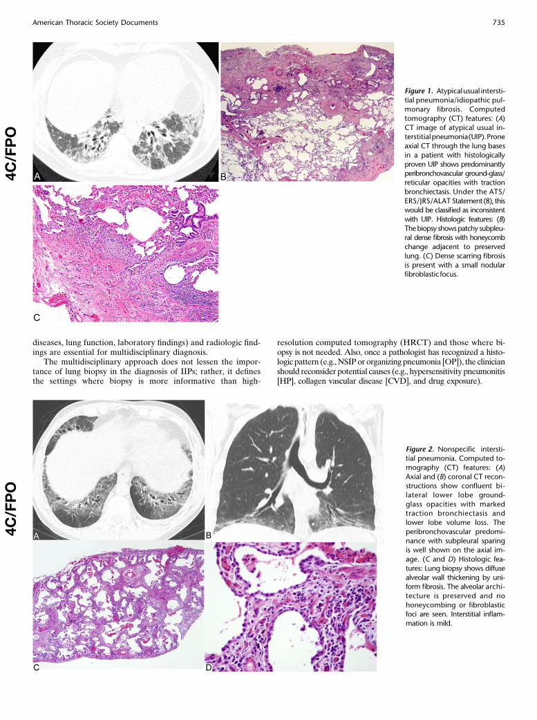
Figure 1. Atypicalusual intersti-
tial pneumonia/idiopathic pul-monary fibrosis. Computed
tomography (CT) features: (A)
CT image of atypical usual in-terstitialpneumonia(UIP).Prone
axial CT through the lung bases
in a patient with histologically
proven UIP shows predominantlyperibronchovascular ground-glass/
reticular opacities with traction
bronchiectasis. Under the ATS/
ERS/JRS/ALAT Statement(8), thiswould be classified as inconsistent
with UIP. Histologic features: (B)
Thebiopsy showspatchy subpleu-ral dense fibrosis with honeycomb
change adjacent to preserved
lung. (C) Dense scarring fibrosis
is present with a small nodularfibroblastic focus.
4C/FPO
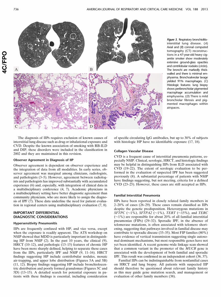
Figure 2. Nonspecific intersti-
tial pneumonia. Computed to-
mography (CT) features: (A)
Axial and (B) coronal CT recon-structions show confluent bi-
lateral lower lobe ground-
glass opacities with markedtraction bronchiectasis and
lower lobe volume loss. The
peribronchovascular predomi-
nance with subpleural sparingis well shown on the axial im-
age. (C and D) Histologic fea-
tures: Lung biopsy shows diffuse
alveolar wall thickening by uni-form fibrosis. The alveolar archi-
tecture is preserved and no
honeycombing or fibroblasticfoci are seen. Interstitial inflam-
mation is mild.
4C/FPO
American Thoracic Society Documents 735
The diagnosis of IIPs requires exclusion of known causes ofinterstitial lung disease such as drug or inhalational exposure andCVD. Despite the known association of smoking with RB-ILDand DIP, these disorders were included in the classification in2002 and they are maintained in this revision.
Observer Agreement in Diagnosis of IIP
Observer agreement is dependent on observer experience andthe integration of data from all modalities. In early series, ob-server agreement was marginal among clinicians, radiologists,and pathologists (3–5). However, agreement between radiolog-ists and pathologists has improved substantially with accumulatedexperience (6) and, especially, with integration of clinical data ina multidisciplinary conference (4, 7). Academic physicians ina multidisciplinary setting have better diagnostic agreement thancommunity physicians, who are more likely to assign the diagno-sis of IPF (7). These data underline the need for patient evalua-tion in regional centers using multidisciplinary evaluation (7, 8).
IMPORTANT DIFFERENTIALDIAGNOSTIC CONSIDERATIONS
Hypersensitivity Pneumonitis
IIPs are frequently confused with HP, and vice versa, exceptwhen the exposure is readily apparent. The ATS workshop onNSIP showed that MDD is particularly important in distinguish-ing HP from NSIP (2). In the past 10 years, the clinical (9),HRCT (10–12), and pathologic (13–15) features of chronic HPhave been more sharply defined, helping to separate these casesfrom the IIPs, particularly IPF and NSIP (9, 11–16). HRCTfindings suggesting HP include centrilobular nodules, mosaicair-trapping, and upper lobe distribution (Figures 5A and 5B)(11, 12). Biopsy findings suggesting HP include bronchiolocen-tric distribution and poorly formed granulomas (Figures 5C and5D) (13–15). A detailed search for potential exposure in pa-tients with these findings is essential, including consideration
of specific circulating IgG antibodies, but up to 30% of subjectswith histologic HP have no identifiable exposure (17, 18).
Collagen Vascular Disease
CVD is a frequent cause of interstitial pneumonia patterns, es-pecially NSIP. Clinical, serologic, HRCT, and histologic findingsmay be helpful in distinguishing IIPs from ILD associated withCVD (19–22). The extent of serologic evaluation to be per-formed in the evaluation of suspected IPF has been suggestedpreviously (8). A substantial percentage of patients with NSIPhave findings suggesting, but not meeting, criteria for a definedCVD (23–25). However, these cases are still accepted as IIPs.
Familial Interstitial Pneumonia
IIPs have been reported in closely related family members in2–20% of cases (26–29). These cases remain classified as IIPsdespite the genetic predisposition. Heterozygous mutations inSFTPC (z1%), SFTPA2 (z1%), TERT (z15%), and TERC(z1%) are responsible for about 20% of all familial interstitialpneumonias (FIPs) (29–32). Sporadic IPF, in the absence oftelomerase mutations, is often associated with telomere short-ening, suggesting that pathways involved in familial disease maycontribute to sporadic disease (33–35). Most FIP families (80%)have evidence of vertical transmission suggesting single autoso-mal dominant mechanisms, but most responsible genes have notyet been identified. A recent genome-wide linkage scan showedthat a common variant in the promoter of the MUCB gene isassociated with the development of both familial and sporadicIPF. This result was confirmed in an independent cohort (36, 37).
Familial IIPs can be indistinguishable from nonfamilial caseson HRCT and lung biopsy. All patients with suspected IIPshould therefore be questioned about relevant family historyas this may guide gene mutation search, and management orevaluation of other family members (38).
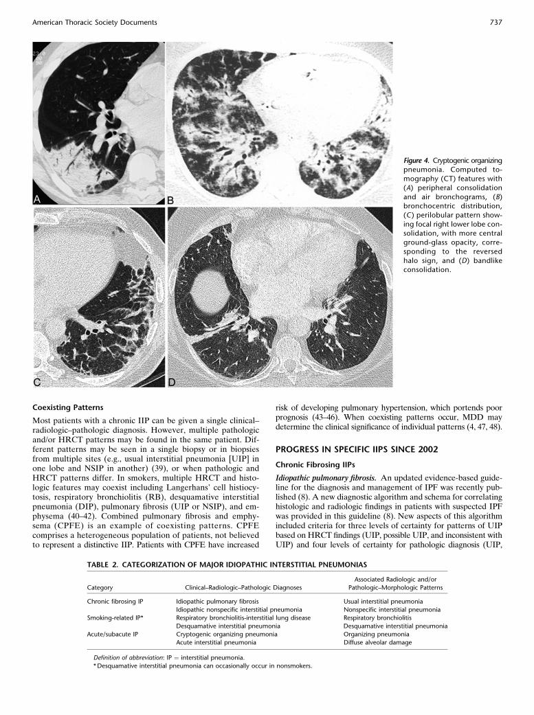
Figure 3. Respiratory bronchiolitis–interstitial lung disease. (A)Axial and (B) coronal computed
tomography (CT) reconstruc-
tions in a 47-year-old heavy cig-
arette smoker show moderatelyextensive ground-glass opacities
and centrilobular nodules (circles).
The bronchi are markedly thick-
walled and there is minimal em-physema. Bronchoalveolar lavage
yielded 91% macrophages. (C)
Histologic features: lung biopsyshows peribronchiolar pigmented
macrophage accumulation and
emphysema. (D) There is mild
bronchiolar fibrosis and pig-mented macrophages within
airspaces.
4C/FPO
736 AMERICAN JOURNAL OF RESPIRATORY AND CRITICAL CARE MEDICINE VOL 188 2013
Coexisting Patterns
Most patients with a chronic IIP can be given a single clinical–radiologic–pathologic diagnosis. However, multiple pathologicand/or HRCT patterns may be found in the same patient. Dif-ferent patterns may be seen in a single biopsy or in biopsiesfrom multiple sites (e.g., usual interstitial pneumonia [UIP] inone lobe and NSIP in another) (39), or when pathologic andHRCT patterns differ. In smokers, multiple HRCT and histo-logic features may coexist including Langerhans’ cell histiocy-tosis, respiratory bronchiolitis (RB), desquamative interstitialpneumonia (DIP), pulmonary fibrosis (UIP or NSIP), and em-physema (40–42). Combined pulmonary fibrosis and emphy-sema (CPFE) is an example of coexisting patterns. CPFEcomprises a heterogeneous population of patients, not believedto represent a distinctive IIP. Patients with CPFE have increased
risk of developing pulmonary hypertension, which portends poorprognosis (43–46). When coexisting patterns occur, MDD maydetermine the clinical significance of individual patterns (4, 47, 48).
PROGRESS IN SPECIFIC IIPS SINCE 2002
Chronic Fibrosing IIPs
Idiopathic pulmonary fibrosis. An updated evidence-based guide-line for the diagnosis and management of IPF was recently pub-lished (8). A new diagnostic algorithm and schema for correlatinghistologic and radiologic findings in patients with suspected IPFwas provided in this guideline (8). New aspects of this algorithmincluded criteria for three levels of certainty for patterns of UIPbased on HRCT findings (UIP, possible UIP, and inconsistent withUIP) and four levels of certainty for pathologic diagnosis (UIP,
Figure 4. Cryptogenic organizingpneumonia. Computed to-
mography (CT) features with
(A) peripheral consolidation
and air bronchograms, (B)bronchocentric distribution,
(C) perilobular pattern show-
ing focal right lower lobe con-solidation, with more central
ground-glass opacity, corre-
sponding to the reversed
halo sign, and (D) bandlikeconsolidation.
TABLE 2. CATEGORIZATION OF MAJOR IDIOPATHIC INTERSTITIAL PNEUMONIAS
Category Clinical–Radiologic–Pathologic Diagnoses
Associated Radiologic and/or
Pathologic–Morphologic Patterns
Chronic fibrosing IP Idiopathic pulmonary fibrosis Usual interstitial pneumonia
Idiopathic nonspecific interstitial pneumonia Nonspecific interstitial pneumonia
Smoking-related IP* Respiratory bronchiolitis-interstitial lung disease Respiratory bronchiolitis
Desquamative interstitial pneumonia Desquamative interstitial pneumonia
Acute/subacute IP Cryptogenic organizing pneumonia Organizing pneumonia
Acute interstitial pneumonia Diffuse alveolar damage
Definition of abbreviation: IP ¼ interstitial pneumonia.
*Desquamative interstitial pneumonia can occasionally occur in nonsmokers.
American Thoracic Society Documents 737
probable, possible, and not UIP) (8). The diagnosis of IPF requires(1) exclusion of other known causes of ILD, (2) the presence ofa UIP pattern on HRCT in patients not subjected to surgical lungbiopsy (SLB), and (3) specific combinations of HRCT and SLBpatterns in patients subjected to SLB (Figures 1A–1C).
Histologic UIP may be associated with atypical HRCT pat-terns (Figures 1A–1C) (8), including extensive ground-glassopacity, nodules, or mosaic attenuation, and after MDD someof these patients will be diagnosed with IPF (49–52). TypicalUIP HRCT pattern is illustrated elsewhere (1, 8). Patients withIPF and definite UIP by HRCT have shorter survival than thosewith indeterminate HRCT findings (49, 51, 53).Idiopathic nonspecific interstitial pneumonia. The diagnostic
criteria for NSIP were summarized in a recent ATS workshop(2). On the basis of the analysis of cases and the availableliterature, this workshop recommended that NSIP be acceptedas a distinct entity among the IIPs, with removal of the term“provisional.” Importantly, the NSIP pattern occurs not only asan idiopathic condition, but also in a variety of settings includ-ing CVD, HP, and drug toxicity, and in some patients withfamilial pulmonary fibrosis. MDD is especially important toestablish the diagnosis of idiopathic NSIP (2).
The most common HRCT abnormality in NSIP is bilateralground-glass opacity (Figures 2A and 2B) (26, 54–62). Irregularreticular opacities with traction bronchiectasis and bronchiolec-tasis occur in approximately 75% of cases (2, 54–62). Subpleuralsparing may be helpful in distinguishing NSIP from UIP (2, 12,52). Consolidation, if present, reflects an OP component and maysuggest CVD. Honeycombing is sparse or absent at presentationbut may increase in prevalence and extent during follow-up (63).
The histologic features include varying amounts of interstitial in-flammation and fibrosis with a uniform appearance (Figures 2C and2D) (2, 64, 65). Most cases of NSIP have a predominantly fibroticpattern of injury with rare cases of isolated cellular NSIP (59, 62,66). OP and honeycomb fibrosis should be inconspicuous or absent.
The prognosis is variable. Some patients improve, others re-main stable or improve on treatment, but some evolve to end-stage fibrosis and eventually die of the disease (2, 63, 67).
Smoking-related IIPs
RB-ILD and desquamative interstitial pneumonia (DIP) repre-sent a histologic spectrum of macrophage accumulation, with thedistinction dependent on the extent and distribution of this pro-cess (and also reflected by the pattern of disease on HRCT).However, clinical presentation, imaging findings, and response totherapy differ and they remain classified separately. In the last de-cade, the term “smoking-related interstitial lung disease” has in-creasingly been used, encompassing most cases of DIP, and nearlyall cases of RB-ILD and Langerhans’ cell histiocytosis (68, 69).Respiratory bronchiolitis–interstitial lung disease. Histologic
RB is always present in current smokers (70) and can be viewedas a physiological response to smoking, which in a few individ-uals becomes extensive enough to result in an interstitial lungdisease (RB-ILD). Characteristic HRCT features are ground-glass opacity and centrilobular nodules (Figures 3A and 3B). Inclinical practice, RB-ILD is increasingly diagnosed without sur-gical lung biopsy in smokers with these HRCT findings andwhere bronchoalveolar lavage demonstrates smokers’ macro-phages and the absence of lymphocytosis (potentially suggestiveof HP in this setting, although HP is uncommon in smokers)(68, 71). The disease course is heterogeneous, with a significantminority having progression despite smoking cessation (72).Desquamative interstitial pneumonia. DIP has been recognized
in nonsmokers (69), perhaps reflecting extension of childhood DIPinto adult life (with the latter often due to surfactant protein [SP]gene mutations) (73, 74). Ten-year survival remains approximately70%, with resistance to treatment in a significant minority.Airspace enlargement with fibrosis. DIP and RB-ILD need to
be distinguished from the smoking-related changes including
Figure 5. Fibrotic hypersensi-tivity pneumonitis. (A) Axial
and (B) coronal computed
tomography (CT) reconstructions
in a 76-year-old bird-keeper withprogressive shortness of breath
over 6 years show upper lung–
predominant subpleural retic-
ulation with some confluentareas of dense opacification,
traction bronchiectasis, and
patchy ground-glass opacities.Honeycombing is not identified.
(C) Histology shows a bronchio-
locentric cellular and fibrosing
interstitial pneumonia. (D) Thereis a patchy cellular interstitial
infiltrate and poorly formed
granulomas.
4C/FPO
738 AMERICAN JOURNAL OF RESPIRATORY AND CRITICAL CARE MEDICINE VOL 188 2013
respiratory bronchiolitis and airspace enlargement with fibrosis(AEF) that have been described in the nonneoplastic lung paren-chyma in lung cancer resection specimens (75, 76). These inci-dental HRCT and histologic findings in smokers are not regardedas a distinct form of IIP, but AEF shows more interstitial fibrosisthan described in the classic definition of emphysema (77). AEFis an incidental histologic or HRCT finding, whereas patientswith CPFE have clinically manifestations reflecting coexistingpatterns of interstitial fibrosing and emphysema.
Acute or Subacute IIPs
IIPs may have an acute or subacute presentation, or an acute ex-acerbation may occur in a previously subclinical or unrecognizedchronic IIP.Cryptogenic organizing pneumonia. COP continues to be in-
cluded in the classification of IIP because of its idiopathic natureand the tendency on occasions to be confused with other forms ofIIP, especially when there is progression to fibrosis. Becausemany cases are secondary, use of the generic term “OP” forthis reaction pattern is suggested with modifiers as appropriate,for example, OP associated with rheumatoid arthritis.
Patients with COP typically present with a subacute illness ofrelatively short duration (median, less than 3 mo) with variabledegrees of cough and dyspnea (78–81). HRCT characteristicallydemonstrates patchy and often migratory consolidation in a sub-pleural, peribronchial, or bandlike pattern (Figures 4A–4D)(82–84), commonly associated with ground-glass opacity(79, 83). Perilobular opacities and reversed halo (or atoll) sign(Figure 4C) may be helpful in suggesting the diagnosis (85, 86).Small unilateral or bilateral pleural effusion may occur in10–30% of patients (83, 84, 86). The OP pattern is a patchy processcharacterized primarily by organizing pneumonia involving alveo-lar ducts and alveoli with or without bronchiolar intraluminal pol-yps. Some cases show more marked interstitial inflammation suchthat there is overlap with cellular NSIP.
The majority of patients recover completely with oral cortico-steroids, but relapse is common (2, 78, 87). Sporadic reports haveidentified a subgroup of patients with OP that does not com-pletely resolve despite prolonged treatment. Some of these casesare characterized by residual or progressive interstitial fibrosis,with or without recurrent episodes of OP (79, 88, 89). It is likelythat some patients reported with fibrotic NSIP fall into this
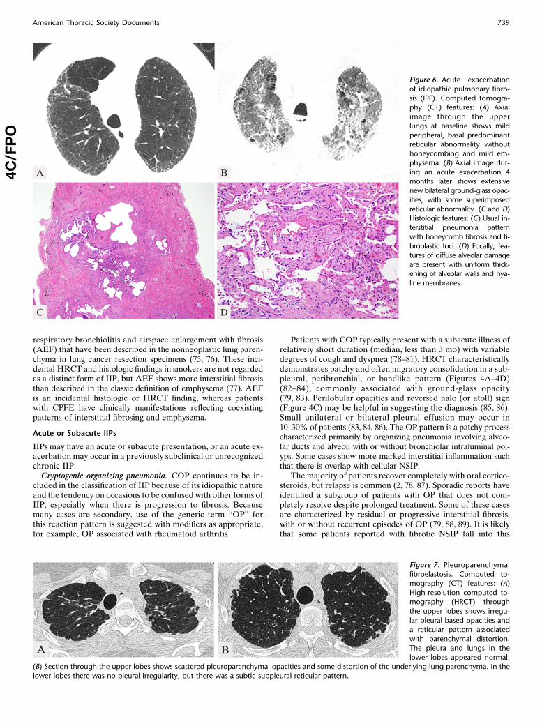
Figure 6. Acute exacerbation
of idiopathic pulmonary fibro-
sis (IPF). Computed tomogra-phy (CT) features: (A) Axial
image through the upper
lungs at baseline shows mildperipheral, basal predominant
reticular abnormality without
honeycombing and mild em-
physema. (B) Axial image dur-ing an acute exacerbation 4
months later shows extensive
new bilateral ground-glass opac-
ities, with some superimposedreticular abnormality. (C and D)
Histologic features: (C) Usual in-
terstitial pneumonia patternwith honeycomb fibrosis and fi-
broblastic foci. (D) Focally, fea-
tures of diffuse alveolar damage
are present with uniform thick-ening of alveolar walls and hya-
line membranes.
4C/FPO
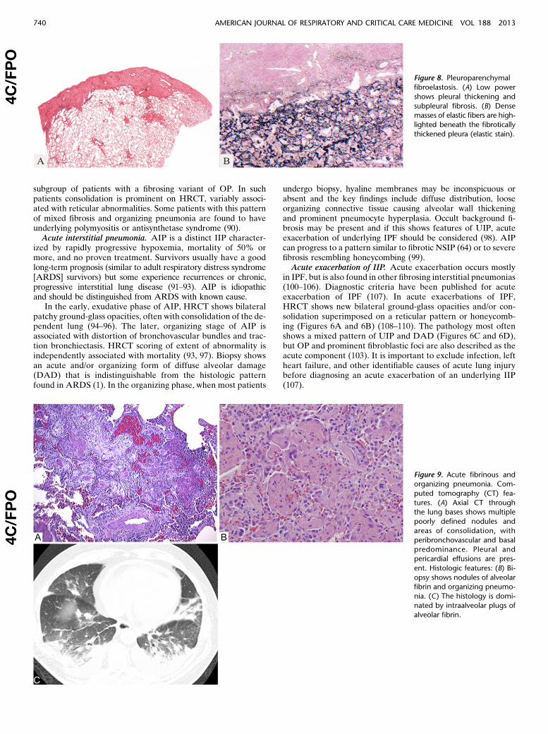
Figure 7. Pleuroparenchymal
fibroelastosis. Computed to-
mography (CT) features: (A)
High-resolution computed to-mography (HRCT) through
the upper lobes shows irregu-
lar pleural-based opacities anda reticular pattern associated
with parenchymal distortion.
The pleura and lungs in the
lower lobes appeared normal.(B) Section through the upper lobes shows scattered pleuroparenchymal opacities and some distortion of the underlying lung parenchyma. In the
lower lobes there was no pleural irregularity, but there was a subtle subpleural reticular pattern.
American Thoracic Society Documents 739
subgroup of patients with a fibrosing variant of OP. In suchpatients consolidation is prominent on HRCT, variably associ-ated with reticular abnormalities. Some patients with this patternof mixed fibrosis and organizing pneumonia are found to haveunderlying polymyositis or antisynthetase syndrome (90).Acute interstitial pneumonia. AIP is a distinct IIP character-
ized by rapidly progressive hypoxemia, mortality of 50% ormore, and no proven treatment. Survivors usually have a goodlong-term prognosis (similar to adult respiratory distress syndrome[ARDS] survivors) but some experience recurrences or chronic,progressive interstitial lung disease (91–93). AIP is idiopathicand should be distinguished from ARDS with known cause.
In the early, exudative phase of AIP, HRCT shows bilateralpatchy ground-glass opacities, often with consolidation of the de-pendent lung (94–96). The later, organizing stage of AIP isassociated with distortion of bronchovascular bundles and trac-tion bronchiectasis. HRCT scoring of extent of abnormality isindependently associated with mortality (93, 97). Biopsy showsan acute and/or organizing form of diffuse alveolar damage(DAD) that is indistinguishable from the histologic patternfound in ARDS (1). In the organizing phase, when most patients
undergo biopsy, hyaline membranes may be inconspicuous orabsent and the key findings include diffuse distribution, looseorganizing connective tissue causing alveolar wall thickeningand prominent pneumocyte hyperplasia. Occult background fi-brosis may be present and if this shows features of UIP, acuteexacerbation of underlying IPF should be considered (98). AIPcan progress to a pattern similar to fibrotic NSIP (64) or to severefibrosis resembling honeycombing (99).Acute exacerbation of IIP. Acute exacerbation occurs mostly
in IPF, but is also found in other fibrosing interstitial pneumonias(100–106). Diagnostic criteria have been published for acuteexacerbation of IPF (107). In acute exacerbations of IPF,HRCT shows new bilateral ground-glass opacities and/or con-solidation superimposed on a reticular pattern or honeycomb-ing (Figures 6A and 6B) (108–110). The pathology most oftenshows a mixed pattern of UIP and DAD (Figures 6C and 6D),but OP and prominent fibroblastic foci are also described as theacute component (103). It is important to exclude infection, leftheart failure, and other identifiable causes of acute lung injurybefore diagnosing an acute exacerbation of an underlying IIP(107).
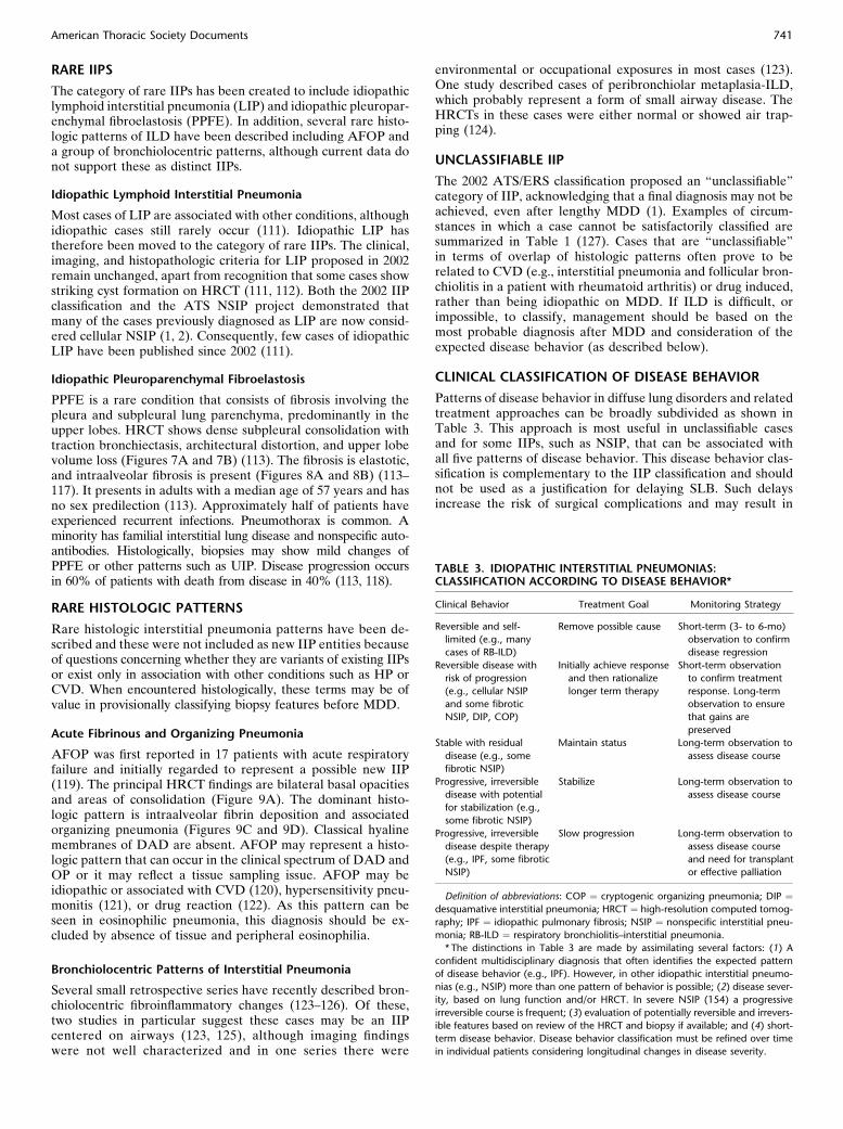
Figure 8. Pleuroparenchymal
fibroelastosis. (A) Low power
shows pleural thickening and
subpleural fibrosis. (B) Densemasses of elastic fibers are high-
lighted beneath the fibrotically
thickened pleura (elastic stain).
4C/FPO
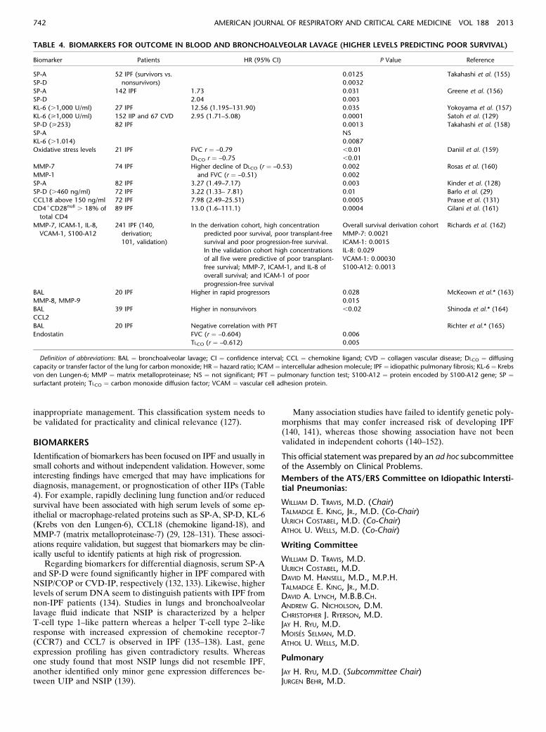
Figure 9. Acute fibrinous andorganizing pneumonia. Com-
puted tomography (CT) fea-
tures. (A) Axial CT throughthe lung bases shows multiple
poorly defined nodules and
areas of consolidation, with
peribronchovascular and basalpredominance. Pleural and
pericardial effusions are pres-
ent. Histologic features: (B) Bi-
opsy shows nodules of alveolarfibrin and organizing pneumo-
nia. (C) The histology is domi-
nated by intraalveolar plugs of
alveolar fibrin.
4C/FPO
740 AMERICAN JOURNAL OF RESPIRATORY AND CRITICAL CARE MEDICINE VOL 188 2013
RARE IIPS
The category of rare IIPs has been created to include idiopathiclymphoid interstitial pneumonia (LIP) and idiopathic pleuropar-enchymal fibroelastosis (PPFE). In addition, several rare histo-logic patterns of ILD have been described including AFOP anda group of bronchiolocentric patterns, although current data donot support these as distinct IIPs.
Idiopathic Lymphoid Interstitial Pneumonia
Most cases of LIP are associated with other conditions, althoughidiopathic cases still rarely occur (111). Idiopathic LIP hastherefore been moved to the category of rare IIPs. The clinical,imaging, and histopathologic criteria for LIP proposed in 2002remain unchanged, apart from recognition that some cases showstriking cyst formation on HRCT (111, 112). Both the 2002 IIPclassification and the ATS NSIP project demonstrated thatmany of the cases previously diagnosed as LIP are now consid-ered cellular NSIP (1, 2). Consequently, few cases of idiopathicLIP have been published since 2002 (111).
Idiopathic Pleuroparenchymal Fibroelastosis
PPFE is a rare condition that consists of fibrosis involving thepleura and subpleural lung parenchyma, predominantly in theupper lobes. HRCT shows dense subpleural consolidation withtraction bronchiectasis, architectural distortion, and upper lobevolume loss (Figures 7A and 7B) (113). The fibrosis is elastotic,and intraalveolar fibrosis is present (Figures 8A and 8B) (113–117). It presents in adults with a median age of 57 years and hasno sex predilection (113). Approximately half of patients haveexperienced recurrent infections. Pneumothorax is common. Aminority has familial interstitial lung disease and nonspecific auto-antibodies. Histologically, biopsies may show mild changes ofPPFE or other patterns such as UIP. Disease progression occursin 60% of patients with death from disease in 40% (113, 118).
RARE HISTOLOGIC PATTERNS
Rare histologic interstitial pneumonia patterns have been de-scribed and these were not included as new IIP entities becauseof questions concerning whether they are variants of existing IIPsor exist only in association with other conditions such as HP orCVD. When encountered histologically, these terms may be ofvalue in provisionally classifying biopsy features before MDD.
Acute Fibrinous and Organizing Pneumonia
AFOP was first reported in 17 patients with acute respiratoryfailure and initially regarded to represent a possible new IIP(119). The principal HRCT findings are bilateral basal opacitiesand areas of consolidation (Figure 9A). The dominant histo-logic pattern is intraalveolar fibrin deposition and associatedorganizing pneumonia (Figures 9C and 9D). Classical hyalinemembranes of DAD are absent. AFOP may represent a histo-logic pattern that can occur in the clinical spectrum of DAD andOP or it may reflect a tissue sampling issue. AFOP may beidiopathic or associated with CVD (120), hypersensitivity pneu-monitis (121), or drug reaction (122). As this pattern can beseen in eosinophilic pneumonia, this diagnosis should be ex-cluded by absence of tissue and peripheral eosinophilia.
Bronchiolocentric Patterns of Interstitial Pneumonia
Several small retrospective series have recently described bron-chiolocentric fibroinflammatory changes (123–126). Of these,two studies in particular suggest these cases may be an IIPcentered on airways (123, 125), although imaging findingswere not well characterized and in one series there were
environmental or occupational exposures in most cases (123).One study described cases of peribronchiolar metaplasia-ILD,which probably represent a form of small airway disease. TheHRCTs in these cases were either normal or showed air trap-ping (124).
UNCLASSIFIABLE IIP
The 2002 ATS/ERS classification proposed an “unclassifiable”category of IIP, acknowledging that a final diagnosis may not beachieved, even after lengthy MDD (1). Examples of circum-stances in which a case cannot be satisfactorily classified aresummarized in Table 1 (127). Cases that are “unclassifiable”in terms of overlap of histologic patterns often prove to berelated to CVD (e.g., interstitial pneumonia and follicular bron-chiolitis in a patient with rheumatoid arthritis) or drug induced,rather than being idiopathic on MDD. If ILD is difficult, orimpossible, to classify, management should be based on themost probable diagnosis after MDD and consideration of theexpected disease behavior (as described below).
CLINICAL CLASSIFICATION OF DISEASE BEHAVIOR
Patterns of disease behavior in diffuse lung disorders and relatedtreatment approaches can be broadly subdivided as shown inTable 3. This approach is most useful in unclassifiable casesand for some IIPs, such as NSIP, that can be associated withall five patterns of disease behavior. This disease behavior clas-sification is complementary to the IIP classification and shouldnot be used as a justification for delaying SLB. Such delaysincrease the risk of surgical complications and may result in
TABLE 3. IDIOPATHIC INTERSTITIAL PNEUMONIAS:CLASSIFICATION ACCORDING TO DISEASE BEHAVIOR*
Clinical Behavior Treatment Goal Monitoring Strategy
Reversible and self-
limited (e.g., many
cases of RB-ILD)
Remove possible cause Short-term (3- to 6-mo)
observation to confirm
disease regression
Reversible disease with
risk of progression
(e.g., cellular NSIP
and some fibrotic
NSIP, DIP, COP)
Initially achieve response
and then rationalize
longer term therapy
Short-term observation
to confirm treatment
response. Long-term
observation to ensure
that gains are
preserved
Stable with residual
disease (e.g., some
fibrotic NSIP)
Maintain status Long-term observation to
assess disease course
Progressive, irreversible
disease with potential
for stabilization (e.g.,
some fibrotic NSIP)
Stabilize Long-term observation to
assess disease course
Progressive, irreversible
disease despite therapy
(e.g., IPF, some fibrotic
NSIP)
Slow progression Long-term observation to
assess disease course
and need for transplant
or effective palliation
Definition of abbreviations: COP ¼ cryptogenic organizing pneumonia; DIP ¼desquamative interstitial pneumonia; HRCT ¼ high-resolution computed tomog-
raphy; IPF ¼ idiopathic pulmonary fibrosis; NSIP ¼ nonspecific interstitial pneu-
monia; RB-ILD ¼ respiratory bronchiolitis–interstitial pneumonia.
* The distinctions in Table 3 are made by assimilating several factors: (1) A
confident multidisciplinary diagnosis that often identifies the expected pattern
of disease behavior (e.g., IPF). However, in other idiopathic interstitial pneumo-
nias (e.g., NSIP) more than one pattern of behavior is possible; (2) disease sever-
ity, based on lung function and/or HRCT. In severe NSIP (154) a progressive
irreversible course is frequent; (3) evaluation of potentially reversible and irrevers-
ible features based on review of the HRCT and biopsy if available; and (4) short-
term disease behavior. Disease behavior classification must be refined over time
in individual patients considering longitudinal changes in disease severity.
American Thoracic Society Documents 741
inappropriate management. This classification system needs tobe validated for practicality and clinical relevance (127).
BIOMARKERS
Identification of biomarkers has been focused on IPF and usually insmall cohorts and without independent validation. However, someinteresting findings have emerged that may have implications fordiagnosis, management, or prognostication of other IIPs (Table4). For example, rapidly declining lung function and/or reducedsurvival have been associated with high serum levels of some ep-ithelial or macrophage-related proteins such as SP-A, SP-D, KL-6(Krebs von den Lungen-6), CCL18 (chemokine ligand-18), andMMP-7 (matrix metalloproteinase-7) (29, 128–131). These associ-ations require validation, but suggest that biomarkers may be clin-ically useful to identify patients at high risk of progression.
Regarding biomarkers for differential diagnosis, serum SP-Aand SP-D were found significantly higher in IPF compared withNSIP/COP or CVD-IP, respectively (132, 133). Likewise, higherlevels of serum DNA seem to distinguish patients with IPF fromnon-IPF patients (134). Studies in lungs and bronchoalveolarlavage fluid indicate that NSIP is characterized by a helperT-cell type 1–like pattern whereas a helper T-cell type 2–likeresponse with increased expression of chemokine receptor-7(CCR7) and CCL7 is observed in IPF (135–138). Last, geneexpression profiling has given contradictory results. Whereasone study found that most NSIP lungs did not resemble IPF,another identified only minor gene expression differences be-tween UIP and NSIP (139).
Many association studies have failed to identify genetic poly-morphisms that may confer increased risk of developing IPF(140, 141), whereas those showing association have not beenvalidated in independent cohorts (140–152).
This official statement was prepared by an ad hoc subcommitteeof the Assembly on Clinical Problems.
Members of the ATS/ERS Committee on Idiopathic Intersti-tial Pneumonias:
WILLIAM D. TRAVIS, M.D. (Chair)TALMADGE E. KING, JR., M.D. (Co-Chair)ULRICH COSTABEL, M.D. (Co-Chair)ATHOL U. WELLS, M.D. (Co-Chair)
Writing Committee
WILLIAM D. TRAVIS, M.D.ULRICH COSTABEL, M.D.DAVID M. HANSELL, M.D., M.P.H.TALMADGE E. KING, JR., M.D.DAVID A. LYNCH, M.B.B.CH.ANDREW G. NICHOLSON, D.M.CHRISTOPHER J. RYERSON, M.D.JAY H. RYU, M.D.MOISES SELMAN, M.D.ATHOL U. WELLS, M.D.
Pulmonary
JAY H. RYU, M.D. (Subcommittee Chair)JURGEN BEHR, M.D.
TABLE 4. BIOMARKERS FOR OUTCOME IN BLOOD AND BRONCHOALVEOLAR LAVAGE (HIGHER LEVELS PREDICTING POOR SURVIVAL)
Biomarker Patients HR (95% CI) P Value Reference
SP-A 52 IPF (survivors vs.
nonsurvivors)
0.0125 Takahashi et al. (155)
SP-D 0.0032
SP-A 142 IPF 1.73 0.031 Greene et al. (156)
SP-D 2.04 0.003
KL-6 (.1,000 U/ml) 27 IPF 12.56 (1.195–131.90) 0.035 Yokoyama et al. (157)
KL-6 (>1,000 U/ml) 152 IIP and 67 CVD 2.95 (1.71–5.08) 0.0001 Satoh et al. (129)
SP-D (>253) 82 IPF 0.0013 Takahashi et al. (158)
SP-A NS
KL-6 (.1.014) 0.0087
Oxidative stress levels 21 IPF FVC r ¼ –0.79 ,0.01 Daniil et al. (159)
DLCO r ¼ –0.75 ,0.01
MMP-7 74 IPF Higher decline of DLCO (r ¼ –0.53)
and FVC (r ¼ –0.51)
0.002 Rosas et al. (160)
MMP-1 0.002
SP-A 82 IPF 3.27 (1.49–7.17) 0.003 Kinder et al. (128)
SP-D (.460 ng/ml) 72 IPF 3.22 (1.33– 7.81) 0.01 Barlo et al. (29)
CCL18 above 150 ng/ml 72 IPF 7.98 (2.49–25.51) 0.0005 Prasse et al. (131)
CD41CD28null . 18% of
total CD4
89 IPF 13.0 (1.6–111.1) 0.0004 Gilani et al. (161)
MMP-7, ICAM-1, IL-8,
VCAM-1, S100-A12
241 IPF (140,
derivation;
101, validation)
In the derivation cohort, high concentration
predicted poor survival, poor transplant-free
survival and poor progression-free survival.
In the validation cohort high concentrations
of all five were predictive of poor transplant-
free survival; MMP-7, ICAM-1, and IL-8 of
overall survival; and ICAM-1 of poor
progression-free survival
Overall survival derivation cohort Richards et al. (162)
MMP-7: 0.0021
ICAM-1: 0.0015
IL-8: 0.029
VCAM-1: 0.00030
S100-A12: 0.0013
BAL 20 IPF Higher in rapid progressors 0.028 McKeown et al.* (163)
MMP-8, MMP-9 0.015
BAL 39 IPF Higher in nonsurvivors ,0.02 Shinoda et al.* (164)
CCL2
BAL 20 IPF Negative correlation with PFT Richter et al.* (165)
Endostatin FVC (r ¼ –0.604) 0.006
TLCO (r ¼ –0.612) 0.005
Definition of abbreviations: BAL ¼ bronchoalveolar lavage; CI ¼ confidence interval; CCL ¼ chemokine ligand; CVD ¼ collagen vascular disease; DLCO ¼ diffusing
capacity or transfer factor of the lung for carbon monoxide; HR ¼ hazard ratio; ICAM ¼ intercellular adhesion molecule; IPF ¼ idiopathic pulmonary fibrosis; KL-6 ¼ Krebs
von den Lungen-6; MMP ¼ matrix metalloproteinase; NS ¼ not significant; PFT ¼ pulmonary function test; S100-A12 ¼ protein encoded by S100-A12 gene; SP ¼surfactant protein; TLCO ¼ carbon monoxide diffusion factor; VCAM ¼ vascular cell adhesion protein.
742 AMERICAN JOURNAL OF RESPIRATORY AND CRITICAL CARE MEDICINE VOL 188 2013
DEMOSTHENES BOUROS, M.D., PH.D.KEVIN K. BROWN, M.D.HAROLD R. COLLARD, M.D.CARLOS ROBALO CORDEIRO, M.D., PH.D.VINCENT COTTIN, M.D., PH.D.MARJOLEIN DRENT, M.D., PH.D.JIM EGAN, M.D., M.B.B.CH.B.A.O.KEVIN FLAHERTY, M.D., M.S.YOSHIKAZU INOUE, M.D., PH.D.DONG SOON KIM, M.D.FERNANDO J. MARTINEZ, M.D., M.S.GANESH RAGHU, M.D.LUCA RICHELDI, M.D., PH.D.DOMINIQUE VALEYRE, M.D.
Radiology
DAVID M. HANSELL, M.D. (Subcommittee Co-Chair)DAVID A. LYNCH, M.B.B.CH. (Subcommittee Co-Chair)TAKESHI JOHKOH, M.D., PH.D.NICOLA SVERZELLATI, M.D.
Pathology
ANDREW G. NICHOLSON, D.M. (Subcommittee Chair)THOMAS V. COLBY, M.D.MASANORI KITAICHI, M.D.JEFFREY MYERS, M.D.
Molecular Biology
MoISES SELMAN, M.D. (Subcommittee Chair)BRUNO CRESTANI, M.D., PH.D.CORY HOGABOAM, PH.D.JAMES LOYD, M.D.
Evidence-based Analysis
CHRISTOPHER J. RYERSON, M.D. (Subcommittee Chair)JEFFREY SWIGRIS, D.O., M.S.
Reference Librarians
ROSALIND F. DUDDEN, M.L.S.SHANDRA PROTZKO, M.S.
Author Disclosures: D.M.H. reported consulting for Astra Zeneca ($1,000–$4,999). T.E.K. reported serving on advisory committees of Immune Works($1–4,999) and Intermune ($50,000–99,999). D.A.L. reported consulting forGilead (no payments), Intermune (no payments), and Perceptive Imaging($25,000–49,999); he received research support from Centocor ($25,000–49,999) and Siemens ($250,0001). A.G.N. reported consulting for Actelion($50,000–99,999) and Boehringer Ingelheim ($10,000–49,999). C.J.R. reportedserving as a speaker and on advisory committees of Intermune ($5,000–24,999),and received research support from Intermune ($50,000–99,999). J.B. reportedserving as a consultant, speaker, and on advisory committees of Actelion($25,000–49,999), and received research support from Actelion ($25,000–49,999); he served as a consultant, speaker, and on advisory committees ofBoehringer Ingelheim ($5,000–24,999) and Intermune ($50,000–99,999); hereceived research support from Intermune ($25,000–49,999). K.K.B. served asa consultant for Almirall ($1–4,999), Amgen ($1–4,999), Array BioPharma ($1–4,999), Genzyme (no payments), GlaxoSmithKline (no payments), Ikaria ($1–4,999), Ironwood ($1–4,999), and Pfizer (no payments); he served on advisorycommittees of Actelion ($5,000–24,999), Array BioPharma ($1–4,999), Boeh-ringer Ingelheim ($5,000–24,999), Centocor ($1–4,999), Fibrogen ($1–4,999),Genentech ($1–4,999), GeNo (no payments), Gilead ($5,000–24,999), Medi-mmune ($1–4,999), Mesoblast ($1–4,999), Promedior ($1–4,999), and Stromedix/Biogen ($1–4,999); he received research support from Actelion ($100,000–249,999),Amgen ($25,000–49,999), Genentech ($25,000–49,999), and Gilead ($5,000–24,999). H.R.C. reported consulting for Boehringer Ingelheim ($5,000–24,999),BMS ($1–4,999), Gilead ($5,000–24,999), and Promedior ($1–4,999); he servedon advisory committees of Fibrogen ($5,000–24,999), Five Prime ($1–4,999), Genoa($1–4,999), Medimmune ($1–4,999), and Mesoblast ($1–4,999), and received re-search support from Genentech ($5,000–24,999) and Intermune (no payments). V.C.reported serving as a speaker and on advisory committees of Intermune ($5,000–24,999); he served on advisory committees of Boehringer Ingelheim ($5,000–24,999)and received research support from Boehringer Ingelheim ($5,000–24,999). B.C.reported consulting for Sanofi ($1–4,999) and serving on advisory committees ofAstra Zeneca ($1,000–4,999) and Intermune ($1–4,999); he was a speaker for Inter-mune ($1–4,999) and Stallergenes ($1–4,999), and received research support from
Intermune ($100,0001). J.E. served on advisory committees of Pfizer (Wyeth)($1–4,999). K.F. served as a speaker for GlaxoSmithKline ($10,000–49,999) and onadvisory committees of Boehringer Ingelheim ($1–9,999), Fibrogen ($1–9,999), andGlaxoSmithKline ($1–9,999); he received research support from Centocor ($50,000–99,999), Immune Works ($50,000–99,999), and Intermune ($100,0001). D.S.K.served on advisory committees of Boehringer Ingelheim ($5,000–24,999). M.K. waspatent holder with Daikin Industries of a process for preparing a fluorine-containingpolymer and a cross-linked fluororubber diaphragm. F.J.M. reported consulting forAlmirall ($1–4,999), American Institute for Research ($1–4,999), Astra Zeneca ($1–4,999), Bayer ($1–4,999), Boehringer Ingelheim ($5,000–24,999), Caden Jennings($1–4,999), Cardiomema (no payments), Forest ($25,000–49,999), GlaxoSmithKline($25,000–49,999), HCRC-TC ($1–4,999), Ikarta ($5,000–24,999), Medimmune ($1–4,999), Merck ($5,000–24,999), Merion-TC ($1–4,999), Novartis ($5,000–24,999),Pearl ($5,000–24,999), Pfizer ($1–4,999), Sudler-Hennessey-TC ($1–4,999), and Ver-tex ($1–4,999); he served as a speaker for Astra Zeneca ($1–4,999), Bayer ($5,000–24,999), Forest ($5,000–24,999), GlaxoSmithKline ($50,000–49,999), andNycomed/Takeda ($50,000–49,999), and on advisory committees of Actelion($5,000–24,999), Bayer ($1–4,999), Biogen/Stromedix ($1–4,999), Janssen ($1–4,999),Mpex (no payments), Nycomed/Takeda ($50,000–99,999), Pfizer ($1–4,999), andVertex ($1–4,999); he received research support from Actelion ($5,000–24,999)and royalties from Informa ($1–4,999). G.R. reported consulting for Actelion($5,000–24,999), Bayer ($1,000–4,999), Boehringer Ingelheim ($1–4,999), Cen-tocor ($1–4,999), Celgene ($1–4,999), Fibrogen ($1–4,999), GlaxoSmithKline ($1–4,999), GeNo ($1–4,999), Gilead ($1–4,999), Intermune ($1–4,999), Promedior($1–4,999), Sanofi-Aventis ($1–4,999), Stromedix ($1–4,999), and Takeda ($1–4,999). L.R. reported consulting for Fibrogen ($1–4,999) and Genentech ($1–4,999), and was a speaker for Boehringer Ingelheim ($1–4,999) and Intermune($5,000–24,999); he served on advisory committees of Boehringer Ingelheim ($1–4,999), Intermune ($1–4,999), GlaxoSmithKline ($1–4,999), Medimmune ($1–4,999), and Sanofi-Aventis ($1–4,999); he received research support from Intermune($50,000–99,999). J.S. consulted for Genentech ($1–4,999) and received researchsupport from Intermune ($50,000–99,999). D.V. served on advisory committees ofActelion ($1–4,999) and as an expert witness for Sanofi-Aventis (,$1,000). W.D.T.,U.C., J.H.R., M.S., A.U.W., D.B., T.V.C., C.R.C., M.D., R.F.D., C.H., Y.I., T.J., J.L., J.M.,S.P., and N.S. reported they had no relevant commercial interests.
Acknowledgment: The committee acknowledges the American Thoracic Society andEuropean Respiratory Society for supporting this project; the staff of the University ofModena and Reggio Emilia, Italy; Shandra Protzko and Rosalin Dudden of the Libraryand Knowledge Services, National Jewish Health, Denver, Colorado; the De-partment of Pathology, VU University Medical Center Amsterdam, Amsterdam,The Netherlands, and the Department of Pathology, Memorial Sloan-KetteringCancer Center, New York, New York for assistance with face-to-face meetings; theATS staff for administrative assistance; and members of the ATS Documentationand Implementation Committee. The committee also acknowledges the followingindividuals who helped review the document: Arata Azuma (Japan), Mary Beth Beasley(United States), Alain Borczuk (United States), Marco Chilosi (Italy), Teri Franks (UnitedStates), Jeffrey Galvin (United States), Katrien Grunberg (The Netherlands), RichardHelmers (United States), Kevin Leslie (United States), Robert Kaner (United States),Heber MacMahon (United States), Nestor Muller (Brazil), David Naidich(United States), Marieke Overbeek (The Netherlands), Lynette Sholl (United States),Zarir F. Udwadia (India), and Dean W. Wallace (United States).
References
1. American Thoracic Society, European Respiratory Society. American
Thoracic Society/European Respiratory Society international mul-
tidisciplinary consensus classification of the idiopathic interstitial
pneumonias. Am J Respir Crit Care Med 2002;165:277–304.
2. Travis WD, Hunninghake G, King TE Jr, Lynch DA, Colby TV, Galvin
JR, Brown KK, Chung MP, Cordier J-F, du Bois RM, et al. Idiopathic
nonspecific interstitial pneumonia: report of an American Thoracic
Society project. Am J Respir Crit Care Med 2008;177:1338–1347.
3. Aziz ZA, Wells AU, Hansell DM, Bain GA, Copley SJ, Desai SR, Ellis
SM, Gleeson FV, Grubnic S, Nicholson AG, et al. HRCT diagnosis
of diffuse parenchymal lung disease: inter-observer variation. Thorax
2004;59:506–511.
4. Flaherty KR, King TE Jr, Raghu G, Lynch JP III, Colby TV, Travis WD,
Gross BH, Kazerooni EA, Toews GB, Long Q, et al. Idiopathic
interstitial pneumonia: what is the effect of a multidisciplinary
approach to diagnosis? Am J Respir Crit Care Med 2004;170:904–910.
5. Nicholson AG, Addis BJ, Bharucha H, Clelland CA, Corrin B, Gibbs
AR, Hasleton PS, Kerr KM, Ibrahim NBN, Stewart S, et al. Inter-
observer variation between pathologists in diffuse parenchymal lung
disease. Thorax 2004;59:500–505.
6. Thomeer M, Demedts M, Behr J, Buhl R, Costabel U, Flower CDR,
Verschakelen J, Laurent F, Nicholson AG, Verbeken EK, et al.;
Idiopathic Pulmonary Fibrosis International Group Exploring N-
Acetylcysteine I Annual (IFIGENIA) Study Group. Multidisciplinary
American Thoracic Society Documents 743
interobserver agreement in the diagnosis of idiopathic pulmonary
fibrosis. Eur Respir J 2008;31:585–591.
7. Flaherty KR, Andrei A-C, King TE Jr, Raghu G, Colby TV, Wells A,
Bassily N, Brown K, du Bois R, Flint A, et al. Idiopathic interstitial
pneumonia: do community and academic physicians agree on
diagnosis? Am J Respir Crit Care Med 2007;175:1054–1060.
8. Raghu G, Collard HR, Egan JJ, Martinez FJ, Behr J, Brown KK, Colby
TV, Cordier JF, Flaherty KR, Lasky JA, et al.; ATS/ERS/JRS/
ALAT Committee on Idiopathic Pulmonary Fibrosis. An official
ATS/ERS/JRS/ALAT statement: idiopathic pulmonary fibrosis:
evidence-based guidelines for diagnosis and management. Am J
Respir Crit Care Med 2011;183:788–824.
9. Selman M. Hypersensitivity pneumonitis: a multifaceted deceiving
disorder. Clin Chest Med 2004;25:531–547, vi.
10. Lynch DA, Newell JD, Logan PM, King TE Jr, Müller NL. Can CT
distinguish hypersensitivity pneumonitis from idiopathic pulmonary
fibrosis? AJR Am J Roentgenol 1995;165:807–811.
11. Silva CI, Churg A, Müller NL. Hypersensitivity pneumonitis: spectrum
of high-resolution CT and pathologic findings. AJR Am J Roentgenol
2007;188:334–344.
12. Silva CIS, Müller NL, Lynch DA, Curran-Everett D, Brown KK, Lee KS,
ChungMP, Churg A. Chronic hypersensitivity pneumonitis: differentiation
from idiopathic pulmonary fibrosis and nonspecific interstitial pneumonia
by using thin-section CT. Radiology 2008;246:288–297.
13. Akashi T, Takemura T, Ando N, Eishi Y, Kitagawa M, Takizawa T,
Koike M, Ohtani Y, Miyazaki Y, Inase N, et al. Histopathologic
analysis of sixteen autopsy cases of chronic hypersensitivity
pneumonitis and comparison with idiopathic pulmonary fibrosis/
usual interstitial pneumonia. Am J Clin Pathol 2009;131:405–415.
14. Churg A, Muller NL, Flint J, Wright JL. Chronic hypersensitivity
pneumonitis. Am J Surg Pathol 2006;30:201–208.
15. Churg A, Sin DD, Everett D, Brown K, Cool C. Pathologic patterns
and survival in chronic hypersensitivity pneumonitis. Am J Surg
Pathol 2009;33:1765–1770.
16. Fenton ME, Cockcroft DW, Wright JL, Churg A. Hypersensitivity
pneumonitis as a cause of airway-centered interstitial fibrosis. Ann
Allergy Asthma Immunol 2007;99:465–466.
17. Sahin H, Brown KK, Curran-Everett D, Hale V, Cool CD, Vourlekis
JS, Lynch DA. Chronic hypersensitivity pneumonitis: CT features
comparison with pathologic evidence of fibrosis and survival.
Radiology 2007;244:591–598.
18. Hanak V, Golbin JM, Ryu JH. Causes and presenting features in 85
consecutive patients with hypersensitivity pneumonitis. Mayo Clin
Proc 2007;82:812–816.
19. Felício CH, Parra ER, Capelozzi VL. Idiopathic and collagen vascular
disease nonspecific interstitial pneumonia: clinical significance of
remodeling process. Lung 2007;185:39–46.
20. Park JH, Kim DS, Park IN, Jang SJ, Kitaichi M, Nicholson AG, Colby
TV. Prognosis of fibrotic interstitial pneumonia: idiopathic versus
collagen vascular disease-related subtypes. Am J Respir Crit Care
Med 2007;175:705–711.
21. Hwang J-H, Misumi S, Sahin H, Brown KK, Newell JD, Lynch DA.
Computed tomographic features of idiopathic fibrosing interstitial
pneumonia: comparison with pulmonary fibrosis related to collagen
vascular disease. J Comput Assist Tomogr 2009;33:410–415.
22. Song JW, Do K-H, Kim M-Y, Jang SJ, Colby TV, Kim DS. Pathologic
and radiologic differences between idiopathic and collagen vascular
disease–related usual interstitial pneumonia. Chest 2009;136:23–30.
23. Corte TJ, Copley SJ, Desai SR, Zappala CJ, Hansell DM, Nicholson
AG, Colby TV, Renzoni E, Maher TM, Wells AU. Significance of
connective tissue disease features in idiopathic interstitial
pneumonia. Eur Respir J 2012;39:661–668.
24. Suda T, Kono M, Nakamura Y, Enomoto N, Kaida Y, Fujisawa T,
Imokawa S, Yasuda K, Hashizume H, Yokomura K, et al. Distinct
prognosis of idiopathic nonspecific interstitial pneumonia (NSIP)
fulfilling criteria for undifferentiated connective tissue disease
(UCTD). Respir Med 2010;104:1527–1534.
25. Kinder BW, Collard HR, Koth L, Daikh DI, Wolters PJ, Elicker B,
Jones KD, King TE Jr. Idiopathic nonspecific interstitial pneumonia:
lung manifestation of undifferentiated connective tissue disease? Am
J Respir Crit Care Med 2007;176:691–697.
26. García-Sancho C, Buendía-Roldán I, Fernández-Plata MR, Navarro C,
Pérez-Padilla R, Vargas MH, Loyd JE, Selman M. Familial
pulmonary fibrosis is the strongest risk factor for idiopathic
pulmonary fibrosis. Respir Med 2011;105:1902–1907.
27. Hodgson U, Laitinen T, Tukiainen P. Nationwide prevalence of sporadic
and familial idiopathic pulmonary fibrosis: evidence of founder effect
among multiplex families in Finland. Thorax 2002;57:338–342.
28. Marshall RP, Puddicombe A, Cookson WO, Laurent GJ. Adult familial
cryptogenic fibrosing alveolitis in the United Kingdom. Thorax 2000;
55:143–146.
29. Barlo NP, van Moorsel CHM, Ruven HJT, Zanen P, van den Bosch
JMM, Grutters JC. Surfactant protein-D predicts survival in patients
with idiopathic pulmonary fibrosis. Sarcoidosis Vasc Diffuse Lung
Dis 2009;26:155–161.
30. Nogee LM, Dunbar AE III, Wert SE, Askin F, Hamvas A, Whitsett JA.
A mutation in the surfactant protein C gene associated with familial
interstitial lung disease. N Engl J Med 2001;344:573–579.
31. Kirwan M, Dokal I. Dyskeratosis congenita, stem cells and telomeres.
Biochim Biophys Acta 2009;1792:371–379.
32. van Moorsel CH, van Oosterhout MF, Barlo NP, de Jong PA, van der
Vis JJ, Ruven HJ, van Es HW, van den Bosch JM, Grutters JC.
Surfactant protein C mutations are the basis of a significant
portion of adult familial pulmonary fibrosis in a Dutch cohort. Am
J Respir Crit Care Med 2010;182:1419–1425.
33. Alder JK, Chen JJL, Lancaster L, Danoff S, Su SC, Cogan JD, Vulto I, Xie
M, Qi X, Tuder RM, et al. Short telomeres are a risk factor for idiopathic
pulmonary fibrosis. Proc Natl Acad Sci USA 2008;105:13051–13056.
34. Cronkhite JT, Xing C, Raghu G, Chin KM, Torres F, Rosenblatt RL,
Garcia CK. Telomere shortening in familial and sporadic pulmonary
fibrosis. Am J Respir Crit Care Med 2008;178:729–737.
35. LawsonWE, Crossno PF, Polosukhin VV, Roldan J, Cheng D-S, Lane KB,
Blackwell TR, Xu C, Markin C, Ware LB, et al. Endoplasmic reticulum
stress in alveolar epithelial cells is prominent in IPF: association with
altered surfactant protein processing and herpesvirus infection. Am J
Physiol Lung Cell Mol Physiol 2008;294:L1119–L1126.
36. Seibold MA, Wise AL, Speer MC, Steele MP, Brown KK, Loyd JE,
Fingerlin TE, Zhang W, Gudmundsson G, Groshong SD, et al. A
common MUC5B promoter polymorphism and pulmonary fibrosis.
N Engl J Med 2011;364:1503–1512.
37. Zhang Y, Noth I, Garcia JG, Kaminski N. A variant in the promoter of
MUC5B and idiopathic pulmonary fibrosis. N Engl J Med 2011;364:
1576–1577.
38. Lawson WE, Loyd JE, Degryse AL. Genetics in pulmonary fibrosis—
familial cases provide clues to the pathogenesis of idiopathic
pulmonary fibrosis. Am J Med Sci 2011;341:439–443.
39. Monaghan H, Wells AU, Colby TV, du Bois RM, Hansell DM,
Nicholson AG. Prognostic implications of histologic patterns in
multiple surgical lung biopsies from patients with idiopathic
interstitial pneumonias. Chest 2004;125:522–526.
40. Ryu JH, Colby TV, Hartman TE, Vassallo R. Smoking-related inter-
stitial lung diseases: a concise review. Eur Respir J 2001;17:122–132.
41. Aubry MC, Wright JL, Myers JL. The pathology of smoking-related
lung diseases. Clin Chest Med 2000;21:11–35, vii.
42. Vassallo R, Jensen EA, Colby TV, Ryu JH, Douglas WW, Hartman
TE, Limper AH. The overlap between respiratory bronchiolitis and
desquamative interstitial pneumonia in pulmonary Langerhans cell
histiocytosis: high-resolution CT, histologic, and functional correla-
tions. Chest 2003;124:1199–1205.
43. Cottin V, Le Pavec J, Prévot G, Mal H, Humbert M, Simonneau G,
Cordier JF; GERM“O”P. Pulmonary hypertension in patients with
combined pulmonary fibrosis and emphysema syndrome. Eur Respir
J 2010;35:105–111.
44. Cottin V, Cordier JF. The syndrome of combined pulmonary fibrosis
and emphysema. Chest 2009;136:1–2.
45. Cottin V, Nunes H, Brillet PY, Delaval P, Devouassoux G, Tillie-Leblond I,
Israel-Biet D, Court-Fortune I, Valeyre D, Cordier JF; Groupe d’Etude et
de Recherche sur les Maladies Orphelines Pulmonaires (GERM“O”P).
Combined pulmonary fibrosis and emphysema: a distinct underrecognised
entity. Eur Respir J 2005;26:586–593.
46. Mejía M, Carrillo G, Rojas-Serrano J, Estrada A, Suárez T, Alonso D,
Barrientos E, Gaxiola M, Navarro C, Selman M. Idiopathic
744 AMERICAN JOURNAL OF RESPIRATORY AND CRITICAL CARE MEDICINE VOL 188 2013
pulmonary fibrosis and emphysema: decreased survival associated
with severe pulmonary arterial hypertension. Chest 2009;136:10–15.
47. Akira M, Inoue Y, Kitaichi M, Yamamoto S, Arai T, Toyokawa K.
Usual interstitial pneumonia and nonspecific interstitial pneumonia
with and without concurrent emphysema: thin-section CT findings.
Radiology 2009;251:271–279.
48. Flaherty KR, Travis WD, Colby TV, Toews GB, Kazerooni EA, Gross
BH, Jain A, Strawderman RL, Flint A, Lynch JP, et al.
Histopathologic variability in usual and nonspecific interstitial
pneumonias. Am J Respir Crit Care Med 2001;164:1722–1727.
49. Flaherty KR, Thwaite EL, Kazerooni EA, Gross BH, Toews GB, Colby
TV, Travis WD, Mumford JA, Murray S, Flint A, et al. Radiological
versus histological diagnosis in UIP and NSIP: survival implications.
Thorax 2003;58:143–148.
50. Sverzellati N, Wells AU, Tomassetti S, Desai SR, Copley SJ, Aziz ZA,
Zompatori M, Chilosi M, Nicholson AG, Poletti V, et al. Biopsy-
proved idiopathic pulmonary fibrosis: spectrum of nondiagnostic
thin-section CT diagnoses. Radiology 2010;254:957–964.
51. Sumikawa H, Johkoh T, Colby TV, Ichikado K, Suga M, Taniguchi H,
Kondoh Y, Ogura T, Arakawa H, Fujimoto K, et al. Computed
tomography findings in pathological usual interstitial pneumonia:
relationship to survival. Am J Respir Crit Care Med 2008;177:433–439.
52. Silva CI, Müller NL, Hansell DM, Lee KS, Nicholson AG, Wells AU.
Nonspecific interstitial pneumonia and idiopathic pulmonary
fibrosis: changes in pattern and distribution of disease over time.
Radiology 2008;247:251–259.
53. Lynch DA, Godwin JD, Safrin S, Starko KM, Hormel P, Brown KK,
Raghu G, King TE Jr, Bradford WZ, Schwartz DA, et al.; Idiopathic
Pulmonary Fibrosis Study Group. High-resolution computed to-
mography in idiopathic pulmonary fibrosis: diagnosis and prognosis.
Am J Respir Crit Care Med 2005;172:488–493.
54. Kim EY, Lee KS, Chung MP, Kwon OJ, Kim TS, Hwang JH.
Nonspecific interstitial pneumonia with fibrosis: serial high-
resolution CT findings with functional correlation. AJR Am J
Roentgenol 1999;173:949–953.
55. Hartman TE, Swensen SJ, Hansell DM, Colby TV, Myers JL, Tazelaar
HD, Nicholson AG, Wells AU, Ryu JH, Midthun DE, et al. Nonspecific
interstitial pneumonia: variable appearance at high-resolution chest CT.
Radiology 2000;217:701–705. [Published erratum appears in Radiology
218:606.]
56. Park JS, Lee KS, Kim JS, Park CS, Suh YL, Choi DL, Kim KJ.
Nonspecific interstitial pneumonia with fibrosis: radiographic and
CT findings in seven patients. Radiology 1995;195:645–648.
57. Cottin V, Donsbeck AV, Revel D, Loire R, Cordier JF. Nonspecific
interstitial pneumonia: individualization of a clinicopathologic entity in
a series of 12 patients. Am J Respir Crit Care Med 1998;158:1286–1293.
58. Nagai S, Kitaichi M, Itoh H, Nishimura K, Izumi T, Colby TV. Idiopathic
nonspecific interstitial pneumonia/fibrosis: comparison with idiopathic
pulmonary fibrosis and BOOP. Eur Respir J 1998;12:1010–1019.
59. Johkoh T, Müller NL, Colby TV, Ichikado K, Taniguchi H, Kondoh Y,
Fujimoto K, Kinoshita M, Arakawa H, Yamada H, et al. Nonspecific
interstitial pneumonia: correlation between thin-section CT findings
and pathologic subgroups in 55 patients. Radiology 2002;225:199–204.
60. Johkoh T, Müller NL, Cartier Y, Kavanagh PV, Hartman TE, Akira M,
Ichikado K, Ando M, Nakamura H. Idiopathic interstitial
pneumonias: diagnostic accuracy of thin-section CT in 129 patients.
Radiology 1999;211:555–560.
61. Nishiyama O, Kondoh Y, Taniguchi H, Yamaki K, Suzuki R, Yokoi T,
Takagi K. Serial high resolution CT findings in nonspecific interstitial
pneumonia/fibrosis. J Comput Assist Tomogr 2000;24:41–46.
62. MacDonald SL, Rubens MB, Hansell DM, Copley SJ, Desai SR, du Bois
RM, Nicholson AG, Colby TV, Wells AU. Nonspecific interstitial
pneumonia and usual interstitial pneumonia: comparative appearances
at and diagnostic accuracy of thin-section CT.Radiology 2001;221:600–605.
63. Akira M, Inoue Y, Arai T, Okuma T, Kawata Y. Long-term follow-up
high-resolution CT findings in non-specific interstitial pneumonia.
Thorax 2011;66:61–65.
64. Katzenstein AL, Fiorelli RF. Nonspecific interstitial pneumonia/
fibrosis: histologic features and clinical significance. Am J Surg
Pathol 1994;18:136–147.
65. Travis WD, Matsui K, Moss J, Ferrans VJ. Idiopathic nonspecific
interstitial pneumonia: prognostic significance of cellular and
fibrosing patterns: survival comparison with usual interstitial
pneumonia and desquamative interstitial pneumonia. Am J Surg
Pathol 2000;24:19–33.
66. Tsubamoto M, Müller NL, Johkoh T, Ichikado K, Taniguchi H, Kondoh
Y, Fujimoto K, Arakawa H, Koyama M, Kozuka T, et al. Pathologic
subgroups of nonspecific interstitial pneumonia: differential diagnosis
from other idiopathic interstitial pneumonias on high-resolution com-
puted tomography. J Comput Assist Tomogr 2005;29:793–800.
67. Park IN, Jegal Y, KimDS, DoKH, Yoo B, Shim TS, Lim CM, Lee SD, Koh
Y, Kim WS, et al. Clinical course and lung function change of idiopathic
nonspecific interstitial pneumonia. Eur Respir J 2009;33:68–76.
68. Hidalgo A, Franquet T, Giménez A, Bordes R, Pineda R, Madrid M.
Smoking-related interstitial lung diseases: radiologic–pathologic
correlation. Eur Radiol 2006;16:2463–2470.
69. Craig PJ, Wells AU, Doffman S, Rassl D, Colby TV, Hansell DM, Du
Bois RM, Nicholson AG. Desquamative interstitial pneumonia,
respiratory bronchiolitis and their relationship to smoking.
Histopathology 2004;45:275–282.
70. Fraig M, Shreesha U, Savici D, Katzenstein AL. Respiratory
bronchiolitis: a clinicopathologic study in current smokers,
ex-smokers, and never-smokers. Am J Surg Pathol 2002;26:647–653.
71. Vassallo R, Ryu JH. Tobacco smoke–related diffuse lung diseases.
Semin Respir Crit Care Med 2008;29:643–650.
72. Portnoy J, Veraldi KL, Schwarz MI, Cool CD, Curran-Everett D,
Cherniack RM, King TE Jr, Brown KK. Respiratory bronchiolitis-
interstitial lung disease: long-term outcome. Chest 2007;131:664–671.
73. Doan ML, Guillerman RP, Dishop MK, Nogee LM, Langston C,
Mallory GB, Sockrider MM, Fan LL. Clinical, radiological and
pathological features of ABCA3 mutations in children. Thorax
2008;63:366–373.
74. Bullard JE, Wert SE, Whitsett JA, Dean M, Nogee LM. ABCA3
mutations associated with pediatric interstitial lung disease. Am J
Respir Crit Care Med 2005;172:1026–1031.
75. Kawabata Y, Hoshi E, Murai K, Ikeya T, Takahashi N, Saitou Y,
Kurashima K, Ubukata M, Takayanagi N, Sugita H, et al.
Smoking-related changes in the background lung of specimens
resected for lung cancer: a semiquantitative study with correlation to
postoperative course. Histopathology 2008;53:707–714.
76. Katzenstein AL, Mukhopadhyay S, Zanardi C, Dexter E. Clinically
occult interstitial fibrosis in smokers: classification and significance
of a surprisingly common finding in lobectomy specimens. Hum
Pathol 2010;41:316–325.
77. Snider GL, Kleinerman J, Thurlbeck WM, Bengali ZH. The definition
of emphysema: report of a National Heart, Lung, and Blood
Institute, Division of Lung Diseases workshop. Am Rev Respir Dis
1985;132:182–185.
78. King TE Jr, Mortenson RL. Cryptogenic organizing pneumonitis: the
North American experience. Chest 1992;102(1, Suppl)8S–13S.
79. Lee JW, Lee KS, Lee HY, Chung MP, Yi CA, Kim TS, Chung MJ.
Cryptogenic organizing pneumonia: serial high-resolution CT find-
ings in 22 patients. AJR Am J Roentgenol 2010;195:916–922.
80. Sen T, Udwadia ZF. Cryptogenic organizing pneumonia: clinical profile
in a series of 34 admitted patients in a hospital in India. J Assoc
Physicians India 2008;56:229–232.
81. Oymak FS, Demirbaş HM, Mavili E, Akgun H, Gulmez I, Demir R,
Ozesmi M. Bronchiolitis obliterans organizing pneumonia: clinical
and roentgenological features in 26 cases. Respiration 2005;72:254–262.
82. Lee JS, Lynch DA, Sharma S, Brown KK, Müller NL. Organizing
pneumonia: prognostic implication of high-resolution computed to-
mography features. J Comput Assist Tomogr 2003;27:260–265.
83. Lee KS, Kullnig P, Hartman TE, Müller NL. Cryptogenic organizing
pneumonia: CT findings in 43 patients. AJR Am J Roentgenol 1994;
162:543–546.
84. Müller NL, Staples CA, Miller RR. Bronchiolitis obliterans organizing
pneumonia: CT features in 14 patients. AJR Am J Roentgenol 1990;
154:983–987.
85. Ujita M, Renzoni EA, Veeraraghavan S, Wells AU, Hansell DM.
Organizing pneumonia: perilobular pattern at thin-section CT. Ra-
diology 2004;232:757–761.
86. Kim SJ, Lee KS, Ryu YH, Yoon YC, Choe KO, Kim TS, Sung KJ.
Reversed halo sign on high-resolution CT of cryptogenic organizing
American Thoracic Society Documents 745
pneumonia: diagnostic implications. AJR Am J Roentgenol 2003;180:
1251–1254.
87. Epler GR, Colby TV, McLoud TC, Carrington CB, Gaensler EA.
Bronchiolitis obliterans organizing pneumonia. N Engl J Med 1985;
312:152–158.
88. Lohr RH, Boland BJ, Douglas WW, Dockrell DH, Colby TV, Swensen
SJ, Wollan PC, Silverstein MD. Organizing pneumonia: features and
prognosis of cryptogenic, secondary, and focal variants. Arch Intern
Med 1997;157:1323–1329.
89. Lazor R, Vandevenne A, Pelletier A, Leclerc P, Court-Fortune I,
Cordier JF; Groupe d’Etudes et de Recherche sur les Maladies
“Orphelines” Pulmonaires (GERM“O”P). Cryptogenic organizing
pneumonia: characteristics of relapses in a series of 48 patients.
Am J Respir Crit Care Med 2000;162:571–577.
90. Fischer A, Swigris JJ, du Bois RM, Lynch DA, Downey GP, Cosgrove GP,
Frankel SK, Fernandez-Perez ER, Gillis JZ, Brown KK. Anti-synthetase
syndrome in ANA and anti–Jo-1 negative patients presenting with idio-
pathic interstitial pneumonia. Respir Med 2009;103:1719–1724.
91. Bouros D, Nicholson AC, Polychronopoulos V, du Bois RM. Acute
interstitial pneumonia. Eur Respir J 2000;15:412–418.
92. Vourlekis JS, Brown KK, Cool CD, Young DA, Cherniack RM, King
TE, Schwarz MI. Acute interstitial pneumonitis: case series and
review of the literature. Medicine (Baltimore) 2000;79:369–378.
93. Ichikado K, Suga M, Müller NL, Taniguchi H, Kondoh Y, Akira M,
Johkoh T, Mihara N, Nakamura H, Takahashi M, et al. Acute
interstitial pneumonia: comparison of high-resolution computed to-
mography findings between survivors and nonsurvivors. Am J Respir
Crit Care Med 2002;165:1551–1556.
94. Primack SL, Hartman TE, Ikezoe J, Akira M, Sakatani M, Müller NL.
Acute interstitial pneumonia: radiographic and CT findings in nine
patients. Radiology 1993;188:817–820.
95. Johkoh T, Müller NL, Taniguchi H, Kondoh Y, Akira M, Ichikado K,
Ando M, Honda O, Tomiyama N, Nakamura H. Acute interstitial
pneumonia: thin-section CT findings in 36 patients. Radiology 1999;
211:859–863.
96. Desai SR, Wells AU, Rubens MB, Evans TW, Hansell DM. Acute
respiratory distress syndrome: CT abnormalities at long-term fol-
low-up. Radiology 1999;210:29–35.
97. Ichikado K, Suga M, Muranaka H, Gushima Y, Miyakawa H,
Tsubamoto M, Johkoh T, Hirata N, Yoshinaga T, Kinoshita Y, et al.
Prediction of prognosis for acute respiratory distress syndrome with
thin-section CT: validation in 44 cases. Radiology 2006;238:321–329.
98. Araya J, Kawabata Y, Jinho P, Uchiyama T, Ogata H, Sugita Y.
Clinically occult subpleural fibrosis and acute interstitial pneumonia a
precursor to idiopathic pulmonary fibrosis? Respirology 2008;13:408–412.
99. Rice AJ, Wells AU, Bouros D, du Bois RM, Hansell DM,
Polychronopoulos V, Vassilakis D, Kerr JR, Evans TW, Nicholson
AG. Terminal diffuse alveolar damage in relation to interstitial
pneumonias: an autopsy study. Am J Clin Pathol 2003;119:709–714.
100. Song JW, Hong SB, Lim CM, Koh Y, Kim DS. Acute exacerbation of
idiopathic pulmonary fibrosis: incidence, risk factors and outcome.
Eur Respir J 2011;37:356–363.
101. Agarwal R, Jindal SK. Acute exacerbation of idiopathic pulmonary
fibrosis: a systematic review. Eur J Intern Med 2008;19:227–235.
102. Silva CIS, Müller NL, Fujimoto K, Kato S, Ichikado K, Taniguchi H,
Kondoh Y, Johkoh T, Churg A. Acute exacerbation of chronic
interstitial pneumonia: high-resolution computed tomography and
pathologic findings. J Thorac Imaging 2007;22:221–229.
103. Churg A, Müller NL, Silva CIS, Wright JL. Acute exacerbation (acute
lung injury of unknown cause) in UIP and other forms of fibrotic
interstitial pneumonias. Am J Surg Pathol 2007;31:277–284.
104. Suda T, Kaida Y, Nakamura Y, Enomoto N, Fujisawa T, Imokawa S,
Hashizume H, Naito T, Hashimoto D, Takehara Y, et al. Acute
exacerbation of interstitial pneumonia associated with collagen
vascular diseases. Respir Med 2009;103:846–853.
105. Park IN, Kim DS, Shim TS, Lim C-M, Lee SD, Koh Y, Kim WS, Kim
WD, Jang SJ, Colby TV. Acute exacerbation of interstitial pneumonia
other than idiopathic pulmonary fibrosis. Chest 2007;132:214–220.
106. Miyazaki Y, Tateishi T, Akashi T, Ohtani Y, Inase N, Yoshizawa Y.
Clinical predictors and histologic appearance of acute exacerbations
in chronic hypersensitivity pneumonitis. Chest 2008;134:1265–1270.
107. Collard HR, Moore BB, Flaherty KR, Brown KK, Kaner RJ, King TE
Jr, Lasky JA, Loyd JE, Noth I, Olman MA, et al.; Idiopathic
Pulmonary Fibrosis Clinical Research Network Investigators.
Acute exacerbations of idiopathic pulmonary fibrosis. Am J Respir
Crit Care Med 2007;176:636–643.
108. Akira M, Kozuka T, Yamamoto S, Sakatani M. Computed tomography
findings in acute exacerbation of idiopathic pulmonary fibrosis. Am J
Respir Crit Care Med 2008;178:372–378.
109. Akira M, Hamada H, Sakatani M, Kobayashi C, Nishioka M,
Yamamoto S. CT findings during phase of accelerated
deterioration in patients with idiopathic pulmonary fibrosis. AJR
Am J Roentgenol 1997;168:79–83.
110. Fujimoto K, Taniguchi H, Johkoh T, Kondoh Y, Ichikado K, Sumikawa
H, Ogura T, Kataoka K, Endo T, Kawaguchi A, et al. Acute
exacerbation of idiopathic pulmonary fibrosis: high-resolution CT
scores predict mortality. Eur Radiol 2012;22:83–92.
111. Cha SI, Fessler MB, Cool CD, Schwarz MI, Brown KK. Lymphoid
interstitial pneumonia: clinical features, associations and prognosis.
Eur Respir J 2006;28:364–369.
112. Johkoh T, Müller NL, Pickford HA, Hartman TE, Ichikado K, Akira
M, Honda O, Nakamura H. Lymphocytic interstitial pneumonia:
thin-section CT findings in 22 patients. Radiology 1999;212:567–572.
113. Reddy TL, Tominaga M, Hansell DM, von der Thusen J Jr, Rassl D,
Parfrey H, Guy S, Twentyman O, Rice A, Maher TM, et al.
Pleuroparenchymal fibroelastosis: a spectrum of histopathological
and imaging phenotypes. Eur Respir J 2012;40:377–385.
114. Becker CD, Gil J, Padilla ML. Idiopathic pleuroparenchymal
fibroelastosis: an unrecognized or misdiagnosed entity? Mod Pathol
2008;21:784–787.
115. Frankel SK, Cool CD, LynchDA, Brown KK. Idiopathic pleuroparenchymal
fibroelastosis: description of a novel clinicopathologic entity. Chest
2004;126:2007–2013.
116. von der Thüsen JH, Hansell DM, Tominaga M, Veys PA, Ashworth
MT, Owens CM, Nicholson AG. Pleuroparenchymal fibroelastosis in
patients with pulmonary disease secondary to bone marrow
transplantation. Mod Pathol 2011;24:1633–1639.
117. Amitani R, Niimi A, Kuze F. Idiopathic pulmonary upper lobe fibrosis
(IPUF) [in Japanese]. Kokyu 1992;11:693–699.
118. Watanabe K, Nagata N, Kitasato Y, Wakamatsu K, Nabeshima K,
Harada T, Hirota T, Shiraishi M, Fujita M. Rapid decrease in
forced vital capacity in patients with idiopathic pulmonary upper
lobe fibrosis. Respir Investig 2012;50:88–97.
119. Beasley MB, Franks TJ, Galvin JR, Gochuico B, Travis WD. Acute
fibrinous and organizing pneumonia: a histological pattern of lung
injury and possible variant of diffuse alveolar damage. Arch Pathol
Lab Med 2002;126:1064–1070.
120. Balduin R, Giacometti C, Saccarola L, Marulli G, Rea F, Bartoli M,
Agostini C, Calabrese F. Acute fibrinous and organizing pneumonia
in a patient with collagen vascular disease “stigma.” Sarcoidosis Vasc
Diffuse Lung Dis 2007;24:78–80.
121. Hariri LP, Mino-Kenudson M, Shea B, Digumarthy S, Onozato M, Yagi
Y, Fraire AE, Matsubara O,Mark EJ. Distinct histopathology of acute
onset or abrupt exacerbation of hypersensitivity pneumonitis. Hum
Pathol 2012;43:660–668.
122. Yokogawa N, Alcid DV. Acute fibrinous and organizing pneumonia as
a rare presentation of abacavir hypersensitivity reaction. AIDS 2007;
21:2116–2117.
123. Churg A, Myers J, Suarez T, Gaxiola M, Estrada A, Mejia M, Selman
M. Airway-centered interstitial fibrosis: a distinct form of aggressive
diffuse lung disease. Am J Surg Pathol 2004;28:62–68.
124. Fukuoka J, Franks TJ, Colby TV, Flaherty KR, Galvin JR, Hayden D,
Gochuico BR, Kazerooni EA, Martinez F, Travis WD. Peribronchiolar
metaplasia: a common histologic lesion in diffuse lung disease and a rare
cause of interstitial lung disease: clinicopathologic features of 15 cases.
Am J Surg Pathol 2005;29:948–954.
125. Yousem SA, Dacic S. Idiopathic bronchiolocentric interstitial
pneumonia. Mod Pathol 2002;15:1148–1153.
126. Mark EJ, Ruangchira Urai R. Bronchiolitis interstitial pneumonitis:
a pathologic study of 31 lung biopsies with features intermediate
between bronchiolitis obliterans organizing pneumonia and usual
interstitial pneumonitis, with clinical correlation. Ann Diagn
Pathol 2008;12:171–180.
746 AMERICAN JOURNAL OF RESPIRATORY AND CRITICAL CARE MEDICINE VOL 188 2013
127. Ryerson CJ, Urbania TH, Richeldi L, Mooney JJ, Lee JS, Jones KD,
Elicker BM, Koth LL, King TE Jr, Wolters PJ, et al. Prevalence and
prognosis of unclassifiable interstitial lung disease. Eur Respir J 2013;
42:750–757.
128. Kinder BW, Brown KK, McCormack FX, Ix JH, Kervitsky A, Schwarz
MI, King TE Jr. Serum surfactant protein-A is a strong predictor of
early mortality in idiopathic pulmonary fibrosis. Chest 2009;135:
1557–1563.
129. Satoh H, Kurishima K, Ishikawa H, Ohtsuka M. Increased levels of
KL-6 and subsequent mortality in patients with interstitial
lung diseases. J Intern Med 2006;260:429–434.
130. Prasse A, Probst C, Bargagli E, Zissel G, Toews GB, Flaherty KR,
Olschewski M, Rottoli P, Müller-Quernheim J. Serum CC-chemokine
ligand 18 concentration predicts outcome in idiopathic pulmonary
fibrosis. Am J Respir Crit Care Med 2009;179:717–723.
131. Prasse A, Pechkovsky DV, Toews GB, Schäfer M, Eggeling S, Ludwig
C, Germann M, Kollert F, Zissel G, Müller-Quernheim J. CCL18 as
an indicator of pulmonary fibrotic activity in idiopathic interstitial
pneumonias and systemic sclerosis. Arthritis Rheum 2007;56:1685–
1693.
132. Ishii H, Mukae H, Kadota J, Kaida H, Nagata T, Abe K, Matsukura S,
Kohno S. High serum concentrations of surfactant protein A in usual
interstitial pneumonia compared with non-specific interstitial pneu-
monia. Thorax 2003;58:52–57.
133. Ohnishi H, Yokoyama A, Kondo K, Hamada H, Abe M, Nishimura K,
Hiwada K, Kohno N. Comparative study of KL-6, surfactant protein-A,
surfactant protein-D, and monocyte chemoattractant protein-1 as se-
rum markers for interstitial lung diseases. Am J Respir Crit Care Med
2002;165:378–381.
134. Casoni GL, Ulivi P, Mercatali L, Chilosi M, Tomassetti S, Romagnoli
M, Ravaglia C, Gurioli C, Gurioli C, Zoli W, et al. Increased levels of
free circulating DNA in patients with idiopathic pulmonary fibrosis.
Int J Biol Markers 2010;25:229–235.
135. Pignatti P, Brunetti G, Moretto D, Yacoub M-R, Fiori M, Balbi B,
Balestrino A, Cervio G, Nava S, Moscato G. Role of the
chemokine receptors CXCR3 and CCR4 in human pulmonary
fibrosis. Am J Respir Crit Care Med 2006;173:310–317.
136. Ishii H, Mukae H, Kadota J, Fujii T, Abe K, Ashitani J, Kohno S.
Increased levels of interleukin-18 in bronchoalveolar lavage fluid
of patients with idiopathic nonspecific interstitial pneumonia. Res-
piration 2005;72:39–45.
137. Choi ES, Pierce EM, Jakubzick C, Carpenter KJ, Kunkel SL, Evanoff
H, Martinez FJ, Flaherty KR, Moore BB, Toews GB, et al. Focal
interstitial CC chemokine receptor 7 (CCR7) expression in
idiopathic interstitial pneumonia. J Clin Pathol 2006;59:28–39.
138. Choi ES, Jakubzick C, Carpenter KJ, Kunkel SL, Evanoff H, Martinez
FJ, Flaherty KR, Toews GB, Colby TV, Kazerooni EA, et al.
Enhanced monocyte chemoattractant protein-3/CC chemokine
ligand-7 in usual interstitial pneumonia. Am J Respir Crit Care Med
2004;170:508–515.
139. Yang IV, Burch LH, Steele MP, Savov JD, Hollingsworth JW,
McElvania-Tekippe E, Berman KG, Speer MC, Sporn TA, Brown
KK, et al. Gene expression profiling of familial and sporadic
interstitial pneumonia. Am J Respir Crit Care Med 2007;175:45–54.
140. Lawson WE, Grant SW, Ambrosini V, Womble KE, Dawson EP, Lane
KB, Markin C, Renzoni E, Lympany P, Thomas AQ, et al. Genetic
mutations in surfactant protein C are a rare cause of sporadic cases
of IPF. Thorax 2004;59:977–980.
141. Kim KK, Flaherty KR, Long Q, Hattori N, Sisson TH, Colby TV,
Travis WD, Martinez FJ, Murray S, Simon RH. A plasminogen
activator inhibitor-1 promoter polymorphism and idiopathic inter-
stitial pneumonia. Mol Med 2003;9:52–56.
142. Markart P, Ruppert C, Wygrecka M, Schmidt R, Korfei M, Harbach H,
Theruvath I, Pison U, Seeger W, Guenther A, et al. Surfactant
protein C mutations in sporadic forms of idiopathic interstitial
pneumonias. Eur Respir J 2007;29:134–137.
143. Zorzetto M, Ferrarotti I, Campo I, Trisolini R, Poletti V, Scabini R,
Ceruti M, Mazzola P, Crippa E, Ottaviani S, et al. NOD2/CARD15
gene polymorphisms in idiopathic pulmonary fibrosis. Sarcoidosis
Vasc Diffuse Lung Dis 2005;22:180–185.
144. Hutyrová B, Pantelidis P, Drábek J, Zůrková M, Kolek V, Lenhart K,
Welsh KI, Du Bois RM, Petrek M. Interleukin-1 gene cluster
polymorphisms in sarcoidosis and idiopathic pulmonary fibrosis. Am
J Respir Crit Care Med 2002;165:148–151.
145. Renzoni E, Lympany P, Sestini P, Pantelidis P, Wells A, Black C,
Welsh K, Bunn C, Knight C, Foley P, et al. Distribution of novel
polymorphisms of the interleukin-8 and CXC receptor 1 and 2 genes
in systemic sclerosis and cryptogenic fibrosing alveolitis. Arthritis
Rheum 2000;43:1633–1640.
146. Zorzetto M, Ferrarotti I, Trisolini R, Lazzari Agli L, Scabini R, Novo
M, De Silvestri A, Patelli M, Martinetti M, Cuccia M, et al.
Complement receptor 1 gene polymorphisms are associated with
idiopathic pulmonary fibrosis. Am J Respir Crit Care Med 2003;
168:330–334.
147. Hodgson U, Tukiainen P, Laitinen T. The polymorphism C5507G of
complement receptor 1 does not explain idiopathic pulmonary
fibrosis among the Finns. Respir Med 2005;99:265–267.
148. Kubistova Z, Mrazek F, Lympany PA, Lagan AL, Arakelyan A,
Kriegova E, Welsh KI, Kolek V, Zatloukal J, Hutyrova B, et al.
The CR1 C5507G polymorphism is not involved in susceptibility to
idiopathic pulmonary fibrosis in two European populations. Tissue
Antigens 2008;72:483–486.
149. Mushiroda T, Wattanapokayakit S, Takahashi A, Nukiwa T, Kudoh S,
Ogura T, Taniguchi H, Kubo M, Kamatani N, Nakamura Y;
Pirfenidone Clinical Study Group. A genome-wide association
study identifies an association of a common variant in TERT with
susceptibility to idiopathic pulmonary fibrosis. J Med Genet 2008;45:
654–656.
150. Selman M, Lin H-M, Montano M, Jenkins AL, Estrada A, Lin Z, Wang
G, DiAngelo SL, Guo X, Umstead TM, et al. Surfactant protein A
and B genetic variants predispose to idiopathic pulmonary fibrosis.
Hum Genet 2003;113:542–550.
151. Falfán-Valencia R, Camarena A, Juárez A, Becerril C, Montano M,
Cisneros J, Mendoza F, Granados J, Pardo A, Selman M. Major
histocompatibility complex and alveolar epithelial apoptosis in
idiopathic pulmonary fibrosis. Hum Genet 2005;118:235–244.
152. Checa M, Ruiz V, Montano M, Velázquez-Cruz R, Selman M, Pardo A.
MMP-1 polymorphisms and the risk of idiopathic pulmonary fibrosis.
Hum Genet 2008;124:465–472.
153. Matsushima H, Takayanagi N, Sakamoto T, Kawata I, Motegi M,
Ubukata M, Yanagisawa T, Sugita Y, Kanazawa M, Kawabata Y.
Pathologic findings both before and after steroid therapy in a case of
desquamative interstitial pneumonia [article in Japanese]. Nihon
Kokyuki Gakkai Zasshi 2001;39:609–614.
154. Latsi PI, du Bois RM, Nicholson AG, Colby TV, Bisirtzoglou D,
Nikolakopoulou A, Veeraraghavan S, Hansell DM, Wells AU.
Fibrotic idiopathic interstitial pneumonia: the prognostic value of
longitudinal functional trends. Am J Respir Crit Care Med 2003;
168:531–537.
155. Takahashi H, Fujishima T, Koba H, Murakami S, Kurokawa K,
Shibuya Y, Shiratori M, Kuroki Y, Abe S. Serum surfactant
proteins A and D as prognostic factors in idiopathic pulmonary
fibrosis and their relationship to disease extent. Am J Respir Crit
Care Med 2000;162:1109–1114.
156. Greene KE, King TE Jr, Kuroki Y, Bucher-Bartelson B, Hunninghake
GW, Newman LS, Nagae H, Mason RJ. Serum surfactant proteins-A
and -D as biomarkers in idiopathic pulmonary fibrosis. Eur Respir J
2002;19:439–446.
157. Yokoyama A, Kondo K, Nakajima M, Matsushima T, Takahashi T,
Nishimura M, Bando M, Sugiyama Y, Totani Y, Ishizaki T, et al.
Prognostic value of circulating KL-6 in idiopathic pulmonary fibrosis.
Respirology 2006;11:164–168.
158. Takahashi H, Shiratori M, Kanai A, Chiba H, Kuroki Y, Abe S.
Monitoring markers of disease activity for interstitial lung diseases
with serum surfactant proteins A and D. Respirology 2006;11:S51–
S54.
159. Daniil ZD, Papageorgiou E, Koutsokera A, Kostikas K, Kiropoulos T,
Papaioannou AI, Gourgoulianis KI. Serum levels of oxidative stress
as a marker of disease severity in idiopathic pulmonary fibrosis.
Pulm Pharmacol Ther 2008;21:26–31.
160. Rosas IO, Richards TJ, Konishi K, Zhang Y, Gibson K, Lokshin AE,
Lindell KO, Cisneros J, Macdonald SD, Pardo A, Sciurba F, Dauber
J, Selman M, Gochuico BR, Kaminski N. MMP1 and MMP7 as
American Thoracic Society Documents 747

potential peripheral blood biomarkers in idiopathic pulmonary
fibrosis. PLoS Med 2008;5:e93.
161. Gilani SR, Vuga LJ, Lindell KO, Gibson KF, Xue J, Kaminski N,
Valentine VG, Lindsay EK, George MP, Steele C, et al. CD28
down-regulation on circulating CD4 T-cells is associated with poor
prognoses of patients with idiopathic pulmonary fibrosis. PLoS One
2010;5:e8959.
162. Richards TJ, Kaminski N, Baribaud F, Flavin S, Brodmerkel C, Horowitz
D, Li K, Choi J, Vuga LJ, Lindell KO, Klesen M, Zhang Y, Gibson
KF. Peripheral blood proteins predict mortality in idiopathic
pulmonary fibrosis. Am J Respir Crit Care Med 2012;185:67–76.
163. McKeown S, Richter AG, O’Kane C, McAuley DF, Thickett DR.
MMP expression and abnormal lung permeability are important
determinants of outcome in IPF. Eur Respir J 2009;33:77–84.
164. Shinoda H, Tasaka S, Fujishima S, Yamasawa W, Miyamoto K, Nakano
Y, Kamata H, Hasegawa N, Ishizaka A. Elevated CC chemokine level
in bronchoalveolar lavage fluid is predictive of a poor outcome of
idiopathic pulmonary fibrosis. Respiration 2009;78:285–292.
165. Richter AG, McKeown S, Rathinam S, Harper L, Rajesh P, McAuley
DF, Heljasvaara R, Thickett DR. Soluble endostatin is a novel
inhibitor of epithelial repair in idiopathic pulmonary fibrosis.
Thorax 2009;64:156–161.
748 AMERICAN JOURNAL OF RESPIRATORY AND CRITICAL CARE MEDICINE VOL 188 2013