an introduction to the structure and behavior of viruses
TRANSCRIPT

SECTION I
AN INTRODUCTION TO THE STRUCTUREAND BEHAVIOR OF VIRUSES
COPYRIG
HTED M
ATERIAL


CHAPTER 1
DEFINING THE ECOLOGY OF VIRUSES�
CHRISTON J. HURST1,2
1Departments of Biology and Music, Xavier University, Cincinnati, OH2Engineering Faculty, Universidad del Valle, Ciudad Universitaria Mel�endez, Santiago de Cali, Valle,Colombia
CONTENTS
1.1 Introduction
1.1.1 What is a Virus?1.1.2 What is Viral Ecology?1.1.3 Why Study Viral Ecology?
1.2 Surviving theGame: TheVirus and it’s Host
1.2.1 Cell Sweet Cell, and Struggles atHome
1.2.2 I Want a Niche, Just Like the Niche,That Nurtured Dear Old Mom andDad
1.2.3 Being Societal
1.3 Steppin’ Out and Taking The A Train:Reaching Out and Touching Someone byVector or Vehicle
1.3.1 “Down and Dirty” (Just Between UsHosts)
1.3.2 “The Hitchhiker” (Finding a Vector)1.3.3 “In a Dirty Glass” (Going There by
Vehicle)1.3.4 Bringing Concepts Together1.3.5 Is There no Hope?
1.4 Why Things Are the Way They Are
1.4.1 To Kill or Not to Kill - A Question ofVirulence
1.4.2 Genetic Equilibrium (versusDisequilibrium)
1.4.3 Uniqueness versus Commonality(There Are Hussies and Floozies inthe Virus World)
1.4.4 Evolution
1.5 Summary (Can There be Conclusions?)AcknowledgementReferences
1.1 INTRODUCTION
The goal of virology is to understand the
viruses and their behavior. Virology is an inter-
esting subject and even has contributed to the
concepts of what we consider to represent
dieties and art. Sekhmet, an ancient Egyptian
goddess, was for a time considered to be the
source of both causation and cure for many of
the diseases that we now know to be caused by
viruses (Figure 1.1). Influenza, a viral-induced
disease of vertebrates, was once assumed to be
caused by the influence of the stars, and that is
represented by the origin of it’s name which
is derived from Italian. The following was a
*This chapter represents a revision of “Defining the ecol-
ogy of viruses”, which appeared as chapter 1 of the book
Viral Ecology, edited by Christon J. Hurst, published in
2000 by Academic Press. All of the artwork contained in
this chapter appears courtesy of Christon J. Hurst.
Studies in Viral Ecology: Microbial and Botanical Host Systems, Volume 1, First Edition. Edited by Christon J. Hurst.� 2011 John Wiley & Sons, Inc. Published 2011 by John Wiley & Sons, Inc.
3

rhyme which children in the United Sates sang
while skipping rope during the influenza pan-
demic of 1918–1919:
I had a little bird
It’s name was Enza
I opened a window
And in-flew-Enza.
(Source: The flu of 1918, by Eileen A Lynch,
The Pennsylvania Gazette November/
December 1998 (http://www.upenn.edu/
gazette/1198/lynch.html).
FIGURE 1.1 Image of Sekhmet, “Bust Fragment from a colossal statue of Sekhmet”, Cincinnati Art Museum,
John J. Emery Fund, Accession #1945.65 Cincinnati, Ohio. Originally the warrior goddess of Upper Egypt,
Sekhmet was for a time believed to be the bringer of disease. She would inflict pestilence if not properly appeased,
and if appeased could cure such illness.
4 DEFINING THE ECOLOGY OF VIRUSES

And a bit more recently an interesting poem
was written about viruses (Source: Michael
Newman, 1984):
“The Virus”
Observe this virus: think how small
Its arsenal, and yet how loud its call;
It took my cell, now takes your cell,
Andwhen it leaveswill take our genes aswell.
Genes that are master keys to growth
That turn it on, or turn it off, or both;
Should it return to me or you
It will own the skeleton keys to do
A number on our tumblers; stage a coup.
But would you kill the us in it,
The sequence that it carries, bit by bit?
The virus was the first to live,
Or lean in that direction; now we give
Attention to its way with locks,
And how its tickings influence our clocks;
Its gears fit in our clockworking,
Its habits of expression have a ring
That makes our carburetors start to ping.
This happens when cells start to choke
As red cells must in monoxic smoke,
When membranes get the guest list wrong
And single-file becomes a teeming throng,
And growth exists for its own sake;
Then soon enough the healthy genes must
break;
If we permit this with our cells,
With molecules abet the clanging bells;
Lend our particular tone to our death knells.
The purpose of this book is to define the
ecology of viruses and, in so doing, try to
approach the question of what life is like from
a “virocentric” (as opposed to our normal
anthropocentric) point of view. Ecology is
defined as the branch of science which
addresses the relationships between an organ-
ism of interest and the other organisms with
which it interacts, the interactions between the
organism of interest and its environment, and
the geographic distribution of the organism of
interest. The objective of this chapter is to
introduce the main concepts of viral ecology.
The remaining chapters of this book set, Stud-
ies in Viral Ecology volumes 1 and 2, will then
address those concepts in greater detail and
illustrate theway inwhich those concepts apply
to various host systems.
1.1.1 What is a Virus?
Viruses are biological entities which possess a
genome composed of either ribonucleic acid
(RNA) or deoxyribonucleic acid (DNA).
Viruses are infectious agents which do not
possess a cellular structure of their own, and
hence are “acellular infectious agents”. Fur-
thermore, the viruses are obligate intracellular
parasites, meaning that they live (if that can be
said of viruses) and replicate within living host
cells at the expense of those host cells. Viruses
accomplish their replication by usurping con-
trol of the host cell’s biomolecular machinery.
Thosewhich are termed “classical viruses”will
form a physical structure termed a “virion” that
consists of their RNA or DNA genome sur-
rounded by a layer of proteins (termed “capsid
proteins”) which form a shell or “capsid” that
protects the genomic material. Together, this
capsid structure and its enclosed genomic
material are often referred to as being a
“nucleocapsid”. The genetic coding for the
capsid proteins generally is carried by the viral
genome. Most of the presently known virus
types code for their own capsid proteins. How-
ever, there are some viruses which are termed
as being “satellite viruses”. The satellite
viruses encapsidatewith proteins that are coded
for by the genome of another virus which
coinfects (simultaneously infects) that same
host cell. That virus which loans its help by
INTRODUCTION 5

giving its capsid proteins to the satellite virus
is termed as being a “helper virus”. The capsid
or nucleocapsid is, in the case of some groups
of viruses, surrounded in turn by one or more
concentric lipid bilayer membranes which are
obtained from the host cell. There exist many
other types of acellular infectious agents
which have commonalities with the classical
viruses in terms of their ecology. Two of these
other types of acellular infectious agents, the
viroids and prions, are included in this book
set and are addressed within their own respec-
tive chapters (Volume 1, chapters 10 and 12).
Viroids are biological entities akin to the
classical viruses and likewise can replicate
only within host cells. The viroids possess
RNA genomes but lack capsid proteins. The
agents which we refer to as prions were once
considered to be nonclassical viruses. How-
ever, we now know that the prions appear to be
aberrant cellular protein products which, at
least in the case of those afflicting mammals,
have acquired the potential to be environmen-
tally transmitted. The natural environmental
acquisition of a prion infection occurs when a
susceptible host mammal ingests the bodily
material of an infected host mammal. The
reproduction of prions is not a replication, but
rather seems to result from a conversion of a
normal host protein into an abnormal form
(Volume 1, chapter 10). The Acidianus two-
tailed virus, currently the sole member of the
viral family Bicaudaviridae, undergoes a mor-
phological maturation following its release
from host cells and this is unique among all
of the biological entities now considered to be
viruses suggesting that this species may rep-
resent the initial discovery of an entirely new
category of biological entities.
1.1.2 What is Viral Ecology?
Ecology is the study of the relationships
between organisms and their surroundings.
Viral ecology is, therefore, the relationship
between viruses, other organisms, and the
environments which a virus must face as it
attempts to comply with the basic biological
imperatives of genetic survival and replica-
tion. As shown in Figure 1.2, interactions
between species and their constituent individ-
ual organisms (biological entities) occur in the
areas where there exist overlaps in the tempo-
ral, physical, and biomolecular (or biochemi-
cal) aspects of the ecological zones of those
different species. Many types of interactions
can develop between species as they share an
environment. One of the possible types of
interactions is predation. When a microorgan-
ism is the predator, that predator is referred to
as being a pathogen and the prey is referred to
as being a host.
When we study viral ecology we can view
the two genetic imperatives that every biologi-
cal entity must face, namely, that it survive and
that it reproduce, in the perspective of a bio-
logical life cycle. A generalized biological life
cycle is presented in Figure 1.3. This type of
cycle exists, in its most basic form, at the level
of the individual virus or individual cellular
being. However, it must be understood that in
the case of a multicellular being this biological
life cycle exists not only at the level of each
individual cell, but also at the tissue or tissue
system level, and at the organ level. This bio-
logical life cycle likewise exists on even larger
scales, where it operates at levels which
describe the existence of each species as a
whole, at the biological genus level, and also
seems to operate further upward to at least the
biological family level. Ecologically, the life
cycles of those different individuals and
respective species which affect one another
will become interconnected both temporaly,
geographically, and biologically. Thus, there
will occur an evolution of the entire biological
assemblage and, in turn, this process of biotic
evolutionwill be obliged to adapt to any abiotic
changes that occur in the environment which
those organisms share. While a species physi-
ologic capacities establish the potential limits
of the niche which it could occupy within this
shared environment, the actual operational
boundaries of it’s niche are more restricted and
defined by it’s interspecies connections and
biological competitions.
6 DEFINING THE ECOLOGY OF VIRUSES

1.1.3 Why Study Viral Ecology?
The interplaywhich occurs between a virus and
the living organisms which surround it, while
all simultaneously pursue their own biological
drive to achieve genetic survival and replica-
tion, creates an interest for studying the
ecology of viruses (Doyle, 1985; Fuller, 1974;
Kuiken et al., 2006; Larson, 1998; Morell,
1997; Zinkernagel, 1996). While examining
this topic, we improve our understanding
of the behavioral nature of viruses as predatory
biological entities. It is important to realize that
in nature both the viruses of macroorganisms
and the viruses of microorganisms normally
FIGURE 1.2 Interactions between organisms (biological entities) occur in the areas where the physical and
chemical ecologies of the involved organisms overlap. Infectious disease is a type of interaction in which a
microorganism acts as a parasitic predator. The microorganism is referred to as a pathogen in these instances.
INTRODUCTION 7

exist in a cycle with their respective hosts.
Under normal conditions, the impact of viruses
upon their natural hosts may be barely apparent
due to factors such as evolutionary coadapta-
tion between the virus and its host (evolution-
ary coadaptation is the process by which
species try to achieve a mutually acceptable
coexistence by evolving in ways which enable
them to adapt to one another). However, when
viruses find access to new types of hosts and
alternate transmission cycles, or when they
encounter a concentrated population of suscep-
tible genetically similar hosts such as occurs
in densely populatedhumancommunities, com-
munities of cultivated plants or animals, or algal
blooms, then the impact of the virus upon its
host population can appear catastrophic
(Nathanson, 1997; Subbarao et al., 1998).
As we study viral ecology we come to
understand not only those interconnections
which exist between the entities of virus and
host, but also the interconnections between
these two entities and any vectors or vehicles
which the virus may utilize. As shown in
Figure 1.4, this interplay can be represented
by the four vertices of a tetrahedron. The pos-
sible routes by which a virus may move from
one host organism to another host organism can
be illustrated as the interconnecting lines
between those vertices which represent two
hosts (present and proximate) plus one vertice
apiece representing the concepts of vector and
vehicle. Figure 1.5, which represents a flattened
form of the tetrahedron shown in the previous
figure (Figure 1.4) can be considered our point
of reference as we move forward in examining
viral ecology. The virus must survive when in
association with the present host and then
successfully move from that (infected) host
organism (center of Figure 1.5) to another host
organism. This movement, or transmission,
may occur via direct contact between the two
host organisms or via routes which involve
vectors and vehicles (Hurst andMurphy, 1996).
Vectors are, by definition, animate (living)
objects. Vehicles are, by definition, inanimate
(non-living) objects. Any virus which utilizes
either vectors or vehicles must possess the
means to survivewhen in association with those
vectors and vehicles in order to sustain its
cycle of transmission within a population of
host organisms. If a virus replicates enough to
increase its populationwhile in associationwith
a vector, then that vector is termed to be
“biological” in nature. If the virus population
does not increase while in association with
a vector, then that vector is termed to be
“mechanical” in nature. Because viruses are
obligate intracellular parasites, and vehicles
are by definition non-living, then we must
assume that the virus cannot increase its popu-
lation while in association with a vehicle.
FIGURE 1.3 Generalized biological life cycle. Ecolog-
ically, the life cycles of different organisms which affect
one another are temporally interconnected.
FIGURE 1.4 The lines connecting the four vertices of
this tetrahedron represent the possible routes by which a
virus can move from one host organism to another host
organism.
8 DEFINING THE ECOLOGY OF VIRUSES

Environmentally, there are several organi-
zational levels at which a virus must function.
The first and most basic of those levels is the
individual host cell. That one cell may com-
prise the entire host organism. Elsewise, that
host cell may be part of a tissue. If within a
tissue, then the tissuewill be contained within a
larger structure termed either a tissue system
(plant terminology) or an organ (plant and
animal terminology). That tissue system or
organ will be contained within an organism.
The host organism is exposed to the open
(ambient) environment, where it is but one part
of a population of other organisms belonging to
its same species. The members of that host
species will be surrounded by populations of
other types of organisms. Those populations of
other types of organisms will be serving as
hosts and vectors for either the same or other
viruses. Each one of these organizational levels
represents a different environment which the
virus must successfully confront. A virus’
affects upon it’s hosts and vectors will draw
responses against which the virus must defend
FIGURE1.5 Viral ecologycanbe representedby this diagram,which represents a flattened formof the tetrahedron
shown in the previous figure (Figure 1.4). Thevirusmust successfullymove froman infected host organism (center of
figure) to another host organism. This movement, or transmission, may occur via direct transfer or via routes which
involve vehicles and vectors. In order to sustain this cycle of transmission within a population of host organisms, the
virus must survive when in association with the subsequently encountered hosts, vehicles and vectors.
INTRODUCTION 9
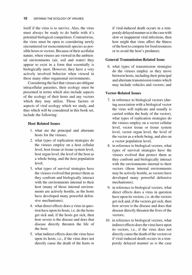
itself if the virus is to survive. Also, the virus
must always be ready to do battle with it’s
potential biological competitors. Contrariwise,
the virus must be open to considering newly
encountered (or reencountered) species as pos-
sible hosts or vectors. Because of their acellular
nature, when viruses are viewed in the ambien-
tal environments (air, soil and water) they
appear to exist in a form that essentially is
biologically inert. However, they have a very
actively involved behavior when viewed in
these many other organismal environments.
Considering the fact that viruses are obligate
intracellular parasites, their ecology must be
presented in terms which also include aspects
of the ecology of their hosts and any vectors
which they may utilize. Those factors or
aspects of viral ecology which we study, and
thus which will be considered in this book set,
include the following:
Host Related Issues
1. what are the principal and alternate
hosts for the viruses;
2. what types of replication strategies do
the viruses employ on a host cellular
level, host tissue or tissue system level,
host organ level, the level of the host as
a whole being, and the host population
level;
3. what types of survival strategies have
the viruses evolved that protect them as
they confront and biologically interact
with the environments internal to their
host (many of those internal environ-
ments are actively hostile, as the hosts
have developed many powerful defen-
sive mechanisms);
4. what direct effects does a virus in ques-
tion have upon its hosts, i.e. do the hosts
get sick and, if the hosts get sick, then
how severe is the disease and does that
disease directly threaten the life of
the host;
5. what indirect effects does the virus have
upon its hosts, i.e., if the virus does not
directly cause the death of the hosts or
if viral-induced death occurs in a tem-
poralydelayedmanner as is the casewith
slow or inapparent viral infections, then
how might that virus affect the fitness
of the host to compete for food resources
or to avoid the host’s predators;
General Transmission-Related Issue
6. what types of transmission strategies
do the viruses employ as they move
between hosts, including their principal
and alternate transmission routes which
may include vehicles and vectors; and
Vector-Related Issues
7. in reference to biological vectors (dur-
ing association with a biological vector
the virus will replicate and usually is
carried within the body of the vector),
what types of replication strategies do
the viruses employ on a vector cellular
level, vector tissue or tissue system
level, vector organ level, the level of
the vector as a whole being, and also on
a vector population level;
8. in reference to biological vectors, what
types of survival strategies have the
viruses evolved that protect them as
they confront and biologically interact
with the environments internal to their
vectors (those internal environments
may be actively hostile, as vectors have
developed many powerful defensive
mechanisms);
9. in reference to biological vectors, what
direct effects does a virus in question
have upon its vectors, i.e. do the vectors
get sick and, if the vectors get sick, then
how severe is the disease and does that
disease directly threaten the lives of the
vectors;
10. in reference to biological vectors, what
indirect effects does the virus have upon
its vectors, i.e., if the virus does not
directly cause the death of the vectors or
if viral-induced death occurs in a tem-
poraly delayed manner as is the case
10 DEFINING THE ECOLOGY OF VIRUSES

with slow or inapparent viral infec-
tions, then how might that virus affect
the fitness of the vectors to compete
for food resources or to avoid the
vector’s predators;
11. in reference to mechanical vectors,
what types of survival strategies have
been evolved by those viruses which
are transmitted by (and during that
event usually carried on the external
surfaces of) mechanical vectors, since
while in association with a mechanical
vector the virus must successfully con-
front any compounds naturally present
on the body surface of the vector plus
confront the passively hostile ambien-
tal environments of either air, water or
soil through which the vector will be
moving; and
Vehicle-Related Issue
12. what types of survival strategies have
been evolved by those viruses which are
transmitted by way of vehicles and
which thereby must successfully con-
front the passively hostile ambiental
environments of either air, water or soil
as the virus itself is transferred through
those environments.
If biological curiosity alone were not a suffi-
cient reason for studying viral ecology, then
perhaps we would study the viruses out of a
desire to both understand them as predators and
to contemplate the ways in which we might
enlist their aid as ecological tools.
1.2 SURVIVING THE GAME:THE VIRUS AND IT’S HOST
Remember that: so long as the virus finds a
new host, whether or not the current host
survives is unimportant. Although it may be
beneficial to not kill a current host until that
host has reproduced to help provide a new
generation of potential host organisms, if the
host to virus ratio is large enough, then even
this latter point may be unimportant.
This section presents in general terms the
relationship between a virus and host. The
generalities of relationships between viruses,
vectors, and vehicles will be discussed in sec-
tion 1.3 of this chapter. The specific subject of
the practical limits to viral virulence in associ-
ation with hosts and vectors will be addressed
in section 1.4 of this chapter.
While in association with a host, the virus
has only one principle goal. This goal is for the
virus to replicate itself to a sufficient level that it
can achieve transmission to another host. This
goal can be attained by one of two basic strate-
gies. The first of these strategies would be a
productive infection, for which five basic pat-
terns can be defined. The second strategywould
be a non-productive infection. The goal of a
productive infection is for the virus to produce
infectious viral particles (those capable of
infecting cells) which are termed “virions”,
during the virus’ association with the current
host. Subsequent spread of the infection to the
next host occurs by transfer of these produced
virions. Contrastingly, some of those agents
which exhibit a non-productive pattern may
either seldom or never produce actual virions.
Thus, the usual goal of a non-productive strat-
egy of infection is to pass the infection to the
next host by directly transferring only the viral
genomic sequences (van der Kuyl et al., 1995).
The patterns of productive infection are:
“Short term - initial” in which viral pro-
duction has only a short term initial
course, after which the viral infection
ends and there no longer is a presence
of that virus within the body of the host
individual although subsequent reinfec-
tion can occur, the outcome from this
pattern of infection depends upon the
virus type and historical exposure to that
type within the host population, the situ-
ation being that in otherwise healthy
members of a multicellular host popula-
tion with which the virus has coevolved,
these infections are usually mild and by
SURVIVING THE GAME: THE VIRUS AND IT’S HOST 11

themselves normally associated with a
fairly low incidence of mortality;
“Recurrent” in which repeated episodes ofviral production occur, this pattern often
has a very pronounced initial period of
viral production, after which the virus
persists in a latent statewithin the body of
the host with periodic reinitiations of
viral production that usually are not life
threatening;
“Increasing to end-stage” in which viral
infection is normally associated with a
slow, almost inocuous start followed by
a gradual progression associated with an
increasing level of viral production and
eventual death of the host, in these
instances death of the host may relate to
destruction of the host’s immunological
defense systems which then results in
death by secondary infections;
“Persistent-episodic” is a pattern that
represents a prolonged nonfatal infection
which may persist for the remainder of
the hosts natural lifetime associated with
a continuous production of virionswithin
the host, but interestingly the infection
only episodically results in symptoms,
the viral genome does not become
quiescent, the host remains infectious
throughout the course of this associative
interaction, and very notably some
members of the family Picobirnaviridae
often produce this pattern of productive
infection;
“Persistent but inapparent” is a pattern
that represents a prolonged nonfatal infec-
tionwhichseeminglyneverresults inovert
symptoms of illness attributable to that
particular virus, the viral genome never
becomes quiescent and viral infections
that follow this pattern are persistently
productive with the host often remaining
infectious for the remainder of their natu-
ral lifetime, with notable examples of
viruses which produce this pattern being
members of the family Anelloviridae, and
it also occurs in certain rare instances of
infection by Human immunodeficiency
virus 2 which is a member of the genus
Lentivirus of the family Retroviridae.
There are two options to the “short term -
initial” pattern. The first option is a very rapid,
highly virulent approach which is termed
“fulminate” (seemingly explosive) and usually
results in the rapid death of the host organism.
This first option usually represents the product
of an encounter between a virus and a host with
which the virus has not coevolved. The second
option is for the virus to be less virulent,
causing an infection which often progresses
more slowly, and appears more benign to the
host. The “recurrent” and “increasing to end-
stage” patterns incorporate latency into their
scheme. Latency is the establishment of a
condition in which the virus remains forever
associated with that individual host organism
and generally shows a slow and possibly only
sporadic replication rate that, for some combi-
nations of virus and host, may never be life
threatening to the host. The strategy of achiev-
ing a non-productive, or virtually non-produc-
tive, pattern of infection involves achieving an
endogenous state (Terzian et al., 2001). Endo-
geny implies that the genome of the virus is
passed through the host’s germ cells to all
offspring of the infected host (van der Kuyl et
al., 1995; Villareal, 1997).
The product of interspecies encounters
between a virus and it’s natural host will usu-
ally lead to a relatively benign (mild, or not
directly fatal), statistically predictable, out-
come that results from adaptive coevolution
between the two species. Still, these normal
relationships do not represent a static coexi-
stance between the virus and the natural host,
but rather a tenuous equilibrium. Both the virus
species and it’s evolved host species will be
struggling to get the upper hand during each of
their encounters (Moineau et al., 1994. The
result will normally be some morbidity and
even somemortality among the host population
as a result of infection by that virus. Yet,
because the virus as a species may not be able
to survive without this natural host species
12 DEFINING THE ECOLOGY OF VIRUSES

(Alexander, 1981), excessiveviral-relatedmor-
tality in the host population is not in the long
term best interest of the virus. Some endoge-
nous viruses have evolved to offer a survival-
related benefit to their natural host, and this can
give an added measure of stability to their
mutual relationship. Two examples of this type
of relationship are the hypovirulence element
associated with some strains of the Chestnut
blight fungus, and the endogenous retroviruses
of placental mammals. The hypovirulence
(reduced virulence) which the virus-derived
genetic elements afford to the fungi that cause
Chestnut blight disease reduce the virulence of
those fungi (Volume 1, chapter 9). This reduced
virulence allows the host tree, and in turn the
fungus, to survive. Placental mammals, includ-
ing humans, permanently have incorporated
species of endogenous retroviruses into the
chromosomes of their genomes. It has been
hypothesized that the incorporation of these
viruses has allowed the evolution of the placen-
tal mammals by suppressing maternal immu-
nity during pregnancy (Villareal, 1997).
However, the impact of a virus upon what
either is, or could become, a natural host
population can sometimes appear catastrophic.
The most disastrous, from the host’s perspec-
tive, are the biological invasions which occur
when that host population encounters a virus
which appears new to the host (Kuiken et
al., 2006). Three categories of events can lead
to biological invasions of a virus into a host
population. These categories are: first, that this
virus species and host species (or sub-popula-
tion of the host species) may never have previ-
ously encountered one another (examples of
this occurring in human populations would be
the introduction of measles into the Pacific
islands and the current introduction of HIV);
second, if there have been previous encounters,
the virus may have since changed to the point
that antigenically it appears new to the host
population (an example of this occurring in
humans would be the influenza pandemic of
1918–1919); and third, that even if the two
species may have had previous encounters, this
subpopulation of the host species subsequently
may have been geographically isolated for
such a length of time that most of the current
host population represents a completely new
generation of susceptible individuals (exam-
ples in humans are outbreaks of viral gastro-
enteritis found in remotely isolated comunities
on small islands as related to the occasional
arrival of ill passengers by aircraft or water-
craft). Sadly, the biological invasion of the HIV
viruses into human populations seems to be
successful (Caldwell and Caldwell, 1996), and
the extreme host death rate associated with this
invasion can be assumed to indicate that the
two species have not had time to coevolve with
one another. The sporadic, but limited, out-
breaks in human populations of viruses such as
those which cause the hemmorrhagic fevers
known as Ebola and Lassa represent examples
of unsuccessful biological invasions. The lim-
ited chain of transmission for these latter two
illnesses (for Lassa, see: Fuller, 1974), with
their serial transfers often being limited to only
two or three hosts in succession, represents
what will occur when a virus species appears
genetically unable to establish a stabile rela-
tionship with a host species. The observation of
extremely virulent and fulminate symptom-
atology, as associated with infections by Lassa
and Ebola in humans, can generally be
assumed to indicate either that the host in
which these drastic symptoms are observed is
not the natural host for those viruses or, at the
very least, that these two species have not had
time to coevolve. In fact, the extreme symp-
tomatology and mortality which result in
humans from Ebola and Lassa fevers seems
to represent an overblown immune response on
the part of the host (Spear, 1998).While having
the death of a host individual occur as the
product of an encounter with a pathogen may
seem like a dire outcome, this outcome repre-
sents a mechanism of defense operating at the
level of the host population. If a particular
infectious agent is something against which
members of the host population could not
easily defend themselves, then it may be better
to have that particular host individual die (and
die very quickly!) to reduce the possible spread
SURVIVING THE GAME: THE VIRUS AND IT’S HOST 13

of the contagion to the other members of the
host population.
1.2.1 Cell Sweet Cell, and Strugglesat Home
As diagramed in Figure 1.6, viruses can
arrive at their new host (solid arrows) either
directly from the previously infected host,
via an intermediate vehicle, or via an inter-
mediate vector. Viral survival in association
with the new host will first depend upon the
virus finding it’s appropriate receptor mole-
cules on the host cell’s surface (Spear, 1998).
After this initial location, the virus must be
capable of entering and modifying the host
cell so that the virus can reproduce within
that cell. If the host is multicellular, then the
virus may first have to successfully navigate
within the body of the host until it finds
the particular host tissue which contains it’s
correct host cells.
Within a multicellular host, the virus may
face anatomically associated barriers includ-
ing membranous tissues in animals. The virus
also may face non-specific, non-immune bio-
logical defenses (Moffat, 1994), including
such chemical factors as the enzymes found
in both tears and saliva, and the acid found in
gastic secretions. The types of anatomical and
non-specific, non-immune defenses encoun-
tered can vary depending upon the viral trans-
mission route and the portal by which the virus
gains entry into the host’s body. After a virus
finds it’s initial host cell and succeeds in
beginning it’s replication, the effects which
the virus has upon the host can then draw a
defensive biological response. The category of
non-specific non-immune responses which a
virus may encounter at this stage include even
such things as changes in host body tempera-
ture for mammals. As if in a game of spy
versus spy, the virus most importantly must
survive the host’s specific immune defenses
FIGURE 1.6 Viruses can arrive at their new host (filled arrows) either directly from the previously infected host,
via an intermediate vehicle, or via an intermediate vector. Viral survival in association with that new host depends
upon: viral replication within that new host, the effects which the virus has upon that host, and the response of that
host to the virus. Successful viral survival in associationwith this new host will allow a possible subsequent transfer
of thevirus (open arrows) to its next host either directly, via a vehicle, or via a vector. This represents a segment from
Figure 1.5.
14 DEFINING THE ECOLOGY OF VIRUSES

(Beck and Habicht, 1996; Gauntt, 1997; Levin
et al., 1999; Litman, 1996; Ploegh, 1998;
Zinkernagel, 1996).
The listing and adequate explanation of
antiviral defense techniques would by itself
be enough to nearly fill a library. But, I will
attempt to summarize some of them here and
help the reader to track those through this
book set.
Molecular antiviral defenses begin at the
most basic level which would be non-specific
mechanisms. These conceptually include
DNA restriction and modification systems
(volume 1, chapter 5), progressing upward
with greater complexity to the use of post
transcriptional processing (Russev, 2007).
Countering these defenses is done by such
techniques as using virally-encoded restric-
tion-like systems to chop-up the DNA genome
of their host cells to provide a ready source of
nucleic acids for the production of progeny
viral genomes. There also are viruses which
try to shut down the the post-transcriptional
defenses, most clearly noticed among some
viruses infective of plants. Plants in fact
heavily rely upon molecular defenses such as
post-transcriptional control, (volume 1, chap-
ter 11) and beyond that technique the plants try
to wall off an infection, essentially trying to
live their lives despite presence of the infec-
tious agent and hoping not to pass the infection
along to their offspring through viral contam-
ination of their germ cells.
Antimicrobial peptides are a defensive
mechanism found in all classes of life, and
represent a main part of the insect defensive
system (volume 2, chapter 10). Higher on the
scale of defensive responses are things which
we term to be immunological in nature (Dani-
lova, 2006). Some of these we term to be
innate, others we call adaptive. A good start-
ing point for this discussion of immunological
responses is the capacity for distinguishing
self versus non-self, accompanied by the capa-
bility for biochemically destroying cells that
are determined to be non-self. This approach
exists from at least the level of fungi (volume
1, chapter 9) upwards for the non-animals, and
among the animals this approach begins with
at least the corals (volume 2, chapter 5).
Determining and acting upon the distinction
of self versus non-self likely may have devel-
oped as a system that helps to support suc-
cessful competition for growth in a crowded
habitat, but it serves well against pathogenic
organisms. As a health issue, this process
sadly plays a role in autoimmune diseases and
we try to suppress it when hoping to use organ
and tissue transplantation to save human lives.
Apoptosis, the targeting of individual cells
within the body of the host for selective
destruction by the host, commonly exists
across the animal kingdom. This mechanism
is used by many invertebrates (volume 2, chap-
ters 6 and 7) as wells as vertebrates to destroy
any virally infected cells which may be present
within their bodies. However, apoptosis is a
weapon that can be used by both of the com-
batants. Using apoptosis to destroy virally-
infected cells before the virus contained within
those cells can assemble progeny virions is an
effective approach when used carefully by the
host. As might be expected, some viruses
therefore defensively try either to shut-down
the process of apoptosis, or at least to shut-
down that process until the virus is ready to use
apoptosis as a mechanism for assisting in
the liberation of assembled virions from the
infected host cell.
Vertebrates, and some of the invertebrates,
have more complex body plans and can use
them with good effectiveness in combating
infections. With the evolutionary development
of more complex body plans, comes the possi-
bility of dedicating cells and even organs to the
task of fighting pathogenic invaders. Those
invertebrates with more complex body plans
are represented in the anti-viral fight by their
use of lymphoid organs to actively collect and
either sequester or actively assault and destroy
the microbial offenders. Some of the aquatic
crustaceans (volume 2, chapter 7) tend to rely
upon sequestering an infection andmust hope to
breed a new generation of their own progeny
before they, themselves, are killed by the infec-
tion which they have sequestered within their
SURVIVING THE GAME: THE VIRUS AND IT’S HOST 15

body. At the same time, the infected parents
must hope not to pass along the sequestered
infection to their offspring through contamina-
tion of their eggs and sperm. Such collection
and sequestration techniques are found upward
through the evolutionary line and likewise used
by the vertebrates. Many viruses have found
ways around these issues, as is the case with
endogenous viruses and retrotransposons that
insert andmaintain themselves in thegenomeof
their host, passingdirectly through thegermcell
line. Some viruses infect and replicated within
the immune cells! Some viruses are shed along
with the eggs of inertebrates and thus are ready
to await the hatching of those offpsring. Still
other viruses, as in the case of viviparous
mammals, simply cross the placenta to infect
the fetus.
Interferons and their homologues are protein
systems which vertebrates have developed and
use effectively against some viruses, and corre-
spondingly many viral groups contain mechan-
isms for suppressing interferon production
(Munoz-Jord�an and Fredericksen, 2010).
Although the “walling-off” of a pathogen still
occurs invertebrates, with an example being the
development of tubercules in some mycobacte-
rial infections, active mechanisms for hunting
down and destroying pathogens and pathogen-
infected cells within their bodies is highly
developed. With vertebrates, the end goal can
be percieved as ridding the body of the pathogen
even if that end goal is not always achieved.
The jawed vertebrates possess immune systems
which are termed adaptive, and these produce
protein antibodies that can be highly specific
(volume 2, chapters 8, 9, 11–14).
Options for surviving the immune defenses
of the host can include such techniques as:
“You don’t knowme” (a virus infecting an
accidental host, in which case a very
rapid proliferation may occur, an exam-
ple being Lassa fever in humans);
“Being very, very quiet” (forming a pattern
of latency in association with the virus’
persistence within that host, an example
being herpesviruses);
“Virus of a thousand faces” (antigen shift-
ing, an example being the lentiviruses);
“Keep to his left, that’s his blind spot”(maintaining low antigenicity, an
approach used by viroids and prions);
“Committing the perfect crime” (infect-
ing the immune system, an approach
taken by many retroviruses and herpes-
viruses); and
“Finding a permanent home” (taking up
permanent genetic residency within the
host and therefore automatically being
transmitted to the host’s progeny, an
approach taken by viroids, endogenous
retroviruses, and LTR retrotransposons).
Each virus must successfully confront it’s
host’s responses while the virus tries to repli-
cate to sufficient numbers that it has a realistic
chance of being transmitted to another candi-
date host. Failure to successfully confront the
host’s responses will result in genetic termina-
tion of the virus and, on a broader scale, such
failure may eventually result in extinction for
that viral species.
1.2.2 I Want a Niche, Just Likethe Niche, That Nurtured Dear OldMom and Dad
The initial tissue type in which a virus repli-
catesmay be linked inextricablywith the initial
transmission mode and portal (or site) of entry
into the body of the host. For example, those
viruses of mammals which are acquired by
fecal - oral transmission tend to initiate their
replication either in the nasopharyngial tissues
or else in the gastrointestinal tissues. There
then are subsequent host tissue and organ types
affected, some of which may be related to the
virus’ efforts at trying to reach it’s proper portal
of exit. Others of the host tissues affected by the
virus may be unrelated to interhost viral trans-
mission, although the affect upon those other
tissues may play a strong role in the severity of
illness which is associated with that viral infec-
tion. An example of the latter would be the
16 DEFINING THE ECOLOGY OF VIRUSES

encephalitic infection of brain neurons in asso-
ciation with echoviral conjunctivitis, an infec-
tion which initially would be acquired from
fomites as part of a fecal-oral transmission
pattern. In this case, the encephalitis causes
nearly all of the associated morbidity but does
not seem to benefit transmission of the virus
(personal observation by author C. J. Hurst).
1.2.3 Being Societal
Successful viral survival in association with
this new host will allow a possible subsequent
transfer of the virus (Figure 1.6, open arrows)
to its next host either directly, via a vehicle, or
via a vector. Themovement of a viral infection
through a population of host organisms can
be examined and mathematically modeled.
An epidemic transmission pattern, character-
ized by a short term, higher than normal rate of
infection within a host population is repre-
sented by the compartmental model shown in
Figure 1.7 (Hurst and Murphy, 1996). An
endemic transmission pattern, characterized
by a long term, relatively constant incidence
rate of infection within a host population is
represented by the compartmental model
shown inFigure 1.8 (Hurst andMurphy, 1996).
1.3 STEPPIN’ OUT AND TAKINGTHE A TRAIN: REACHING OUTAND TOUCHING SOMEONEBY VECTOR OR VEHICLE
Remember that: host-vector choices, cycles
and vehicle utilizations as they exist today
may (and probably do!) reflect evolutionary
progression from prior species interactions
and ecological relationships.
After a virus has successfully replicated
within the body of it’s current (present) host,
it must seek successful transmission to it’s
next (proximate) host. The resulting chain of
transmission usually is the end-all of viral
reproduction. These are three basic
approaches by which this can be attained:
transmission by direct contact between the
present and proximate hosts, transmission
mediated by a vector (Brogdon and
McAllister, 1998; Hurst and Murphy, 1996;
Mills and Childs, 1998), and transmission
mediated by a vehicle (Hurst and Murphy,
1996). While considering these approaches, it
is important to keep in mind that the chains
of transmission originate by random chance
followed by evolution.
1.3.1 “Down and Dirty”(Just Between Us Hosts)
This heading is one which can be used to
describe host to host transmission (transmis-
sion by host to host contact). While this is one
of the most notorious, it is not the most
common route of viral transmission between
animals. This route only serves to a limited
extent in microbes. Even worse, this route
essentially does not seem to function in vas-
cular plants due to the relative immobility of
those hosts.
1.3.2 “The Hitchhiker”(Finding a Vector)
Transmission by vectors may be the most
prevalent route by which the viruses of plants
are spread among their hosts. This route
clearly also exists for some viruses of animals.
However, this route has not yet been defined
in terms of viruses which infect microbes.
Vectors are, by definition, animate objects,
and more specifically they are live organisms.
Being a vector implies, although by definition
does not require, that the entity serving as
vector has self-mobility. Thus, plants could
serve by definition as vectors, although when
we consider the topic of viral vectors we
usually tend to think in terms of the vectors
as being invertebrate animals. Vertebrate ani-
mals can also serve as vectors, as likewise can
some cellular microbes.
There are two categories of vectors: biolog-
ical and mechanical. As was stated earlier, if
the virus increases it’s numbers while in asso-
ciation with a vector, then that vector is termed
STEPPIN’OUTANDTAKINGTHEATRAIN:REACHINGOUTANDTOUCHINGSOMEONEBYVECTORORVEHICLE 17

as being biological. Conversely, the vector is
termed to be mechanical if the virus does not
increase it’s numbers while in association with
that vector. Beyond this there lie some deeper
differences between mechanical and biological
vectors. These differences include the fact that
the acquisition of a virus by a biological vector
usually involves a feeding process. Phagic
habits of the biological vector result in the
virus being acquired from an infected host
when the vector ingests virally contaminated
host body materials acquired through a bite or
sting. Subsequent transfer of the infection from
the contaminated biological vector to the virus’
next host occurs when the biological vector
wounds and feeds upon the next host. Actual
transference of the virus to that next host occurs
incidentally when the vector contaminates the
wound by discharging viruses contained either
in the vector’s saliva, regurgitated stomach or
intestinal contents, or else discharged feces and
urine. Essentially any animal is capable of
serving as a potential biological vector pro-
vided that the wound which it inflicts while
feeding upon a host plant or animal will not
result in the death of that new host until the
virus would have had the chance to replicate
within and subsequently be transmitted onward
from that new host. There are many issues
surrounding the question of what makes a good
biological vector. These issues include: physi-
cal contact between the virus’ host and the
potential vector during a feeding event, viral
reproduction within that potential vector, and
that the infected vector be able to survive long
enough to transmit the virus to a new host.
It also helps if there is some factor driving the
vector to pass along the infection, such as the
FIGURE 1.7 Epidemic transmission of a virus within a host population is represented by this type of
compartment model (Hurst and Murphy, 1996). Each of the boxes, referred to as compartments, represents a
decimal fraction of the host population with the sum of those decimal fractions equaling 1.0. The compartments
which represent actively included members of the host population are those labeled susceptible, infectious, and
immune. This model incorporates only a single category of removed individuals, representing those whose demise
was due to infection related mortality. The solid arrows represent the rates at which individual members of the host
species move between the different compartments during the course of an epidemic. Those rates of movement are
often expressed in terms of individuals per day as described by Hurst andMurphy (1996). Used with permission of
the author and Cambridge University Press.
18 DEFINING THE ECOLOGY OF VIRUSES

19

virus finding it’s way into the vector’s saliva, or
the virus increasing the physical aggres-
siveness of the vector.
The fact that biological vectors usually
acquire the viral contaminant while wounding
and ingesting tissues from an infected host
brings us to another distinguishing difference
between biological and mechanical vectors:
viral contamination of a biological vector
usually is associated with the virus being
carried internal to the body of the vector.
Replication of the virus then occurs within
the body of the biological vector. Contrast-
ingly, viral contamination of a mechanical
vector usually occurs on the external surface
of the vector and the virus subsequently tends
to remain on the external surface of the
mechanical vector. One possible example of
mechanical vectoring would be the acquisition
of plant viruses by pollinating animals such as
bees and bats during their feeding process.
These pollinators can serve as mechanical
vectors if subsequently they are able to pas-
sively transfer the virus from their body sur-
face to the next plant from which they will
feed. In the case of these pollinators, the
acquired virus presumably is carried external
to the pollinator’s body. Conversely, it is pos-
sible that a plant being visited by a pollinator
might become contaminated by viruses afflict-
ing that pollinator, and the plant could then
passsively serve as a mechanical vector
if subsequent pollinators should become
infected when they visit that plant. Biting flies
can serve as biological vectors if, during feed-
ing, they ingest a pathogen which can replicate
in association with that fly and then be passed
onward when the fly bites it’s next victim
(Hurst and Murphy, 1996). Non-biting flies
can passively serve as mechanical vectors if
they feed upon contaminated material and then
subsequently transmit those microbial con-
taminants to the food of a new host without
that pathogen having been able to replicate
while in association with the non-biting fly
(Hurst and Murphy, 1996). Arthropods such as
wasps, which repeatedly can sting multiple
animals, could serve as mechanical vectors
by transporting viruses on the surfaces of their
stingers. Also, passive surface contamination
of pets that occurs unrelated to a feeding event
can result in the pets serving as mechanical
vectors (Hurst and Murphy, 1996).
When a virus is transported inside the body
of the vector, then that transportation is referred
to as being an “internal carriage”. Contrast-
ingly, transportation of a virus on the external
body surfaces of a vector is referred to as being
an “external carriage”. As will be described in
volume 1, chapter 11, there are some plant
viruses which are transported through internal
carriage by invertebrates that represent
mechanical vectors (because the virus does not
increase its population level when in associa-
tion with those invertebrates). Thus, although
the biological vectoring of a virus usually
involves internal carriage, the fact of internal
carriage does not alone always indicate that
FIGURE1.8 Endemic transmission of a viruswithin a host population is represented by this type of compartment
model (Hurst andMurphy, 1996). This model is essentially an extension of the model presented in Figure 1.7. This
model contains the same three compartments (susceptible, infectious, and immune) representing actively included
individuals and the category of individuals removed by infection relatedmortality as were described for Figure 1.7.
Thismodel differs in that itmust also consider thevarious possible categories of live removed individualswhich can
move into and out from the compartments of actively included individuals. Their removal represents the fact that
they do not interact with the actively included individuals in such a way that the virus can reach them, often due to
spatial isolation. This model also includes the fact that the immune status of individuals can naturally wane or
diminish with time such that immune individuals return to the compartment labeled susceptible; production of host
progeny, representing reproductive success of the members of the host species; natural mortality, as a means of
removingmembers of the population; and the possible use of vaccination to circumvent the infectious process plus
the associated vaccine - related mortality. Please notice that the progeny of infectious individuals may be
susceptible, infectious, or immune at the time of their birth depending upon the type of virus which is involved
and whether or not that viral infection is passed to the progeny. Used with permission of the author and Cambridge
University Press.
3
20 DEFINING THE ECOLOGY OF VIRUSES

the vectoring is biological. Humans, interest-
ingly, can serve as mechanical vectors via
internal carriage for plant viruses that would
be consumed with food and later excreted in
feces (Zhang et al., 2006).
Because a virus must (by definition!) repli-
cate in association with the biological vector;
we can view the viral - vector association
(Figure 1.9) in the same manner as was done
for that of a virus and it’s host (Figure 1.6).
Indeed, it often is difficult to know which
species is actually the viral host and which is
actually the viral vector; to distinguish which is
the victim and which serves as the messenger.
Traditionally, we have often taken the view that
humans are a high form of life and that there is a
decreasing heirarchy down to the microbes.
From this traditional, and sadly very anthropo-
centric, viewpoint we might assume that any
living thing that transmits a virus between
humans must be the vector as humans surely
must be in the respectible position of serving as
the host. Another version of this philosophy
would consider a vertebrate to be the host and
any invertebrate to be the vector. Still a third
version has been based upon relative size, with
the largest creature considered as the host and
the smaller considered as the vector. Since we
stated earlier that this chapter is intended to
consider life from a virocentric perspective, we
could easily accept the virocentric view which
finds that there may be no clear distinction
FIGURE 1.9 This figure addresses viral association with a biological vector and represents a segment from
Figure 1.5. Vectors are, by definition, animate objects and are categorized either as ’biological’, meaning that the
virus increases in number during association with that vector, or ’mechanical’, meaning that the virus does not
increase in number during association with that vector. Biological vectors seem to have far greater importance than
do mechanical vectors in terms of the spread of viral infections. Viruses can arrive at the biological vector (filled
arrows) either directly froman infected host or via an intermediatevehicle. Transmission of thevirus, via this vector,
to a new host (or perhaps more accurately the ’next’ host since, in the case of viruses, biological vectors may be
considered as alternate hosts) requires that the virus both survive and replicate while in association with that
biological vector. Thus, examining viral survival in association with a biological vector also involves considering
the effects which viral replication has upon that vector and the response of that vector to the virus. Successful viral
survival in association with the vector will allow a possible subsequent transfer of the virus to its next host either
directly or via a vehicle (open arrows).
STEPPIN’OUTANDTAKINGTHEATRAIN:REACHINGOUTANDTOUCHINGSOMEONEBYVECTORORVEHICLE 21

between host and vector. Rather, any biological
vector can likewise be viewed as a host. The
argument as to which one, the traditional host
or traditional vector, really serves as the host
would then become moot.
Because many types of viruses are capable
of infecting more than a single species of host,
we are also left to ponder about determining
which is the principle host versus those which
serve as alternate hosts. Settlement of the dis-
tinction asked by this latter question is usually
done by examining the comparative virulence
of the virus in the different types of hosting
species. That species for which the virus seems
less virulent is assumed to be the more natural,
most coevolved, host. It then is assumed that
the species for which the virus seems to have
greater virulence are alternate hosts. While
trying to appreciate this conundrum, it must
be understood that from a virocentric perspec-
tive both the principle and alternate hosts, as
well as any biological vectors utilized by a
virus, will all represent hosting species, and
thus we may never be able to sort out the
answers. Any further discussion of this partic-
ular issue is best left to only the most insistent
of philosophers! Perhaps the only things left to
be said of this issue are that examples of the
transmission of a virus by a biological vector
are represented in Figure 1.10, and that eco-
logical interactions between a virus and it’s
principle hosts, alternate hosts, and biological
vectors can be represented by the example
shown in Figure 1.11.
FIGURE1.10 The transmission of a virus via a biological vector can be represented by this diagram. The virus is
acquired as the biological vector feeds upon natural bodily fluids or else enzymatically liquified bodily components
of the infected host. Subsequent transmission of the virus to a new host results when the vector releases
contaminated excretions or secretions while feeding upon that new host.
22 DEFINING THE ECOLOGY OF VIRUSES
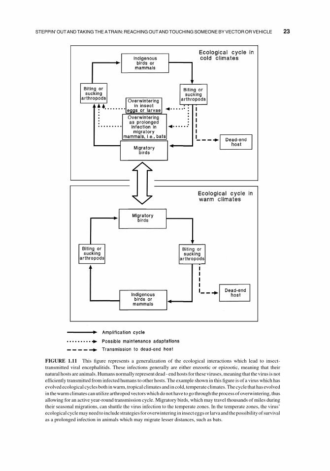
FIGURE 1.11 This figure represents a generalization of the ecological interactions which lead to insect-
transmitted viral encephalitids. These infections generally are either enzootic or epizootic, meaning that their
natural hosts are animals.Humans normally represent dead - end hosts for theseviruses,meaning that thevirus is not
efficiently transmitted from infected humans to other hosts. The example shown in this figure is of a virus which has
evolved ecological cycles both inwarm, tropical climates and in cold, temperate climates.Thecycle that has evolved
in thewarmclimates canutilize arthropodvectorswhichdonot have to go through theprocess of overwintering, thus
allowing for an active year-round transmission cycle. Migratory birds, which may travel thousands of miles during
their seasonal migrations, can shuttle the virus infection to the temperate zones. In the temperate zones, the virus’
ecological cyclemayneed to includestrategies foroverwintering in insecteggsor larvaand thepossibilityof survival
as a prolonged infection in animals which may migrate lesser distances, such as bats.
STEPPIN’OUTANDTAKINGTHEATRAIN:REACHINGOUTANDTOUCHINGSOMEONEBYVECTORORVEHICLE 23

1.3.3 “In a Dirty Glass”(Going There by Vehicle)
Viruses also can be transmitted by vehicles.
Vehicles are, by definition, inanimate objects.
More specifically, the termvehicle applies to all
objects other than living organisms. There are
four general categories of vehicles and these
are: foods, water, fomites (pronounced fo mi
tez, defined as contaminated environmental
surfaces which can serve in the transmission
of pathogens), and aerosols. Figure 1.12 repre-
sents viral association with a vehicle. Trans-
mission of the virus, via a vehicle, to a new host
first requires contamination of that vehicle
(shown by the filled arrows in Figure 1.12).
Thevirusmust then survivewhile in association
with the vehicle. Because viruses are by defini-
tion obligate intracellular parasites, and by
definition vehicles are non-living, then a virus
neither can replicate on nor within a vehicle.
Likewise, because vehicles are by definition
non-living, we do not expect that any specific
antiviral response will be produced by the
vehicle. Transference of the virus to its next
host can occur either directly or via a vector
(shown as the open arrows in Figure 1.12). One
possible indication as to the difference between
a vector and a vehicle is that, while a live
mosquito can serve as a biological vector, after
it’s death that same mosquito instead repre-
sents a vehicle. The transmission of a virus via a
vehicle can be represented by the diagram
shown in Figure 1.13. Acquisition of the virus
by the next host or vector from that contami-
nated vehicle results from either ingestion of
the vehicle (associated with foods and water),
surface contact with either contaminated water
or a contaminated solid object (a fomite), or
inhalation (aerosols). Although, from a human
perspective, we might tend to associate water-
borne transmission with animals and in partic-
ular human diseases (volume 2 chapter 13); the
waterborne approach will play a major role in
viral transmission for viruses that infect
cyanobacteria (volume 1 chapter 6), algae
(volume 1 chapter 7) and seaweeds (volume 1
FIGURE 1.12 This figure addresses viral association with a vehicle and represents a segment from Figure 1.5.
Viral transmission between hosts can occur by means of a vehicle. Vehicles are by definition inanimate objects.
Viral contaminants can reach the vehicle (filled arrows) either directly from an infected host or via an
intermediate vector. Transmission of the virus, via this vehicle, to a new host requires that the virus survive
in association with the vehicle. Transference of the virus to its next host can occur either directly or via a vector
(open arrows).
24 DEFINING THE ECOLOGY OF VIRUSES

chapter 8). The are even viruses of terrestrial
plants, including some carmoviruses of the viral
family Tombusviridae, which seem as though
they might be transmitted by water. The list of
vehicles associatedwith viral transmission even
includes agricultural tools and other work
implements. The topic of vehicle-associated
transmission of pathogens is discussed at length
in the reference by Hurst and Murphy (1996).
1.3.4 Bringing Concepts Together
Biological entities exist over a spectrum of
complexities, ranging from the viruses, viroids
and prions (yes, even the prions are biological
entities!) to multicellular organisms. The pro-
cess of maintaining the viability of even the
largest of organisms is, and perhaps must, be
organized at small levels. Biologically, this has
been achieved by a highly evolved process of
internal compartmentalization of functions
with a systemic coordination. If we consider
for a moment one of the most enormous of the
currently livingmulticelled organisms, the blue
whale (Balaenoptera musculus), we notice that
this kind of compartmentalization and coordi-
nation begins all of theway down at the level of
the subcellular structures and organelles within
each individual cell. The compartmentalization
and coordination then continue upward
through a number of levels including the vari-
ous individual types of cells, the tissues into
which those cells are organized, the organs
which the tissues comprise, and finally the total
internal coordination of all of these through
nerve signaling and hormonal regulation. At
every one of these biological levels there is a
“taking from” and a “leaving behind” exchange
of material with respect to the immediate sur-
rounding environment. This results in the exis-
tence of dramatic environmental differences at
all levels, even down to the many microenvir-
onments which exist within the organizational
regions of a single cell.
FIGURE 1.13 The transmission of a virus via a vehicle can be represented by this diagram. Food items can be
contaminated by the action of an infected host. Alternatively, the food in question may actually be the body of an
infected host that subsequently is consumed by a susceptible, predatory new host. Viral contaminants present in
water can be acquired by a new host either directly, as the result of external or internal exposure to the contaminated
water including ingestion of the water; or indirectly, following contact between the new host and an environmental
surface (serving as a secondary, intermediate vehicle) that has been contaminated by that water. Fomites are solid
environmental (non-food) objects whose surfaces may be involved in the transfer of infectious agents. Viral
aerosols may result in the infection of a new host either directly through inhalation of the aerosol, or indirectly
following contact between the new host and some other vehicle (either food, water, or a fomite) contaminated by
that aerosol.
STEPPIN’OUTANDTAKINGTHEATRAIN:REACHINGOUTANDTOUCHINGSOMEONEBYVECTORORVEHICLE 25

Every virus must try to comply with the
basic biological imperatives of genetic sur-
vival and replication. While complying with
these imperatives the viruses must, as obligate
intracellular parasites, not only face but also
survivewithin and successfully be transported
through the various environments which are
internal to the host. Those viruses which are
transmitted by biological vectors must also
have evolved the capability to survive and be
transported through internal environments
faced within the vector. Viruses which are
transmitted by mechanical vectors generally
must possess an additional evolved ability to
survive on the surface of that vector. Likewise,
both those viruses transmitted by mechanical
vectors and viruses transmitted by vehicles
must possess the ability to survive exposure to
natural ambiental environments encountered
either in the atmosphere, hydrosphere or lith-
osphere. These numerous environments are
summarized in Figure 1.14. Conditions con-
fronted at the interface zones, as indicated by
the dashed lines in Figure 1.14, represent areas
of still additional environmental complexity.
While viruses appear biologically inert when
viewed in the ambiental environments, they
display their biology and interact with their
surroundings when they reach the environ-
ments internal to their hosts and biological
vectors.
The adaptability of a species in terms of its
biological cycle and biological needs will
determine that species’ potential distribution
range. This potential distribution range is lim-
ited in actuality to a smaller range based upon
interspecies relationships and competitions.
Ourselves being large multicellular creatures,
we humans normally think of a distribution
range as being geographical in nature. As
microbiologists, many of us have come to
understand the concept of distribution range
in finer detail; an example being the depth
within a body of water where a particular
species of microorganism normally will be
found. At the level of viral ecology, the concept
of species distribution range encompasses
everything from tissue and organ tropisms
(those tissues and organs which a virus seems
to attack preferentially) upwards to the geo-
graphical availability of host species, vector
species, and the prevailing directional flow of
appropriate vehicles such as air and water. The
larger, geographical end of this scale is repre-
sented in Figure 1.15.
While considering the factors addressed in
Figure 1.15, it is important to keep in mind that
albeit the virus’ election of hosts, vectors, and
routes of transmission would all originate by
random chance, the attainment of reliable con-
tinued viral success would require that such
random selection events be followed and
strengthened by evolution. This explains the
reason why viruses do not appear suddenly to
develop the ability to use a different vehicle.
Indeed, it is perhaps likely that in order to use a
vehicle such as air or water, the virus must have
preadapted itself to the conditions which it will
encounter in association with that vehicle.
Nearly each individual species of virus which
achieves transmission by vehicles, seems
invariably to use only one type of vehicle. This
trait likewise seems to hold true for all species
belonging to any given viral genus. Further-
more, this identification seems to nearly always
hold true at the level of viral family. In fact, this
is one of the defining characteristics of the
ecology of a viral group. The only virus which
seems to have evolved the ability to utilize
more than a single vehicle is the Hepatitis
A virus (Hurst and Murphy, 1996), which has
evolved a most remarkable ability to be effec-
tively transmitted both bywater and on fomites.
Perhaps accordingly, the Hepatitis A virus cur-
rently exists in a genus (Hepatovirus) of its
own. We should not be surprised if we eventu-
ally would discover other members of that viral
genus, and subsequently discern those other
members to likewise use these same two vehi-
cles. It is for these reasons, that fears expressed
in the public press that viruses such as Ebola
will suddenly take flight and be transmitted
over large distances via aerosol transmission
amount to nothing more than frightening spec-
ulation. Why is it just speculation? Because
that route of transmission is not a part of the
26 DEFINING THE ECOLOGY OF VIRUSES

virus’ ecology. Invasive medical devices such
as syringes, endoscopes and other surgical
implements, plus transplanted animal tissues
including transfused blood and blood products,
and grafted plantmaterial, represent exceptions
to this rule. These devices and transplanted
tissues represent unnatural vehicles which, by
their nature, allow the virus an abnormal access
to the interior of a new host (Hurst and
Murphy, 1996). Any virus which would
naturally be transmissible by direct contact
with either an infected host or any type of
FIGURE 1.14 This figure integrates the concepts of host, vehicle and biological vector by representing the
environments potentially faced by a virus. As obligate intracellular parasites, the viruses must face, survivewithin,
and successfully be transported through environments which are internal to the host. Those viruses which are
transmitted by biological vectors must also have evolved the capability to survive and be transported through
internal environments faced within the vector. Viruses which are transmitted by vehicles and mechanical vectors
must additionally possess an evolved ability to survive in natural ambiental environments (atmosphere, hydro-
sphere and lithosphere). Conditions confronted at the interface zones, as indicated by dashed lines, represent areas
of additional environmental complexity.
STEPPIN’OUTANDTAKINGTHEATRAIN:REACHINGOUTANDTOUCHINGSOMEONEBYVECTORORVEHICLE 27

FIGURE1.15 This figure presents a hypothetical example of theway inwhich the ecology of a virus is delineated
by the spatial relationships between its potential hosts, vectors, and vehicles. The figure represents a viral infection
existing in a watershed basin whose area covers tens of millions of hectares. An assumption is made that the four
potential indigenous host populations and three potential indigenous vector populations are terrestrial organisms
whose ecological areas are delineated and that these organisms do not migrate outside of their own respective
ecological areas. Indigenous host populations 1, 2, and 3 reside in riverine ecological areas within the basin.
Indigenous vector population B has a highland ecology, while vector population C has a lowland ecology, and both
of these vector populations residewithin the basin. Indigenous vector population A and indigenous host population
4 are excluded fromparticipation in the viral infection cycle due to their geographical isolation and, because of their
28 DEFINING THE ECOLOGY OF VIRUSES

vector can also be transmitted by one of these
unnatural vehicular routes.
Viruses occasionally will appear in associa-
tion with “apparently new” (unexpected) hosts
andbiological vectors.These latter occurrences
with unexpected hosts or vectors represent the
identification of sporadic events which occur
when geographical boundaries are breached
by the movement of those potential hosts and
vectors for which the virus in question already
has a preevolved disposition. These pree-
volved dispositions may represent, at some
basic level, the renewal of old acquaintances
between a virus, vector, and host. Alterna-
tively, if these particular viral, host, and vector
species truely never have met before, then an
important aspect which can factor into these
encounters is the biological relatedness
between these “apparently new” hosts or vec-
tors and those other hosts or vectors which the
virus more normally would use.
1.3.5 Is There no Hope?
Many host-related factors do play a role in the
transmission of viral-induced illnesses. These
include:
“Finding the wrong host”– the “oops” or
accidental occurrence factor wherein
viruses occasionally will encounter and
successfully infect living beings other
than their natural hosting species, an
event which represents a mistake not
only for the host (which often will be
fated to die for want of having inherited
an evolved capability to mount an effec-
tive defense against that virus) but also is
a mistake for the virus (which often will
not be able to subsequently find one of its
natural hosts and hence also loses it’s
existence);
“Only the good die young”– culling the
herd for communal protection can have
some advantage for the host population
as a whole if those individuals that dem-
onstrate a lesser ability to resist the virus
are weakened enough by the infection
that they then are more easily killed by
predators (this is an act that both reduces
the likelihood that other members of the
host population will become infected by
that virus strain and alsomay improve the
gene pool of the host species by selec-
tively eliminating it’s most susceptible
members);
“Being your own worst enemy”– behav-
ioral opportunities for disease transmis-
sion do exist, and ethnic or social customs
often play a role in disease transmission
(including the probable reality that a lack
of male circumcision has spelled disaster
for the human population of Africa by
facilitating the heterosexual transmission
of HIV) (Caldwell and Caldwell, 1996),
and in fact most of those vector borne
diseases that aflict humans can be avoided
by changes in host behavior.
If we view this situation from the human per-
spective, there does exist a basis for hope in
terms of the health of hosts. Ourmost important
geographical exclusion from the basin, we do not need to be concerned with the nature of their ecological zones.
Vector population B is capable of interacting in a cycle of transmission involving host population 2. Vector
population C is capable of interacting in a cycle of transmission invloving host populations 1 and 2. None of the
indigenous vector populations is capable of interacting in a cycle of transmission involving host population 3. A
virus capable of being transmitted by surface waters could move from host population 3 to host population 2, since
host population 2 is located downstream of host population 3. That same surfacewaterborne route could not spread
the virus to host population 1, because host population 1 is not situated downstreamof either host populations 2 or 3.
Likewise, neither could the surfacewaterborne route spread thevirus in a upstreamdirection fromhost population 1
to host population 2, nor from host population 2 to host population 3. Alternatively, a migratory host or vector
population could carry the virus from host population 1 to host populations 2 and 3, as likewise could air flow if the
virus is capable of being transmitted as an aerosol.
3
STEPPIN’OUTANDTAKINGTHEATRAIN:REACHINGOUTANDTOUCHINGSOMEONEBYVECTORORVEHICLE 29

advantage lies in the use of barriers, which
represent a very effective means by which we
can reduce the transmission of all types of
infectious agents. Barriers can be classified by
their nature as physical (Table 1.1), chemical
(Table 1.2), and biological (Table 1.3). In many
cases, these barriers already exist in nature.
Natural examples of barriers include both high
and low temperatures (thermal, a physical bar-
rier), sunlight (radiation, a physical barrier),
the natural salinity of water (both osmotic, a
physical barrier and also dessicant, a chemical
barrier), and ecological competition (competi-
tive, a biological barrier). The intentional use of
barriers can involve both individual and com-
bined applications. One example of a combined
barrier application is the retorting of canned
products, a process which employs a combina-
tion of elevated temperature and hydrostatic
pressure to achieve either disinfection or ster-
ilization (this process is similar to autoclaving).
Many of these barrier concepts, such as filtra-
tion acting as a physical barrier, can be applied
at different levels. For example: some particle
exclusionfiltrationdevices have pore sizes small
enough that they can act as a filtration barrier
against virus particles themselves; natural latex
condoms and disposable gloves act as filtration
barriers against a liquid vehicle (they contain
poreswhich are larger than thevirus particles yet
smaller than the droplets of liquid in which the
virus is contained); window screens and mos-
quito netting act as filtration barriers against
flying vectors; andwalls, fences, doors and gates
can act as filtration barriers against infected
hosts. The ingestion of food and water is asso-
ciated with digestive treatments such as pH
changes and secreted enzymes, both of which
represent chemical barriers. When viewed from
the virocentric perspective, the use of barrier
techniques for preventing viral transmission
would represent cause for despair instead of
hope. There is, however, a notable exception
represented by the idea of some viruses such as
the polyhedrin- forming members of the viral
families Baculoviridae and Reoviridae seem to
require digestive treatment as an aid to their
infectivity for their insect hosts.
TABLE 1.1 Categories of Physical Barriers
Thermal
Acoustic (usually ultrasonic)
Pressure
barometric
hydrostatic
osmotic
Radiation
electronic
neutronic
photonic
protonic
Impaction (includes gravitational)
Adhesion (adsorption)
electrostatic
van der Waals
Filtration (size exclusion)
Geographic features
Atmospheric factors (includes such meterological
aspects as humidity, precipitation, and prevailing
winds)
TABLE 1.2 Categories of Chemical Barriers
Ionic (includes pH and salinity)
Surfactant
Oxidant
Alkylant
Desiccant
Denaturant
TABLE 1.3 Categories of Biological Barriers
Immunological (includes specific as well as
nonspecific)
naturally induced (intrinsic response)
naturally transferred (lacteal, transovarian,
transplacental, etc.)
artificially transferred (includes injection with
antiserum and tissue transfers such as transfusion
and grafting)
Biomolecular resistance (not immune-related)
lack of receptor molecules
molecular attack mechanisms (includes nucleo-
tide-based restrictions)
antibiotic compounds (metabolic inhibitors,
either intrinsic or artificially supplied)
Competitive (other species in ecological competi-
tion with either the virus, its vectors, or its hosts)
30 DEFINING THE ECOLOGY OF VIRUSES

1.4 WHY THINGS ARE THE WAYTHEY ARE
The ability of a virus to pass on it’s genetic
content is the key consideration of thevirus.We
now understand how this gets done on a molec-
ular level. What still remains to be understood
are how this thing gets done and has come about
at the species level.
1.4.1 To Kill or Not to Kill - A Questionof Virulence
One of the nagging questions which a virus
must face is what should be the extent of it’s
virulence, i.e., whether or not it should kill
it’s hosts and biological vectors as a conse-
quence of their encounters (Ewald, 1993;
Lederberg, 1997). When considered in purely
evolutionary terms, virulence is the ability of
the disease agent to reduce reproductive fit-
ness of that host. The relative virulence of a
virus with respect to one of it’s hosts or
biological vectors is generally presumed to
be a marker of co-evolution. More specifi-
cally stated, it seems that the less virulent is
the virus for one of it’s hosts or vectors, the
more greatly coevolved is the relationship.
Why should this be so? It should clearly be
the case that, were a virus to infect an indi-
vidual member of a host or biological vector
population prior to that individual having
reached reproductive age, it would be in the
virus’ best interest to not kill that host or
vector. Contrariwise, in a very strict sense,
death of that host or biological vector should
not matter to the virus if that individual host
or biological vector has passed the end of the
normal reproductive lifespan. The reason for
this latter philosophy is that, even if this
particular host were to survive, it would not
produce more susceptible offspring. Addi-
tionally, within each species of potential host
or biological vector, there would be a strong
genetic drive to enable their infants to mount
sufficient immunological defense so as to
reach the age of reproductive maturity. That
same genetic drive does not, by definition, act
upon the preservation of individuals who
have passed their reproductive years. One
example of the result from interaction of
these forces is the fact that infections caused
by the Hepatitis A virus can go nearly unno-
ticed in human infants, yet Hepatitis A virus
infections can be disastrous in human adults.
Figure 1.16 represents the question of how
the success of a virus relates to its’ virulence.
The virus will not be successful if the result of
viral infection is too deleterious in terms of
affecting the ability of the present host or
biological vector to survive before that virus
has been able to achieve transmission to it’s
next host or biological vector.
1.4.2 Genetic Equilibrium(versus Disequilibrium)
One of the hallmarks of relationships
between virus species and their host species
is their apparent goal of reaching a mutually
acceptable genetically-based equilibrium
(Dennehy et al., 2006; Lederberg, 1997;
Zinkernagel, 1996). Some viruses also seem
to have interchanged genetic material with
their hosts while striving to evolutionarily
reach a level of mutual coexistence.
There are many considerations associated
with an apparent genetic equilibrium. In most
instances of endemic viral infection in popu-
lations of a coevolved host or biological
vector, the infections appear relatively unno-
ticed or relatively innocuous. This may
change when the virus encounters a concen-
trated population of genetically similar sus-
ceptible hosts or biological vectors concen-
trated within a small radius, perhaps resulting
in an epidemic. It also may change when the
virus invades a population of novel hosts
or vectors (hosts or vectors to which that
virus appears to be new); this is termed a
“biological invasion”. Excessive virulence
may represent reduced genetic fitness with
respect to the virus, host, or biological vector.
Limited virulence on the part of the virus
seems to represent a state of coevolution but
with some remaining flux in the virus-host
WHY THINGS ARE THE WAY THEY ARE 31

interaction. This state may have a beneficial
effect by acting as a genetic screening upon
both the host species and the viral species.
In contrast, avirulence may represent a far
more evolved steady state, although evolu-
tionarily it may not be the final state, between
the viral and host populations. Avirulence is
normally acquired by repeated successive
passage of the virus through members of a
host or biological vector population.
What are the considerations associated with
an apparent genetic disequilibrium? If the virus
seems to make all of the members of a species
extremely sick, then presumably it normally
may not be hosted or vectored by that species.
If a virus causes a reduction in the genetic fitness
of the host (ability of the host to pass on it’s
genetic heritage) then the virus is viewed as
being in disequilibrium with the host. Incom-
patible genetic differences may both fuel the
fires of virulence and allow a constant state of
genetic disequilibrium to exist. Genetic equilib-
ria need time to establish. Constant disequilib-
rium may be viewed as a competitive strategy
effected via “Evolutionary Cheating” (included
in lovingmemoryofDr.AlexFrasierwho taught
me to understand evolution). Evolutionary
cheating involves finding ways to change the
rules of fair competition and thereby tilt the
playing field in favor of your species. One good
FIGURE 1.16 This figure represents the question of how the success of a virus relates to its’ virulence. Success
requires that the virus replicate within the bodies of its hosts and any biological vectors to concentrations which
are high enough that the virus has a reasonable chance of being passed onward to infect either its next host or its
next biological vector. The virus will not be successful if, within this period of replication, the result of viral
infection is too deleterious in terms of affecting the ability of the present host or biological vector to survive
within its own respective ecological niche. The survival requirements of those potential hosts and biological
vectors include: the respective ability of those hosts and biological vectors to compete for their essential needs;
their ability to survive within their own vital ambiental range as defined by factors such as temperature, plus
either humidity and altitude (if terrestrial) or depth and salinity (if aquatic); and their ability to avoid being
consumed by predatory individuals.
32 DEFINING THE ECOLOGY OF VIRUSES

example of evolutionary cheating would be to
eat your competing species. Viruses tend to
steal genes from their hosts (Balter, 1998), and
this would represent another example of evolu-
tionary cheating.
1.4.3 Uniqueness versusCommonality (There Are Hussiesand Floozies in the Virus World)
1.4.3.1 Numbers of Major Viral Groups(Viral Families and Floating Genera)Affecting Different Host Categories:From examining the list of approved viral
taxonomic groups published by the ICTV
(International Committee on Taxonomy of
Viruses, Master Species list for November
2009, which is available as 2009_5F00_v3 on
their website http://www.ictvdb.org/) is was
possible to determine the host ranges of the
100 major viral groups (88 families plus 12
unassigned or ‘floating’ genera). These groups
are listed alphabetically in Table 1 of Chapter 2
(if you are curious, searching each of those
2,289 viral species on the internet took 8 days
of diligence). From that knowledge, the relative
specificity of those major viral groups can be
ascertained with regard to the host categories
for which they are infective. Each of the major
viral groups was associated only with either
prokaryotic hosts or eukaryotic hosts. As such,
none of the major viral groups crossed the
imposing biochemical divide between prokar-
yotes and eukaryotes.
1.4.3.1.1 Prokaryotic Host CategoriesThere are 18 known major viral groups that are
associated with prokaryotic hosts, and summa-
rizing these by category of host the results are:
Archaea - a total of 10 major viral groups
contain member species which infect
archaea, with 8 of those viral groups
being unique to only this host category,
and the other 2 viral groups being com-
mon which means that they include viral
species infective of additional host
categories;
Bacteria - a total of 10 major viral groups
contain member species which infect
bacteria, with 7 of those viral groups
being unique to only this host category,
and the other 3 viral groups being com-
mon which means that they include viral
species infective of additional host
categories;
Cyanobacteria - a total of 2 major viral
groups contain member species which
infect cyanobacteria with none of those
viral groups being unique to only this
host category.
Among those major viral groups associated
with prokaryotes, we can assess which groups
have commonality as expressed in terms of
their possesing a general capacity for associa-
tion with more than one host category (the
hussies!), and those are:
1 viral group is common to Archaea þBacteria
1 viral group is common to ArchaeaþBacteriaþCyanobacteria
1 viral group is common to Bacteria þCyanobacteria
1.4.3.1.2 Eukaryotic Host CategoriesThere are 82 known major viral groups that are
associated with eukaryotic hosts, and sum-
marizing these by category of host the results
are:
Algae – a total of4major viral groups contain
member specieswhich infect algae,with1
of those viral groups being unique to only
this host category, and the other 3 viral
groups being common which means that
they include viral species infective of
additional host categories;
Fungi – a total of 14 major viral groups
contain member species which infect
fungi, with 6 of those viral groups being
unique to only this host category, and the
other 8 viral groups being common
which means that they include viral
WHY THINGS ARE THE WAY THEY ARE 33

species infective of additional host
categories;
Invertebrates – a total of 22 major viral
groups contain member species which
infect invertebrates, with 9 of those viral
groups being unique to only this host
category, and the other 13 viral groups
being common which means that they
include viral species infective of addi-
tional host categories;
Plants – a total of 33 major viral groups
contain member species which infect
plants, with 25 of those viral groups being
unique to only this host category, and the
other 8 viral groups being commonwhich
means that they include viral species
infective of additional host categories;
Protozoa – a total of 3 major viral groups
contain member species which infect pro-
tozoans, with 1 of thoseviral groups being
unique to only this host category, and the
other 2 viral groups being commonwhich
means that they include viral species
infectiveof additional host categories; and
Vertebrates – a total of 33major viral groups
contain member species which infect
vertebrates, with 22 of those viral groups
being unique to only this host category,
and the other 11 viral groups being
common which means that they include
viral species infective of additional host
categories.
Among those major viral groups associated
with eukaryotes, we can assess which groups
have commonality as expressed in terms of the
general capacity for association withmore than
one host category (the hussies!), and those are:
Viruses Infecting only Microbial or Botani-
cal Hosts
1 viral group is common to Algae þProtozoa
1 viral group is common to Fungi þProtozoa
3 viral groups are common to PlantsþFungi
Viruses Infecting Invertebrate Animal
Hosts
1 viral group is common to Invertebrates þFungiþ Plants
1 viral group is common to Invertebrates
þ Fungiþ PlantsþAlgae
Viruses Infecting Vertebrate Animal Hosts
7 viral groups are common to Invertebrates
þVertebrates
1 viral group is common to Invertebrates
þVertebratesþ Fungi
2 viral groups are common to Invertebrates
þVertebratesþ Plants
1 viral group is common to InvertebratesþVertebratesþ Fungiþ PlantsþAlgae
The absolute floozies were the Reoviridae, a
viral family that produces infectious virions
and presently is known to have representation
in five host categories of eukaryotes excepting
only the protozoa; and the Metaviridae and
Pseudoviridae which are the two viral families
that represent LTR (long terminal repeat) retro-
transposons and are known to each be associ-
ated with four host categories of eukaryotes.
Table 1.4 gives an asessment of relative
specificity in terms of the percentage of major
viral groups that were determined associated
with (unique to) only a single host category,
plus those major viral groups that were associ-
ated with only one additional host category.
The absolute numbers of viral groups associ-
ated with each host category differed, with the
greatest numbers of viral groups being known
for vertebrates and plants. This relative wealth
of information may be an absolute indication
that in fact somehost categories aremore fertile
ground for the evolution of new viral groups,
but there also is an important associated truth
which is that this difference in numbers of
identified viral groups likely reflects the far
greater amount of time and money that have
been spent on researching viruses of verte-
brates and plants. Among the eukaryotic host
categories, those major viral groups infective
for plants and vertebrates tended to be more
34 DEFINING THE ECOLOGY OF VIRUSES

unique, ranging from 67–76%, with the extent
of uniqueness being either 43% or less for viral
groups associated with the other categories of
eukaryotes. Among the prokaryotic host cate-
gories, those major viral groups infective for
archaea and bacteria tended to be more unique,
ranging from 70–80%, while the extent of
uniquenesswas zero for viral groups associated
with the only other category of prokaryotes,
whichwas the cyanobacteria. Thevastmajority
of themajor viral groups eitherwere unique to a
single host category or common to only one
additional host category (71–100%) except for
the viruses of algae and cyanobacteria (50%).
1.4.3.2 What Might be Reflected WhenWe Look at the Concept of Uniquenessversus Commonality for the Major ViralGroups? Figure 1.17 gives a visual repre-
sentation for this concept of assessing unique-
ness versus commonality. The most obvious
separation was observed to be an apparently
absolute distinction between those major viral
groups associated with eukaryotic host cate-
gories (Figure 1.17a) versus prokaryotic host
categories (Figure 1.17b). The second most
obvious separation is not quite as absolute, but
nevertheless represents a clear distinction
between viral groups associated with animals
versus non-animals. Among those major viral
groups associated with animals, the majority
of commonalities were limited to the host
categories of vertebrates and invertebrates,
with only a relatively small percentage of
those viral groups extending between the ani-
mals and non-animals. Among those major
viral groups associated with non-animals, the
majority of commonalities were between the
host categories of fungi and plants. Half of
those viral groups which were common to
fungi and plants were able to cross the divide
into invertebrates.
Invertebrates often serve as biological vec-
tors for viruses, and this accounts for many, but
not all, of the viral group associations which
exist between the host categories of inverte-
brates and either vertebrates, fungi, or plants.
It also is very possible that the apparent separa-
tions or ‘divides’ visualized as we examine
Figures 1.17a. and 1.17b. can give us clues as
towhen the presently knownmajor viral groups
evolved, i.e., that all presently known viral
groups may have arisen since the separation
of prokaryotes and eukaryotes, with there being
a second major point representing the develop-
ment of animals.
TABLE 1.4 Relative Specificity of the Viral Taxonomic Groups as Compared by Category of Host
Host Category
Viral groups
unique to that
host category
Viral groups
common to one
additional host
category
Summary of viral groups
either unique to that host
category or common to just
one additional host category
Eukaryotes
Algae 25% (1 of 4) 25% (1 of 4) 50% (2 of 4)
Fungi 43% (6 of 14) 29% (4 of 14) 71% (10 of 14)
Invertebrates 41% (9 of 22) 32% (7 of 22) 73% (16 of 22)
Plants 76% (25 of 33) 9% (3 of 33) 85% (28 of 33)
Protozoa 33% (1 of 3) 67% (2 of 3) 100% (3 of 3)
Vertebrates 67% (22 of 33) 21% (7 of 33) 88% (29 of 33)
Prokarytoes
Archaea 80% (8 of 10) 10% (1 of 10) 90% (9 of 10)
Bacteria 70% (7 of 10) 20% (2 of 10) 90% (9 of 10)
Cyanobacteria 0% (0 of 2) 50% (1 of 2) 50% (1 of 2)
The viral taxonomic groups represented in this table are the 88 families and 12 floating genera currently listed by the ICTV
(InternationalCommittee onTaxonomyofViruses,Master species list ofNovember 2009 (2009_5F00_v3)which is available
on the website http://www.ictvdb.org/) and those groups are listed along with their host ranges in Table 1 of Chapter 2).
WHY THINGS ARE THE WAY THEY ARE 35

1.4.4 Evolution
As we look at the relationships between
viruses and their hosts and vectors, we might
ask ourselves that age-old question of “Which
came first, the virus or the cell?” (Koonin
et al., 2006). It is perhaps more likely that
the viruses and cells arose simultaneously.
Presumably they have been struggling to come
to terms for a long time, (Claverie, 2006;
Forterre and Prangishvili, 2009). We do not
FIGURE1.17 This figure represents the number of major viral groups, those having the taxonomic classification
level of either family or unassigned “floating” genus, know to be associated with eukaryotic host categories
(Figure 1.17a) and prokaryotic host categories (Figure 1.17b). The boxes represent host categories. The circles
represent interconnections, which are zones that illustrate the fact that many of the major virus groups overlap and
are common to more than a single host category. The areas within the boxes and circles are in relative proportion to
the numbers of viral families and floating genera being represented, thus giving a visual presentation of viral
diversity. The connecting lines represent possibilities for viral-mediated gene flowbetweenhost categories. To date,
there are no viral families or floating genera known capable of crossing the boundary between eukaryotic and
prokaryotic host categories. The names of the virus families and floating genera are listed in Table 1 of chapter 2.
36 DEFINING THE ECOLOGY OF VIRUSES

know either to what, or to where, the viruses
are leading. Although in a true biological
sense it is not necessary for the viruses to
“lead” anywhere. From a virocentric view, a
perfectly organized virus reproducing from
host to host (perhaps with a few vectors
included for spice) and transmitting its genetic
information over time is a sufficient trend.
In considering the evolution of viruses, we
must remember the wisdom of Niles
Eldredge (1991), that no existing biological
entity can be said to represent an end product
of evolution. Rather, it is only the extinct
biological life forms that clearly can be said
to have represented end products of evolu-
tion. Likewise, we do not and perhaps never
may know if viruses arose only once or else
have arisen at many times, with their evolu-
tionary arisal bounded only by the practical
limits of some definable adaptive zone.
Understanding this comes from the realiza-
tion that thus far, sabre-toothed cats have
evolved at three different times during history
and that they evolved from different lineages
(Eldredge, 1991). Their evolution at each
time would have corresponded to the opening
of the appropriate niche, and each of their
extinctions would have corresponded to the
closing of that niche. For just as it is true that
the availability of a niche can drive evolution,
so too can the closure of a niche drive
extinction.
Although the lack of viral fossils restricts
our efforts at following the evolution of viruses,
we can draw hypotheses by looking at parallels
between a few of the virus groups and their
hosts. To begin this process, we have seen that
some of the presently existing viral families
(we know nothing about those viral families
that may be extinct) seem restricted to different
host groups. It is likely that as time has gone by,
these viruses and their hosts have coevolved
and perhaps even undergone phylogenation
(the evolution of phylogenetic groupings) in
parallel. For example, those viruses which we
know as the Myoviridae seem restricted to
FIGURE 1.17 (Continued).
WHY THINGS ARE THE WAY THEY ARE 37

infecting prokaryotic cells. This could suggest
either that the ancestors of the Myoviridae are
relatively new or else relatively ancient. Mem-
bers of the Siphoviridae, which also infect
prokaryotes, have developed a relatively stabile
mechanism of endogeny (in their case referred
to as lysogeny), which may be suggestive of
these viruses having had a long period of coevo-
lutionary adaptation with their host cells. We
can see that the viroids of plants, which geneti-
cally bear a link to the viruses (chapter 2
addresses viral taxonomy, and prions are spe-
cifically addressed in chapter 12 of volume 1)
seemingly have developed such a highly
evolved endogenous state that they never pro-
duce anything resembling a virion and indeed
may not use or even need a natural route of
transmission because they remain internal to
their host. Additional examination of the exist-
ing viral groups, and the establishment of par-
allels between these and the known evolution of
animal phyla, reveals that virus groups such as
the Iridoviridae, which do produce virions,
seem restricted to invertebrates and poikilother-
mic vertebrates. This latter examination could
lead to the suggestion that ancestors of the
iridoviruses followed the animal phylogenation
pathway upward to a point just short of the
evolution of euthermia. The retroviruses have
gone onward to infect euthermic animals, and it
has been hypothesized that at least some retro-
viruses have coevolved with their hosts to the
extent that they allowed development of the
placental mammals (Villareal, 1997).
Why are the viruses still around? The
viruses might serve as an evolutionary benefit
to the cellular organisms by gradually transfer-
ring genetic information between different
sources and serving as a source for genomic
development (de Lima F�avaro et al., 2005;
Piskurek and Okada, 2007; Todorovska, 2007;
Williams, 1996). Perhaps this is the reason why
their hosting species continue allowing the
viruses to exist. Perhaps the pure beauty of a
virus, when viewed as an evolutionary element,
is that it can break free from one host to enter
another host. Gradually, that virus could
coevolve until at last it might settle upon a
permanent home as some endogenous genetic
element within a single hosting species. Alter-
natively, the virusmay play the role of eternally
being a rebel in search of a cause. Oh, to be so
free as a species!
What will the viruses become with time?
As stated above, in a strictly evolutionary sense
it is not necessary for the viruses to be leading
to anywhere. However, if we can draw parallels
and make the assumption that the relationship
between virus and host moves with time
towards avirulence and an eventual genetic
equilibrium, then we can make hypotheses.
Perhaps some of the viruses will indeed con-
tinue the way of being predatory outsiders.
Others, however, seem destined for symbiosis
and thus to become a part of us. We see at least
two clues pointing to the latter type of destiny.
One of these lies in Villareal’s hypothesis
(Villareal, 1997) that by evolving to have the
same biological agenda as their placentalmam-
malian hosts, the endogenous retroviruses have
symbiotically joined with their hosts to create a
single species. The hypovirulence elements of
the fungi which cause Chestnut blight disease
are another clue (Volume 1, Chapter 9), these
elements apparently evolved from a virus and
seemingly have achieved symbiosis. The hypo-
virulence elements sustain their existence by
reducing the virulence of their host fungi,
so that in turn the host fungus does not kill the
tree upon which the fungus feeds, enabling all
to survive.
Alas, it might also be true that the evolution
of viruses represents a question which we
cannot yet even try to answer.
1.5 SUMMARY (CAN THEREBE CONCLUSIONS?)
The ecology of a virus primarily consists of
it’s interactions with the organisms that serve
as it’s hosting species (principle hosts, alter-
nate hosts, and vectors. The routes by which
viruses achieve transmission between these
other organisms represent a second aspect
of the ecology of viruses. Furthermore, an
38 DEFINING THE ECOLOGY OF VIRUSES

examination of the interactions between a
virus and it’s hosts and biological vectors
brings up many questions. Principle among
these questions is the reason why the outcome
of viral infections sometimes appears to be
so disastrous, and yet at other times appears
unnoticeable.
One of the founding principles in biology is
that natural selection serves as the basis for the
population dynamics which produce the many
different outcomes that we observe as scien-
tists. When we use this principle as the lense
throughwhich to examine interactions between
viruses and their host and vector species,
we notice that many possible strategies exist,
more than can be explained. The strategies
which we do find in evidence began at random
and exist because selection has not done away
with them. While we do not know how the
viruses have arisen, or what will be their des-
tiny, we can assume that there may be viruses
for as long as there are cells.
ACKNOWLEDGEMENT
I am thankful to my friend and former col-
league Dr. H. D. Alan Lindquist for his assis-
tance with the preparation of a much older
version of this chapter. My hopes and best
wishes go out to him.
REFERENCES
Alexander, M. (1981). Why microbial predators and
parasites do not eliminate their prey and hosts.
Ann. Rev. Microbiol. 35, 113–133.
Balter, M. (1998). Viruses have many ways to be
unwelcome guests. Science 280, 204–205.
Beck,G. andHabicht, G. S. (1996). Immunity and the
invertebrates. Scientific American 275(5), 60–66.
Brogdon, W. G. and McAllister, J. C. (1998). Insec-
ticide resistance and vector control. Emerging
Infectious Diseases 4, 605–613.
Caldwell, J. C. and Caldwell, P. (1996). The African
AIDS epidemic. Scientific American 274(3),
62–68.
Claverie, J.-M. (2006). Viruses take center stage in
cellular evolution. Genome Biology 7(6), 110
(5 pages).
Danilova, N. (2006). The Evolution of Immune
Mechanisms. Journal of Experimental Zoology
306B, 496–520.
de LimaF�avaro, L. C., deAraujo,W. L., deAzevedo,
J. L., and Paccola-Meirelles, L. D. (2005). The
biology and potential for genetic research of
transposable elements in filamentous fungi.
Genetics and Molecular Biology 28(4), 804–813.
Dennehy, J. J., Friedenberg, N. A., Holt, R. D., and
Turner, P. E. (2006). Viral Ecology and the Main-
tenance of Novel Host Use. American Naturalist
167, 429–439.
Doyle, J. (1985). “Altered Harvest”. Viking, New
York.
Eldredge, N. (1991). “Fossils: the Evolution and
Extinction of Species”, pp. 4–30. Harry N.
Abrams, New York.
Ewald, P. W. (1993). The evolution of virulence.
Scientific American 268(4), 86–93.
Forterre, P. and Prangishvili, D. (2009). The great
billion-year war between ribosome- and capsid-
encoding organisms (cells and viruses) as the
major source of evolutionary novelties. Ann. N.
Y. Acad. Sci. 1178, 65–77.
Fuller, J. G. (1974). “Fever!: the Hunt for a New
Killer Virus”. Reader’s Digest Press, New York.
Gauntt, C. J. (1997). Nutrients often influence viral
diseases. ASM News 63, 133–135.
Hurst, C. J. and Murphy, P. A. (1996). The transmis-
sion and prevention of infectious disease.
In “Modeling Disease Transmission and It’s
Prevention by Disinfection” (C. J. Hurst, ed.),
pp. 3–54. Cambridge University Press,
Cambridge.
Koonin, E. V., Senkevich, T. G., and Dolja, V. V.
(2006). The ancient virus world and evolution of
cells. Biology Direct 1, 29 (27 pages).
Kuiken, T., Holmes, E. C., McCauley, J., Rimmelz-
waan, G. F., Williams, C. S., and Grenfell, B. T.
(2006). Host Species Barriers to Influenza Virus
Infections. Science 312, 394–397.
Larson, G. (1998). “There’s a Hair in my Dirt! a
Worm’s Story”. HarperCollins, New York.
Lederberg, J. (1997). Infectious disease as an evolu-
tionary paradigm. Emerging Infectious Diseases
3, 417–423.
REFERENCES 39

Levin, B. R., Lipsitch, M., and Bonhoeffer, S.
(1999). Population biology, evolution, and infec-
tious disease: convergence and synthesis. Science
283, 806–809.
Litman, G. W. (1996). Sharks and the origins of
vertebrate immunity. Scientific American 275(5),
67–71.
Mills, J. N. and Childs, J. E. (1998). Ecologic studies
of rodent reservoirs: their relevance for human
health. Emerging Infectious Diseases 4, 529–537.
Moffat, A. S. (1994). Mapping the sequence of
disease resistance. Science 265, 1804–1805.
Moineau, S., Pandian, S., and Klaenhammer, T. R.
(1994).Evolutionof a lytic bacteriophageviaDNA
acquisition from the Lactococcus lactis chromo-
some. Appl. Environ. Microbiol. 60, 1832–1841.
Morell, V. (1997). Return of the forest. Science 278,
2059.
Munoz-Jord�an, J. L. and Fredericksen, B. L. (2010).HowFlaviviruses Activate and Suppress the Inter-
feron Response. Viruses 2, 676–691.
Nathanson, N. (1997). The emergence of infectious
diseases: societal causes and consequences. ASM
News 63, 83–88.
Newman, M. (1984). The Virus. Focus 6(3), 14. July
1984, Bethesda Research Laboratories, Gaithers-
burg, Maryland.
Piskurek, O. and Okada, N. (2007). Poxviruses as
possible vectors for horizontal transfer of retro-
posons from reptiles to mammals. Proc. Natl.
Acad. Sci. 104(29), 12046–12051.
Ploegh, H. L. (1998). Viral strategies of immune
evasion. Science 280, 248–253.
Russev, G. (2007). RNA Interference. Biotechnology
& Biotechnological Equipment 21(3), 283–285.
Spear, P. G. (1998). Awelcome mat for leprosy and
lassa fever. Science 282, 1999–2000.
Subbarao,K.,Klimov,A.,Katz, J., Regnery,H., Lim,
W., Hall, H., Perdue, M., Swayne, D., Bender, C.,
Huang, J., Hemphill, M., Rowe, T., Shaw, M., Xu,
X., Fukuda, K. and Cox, N. (1998). Characteriza-
tion of an avian influenza A (H5N1) virus isolated
from a child with a fatal respiratory illness. Sci-
ence 279, 393–396.
Terzian, C., P�elisson, A., and Bucheton, A. (2001).
Evolution and phylogeny of insect endogenous
retroviruses. BioMed Central Evol. Biol. 1, 3
(8 pages).
Todorovska, E. (2007). Retrotransposons and
their role in plant-genome evolution. Biotech-
nology & Biotechnological Equipment 21(3),
294–305.
van der Kuyl, A. C., Dekker, J. T., and Goudsmit,
J. (1995). Distribution of baboon endogenous
virus among species of african monkeys sug-
gests multiple ancient cross-species transmis-
sions in shared habitats. J. Virol. 69,
7877–7887.
Villareal, L. P. (1997). On viruses, sex, and mother-
hood. J. Virol. 71, 859–865.
Williams, N. (1996). Phage transfer: a new player
turns up in cholera infection. Science 272,
1869–1870.
Zhang, T., Breitbart,M., Lee,W.-H., Run, J.-Q.,Wei,
C.L., Soh, S. W. L., Hibberd, M. L., Liu, E. T.,
Rohwer, F., and Ruan, Y. (2006). RNA viral
community in human feces: prevalence of plant
pathogenic viruses. PloS Biol 4(1), e3.
Zinkernagel, R. M. (1996). Immunology taught by
viruses. Science 271, 173–178.
40 DEFINING THE ECOLOGY OF VIRUSES