an in vitro vesicle formation assay reveals cargo clients
TRANSCRIPT

An in vitro vesicle formation assay reveals cargoclients and factors that mediate vesicular traffickingYan Huanga,1
, Haidi Yinb,c,1, Baiying Lid,1, Qian Wub,c, Yang Liua
, Kristina Poljake, Julija Maldutytee,Xiao Tanga
, Mo Wanga, Zhixiao Wua, Elizabeth A. Millere, Liwen Jiangd, Zhong-Ping Yaob,c,2
,and Yusong Guoa,f,g,2
aDivision of Life Science and State Key Laboratory of Molecular Neuroscience, The Hong Kong University of Science and Technology, Hong Kong, China;bState Key Laboratory of Chemical Biology and Drug Discovery, Research Institute for Future Food, Department of Applied Biology and ChemicalTechnology, The Hong Kong Polytechnic University, Hung Hom, Kowloon, Hong Kong Special Administrative Region, China; cState Key Laboratory ofChinese Medicine and Molecular Pharmacology (Incubation), Shenzhen Key Laboratory of Food Biological Safety Control, Shenzhen Research Institute ofHong Kong Polytechnic University, Shenzhen 518057, China; dSchool of Life Sciences, Centre for Cell and Developmental Biology, State Key Laboratory ofAgrobiotechnology, The Chinese University of Hong Kong, Shatin, New Territories, Hong Kong, China; eCell Biology Division, Medical Research CouncilLaboratory of Molecular Biology, CB2 0QH, Cambridge, United Kingdom; fShenzhen Research Institute, The Hong Kong University of Science andTechnology, 518057 Shenzhen, China; and gHong Kong Branch of Southern Marine Science and Engineering Guangdong Laboratory (Guangzhou),The Hong Kong University of Science and Technology, Clear Water Bay, Hong Kong, China
Edited by Peter J. Novick, University of California San Diego, La Jolla, CA, and approved July 13, 2021 (received for review January 21, 2021)
The fidelity of protein transport in the secretory pathway relies onthe accurate sorting of proteins to their correct destinations. Todeepen our understanding of the underlying molecular mechanisms,it is important to develop a robust approach to systematicallyreveal cargo proteins that depend on specific sorting machinery tobe enriched into transport vesicles. Here, we used an in vitro assaythat reconstitutes packaging of human cargo proteins into vesiclesto quantify cargo capture. Quantitative mass spectrometry (MS)analyses of the isolated vesicles revealed cytosolic proteins thatare associated with vesicle membranes in a GTP-dependent man-ner. We found that two of them, FAM84B (also known as LRATdomain containing 2 or LRATD2) and PRRC1, contain proline-richdomains and regulate anterograde trafficking. Further analysesrevealed that PRRC1 is recruited to endoplasmic reticulum (ER) exitsites, interacts with the inner COPII coat, and its absence increasesmembrane association of COPII. In addition, we uncovered cargoproteins that depend on GTP hydrolysis to be captured into vesicles.Comparing control cells with cells depleted of the cargo receptors,SURF4 or ERGIC53, we revealed specific clients of each of these twoexport adaptors. Our results indicate that the vesicle formation as-say in combination with quantitative MS analysis is a robust andpowerful tool to uncover novel factors that mediate vesicular traf-ficking and to uncover cargo clients of specific cellular factors.
cargo sorting | secretory pathway | intracellular protein transport |COPII | cargo receptor
The eukaryotic secretory pathway plays important roles in de-livering a variety of newly synthesized proteins to their specific
resident compartments. The fidelity of protein transport in thesecretory pathway depends on accurate sorting of specific cargoproteins into transport vesicles. Defects in cargo sorting cause proteinmistargeting and induce defects in establishing cell polarity, im-munity, as well as other physiological processes (1).A variety of cytosolic proteins are recruited to the membrane
and play important roles in the protein sorting process. These cytosolicproteins include small GTPases of the Arf family and cargo adaptors(1, 2). The Arf family GTPases cycle between a GDP-bound cytosolicstate and a GTP-bound state. Upon GTP binding, Arf proteins un-dergo conformational changes in which the N-terminal amphipathichelix is exposed to bind membranes and the switch domains changetheir conformation to recruit various cytosolic cargo adaptors. Oncerecruited onto the membranes, these cargo adaptors recognize sort-ing motifs on the cargo proteins. This recognition step is importantfor efficiently capturing cargo proteins into vesicles.The Arf family protein, Sar1, regulates packaging of cargo pro-
teins into vesicles at the endoplasmic reticulum (ER). GTP-boundSar1 mediates membrane recruitment of the coat protein complex
II (COPII) to capture cargo proteins (2). Soluble cargo proteins inthe lumen of the ER cannot be directly recognized by COPII coatand such proteins are thought to be linked to the cargo sortingmachinery on the cytosolic side by transmembrane cargo receptors.One cargo receptor in mammalian cells, ERGIC53, is a mannoselectin and functions in capturing specific N-linked glycoproteins inthe lumen of the ER (3). ERGIC53 regulates ER export of bloodcoagulation factors V and VIII, a cathepsin-Z–related protein, andalpha1-antittrypsin (4–7). Another cargo receptor, SURF4, bindsamino-terminal tripeptide motifs of soluble cargo proteins andregulates ER export of soluble cargo proteins, including the yolkprotein VIT-2 in Caenorhabditis elegans (8), and PCSK9 andapolipoprotein B in mammalian cells (9–11).Although significant progress has been made in understanding
the general steps of cargo sorting, the spectrum of cargo clientsof a specific Arf family member, cargo adaptor, or cargo receptorremains largely underinvestigated. To deepen our understand-ing of protein sorting in the secretory pathway, it is important to
Significance
Protein sorting in the secretory pathway is a fundamentally im-portant cellular process, but the clients of a specific cargo sortingmachinery remains largely underinvestigated. Here, utilizing avesicle formation assay to profile proteins associated with vesi-cles, we identified cytosolic proteins that are associated withvesicle membranes in a GTP-dependent manner or that interactwith GTP-bound Sar1A. We found that two of them, FAM84B andPRRC1, regulate anterograde trafficking. Moreover, we revealedspecific clients of two export adaptors, SURF4 and ERGIC53. Theseanalyses demonstrate that our approach is powerful to identifyfactors that regulate vesicular trafficking and to uncover clientsof specific cargo receptors, providing a robust method to revealinsights into the secretory pathway.
Author contributions: Y.H. and Y.G. designed research; Y.H., H.Y., B.L., Q.W., Y.L., K.P.,J.M., X.T., M.W., Z.W., and Y.G. performed research; E.A.M., L.J., Z.-P.Y., and Y.G. contrib-uted new reagents/analytic tools; Y.H., H.Y., E.A.M., L.J., Z.-P.Y., and Y.G. analyzed data;and Y.H., H.Y., E.A.M., Z.-P.Y., and Y.G. wrote the paper.
The authors declare no competing interest.
This article is a PNAS Direct Submission.
This open access article is distributed under Creative Commons Attribution License 4.0(CC BY).1Y.H., H.Y., and B.L. contributed equally to this work.2To whom correspondence may be addressed. Email: [email protected] or [email protected].
This article contains supporting information online at https://www.pnas.org/lookup/suppl/doi:10.1073/pnas.2101287118/-/DCSupplemental.
Published August 25, 2021.
PNAS 2021 Vol. 118 No. 35 e2101287118 https://doi.org/10.1073/pnas.2101287118 | 1 of 12
CELL
BIOLO
GY
Dow
nloa
ded
by g
uest
on
Feb
ruar
y 18
, 202
2

develop a robust approach to systematically reveal cargo proteinsthat depend on a specific factor to be efficiently packaged intovesicles. Revealing this will provide significant insight into thefunctions and the specificity of cargo sorting. Since distinct cyto-solic proteins are recruited to membranes by different GTP-boundArf family proteins, systematic approaches are needed to char-acterize budding events associated with a specific GTP-bound Arffamily protein.A cellular imaging approach, pairing analysis of cargo recep-
tors (PAIRS), has been utilized to identify the spectrum of cargoproteins that depend on a specific cargo receptor for ER exportin yeast. This analysis focused on around 150 cargo moleculeslabeled with fluorescent tags (12). An in vitro assay that recon-stitutes packaging of cargo proteins into vesicles has been used toreveal protein profiles of vesicles budded with purified COPII orCOPI proteins (13). However, this analysis did not identify anynon-ER resident transmembrane proteins or secretory proteins
(13). This is possibly due to an unappreciated requirement forother cytosolic factors in addition to the COP coats. Affinitychromatography has been utilized to reveal cytosolic proteins thatspecifically interact with GTP-bound Arf or Rab proteins (14–16).In this approach, the membranes are disrupted, which mightpreclude identification of membrane-associated effectors. Thus, itis important to develop additional approaches to reveal novelcytosolic proteins that associate with GTP-bound Arf proteins onmembranes.Here, we used an in vitro assay to reconstitute packaging of cargo
proteins into transport vesicles utilizing rat liver cytosol (RLC) as asource of cytosolic proteins. Analysis of vesicle fractions by quan-titative mass spectrometry (MS) revealed cytosolic proteins that areassociated with vesicles dependent on GTP or GTP-bound Sar1A,and that regulate protein trafficking. One of the identified proteins,PRRC1, regulates membrane association of the COPII coat andfacilitates ER-to-Golgi trafficking. We also revealed cargo proteins
Fig. 1. A large-scale in vitro vesicle formation assayfor proteomic analysis. (A) Diagram demonstratingthe experimental procedures for the vesicle forma-tion assay. (B–D) Visualization of the morphology ofthe buoyant membrane structures formed in thebudding reaction. The buoyant membranes wereisolated by density gradient flotation and analyzedby negative staining TEM. (C′ and D′) The magnifiedviews of the indicted areas in C and D. (Scale bar,100 nm.) (E) Quantification of the diameter of donutshape structures from three biological repeats(mean ± SD *****P < 0.00001). (F–H) The vesicleformation assay was performed using the indicatedreagents. Vesicle fractions were analyzed by immu-noblot (F and G) or Coomassie blue staining (H).ATPrS: ATP regeneration system. (I and J) The vesicleformation assay was performed in the presence ofGTP (I) or GMPPNP (J). The vesicle fractions wereevaluated by density gradient flotation. (K and L)The vesicle formation assay was performed using theindicated reagents. The vesicle fraction was analyzedby immunoblot using the indicated antibodies. Datashown in F, G, and K are representative example ofthree biological repeats.
2 of 12 | PNAS Huang et al.https://doi.org/10.1073/pnas.2101287118 An in vitro vesicle formation assay reveals cargo clients and factors that mediate vesicular
trafficking
Dow
nloa
ded
by g
uest
on
Feb
ruar
y 18
, 202
2

that depend on specific cargo receptors, ERGIC53 or SURF4, to beefficiently packaged into vesicles. Our study indicates that the ves-icle formation assay is a robust tool to reveal functional roles ofspecific factors in protein sorting, and to uncover novel factors thatregulate vesicular trafficking in the secretory pathway.
ResultsAn In Vitro Reconstituted Vesicle Formation Assay for Proteomic Analysis.An in vitro vesicle formation assay to reconstitute packaging of cargoproteins into vesicles from mammalian cells has been well established(17–21). We sought to perform this assay in HEK293T cells on alarge scale and then perform proteomic analysis on the isolatedvesicles. The general procedures of the vesicle formation assay areshown in Fig. 1A. Briefly, HEK293T cells were permeabilized bydigitonin, after which the semi-intact cells were washed with bufferto remove cytosolic proteins. Washed semi-intact cells were thenincubated at 30 °C with RLC, GTP, and an ATP regeneration system(ATPrS). The small vesicles released during this incubation wereseparated from the heavy donor membranes by medium speedcentrifugation. The supernatant containing the vesicle fraction wasadjusted to 35% Opti-Prep and overlaid with layers of 30% Opti-Prep and the reaction buffer. The samples were then centrifugedto float the vesicles away from cytosolic proteins that are not as-sociated with membranes. Two control experiments were per-formed: one performed in the absence of GTP and ATPrS and theother performed in the presence of a nonhydrolyzable analog ofGTP, GMPPNP.We performed negative stain electron microscopy to visualize
the morphology of the buoyant membrane structures producedin the vesicle budding reaction. We detected numerous smallmembrane structures with an average diameter of 67 nm (Fig. 1 Cand E). When we performed the vesicle budding reaction in theabsence of GTP and ATPrS or in the presence of GMPPNP, thenumber of vesicles was greatly reduced (Fig. 1 B andD). The averagediameter of the membrane structures produced in the presence ofGMPPNP was significantly reduced to 54 nm (Fig. 1E, magnifiedviews in Fig. 1 C′ and D′). These analyses are consistent with theslowly sedimenting membranes in the budding reaction representingtransport vesicles rather than fragments of the ER or Golgi.The buoyant membranes were analyzed by immunoblotting
with antibodies against standard cargo proteins in COPII vesicles,Sec22B (a tSNARE), and ERGIC53. Capture of ERGIC53 andSec22B into the vesicle fraction was enhanced by the ATPrS andGTP (Fig. 1F, compare lanes 1 and 2) and reduced in the presenceof GMPPNP (Fig. 1F, compare lanes 2 and 3), suggesting thatGTP hydrolysis is important for efficient packaging of cargoproteins into transport vesicles. In contrast, vesicle coat proteins,including the γ- and μ-subunits of the adaptor complex 1 (AP1γ1and AP1μ1) and the inner COPII subunit Sec23A/B, were morerobustly associated with the vesicle fraction in the presence ofGMPPNP (Fig. 1F, compare lanes 2 and 3). The ER residentprotein, calreticulin, was not detected in the vesicle fraction in allof the experimental groups (Fig. 1G). These results confirm thatGTP hydrolysis permits release of AP-1 and COPII from mem-branes (1, 2), and that this recycling is important to sustain efficientvesicle formation. We next analyzed the proteins in the buoyantvesicle fractions by sodium dodecyl sulphate-polyacrylamide gelelectrophoresis (SDS-PAGE) and Coomassie blue staining (Fig. 1H),noting distinct protein compositions for the different reaction con-ditions. Again, the pattern of protein recovery is consistent with coatproteins stabilized in the presence of GMPPNP and more robustvesicle release in the context of ATPrS and GTP. Finally, we assessedthe distribution of cargo and coat proteins throughout the OptiPrepgradient, finding Sec22B and ERGIC53 enriched in the top fraction(Fig. 1 I and J). We detected Sec23A/B in the floated fraction onlywhen the vesicle formation assay was performed in the presence ofGMPPNP (Fig. 1 I and J).
Since cytosol was used as the source of coat proteins in theseexperiments, multiple types of vesicles may be formed. Sec22Band ERGIC53 were packaged into the vesicle fraction with puri-fied COPII, albeit with reduced efficiency compared to reactionswith cytosol (Fig. 1K, compare lanes 1 and 4). Purified COPIIproteins have previously been shown to promote vesicular releaseof ER-Golgi cargo proteins (17, 22). Consistently, when RLC atlow concentration was supplemented with purified COPII, releaseof Sec22B and ERGIC53 into vesicles was enhanced (Fig. 1K,compare lane 5 with lanes 3 and 4), whereas calreticulin was notdetected (Fig. 1L). These analyses indicate that some proteins inRLC work together with purified COPII to promote packaging ofcargo proteins into vesicles. Therefore, we utilized cytosol pre-pared from rat liver to provide a source of cytosolic proteins in thevesicle formation assay for our subsequent quantitative analysis.
Identification of Cytosolic Proteins Associated with Vesicles in aGTP-Dependent Manner. Immunogold labeling experiments in-dicated that many of the vesicular structures produced in thepresence of GMPPNP were labeled with antibodies against AP1γ1or the outer COPII subunit, Sec31A (Fig. 2A). In contrast, we didnot detect vesicular structures produced in the presence of GTPthat were labeled by these antibodies. Interestingly, the averagediameter of vesicular structures labeled by AP1γ1 was significantlylower than those labeled by Sec31A (66 nm vs. 77 nm, Fig. 2B),suggesting AP-1–coated vesicles are smaller than COPII-coatedvesicles.To gain a comprehensive view of cytosolic proteins that are
associated with vesicle membranes in a GTP-dependent manner,we performed label-free quantitative mass spectrometry to com-pare protein profiles of the vesicle fractions in GTP vs. GMPPNPtreatment conditions based on three biological repeats. A total of1,285 proteins were identified and quantified, all of which had twoor more unique peptides with a false discovery rate (FDR) of <0.01and were successfully quantified in all of the three biological repeats(SI Appendix, Table S1, sheet 1). The fold changes of the identifiedproteins in the GMPPNP group compared with the GTP groupwere quantified. Based on protein abundance, a P value was cal-culated and plotted against the mean log2 fold changes. Proteinswith a fold change of >2 and a P value of <0.05 are considered assignificant hits. Through this approach, 54 proteins were identifiedas having more than twofold enrichment in the GMPPNP groupover the GTP group (P < 0.05, Fig. 2C, area B; proteins identifiedusing the protein sequence database of Homo sapiens are indi-cated in round shapes and additional proteins identified using thedatabase of Rattus norvegicus are indicated in triangle shapes; SIAppendix, Table S1, sheet 2). In addition, a permutation-basedFDR (q value) was calculated (23). Most identified hits (excepttwo proteins) showed q value of <0.05 (SI Appendix, Table S1,sheet 2). A total of 36 proteins (67%) were known Arf familyproteins, Rab proteins, and cargo adaptors (Fig. 2C, area B, markedin pink).A stoichiometric analysis of the cargo proteins and cargo
adaptors is shown in Fig. 2 D and E. Cargo adaptors, Arf, and Rabproteins, were significantly enriched in the GMPPNP group com-pared with the GTP group (P < 0.01) (Fig. 2D, marked in pink) andconstituted 65% of the top 20 most abundant proteins (13 out of 20proteins) in the GMPPNP group (Fig. 2F, marked in pink). Weidentified several cytosolic proteins in addition to Arf, Rab, andknown cargo adaptor proteins that are associated with vesicles in aGTP-dependent manner (Fig. 2C, area B, marked in blue and SIAppendix, Table S1, sheet 4). We hypothesize that these proteinsmay be cargo adaptors or proteins associated with vesicle coats.Proteins highlighted with double asterisks are those first predictedby the present study to be associated with coated vesicles. Proteinshighlighted with a single asterisk were predicted to be associated with
Huang et al. PNAS | 3 of 12An in vitro vesicle formation assay reveals cargo clients and factors that mediate vesiculartrafficking
https://doi.org/10.1073/pnas.2101287118
CELL
BIOLO
GY
Dow
nloa
ded
by g
uest
on
Feb
ruar
y 18
, 202
2

clathrin-coated vesicles in previous studies (24–26). The cellularfunctions of the majority of these proteins are unclear.
Identification of Cargo Proteins Enriched in Vesicles in a GTP-Hydrolysis–Dependent Manner. Next, we characterized cargo proteins that arepackaged into vesicles in the vesicle formation assay. We definedcargo proteins as soluble secretory proteins or transmembraneproteins that are localized at the Golgi, endosomes, lysosomes, or
plasma membrane. For simplification, we classified transmembranecargo receptors as cargo proteins. Cargo proteins, contrary tocargo adaptor proteins, were enriched in the GTP group (P <0.01) (Fig. 2E, marked in green) and constituted 55% of the top20 most abundant proteins (11 out of 20 proteins) in the GTPgroup (Fig. 2F, marked in green). Further analysis indicates that4% (51 proteins) of the proteins identified in the vesicle fractionwere predicted by Uniprot annotation to be soluble secretory
Fig. 2. Identification of cytosolic proteins that are as-sociated with vesicles in a GTP-dependent manner andcargo proteins that are packaged into vesicles in a GTP-hydrolysis–dependent manner. (A) Immunogold TEM wasperformed using AP1γ1 and Sec31A antibodies to labelthe donut shape structures produced in the presence ofGMPPNP. (Scale bar, 50 nm.) (B) Quantification of thediameter of the donut shape structures labeled by anti-bodies against AP1γ1 and Sec31A (mean ± SD *P < 0.05,from two biological repeats). (C) The vesicle formationassay was performed in the presence of GTP or GMPPNP.The isolated vesicles in each experimental group wereresuspended in RapiGest SF surfactant. The proteins inthe vesicle fractions were trypsin digested and analyzedby label-free mass spectrometry. A total of 1,285 proteinswere identified in both experimental groups. The log2ratio of the abundance of each identified protein in thevesicles prepared in the presence of GMPPNP over that inthe vesicles prepared in the presence of GTP was plottedon the x axis and the –log10 P value of the difference wasplotted on the y axis. (D and E) Histogram of the log2abundance of the human proteins identified in the ves-icle fraction produced in the presence of GMPPNP (D) orGTP (E). (F) The list of the top 20 abundant human pro-teins in the GMPPNP group or in the GTP group. (G)Number of proteins categorized based on predictionsfrom Uniprot.
4 of 12 | PNAS Huang et al.https://doi.org/10.1073/pnas.2101287118 An in vitro vesicle formation assay reveals cargo clients and factors that mediate vesicular
trafficking
Dow
nloa
ded
by g
uest
on
Feb
ruar
y 18
, 202
2

cargo proteins (Fig. 2G and SI Appendix, Table S1, sheet 5). Atotal of 37% (482 proteins, SI Appendix, Table S1, sheet 5) of theproteins identified in the vesicle fraction were predicted to betransmembrane proteins. Among the predicted transmembraneproteins, 179 are predicted to show Golgi localization, 21 proteinscan be secreted presumably in extracellular vesicles, 123 proteinsare predicted to show plasma membrane localization, and 62proteins show endosomal and lysosomal localization (Fig. 2G andSI Appendix, Table S1, sheet 5). We also detected several trans-membrane proteins that are predicted to be located at the mi-tochondria or peroxisome in the vesicle fraction (Fig. 2G and SIAppendix, Table S1, sheet 5). These proteins may be associatedwith mitochondria- or peroxisome-derived vesicles.Our analyses indicate that the abundance of certain cargo
proteins are more enriched in the vesicle fraction when the vesicleformation assay is performed in the presence of GTP than in thepresence of GMPPNP. We found that 216 proteins showed morethan twofold enrichment in the GTP group over the GMPPNPgroup (P < 0.05, Fig. 2C, area A and SI Appendix, Table S1, sheet3). The q values of most of the identified hits (except one protein)are <0.05 (SI Appendix, Table S1, sheet 3). A total of 72% (156 intotal) of proteins among the 216 identified proteins in area A arepredicted by Uniprot to be transmembrane proteins: 110 of themare predicted to show Golgi localization, 11 of the predicted trans-membrane proteins can be secreted, 21 proteins are predicted toshow plasma membrane localization, and 8 show endosomal andlysosomal localization (Fig. 2G and SI Appendix, Table S1, sheet6). Six of the identified proteins in area A are soluble secretorycargo proteins (Fig. 2G). We propose that these transmembraneproteins and soluble secretory proteins are cargo proteins that arepackaged into vesicles in a GTP-hydrolysis–dependent manner.
FAM84B/LRATD2 Associates with Vesicles in a GTP-Dependent Mannerand Regulates ER-to-Golgi Transport of EGFR.We next sought to performexperiments to verify one of the identified hits, FAM84B or LRATdomain-containing 2 (LRATD2), that associates with vesicle mem-branes in a GTP-dependent manner (Fig. 2C). FAM84B/LRATD2is predicted to be found in clathrin-coated vesicles and partiallycolocalized with AP1γ1 (24, 25). Western blot analysis confirmedthat FAM84B/LRATD2 was significantly enhanced in the vesiclefraction when the incubation was conducted in the presence ofGMPPNP (Fig. 3A, compare lanes 2 and 3). HA-tagged FAM84B/LRATD2 (FAM84B-HA) was partially located at the cytoplasmand partially located at the juxtanuclear Golgi area colocalized withTGN46 (SI Appendix, Fig. S1A). After cells were treated with dig-itonin to remove the cytosolic pool, FAM84B/LRATD2 was par-tially located at the juxtanuclear Golgi area and partially located atthe membrane structures in the cell periphery, presumably the ER(SI Appendix, Fig. S1 B and C). FAM84B-HA coimmunoprecipitatedwith AP1γ1 and Sec23A/B, but not Sar1A in the presence of a cross-linker (Fig. 3B).We analyzed the role of FAM84B/LRATD2 in anterograde
trafficking. We selected a transmembrane cargo protein, epidermalgrowth factor receptor (EGFR) and two soluble secretory cargoproteins, insulin-like growth factor II (IGF2) and the N-terminalfragment of sonic hedgehog (ShhN) (27). We analyzed traffickingof EGFR, ShhN, and IGF2 through a retention using selective hook(RUSH) transport assay (28, 29). In the RUSH assay, HEK293T cellswere transfected with a plasmid encoding human EGFR or mouseShhN or human IGF2 tagged with EGFP and the streptavidinbinding peptide (SBP) (SBP-EGFP-EGFR or SBP-EGFP-ShhNor SBP-EGFP-IGF2-HA). This plasmid also encodes streptavidinfused to a C-terminal ER retention signal (Lys-Asp-Glu-Leu;Str-KDEL) (SI Appendix, Fig. S2 A,H, and O). Due to the bindingbetween streptavidin and SBP, these cargo proteins were retainedat the ER colocalized with an ER-located protein, the loop tailmutant of Vangl2 (HA-Vangl2D255E) (17) (SI Appendix, Fig. S2 B–D
and I–K), or with an ER-located protein, Myc-atlastin-1 (SI Appendix,Fig. S2 P–R). In this condition, SBP-EGFP-ShhN was largelycolocalized with the peripheral membranous structures marked byFAM84B-HA in the presence of digitonin (SI Appendix, Fig. S1D).When cells were incubated with biotin, SBP is released from strep-tavidin, thereby releasing the cargo proteins from the ER (SI Ap-pendix, Fig. S2 A, H, and O). Thirty minutes after biotin treatment,SBP-EGFP-EGFR, SBP-EGFP-ShhN, and SBP-EGFP-IGF2-HA were delivered to the juxtanuclear area colocalized with theTGN marker, TGN46 (SI Appendix, Fig. S2 E–G, L–N, and S–U).Knockdown of FAM84B/LRATD2 caused a significant delay ofEGFR transport from the ER to the Golgi in the RUSH transportsystem (Fig. 3C and SI Appendix, Fig. S3 A and B). The defects wererescued by expressing a siRNA-resistant construct of FAM84B-HA(Fig. 3D and E). In contrast, knockdown of FAM84B/LRATD2 didnot cause defects in the ER-to-Golgi transport of SBP-EGFP-ShhNand SBP-EGFP-IGF2-HA (Fig. 3 F–I). These analyses indicatethat FAM84B/LRATD2 is important for ER-to-Golgi transport ofEGFR but not ShhN and IGF2.
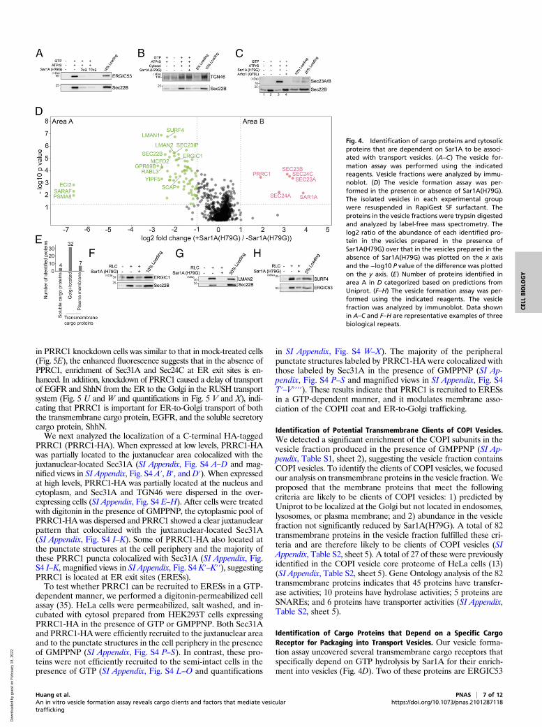
Identification of Cargo Proteins and Cytosolic Proteins that AreDependent on Sar1A for Their Association with Transport Vesicles.The experiment performed in the presence of GTP or GMPPNPrevealed proteins that depend broadly on a group of GTP-bindingproteins, such as Arf family proteins and Rab proteins, to beassociated with vesicles. Next, we sought to utilize this assay toidentify cytosolic proteins and cargo proteins that depend on aspecific GTP-binding protein to be incorporated into transportvesicles. We focused our analysis on the Arf family member,Sar1, which initiates the assembly of the COPII coat at the ER(2). Sar1 has two isoforms in mammalian cells: Sar1A and Sar1B(2). The H79G mutation locks Sar1A in its GTP-bound form andinhibits COPII-dependent ER export (30). Consistent with pre-vious reports, Sar1A(H79G) significantly abolished the vesicularcapture of Sec22B and ERGIC53 (Fig. 4A). In contrast, Sar1-A(H79G) did not interfere with the vesicular release of TGN46(Fig. 4B), a cargo protein that cycles between the plasma membraneand the Golgi (31). Moreover, we found that Sar1A(H79G) en-hanced the membrane association of Sec23A/B (Fig. 4C, comparelanes 3 and 2). In contrast, the dominant active form of anothersmall GTPase, Arfrp1(Q79L), did not enhance the membraneassociation of Sec23A/B (Fig. 4C, compare lanes 4 and 3). Theseanalyses suggest that our vesicle formation assay recapitulates thespecific functions of Sar1A in mediating assembly of COPII coatproteins and in regulating packaging of cargo proteins into COPIIvesicles.We propose that proteins that are significantly reduced in the
presence of Sar1A(H79G) are cargo proteins associated with COPIIvesicles, and that cytosolic proteins that are significantly enhancedin the presence of Sar1A(H79G) are COPII coat proteins orproteins that directly or indirectly interact with COPII coat. Wetherefore performed our vesicle formation assay at a large scale inthe presence or absence of Sar1A(H79G). Proteins in the vesiclefractions were quantified by label-free mass spectrometry (Fig. 4D).A total of 1,223 proteins were identified and quantified, all of whichhad two or more unique peptides (FDR< 0.01) and were successfullyquantified in all of the three biological repeats (SI Appendix, TableS2, sheet 1). This analysis indicates that the vast majority of pro-teins that are significantly enriched in vesicles generated in thepresence of Sar1A(H79G) are subunits of the COPII coat (morethan twofold enrichment, P < 0.05, Fig. 4D, area B and SI Ap-pendix, Table S2, sheet 2). However, a cytosolic protein in additionto COPII subunits was significantly enriched in the Sar1A(H79G)condition (Fig. 4D, area B and SI Appendix, Table S2, sheet 2, pro-teins identified using the protein sequence database of H. sapiens areindicated in round shapes and no additional proteins were identified
Huang et al. PNAS | 5 of 12An in vitro vesicle formation assay reveals cargo clients and factors that mediate vesiculartrafficking
https://doi.org/10.1073/pnas.2101287118
CELL
BIOLO
GY
Dow
nloa
ded
by g
uest
on
Feb
ruar
y 18
, 202
2

using the database of R. norvegicus). All of the identified hits showeda q value of <0.05 (SI Appendix, Table S2, sheet 2).Seventy-three proteins were identified with significant en-
richment in the untreated group over the Sar1A(H79G) group(more than twofold enrichment, P < 0.05, Fig. 4D, area A and
SI Appendix, Table S2, sheet 3). A total of 62 of these proteinsshowed a q value of <0.05 (SI Appendix, Table S2, sheet 3). A totalof 50 of these 62 proteins are predicted to be transmembraneproteins and 4 were soluble cargo proteins that are secretory orlocated at the Golgi (Fig. 4E and SI Appendix, Table S2, sheet 4).Many of the transmembrane proteins were predicted to showplasma membrane and Golgi localization (Fig. 4E and SI Appen-dix, Table S2, sheet 4). None of these transmembrane proteinswere predicted to show mitochondria or peroxisome localizations.All of the SNARE proteins identified in area B (Sec22A, Sec22B,STX5, GOSR2, and BET1) mediate ER-to-Golgi trafficking. Atotal of 33 among the 62 hits were identified to be associated withCOPII vesicles reconstituted with purified COPII componentscontaining specific Sec24 isoforms (13) (SI Appendix, Table S2,sheet 3). A total of 29 proteins were not identified in the previousstudy (SI Appendix, Table S2, sheet 3, highlighted with single as-terisks) and many of them are transmembrane proteins that arepredicted to show Golgi or plasma membrane localization. Westernblot analysis confirmed that three transmembrane proteins,ERGIC1, SURF4, and LMAN2 were present in the vesiclefraction and their vesicular release was significantly reduced bySar1A(H79G) (Fig. 4 F–H), indicating that they are packaged inCOPII vesicles. Several cytosolic proteins were identified in areaA, including RabL3, Sec23IP, and SCFD1. SCFD1 and Sec23IPhave been shown to regulate ER-to-Golgi trafficking in mamma-lian cells (32–34). The role of RabL3 in ER-to-Golgi traffickingremains to be investigated.In summary, these analyses revealed candidate cargo proteins
that are packaged into COPII vesicles and candidate cytosolicproteins that are associated with COPII-coated vesicles. Moreover,these analyses indicate that our approach is robust in revealing ef-fector proteins that are associated with GTP-bound Arf familyproteins on vesicle membranes.
PRRC1 Is Recruited to Vesicle Membranes by GTP-Bound Sar1A,Located at the ER Exit Sites and Regulates ER-to-Golgi Trafficking.A proline-rich domain-containing protein, PRRC1, was identifiedby our analysis to be recruited to vesicles by GTP-bound Sar1A(Fig. 4D, area B). The cellular function of PRRC1 is unknown.Western blot analysis confirmed that vesicles produced in the pres-ence of Sar1A(H79G) contain higher levels of PRRC1 (Fig. 5A). Wetherefore tested whether PRRC1 was a binding partner of Sar1Autilizing a GST-pulldown approach. Purified GST-tagged human Sar1Awith its N-terminal amphipathic helix removed (GST-Sar1AΔ1–17)was loaded with GDP or GMPPNP and then incubated with RLC.The concentration of RLC in the reaction mixture was equal tothat used in the vesicle formation assay. After incubation, proteinsthat bound to Sar1A in a GTP-dependent manner were eluted withethylenediaminetetraacetic acid (EDTA). Western blot analysis ofthe eluted fraction indicated that Sec23A/B was specifically de-tected in the eluate of GMPPNP-loaded but not GDP-loaded GST-Sar1AΔ1–17 immobilized on glutathione beads (Fig. 5B, comparelanes 1 and 2). In contrast, PRRC1 was not detected in the eluate ofGMPPNP-loaded GST-Sar1AΔ1–17 (Fig. 5B). This GST pulldownoccurred in the absence of a lipid bilayer, whereas the vesicle for-mation assay preserves the lipid bilayers of the ER and the Golgi.We propose that the vesicle formation assay has the advantage ofrevealing protein–protein interactions that take place on lipid bi-layers. The interaction between PRRC1 and Sar1A might thereforerequire the intact membranes.Coimmunoprecipitation experiments indicate that FLAG-tagged
PRRC1 interacted with the inner COPII subunit, Sec23A/B, but notwith the outer COPII subunit, Sec31A (Fig. 5 C and D, comparelanes 1 and 2). Knockdown of PRRC1 caused a significant en-hancement of the total cellular immunofluorescence signal asso-ciated with Sec31A and Sec24C but not Golgin97 (Fig. 5 E–Q,quantifications in Fig. 5 R–T). Since the expression level of Sec31A
Fig. 3. FAM84B/LRATD2 is recruited to vesicle membranes in a GTP-dependentmanner, interacts with AP1γ1 and Sec23A/B, and regulates ER-to-Golgi transportof EGFR but not ShhN or IGF2. (A) The vesicle formation assay was performedusing the indicated reagents. The proteins in the vesicle fraction were analyzedby Western blot. (B) HEK293T cells expressing the FAM84B-HA were treated in2 mM dithiobis(succinimidyl propionate) (DSP), and the cell lysates were incu-bated with beads conjugated with HA antibodies. After incubation, the boundproteins were analyzed by Western blot using the indicated antibodies. (C)HEK293T cells were transfected with control siRNA or siRNA against FAM84B/LRATD2. Day 3 after transfection, cells were lysed and analyzed by Westernblot. (D, F, and H) HEK293T cells were transfected with control siRNA or siRNAagainst FAM84B/LRATD2. Twenty-four hours after transfection, cells weretransfected with plasmids encoding the indicted constructs. On day 3 afterknockdown, cells were incubated with biotin and cycloheximide for 15min andthe localization of the cargo proteins was analyzed by fluorescent microscope.(Scale bar, 10 μm.) (E, G, and I) Quantifications of the percentage of cellsshowing Golgi-localized cargo proteins in each experimental group (mean ±SD; n = 3; >100 cells counted for each experiment). ****P < 0.0001; *****P <0.00001; n.s., not significant. Data shown in A–C are representative examples ofthree biological repeats.
6 of 12 | PNAS Huang et al.https://doi.org/10.1073/pnas.2101287118 An in vitro vesicle formation assay reveals cargo clients and factors that mediate vesicular
trafficking
Dow
nloa
ded
by g
uest
on
Feb
ruar
y 18
, 202
2
in PRRC1 knockdown cells was similar to that in mock-treated cells(Fig. 5E), the enhanced fluorescence suggests that in the absence ofPPRC1, enrichment of Sec31A and Sec24C at ER exit sites is en-hanced. In addition, knockdown of PRRC1 caused a delay of transportof EGFR and ShhN from the ER to the Golgi in the RUSH transportsystem (Fig. 5 U and W and quantifications in Fig. 5 V and X), indi-cating that PRRC1 is important for ER-to-Golgi transport of boththe transmembrane cargo protein, EGFR, and the soluble secretorycargo protein, ShhN.We next analyzed the localization of a C-terminal HA-tagged
PRRC1 (PRRC1-HA). When expressed at low levels, PRRC1-HAwas partially located to the juxtanuclear area colocalized with thejuxtanuclear-located Sec31A (SI Appendix, Fig. S4 A–D and mag-nified views in SI Appendix, Fig. S4 A′, B′, and D′). When expressedat high levels, PRRC1-HA was partially located at the nucleus andcytoplasm, and Sec31A and TGN46 were dispersed in the over-expressing cells (SI Appendix, Fig. S4 E–H). After cells were treatedwith digitonin in the presence of GMPPNP, the cytoplasmic pool ofPRRC1-HA was dispersed and PRRC1 showed a clear juxtanuclearpattern that colocalized with the juxtanuclear-located Sec31A(SI Appendix, Fig. S4 I–K). Some of PRRC1-HA also located atthe punctate structures at the cell periphery and the majority ofthese PRRC1 puncta colocalized with Sec31A (SI Appendix, Fig.S4 I–K, magnified views in SI Appendix, Fig. S4 K′–K′′), suggestingPRRC1 is located at ER exit sites (ERESs).To test whether PRRC1 can be recruited to ERESs in a GTP-
dependent manner, we performed a digitonin-permeabilized cellassay (35). HeLa cells were permeabilized, salt washed, and in-cubated with cytosol prepared from HEK293T cells expressingPRRC1-HA in the presence of GTP or GMPPNP. Both Sec31Aand PRRC1-HA were efficiently recruited to the juxtanuclear areaand to the punctate structures in the cell periphery in the presenceof GMPPNP (SI Appendix, Fig. S4 P–S). In contrast, these pro-teins were not efficiently recruited to the semi-intact cells in thepresence of GTP (SI Appendix, Fig. S4 L–O and quantifications
in SI Appendix, Fig. S4 W–X). The majority of the peripheralpunctate structures labeled by PRRC1-HA were colocalized withthose labeled by Sec31A in the presence of GMPPNP (SI Ap-pendix, Fig. S4 P–S and magnified views in SI Appendix, Fig. S4T′–V′′′′). These results indicate that PRRC1 is recruited to ERESsin a GTP-dependent manner, and it modulates membrane asso-ciation of the COPII coat and ER-to-Golgi trafficking.
Identification of Potential Transmembrane Clients of COPI Vesicles.We detected a significant enrichment of the COPI subunits in thevesicle fraction produced in the presence of GMPPNP (SI Ap-pendix, Table S1, sheet 2), suggesting the vesicle fraction containsCOPI vesicles. To identify the clients of COPI vesicles, we focusedour analysis on transmembrane proteins in the vesicle fraction. Weproposed that the membrane proteins that meet the followingcriteria are likely to be clients of COPI vesicles: 1) predicted byUniprot to be localized at the Golgi but not located in endosomes,lysosomes, or plasma membrane; and 2) abundance in the vesiclefraction not significantly reduced by Sar1A(H79G). A total of 82transmembrane proteins in the vesicle fraction fulfilled these cri-teria and are therefore likely to be clients of COPI vesicles (SIAppendix, Table S2, sheet 5). A total of 27 of these were previouslyidentified in the COPI vesicle core proteome of HeLa cells (13)(SI Appendix, Table S2, sheet 5). Gene Ontology analysis of the 82transmembrane proteins indicates that 45 proteins have transfer-ase activities; 10 proteins have hydrolase activities; 5 proteins areSNAREs; and 6 proteins have transporter activities (SI Appendix,Table S2, sheet 5).
Identification of Cargo Proteins that Depend on a Specific CargoReceptor for Packaging into Transport Vesicles. Our vesicle forma-tion assay uncovered several transmembrane cargo receptors thatspecifically depend on GTP hydrolysis by Sar1A for their enrich-ment into vesicles (Fig. 4D). Two of these proteins are ERGIC53
Fig. 4. Identification of cargo proteins and cytosolicproteins that are dependent on Sar1A to be associ-ated with transport vesicles. (A–C) The vesicle for-mation assay was performed using the indicatedreagents. Vesicle fractions were analyzed by immu-noblot. (D) The vesicle formation assay was per-formed in the presence or absence of Sar1A(H79G).The isolated vesicles in each experimental groupwere resuspended in RapiGest SF surfactant. Theproteins in the vesicle fractions were trypsin digestedand analyzed by label-free mass spectrometry. Thelog2 ratio of the abundance of each identified pro-tein in the vesicles prepared in the presence ofSar1A(H79G) over that in the vesicles prepared in theabsence of Sar1A(H79G) was plotted on the x axisand the −log10 P value of the difference was plottedon the y axis. (E) Number of proteins identified inarea A in D categorized based on predictions fromUniprot. (F–H) The vesicle formation assay was per-formed using the indicated reagents. The vesiclefraction was analyzed by immunoblot. Data shownin A–C and F–H are representative examples of threebiological repeats.
Huang et al. PNAS | 7 of 12An in vitro vesicle formation assay reveals cargo clients and factors that mediate vesiculartrafficking
https://doi.org/10.1073/pnas.2101287118
CELL
BIOLO
GY
Dow
nloa
ded
by g
uest
on
Feb
ruar
y 18
, 202
2

and SURF4 (Fig. 4 A and H). To reveal the client repertoire ofERGIC53 and SURF4, we performed the vesicle formation assayusing donor membranes prepared from genome-engineeredERGIC53 knockout (KO) HEKTrex cells or SURF4 knockoutHEKTrex cells. Western blot analysis confirmed the absence ofERGIC53 or SURF4 in the vesicles generated from the corre-sponding KO donor membranes (Fig. 6 A and B). We did notdetect obvious changes in the localization of ERGIC53 in SURF4KO cells (SI Appendix, Fig. S5 A–C) or changes in the localizationof SURF4 in ERGIC53 KO cells (SI Appendix, Fig. S5 D–F).Similarly, KO cells showed no obvious defects in ER morphology,marked by HA-Vangl2D255E (SI Appendix, Fig. S5 G–I) or the mem-brane association of COPII, marked by Sec31A (SI Appendix, Fig. S5
J–L). The cis-Golgi marker, GM130, was mainly located at thejuxtanuclear region in wild-type (WT) and KO cells (SI Appendix,Fig. S5 M–O). GM130 in around 10% of WT cells and 30% ofSURF4 KO cells or 30% of ERGIC53 KO cells located to somepunctate structures in addition to the juxtanuclear localization(SI Appendix, highlighted by asterisks in SI Appendix, Fig. S5 N andO). Consistent with a previous report (36), we did not detect in-duction of the unfolded protein response (UPR) in SURF4 KO andERGIC53 KO cells (SI Appendix, Fig. S5P).Vesicles were generated from KO or WT donor membranes
incubated with rat liver cytosol. Proteins in the vesicle fractionswere then analyzed by label-free quantitative mass spectrometry.A total of 815 proteins were identified and quantified, all of which
Fig. 5. PRRC1 interacts with Sec23A/B and knockdown increasesmembrane association of Sec31A and Sec24C and decreasesER-to-Golgi transport of EGFR and ShhN. (A) The vesicle formationassay was performed using the indicated reagents. Vesicle fractionswere analyzed by immunoblot. (B) GST-Sar1AΔ1–17 was loaded withGDP or GMPPNP and then incubated with rat liver cytosol. Afterincubation, proteins that bound to Sar1A in a nucleotide-dependentmanner were eluted with EDTA. The eluted fraction and the pro-teins left on beads after elution were analyzed by immunoblot.(C and D) M2 agarose beads were incubated with cell lysates fromHEK293T cells expressing the PRRC1-FLAG. After incubation, thebound proteins were eluted with 3× FLAG peptides and analyzed byWestern blot using the indicated antibodies. (E) HEK293T cells weretransfected with control siRNA or siRNA against PRRC1. Day 3 aftertransfection, cells were lysed and analyzed by Western blot. (F–Q)HEK293T cells were transfected with control siRNA (F–H, L–N) orsiRNA against PRRC1 (I–K, O–Q). Day 3 after transfection, the local-izations of Sec31A, Sec24C, and Golgin97 were analyzed by immuno-fluorescence. (Scale bar, 10 μm.) The magnified views of theindicated area in F, I, L, and O are shown in F′, I′, L′, and O′. (R–T)Quantifications of the total fluorescent level of Sec31A (R), Sec24C(S), and Golgin97 (T) per cell (mean ± SD; n = 3; >125 cells from ninerandom imaging fields counted for each experiment). In each ex-periment, the total fluorescent level was normalized to that in mockcells. **P < 0.01; NS, not significant. (U and W) HEK293T cells weretransfected with control siRNA or siRNA against PRRC1. Twenty-four hours after transfection, cells were retransfected with plas-mids encoding the indicated construct. On day 3 after knockdown,cells were incubated with biotin for the indicated time and the lo-calization of the indicated protein was analyzed by fluorescent mi-croscope. (Scale bar, 10 μm.) (V and X) Quantifications of thepercentage of cells showing Golgi-localized EGFR or ShhN in cellstreated with control siRNA or siRNA against PRRC1 (mean ± SD; n =3; >100 cells counted for each experiment). ***P < 0.001; **P < 0.01.Data shown in A, B, D, and E are representative examples of threebiological repeats.
8 of 12 | PNAS Huang et al.https://doi.org/10.1073/pnas.2101287118 An in vitro vesicle formation assay reveals cargo clients and factors that mediate vesicular
trafficking
Dow
nloa
ded
by g
uest
on
Feb
ruar
y 18
, 202
2
had two or more unique peptides (FDR < 0.01) and were suc-cessfully quantified in all three biological replicates of the exper-imental groups (SI Appendix, Table S3, sheet 1). In each case,several proteins were significantly reduced in the vesicle fractionof the KO reaction compared to WT (area A, Fig. 6 C and D andSI Appendix, Table S3, sheet 2). Transmembrane proteins that arepredicted to show Golgi or plasma membrane localization arehighlighted in red and soluble secretory proteins highlighted ingreen. Additional cargo proteins were significantly reduced in thevesicle fraction in the KO vesicles compared to WT when thethreshold was changed from 0.5 to 0.6 (area A, Fig. 6 C and D andSI Appendix, Table S3, sheet 2).Fourteen proteins, including ERGIC53, were underrepresented
in the ERGIC53 KO condition relative to wild type (fold change <0.6; P < 0.05, SI Appendix, Table S3, sheet 2). Five of them showeda q value of <0.05. Removing ERGIC53, we defined the remainingfour proteins as ERGIC53 clients (highlighted in Fig. 6C, area Aand SI Appendix, Table S3, sheet 2). One of these proteins, mul-tiple coagulation factor deficiency protein 2 (MCFD2), is a knownERGIC53 interactor (37). The other three proteins may be novelinteractors of ERGIC53. Among the remaining proteins with a qvalue of >0.05, coagulation factor V (FV) (q value = 0.1), is aknown cargo client of ERGIC53 (SI Appendix, Table S3, sheet 2)(3). MCFD2 forms a complex with ERGIC53 to facilitate thetransport of coagulation factors V and VIII (FVIII) from the ERto the Golgi (37, 38). We next examined the abundance of the fourERGIC53 clients in vesicles made from SURF4 KO cells(Fig. 6E). Three ERGIC53 clients were not similarly depleted inthe SURF4 KO condition (Fig. 6E, above the green dotted line),suggesting that these cargo proteins are dependent on ERGIC53but not SURF4 for efficient packaging into vesicles. Moreover,ERGIC53 is required for retention of MCFD2 in the early secretorytransport pathway (39). Immunoblot analysis confirmed that pack-aging of HA-tagged MCFD2 into vesicles was abrogated inERGIC53 KO cells (Fig. 6G, lane 2) but not in SURF4 KO cells(Fig. 6I, lane 2). Exogenously expressing ERGIC53 in ERGIC53KO cells rescued packaging of MCFD2 into transport vesicles, andadding Sar1A(H79G) blocked this rescue (Fig. 6H, lanes 2 and 3).Using similar criteria, 19 proteins were underrepresented in
the SURF4 KO condition relative to wild type (fold change < 0.6;P < 0.05). Most of them (except one protein) showed a q valueof <0.05 (SI Appendix, Table S3, sheet 2). Removing SURF4, wedefined the remaining 17 proteins as SURF4 clients (highlightedin Fig. 6D, area A and SI Appendix, Table S3, sheet 2). A totalof 16 out of 17 SURF4 clients were unaffected by the loss ofERGIC53 (Fig. 6F). Commercial antibodies against two of the tophits, NUCB1 and NUCB2, confirmed that these two proteins arepackaged into vesicles, and the budding efficiency was reduced bySar1A(H79G) (Fig. 6 J and M, compare lanes 2 and 3). Efficiencyof NUCB1 and NUCB2 packaging was greatly reduced in SURF4KO cells (Fig. 6 K and N, lane 2), whereas vesicular release ofthese two cargo proteins was not affected in ERGIC53 KO cells(Fig. 6 L and O, lane 2). Consistent with the observation fromSURF4 KO cells, knockdown of SURF4 by siRNA (SI Appendix,Fig. S6E) also significantly reduced the efficiency of packaging ofNUCB1 and NUCB2 into transport vesicles (SI Appendix, Fig.S6 A and B, compare lanes 2 and 6, quantifications in SI Appendix,Fig. S6 C and D). NUCB1 and NUCB2 coimmunoprecipitatedwith SURF4-HA in the presence of a cross-linker (SI Appendix,Fig. S6F), suggesting these cargo proteins interact with SURF4.Altogether, our analyses revealed specific transmembrane andsoluble cargo proteins that depend on SURF4 or ERGIC53 tobe packaged into transport vesicles. These analyses indicatethat our method is a robust approach to reveal the clients of aspecific transmembrane cargo receptor.
DiscussionGTP-binding proteins, including Arf family proteins and Rabfamily proteins, play critical roles in mediating membrane recruit-ment of cytosolic factors to regulate cargo sorting and vesicle for-mation (14–16). Affinity chromatography is a traditional approachto identify these cytosolic factors. Here, we utilized the vesicleformation assay to investigate this aspect. A benefit of our approachis that membranes are preserved and our analysis indicates that thisapproach can reveal protein–protein interactions that take place onlipid bilayers. Through this approach, we identified cytosolic factorsthat are associated with vesicle membranes in a GTP-dependentmanner or that interact with GTP-bound Sar1A on vesicle mem-branes. These cytosolic proteins may function as cargo adaptors ormay associate with vesicle coats to regulate cargo sorting.Two of these cytosolic proteins, FAM84B/LRATD2 and PRRC1,
were shown to regulate ER-to-Golgi transport of newly synthesizedEGFR. FAM84B/LRATD2 contains a LRAT (lecithin:retinalacyltransferase) domain. This domain is present in the H-Ras–likesuppressor (HRASLS) family. The expression of FAM84B/LRATD2is up-regulated during prostate cancer progression and in preclinicaland esophageal squamous cell carcinoma tumors (40, 41). FAM84B/LRATD2 is shown to promote prostate tumorigenesis (42). PRRC1 ispredicted to have protein kinase A regulatory subunit binding activity.Our results indicate that PRRC1 is recruited to ER exit sites in aGTP-dependent manner; associates with vesicle membranes in thepresence of GTP-bound Sar1A; and down-regulates membranerecruitment of the COPII subunit, Sec24C and Sec31A. PRRC1contains a proline-rich domain. The proline-rich region of Sec31interacts with Sec23 (43–45), suggesting that PRRC1 may directlyinteract with Sec23 to perform its function. FAM84B/LRATD2also contains a proline-rich domain but its function remains to befurther investigated.We found several further Arf and Rab proteins, besides Sar1A,
whose abundances were significantly increased in the vesiclefraction produced in the presence of GMPPNP. It would beinteresting to utilize our approach to reveal the cytosolic effectorproteins that depend on these proteins to associate with transportvesicles. Our approach can also be performed in the presence ofother vesicle-associated extrinsic factors, which would further re-veal binding partners on vesicle membranes.In addition to cytosolic proteins associated with vesicles, our
approach is powerful in revealing cargo proteins that depend ona specific factor to be enriched into transport vesicles. In this study,we found cargo protein enrichment into vesicles depends on GTPhydrolysis by Sar1A. The protein composition of vesicles producedby purified COPII coats has been analyzed (13), which revealedseveral COPII clients that depend on specific isoforms of the COPIIcargo-binding subunit, Sec24 (13). Release of the COPII clients,Sec22B and ERGIC53, into vesicles was greatly enhanced when thevesicle formation assay was performed using purified COPII sup-plemented with a low concentration of RLC (Fig. 1K). Using RLCas the source of cytosolic proteins, we found several non-ER resi-dent cargo proteins that were not identified in the previous study.These cargo proteins may depend on cytosolic factors in addition toCOPII in the RLC to be efficiently packaged into COPII vesicles.Utilizing donor membranes from wild-type cells or cells depleted
of a specific cargo receptor, we revealed clients of ERGIC53 andSURF4. Similar analysis will facilitate the identification of clients ofother cargo receptors, thereby providing important information onthe specificity of protein sorting and also revealing insight into thefunctional role of specific cellular factors. Another application ofour approach is to analyze the protein composition of the vesiclesproduced from cells under different physiological conditions suchas starvation. This could provide important insight into how vesi-cles contribute to establish and maintain a specific physiologicalcondition. A caveat of our assay is that it relies on identification ofcargo proteins that are actively produced by cells. Another caveat
Huang et al. PNAS | 9 of 12An in vitro vesicle formation assay reveals cargo clients and factors that mediate vesiculartrafficking
https://doi.org/10.1073/pnas.2101287118
CELL
BIOLO
GY
Dow
nloa
ded
by g
uest
on
Feb
ruar
y 18
, 202
2

Fig. 6. Identification of cargo proteins that depend on ERGIC53 or SURF4 for being packaged into transport vesicles. (A and B) The vesicle formation wasperformed using wild-type HEK293TRex cells and ERGIC53 KO HEK293TRex cells (A) or SURF4 KO HEK293TRex cells (B). The vesicle fraction was then analyzedby immunoblot. (C and D) The vesicle formation assay was performed using wild-type HEK293TRex cells and ERGIC53 KO HEK293TRex cells (C) or SURF4 KOHEK293TRex cells (D). The isolated vesicles in each experimental group were resuspended in RapiGest SF surfactant. The proteins in the vesicle fractions weretrypsin digested and analyzed by label-free mass spectrometry. The log2 ratio of the abundance of each identified protein in the vesicles prepared fromERGIC53 KO or SURF4 KO cells over that in the vesicles prepared in wild-type cells was plotted on the x axis and the −log10 P value of the difference wasplotted on the y axis. (E) The fold change of the identified ERGIC53 client was compared with the fold change of the abundance of these proteins in thevesicle fraction prepared from the WT cells and SURF4 KO cells. (F) The fold change of identified SURF4 client was compared with the fold change ofthe abundance of these proteins in the vesicle fraction prepared from the WT cells and ERGIC53 KO cells. Dotted line indicates the fold change of 0.75. (G–O)The vesicle formation was performed using the indicated cells. Vesicle fractions were then analyzed by immunoblot. Data shown in G–O are representativeexamples of three biological repeats. The single asterisks in panels G–I indicate the nonspecific bands recognized by anti-ERGIC53 antibodies.
10 of 12 | PNAS Huang et al.https://doi.org/10.1073/pnas.2101287118 An in vitro vesicle formation assay reveals cargo clients and factors that mediate
vesicular trafficking
Dow
nloa
ded
by g
uest
on
Feb
ruar
y 18
, 202
2

of our assay is that the donor membranes to produce vesicles arefrom digitonin-permeablized cells, so that the vesicles producedoriginate from multiple different organelles. It would be inter-esting to perform this assay in cell lines that highly secrete a varietyof cellular factors to identify cargo proteins and utilize purified ERor Golgi membranes as the donor membranes for the vesicleformation assay.In summary, our study demonstrates that the vesicle formation
assay in combination with quantitative mass spectrometry anal-ysis is powerful to identify cytosolic proteins that associate withvesicle membranes to regulate vesicular trafficking and to un-cover cargo clients of a specific cellular factor, providing a robusttool to reveal insights into the secretory pathway.
Materials and MethodsTransfection, Immunofluorescence, and Permeabilized Cell Assays. Transfectionwas performed using Lipofectamine 2000 (Invitrogen) or polyethyleneimine(PEI). Immunofluorescence was performed as described previously (20). Im-ages were acquired with a Zeiss Axioobserver Z1 microscope system. Quan-tifications of the total fluorescence of Sec31A and Gogin97 were performedas described using ImageJ (46). Permeabilized cell assays were performed asdescribed previously (35).
In Vitro Vesicle Formation Assay. In vitro vesicle formation assay was per-formed as described previously (20).
Vesicle Immunogold Labeling and Negative Staining for Transmission ElectronMicroscopy Analysis. Negative staining transmission electron microscopy(TEM) analysis and immunogold labeling were performed as describedpreviously (47).
Protein Purifications, Nucleotide Loading, and GST Pulldown. Purification ofHis-tagged proteins from Escherichia coli was performed as described pre-viously (48). Purification of GST-tagged protein was performed as described(16). The nucleotide loading and GST pulldown experiment was performedas described previously (14, 16).
RUSH Assay.HEK293T cells were transfectedwith plasmids encoding the RUSHconstruct of a specific cargo protein for 24 h. To release the RUSH construct ofthe cargo protein from the ER, cells were treated with 100 ng/μL cyclohex-imide (Sigma-Aldrich) and 40 μM D-biotin (Sigma-Aldrich) for the indicatedtime. Cells were then washed with phosphate-buffered saline (PBS), fixedwith 4% paraformaldehyde (PFA), washed with PBS and mounted ontoslides with ProLong Gold antifade mountant (Thermo Fisher).
Sample Preparation for Label-Free Quantitative MS Analysis, Liquid Chromatography-MSAnalysis, and MS Data Analysis. These procedures were performed as describedpreviously (19).
Statistical Analysis of MS Data. Student’s t test was used to compare thesignificant changes between two experimental groups based on the proteinabundance values of the identified protein in three biological repeats in thetwo experimental groups. In addition, a permutation-based FDR with an s0value of 0.01 was calculated using Perseus software (23). The identifiedproteins shown in Fig. 2C, areas A and B, Fig. 4D, areas A and B, Fig. 6C, areaA, and Fig. 6D, area A were referred to as the identified hit proteins. Theaverage abundance of the identified hit proteins that are less than 200,000in both experimental groups was removed from the list of the identified hitproteins. The average abundance of each identified hit protein was calcu-lated as the average of the absolute abundance of this protein in the threereplicated experiments.
The abundance of proteins in the vesicle fraction isolated in the presenceof GTP or GMPPNP was plotted using a histogram as previously reported (49).The enrichment analysis for categories of identified proteins was performedbased on a Fisher’s exact test with a Benjamini–Hochberg FDR threshold of0.02 using Perseus software.
ATF6-Luciferase UPR Assay. This assay was performed as described previously (51).
Data Availability. The mass spectrometry proteomics data have been de-posited to the ProteomeXchange Consortium via the PRIDE (50) partnerrepository with the dataset identifier PXD026081.
ACKNOWLEDGMENTS.We thank Dr. Randy Schekman (University of California,Berkeley) for providing purified COPII components and antibodies againstCOPII components. We thank Dr. Xiao-wei Chen (Peking University) forantibodies against SURF4. This work was supported by Hong Kong ResearchGrants Council (RGC) grants 16102921, 26100315, 16101116, 16102218, 16103319,AoE/M-05/12, C4002-17G, and C4012-16E (to Y.G.). This workwas also supported bygrants from the National Natural Science Foundation of China (NSFC31871421and NSFC3207050042 to Y.G. and NSFC 81601828 to H.Y.) and a Free ExploreProject from the Shenzhen Science and Technology Innovation Committee(JCYJ20180306174847511 to Y.G.). This study was supported in part by theInnovation and Technology Commission (ITCPD/17-9 to Y.G.). This study wasalso partially supported by the Hong Kong Branch of Southern Marine Scienceand Engineering Guangdong Laboratory (Guangzhou) (SMSEGL20SC01 toY.G.). In addition, this work was supported by the Medical Research Council(MC_UP_1201/10) to E.A.M., and Z.-P.Y. is supported by Hong Kong RGC grantsR5013-19, R4005-18, C5031-14E, 15304117, 15304020, and the UniversityResearch Facility in Chemical and Environmental Analysis and UniversityResearch Facility in Life Sciences of PolyU. L.J. is supported by grants fromHong Kong RGC (C4012-16E, C4002-17G, C4033-19E, and AoE/M-05/12).
1. Y. Guo, D. W. Sirkis, R. Schekman, Protein sorting at the trans-Golgi network. Annu.
Rev. Cell Dev. Biol. 30, 169–206 (2014).
2. M. C. Lee, E. A. Miller, J. Goldberg, L. Orci, R. Schekman, Bi-directional protein
transport between the ER and Golgi. Annu. Rev. Cell Dev. Biol. 20, 87–123 (2004).
3. J. Dancourt, C. Barlowe, Protein sorting receptors in the early secretory pathway.
Annu. Rev. Biochem. 79, 777–802 (2010).
4. W. C. Nichols et al., Mutations in the ER-Golgi intermediate compartment protein
ERGIC-53 cause combined deficiency of coagulation factors V and VIII. Cell 93, 61–70
(1998).
5. F. Vollenweider, F. Kappeler, C. Itin, H. P. Hauri, Mistargeting of the lectin ERGIC-53 to
the endoplasmic reticulum of HeLa cells impairs the secretion of a lysosomal enzyme.
J. Cell Biol. 142, 377–389 (1998).
6. C. Appenzeller, H. Andersson, F. Kappeler, H. P. Hauri, The lectin ERGIC-53 is a cargo
transport receptor for glycoproteins. Nat. Cell Biol. 1, 330–334 (1999).
7. B. Nyfeler et al., Identification of ERGIC-53 as an intracellular transport receptor of
alpha1-antitrypsin. J. Cell Biol. 180, 705–712 (2008).
8. K. Saegusa, M. Sato, N. Morooka, T. Hara, K. Sato, SFT-4/Surf4 control ER export of
soluble cargo proteins and participate in ER exit site organization. J. Cell Biol. 217,
2073–2085 (2018).
9. Y. Yin et al., Surf4 (Erv29p) binds amino-terminal tripeptide motifs of soluble cargo
proteins with different affinities, enabling prioritization of their exit from the en-
doplasmic reticulum. PLoS Biol. 16, e2005140 (2018).
10. B. T. Emmer et al., The cargo receptor SURF4 promotes the efficient cellular secretion
of PCSK9. eLife 7, e38839 (2018).
11. X. Wang et al., Receptor-mediated ER export of lipoproteins controls lipid homeo-
stasis in mice and humans. Cell Metab. 33, 350–366.e7 (2020).
12. Y. Herzig, H. J. Sharpe, Y. Elbaz, S. Munro, M. Schuldiner, A systematic approach to
pair secretory cargo receptors with their cargo suggests a mechanism for cargo se-
lection by Erv14. PLoS Biol. 10, e1001329 (2012).
13. F. Adolf et al., Proteomic profiling of mammalian COPII and COPI vesicles. Cell Rep. 26,
250–265.e5 (2019).
14. S. Christoforidis, M. Zerial, Purification and identification of novel Rab effectors using
affinity chromatography. Methods 20, 403–410 (2000).
15. H. Jin et al., The conserved Bardet-Biedl syndrome proteins assemble a coat that
traffics membrane proteins to cilia. Cell 141, 1208–1219 (2010).
16. Y. Guo, G. Zanetti, R. Schekman, A novel GTP-binding protein-adaptor protein
complex responsible for export of Vangl2 from the trans Golgi network. eLife 2,
e00160 (2013).
17. J. Merte et al., Sec24b selectively sorts Vangl2 to regulate planar cell polarity during
neural tube closure. Nat. Cell Biol. 12, 41–46; sup pp. 41–48 (2010).
18. J. Kim et al., Biogenesis of gamma-secretase early in the secretory pathway. J. Cell
Biol. 179, 951–963 (2007).
19. L. Niu et al., Atlastin-mediated membrane tethering is critical for cargo mobility and exit
from the endoplasmic reticulum. Proc. Natl. Acad. Sci. U.S.A. 116, 14029–14038 (2019).
20. T. Ma et al., A mechanism for differential sorting of the planar cell polarity proteins
Frizzled6 and Vangl2 at the trans-Golgi network. J. Biol. Chem. 293, 8410–8427
(2018).
21. X. Tang et al., Molecular mechanisms that regulate export of the planar cell-polarity
protein Frizzled-6 out of the endoplasmic reticulum. J. Biol. Chem. 295, 8972–8987
(2020).
22. L. Yuan, S. J. Kenny, J. Hemmati, K. Xu, R. Schekman, TANGO1 and SEC12 are co-
packaged with procollagen I to facilitate the generation of large COPII carriers. Proc.
Natl. Acad. Sci. U.S.A. 115, E12255–E12264 (2018).
Huang et al. PNAS | 11 of 12An in vitro vesicle formation assay reveals cargo clients and factors that mediate vesiculartrafficking
https://doi.org/10.1073/pnas.2101287118
CELL
BIOLO
GY
Dow
nloa
ded
by g
uest
on
Feb
ruar
y 18
, 202
2

23. S. Tyanova et al., The Perseus computational platform for comprehensive analysis of
(prote)omics data. Nat. Methods 13, 731–740 (2016).
24. G. H. Borner et al., Multivariate proteomic profiling identifies novel accessory proteins
of coated vesicles. J. Cell Biol. 197, 141–160 (2012).
25. J. Hirst et al., Distinct and overlapping roles for AP-1 and GGAs revealed by the
“knocksideways” system. Curr. Biol. 22, 1711–1716 (2012).
26. J. Hirst et al., Contributions of epsinR and gadkin to clathrin-mediated intracellular
trafficking. Mol. Biol. Cell 26, 3085–3103 (2015).
27. Y. Li, H. Zhang, Y. Litingtung, C. Chiang, Cholesterol modification restricts the spread
of Shh gradient in the limb bud. Proc. Natl. Acad. Sci. U.S.A. 103, 6548–6553 (2006).
28. G. Boncompain et al., Synchronization of secretory protein traffic in populations of
cells. Nat. Methods 9, 493–498 (2012).
29. Y. Mao et al., The exocyst functions in niche cells to promote germline stem cell
differentiation by directly controlling EGFR membrane trafficking. Development 146,
dev174615 (2019).
30. M. Aridor, S. I. Bannykh, T. Rowe, W. E. Balch, Sequential coupling between COPII and
COPI vesicle coats in endoplasmic reticulum to Golgi transport. J. Cell Biol. 131,
875–893 (1995).
31. S. Ponnambalam et al., Primate homologues of rat TGN38: Primary structure, ex-
pression and functional implications. J. Cell Sci. 109, 675–685 (1996).
32. Y. S. Ong, B. L. Tang, L. S. Loo, W. Hong, p125A exists as part of the mammalian Sec13/
Sec31 COPII subcomplex to facilitate ER-Golgi transport. J. Cell Biol. 190, 331–345
(2010).
33. C. Dascher, W. E. Balch, Mammalian Sly1 regulates syntaxin 5 function in endoplasmic
reticulum to Golgi transport. J. Biol. Chem. 271, 15866–15869 (1996).
34. C. Nogueira et al., SLY1 and Syntaxin 18 specify a distinct pathway for procollagen VII
export from the endoplasmic reticulum. eLife 3, e02784 (2014).
35. F. Yang, T. Li, Z. Peng, Y. Liu, Y. Guo, The amphipathic helices of Arfrp1 and Arl14 are
sufficient to determine subcellular localizations. J. Biol. Chem. 295, 16643–16654
(2020).
36. A. Ordóñez, H. P. Harding, S. J. Marciniak, D. Ron, Cargo receptor-assisted endo-
plasmic reticulum export of pathogenic α1-antitrypsin polymers. Cell Rep. 35, 109144
(2021).
37. B. Zhang, R. J. Kaufman, D. Ginsburg, LMAN1 and MCFD2 form a cargo receptor
complex and interact with coagulation factor VIII in the early secretory pathway.
J. Biol. Chem. 280, 25881–25886 (2005).
38. B. Zhang et al., Bleeding due to disruption of a cargo-specific ER-to-Golgi transport
complex. Nat. Genet. 34, 220–225 (2003).
39. B. Nyfeler, B. Zhang, D. Ginsburg, R. J. Kaufman, H. P. Hauri, Cargo selectivity of the
ERGIC-53/MCFD2 transport receptor complex. Traffic 7, 1473–1481 (2006).
40. C. Cheng et al., Genomic analyses reveal FAM84B and the NOTCH pathway are as-
sociated with the progression of esophageal squamous cell carcinoma. Gigascience 5,
1 (2016).
41. N. Wong et al., Upregulation of FAM84B during prostate cancer progression. Onco-
target 8, 19218–19235 (2017).
42. Y. Jiang et al., FAM84B promotes prostate tumorigenesis through a network alter-
ation. Ther. Adv. Med. Oncol. 11, 1758835919846372 (2019).
43. C. A. Shugrue et al., Identification of the putative mammalian orthologue of Sec31P,
a component of the COPII coat. J. Cell Sci. 112, 4547–4556 (1999).
44. D. A. Shaywitz, P. J. Espenshade, R. E. Gimeno, C. A. Kaiser, COPII subunit interactions
in the assembly of the vesicle coat. J. Biol. Chem. 272, 25413–25416 (1997).
45. V. G. Stancheva et al., Combinatorial multivalent interactions drive cooperative as-
sembly of the COPII coat. J. Cell Biol. 219, e202007135 (2020).
46. Y. Guo, A. D. Linstedt, COPII-Golgi protein interactions regulate COPII coat assembly
and Golgi size. J. Cell Biol. 174, 53–63 (2006).
47. Y. C. Tse et al., Identification of multivesicular bodies as prevacuolar compartments in
Nicotiana tabacum BY-2 cells. Plant Cell 16, 672–693 (2004).
48. Y. Guo, V. Punj, D. Sengupta, A. D. Linstedt, Coat-tether interaction in Golgi orga-
nization. Mol. Biol. Cell 19, 2830–2843 (2008).
49. J. Cox, M. Mann, 1D and 2D annotation enrichment: A statistical method integrating
quantitative proteomics with complementary high-throughput data. BMC Bio-
informatics 13(suppl. 16), S12 (2012).
50. Y. Perez-Riverol et al., The PRIDE database and related tools and resources in 2019:
Improving support for quantification data. Nucleic Acids Res. 47, D442–D450 (2019).
51. J. Shen, R. Prywes, ER stress signaling by regulated proteolysis of ATF6. Methods 35,
382–389 (2005).
12 of 12 | PNAS Huang et al.https://doi.org/10.1073/pnas.2101287118 An in vitro vesicle formation assay reveals cargo clients and factors that mediate
vesicular trafficking
Dow
nloa
ded
by g
uest
on
Feb
ruar
y 18
, 202
2