an improved method for western blotting when extracting
TRANSCRIPT
Instructions for use
Title An improved method for western blotting when extracting proteins from mammalian cells cultured on a collagen gelunder serum-free conditions
Author(s) Ishihara, Seiichiro; Mizutani, Takeomi; Kawabata, Kazushige; Haga, Hisashi
Citation Cytotechnology, 68(1), 25-32https://doi.org/10.1007/s10616-014-9766-4
Issue Date 2016-01
Doc URL http://hdl.handle.net/2115/63946
Rights The final publication is available at Springer via http://dx.doi.org/10.1007/s10616-014-9766-4
Type article (author version)
File Information Manuscript_revised_final.pdf
Hokkaido University Collection of Scholarly and Academic Papers : HUSCAP
1
Title 1
An improved method for western blotting when extracting proteins from mammalian cells 2
cultured on a collagen gel under serum-free conditions 3
4
Seiichiro Ishiharaa, b, Takeomi Mizutania, Kazushige Kawabataa, and Hisashi Hagaa, b, * 5
6
aTransdisciplinary Life Science Course, Faculty of Advanced Life Science, Hokkaido University, 7
N10-W8, Kita-ku, Sapporo 060-0810, Japan 8
bResearch Center for Cooperative Projects, Hokkaido University Graduate School of Medicine, 9
Sapporo, Japan 10
11
12
13
14
15
16
17
*To whom correspondence should be addressed: 18
E-mail address: [email protected] (H. Haga). 19
Tel / Fax: +81 11 706 4909. 20
21

2
Abstract 1
Western blotting is a widely used method for detection and quantification of specific proteins 2
extracted from mammalian cells. In the conventional method of protein extraction, we found 3
that collagen-containing gels interfered with detection of the p65 protein (one of the subunits in 4
the NF-B family of proteins) in human lung adenocarcinoma A549 cells cultured on a collagen 5
gel containing serum. In contrast, the collagen gels did not affect detection of the GAPDH 6
protein. Then, we established an improved method for preparation of protein extracts (using 7
trichloroacetic acid fixation and collagenase treatment) from the cells cultured on the collagen 8
gel. Using the improved method, we were able to detect p65 proteins without a loss in A549 9
cells cultured on a collagen gel under serum-free conditions, but we could not if serum was 10
present in cell culture. Thus, using western blotting and serum-free culture conditions, we 11
succeeded in comparing the p65 expression between the cells grown in a plastic dish and cells 12
grown on a collagen gel. 13
14
Keywords 15
western blotting; collagen gel; serum; trichloroacetic acid; collagenase; p65 16
17
3
Introduction 1
Western blotting (WB) is one of the well-established methods for detecting and quantifying 2
specific proteins (Laemmli 1970; Towbin et al. 1979). A recent study reported that substrate 3
stiffness affects cell behavior. For example, the rigidity of a collagen matrix regulates collective 4
motility and the expression of adhesion proteins (Wang et al. 2003; Haga et al. 2005). 5
Furthermore, stiffer substrates may enhance cancer progression (Paszek et al. 2005, Levental et 6
al. 2009). Accordingly, quantitative analysis of specific proteins in the cells cultured on stiff and 7
soft substrates is gaining wider interest. In particular, the proteins that regulate intracellular 8
biochemical signaling affected by substrate stiffness have been analyzed. NF-B is one of the 9
regulators of such signaling and plays important roles in inflammatory reactions and cancer 10
progression (Karin et al. 2002; Li and Verma 2002; Ishihara et al. 2013). We focused on the p65 11
protein, one of the NF-B subunits. 12
In this study, we found that collagen-containing gels hamper detection of the p65 protein 13
in the cells cultured on a collagen gel containing serum. Then, we established an improved 14
method for western blotting using trichloroacetic acid (TCA) fixation and collagenase treatment 15
to extract proteins from mammalian cells cultured on a collagen gel under serum-free conditions. 16
TCA is used as a fixative of cells or tissues for various experiments, such as western blotting 17
and histochemical analysis (Cafruny 1957; Musial and Eissa 2001). Although the mechanism 18
has not been identified yet, TCA fixation is thought to be caused by reactions between the 19
negatively charged chloroacetate groups of TCA and positively charged amino groups of the 20
proteins. Collagenases are enzymes that hydrolyze the triple helical region of collagens (Bond 21
and Van Wart 1984). Thus, by means of the collagenase treatment, we were able to dissolve the 22
collagen gel and remove it from cell lysates. In previous studies, researchers often lysed the 23
cells without trichloroacetic acid and collagenase treatment for western blotting (Wang et al. 24

4
2003; Engler et al. 2006; Tsutsumi et al. 2009; Wu et al. 2009). We refer to this approach as “the 1
conventional method” in this study. Compared to the conventional method, our improved 2
method allowed for detection of the band of p65 proteins without losses. 3
4

5
Materials and methods 1
Cell culture and material preparation 2
The A549 human lung adenocarcinoma cell line (American Type Culture Collection, Manassas, 3
VA), HT1080 human fibrosarcoma cell line (Riken Cell Bank, Tsukuba, Japan), and NIH-3T3 4
mouse fibroblast cell line (Riken Cell Bank) were cultured in Dulbecco’s Modified Eagle 5
Medium (DMEM; Sigma, St Louis, MO) containing 10% fetal bovine serum (BIST TECH, 6
Equitech Bio Inc., Kerrville, TX or Biowest, Nuaillé, France) and 1% antibiotic/antimycotic 7
solution (Sigma). Mouse primary fibroblasts were isolated from the abdominal subcutaneous 8
tissue of a male Slc:ICR retired mouse (Japan SLC, Inc., Hamamatsu, Japan). The animal 9
experiment was strictly compliant with the animal care guidelines of Hokkaido University. The 10
cells were incubated at 37 °C in a humidified incubator with 5% CO2. Each cell type was 11
passaged and used for experiments unless the cells showed aberrant morphology. For western 12
blotting, 105 cells were seeded on a 35 mm plastic dish (>1 GPa) coated with type I collagen 13
(Cell matrix I-C; Nitta Gelatin, Osaka, Japan) or on a 500 L of type I collagen gel (1.6 mg/mL, 14
approximately 0.6 kPa; Cell matrix I-P, Nitta Gelatin) in a 35 mm plastic dish. The cells were 15
cultured for 24 h for western blotting. For serum-free culture, we seeded 105 cells in a culture 16
dish filled with serum-free DMEM and cultured them for 24 h. To add a collagen gel to a cell 17
lysate, we prepared 500 L of type I collagen gel (1.6 mg/mL, approximately 0.6 kPa; Cell 18
matrix I-P, Nitta Gelatin) in a 35-mm plastic dish. The gel was covered with serum-containing 19
or serum-free DMEM and incubated at 37 °C in a humidified incubator with 5% CO2 for 24 h. 20
21
Antibodies 22
For western blotting, we used the following antibodies: an anti-NF-B p65 antibody (IBL, 23
Fujioka, Japan; cat. # 18667), anti-GAPDH (Ambion, Foster City, CA; cat. # 1103016), 24
6
anti-integrin 1 (BD BioScience, San Jose, CA; cat. # 610467), anti-cyclin E1 (Cell Signaling 1
Tech, Danvers, MA; cat. # 4129), HRP-conjugated anti-rabbit IgG (Cell Signaling Tech; cat. # 2
7074), and an HRP-conjugated anti-mouse IgG antibody (Bio-Rad, Hercules, CA; cat. # 3
70-6516). 4
5
Western blotting 6
In the conventional method for extraction of proteins from the cells cultured in a plastic dish 7
and on a collagen gel, we removed the medium from the cell sample and lysed the cells with 8
200 μL of SDS sample buffer (0.25 M Tris-HCl pH 8.8, 5% dithiothreitol, 2.3% SDS, 10% 9
glycerol, 0.01% bromophenol blue). This SDS sample buffer was used for lysing the whole-cell 10
extract. After that, the cell lysate was homogenized, sonicated using an ultrasonic homogenizer 11
(UH-50; SMT Company, Tokyo, Japan), and then boiled at 95 °C for 5 min. 12
In the improved method for extraction of proteins from the cells cultured in a plastic dish, 13
we removed the media from the cell sample and added 500 μL of 10% trichloroacetic acid (TCA, 14
Sigma) in phosphate-buffered saline (PBS). The sample was incubated for 3 min on ice and 15
washed 3 times with 1 mL of PBS on ice. We lysed the cells with 200 μL of SDS sample buffer. 16
After that, the cell lysate was homogenized, sonicated using the ultrasonic homogenizer (UH-50, 17
SMT company, Tokyo, Japan), and then boiled at 95 °C for 5 min. 18
In the improved method for extraction of proteins from the cells cultured on a collagen 19
gel, we removed the medium from the cell sample and added 500 μL of 10% TCA in PBS. Then, 20
the sample was incubated for 3 min on ice and washed 3 times with 1 mL of PBS on ice. Next, 21
we added 250 μL (half a volume of the collagen gel) of 0.1% collagenase-L (Nitta Gelatin) in 22
PBS to the sample and incubated the mixture at 37 °C for 1 h to allow for digestion of the 23
collagen gel. The cells were collected into microtubes and centrifuged at 14,000 revolutions per 24
7
minute (17,700 g) for 3 min at 4 °C. After removing the supernatant, we lysed the cells with 200 1
μL of SDS sample buffer. After that, the cell lysate was homogenized, sonicated using the 2
ultrasonic homogenizer (UH-50, SMT company, Tokyo, Japan), and then boiled at 95 °C for 5 3
min. 4
We used 10% polyacrylamide gels for SDS-PAGE (20 mA per gel, 1 h). We applied the 5
same volume (4 L as usual) of cell lysates to wells of the polyacrylamide gel in order to 6
compare the protein expression among the cell lysates. After SDS-PAGE, blotting to 7
polyvinylidene difluoride membranes was performed (92 mA per gel, 30 min for p65 and 8
GAPDH, 60 min for integrin 1 and cyclin E1). After the blotting, the membranes were 9
incubated in 5% skim milk in Tris-buffered saline plus Tween 20 (TBS-T) for 1 h at room 10
temperature. The membranes were incubated with primary antibodies (p65 1:1,000, GAPDH 11
1:1,000,000–1:10,000,000, integrin 1 1:10,000, and cyclin E1 1:100,000) for 1 h at room 12
temperature and/or overnight at 4 °C. After 3 washes with TBS-T, the membranes were 13
incubated with secondary antibodies (HRP-conjugated anti-rabbit IgG antibody, 1:10,000 14
dilution for p65; HRP-conjugated anti-mouse IgG antibody, 1:200,000 dilution, for GAPDH; 15
and HRP-conjugated anti-mouse IgG antibody, 1:10,000 dilution, for integrin 1 and cyclin E1) 16
for 1 h at room temperature. Signals were detected with Immobilon Western Chemiluminescent 17
HRP substrate (Millipore, Billerica, MA). The relative expression of the proteins was assessed 18
by means of the Image J software (National Institutes of Health, USA). Statistical analysis was 19
performed using Student’s or Welch’s t test. 20
21
8
Results and discussion 1
First, we examined the expression of p65 and GAPDH proteins in the cells on a collagen gel as 2
a soft substrate and that on a plastic dish as a stiff substrate in the presence of serum, by means 3
of WB using the conventional method for preparing cell extracts (Fig. 1a). Extracts of the cells 4
cultured in a plastic dish and on a collagen gel using this method are labeled in the figure as P 5
and G respectively. GAPDH served as an internal control. The intensity of p65 bands in P was 6
higher than in G (Fig. 1b). We elucidated whether this difference is due to the different level of 7
p65 expression when the cells are on different substrates or due to the experimental artifacts 8
related to the collagen gel. To this end, we added a collagen gel to the cell lysate prepared from 9
the cells on a plastic dish. The collagen gel was preincubated with serum-containing media to 10
mimic the collagen gel in cell culture with serum-containing media and to assess the effects of 11
serum in the gel on the efficiency of protein detection. This condition was abbreviated as P-G 12
(Fig. 1a). The p65 band of condition P showed higher intensity compared to P-G (Fig. 1b). 13
These results indicated that collagen gels preincubated with serum-containing media worsened 14
p65 detection and made it impossible to compare the expression of p65 in the cells on a collagen 15
gel as a soft substrate with that on a plastic dish as a stiff substrate by means of WB. 16
To avoid the artificial deterioration of p65 detection in the cells cultured on a collagen gel, 17
we improved the method of cell extraction by introducing trichloroacetic acid (TCA) fixation 18
and collagenase treatment (Fig. 2a). We tested whether the improved method eliminates the 19
problems with p65 detection caused by the collagen gel. At the same time, the effect of serum in 20
a collagen gel on p65 detection was examined. We prepared 3 protein extracts from the cells on 21
a plastic dish cultured with serum-containing media. We prepared the following samples: P(+): 22
protein extraction without a collagen gel, P-G(+/+): extraction when a collagen gel was added 23
along with preincubation with serum-containing media, and P-G(+/-): extraction when a 24

9
collagen gel was added, but preincubation with serum-free media. P(+) was implemented using 1
the improved method for protein extraction from the cells cultured on a plastic dish (shown as P 2
in Fig. 2a) whereas P-G(+/+) and P-G(+/-) were implemented using the method for protein 3
extraction from the cells on a plastic dish with a collagen gel added later (shown as P-G in Fig. 4
2a). The WB results indicated that P(+) displayed more intense p65 bands than did P-G(+/+), 5
whereas P(+) showed almost the same intensity of the p65 bands compared to P-G(+/-) (Fig. 6
2b). These results indicated that we enabled detection of p65 bands without losses in the cells 7
cultured with a serum-free collagen gel when we used the improved method. In contrast, p65 8
detection was compromised in the cells cultured with a collagen gel preincubated with 9
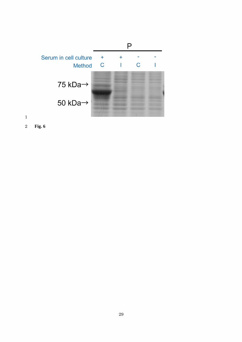
serum-containing media when we used the improved method. Coomassie brilliant blue staining 10
revealed that the P-G(+/+) resulted in remarkable intensity of the protein bands, whose 11
molecular weight was 50–75 kDa, whereas the other approaches did not (Fig. 3). This protein 12
may be albumin, which is a 66 kDa protein and a major component of serum (Peters 1977). 13
Thus, albumin may interfere with p65 detection by blocking the reaction between the antibody 14
and p65 as an antigen. 15
Next, we compared the conventional method with the improved method in the detection 16
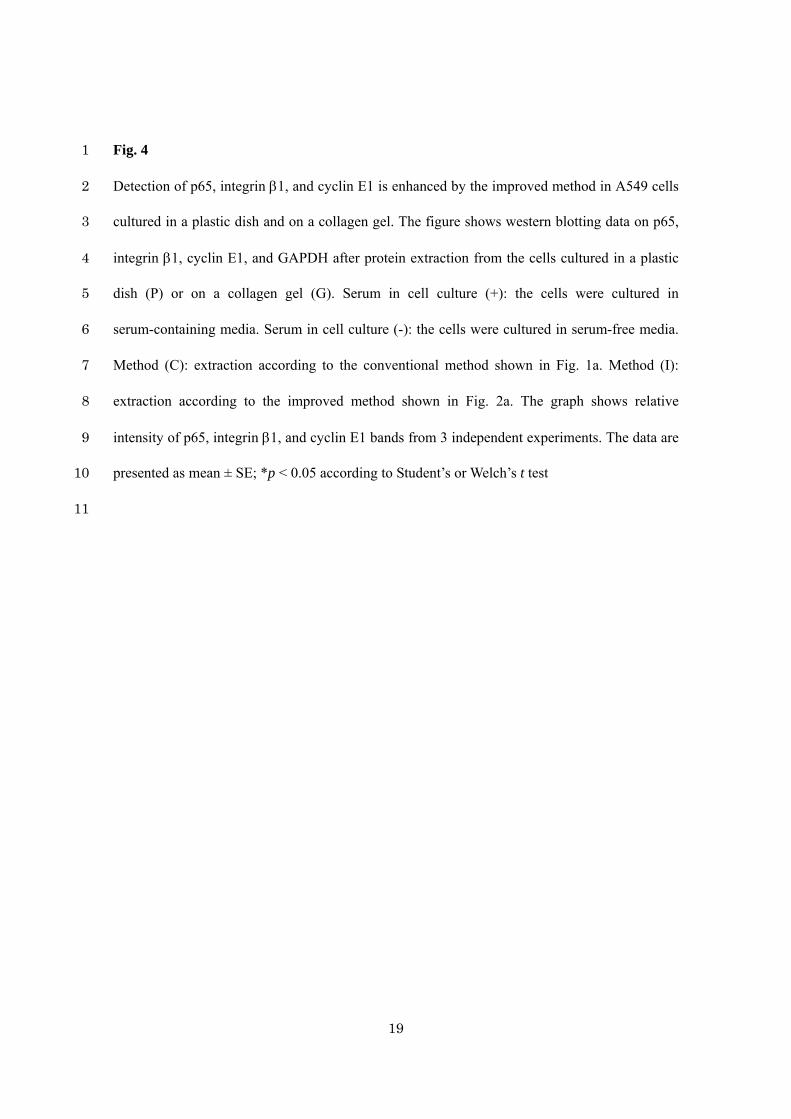
of proteins in A549 cells to assess the possible advantages of the improved method. In both 17
types of samples (plastic dish and collagen gel), the improved method enhanced the detection of 18
p65 compared to the conventional method when cells were cultured in the presence of serum 19
(Fig. 4). Under serum-free conditions, the improved method enhanced the detection of p65 in 20
the samples from cells grown on a collagen gel (Fig. 4). Furthermore, detection of integrin 1 21
and cyclin E1 was also bettered by the improved method in the samples from cells grown on a 22
collagen gel, except for integrin 1 in the cells grown under serum-free conditions (Fig. 4). 23
These results demonstrated that the improved method enhances protein detection in A549 cells 24
10
grown in a plastic dish in the presence of serum or on a collagen gel either with or without 1
serum. 2
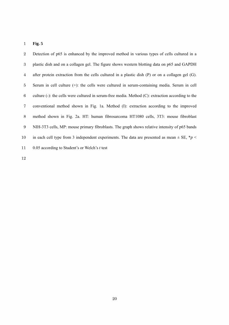
For further confirmation of the advantage of the improved method, we attempted to detect 3
p65 and GAPDH in different batches of A549 cells using the conventional or the improved 4
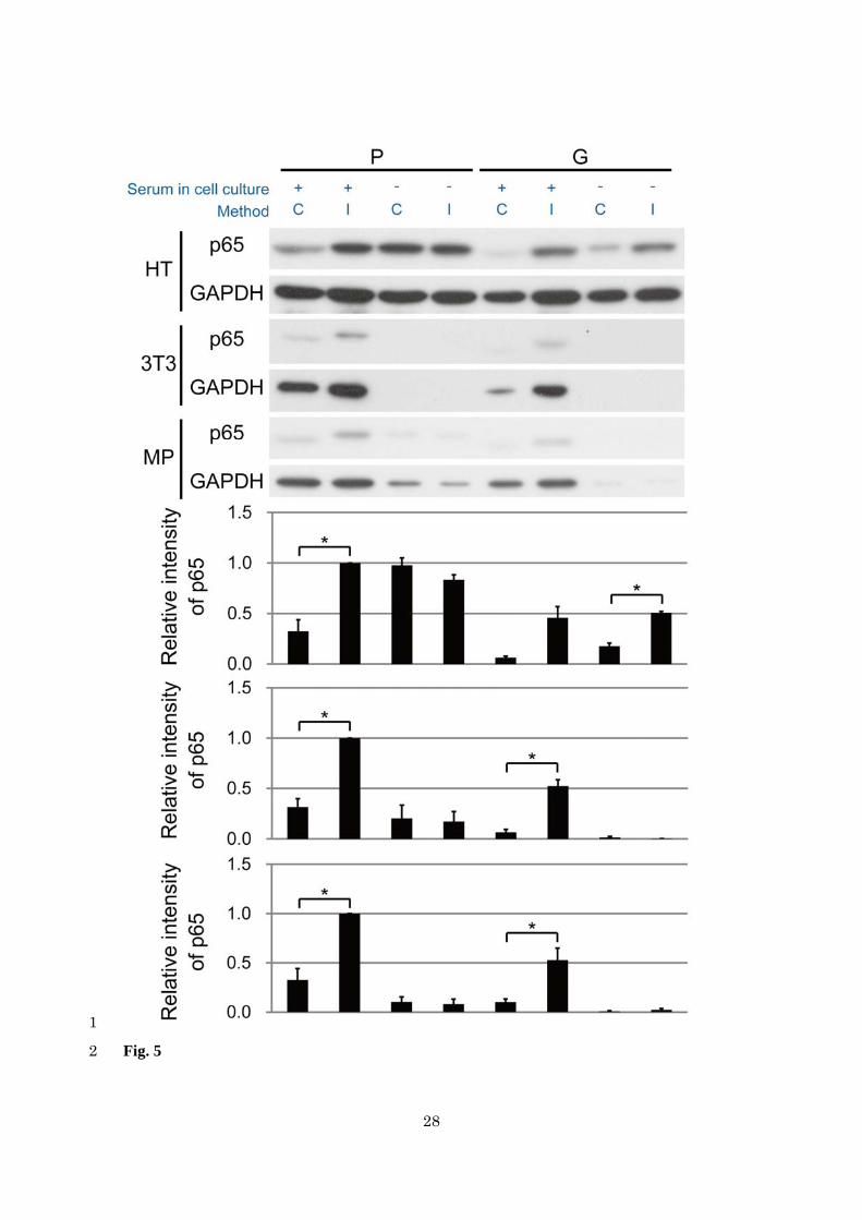
method. We prepared cell extracts from HT1080 human fibrosarcoma cells, NIH-3T3 mouse 5
fibroblast cells, and mouse primary fibroblasts. Just like in A549 cells, p65 detection was 6
enhanced by the improved method in those other cell lines when they were cultured in a plastic 7
dish and on a collagen gel in the presence of serum (Fig. 5). Under serum-free conditions, 8
because NIH-3T3 cells and mouse primary fibroblasts could barely adhere to a plastic dish and 9
a collagen gel, we could not detect distinctive bands of p65 and GAPDH. Overall, in several 10
types of cells, the improved method resulted in more visible bands of p65 compared to the 11
conventional method. 12
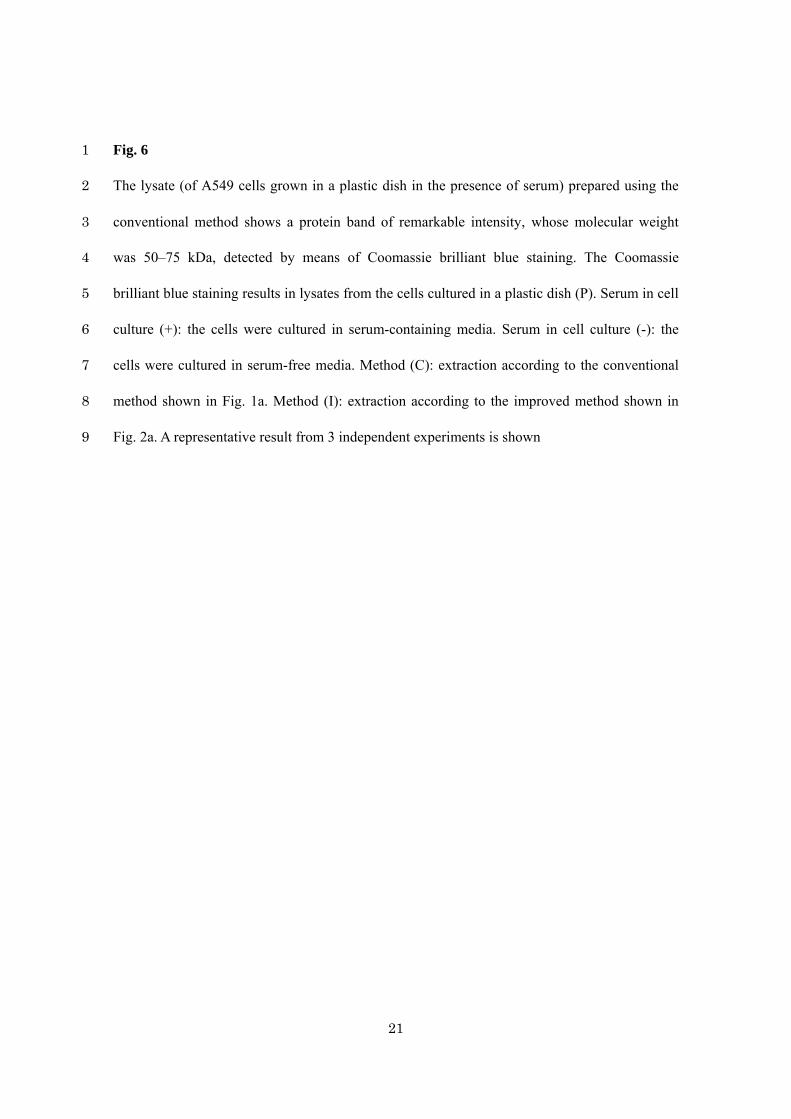
Our results suggest that the detection of p65 is impeded by a collagen gel and serum. Thus, 13
the p65 bands—from the A549 and HT1080 cells grown on a collagen gel under serum-free 14
conditions—were more visible after the improved method, which can exclude a collagen gel 15
from lysates (Fig. 4 and 5). Therefore, a collagen gel impedes the detection of p65 in western 16
blotting. In addition, we prepared lysates of A549 and HT1080 cells grown in a plastic dish in 17
the presence of serum using the conventional method (which cannot exclude serum proteins 18
from lysates; Fig. 6). This approach resulted in weaker bands of p65 compared to lysates of the 19
cells grown in a plastic dish under serum-free conditions (Fig. 4 and 5). These results suggest 20
that serum hampers p65 detection. Thus, removal of collagen gel and serum is important for 21
sensitive detection of p65 proteins (without losses). 22
In contrast to p65 detection, GAPDH detection was almost the same for the lysates 23
prepared according to the conventional and the improved method, in each culture condition. 24
11
This may be because the collagen gel and serum affect protein detection depending on the 1
properties of a protein under study, such as molecular weight. Indeed, molecular weight of 2
GAPDH (approximately 37 kDa) is substantially less than that of p65 (approximately 65 kDa). 3
Thus, detection of a protein may be affected by a collagen gel and serum, depending on the 4
protein’s molecular weight. 5
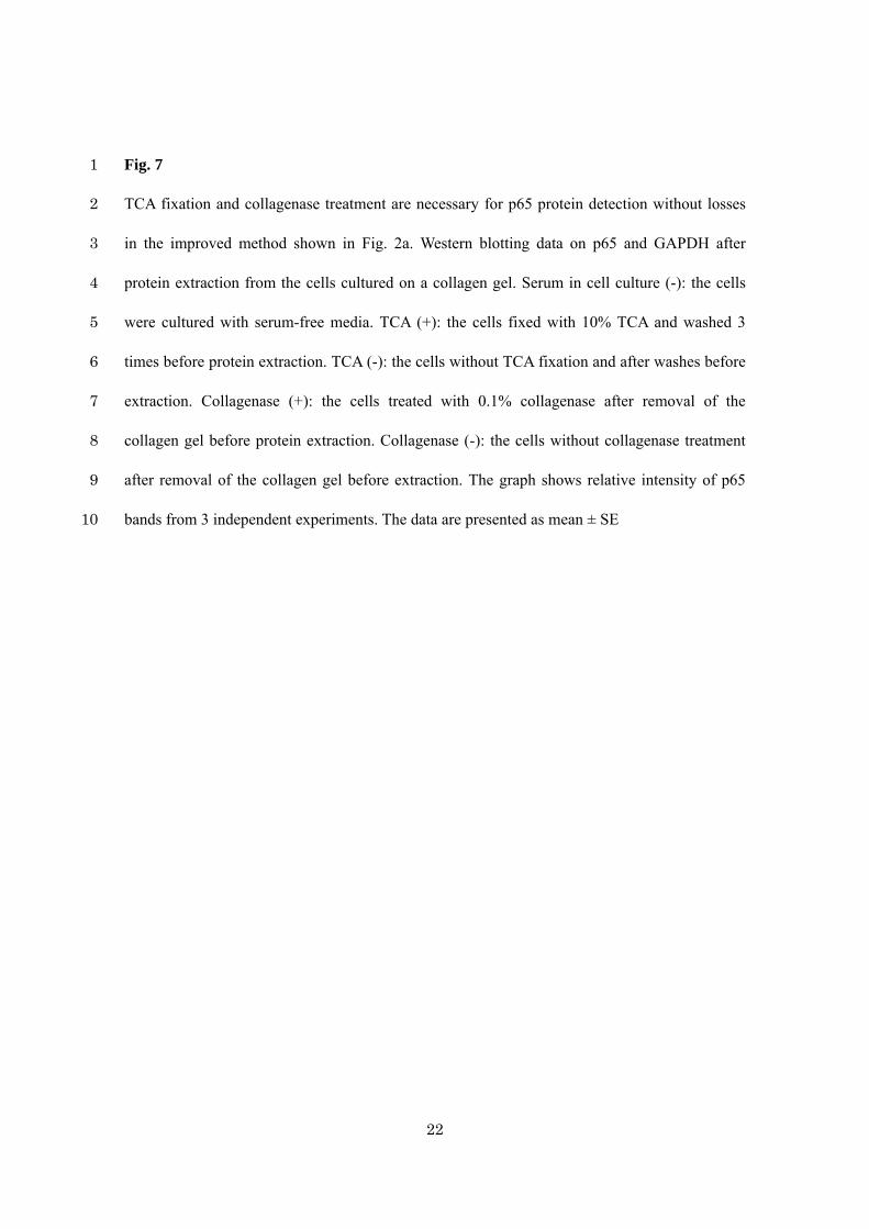
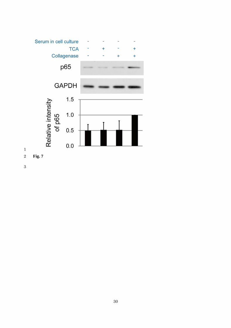
Next, we examined the necessity of TCA fixation and collagenase treatment in the 6
improved method for the lossless protein detection in the cells cultured on a collagen gel under 7
serum-free conditions. We compared the following samples: a sample treated with collagenase 8
after TCA fixation to a sample without TCA fixation and collagenase treatment, a sample treated 9
only with TCA, and a sample treated only with collagenase. The former showed stronger 10
intensity of the p65 bands, although the differences were not significant (Fig. 7). Thus, both 11
TCA fixation and collagenase treatment were necessary for the protein detection without losses 12
in the improved method. 13
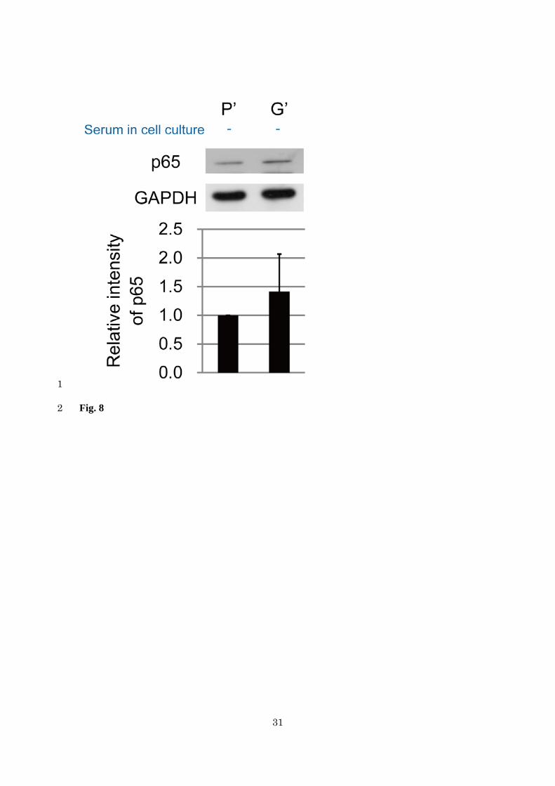
Finally, using the improved method, we compared the expression of the p65 protein in 14
A549 cells cultured with serum-free media on a plastic dish with that on a collagen gel. The 15
expression level of p65 protein was almost the same (Fig. 8). This result suggested that the 16
difference in substrate stiffness does not affect the expression of the p65 protein in A549 cells in 17
serum-free culture. 18
We showed here that the method including TCA fixation and collagenase treatment 19
improved protein detection. In previous studies, conventional cell lysis methods, which do not 20
involve TCA fixation and collagenase treatment, were often used for western blotting (Engler et 21
al. 2006; Tsutsumi et al. 2009; Wu et al. 2009). In these studies, the cells were cultured on 22
polyacrylamide gels, plastic plates, or dishes. Under these culture conditions, conventional 23
methods may not be particularly problematic because there is almost no effect of collagen gels 24

12
on protein detection. In contrast, in our experiments, including cell culture on a collagen gel, the 1
improved method is essential for sensitive detection of specific proteins such as p65, integrin 1, 2
and cyclin E1 without losses caused by collagens. In western blotting analysis, our improved 3
method is a powerful way to detect proteins in cells cultured with a collagen gel. 4
5

13
Acknowledgements 1
This study was supported by Grants-in-Aid for Scientific Research (B) (24390285), Scientific 2
Research (C) (26430104) and Scientific Research in Innovative Areas (25127701) to H.H., 3
Young Scientists (B) (23770167) and Scientific Research in Innovative Areas (24106502) to 4
T.M., Scientific Research (B) (25287106) to K.K. from the Japanese Ministry of Education, 5
Culture, Sports, Science, and Technology, Young Scientists (B) (10719933) to S.I. The authors 6
thank Sawa Kobayashi for technical assistance.7

14
References 1
Bond MD, Van Wart HE (1984) Purification and separation of individual collagenases of 2
Clostridium histolyticum using red dye ligand chromatography. Biochemistry 23:3077-3085. 3
Cafruny EJ (1957) Studies on tissue fixation: the penetration of trichloracetic acid solutions into 4
rat tissues. J Histochem Cytochem 5:414-419. 5
Engler AJ, Sen S, Sweeney HL, Discher DE (2006) Matrix elasticity directs stem cell lineage 6
specification. Cell 126:677-689. 7
Haga H, Irahara C, Kobayashi R, Nakagaki T, Kawabata K (2005) Collective movement of 8
epithelial cells on a collagen gel substrate. Biophys J 88:2250-2256. 9
Ishihara S, Yasuda M, Harada I, Mizutani T, Kawabata K, Haga H (2013) Substrate stiffness 10
regulates temporary NF-kappaB activation via actomyosin contractions. Exp Cell Res 11
319:2916-2927. 12
Karin M, Cao Y, Greten FR, Li ZW (2002) NF-kappaB in cancer: from innocent bystander to 13
major culprit. Nat Rev Cancer 2:301-310. 14
Laemmli UK (1970) Cleavage of structural proteins during the assembly of the head of 15
bacteriophage T4. Nature 227:680-685. 16
Levental KR, Yu H, Kass L, Lakins JN, Egeblad M, Erler JT, Fong SF, Csiszar K et al (2009) 17
Matrix crosslinking forces tumor progression by enhancing integrin signaling. Cell 18
139:891-906. 19
Li Q, Verma IM (2002) NF-kappaB regulation in the immune system. Nat Rev Immunol 20
2:725-734. 21
Musial A, Tony Eissa N (2001) Inducible nitric-oxide synthase is regulated by the proteasome 22
degradation pathway. J Biol Chem 276:24268-24273. 23
Paszek MJ, Zahir N, Johnson KR, Lakins JN, Rozenberg GI, Gefen A, Reinhart-King CA, 24
15
Margulies SS et al (2005) Tensional homeostasis and the malignant phenotype. Cancer Cell 1
8:241-254. 2
Peters T (1977) Serum albumin: recent progress in the understanding of its structure and 3
biosynthesis. Clin Chem 23:5-12. 4
Towbin H, Staehelin T, Gordon J (1979) Electrophoretic transfer of proteins from 5
polyacrylamide gels to nitrocellulose sheets: procedure and some applications. Proc Natl Acad 6
Sci U S A 76:4350-4354. 7
Tsutsumi K, Tsuda M, Yazawa N, Nakamura H, Ishihara S, Haga H, Yasuda M, Yamazaki R, 8
Shirato H, Kawaguchi H, Nishioka T, Ohba Y (2009) Increased motility and invasiveness in 9
tumor cells that survive 10 Gy irradiation. Cell Struct. Funct. 34:89-96. 10
Wang YK, Wang YH, Wang CZ, Sung JM, Chiu WT, Lin SH, Chang YH, Tang MJ (2003) 11
Rigidity of collagen fibrils controls collagen gel-induced down-regulation of focal adhesion 12
complex proteins mediated by alpha2beta1 integrin. J Biol Chem 278:21886-21892. 13
Wu X, Guo R, Chen P, Wang Q, Cunningham PN (2009) TNF induces caspase-dependent 14
inflammation in renal endothelial cells through a Rho- and myosin light chain 15
kinase-dependent mechanism. Am J Physiol Renal Physiol 297:F316-F326. 16
17
16
Figure Captions 1
Fig. 1 2
In the conventional method, a collagen gel interferes with detection of the p65 protein in human 3
lung adenocarcinoma A549 cells cultured on a serum-containing collagen gel. (a) A 4
conventional procedure of preparing the protein extracts from the cells cultured on a plastic dish 5
or on a collagen gel. The cells were cultured with serum-containing media. P: protein extraction 6
from the cells cultured on a plastic dish. G: extraction from the cells cultured on a collagen gel. 7
P-G: extraction from the cells cultured on a plastic dish: a collagen gel was added to the 8
extraction to assess the artificial deterioration of the protein detection because of the collagen 9
gel. In the P-G method, the collagen gel was preincubated with a serum-containing medium to 10
evaluate the effect of serum in the gel on the protein detection. (b) Western blotting results for 11
p65 and GAPDH in the samples prepared using the conventional procedure shown in (a). Serum 12
in cell culture (+): the cells were cultured with a serum-containing medium. Serum in added gel 13
(+): a P-G sample means that a collagen gel was added along with preincubation with a 14
serum-containing medium. The graph shows relative intensity of p65 bands from 3 independent 15
experiments. The data are presented as mean ± SE; *p < 0.05 according to Welch’s t test16
17
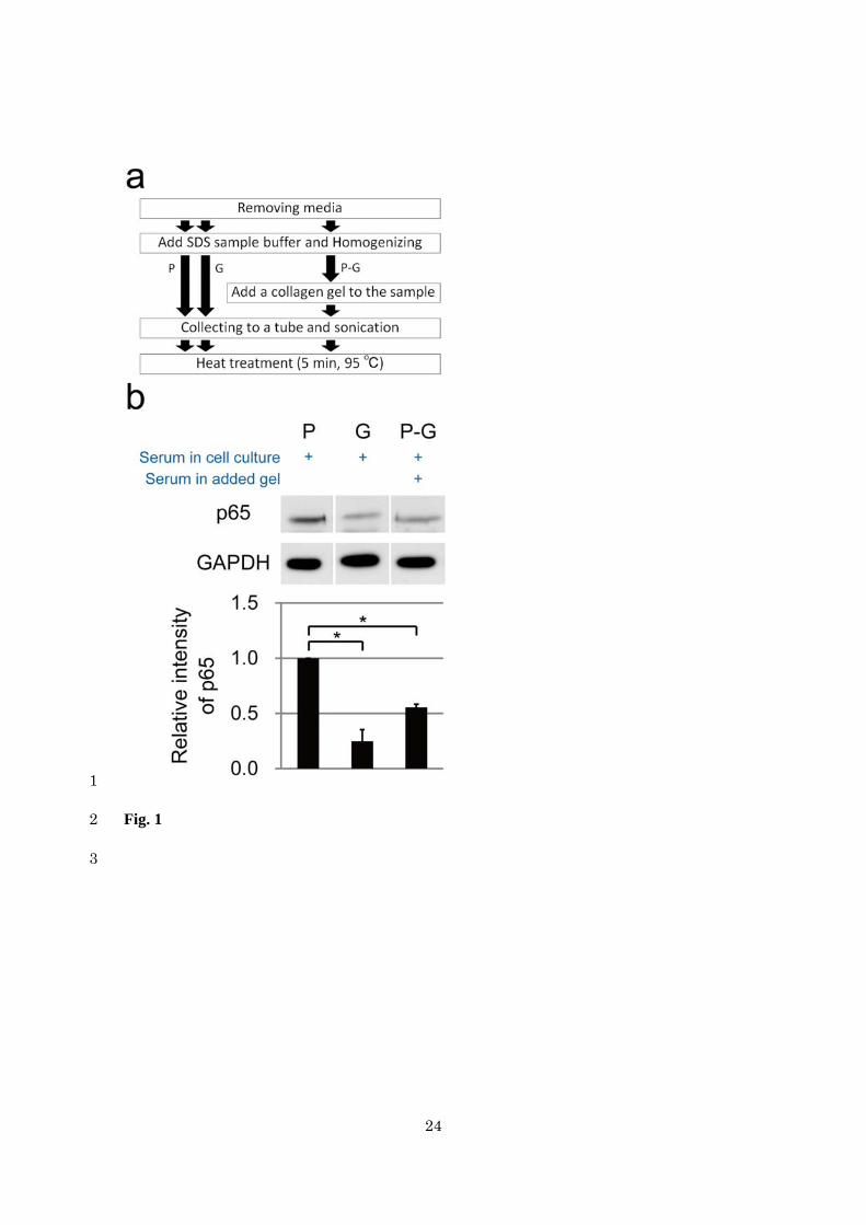
Fig. 2 1
An improved method allows us to detect the p65 protein without losses during protein 2
extraction from lung adenocarcinoma A549 cells cultured on a collagen gel under serum-free 3
conditions. (a) An improved procedure of preparing the extracts from the cells cultured on a 4
plastic dish or on a collagen gel. The added steps in the improved procedure are highlighted in 5
red. P: protein extraction from the cells cultured on a plastic dish. G: extraction from the cells 6
cultured on a collagen gel. P-G: extraction from the cells cultured on a plastic dish, where a 7
collagen gel was added to the samples before collagenase treatment. (b) GAPDH and p65 data 8
from western blotting after the P and P-G methods where the samples were prepared using the 9
improved procedure shown in (a). Serum in cell culture (+): the cells were cultured with 10
serum-containing media. Serum in added gel (+): P-G means that a collagen gel was added 11
before collagenase treatment, and the collagen gel was preincubated with a serum-containing 12
medium. Serum in added gel (-): P-G means that a collagen gel was added before collagenase 13
treatment, and the collagen gel was preincubated with a serum-free medium. The graph shows 14
the relative intensity of p65 bands from 3 independent experiments. The data are presented as 15
mean ± SE; *p < 0.05 according to Welch’s t test 16
17
18
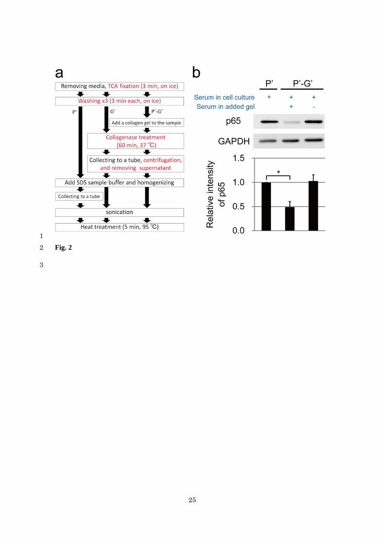
Fig. 3 1
The P-G sample (where we added a collagen gel preincubated with a serum-containing 2
medium) shows a protein band of remarkable intensity, whose molecular weight was 50–75 kDa, 3
detected by means of Coomassie brilliant blue staining. The Coomassie brilliant blue staining 4
results in the P and P-G samples prepared using the improved procedure shown in Fig. 2a. P: 5
protein extraction from the cells cultured on a plastic dish. P-G: extraction from the cells 6
cultured on a plastic dish, where a collagen gel was added to the samples before collagenase 7
treatment. Serum in cell culture (+): the cells were cultured with serum-containing media. 8
Serum in added gel (+): the P-G sample where we added a collagen gel before collagenase 9
treatment, and the collagen gel was preincubated with a serum-containing medium. Serum in the 10
added gel (-): the P-G sample where we added a collagen gel before collagenase treatment, and 11
the collagen gel was preincubated with a serum-free medium. A representative result from 3 12
independent experiments is shown13
19
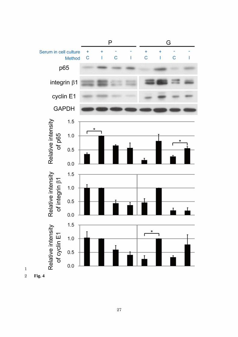
Fig. 4 1
Detection of p65, integrin 1, and cyclin E1 is enhanced by the improved method in A549 cells 2
cultured in a plastic dish and on a collagen gel. The figure shows western blotting data on p65, 3
integrin 1, cyclin E1, and GAPDH after protein extraction from the cells cultured in a plastic 4
dish (P) or on a collagen gel (G). Serum in cell culture (+): the cells were cultured in 5
serum-containing media. Serum in cell culture (-): the cells were cultured in serum-free media. 6
Method (C): extraction according to the conventional method shown in Fig. 1a. Method (I): 7
extraction according to the improved method shown in Fig. 2a. The graph shows relative 8
intensity of p65, integrin 1, and cyclin E1 bands from 3 independent experiments. The data are 9
presented as mean ± SE; *p < 0.05 according to Student’s or Welch’s t test 10
11
20
Fig. 5 1
Detection of p65 is enhanced by the improved method in various types of cells cultured in a 2
plastic dish and on a collagen gel. The figure shows western blotting data on p65 and GAPDH 3
after protein extraction from the cells cultured in a plastic dish (P) or on a collagen gel (G). 4
Serum in cell culture (+): the cells were cultured in serum-containing media. Serum in cell 5
culture (-): the cells were cultured in serum-free media. Method (C): extraction according to the 6
conventional method shown in Fig. 1a. Method (I): extraction according to the improved 7
method shown in Fig. 2a. HT: human fibrosarcoma HT1080 cells, 3T3: mouse fibroblast 8
NIH-3T3 cells, MP: mouse primary fibroblasts. The graph shows relative intensity of p65 bands 9
in each cell type from 3 independent experiments. The data are presented as mean ± SE, *p < 10
0.05 according to Student’s or Welch’s t test 11
12
21
Fig. 6 1
The lysate (of A549 cells grown in a plastic dish in the presence of serum) prepared using the 2
conventional method shows a protein band of remarkable intensity, whose molecular weight 3
was 50–75 kDa, detected by means of Coomassie brilliant blue staining. The Coomassie 4
brilliant blue staining results in lysates from the cells cultured in a plastic dish (P). Serum in cell 5
culture (+): the cells were cultured in serum-containing media. Serum in cell culture (-): the 6
cells were cultured in serum-free media. Method (C): extraction according to the conventional 7
method shown in Fig. 1a. Method (I): extraction according to the improved method shown in 8
Fig. 2a. A representative result from 3 independent experiments is shown9
22
Fig. 7 1
TCA fixation and collagenase treatment are necessary for p65 protein detection without losses 2
in the improved method shown in Fig. 2a. Western blotting data on p65 and GAPDH after 3
protein extraction from the cells cultured on a collagen gel. Serum in cell culture (-): the cells 4
were cultured with serum-free media. TCA (+): the cells fixed with 10% TCA and washed 3 5
times before protein extraction. TCA (-): the cells without TCA fixation and after washes before 6
extraction. Collagenase (+): the cells treated with 0.1% collagenase after removal of the 7
collagen gel before protein extraction. Collagenase (-): the cells without collagenase treatment 8
after removal of the collagen gel before extraction. The graph shows relative intensity of p65 9
bands from 3 independent experiments. The data are presented as mean ± SE10
23
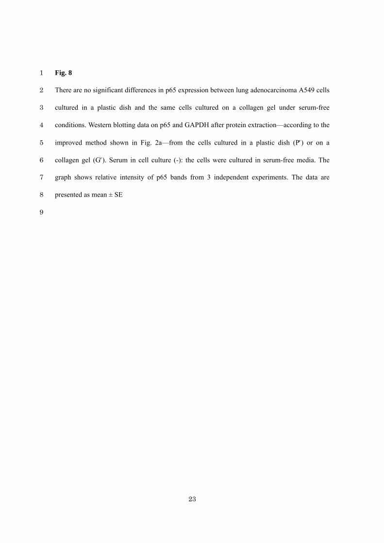
Fig. 8 1
There are no significant differences in p65 expression between lung adenocarcinoma A549 cells 2
cultured in a plastic dish and the same cells cultured on a collagen gel under serum-free 3
conditions. Western blotting data on p65 and GAPDH after protein extraction—according to the 4
improved method shown in Fig. 2a—from the cells cultured in a plastic dish (P) or on a 5
collagen gel (G). Serum in cell culture (-): the cells were cultured in serum-free media. The 6
graph shows relative intensity of p65 bands from 3 independent experiments. The data are 7
presented as mean ± SE 8
9
24
1
Fig. 1 2
3
25
1
Fig. 2 2
3
26
1
Fig. 3 2
3
27
1
Fig. 42
28
1
Fig. 52
29
1
Fig. 62
30
1
Fig. 7 2
3
31
1
Fig. 8 2