an expression signature as an aid to the histologic...
TRANSCRIPT

Personalized Medicine and Imaging
An Expression Signature as an Aid to theHistologic Classification of Non–Small CellLung CancerLuc Girard1,2,8, Jaime Rodriguez-Canales3, Carmen Behrens4, Debrah M. Thompson5,Ihab W. Botros5, Hao Tang6, Yang Xie6,8, Natasha Rekhtman7,William D. Travis7,Ignacio I.Wistuba3,4, John D. Minna1,2,8,9, and Adi F. Gazdar1,8,10
Abstract
Purpose: Most non–small cell lung cancers (NSCLC) are nowdiagnosed from small specimens, and classification using stan-dard pathology methods can be difficult. This is of clinicalrelevance as many therapy regimens and clinical trials are histol-ogy dependent. The purpose of this study was to develop anmRNA expression signature as an adjunct test for routine histo-pathologic classification of NSCLCs.
Experimental Design: A microarray dataset of resected adeno-carcinomas (ADC) and squamous cell carcinomas (SCC) wasused as the learning set for an ADC-SCC signature. The CancerGenome Atlas (TCGA) lung RNAseq dataset was used for valida-tion. Another microarray dataset of ADCs and matched nonma-lignant lung was used as the learning set for a tumor versusnonmalignant signature. The classifiers were selected as the mostdifferentially expressed genes and sample classification was deter-mined by a nearest distance approach.
Results: We developed a 62-gene expression signature thatcontained many genes used in immunostains for NSCLC typing.It includes 42 genes that distinguish ADC from SCC and 20 genesdifferentiating nonmalignant lung from lung cancer. Testing ofthe TCGA and other public datasets resulted in high predictionaccuracies (93%–95%). In addition, a prediction score wasderived that correlates both with histologic grading and progno-sis. We developed a practical version of the Classifier using theHTG EdgeSeq nuclease protection–based technology in combi-nation with next-generation sequencing that can be applied toformalin-fixed paraffin-embedded (FFPE) tissues and smallbiopsies.
Conclusions:Our RNA classifier provides an objective, quan-titative method to aid in the pathologic diagnosis of lungcancer. Clin Cancer Res; 22(19); 4880–9. �2016 AACR.
IntroductionMost cancers of the lung are carcinomas, and they may be
divided into two broad categories, small-cell carcinoma (SCLC,
10%–20%) and non–small cell carcinoma (NSCLC, 80%–90%),differing in their biology, clinical presentation, and therapy (1).While there are rare types of NSCLCs, the vast majority fall intothree categories: adenocarcinomas (ADC), squamous cell carci-nomas (SCC), and large-cell carcinomas (LCC). ADCs are carci-nomas that form glands, papillary structures, grow in a lepidicpattern, or secrete mucin. As the lung is a complex organ withcentral and peripheral compartments having different histologiesand functions (2), there are multiple subtypes of ADCs (3). SCCsare believed to arise frommetaplastic cells in the large airways, asthere are no squamous cells in the normal respiratory tract. LCCsare undifferentiated NSCLCs that do not show morphologic orimmunostaining evidence of glandular or squamous differenti-ation. Recent studies confirmed that most or all LCCs lackingneuroendocrine features could be assigned to other NSCLC types,with only a small number of true null phenotype cases remaining(4, 5).
Originally, the clinical management of the major forms ofNSCLC was similar, and the main clinical task required of thepathologist was the separation of NSCLC from SCLC (6). How-ever, during the past decade, the therapy ofNSCLChas undergonea paradigm shift, as we are rapidly moving from an era of empiri-cal therapy to one of personalized therapy ("precisionmedicine")based on mutational patterns and tumor classification (7). Ofinterest, the known oncogenic "driver" mutations for ADC andSCC are almost completely different (8) and the selection of both
1Hamon Center for Therapeutic Oncology Research, University ofTexas Southwestern Medical Center, Dallas, Texas. 2Department ofPharmacology, University of Texas Southwestern Medical Center, Dal-las, Texas. 3Department of Translational Molecular Pathology, Univer-sity of Texas MD Anderson Cancer Center, Houston, Texas. 4Depart-ment of Thoracic/Head and Neck Medical Oncology, University ofTexas MD Anderson Cancer Center, Houston, Texas. 5HTG MolecularDiagnostics, Tucson, Arizona. 6Department of Clinical Science, Uni-versity of Texas Southwestern Medical Center, Dallas, Texas. 7Depart-ment of Thoracic Pathology, Memorial Sloan Kettering Cancer Center,New York, New York. 8Simmons Cancer Center, University of TexasSouthwestern Medical Center, Dallas, Texas. 9Department of InternalMedicine, University of Texas Southwestern Medical Center, Dallas,Texas. 10Department of Pathology, University of Texas SouthwesternMedical Center, Dallas, Texas.
Note: Supplementary data for this article are available at Clinical CancerResearch Online (http://clincancerres.aacrjournals.org/).
Corresponding Author: Adi F. Gazdar, Hamon Center for Therapeutic OncologyResearch, University of Texas Southwestern Medical Center at Dallas, 6000Harry Hines Blvd., Dallas, TX 75390-8593. Phone: 214-648-4921; Fax: 214-648-4940; E-mail: [email protected]
doi: 10.1158/1078-0432.CCR-15-2900
�2016 American Association for Cancer Research.
ClinicalCancerResearch
Clin Cancer Res; 22(19) October 1, 20164880
on June 27, 2018. © 2016 American Association for Cancer Research. clincancerres.aacrjournals.org Downloaded from
Published OnlineFirst June 28, 2016; DOI: 10.1158/1078-0432.CCR-15-2900

conventional chemotherapy and targeted therapy may be influ-enced by theNSCLC subtype (6, 9).Whilemost targeted therapiesfor NSCLC are directed at ADCs or nonsquamous histologies, theimportance of the correct diagnosis of squamous tumors bypathologic or molecular methods is gaining recognition (10).Hence, the precise histologic classification of NSCLC is of crucialclinical importance.
Other major developments in lung cancer management are theability to obtain CT-guided needle and small core biopsies (usu-ally allowing a pathologic diagnosis to be made irrespective ofanatomic location) and imaging studies that have greatlyincreased the accuracy of preoperative staging. These advanceshave considerably reduced the number of futile lung cancerresections and staging that relied on postoperative pathologicresults. However, one practical disadvantage is that most initiallung cancer diagnoses (�70%) now have to be made from smallbiopsies or cytologic specimens (6). While the pathologic classi-fication of NSCLC is relatively straightforward if there is anadequate tumor specimen and the tumor is well or moderatelydifferentiated, the diagnosis of poorly differentiated tumors,particularly from small specimens,may be challenging. Followingrecommendations by the latest version of theWHOClassification(1) pathologists now use combinations of immunostains, andvarious algorithms have been proposed to assist in the accurateclassification of NSCLC (3, 9, 11, 12). Despite these improve-ments, not all poorly differentiated NSCLCs in small biopsy orcytology samples can be classified into ADCs and SCCs and theyare usually referred to as NSCLC-not otherwise specified (NSCLC-NOS; refs. 6, 9). As a side note, this term should be reserved to thelung cancer specimens of small size and should not be applied tosurgical resections or large biopsies; instead, resected tumors thatcannot be classified as specific forms of NSCLC should be clas-sified as LCC (13).
To improve the classification of lung cancer and reduce potentialobserver bias and variability, we developed and validated anmRNA
expression–based classification of NSCLC utilizing large datasetsof resected NSCLC tumors with expert pathology review. We havefurther developed a practical version of the Classifier based onthe HTG EdgeSeq technology that, among other advantages, canbe applied reproducibly to FFPE and core-needle biopsies.
Materials and MethodsPatient tumor samples
Three cohorts of lung cancer or nonmalignant lung specimenswere used to derive and test theRNAclassifiers: a set of 275NSCLCspecimens obtained from the Pathology Core at MD AndersonCancer Center (MDACC, Houston, TX), consisting of 183 ADC,80 SCC, and 12 tumors of other subtypes; a set of 83 pairs of lungADCs and matched nonmalignant lung tissues obtained fromBritish Columbia Cancer Research Centre (Vancouver, BritishColumbia, Canada) in collaboration with Early DetectionResearch Network (EDRN) and the Canary Foundation; and aset of 979 NSCLCs (490 ADC, 489 SCC) and nonmalignant lungtissues (n ¼ 108) from The Cancer Genome Atlas (TCGA).Reference pathologists for the tumor specimen diagnoses wereI.I. Wistuba and J. Rodriguez-Canales for the MDACC set, A.F.Gazdar for the EDRN/Canary set, and the TCGA Lung CancerPathology Panel (W.D. Travis andN. Rekhtman) for the TCGA set.For most of the tumors analyzed in this study, immunostainingwas not utilized for pathologic classification. Histologic typing ofthe TCGA set was performed by light microscopy according to theprevious WHO Classification (14).
Expression profiling datasetsMDACC set. Frozen tissues from NSCLC tumors resected atMDACC were used to generate multiple 5-mm thick sections.Representative tissue sections were hematoxylin and eosin (H&E)stained and reviewed to estimate the percentage of tumor andnonmalignant cells. About 5 to 10 sections were processed toextract RNA, whose quality was assessed on Nano Series II RNALAB-chips using Agilent Bioanalyzer 2100 (Agilent Technologies,Inc.). Cases were selected with the following defined character-istics: tumor (vs. nonmalignant) � 70%, malignant cells (vs.stromal cells) � 30%, RNA Integrity Number (RIN) � 4 (range0–10). RNA samples were shipped to University of Texas South-westernMedical Center (UT Southwestern; Dallas, TX) for expres-sion profiling. Five-hundred nanograms of RNAwere labeled andhybridized to the Illumina BeadChip array HumanWG-6 V3.Array data were preprocessed using the R package mbcb (15) forbackground correction. The arrays were then log-transformed andquantile-normalized. This dataset was submitted to Gene Expres-sion Omnibus (GEO) under the accession number GSE41271.
EDRN/Canary set. RNA was prepared with TRIzol (Invitrogen) aspreviously described from ADCs resected in British Columbia,Canada and collected by Dr. Stephen Lam (British Columbia Can-cer Agency, Vancouver, BC, Canada) (16, 17). Profiling was doneon Illumina HumanWG-6 V3 BeadChips at UT Southwestern(Dallas, TX) andprocessed similarly to theMDACC set. This datasetwas submitted to GEO under the accession number GSE75037.
TCGA set. RNAseq data were downloaded from the TCGA portal(18). The archive filenameswere unc.edu_LUAD.IlluminaHiSeq_R-NASeqV2.1.13.0 for ADC and unc.edu_LUSC.IlluminaHiSeq_RNA-SeqV2.1.10.0 for SCC. The extracted files consisted of 548 and 539
Translational Relevance
Personalized therapy and entry into clinical trials for non–small lung cancer (NSCLC) are heavily dependent on accuratehistologic classification. While most cases can be classifiedusing routine pathology review of hematoxylin and eosin(H&E) and immunostains, examination of the U.S. cancerregistries indicates that many cases of NSCLC remain unclas-sified. To address this important clinical problem, we devel-oped and validated a quantitative gene expression signaturefor the highly accurate classification of NSCLC. As the signa-ture score reflects differentiation, it also provides prognosticinformation for early-stage resected NSCLC, and thus mayhelp identify patients who would benefit from adjuvant che-motherapy. In addition, we developed a next-generationsequencing (NGS) laboratory assay utilizing HTG MolecularDiagnostic's HTG EdgeSeq chemistry and demonstrated thatits performance is similar to the original microarray–basedclassifier. Importantly, the NGS classifier can be applied repro-ducibly to clinically challenging sample types, such as forma-lin-fixed paraffin-embedded (FFPE)materials and core-needlebiopsies.
Molecular Classification of NSCLC
www.aacrjournals.org Clin Cancer Res; 22(19) October 1, 2016 4881
on June 27, 2018. © 2016 American Association for Cancer Research. clincancerres.aacrjournals.org Downloaded from
Published OnlineFirst June 28, 2016; DOI: 10.1158/1078-0432.CCR-15-2900

samples, respectively. Using the barcode key provided (19), wefound that the first set had 490 ADCs and 58 nonmalignant lungsamples and the second set had 489 SCCs and 50 nonmalignantsamples.
FFPE material. FFPE specimens from resected NSCLC (n ¼ 35),nonmalignant lung tissue (n ¼ 11), or core-needle biopsies (n ¼36) were collected at MDACC (Houston, TX). They were then cutin a microtome at 5-mm thick sections, mounted on glass slidesusing nuclease-free conditions, and sent to HTG labs for EdgeSeqprofiling (see below).
Statistical analysisA Sweave report documenting all statistical steps (written in
R code) pertaining to this article is available from the authorson request. In brief, classifiers were generated from the trainingset's two classes by generating a volcano plot and selecting the nmost significantly overexpressed genes in the two classes, wheren is optimized by 5-fold stratified cross validation with 100iterations (which resulted in n ¼ 21 for the ADC-SCC classifier,and n ¼ 10 for the tumor–nonmalignant classifier, see Supple-mentary Fig. S1). We thus obtained a 2n-gene classifier (42genes for ADC-SCC classification, 20 genes for tumor–nonma-lignant classification). Classification was determined by a near-est distance approach that compares each sample's expressionvalues for the classifier genes to the mean expression values inthe training set's two classes (called class centroids). Pearsoncorrelation was used as a similarity measure. The relativemagnitude of this measure determined the class prediction,that is, if the Pearson correlation was greater with the ADCcentroid than with the SCC centroid, then the sample waspredicted to be ADC, and vice versa. A correlation plot wasgenerated from each sample's correlation pair (correlation withADC, correlation with SCC). A score for each sample wascalculated as the signed distance from its plotted location tothe diagonal where the correlations are equal. After normali-zation, this score ranged from �1 to þ1. A positive scoreindicates ADC prediction while a negative score indicates SCCprediction. The magnitude of these scores can be viewed as anestimate of the prediction's confidence.
HTG EdgeSeq assayThe assay couples quantitative nuclease protection (qNPA) with
next-generation sequencing (NGS) to measure gene expression insmall FFPEor frozen sampleswithout RNA extraction (20). Amoredetailed description of the assay is available in the SupplementaryMaterial. Briefly, the FFPE specimens from MDACC were scrapedinto tubes and lysed in HTG's lysis buffer, followed by the intro-duction of gene-specific DNA nuclease protection probes (NPP).After allowing the NPPs to hybridize to their target RNAs, whichcan be both soluble or cross-linked in the biological matrix, S1nuclease is added which removes excess unhybridized NPPs andRNAs, leaving behind only NPPs hybridized to their target RNAs.Thus, a stoichiometric conversion of the target RNA to the NPPsis achieved, producing a virtual 1:1 ratio of NPP to RNA. The qNPAsteps are automated on the HTG EdgeSeq processor, which isfollowed by PCR to add sequencing adaptors and tags. The labeledsamples are pooled, cleaned, and sequenced on an NGS platformusing standard protocols. Data from the NGS instrument areprocessed and reported by the HTG EdgeSeq parser software.Supplementary Fig. S6 shows an example of the assay results for
25 FFPE samples used in this study. Good dynamic range andreproducibility were obtained, which indicates that the assay isboth sensitive and specific.
ResultsSEER-based classification of NSCLC
We examined the Surveillance, Epidemiology and End Results(SEER) database for lung cancer classifications which covers theyears 2008–2012 (21) and identified 227,000 lung cancer cases,of which 83.4% were classified as NSCLC, 13.3% as SCLC, and3.1% were unclassified (Carcinoma-NOS). Of the NSCLCs,51.9% were ADCs, 27.1% were SCCs, 2.5% were LCCs, 5.9%were other forms of NSCLC, and 12.6% were not classified(NSCLC-NOS). From these figures, a total of 13.6%of lung cancercases, which after sampling adjustment represent about 22,000patients yearly, were not histologically classified. These patientstherefore were not eligible for histology type–based therapies.
ADC-SCC signature: training on the MDACC setTo build a classifier distinguishing ADC and SCC, we used the
MDACCdataset which containsmRNAprofiles for 183 ADCs and80 SCCs from surgically resected frozen specimens. Forty-twogenes differentially expressed between the two subtypes wereselected from a volcano plot (Fig. 1A and B; Supplementary TableS1). These genes were highly significant with expression folddifferences > 2.6 and t test P value < 10�18 (FDR < 10�16). Someof theproteins encodedby the significant genes are alreadyused asimmunostains in clinical diagnostic procedures and include highmolecular weight keratins (KRT), NKX2-1 (TITF1), TP63, andDSG3 (desmoglein 3).
We defined the Classifier using this training set as follows: wefirst calculated thePearson correlations between each sample's 42-gene signature expression values and the mean expression valuesof the same 42 genes in the ADC group and in the SCC group (theclass centroids). A "correlation plot" was generated (Fig. 1C)where each point represents the pair of correlation values asso-ciated with each sample. On the straight line y ¼ x, the twocorrelations are equal. Below this line, the correlation with theADC group is higher than with the SCC group, and vice versa. Foreach point we defined a score as
(Correl ADC – Correl SCC)/2 (range: �1 to þ1)
This score is proportional to the distance from each point to thedividing line (arrows in Fig. 1C; also plotted vertically in Fig. 1D).We interpret positive scores as predicting ADC histology whilenegative scores predict SCC histology. The two dotted lines arecutoff scores set at
� SD [abs(all scores)]
where SD is the standard deviation and abs is the absolutevalue. This cutoff is equal to� 0.17 in the MDACC set and can beviewed as a prediction threshold: values above 0.17 are predictedto be ADC while values below �0.17 are predicted to be SCC.Intermediate values are predicted as poorly differentiated (seebelow). Using these definitions, 170 of 183 ADCs (93%; originalhistologic review) had positive scores and were thus correctlyclassified, while 72 of 80 SCCs (90%) had negative score andwerecorrected classified. Overall, the accuracy within this training setwas 92% (Table 1).
Girard et al.
Clin Cancer Res; 22(19) October 1, 2016 Clinical Cancer Research4882
on June 27, 2018. © 2016 American Association for Cancer Research. clincancerres.aacrjournals.org Downloaded from
Published OnlineFirst June 28, 2016; DOI: 10.1158/1078-0432.CCR-15-2900

The discrepancies and borderline classified cases (with scoreslower than the specified cutoffs) were re-evaluated by the MDAnderson pathologists using immunostains when appropriate:these pathologists on re-review felt that about half of thediscrepant diagnoses should be changed, with an improvedaccuracy of 95%. However, we stress that no change was done
to the Classifier itself, which was based on the original histo-logic diagnosis.
ADC-SCC signature: testing on the TCGA setAs a validation set, we used the TCGA-lung RNAseq data. Scores
were calculated as before and values larger than 0.17 predicted
−6 −4 −2 6420
010
2030
40
Log2 Ratio (ADC vs. SCC)
− Lo
g10
(T T
est P
val
ue)
NKX2−1SOX2*
TP63KRT5
KRT6A
DSG3
NAPSA*
KRT13
KRT16
KRT17
SCC Genes ADC Genes
* Not part of the42-gene signature
−1.0 −0.5 0.5 1.0
−1.0
−0.5
0.5
1.0
Correl ADC
Cor
rel S
CC
ADCSCCOther NSCLC
Diagnoses
Neg Score (SCC)
Pos Score (ADC)
−0.8
−0.6
−0.4
−0.2
0.0
0.2
0.4
0.6
0.8A
DC
−SC
C S
core
s
Classification
ADC
NSCLC−PD Favor ADC
NSCLC−PD Favor SCC
SCC
ADCSCCOther NSCLC
Diagnoses
A B
C D
Low expression High expression
DAPL1
DSTPNCKCOL7A1SOX15GOLT1A
ACSL5
TESCRORCFMO5SMPDL3B
HNF1B
KRT5KRT6ATP63CALML3PKP1KRT6BKRT17CLCA2DSG3VSNL1SERPINB5KRT16KRT13KRT6C
DSC3FAM83B
DPP4CAPN8ALDH3B1SFTA2
ABCC6NKX2−1STK32AHPNKCNK5CDH15
CLDN3PRR15LLGSNSPINK1
ADC SCC
Figure 1.
A, a volcano plot shows that many genes are significantly different between ADC and SCC. Among these are several genes for immunostains typicallyused by pathologists, including high molecular weight keratins (KRTs), TP63, DSG3, and TITF1 (NKX2-1). Color coding is related to the distance of each point to theplot's origin and shows significance levels (red: highly significant). B, heatmap of ADC-SCC signature in the MDACC dataset. Red, high mRNA expression;green, low mRNA expression. Twenty-one genes overexpressed in ADC and twenty-one genes overexpressed in SCC were selected for this signature. Blue labels,genes known to be relevant to lung cancer pathogenesis. Arrows, commonly used immunostains; C, "Correlation plot" where each point represents thePearson correlation values between the 42-gene signature expression of individual samples and the mean expression of ADCs (x-axis) and SCCs (y-axis). Thescores shown as arrows are defined as (Correl ADC – Correl SCC)/2. They are proportional to the distances from each point to the line y ¼ x. Dotted linesrepresent cut-off score values below which the samples are thought to be less well-differentiated. Color coding in this and similar plots represent pathologicdiagnoses. D, "Score plot" where the y-axis represents the scores calculated from C. PD, poorly differentiated. Dotted lines are the score cutoffs.
Molecular Classification of NSCLC
www.aacrjournals.org Clin Cancer Res; 22(19) October 1, 2016 4883
on June 27, 2018. © 2016 American Association for Cancer Research. clincancerres.aacrjournals.org Downloaded from
Published OnlineFirst June 28, 2016; DOI: 10.1158/1078-0432.CCR-15-2900

ADC while values lower than �0.17 predicted SCC (Fig. 2).Intermediate values predicted poorly differentiated tumors favor-ing ADC (positive low scores) or SCC (negative low scores; seebelow). Including these lower scores, we thus obtained 97%correct prediction for the ADC set of samples (475 of 490), and93% correct prediction for the SCC set of samples (456 of 489;overall: 95%; Table 1). Interestingly, the nonmalignant lungtissues largely fell into the same classification group as the ADCs.
On the basis of these class prediction results, selected caseswerere-examined by pathologists from the TCGA studies. Many of theSCCs that were scored as ADCs turned out on re-examination tobe called NSCLC-NOS or other subtypes such as undifferentiatedLCCs (Fig. 2). Using these revised diagnoses, the predictionaccuracy for the ADC group was unchanged at 97%, while the
accuracy for the SCC group increased from 93% to 96% (overallaccuracy: 97%, Table 1).
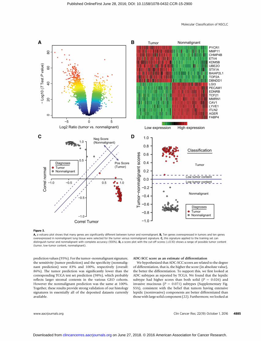
Tumor–nonmalignant signatureAs observed above, most of the nonmalignant lung samples in
the TCGA set were classified as "ADC". We thus generated anadditional signature to distinguish tumor from nonmalignantlung. As a training set,we chose theEDRN/Canary set consisting of83 pairs of ADC specimens and matched nonmalignant lungcontrols. Using the same method as for the ADC-SCC signature,we generated a volcano plot (Fig. 3A) and ranked differentiallyexpressed genes between tumor and nonmalignant using theirdistance to the plot's origin. Twenty genes were selected as mostdifferentially expressed (10 overexpressed in the tumor group, 10overexpressed in the nonmalignant group; Fig. 3B; Supplemen-tary Table 1). A correlation plot (Fig. 3C) shows that the twogroups are clearly separated in the training set (100% correctclassification; Table 1), and a score ranging from �1 (nonmalig-nant) to þ1 (tumor) can be computed (Fig. 3C and D).
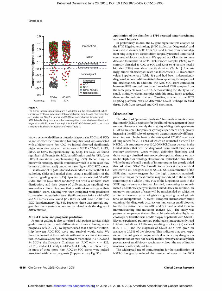
As a validation set for the tumor–nonmalignant signature, weagain used the TCGA dataset. Fig. 4 shows the score values with100% prediction accuracy for the nonmalignant group and 98%prediction accuracy for the tumor group (overall: 98%; Table 1).This shows that a 20-gene signature is sufficient to differentiateNSCLCs from nonmalignant lung with high accuracy.
The ADC-SCC and tumor–nonmalignant prediction scores canbe combined in a 2D plot that clearly segregates the three groups(Supplementary Fig. S2).
Validation in public datasetsTo further validate the ADC-SCC and tumor–nonmalignant
signatures we looked at several public mRNA expression datasetscontaining sufficiently large numbers of NSCLC samples (n > 20each) and deposited in GEO. The selected 22 datasets contained1,560 ADCs, 732 SCCs, and 340 nonmalignant lung tissues.
Both signatures gave highly accurate predictions (Supplemen-tary Table S2). For the ADC-SCC signature, the average sensitivity(ADC prediction) and specificity (SCC prediction) were 95% and89%, respectively (overall: 93%), similar to the TCGA test set
Table 1. Prediction accuracies of ADC-SCC and tumor–nonmalignant signatures
Signature type Dataset Expression platform Training or testing Sensitivity Specificity Accuracy
ADC-SCC SPORE/MDACCa Illumina WG-6 V3 Training 170/183 (93%)c 72/80 (90%)d 242/263 (92%)ADC-SCC SPORE/MDACCb Illumina WG-6 V3 Training 170/178 (96%) 78/83 (94%) 248/261 (95%)ADC-SCC TCGAa RNAseq Testing 475/490 (97%) 456/489 (93%) 931/979 (95%)ADC-SCC TCGAb RNAseq Testing 423/437 (97%) 437/453 (96%) 860/890 (97%)ADC-SCC EDRN/Canary Illumina WG-6 V3 Testing 82/83 (99%) NA 82/83 (99%)
Tumor–nonmalignant EDRN/Canary Illumina WG-6 V3 Training 83/83 (100%)e 83/83 (100%)f 166/166 (100%)Tumor–nonmalignant TCGA RNAseq Testing 959/979 (98%) 108/108 (100%) 1067/1087 (98%)Tumor–nonmalignant SPORE/MDACC Illumina WG-6 V3 Testing 252/275 (92%) NA 252/275 (92%)
ADC-SCC FFPE Resected HTG EdgeSeq Testing 16/17 (94%) 18/18 (100%) 34/35 (97%)ADC-SCC FFPE CNB HTG EdgeSeq Testing 15/19 (79%) 17/17 (100%) 32/36 (89%)Tumor–nonmalignant FFPE Resected HTG EdgeSeq Testing 33/35 (94%) 11/11 (100%) 44/46 (96%)Tumor–nonmalignant FFPE CNB HTG EdgeSeq Testing 35/36 (97%) NA 35/36 (97%)
Abbreviation: CNB, core-needle biopsy.aOriginal histopathologic diagnosis.bRevised histopathologic diagnosis.cPredicted ADC (score > 0)/Diagnostic ADC.dPredicted SCC (score < 0)/Diagnostic SCC.ePredicted tumor (score > 0)/Diagnostic tumorfPredicted nonmalignant (score < 0)/Diagnostic nonmalignant
−0.8
−0.6
−0.4
−0.2
0.0
0.2
0.4
0.6
0.8
AD
C−S
CC
Sco
res
TCGA Beforerevision
TCGA Afterrevision
EDRN/Canary
Accuracy: 95% 97% 99%
Prediction
ADC
NSCLC−PD Favor ADC
NSCLC−PD Favor SCC
SCC
ADCSCCOther NSCLCNonmalignant
Diagnoses
Figure 2.
The ADC-SCC signature is validated on the TCGA and EDRN/Canary datasetsand shown as score plots as in Fig. 1D. The prediction accuracies for TCGA(before revision) were 97% (ADC) and 93% (SCC). Overall accuracy: 95% (Table1). These accuracieswere 97%and 96%, respectively (overall: 97%) after revisionof diagnosis of selected TCGA cases which included many NSCLC-NOS (blue).The prediction accuracy for the EDRN/Canary dataset which consists of ADCsonly was 99%.
Girard et al.
Clin Cancer Res; 22(19) October 1, 2016 Clinical Cancer Research4884
on June 27, 2018. © 2016 American Association for Cancer Research. clincancerres.aacrjournals.org Downloaded from
Published OnlineFirst June 28, 2016; DOI: 10.1158/1078-0432.CCR-15-2900
prediction values (95%). For the tumor–nonmalignant signature,the sensitivity (tumor prediction) and the specificity (nonmalig-nant prediction) were 83% and 100%, respectively (overall:86%). The tumor prediction was significantly lower than thecorresponding TCGA test set prediction (98%), which probablyreflects larger stromal contents in the various GEO cohorts.However the nonmalignant prediction was the same at 100%.Together, these results provide strong validation of our histologysignatures in essentially all of the deposited datasets currentlyavailable.
ADC-SCC score as an estimate of differentiationWehypothesized that ADC-SCC scores are related to the degree
of differentiation, that is, the higher the score (in absolute value),the better the differentiation. To support this, we first looked atADC subtypes as reported by TCGA. We found that the lepidicsubtype had higher scores than both solid (P ¼ 0.026) andinvasive mucinous (P ¼ 0.071) subtypes (Supplementary Fig.S3A), consistent with the belief that tumors having extensivelepidic (noninvasive) components are better differentiated thanthosewith large solid component (22). Furthermore,we looked at
−5 50
020
4060
80
Log2 Ratio (tumor vs. nonmalignant)
− Lo
g10
(T T
est P
val
ue)
Low expression High expression
FABP4AGERITLN2LYVE1CAV1MMRN1TCF21EDNRBPECAM1LGI3DBNDD1TOP2ABAIAP2L1STX1AUBE2OKDM5BETV4CHMP4BMMP11PYCR1
Tumor Nonmalignant
−1.0 −0.5 0.5 1.0
−1.0
−0.5
0.5
1.0
Correl Tumor
Cor
rel n
orm
al TumorNonmalignant
Diagnoses
Neg Score(Nonmalignant)
Pos Score(Tumor)
−1.0
−0.8
−0.6
−0.4
−0.2
0.0
0.2
0.4
0.6
0.8
1.0Tu
mor
−non
mal
igna
nt s
core
s Classification
Tumor
Low tumor contentLow tumor content
Nonmalignant
TumorNonmalignant
Diagnoses
A B
C D
Figure 3.
A, a volcano plot shows that many genes are significantly different between tumor and nonmalignant. B, Ten genes overexpressed in tumors and ten genesoverexpressed in nonmalignant lung tissue were selected for the tumor versus nonmalignant signature. C, this signature applied to the training set candistinguish tumor and nonmalignant with complete accuracy (100%). D, a score plot with the cut-off scores (�0.10) shows a range of possible tumor content(tumor, low-tumor content, nonmalignant).
Molecular Classification of NSCLC
www.aacrjournals.org Clin Cancer Res; 22(19) October 1, 2016 4885
on June 27, 2018. © 2016 American Association for Cancer Research. clincancerres.aacrjournals.org Downloaded from
Published OnlineFirst June 28, 2016; DOI: 10.1158/1078-0432.CCR-15-2900
known genes with differentmutational spectra in ADCs and SCCsto see whether their mutation (or amplification) was associatedwith a higher score. For ADC, we indeed observed significantlyhigher scores for cases with mutations in EGFR, CTNNB1, HER2,BRAF, or KRAS (Supplementary Fig. S3B). For SCC, we foundsignificant differences for SOX2 amplification and for NFE2L2 orPIK3CA mutations (Supplementary Fig. S3C). Hence, lung tu-mors with histology-specificmutations (which in some casesmaybe more differentiated) tended to have higher ADC-SCC scores.
Finally, one of us (AFG) looked at a random subset of the TCGApathology slides and graded them using a modification of thestandard grading system (23). Specifically, we selected 50 ADCslides and 50 SCC slides randomly but with a uniform scoredistribution, and their degree of differentiation (grading) wasassessed in a blinded fashion, that is, without knowledge of theirprediction score. Grading was then compared with predictionscores using two statistical tests. Significant associationswithADCand SCC scores were found (P < 0.03 for ADC and P < 10�4 forSCC; Supplementary Fig. S4). Together, these data strongly sug-gest that the signature scores are correlated with the degree ofdifferentiation.
ADC-SCC score and prognosis predictionAs tumor grading is also correlated with patient survival (high
grade tumors, i.e. poorly differentiated tumors, having worseprognosis; refs. 23, 24), we hypothesized that a similar relation-ship between ADC-SCC score and survival would exist. Wetherefore looked at three cohorts with available clinical informa-tion: theMDACC set (non-neoadjuvant cases only: 145ADCs and64 SCCs), the Director's Challenge set (ADC only; n ¼ 423;ref. 25), and a SCC study (GSE4573; SCC only; n ¼ 106; ref. 26).In most of these cases, high ADC or SCC scores were indeedassociated with better prognosis (Supplementary Fig. S5).
Application of the classifier to FFPE-resected tumor specimensand small biopsies
In preliminary studies, the 62-gene signature was adapted tothe HTG EdgeSeq technology (HTG Molecular Diagnostics) andwas used to classify ADC from SCC and tumor from nonmalig-nant lung using FFPE sections from surgically resected tumors andcore-needle biopsy specimens. We applied our Classifier to thesedata and found that 34 of 35 FFPE-resected samples (97%) werecorrectly classified as ADC or SCC and 32 of 36 FFPE core-needlebiopsies (89%) were also correctly classified (Table 1). Interest-ingly, almost all discrepant cases had low scores (< 0.1 in absolutevalue; Supplementary Table S3) and had been independentlydiagnosed as poorly differentiated, thus explaining themajority ofthe discrepancies. In addition, the ADC-SCC score correlationbetween FFPE resected tumors and matched CNB samples fromthe same patients was r ¼ 0.98, demonstrating the ability to usesmall, clinically relevant samples with this assay. Taken together,these results indicate that our Classifier, adapted to the HTGEdgeSeq platform, can also determine NSCLC subtype in fixedtissue, both from resected and CNB specimens.
DiscussionThe advent of "precision medicine" has made accurate classi-
fication ofNSCLC a necessity for the clinicalmanagement of thesetumors. However, currently the majority of diagnostic specimens(�70%) are small biopsies or cytologic specimens (27), greatlyincreasing the difficulty of accurately diagnosing poorly differen-tiated tumors. On the basis of the anticipated 225,000 new casesof lung cancer for 2016 (28), of which an estimated 85% will beNSCLC, this amounts to over 130,000NSCLC cases per year in theUnited States that will be diagnosed from small biopsies orcytology specimens. Cases without definitive diagnoses, andthose wrongly classified, may not receive optimal therapy or maynot be eligible for histology classification–restricted clinical trials.While the use of small panels of immunostains has greatly aidedthis task, about 5%–10% of small biopsy cases at major medicalcenters will still be signed out asNSCLC-NOS. Examination of theSEER data registry suggests that the high diagnostic standardspresent at major medical centers may not extend to the medicalcommunity as a whole. Thus, 14% of the lung cancer cases in theSEER registry were not further classified, amounting to an esti-mated 22,000 cases per year in the United States. In addition, anunknown percentage of cases will be misclassified or subject toarbitrary diagnosis by pathologists using varying pathologic cri-teria or interpretation. A recent European interobserver studyexamined the diagnostic accuracy on lung cancer small biopsiesfor the distinction between ADC and SCC and related these toimmunostaining and mutation analysis (29). The study wasperformed on prospectively collected biopsies obtained by bron-choscopy or transthoracic needle biopsy of patients with NSCLC.Eleven experienced pulmonary pathologists independently readH&E-stained slides of 110 cases, resulting in a kappa (k) value of0.55 � 0.10 and the diagnosis of NSCLC-NOS was given onaverage to 29.5% of the biopsies. This indicates that even expe-rienced pathologists at major medical centers may disagree oninterpretation or may not be able to fully classify a relatively highpercentage of small biopsy specimens without the use of immu-nostains or other adjunct tests.
The widespread use of immunostains for the classification ofNSCLC has greatly reduced the number of cases in the NOS
−0.9
−0.7
−0.5
−0.3
−0.1
0.1
0.3
0.5
0.7
0.9Tu
mor
−non
mal
igna
nt s
core
s
TCGA MDACCAccuracy: 98% 92%
Prediction
Tumor
Low tumor contentLow tumor content
Nonmalignant
ADCSCCOther NSCLCNonmalignant
Diagnoses
Figure 4.
The tumor–nonmalignant signature is validated on the TCGA dataset, whichconsists of 979 lung tumors and 108 nonmalignant lung tissues. The predictionaccuracies are 98% for tumors and 100% for nonmalignant lung (overall:98%, Table 1). Many tumor samples have negative scores which could be due tolarger stromal infiltration. A score plot for the MDACC dataset, which has tumorsamples only, shows an accuracy of 92% (Table 1).
Girard et al.
Clin Cancer Res; 22(19) October 1, 2016 Clinical Cancer Research4886
on June 27, 2018. © 2016 American Association for Cancer Research. clincancerres.aacrjournals.org Downloaded from
Published OnlineFirst June 28, 2016; DOI: 10.1158/1078-0432.CCR-15-2900

category (30) andmost of these tumors can nowbe classifiedwitha single SCC and a single ADC marker (1). These findings led thenew WHO Classification to recommend using immunostainingfor SCCmarkers such as TP63 or its isoform p40 (deltaNp63) andhigh molecular weight keratins as well as ADC markers such asNKX2-1 (TTF-1) and Napsin A to classify poorly differentiatedlung cancers including NSCLC-NOS (1, 31). However, interpre-tation of immunostains is not uniform and alternativeapproaches to lung cancer classification are being explored asadjunct tools to aid the pathologic diagnosis of lung cancers.These methods include digital nuclear imaging, mutation anal-ysis, copy number variations, and various other molecular meth-ods, either singly or in combination (32–34).
In this report, we developed and validated a gene expressionclassifier from a training set consisting of 263 surgically resectedtumors to accurately and nonsubjectively separate ADC fromSCC. The list of top differentially expressed genes heavily favoredSCC, possibly reflecting the greater pathologic heterogeneity andmolecular complexity of ADCs and their multiple subtypes (3,11). Thus, we selected an equal number of top genes significantlyoverexpressed in ADCs (n ¼ 21) and SCCs (n ¼ 21) so as not tobias the selection in favor of one NSCLC type. Not surprisingly,many of the selected genes are among the most frequently usedand reliable immunostains in routine pathologic practice (Fig. 1B,red arrows) or are known to play a role in lung cancer or in oneof the major subtypes (Fig. 1B, blue labels). We validated theClassifier using the TCGA lung cancer datasets, which were avail-able on a different platform (RNAseq) than our training set(Illumina BeadArray). We obtained very high prediction accura-cies (95%) in spite of the fact that a fraction of the TCGAdiagnostic materials were found to be of less than optimalquality (e.g. frozen sections instead of permanently fixed H&Eslides) and in spite of the partially subjective nature of path-ologic diagnosis (29). In fact, a significant limitation to theTCGA project was that the materials for immunostaining werenot always available. Nevertheless, N. Rekhtman and W.D.Travis, who are the TCGA reference pathologists, reviewed thediscrepancies, and this resulted in even better classificationaccuracy (Table 1, "Revised histopathologic diagnosis").
Interestingly, several nonmalignant lung TCGA specimenswereclassified as ADC by the signature, so we used the EDRN/Canarydataset to develop another classifier, containing 20 genes, thatseparated tumor cells from nonmalignant lung with high accu-racy. The combined 62-gene signature could now segregate ADC,SCC, and nonmalignant lung in this TCGA test set.
There are no squamous cells in the normal lung. Squamousmetaplasia arises as the result of noxious stimuli such as tobaccoexposure, mechanical trauma, inflammation, or infection. Manyof the SCC-associated classifier genes are involved in squamousdifferentiation, including basal (stem) cell proliferation, expres-sion of high molecular weight keratins, desmosome formation,calcium regulation or cornified envelope formation (35–37).ADCs demonstrate considerable heterogeneity of morphologicand biologic subtypes (31). However, most of the ADC genes hadrelevance to lung cancer or were known to be ADC-specific. BothNKX2-1 and NAPSA are routinely used in many pathology clas-sification schemes; however the latter, with a rank of 177, was notpart of the top 21 genes overexpressed in the ADC group, and wasnot used in the Classifier. Our data indicate that other genes in theClassifier, such as the trypsin inhibitor SPINK1 which is alreadyknown to be overexpressed in lung ADCs (38), may represent
good candidates for new immunostains in pathologic diagnosis,provided sensitive and specific antibodies are available. For SCCidentification, the Classifier selected several high molecularweight KRTs as well as TP63 among the top genes, but excludedSOX2, a gene frequently amplified in SCCs, although it was alsosignificantly overexpressed in SCC (rank ¼ 46; refs. 9, 39, 40).
The Classifier can also provide a score that reflects the degree ofdifferentiation. In support of this, we observed that NSCLCs withmutations that are specific for ADCs (EGFR, KRAS, and others) orSCCs (SOX2 amplification, NFE2L2 mutation) tended to havehighermagnitude scores than tumors thatwerewild-type for thesemutations or amplifications (Supplementary Fig. S3B and S3C).In addition, the lepidic subtype of ADCs which is believed to bemore differentiated also had a relatively higher score (Supple-mentary Fig. S3A). Finally, evaluation of tumor grade from TCGAhistopathology slides revealed a strong concordance betweenprediction score and histologic grading (high scorewas associatedwith better differentiation). Thus, our Classifier can be interpretedboth qualitatively and quantitatively.
Consistent with the association between histologic gradingand survival, our signature turned out to have prognostic valueas well (high scores being associated with better survival). Thus,this Classifier has the additional advantage of being of progno-stic importance andmay be useful in selecting the subpopulationof curative resected lung cancer patients that will benefit fromadjuvant therapy.
Previous ADC-SCC gene signatures have been reported (41–46) and about 10%–45% of the genes in these signatures overlapwith ours. Two of these signatures were formally developed asclassifiers, with external tumor set validation. The first, from Houand colleagues (44), comprises 50 unique genes (15 of whichoverlappedwith our signature) andwere validated in one externaldataset with a prediction accuracy of 84%. To directly comparethis classifier with our own, we tested it in TCGA RNAseq datausing the class centroids provided by the study and Pearsoncorrelation to predict the class. The resulting prediction had anaccuracy of 92% (sensitivity, 99%; specificity, 84%) while ourClassifier showed 95% accuracy (97% sensitivity, 93% specific-ity). The second study, from Wilkerson and colleagues (46), had15 genes (4 overlapping with our signature) and a reportedprediction accuracy of 81% in external validation. Using theTCGA validation test, this corresponded to an accuracy of 92%(sensitivity: 90%, specificity 95%). Our current study thus offersthe following advantages over prior ones: (i) a slightly betteroverall accuracy; (ii) a balance between sensitivity and specificity;(iii) the ability to distinguish nonmalignant from lung cancer; (iv)validation in a larger number of publicNSCLCexpressiondatasetswith high prediction accuracies (93% for the ADC-SCC classifi-cation); (v) the quantitative aspect of our Classifier and itscorrelation with differentiation and prognosis (this point alsosupports removing the term LCC and replacing it with poorlydifferentiated NSCLC); (vi) the ability, as mentioned below, toclassify small biopsy samples and FFPE materials, using technol-ogy that can be transferred to a CLIA-certified environment.
While some pathologists may question the necessity for amolecular classification of NSCLC, the large number of nonclas-sified cases, and the potential lack of diagnostic reproducibilityeven among experienced lung cancer pathologists, point to thevalue of a nonsubjective test. This may even be a necessity ininstitutions or countries where immunostains are not routinelyused and where staff pathologists may apply highly variable
Molecular Classification of NSCLC
www.aacrjournals.org Clin Cancer Res; 22(19) October 1, 2016 4887
on June 27, 2018. © 2016 American Association for Cancer Research. clincancerres.aacrjournals.org Downloaded from
Published OnlineFirst June 28, 2016; DOI: 10.1158/1078-0432.CCR-15-2900

diagnostic criteria. An especially relevant use of a molecularclassificationwouldbe for largemultinational clinical trialswhereno central pathology review is available. Wewill also need furtherevaluation in a set of cases that have been diagnosed utilizingestablished immunohistochemical methods recommended bythe 2015 WHO Classification. Unfortunately, these new criteriacould not be applied to the datasets evaluated in this study.
Finally, to demonstrate the potential clinical applicability ofour Classifier, we have developed an extraction-free, highly sen-sitive, automated, and cost-effective NGS version based on theHTG EdgeSeq technology and have shown that its accuracy issimilar to the original microarray-based Classifier (Table 1). Infact, the NGS classifier can be reproducibly applied to commonlyavailable clinical specimens, including FFPE materials and core-needle biopsies.
In summary, we have developed and validated a sensitive andspecific gene expression classifier for NSCLC that distinguishesADC from SCC, and lung cancer from normal lung. The Classifierwas shown tobe largely independent of themajor gene expressionplatforms in commonusage.Most of the genes in theClassifier arerelevant to lung cancer or are known to be differentially expressedin NSCLC. The development and further validation of a practicaland cost effective FFPE-based CLIA-certified version has thepotential to lead to a widespread clinical application of theClassifier.
Disclosure of Potential Conflicts of InterestD.M. Thompson and I.W. Botros have ownership interest (including
patents) in HTG Molecular Diagnostics. No potential conflicts of interestwere disclosed by the other authors.
Authors' ContributionsConception and design: L. Girard, D.M. Thompson, H. Tang, J.D. Minna,A.F. GazdarDevelopment of methodology: L. Girard, D.M. Thompson, I.W. Botros,I.I. Wistuba, J.D. Minna, A.F. GazdarAcquisition of data (provided animals, acquired and managed patients,provided facilities, etc.): L. Girard, J. Rodriguez-Canales, C. Behrens,I.W. Botros, W.D. Travis, I.I. Wistuba, A.F. GazdarAnalysis and interpretation of data (e.g., statistical analysis, biostatistics,computational analysis): L. Girard, D.M. Thompson, I.W. Botros, H. Tang,Y. Xie, W.D. Travis, I.I. Wistuba, J.D. Minna, A.F. GazdarWriting, review, and/or revisionof themanuscript: L.Girard,D.M. Thompson,I.W. Botros, H. Tang, Y. Xie, N. Rekhtman, W.D. Travis, I.I. Wistuba, J.D. Minna,A.F. GazdarAdministrative, technical, or material support (i.e., reporting or organizingdata, constructing databases): L. Girard, A.F. GazdarStudy supervision: J.D. Minna, A.F. Gazdar
AcknowledgmentsWewish to thankHTGMolecularDiagnostics Vice Presidents JohnWineman
and Patrick Roche for their support and contribution to this work.
Grant SupportThis work was generously supported by the NCI Specialized Program in
Research Excellence (SPORE) in Lung Cancer, P50CA70907, the LungevityFoundation, theNCI EarlyDetectionResearchNetwork (EDRN),U01CA086402,and theCanary Foundation. TheHTGEdgeSeqworkwas supportedbyNIHgrantR44HG005949.
The costs of publication of this articlewere defrayed inpart by the payment ofpage charges. This article must therefore be hereby marked advertisement inaccordance with 18 U.S.C. Section 1734 solely to indicate this fact.
Received December 1, 2015; revised June 1, 2016; accepted June 12, 2016;published OnlineFirst June 28, 2016.
References1. Travis W, Brambilla E, Burke A, Marx A, Nicholson A. WHO Classification
of Tumours of the Lung, Pleura, Thymus and Heart. 4th ed. Lyon, France:International Agency for Research on Cancer; 2015.
2. Sun S, Schiller JH, Gazdar AF. Lung cancer in never smokers–a differentdisease. Nat Rev Cancer 2007;7:778–90.
3. Travis W, Brambilla E, Noguchi M, Geisinger K, Beer D, Powell C, et al. Thenew IASLC/ATS/ERS internationalmultidisciplinary lung adenocarcinomaclassification. J Thorac Oncol 2009;4:244–85.
4. Rekhtman N, Tafe LJ, Chaft JE, Wang L, Arcila ME, Colanta A, et al.Distinct profile of driver mutations and clinical features in immuno-marker-defined subsets of pulmonary large-cell carcinoma. Mod Pathol2013;26:511–22.
5. TheClinical LungCancerGenomeProject (CLCGP) andNetworkGenomicMedicine (NGM). A genomics-based classification of human lung tumors.Sci Translat Med 2013;5:209ra153.
6. Gazdar AF. Should we continue to use the term non-small-cell lung cancer?Ann Oncol 2010;21 Suppl 7:vii225–vii9.
7. Gazdar AF, Minna JD. Precision medicine for cancer patients: lessonslearned and the path forward. J Natl Cancer Inst 2013;105:1262–3.
8. ShamesDS,Wistuba II. The evolving genomic classificationof lung cancer. JPathol 2014;232:121–33.
9. Cagle PT, Allen TC, Dacic S, Beasley MB, Borczuk AC, Chirieac LR, et al.Revolution in lung cancer: new challenges for the surgical pathologist. ArchPathol Lab Med 2011;135:110–6.
10. Oliver TG, Patel J, Akerley W. Squamous non-small cell lung cancer as adistinct clinical entity. J Clin Oncol 2015;38:220–6.
11. Kerr KM. Pathologist and molecular biologist, ever the twain shall meet?Lung Cancer 2009;63:161–3.
12. Rekhtman N, Ang DC, Sima CS, Travis WD, Moreira AL. Immunohisto-chemical algorithm for differentiation of lung adenocarcinoma and squa-mous cell carcinoma based on large series of whole-tissue sections withvalidation in small specimens. Mod Pathol 2011;24:1348–59.
13. Loo PS, Thomas SC, Nicolson MC, Fyfe MN, Kerr KM. Subtyping ofundifferentiated non-small cell carcinomas in bronchial biopsy specimens.J Thorac Oncol 2010;5:442–7.
14. Travis WD, Brambilla E, Muller-Hermelink HK, Harris CC. Pathology andgenetics of tumours of the lung, pleura, thymus, and heart. Lyon, France:International Agency for Research on Cancer; 2004.
15. Ding LH, Xie Y, Park S, Xiao G, Story MD. Enhanced identification andbiological validation of differential gene expression via Illumina whole-genome expression arrays through the use of themodel-based backgroundcorrection methodology. Nucleic Acids Res 2008;36:e58.
16. Selamat SA, Chung BS, Girard L, Zhang W, Zhang Y, Campan M, et al.Genome-scale analysis of DNA methylation in lung adenocarcinoma andintegration with mRNA expression. Genome Res 2012;22:1197–211.
17. Tam KW, Zhang W, Soh J, Stastny V, Chen M, Sun H, et al. CDKN2A/p16 in-activation mechanisms and their relationship to smoke exposure and molec-ular features in non-small-cell lung cancer. J Thorac Oncol 2013;8:1378–88.
18. The Cancer Genome Atlas. The Cancer Genome Atlas - Data Portal; 2016[cited 2016 April 12]. Available from: https://tcga-data.nci.nih.gov/tcga/tcgaHome2.jsp.
19. National Cancer Institute TCGA Barcode; 2016 [cited 2016 April 12].Available from: https://wiki.nci.nih.gov/display/TCGA/TCGAþbarcode.
20. HTG EdgeSeq Chemistry [cited 2016 April 12]. Available from: https://www.htgmolecular.com/science/htg-edgeseq-chemistry.
21. Surveillance Epidemiology and End Results. SEERCancer Statistics Review,1975–2012; 2016 [cited 2016 April 12]. Available from: http://seer.cancer.gov/csr/1975_2012/.
22. TravisWD.Classificationof lungcancer. SeminRoentgenol2011;46:178–86.23. Cancer Grading Manual. Editors: Damjanov I, Fan F. 2nd ed: Springer;
2013, 220 pages.24. National Cancer Institute. Tumor grade fact sheet;2016 [cited 2016 April
12]. Available from: http://www.cancer.gov/about-cancer/diagnosis-stag-ing/prognosis/tumor-grade-fact-sheet.
Girard et al.
Clin Cancer Res; 22(19) October 1, 2016 Clinical Cancer Research4888
on June 27, 2018. © 2016 American Association for Cancer Research. clincancerres.aacrjournals.org Downloaded from
Published OnlineFirst June 28, 2016; DOI: 10.1158/1078-0432.CCR-15-2900

25. Shedden K, Taylor JM, Enkemann SA, Tsao MS, Yeatman TJ, Gerald WL,et al. Gene expression-based survival prediction in lung adenocarcinoma: amulti-site, blinded validation study. Nat Med 2008;14:822–7.
26. Raponi M, Zhang Y, Yu J, Chen G, Lee G, Taylor JM, et al. Gene expressionsignatures for predicting prognosis of squamous cell and adenocarcinomasof the lung. Cancer Res 2006;66:7466–72.
27. Travis WD, Rekhtman N. Pathological diagnosis and classification of lungcancer in small biopsies and cytology: strategic management of tissue formolecular testing. Semin Respir Crit Care Med 2011;32:22–31.
28. Siegel RL, Miller KD, Jemal A. Cancer statistics, 2016. CA Cancer J Clin2016;66:7–30.
29. Thunnissen E, Noguchi M, Aisner S, Beasley MB, Brambilla E, Chirieac LR,et al. Reproducibility of histopathological diagnosis in poorly differenti-ated NSCLC: an international multiobserver study. J Thorac Oncol2014;9:1354–62.
30. Lewis DR, Check DP, Caporaso NE, Travis WD, Devesa SS. US lung cancertrends by histologic type. Cancer 2014;120:2883–92.
31. TravisWD, Brambilla E,NoguchiM,NicholsonAG,Geisinger KR, Yatabe Y,et al. International association for the study of lung cancer/americanthoracic society/european respiratory society international multidisciplin-ary classification of lung adenocarcinoma. J Thorac Oncol 2011;6:244–85.
32. Thunnissen EB,Diegenbach PC. Classification of lung carcinomabymeansof digital nuclear image analysis. Anal Quant Cytol Histol 1986;8:301–4.
33. Bishop JA, Benjamin H, Cholakh H, Chajut A, Clark DP, Westra WH.Accurate classification of non-small cell lung carcinoma using a novelmicroRNA-based approach. Clin Cancer Res 2010;16:610–9.
34. Ramani RG, Jacob SG. Improved classification of lung cancer tumors basedon structural andphysicochemical properties of proteins using dataminingmodels. PLoS One 2013;8:e58772.
35. Ratushny V,GoberMD,Hick R, Ridky TW, Seykora JT. Fromkeratinocyte tocancer: the pathogenesis and modeling of cutaneous squamous cell car-cinoma. J Clin Invest 2012;122:464–72.
36. Baroni A, Buommino E, De Gregorio V, Ruocco E, Ruocco V, Wolf R.Structure and function of the epidermis related to barrier properties. ClinDermatol 2012;30:257–62.
37. Fuchs E.Scratching the surface of skin development. Nature 2007;445:834–42.
38. Lazar V, SuoC,OrearC, vandenOord J, BaloghZ,Guegan J, et al. Integratedmolecular portrait of non-small cell lung cancers. BMC Med Genomics2013;6:53.
39. Travis WD, Brambilla E, Noguchi M, Nicholson AG, Geisinger K, Yatabe Y,et al. Diagnosis of lung cancer in small biopsies and cytology: implicationsof the 2011 International Association for the Study of Lung Cancer/American Thoracic Society/European Respiratory Society classification.Arch Pathol Lab Med 2013;137:668–84.
40. Travis WD, Brambilla E, Noguchi M, Nicholson AG, Geisinger K, Yatabe Y,et al. Diagnosis of lung adenocarcinoma in resected specimens: implica-tions of the 2011 International Association for the Study of Lung Cancer/American Thoracic Society/European Respiratory Society classification.Arch Pathol Lab Med 2013;137:685–705.
41. Hofmann HS, Bartling B, Simm A, Murray R, Aziz N, Hansen G, et al.Identification and classification of differentially expressed genes in non-small cell lung cancer by expression profiling on a global human 59.620-element oligonucleotide array. Oncol Rep 2006;16:587–95.
42. Rohrbeck A, Neukirchen J, Rosskopf M, Pardillos GG, Geddert H,Schwalen A, et al. Gene expression profiling for molecular distinctionand characterization of laser captured primary lung cancers. J TranslMed 2008;6:69.
43. Kuner R, Muley T, Meister M, RuschhauptM, Buness A, Xu EC, et al. Globalgene expression analysis reveals specific patterns of cell junctions in non-small cell lung cancer subtypes. Lung Cancer 2009;63:32–8.
44. Hou J, Aerts J, den Hamer B, van Ijcken W, den Bakker M, Riegman P, et al.Gene expression-based classification of non-small cell lung carcinomasand survival prediction. PLoS One 2010;5:e10312.
45. Sanchez-Palencia A, Gomez-Morales M, Gomez-Capilla JA, Pedraza V,Boyero L, Rosell R, et al.Gene expressionprofiling reveals novel biomarkersin nonsmall cell lung cancer. Int J Cancer 2011;129:355–64.
46. Wilkerson MD, Schallheim JM, Hayes DN, Roberts PJ, Bastien RR, MullinsM, et al. Prediction of lung cancer histological types by RT-qPCR geneexpression in FFPE specimens. J Mol Diagn 2013;15:485–97.
www.aacrjournals.org Clin Cancer Res; 22(19) October 1, 2016 4889
Molecular Classification of NSCLC
on June 27, 2018. © 2016 American Association for Cancer Research. clincancerres.aacrjournals.org Downloaded from
Published OnlineFirst June 28, 2016; DOI: 10.1158/1078-0432.CCR-15-2900
2016;22:4880-4889. Published OnlineFirst June 28, 2016.Clin Cancer Res Luc Girard, Jaime Rodriguez-Canales, Carmen Behrens, et al.
Small Cell Lung Cancer−of Non An Expression Signature as an Aid to the Histologic Classification
Updated version
10.1158/1078-0432.CCR-15-2900doi:
Access the most recent version of this article at:
Material
Supplementary
http://clincancerres.aacrjournals.org/content/suppl/2016/07/29/1078-0432.CCR-15-2900.DC2
Access the most recent supplemental material at:
Cited articles
http://clincancerres.aacrjournals.org/content/22/19/4880.full#ref-list-1
This article cites 38 articles, 3 of which you can access for free at:
Citing articles
http://clincancerres.aacrjournals.org/content/22/19/4880.full#related-urls
This article has been cited by 3 HighWire-hosted articles. Access the articles at:
E-mail alerts related to this article or journal.Sign up to receive free email-alerts
Subscriptions
Reprints and
To order reprints of this article or to subscribe to the journal, contact the AACR Publications Department at
Permissions
Rightslink site. Click on "Request Permissions" which will take you to the Copyright Clearance Center's (CCC)
.http://clincancerres.aacrjournals.org/content/22/19/4880To request permission to re-use all or part of this article, use this link
on June 27, 2018. © 2016 American Association for Cancer Research. clincancerres.aacrjournals.org Downloaded from
Published OnlineFirst June 28, 2016; DOI: 10.1158/1078-0432.CCR-15-2900