an exome sequencing study of 10 families with iga …...immunoglobulin a nephropathy (igan) is a...
TRANSCRIPT

Experimental Nephrology and Genetics: Research Article
Nephron
An Exome Sequencing Study of 10 Families with IgA Nephropathy
Caragh P. Stapleton
a Claire Kennedy
b Neil K. Fennelly
c Susan L. Murray
b
Dervla M. Connaughton
d Anthony M. Dorman
c Brendan Doyle
c
Gianpiero L. Cavalleri
a Peter J. Conlon
b, e a
School of Pharmacy and Biomolecular Sciences, Royal College of Surgeons in Ireland, Dublin, Ireland; b Department of Nephrology and Transplantation, Beaumont Hospital, Dublin, Ireland; c Department of Pathology, Beaumont Hospital, Dublin, Ireland; d Department of Medicine, Boston Children’s Hospital, Harvard Medical School, Boston, MA, USA; e Department of Medicine, Royal College of Surgeons in Ireland, Dublin, Ireland
Received: January 22, 2019Accepted after revision: September 18, 2019Published online: December 19, 2019
Gianpiero L. Cavalleri or Peter J. ConlonDepartment of Molecular and Cellular TherapeuticsRoyal College of Surgeons in Ireland123 St Stephens Green, Dublin 2 (Ireland)E-Mail gcavalleri @ rcsi.ie or peterconlon @ beaumont.ie
© 2019 S. Karger AG, Basel
E-Mail [email protected]/nef
DOI: 10.1159/000503564
KeywordsGenetics · Genetic diseases · Exome sequencing · Glomerular nephropathy · Immunoglobulin A nephropathy
AbstractBackground: Immunoglobulin A nephropathy (IgAN) is a heterogeneous disorder with a strong genetic component. The advent of whole exome sequencing (WES) has acceler-ated the discovery of genetic risk factors underlying familial disorders. Objectives: We set out to test whether damaging variants in known kidney disease genes explain a proportion of IgAN cases recruited in Ireland. Methods: We performed WES in 10 Irish families with multiple affected members hav-ing kidney disease where at least one member had biopsy confirmed IgAN. Candidate variants were identified based on being shared between affected family members, minor allele frequency, function and predicted pathogenicity. Pathogenicity of variants was determined according to American College of Medical Genetics and Genomics guide-lines. Results: We detected candidate variants in 3 of 10 fam-ilies. We identified a likely pathogenic variant in COL4A5 in one family and a variant of unknown significance (VUS) in COL4A3 in another. Variants in COL4A5 and COL4A3 are
known to cause Alport syndrome. In the third family, we identified a VUS in LMX1B, a gene associated with Nail-patel-la syndrome. Conclusions: We identified a number of cases of familial IgAN where the families harbored variants in known kidney disease-related genes indicating that poten-tially a number of cases of familial IgAN are mistaken for oth-er familial kidney disorders. However, the majority of families studied did not carry a candidate variant in a known kidney disease causing gene indicating that there may be > 1 under-lying genetic mechanism present in these families.
© 2019 S. Karger AG, Basel
Introduction
Immunoglobulin A nephropathy (IgAN) is a hetero-geneous disorder defined by the presence of immuno-globulin A (IgA) deposits on immunofluorescence of kid-ney biopsies. It is the most common form of glomerulo-nephritis in the world and can occur de novo or associated with a multitude of different disease types [1].
G.L.C. and P.J.C. contributed equally to this work.
Dow
nloa
ded
by:
Ver
lag
S. K
AR
GE
R A
G, B
AS
EL
172.
16.7
.76
- 12
/23/
2019
10:
36:1
3 A
M
Stapleton et al.Nephron2DOI: 10.1159/000503564
The condition varies greatly in progression and severity [2] ranging from individuals developing end-stage renal disease to cases of asymptomatic mesangial IgA deposits seen post-mortem [3]. It affects people of all ages but most commonly in early adulthood [1]. More males are affected than females, with a reported ratio of approxi-mately 2: 1 [4, 5].
There are many pieces of evidence which suggest that genetic factors play a major role in disease occurrence and progression. These include many reports over the last number of decades, of clustering in families and different prevalence of the disease across ethnicities living in simi-lar environments [6, 7]. Some of the reports of IgAN within families suggest a Mendelian pattern of inheri-tance [6], however, many of these families show incom-plete penetrance, which fits with a multifactorial disease model and suggests that additional factors (either envi-ronmental or genetic) beyond a single causal mutational are needed to trigger the onset of the disease [6].
Despite > 30 years of genetic analysis of IgAN, no clear causative gene has been identified. Linkage studies of families with IgAN have identified significantly associ-ated loci including 3q24–23, 6q22–23 and 2q36 [8–10]. In spite of a number of linked loci, in the majority of these families, the causal gene has remained elusive.
In the last decade, a handful of next-generation se-quencing studies have been carried out to identify candi-date genes for IgAN. One such study carried out whole exome sequencing (WES) in 10 families with IgAN. They identified 6 candidate disease-causing variants in the genes MYCT1, DEFA4, CARD8 and ZNF543 [11]. An-other WES study identified a novel deleterious variant in
the gene SPRY2 that segregated with the disease in a large Sicilian family presenting with autosomal dominant IgAN. SPRY2 is part of the MAPK/ERK pathway; how-ever, the biological link between IgAN and MAPK/ERK pathway is yet to be explained [12].
A WES study by Cox et al. [13] examined rare variants segregating with IgAN in 16 families composed of 240 IgAN cases and 113 controls. They identified 23 genes with candidate disease-causing variants that were func-tionally related to a large immune-related network of pathways. They hypothesized that IgAN disease status may be influenced by a variety of mutations influencing one over-arching immune-related network [13].
To further expand upon these studies, we conducted WES on 10 Irish families who presented with multiple af-fected family members having severe kidney disease (stage 3 or above) in whom at least one member had bi-opsy confirmed IgAN (Table 1). In this study, we identi-fied that a number of cases of familial IgAN are in fact likely mistaken for other familial kidney disorders. Our findings illustrate the role of molecular diagnosis in ac-curate disease classification.
Materials and Methods
Selection of FamiliesThe Irish Kidney Gene Project is a national cross-sectional
analysis of 1,809 patients who attended renal clinics and dialysis units in Ireland. Part of the project involved characterisation of family history among patients with kidney disease [14]. The Irish Kidney Gene Project and several other data sources were accessed to identify all cases for inclusion in this study of familial IgAN.
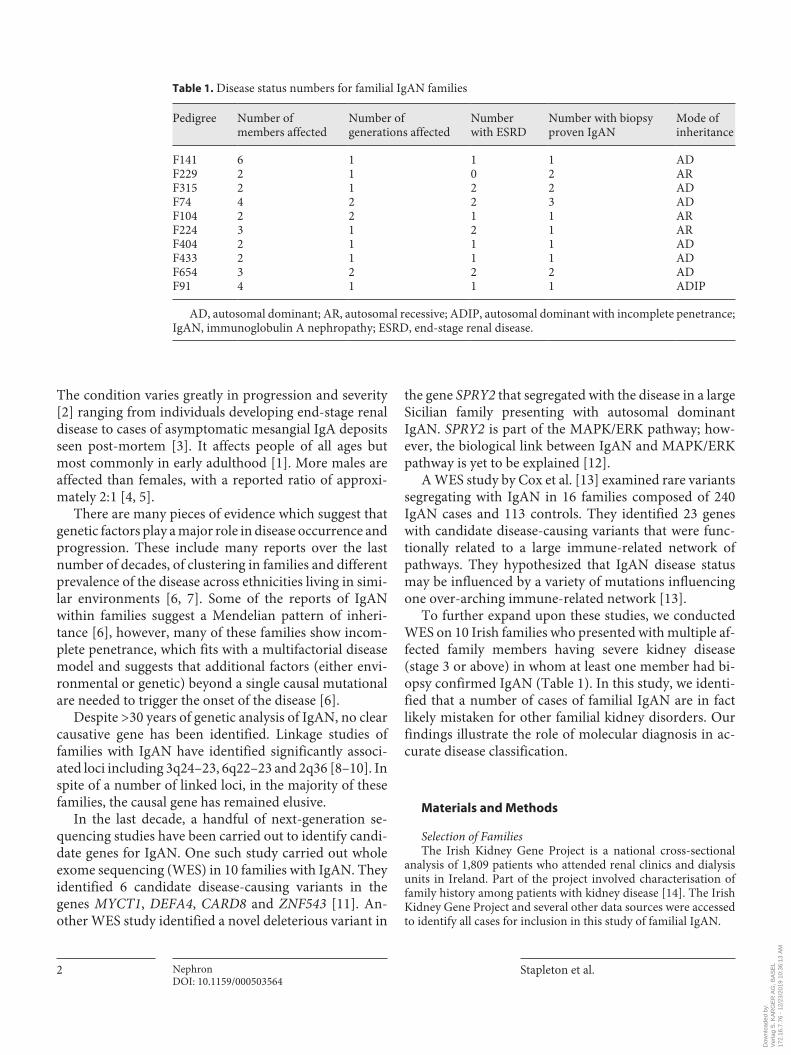
Table 1. Disease status numbers for familial IgAN families
Pedigree Number ofmembers affected
Number ofgenerations affected
Numberwith ESRD
Number with biopsyproven IgAN
Mode ofinheritance
F141 6 1 1 1 ADF229 2 1 0 2 ARF315 2 1 2 2 ADF74 4 2 2 3 ADF104 2 2 1 1 ARF224 3 1 2 1 ARF404 2 1 1 1 ADF433 2 1 1 1 ADF654 3 2 2 2 ADF91 4 1 1 1 ADIP
AD, autosomal dominant; AR, autosomal recessive; ADIP, autosomal dominant with incomplete penetrance; IgAN, immunoglobulin A nephropathy; ESRD, end-stage renal disease.
Dow
nloa
ded
by:
Ver
lag
S. K
AR
GE
R A
G, B
AS
EL
172.
16.7
.76
- 12
/23/
2019
10:
36:1
3 A
M
Exome Sequencing in 10 Families with IgAN
3NephronDOI: 10.1159/000503564
All patients in this study provided informed written consent. The study protocol and design were subject to appropriate Ethics Committee approval at Beaumont Hospital, Dublin, Ireland (re-search Ethics Committee reference: 12/75).
Families presenting with IgAN were identified through a clini-cal analysis carried out by Fennelly et al. [15]. Patients were se-lected on the basis that they had a clinical picture compatible with IgAN predominantly. Specifically patients with advanced liver dis-ease that is associated with IgAN or patients with other disease that can show IgAN deposition such as Lupus or membranous ne-phropathy were excluded. In this study, 8,033 kidney biopsies per-formed at Beaumont Hospital over the period 1986–2016 were re-viewed and identified 1,283 cases of IgAN. Of these cases, 23 (1.8%) were found to have a family history of kidney disease and were classed as familial IgAN. These 23 cases represented 14 different families. The families were then contacted and blood/saliva was collected on any available, consented family member by the staff at Beaumont Hospital. DNA from these patients was obtained at Beaumont Hospital from these collected blood or saliva samples and processed through the Rare Kidney Disease Biobank at St. James Hospital, Dublin, Ireland. We then chose families who had at least one family member with biopsy proven IgAN and one oth-er family member with either biopsy-proven IgAN or end-stage renal disease as previously described [15] and selected families that had at least 2 affected members with DNA available. This left us with 10 families for the exome sequencing analysis. All unaffected individuals that had gene sequencing were labelled unaffected on the basis of negative dipstick for significant blood or proteinuria. All families recruited were Irish and were reported as non-consan-guineous.
Ten families were selected for exome sequencing, labelled F104, F141, F229, F315, F74, F404, F433, F654, F224, and F91. In family F141, we sequenced 3 affected members. In family F104, we se-quenced one affected member and in the remaining 8 families, we sequenced 2 affected members (F229, F315, F74, F404, F433, F654, F224, and F91). Families were selected for exome sequencing on the basis of meeting the aforementioned phenotype criteria and also having sufficient DNA to carry out exome sequencing and test for segregation, as required.
WES AnalysisWe exome sequenced the 20 individuals from 10 families to
100X coverage using either an Ion Proton platform (n = 6, fami-lies = F229, F315 and F91) or an Illumina Hiseq platform (n = 14, families = F141, F74, F104, F224, F404, F433 and F654). Six indi-viduals were sequenced using the SureSelect XT Human All Exon V6 + UTRs Kit (members from F141, F74 and F104), 8 were se-quenced using the SureSelect XT Human All Exon V6 Kit (mem-bers from F224, F404, F433 and F654), 2 were sequenced with the Ion TargetSeq Exome (both individuals in F91) and the remaining 4 were sequenced using the Ion Ampliseq exome RDY kit (families F229 and F315).
Annotating VariantsFollowing alignment and quality control of exome data (online
suppl. Methods; for all online suppl. material, see www.karger.com/doi/10.1159/000503564), VCF files were annotated using Annovar [16]. Candidate variants were defined as variants that were shared between affected family members, functional, rare and predicted deleterious (online suppl. Methods).
Candidate variants and where relevant, the associated genes were cross-referenced with the Online Mendelian Inheritance in Man catalog, Clinvar (version 20150330), gnomad, missense toler-ance ratio Gene Viewer and Residual Variation Intolerance Score [17–21].
We incorporated clinical data using Online Mendelian Inheri-tance in Man and Clinvar to see if any of the candidate variants were thought to be involved in kidney disease or had been previ-ously been reported as pathogenic. We then re-examined the ped-igree information to select variants that fit the disease model. Fi-nally, we used the American College of Medical Genetics and Ge-nomics (ACMG) guidelines to annotate whether or not the variant was considered pathogenic, likely pathogenic, uncertain signifi-cance, likely benign or benign [22].
We used Sanger sequencing to confirm candidate variants and to test for segregation (online suppl. Methods).
Searching for Variants Across FamiliesFamilies in which we did not identify a candidate variant in a
gene previously linked with renal disease were assessed for candi-date variants found in genes shared across these families. Genes with a candidate variant found in > 1 family were selected. Artefacts of sequencing were identified through examination of the bam files using the Integrative Genomics Viewer [23].
Results
We performed WES on 20 individuals across 10 fami-lies (Table 1). The average age in the affected individuals (n = 30) across these 10 families was 41 (SD 15.8 years), with 22 males and 8 females (online suppl. Table 1 for further details on affected family members). The average age in the unaffected individuals (n = 26) across these 10 families was 58 (SD 11.6 years), with 8 males and 17 fe-males. In 7 families (F224, F91, F404, F433, F654, F229 and F315), we did not identify candidate variants in genes known to cause kidney disease, or that were found across multiple families in biologically relevant genes (online suppl. Table 2 for list of qualifying variants found in these families). In 3 families (F141, F74 and F104), we identi-fied candidate mutations that were found in genes known to cause kidney disease (Table 2).
Exome Sequencing: Family 141Family 141 (F141) is a multi-generational family with
multiple affected members presenting with blood and proteinuria. In one member a kidney biopsy confirmed IgA deposition in the mesangium. There was clear male to male transmission suggesting an autosomal dominant mode of inheritance (Fig. 1). The proband (IV.1) in this family was diagnosed (via biopsy) with IgAN and mild arteriosclerosis/arteriolar sclerosis (Fig. 2). The pro-band’s father (III.6) was diagnosed with focal prolifera-
Dow
nloa
ded
by:
Ver
lag
S. K
AR
GE
R A
G, B
AS
EL
172.
16.7
.76
- 12
/23/
2019
10:
36:1
3 A
M
Stapleton et al.Nephron4DOI: 10.1159/000503564
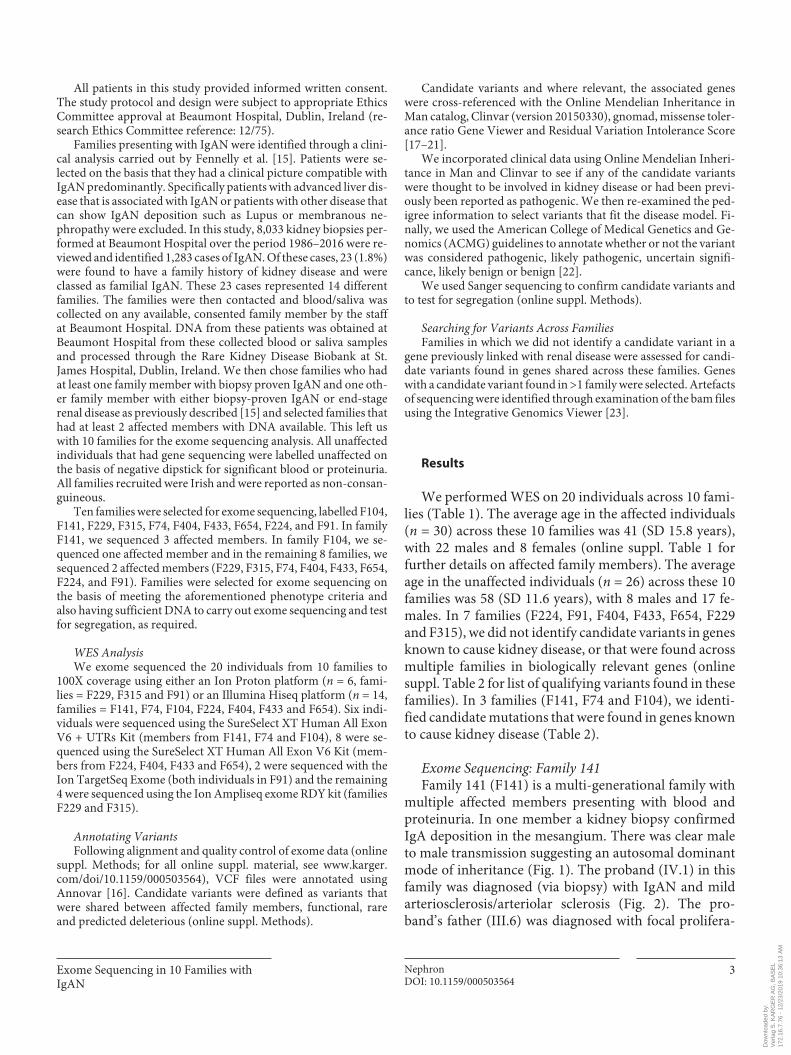
tive glomerular nephropathy, thin glomerular basement membrane and mild arteriolar sclerosis. The proband’s grandmother (II.1) was reported to have “scarred kid-neys” but the reason for this scarring was not reported. The proband’s sister (IV.2) had signs of early stage chron-ic kidney disease with both blood and proteins present in the urine. The proband’s paternal uncle (III.5) was also found to have kidney disease with urinalysis showing presence of protein and haemolysed blood and upon bi-opsy showed signs of thin basement membrane nephrop-athy (Fig. 2). The proband’s aunt (III.3) also presented
with 2+ blood in the urinalysis. A cousin (IV.3) was re-ported as affected, but exact details on this individual are unknown.
Three of the affected individuals were selected for WES (Fig. 1) – the proband (IV.1), the sister (IV.2) and the affected uncle (III.5). In F141, we identified a likely pathogenic variant (according to ACMG standards) in the gene COL4A5, a gene known to cause X-linked Alport syndrome [24]. This variant was present in IV2 and III5 but not in the proband. This variant, rs104886228: G>A, was found on chromosome X in the 38th exon of COL4A5.
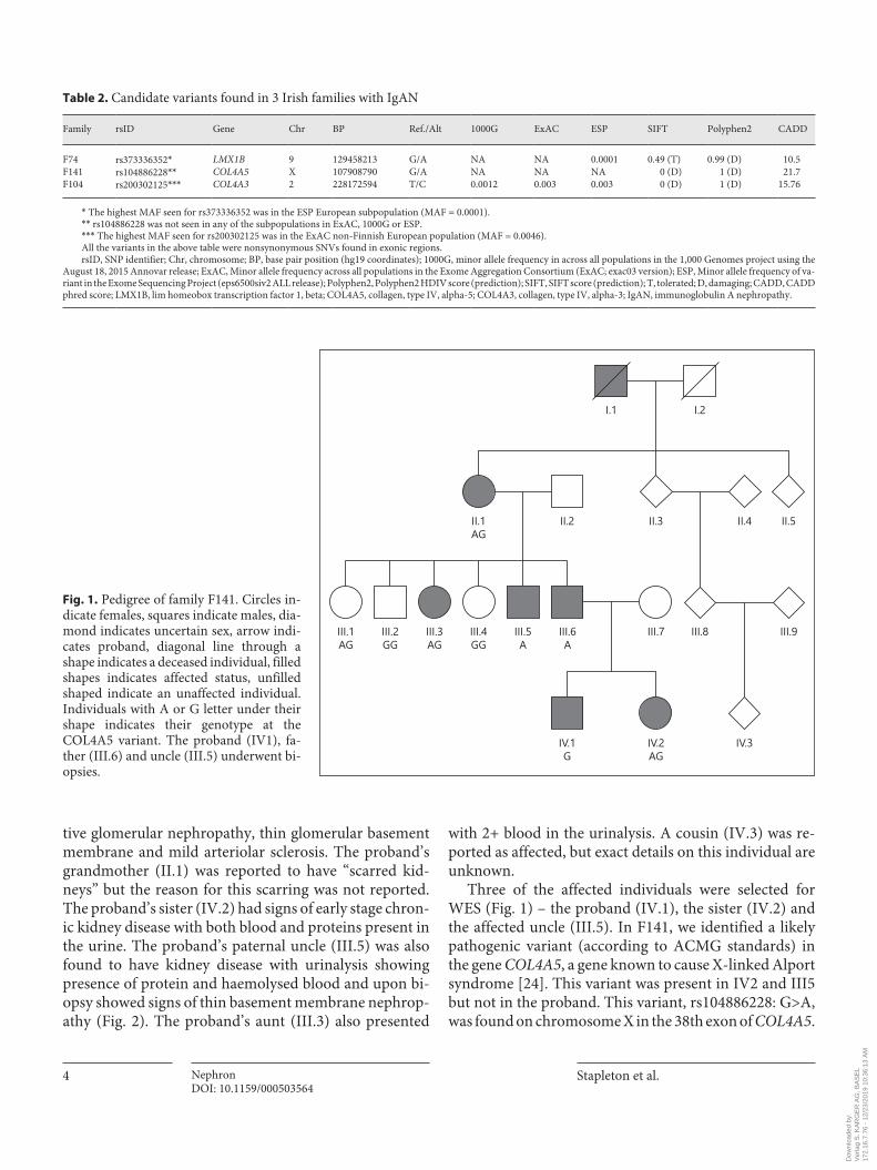
Table 2. Candidate variants found in 3 Irish families with IgAN
Family rsID Gene Chr BP Ref./Alt 1000G ExAC ESP SIFT Polyphen2 CADD
F74 rs373336352* LMX1B 9 129458213 G/A NA NA 0.0001 0.49 (T) 0.99 (D) 10.5F141 rs104886228** COL4A5 X 107908790 G/A NA NA NA 0 (D) 1 (D) 21.7F104 rs200302125*** COL4A3 2 228172594 T/C 0.0012 0.003 0.003 0 (D) 1 (D) 15.76
* The highest MAF seen for rs373336352 was in the ESP European subpopulation (MAF = 0.0001).** rs104886228 was not seen in any of the subpopulations in ExAC, 1000G or ESP.*** The highest MAF seen for rs200302125 was in the ExAC non-Finnish European population (MAF = 0.0046).All the variants in the above table were nonsynonymous SNVs found in exonic regions. rsID, SNP identifier; Chr, chromosome; BP, base pair position (hg19 coordinates); 1000G, minor allele frequency in across all populations in the 1,000 Genomes project using the
August 18, 2015 Annovar release; ExAC, Minor allele frequency across all populations in the Exome Aggregation Consortium (ExAC; exac03 version); ESP, Minor allele frequency of va-riant in the Exome Sequencing Project (eps6500siv2 ALL release); Polyphen2, Polyphen2 HDIV score (prediction); SIFT, SIFT score (prediction); T, tolerated; D, damaging; CADD, CADD phred score; LMX1B, lim homeobox transcription factor 1, beta; COL4A5, collagen, type IV, alpha-5; COL4A3, collagen, type IV, alpha-3; IgAN, immunoglobulin A nephropathy.
I.1
II.1AG
III.1AG
III.2GG
III.3AG
III.4GG
III.5A
III.6A
IV.1G
IV.2AG
IV.3
III.7 III.8 III.9
II.2 II.3 II.4 II.5
I.2
Fig. 1. Pedigree of family F141. Circles in-dicate females, squares indicate males, dia-mond indicates uncertain sex, arrow indi-cates proband, diagonal line through a shape indicates a deceased individual, filled shapes indicates affected status, unfilled shaped indicate an unaffected individual. Individuals with A or G letter under their shape indicates their genotype at the COL4A5 variant. The proband (IV1), fa-ther (III.6) and uncle (III.5) underwent bi-opsies.
Dow
nloa
ded
by:
Ver
lag
S. K
AR
GE
R A
G, B
AS
EL
172.
16.7
.76
- 12
/23/
2019
10:
36:1
3 A
M
Exome Sequencing in 10 Families with IgAN
5NephronDOI: 10.1159/000503564
This nucleotide change was a nonsynonymous single nu-cleotide variation (SNV) predicted to result in an amino-acid change (glycine to serine) at position 1,143 of the COL4A5 protein p.(Gly1143Ser). We interpreted this variant according to ACMG criteria as “likely pathogen-ic” (Table 3).
We went on to genotype the variant in 9 family mem-bers (6 affected and 3 unaffected) to test for segregation. The “likely pathogenic” (“A”) allele was found in to seg-regate with affected status along the proband’s father’s side of the family (Fig. 1, online suppl. Fig. 1). As this vari-ant is on the X chromosome, the father cannot pass the mutation to the proband indicating that there may be 2 very similar diseases segregating in 1 family. Notably, there was one healthy female carrier (III.1) who was found to have a clear urinalysis (urinalysis carried out at age 44). Due to potential X-inactivation [25, 26] and the segregation pattern of the variant, we did not consider cosegregation data as evidence towards ACMG. This ped-igree appears to demonstrate sporadic IgAN within a family with Alport mutations. This case demonstrates the
difficulty of studying familial kidney disease where 2 kid-ney disease patterns occur within the 1 family or where a family member is a phenocopy with a different disease.
Exome Sequencing: Family 104Family 104 (F104) presented with 2 affected brothers
with a recessive inheritance pattern (Fig. 3). The first af-fected brother (II.1) presented with hematuria and pro-teinuria at the age of 29 and was diagnosed with IgAN upon biopsy. He progressed to end-stage kidney disease undergoing transplant approximately 6 years after his IgAN diagnosis. The second brother (II.3) was also diag-nosed with IgAN. No other family members reported kid-ney disease.
In F104, a variant of unknown significance (VUS, ac-cording to ACMG guidelines), rs200302125: T>C, was identified in the non-collagenous domain of COL4A3 (Table 2, Fig. 3). Here, a nonsynonymous SNV was pre-dicted to result in the replacement of a leucine with a pro-line at amino acid position 1474 p.(Leu1474Pro). COL4A3 is associated with autosomal dominant and recessive
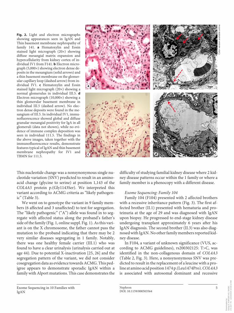
Fig. 2. Light and electron micrographs showing appearances seen in IgAN and Thin basement membrane nephropathy of family 141. a Hematoxylin and Eosin stained light micrograph (20×) showing diffuse mesangial matrix expansion and hypercellularity from kidney cortex of in-dividual IV1 from F141. b Electron micro-graph (5,000×) showing electron dense de-posits in the mesangium (solid arrows) and a thin basement membrane on the glomer-ular capillary loop (dashed arrow) from in-dividual IV1. c Hematoxylin and Eosin stained light micrograph (20×) showing a normal glomerulus in individual III.5. d Electron micrograph (10,000×) showing a thin glomerular basement membrane in individual III.5 (dashed arrow). No elec-tron dense deposits were found in the me-sangium of III.5. In individual IV1, immu-nofluorescence showed global and diffuse granular mesangial positivity for IgA in all glomeruli (data not shown), while no evi-dence of immune complex deposition was seen in individual 111.5. The findings in the above images, taken together with the immunofluorescence results, demonstrate features typical of IgAN and thin basement membrane nephropathy for IV1 and TBMN for 111.5.
Colo
r ver
sion
avai
labl
e on
line
c d
ba
Dow
nloa
ded
by:
Ver
lag
S. K
AR
GE
R A
G, B
AS
EL
172.
16.7
.76
- 12
/23/
2019
10:
36:1
3 A
M

Stapleton et al.Nephron6DOI: 10.1159/000503564
forms of Alport syndrome as well as autosomal dominant benign familial hematuria [27–32].
The minor allele frequency was high for a disease-caus-ing heterozygote variant (0.12% MAF in 1000G_All and 0.2% in gnomad). Several computational tools predicted the variant to be damaging (SIFT_pred = D, Polyphen2_HDIV_pred = D, MutationTaster_pred = D, CADD_phred = 15.76). The variant met a number of criteria for ACMG, as laid out in Table 4. However, the evidence was not sufficient to call this variant as pathogenic, and so at the time of writing, this variant comes under the VUS category.
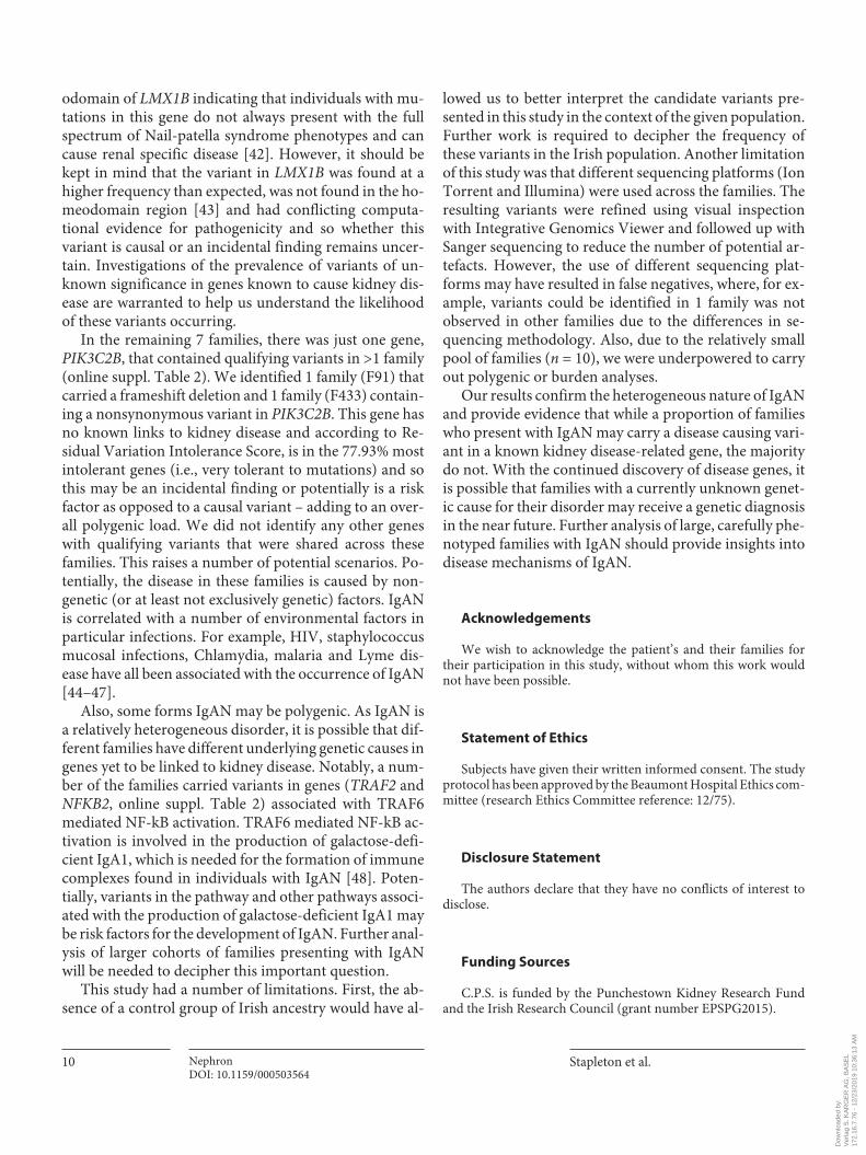
Exome Sequencing: Family 74The third family, family 74 (F74), presented with an
autosomal dominant inheritance pattern with 4 affected individuals across 2 generations (Fig. 4). The proband (II.11) presented with biopsy confirmed IgAN and dia-betic nephropathy, which progressed to end-stage kidney disease and required the patient to undergo transplant.
Table 3. Criteria met by COL4A5 variant found in F141
ACMG criteria met Evidence
PM1 – Located in a mutational hot spot and/or criticaland well-established functional domain (e.g., the activesite of an enzyme) without benign variation
Variant was found in the in the collagenous domain of the α5 (IV) chain. Glycine substitutions in this region are known to havedeleterious effects on the function of the protein [35].
PM2 – Absent from controls (or at an extremely lowfrequency if recessive) in Exome Sequencing Project, 1,000 Genomes Project, or Exome Aggregation Consortium
Variant not found in gnomad, 1000G_ALL, ExAC_ALL orESP6500siv2_ALL
PM5 - Novel missense change at an amino acid residuewhere a different missense change determined to bepathogenic has been seen before
Variant was at the same codon but causing a different amino acid(glycine to aspartate) change that had previously been found in a family with Alport syndrome and is reported as pathogenic in OMIM [34]. This variant was located in the collagenous domain of the α5 (IV) chain and was found interfere with the maintenance of the triple helicalformation of the collagen molecule which leads to increasedpermeability and weakening of the glomerular basement membrane
PP2 – Missense variant in a gene that has a low rate ofbenign missense variation and in which missensevariants are a common mechanism of disease
COL4A5 has an RVIS score = 6.3%, meaning this gene is amongst the top 6.3% most intolerant genes in the human genome (based upon the amount of common variation found in that gene) [18].
PP3 – Multiple lines of computational evidence supporta deleterious effect on the gene or gene product(conservation, evolutionary, splicing impact, etc.)
Variant was predicted damaging (D) by SIFT_pred, Polyphen2_HDIV_pred and MutationTaster_pred. CADD_phred score of 21.7
PP5 – Reputable source recently reports variant aspathogenic, but the evidence is not available to thelaboratory to perform an independent evaluation
Previous studies identified variant as pathogenic in families withAlport syndrome, however this variant not reported in OMIM [35, 49].
The above table lays out the evidence for calling the COL4A5 variant found in F141 as likely pathogenic according to ACMG guide-lines.
PM, moderate pathogenic criterion; PP, supporting pathogenic criterion; ACMG, American College of Medical Genetics and Geno-mics; OMIM, Online Mendelian Inheritance in Man.
II.1TC
II.2TT
I.1 I.2TT
II.3TC
II.4
Fig. 3. Pedigree of family F104. Circles indicate females, squares indicate males, diamond indicates uncertain sex, arrow indicates proband, filled shapes indicates affected status, unfilled shaped in-dicate an unaffected individual. Individuals with T or C letter un-der their shape indicates their genotype at the COL4A3 variant. Individual II.1 underwent biopsy.
Dow
nloa
ded
by:
Ver
lag
S. K
AR
GE
R A
G, B
AS
EL
172.
16.7
.76
- 12
/23/
2019
10:
36:1
3 A
M
Exome Sequencing in 10 Families with IgAN
7NephronDOI: 10.1159/000503564
The proband’s nephew (III.1) presented with biopsy con-firmed IgAN and mild arteriosclerosis/arteriolarsclerosis (Fig. 5). The second nephew (III.2) of the proband self-reported as healthy, however, urinalysis showed haemo-lysed blood. The proband’s brother (II.1) presented with CKD and on renal biopsy IgA deposits were found in the glomerular mesangium (Fig. 5).
Two individuals in F74 were selected for WES (II.1 and III.1). We identified a candidate variant, rs373336352: G>A, in the gene LMX1B, a gene associated with Nail-patella syndrome (Table 2, Fig. 4) [33]. This variant, was a nonsynonymous SNV which was predicted to cause an amino acid change from glycine to serine, p.(Gly339Ser). Although this variant was also found to segregate with the disease in affected family members, it had mixed results for computational evidence supporting a deleterious ef-fect on the gene or gene product (CADD Phred score of
10.5, Polyphen2_HDIV_pred = D, MutationTaster = D, SIFT_pred = T) and had a higher MAF than would be ex-pected for a pathogenic variant. As such, it was classified as VUS (Table 5). It was not possible to obtain DNA sam-ples for unaffected family members in F74 and therefore only affected family members were sequenced.
Discussion
We analysed exome sequence data from 10 Irish fami-lies presenting with IgAN, filtering for rare, functional, exonic or splicing, predicted damaging variants that were shared between affected members of the family. We de-tected an ACMG-satisfying disease causing mutation in one of the 10 families, with VUS found in kidney-disease-related genes in another 2 families.
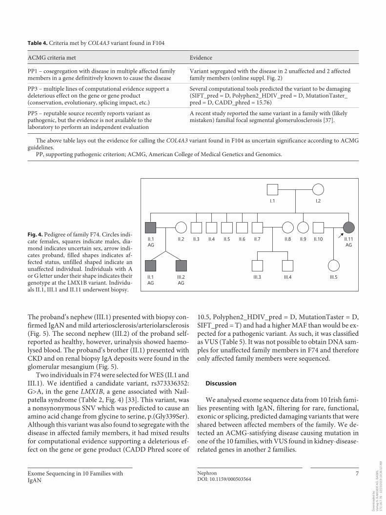
Table 4. Criteria met by COL4A3 variant found in F104
ACMG criteria met Evidence
PP1 – cosegregation with disease in multiple affected family members in a gene definitively known to cause the disease
Variant segregated with the disease in 2 unaffected and 2 affected family members (online suppl. Fig. 2)
PP3 – multiple lines of computational evidence support adeleterious effect on the gene or gene product(conservation, evolutionary, splicing impact, etc.)
Several computational tools predicted the variant to be damaging (SIFT_pred = D, Polyphen2_HDIV_pred = D, MutationTaster_pred = D, CADD_phred = 15.76)
PP5 – reputable source recently reports variant aspathogenic, but the evidence is not available to thelaboratory to perform an independent evaluation
A recent study reported the same variant in a family with (likely mistaken) familial focal segmental glomerulosclerosis [37].
The above table lays out the evidence for calling the COL4A3 variant found in F104 as uncertain significance according to ACMG guidelines.
PP, supporting pathogenic criterion; ACMG, American College of Medical Genetics and Genomics.
I.1
II.1AG
II.1AG
III.2AG
III.3 III.4 III.5
II.2 II.3 II.4 II.5 II.6 II.7 II.8 II.9 II.10 II.11AG
I.2
Fig. 4. Pedigree of family F74. Circles indi-cate females, squares indicate males, dia-mond indicates uncertain sex, arrow indi-cates proband, filled shapes indicates af-fected status, unfilled shaped indicate an unaffected individual. Individuals with A or G letter under their shape indicates their genotype at the LMX1B variant. Individu-als II.1, III.1 and II.11 underwent biopsy.
Dow
nloa
ded
by:
Ver
lag
S. K
AR
GE
R A
G, B
AS
EL
172.
16.7
.76
- 12
/23/
2019
10:
36:1
3 A
M

Stapleton et al.Nephron8DOI: 10.1159/000503564
IgAN is the most common form of glomerulonephritis worldwide and likely has multiple different forms, from monogenic to multifactorial. Despite strong evidence from familial segregation, to date a clearly monogenic form has yet to be fully characterised. Given the relative frequency of IgAN, it is also clear that IgA deposits may be present on kidney biopsies with a second clear renal pathological diagnosis. This points to the difficulty in an-alysing kidney pathology where IgA deposition may be present but other disease mechanisms are the predomi-nate lesion affecting kidney structure. In the families pre-sented in this study, there was clear evidence of IgA de-position thus making the criteria for a diagnosis of IgAN in extended families with advanced kidney disease.
In our analysis, we did not identify any potentially causal qualifying variants in 7 of the families. We made a clear molecular diagnosis in one case and in 2 further cas-es, we identified variants of uncertain significance. In family F141, a likely pathogenic mutation was found in the COL4A5 gene (notably, this variant did not meet the criteria originally set out in this study as it was not present in the proband). Mutations in this gene are commonly associated with Alport syndrome. This variant and a mu-tation resulting in a different amino acid change at the same codon had previously been identified as pathogenic in families with Alport syndrome [34, 35]. In F141, the father’s biopsy report diagnosis was focal proliferative glomerular nephropathy with mild arteriolar sclerosis
Fig. 5. Light and electron micrographs showing appearances seen in IgAN of fam-ily 74. a Hematoxylin and Eosin stained light micrograph (20×) showing areas of mesangial hypercellularity and mesangial matrix expansion (arrows) from kidney cortex of individual II.11. The inset shows one of these areas at higher magnification (40×). e Hematoxylin and Eosin stained light micrographs (20×) showing diffuse mesangial matrix hypercellularity and ex-pansion from kidney cortex of individuals II.1 and III.1, respectively. b, d, e Electron micrographs (15,000×) showing electron vdense deposits in the mesangium (arrows) and also capillary loops (b). The diamonds show the glomerular capillary loop spaces. Immunofluorescence for all patients dem-onstrated global and diffuse granular me-sangial positivity for IgA (data not shown). The findings in the above images, taken to-gether with the immunofluorescence re-sults, demonstrate the features typical of IgAN.
Colo
r ver
sion
avai
labl
e on
line
c d
e f
ba
Dow
nloa
ded
by:
Ver
lag
S. K
AR
GE
R A
G, B
AS
EL
172.
16.7
.76
- 12
/23/
2019
10:
36:1
3 A
M

Exome Sequencing in 10 Families with IgAN
9NephronDOI: 10.1159/000503564
and the uncles’ biopsy showed thin basement membrane nephropathy (Fig. 2). In comparison, the proband’s bi-opsy report showed IgAN.
This finding indicates that the father (and associated carriers of the COL4A5 variant) had a similar, yet differ-ent, cause of kidney disease compared to the proband, who has clear features of IgAN and does not carry the COL4A5 variant. Deafness, a clinical phenotype often found in individuals with Alport syndrome, was not found in this family [36]. This highlights the range and diversity in the phenotypes associated with type 4 colla-gen mutations. The proband in this family had clear evi-dence of IgAN with multiple affected family members with overt kidney disease which was likely resulting from collagen mediated disease. This family illustrates the dif-ficulty in diagnosing familial kidney disease as there was clear evidence of 2 distinct pathogenic processes coexist-ing within the 1 family (IgAN and collagen basement membrane disease).
In family F104, we identified a VUS in COL4A3. This gene has previously been shown to cause autosomal re-cessive and dominant Alport syndrome as well as autoso-mal dominant benign familial haematuria [28]. This vari-ant was recently reported in a family diagnosed with focal segmental glomerular sclerosis indicating that the spec-trum of disease caused by mutations in COL4A3 may be wider than previously thought [37]. However, the variant had a minor allele frequency that is higher than what we would expect for a causal heterozygous pathogenic vari-ant. That being said, kidney disease is relatively common in any population. According to a 2010 study, the age-standardized global prevalence of CKD in adults (mini-
mum age of 20) is 10.4% in men and 11.8% in women [38]. Therefore, it is possible that variants with relatively high minor allele frequencies, may in fact be causal, or contributing risk. Also, kidney disease has an extremely broad age of onset, becoming more common as age in-creases [39]. This means that variants causing kidney dis-ease may not be under the same selective pressure as vari-ants causing disease with earlier ages of onset as the mean age for end stage kidney disease in patients with IgAN is past the average age for child bearing (mean age recently reported as 42.8 [40]). Therefore, variants causing the dis-ease could be at relatively high frequencies in the general population. However, without further evidence we can-not class the mutation found in this family as pathogenic and so whether this variant is causal or an incidental find-ing has yet to be determined.
In the third family, F74, we identified a variant in the gene LMX1B, known to cause autosomal dominant Nail-patella syndrome. This syndrome is characterised by dys-plastic nails, absent or hypoplastic patellae and, in a num-ber of cases, nephropathy resembling glomerulonephri-tis. LMX1B is binds the enhancers of COL4A4 [41]. In mice with LMX1B knock-out mutations (–/–), there is a strong decrease in the alpha-3 and -4 chains of type IV collagen [41]. Similar to that of Alport syndrome, the dys-regulation of the alpha-3 and -4 chains of type IV collagen results in glomerulopathy. It is thought that haploinsuf-ficiency is the pathogenic mechanism of Nail-patella syn-drome, with the disease following an autosomal domi-nant inheritance pattern [33]. A study examining indi-viduals with nail patella-like renal disease reported a number of families who carried a mutation in the home-
Table 5. Criteria met by LMX1B variant found in F74
ACMG criteria met Evidence
PP1 – cosegregation with disease in multiple affected familymembers in a gene definitively known to cause the disease
The variant segregated with all 4 affected family members (online suppl. Fig. 3)
PP2 – Missense variant in a gene that has a low rate ofbenign missense variation and in which missense variantsare a common mechanism of disease
LMX1B has RVIS score of 11.06% and a number of missensevariants reported as pathogenic in OMIM
BS1 Allele frequency is greater than expected for thedisorder
Variant was not found in 1,000 genomes, EXAC or gnomad databases but had a frequency of 0.0001 in the NHLBI-ESP project with 6,500 exomes. The variant was found in one individual in this database
The above table lays out the evidence for calling the LMX1B variant found in F74 as uncertain significance according to ACMG gui-delines.
PP, supporting pathogenic criterion; BS, strong benign criterion; OMIM, Online Mendelian Inheritance in Man; ACMG, American College of Medical Genetics and Genomics.
Dow
nloa
ded
by:
Ver
lag
S. K
AR
GE
R A
G, B
AS
EL
172.
16.7
.76
- 12
/23/
2019
10:
36:1
3 A
M
Stapleton et al.Nephron10DOI: 10.1159/000503564
odomain of LMX1B indicating that individuals with mu-tations in this gene do not always present with the full spectrum of Nail-patella syndrome phenotypes and can cause renal specific disease [42]. However, it should be kept in mind that the variant in LMX1B was found at a higher frequency than expected, was not found in the ho-meodomain region [43] and had conflicting computa-tional evidence for pathogenicity and so whether this variant is causal or an incidental finding remains uncer-tain. Investigations of the prevalence of variants of un-known significance in genes known to cause kidney dis-ease are warranted to help us understand the likelihood of these variants occurring.
In the remaining 7 families, there was just one gene, PIK3C2B, that contained qualifying variants in > 1 family (online suppl. Table 2). We identified 1 family (F91) that carried a frameshift deletion and 1 family (F433) contain-ing a nonsynonymous variant in PIK3C2B. This gene has no known links to kidney disease and according to Re-sidual Variation Intolerance Score, is in the 77.93% most intolerant genes (i.e., very tolerant to mutations) and so this may be an incidental finding or potentially is a risk factor as opposed to a causal variant – adding to an over-all polygenic load. We did not identify any other genes with qualifying variants that were shared across these families. This raises a number of potential scenarios. Po-tentially, the disease in these families is caused by non-genetic (or at least not exclusively genetic) factors. IgAN is correlated with a number of environmental factors in particular infections. For example, HIV, staphylococcus mucosal infections, Chlamydia, malaria and Lyme dis-ease have all been associated with the occurrence of IgAN [44–47].
Also, some forms IgAN may be polygenic. As IgAN is a relatively heterogeneous disorder, it is possible that dif-ferent families have different underlying genetic causes in genes yet to be linked to kidney disease. Notably, a num-ber of the families carried variants in genes (TRAF2 and NFKB2, online suppl. Table 2) associated with TRAF6 mediated NF-kB activation. TRAF6 mediated NF-kB ac-tivation is involved in the production of galactose-defi-cient IgA1, which is needed for the formation of immune complexes found in individuals with IgAN [48]. Poten-tially, variants in the pathway and other pathways associ-ated with the production of galactose-deficient IgA1 may be risk factors for the development of IgAN. Further anal-ysis of larger cohorts of families presenting with IgAN will be needed to decipher this important question.
This study had a number of limitations. First, the ab-sence of a control group of Irish ancestry would have al-
lowed us to better interpret the candidate variants pre-sented in this study in the context of the given population. Further work is required to decipher the frequency of these variants in the Irish population. Another limitation of this study was that different sequencing platforms (Ion Torrent and Illumina) were used across the families. The resulting variants were refined using visual inspection with Integrative Genomics Viewer and followed up with Sanger sequencing to reduce the number of potential ar-tefacts. However, the use of different sequencing plat-forms may have resulted in false negatives, where, for ex-ample, variants could be identified in 1 family was not observed in other families due to the differences in se-quencing methodology. Also, due to the relatively small pool of families (n = 10), we were underpowered to carry out polygenic or burden analyses.
Our results confirm the heterogeneous nature of IgAN and provide evidence that while a proportion of families who present with IgAN may carry a disease causing vari-ant in a known kidney disease-related gene, the majority do not. With the continued discovery of disease genes, it is possible that families with a currently unknown genet-ic cause for their disorder may receive a genetic diagnosis in the near future. Further analysis of large, carefully phe-notyped families with IgAN should provide insights into disease mechanisms of IgAN.
Acknowledgements
We wish to acknowledge the patient’s and their families for their participation in this study, without whom this work would not have been possible.
Statement of Ethics
Subjects have given their written informed consent. The study protocol has been approved by the Beaumont Hospital Ethics com-mittee (research Ethics Committee reference: 12/75).
Disclosure Statement
The authors declare that they have no conflicts of interest to disclose.
Funding Sources
C.P.S. is funded by the Punchestown Kidney Research Fund and the Irish Research Council (grant number EPSPG2015).
Dow
nloa
ded
by:
Ver
lag
S. K
AR
GE
R A
G, B
AS
EL
172.
16.7
.76
- 12
/23/
2019
10:
36:1
3 A
M
Exome Sequencing in 10 Families with IgAN
11NephronDOI: 10.1159/000503564
Author Contributions
N.K.F., C.K., D.M.C., and S.L.M. recruited patients, compiled pedigrees, gathered patient phenotype data and collected blood/saliva samples; C.P.S., G.L.C., and P.J.C. designed the analyses;
C.P.S. carried out DNA sample preparation and data analysis. G.L.C. and P.J.C. supervised the study; C.P.S. wrote the manu-script with consultation and guidance from G.L.C., P.J.C., and N.K.F.; N.K.F., A.M.D., and B.D. imaged and analysed biopsies. All authors reviewed and gave input to the final manuscript.
References
1 D’Amico G. The commonest glomerulone-phritis in the world: IgA nephropathy. Q J Med. 1987 Sep; 64(245): 709–27.
2 Hsu SI, Feehally J. The molecular basis of IgA nephropathy. In: Mount DB, Pollak MR, edi-tors. Molecular and genetic basis of renal dis-ease : a companion to Brenner & Rector’s the kidney. Philadelphia: Saunders/Elsevier; 2008. pp. 481–97.
3 Waldherr R, Rambausek M, Duncker WD, Ritz E. Frequency of mesangial IgA deposits in a non-selected autopsy series. Nephrol Dial Transplant. 1989; 4(11): 943–6.
4 Wyatt RJ, Julian BA. IgA nephropathy. N Engl J Med. 2013 Jun; 368(25): 2402–14.
5 Schena FP, Cerullo G, Rossini M, Lanzilotta SG, D’Altri C, Manno C. Increased risk of end-stage renal disease in familial IgA nephropa-thy. J Am Soc Nephrol. 2002 Feb; 13(2): 453–60.
6 Kiryluk K, Julian BA, Wyatt RJ, Scolari F, Zhang H, Novak J, et al. Genetic studies of IgA nephropathy: past, present, and future. Pedi-atr Nephrol. 2010 Nov; 25(11): 2257–68.
7 Hall YN, Fuentes EF, Chertow GM, Olson JL. Race/ethnicity and disease severity in IgA ne-phropathy. BMC Nephrol. 2004 Sep; 5(1): 10.
8 Gharavi AG, Yan Y, Scolari F, Schena FP, Fra-sca GM, Ghiggeri GM, et al. IgA nephropathy, the most common cause of glomerulonephri-tis, is linked to 6q22-23. Nat Genet. 2000 Nov;
26(3): 354–7. 9 Bisceglia L, Cerullo G, Forabosco P, Torres
DD, Scolari F, Di Perna M, et al.; European IgAN Consortium. Genetic heterogeneity in Italian families with IgA nephropathy: sug-gestive linkage for two novel IgA nephropa-thy loci. Am J Hum Genet. 2006 Dec; 79(6):
1130–4.10 Paterson AD, Liu XQ, Wang K, Magistroni R,
Song X, Kappel J, et al. Genome-wide linkage scan of a large family with IgA nephropathy localizes a novel susceptibility locus to chro-mosome 2q36. J Am Soc Nephrol. 2007 Aug;
18(8): 2408–15.11 Liu R, Hu B, Li Q, Jing X, Zhong C, Chang Y,
et al. Novel genes and variants associated with IgA nephropathy by co-segregating with the disease phenotypes in 10 IgAN families. Gene. 2015 Oct; 571(1): 43–51.
12 Milillo A, La Carpia F, Costanzi S, D’Urbano V, Martini M, Lanuti P, et al. A SPRY2 muta-tion leading to MAPK/ERK pathway inhibi-tion is associated with an autosomal domi-nant form of IgA nephropathy. Eur J Hum Genet. 2015 Dec; 23(12): 1673–8.
13 Cox SN, Pesce F, El-Sayed Moustafa JS, Sal-lustio F, Serino G, Kkoufou C, et al.; European IgAN Consortium. Multiple rare genetic vari-ants co-segregating with familial IgA ne-phropathy all act within a single immune-re-lated network. J Intern Med. 2017 Feb; 281(2):
189–205.14 Connaughton DM, Bukhari S, Conlon P, Cas-
sidy E, O’Toole M, Mohamad M, et al. The Irish Kidney Gene Project—Prevalence of Family History in Patients with Kidney Dis-ease in Ireland. Nephron. 2015; 130(4): 293–301.
15 Fennelly NK, Kennedy C, Jenkinson AC, Connaughton DM, Stapleton C, Dorman AM, et al. Clinical Heterogeneity in Familial IgA Nephropathy. Nephron. 2018; 139(1): 63–9.
16 Wang K, Li M, Hakonarson H. ANNOVAR: functional annotation of genetic variants from high-throughput sequencing data. Nu-cleic Acids Res. 2010 Sep; 38(16):e164.
17 Hamosh A, Scott AF, Amberger J, Valle D, McKusick VA. Online Mendelian Inheritance in Man (OMIM). Hum Mutat. 2000; 15(1):
57–61.18 Petrovski S, Wang Q, Heinzen EL, Allen AS,
Goldstein DB. Genic intolerance to function-al variation and the interpretation of personal genomes. PLoS Genet. 2013; 9(8):e1003709.
19 Traynelis J, Silk M, Wang Q, Berkovic SF, Liu L, Ascher DB, et al. Optimizing genomic med-icine in epilepsy through a gene-customized approach to missense variant interpretation. Genome Res. 2017 Oct; 27(10): 1715–29.
20 Lek M, Karczewski KJ, Minikel EV, Samocha KE, Banks E, Fennell T, et al.; Exome Aggre-gation Consortium. Analysis of protein-cod-ing genetic variation in 60,706 humans. Na-ture. 2016 Aug; 536(7616): 285–91.
21 Landrum MJ, Lee JM, Benson M, Brown GR, Chao C, Chitipiralla S, et al. ClinVar: improv-ing access to variant interpretations and sup-porting evidence. Nucleic Acids Res. 2018 Jan; 46(D1):D1062–7.
22 Richards S, Aziz N, Bale S, Bick D, Das S, Gast-ier-Foster J, et al.; ACMG Laboratory Quality Assurance Committee. Standards and guide-lines for the interpretation of sequence vari-ants: a joint consensus recommendation of the American College of Medical Genetics and Ge-nomics and the Association for Molecular Pa-thology. Genet Med. 2015 May; 17(5): 405–24.
23 Robinson JT, Thorvaldsdóttir H, Wenger AM, Zehir A, Mesirov JP. Variant Review
with the Integrative Genomics Viewer. Can-cer Res. 2017 Nov; 77(21):e31–4.
24 Lemmink HH, Schröder CH, Monnens LA, Smeets HJ. The clinical spectrum of type IV collagen mutations. Hum Mutat. 1997; 9(6):
477–99.25 Nakanishi K, Iijima K, Kuroda N, Inoue Y,
Sado Y, Nakamura H, et al. Comparison of alpha5(IV) collagen chain expression in skin with disease severity in women with X-linked Alport syndrome. J Am Soc Nephrol. 1998 Aug; 9(8): 1433–40.
26 Rheault MN, Kren SM, Hartich LA, Wall M, Thomas W, Mesa HA, et al. X-inactivation modifies disease severity in female carriers of murine X-linked Alport syndrome. Nephrol Dial Transplant. 2010 Mar; 25(3): 764–9.
27 Storey H, Savige J, Sivakumar V, Abbs S, Flinter FA. COL4A3/COL4A4 mutations and features in individuals with autosomal reces-sive Alport syndrome. J Am Soc Nephrol. 2013 Dec; 24(12): 1945–54.
28 Webb BD, Brandt T, Liu L, Jalas C, Liao J, Fedick A, et al. A founder mutation in CO-L4A3 causes autosomal recessive Alport syn-drome in the Ashkenazi Jewish population. Clin Genet. 2014 Aug; 86(2): 155–60.
29 Heidet L, Arrondel C, Forestier L, Cohen-Sol-al L, Mollet G, Gutierrez B, et al. Structure of the human type IV collagen gene COL4A3 and mutations in autosomal Alport syndrome. J Am Soc Nephrol. 2001 Jan; 12(1): 97–106.
30 van der Loop FT, Heidet L, Timmer ED, van den Bosch BJ, Leinonen A, Antignac C, et al. Autosomal dominant Alport syndrome caused by a COL4A3 splice site mutation. Kidney Int. 2000 Nov; 58(5): 1870–5.
31 Badenas C, Praga M, Tazón B, Heidet L, Ar-rondel C, Armengol A, et al. Mutations in the-COL4A4 and COL4A3 genes cause familial benign hematuria. J Am Soc Nephrol. 2002 May; 13(5): 1248–54.
32 Mochizuki T, Lemmink HH, Mariyama M, Antignac C, Gubler MC, Pirson Y, et al. Iden-tification of mutations in the alpha 3(IV) and alpha 4(IV) collagen genes in autosomal re-cessive Alport syndrome. Nat Genet. 1994 Sep; 8(1): 77–81.
33 Bongers EM, de Wijs IJ, Marcelis C, Hoefsloot LH, Knoers NV. Identification of entire LMX1B gene deletions in nail patella syn-drome: evidence for haploinsufficiency as the main pathogenic mechanism underlying dominant inheritance in man. Eur J Hum Genet. 2008 Oct; 16(10): 1240–4.
Dow
nloa
ded
by:
Ver
lag
S. K
AR
GE
R A
G, B
AS
EL
172.
16.7
.76
- 12
/23/
2019
10:
36:1
3 A
M
Stapleton et al.Nephron12DOI: 10.1159/000503564
34 Zhou J, Hertz JM, Tryggvason K. Mutation in the alpha 5(IV) collagen chain in juvenile-onset Alport syndrome without hearing loss or ocular lesions: detection by denaturing gradient gel electrophoresis of a PCR prod-uct. Am J Hum Genet. 1992 Jun; 50(6): 1291–300.
35 Renieri A, Meroni M, Sessa A, Battini G, Ser-belloni P, Torri Tarelli L, et al. Variability of clinical phenotype in a large Alport family with Gly 1143 Ser change of collagen alpha 5(IV)-chain. Nephron. 1994; 67(4): 444–9.
36 Wang Y, Sivakumar V, Mohammad M, Colville D, Storey H, Flinter F, et al. Clinical and genetic features in autosomal recessive and X-linked Alport syndrome. Pediatr Nephrol. 2014 Mar; 29(3): 391–6.
37 Malone AF, Phelan PJ, Hall G, Cetincelik U, Homstad A, Alonso AS, et al. Rare heredi-tary COL4A3/COL4A4 variants may be mistaken for familial focal segmental glo-merulosclerosis. Kidney Int. 2014 Dec;
86(6): 1253–9.38 Mills KT, Xu Y, Zhang W, Bundy JD, Chen
CS, Kelly TN, et al. A systematic analysis of worldwide population-based data on the
global burden of chronic kidney disease in 2010. Kidney Int. 2015 Nov; 88(5): 950–7.
39 Glassock RJ, Oreopoulos DG. Aging and chronic kidney disease. Nephron Clin Pract. 2011; 119(Suppl 1):c1.
40 Komatsu H, Kikuchi M, Nakagawa H, Fukuda A, Iwakiri T, Toida T, et al. Long-term sur-vival of patients with IgA nephropathy after dialysis therapy. Kidney Blood Press Res. 2013; 37(6): 649–56.
41 Morello R, Zhou G, Dreyer SD, Harvey SJ, Ninomiya Y, Thorner PS, et al. Regulation of glomerular basement membrane collagen ex-pression by LMX1B contributes to renal dis-ease in nail patella syndrome. Nat Genet. 2001 Feb; 27(2): 205–8.
42 Hall G, Lane B, Chryst-Ladd M, Wu G, Lin JJ, Qin X, et al. Dysregulation of WTI (-KTS) is Associated with the Kidney-Specific Effects of the LMX1B R246Q Mutation. Sci Rep. 2017 Jan; 7(1): 39933.
43 Dreyer SD, Zhou G, Baldini A, Winterpacht A, Zabel B, Cole W, et al. Mutations in LMX1B cause abnormal skeletal patterning and renal dysplasia in nail patella syndrome. Nat Genet. 1998 May; 19(1): 47–50.
44 Rafieian-Kopaei M, Nasri H, Alizadeh F, Ataei B, Baradaran A. Immunoglobulin A Ne-phropathy and Malaria falciparum Infection; a Rare Association. Iran J Public Health. 2013 May; 42(5): 529–33.
45 Satoskar AA, Nadasdy G, Plaza JA, Sedmak D, Shidham G, Hebert L, et al. Staphylococcus in-fection-associated glomerulonephritis mim-icking IgA nephropathy. Clin J Am Soc Nephrol. 2006 Nov; 1(6): 1179–86.
46 Rollino C, Vischini G, Coppo R. IgA ne-phropathy and infections. J Nephrol. 2016 Aug; 29(4): 463–8.
47 Beaufils H, Jouanneau C, Katlama C, Sazdo-vitch V, Hauw JJ. HIV-associated IgA ne-phropathy–a post-mortem study. Nephrol Dial Transplant. 1995; 10(1): 35–8.
48 Wu MY, Chen CS, Yiang GT, Cheng PW, Chen YL, Chiu HC, et al. The Emerging Role of Pathogenesis of IgA Nephropathy. J Clin Med. 2018 Aug; 7(8): 7.
49 Renieri A, Bruttini M, Galli L, Zanelli P, Neri T, Rossetti S, et al. X-linked Alport syndrome: an SSCP-based mutation survey over all 51 exons of the COL4A5 gene. Am J Hum Genet. 1996 Jun; 58(6): 1192–204.
Dow
nloa
ded
by:
Ver
lag
S. K
AR
GE
R A
G, B
AS
EL
172.
16.7
.76
- 12
/23/
2019
10:
36:1
3 A
M