an early slide collection: exploring a connection to ... · pdf filean early slide collection:...
TRANSCRIPT

An Early Slide Collection: Exploring a Connection to Quekett
Howard Lynk Introduction Early in 2014, a rather plain looking cloth covered box of old microscope slides sold at public auction in Sydney, Australia. The description from auction house Vickers & Hoad’s sale catalogue listed them simply as “Cased box of various slides, approximately 12cm H. x 30cm W. x 15cm D.” and included a single picture (Fig.1). They drew little interest, selling to an online bidder for $50AUD (approximately £24BP or $37USD).
The buyer, an individual dealer in the UK, had come across them while searching online for vintage and antique items to purchase and resell. Upon receiving the lot from Australia, the dealer soon offered them for sale again through a well known online auction site, where I acquired them. With better photographs of the individual slides included as part of the auction description, it was evident they were potentially something very special. The Collection totals 138 slides, all but one with paper labels.
Fig.1 Online auction catalogue page showing single image for Collection from auction house Vickers & Hoad, Sydney, Australia.
An Early Slide Collection: Exploring a Connection to Quekett ~ Howard Lynk
1 of 25 Original version published in the Winter 2015 Quekett Journal of Microscopy, Issue 42, pages 491-510 Republished with author's permission in Micscape Magazine, August 2016
www.micscape.org
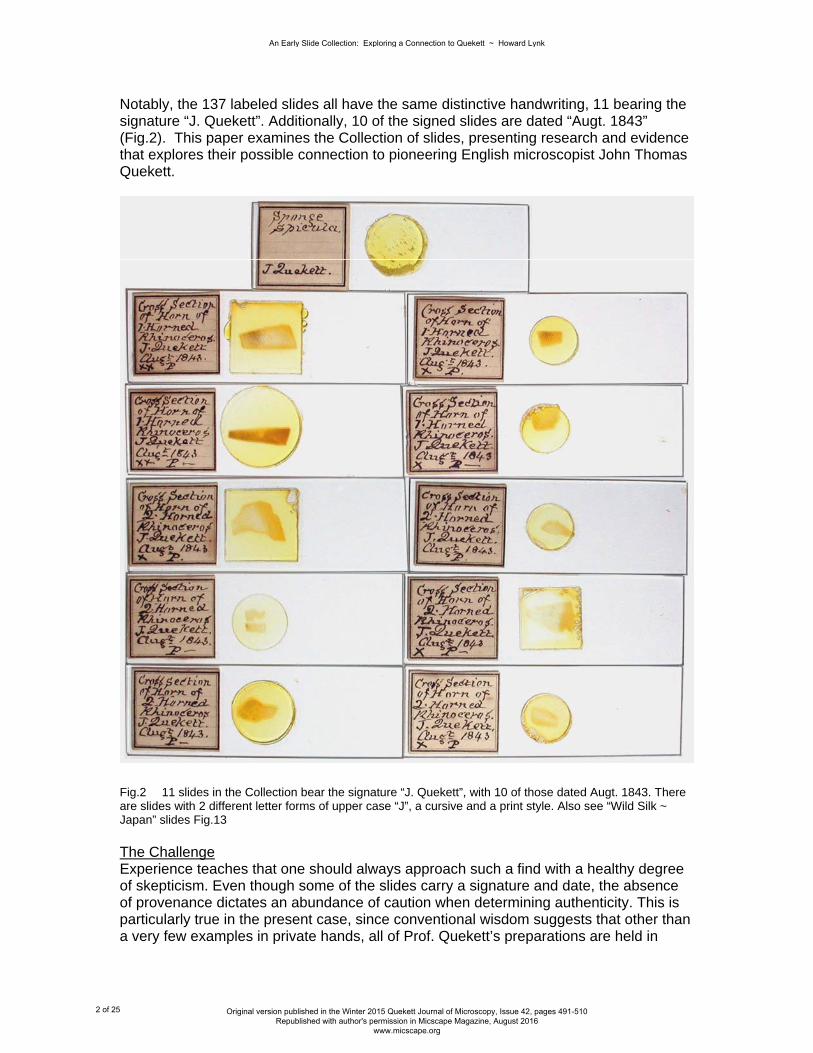
Notably, the 137 labeled slides all have the same distinctive handwriting, 11 bearing the signature “J. Quekett”. Additionally, 10 of the signed slides are dated “Augt. 1843” (Fig.2). This paper examines the Collection of slides, presenting research and evidence that explores their possible connection to pioneering English microscopist John Thomas Quekett.
Fig.2 11 slides in the Collection bear the signature “J. Quekett”, with 10 of those dated Augt. 1843. There are slides with 2 different letter forms of upper case “J”, a cursive and a print style. Also see “Wild Silk ~ Japan” slides Fig.13 The Challenge Experience teaches that one should always approach such a find with a healthy degree of skepticism. Even though some of the slides carry a signature and date, the absence of provenance dictates an abundance of caution when determining authenticity. This is particularly true in the present case, since conventional wisdom suggests that other than a very few examples in private hands, all of Prof. Quekett’s preparations are held in
An Early Slide Collection: Exploring a Connection to Quekett ~ Howard Lynk
2 of 25 Original version published in the Winter 2015 Quekett Journal of Microscopy, Issue 42, pages 491-510 Republished with author's permission in Micscape Magazine, August 2016
www.micscape.org

museums (primarily the Hunterian Museum, Royal College of Surgeons, London)[1]. A heretofore unknown Collection of slides prepared by Quekett coming to light in Australia after 170 years begs an explanation! How could this have happened? My efforts to answer that question, as well as either verify or rule out these slides as Quekett’s, involved research in several different but complementary directions. First thoughts included a possible family connection. Perhaps a descendant of Prof. Quekett emigrated to Australia with the box of slides as a family heirloom, their significance lost over time. Genealogical research was initiated to investigate this possibility. Another line of inquiry focused on the consistent handwriting seen on the slide labels. This involved careful comparison to the handwriting on known examples of Quekett’s microscopic preparations, as well as handwritten notes and letters. Finally, our investigation involved comparison of the specimens seen on the slides with work and interests Quekett was known to have pursued during the relevant time period. A variety of different source materials were used, including Prof. Quekett’s journals and publications. These areas of research and the results of my inquiries will be described and presented in detail as we proceed. Towards that end, let us first examine the slides and their case. The Collection Prior to focusing on individual slides, it is instructive to view them as a group. As found, the Collection consists of 138 glass microscope slides, each approximately 1” x 3” in size. The slides are all made of glass consistent with that seen produced in England in the 1840s; the glass shows wide variation in colour tint, thickness and uniformity. All but a few of the slides have the specimens mounted under thin glass covers of various shapes and sizes, including irregular pieces, using Canada balsam. On most, the balsam mountant has yellowed and darkened with age, comparable to other preparations from this time period. While a few of the slides are carefully finished with beveled and polished edges, most are not, suggesting they were prepared as non~commercial “working” mounts. The overall impression of the majority of preparations is one of functionality, the apparent primary objective being effective mounting of the specimens for study. Visual presentation and finish of the slides was evidently of less importance. The slides are housed in a sturdy purpose built case with fabric hinged top and drop front door (Fig.3). There is a single large brass hook and eye fastener. The case contains 12 wooden trays of 12 slide capacity each, thus capable of holding 144 slides. While of a somewhat unusual design, the case is typical mid 19th century construction, with heavy blue~green faux shagreen fabric over wood. Each of the 12 trays are consecutively numbered beginning with 1, using small paper labels. The exception is tray 12, which carries the number 19. This suggests more than one such case originally existed, each with its own set of uniquely numbered trays. Also included in the case were 8 pieces of old newspaper, cut to the same size as the slide trays. These can be identified as being from a London newspaper, dated 1903. They were obviously added at some point as spacers between the slide trays to prevent movement and help protect the slides. All of the slides but one have a single paper label, being either of two styles. 113 have a simple commercially printed label, 1 inch square with 6 lines for specimen details. The remaining 24 have hand cut labels of plain paper varying in size from approximately ½” to nearly 1” in either dimension. There is a single slide with no paper label, having
An Early Slide Collection: Exploring a Connection to Quekett ~ Howard Lynk
3 of 25 Original version published in the Winter 2015 Quekett Journal of Microscopy, Issue 42, pages 491-510 Republished with author's permission in Micscape Magazine, August 2016
www.micscape.org
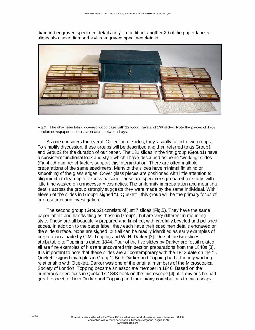
diamond engraved specimen details only. In addition, another 20 of the paper labeled slides also have diamond stylus engraved specimen details.
As one considers the overall Collection of slides, they visually fall into two groups. To simplify discussion, these groups will be described and then referred to as Group1 and Group2 for the duration of our paper. The 131 slides in the first group (Group1) have a consistent functional look and style which I have described as being “working” slides (Fig.4). A number of factors support this interpretation. There are often multiple preparations of the same specimens. Many of the slides have minimal finishing or smoothing of the glass edges. Cover glass pieces are positioned with little attention to alignment or clean up of excess balsam. These are specimens prepared for study, with little time wasted on unnecessary cosmetics. The uniformity in preparation and mounting details across the group strongly suggests they were made by the same individual. With eleven of the slides in Group1 signed “J. Quekett”, this group will be the primary focus of our research and investigation. The second group (Group2) consists of just 7 slides (Fig.5). They have the same paper labels and handwriting as those in Group1, but are very different in mounting style. These are all beautifully prepared and finished, with carefully beveled and polished edges. In addition to the paper label, they each have their specimen details engraved on the slide surface. None are signed, but all can be readily identified as early examples of preparations made by C.M. Topping and W. H. Darker [2]. One of the two slides attributable to Topping is dated 1844. Four of the five slides by Darker are fossil related, all are fine examples of his rare uncovered thin section preparations from the 1840s [3]. It is important to note that these slides are all contemporary with the 1843 date on the “J. Quekett” signed examples in Group1. Both Darker and Topping had a friendly working relationship with Quekett. Darker was one of the original members of the Microscopical Society of London; Topping became an associate member in 1846. Based on the numerous references in Quekett’s 1848 book on the microscope [4], it is obvious he had great respect for both Darker and Topping and their many contributions to microscopy.
Fig.3 The shagreen fabric covered wood case with 12 wood trays and 138 slides. Note the pieces of 1903 London newspaper used as separators between trays.
An Early Slide Collection: Exploring a Connection to Quekett ~ Howard Lynk
4 of 25 Original version published in the Winter 2015 Quekett Journal of Microscopy, Issue 42, pages 491-510 Republished with author's permission in Micscape Magazine, August 2016
www.micscape.org

Fig.4 A selection of 16 preparations from the Group1 slides. A wide variety of natural history specimens are represented.
An Early Slide Collection: Exploring a Connection to Quekett ~ Howard Lynk
5 of 25 Original version published in the Winter 2015 Quekett Journal of Microscopy, Issue 42, pages 491-510 Republished with author's permission in Micscape Magazine, August 2016
www.micscape.org

Fig.5 The 7 Group2 preparations by Darker and Topping are shown. Slides 1, 2, 4, 5, & 6 on the top row from right are by Darker; 3 is by Topping. Each slide 3 - 6 is also shown directly below on the bottom row, photographed with a dark background to accentuate the engraved writing. Slides 2 (dated 1844) and 3 on the bottom row are by Topping. Note unusual bone slide (6) engraved by Darker “St. Bartholo”. (Slide 1 on bottom row is a Group1 slide with engraved name “Mr. Ince”) The preparations in Group1 are a most interesting selection of natural history specimens. They range from the ordinary to the exotic. Mounts of common objects such as moth wing scales are found side by side with rare and unusual specimens sourced from locations around the globe. The table below (Fig.6) gives some idea of the wide range of preparations represented within Group1.
An Early Slide Collection: Exploring a Connection to Quekett ~ Howard Lynk
6 of 25 Original version published in the Winter 2015 Quekett Journal of Microscopy, Issue 42, pages 491-510 Republished with author's permission in Micscape Magazine, August 2016
www.micscape.org

Quantity Type Specimens
13 Animal Sections: Rhinoceros Horn, Whale Tooth; Mouse Whisker, Etc.
45 Botanical Various Dissections: Durio, Onosma, Deutzia, Equisetum, Seringa, Alyssum; Var. Starches, Raphides, etc.; Cotton Fiber
5 Infusoria Various Recent and Fossil from several UK locations; Guano
19 Insect Various Scales: Lepisma (Test), Moth; Dissections: Moth, Butterfly; Parasites: Pulex, Burying Beetle, Pediculus; Wild Silk
46 Marine Various Sponge and Spicula; Foraminifera; Cellularia; Sections of Echinus Spines; Var. Gorgonia; Palates; Var. Fish Scales
3 Other Selenite Fragments, Santonine, Iodide of Lead
Fig. 6 The 131 preparations in Group1 are a broad cross section of Natural History Specimens.
In addition to label details giving specimen name and collection location, many of the preparations have one or more letters or symbols in the same hand, usually along the bottom edge of the label. The meaning of “P” seems obvious, usually standing for Polarize or Polariscope; others are less so. It is possible that a grading or quality classification was assigned, denoted by the “x”, “xx”, or “Xx” seen on many of the slides. The significance or meaning of other symbols such as the stylized script “Q”, backwards facing “?”, large asterisk or star “*”, “V-1” and “Y”, or the Roman numerals “I”, “II”, and “III”, can only be guessed at (Fig.7). Notably, 93 of the 131 slides in Group1 have a “P” character on their label. Many of these are not specimens one would associate with the use of polarized light; a fact suggesting the need to confirm that my initial interpretation of the “P” designation was accurate. This was simply accomplished by examining a selection of those slides between crossed polarizing filters (Polariscope). In each case I was rewarded with the enhanced and often colourful view one expects to see with such objects. In light of the apparent connection between this Collection and Prof. Quekett, several paragraphs in his 1848 book “Practical Treatise on the Use of the Microscope” [4] take on an increased significance. In the chapter on polarized light he recommends and encourages the use of crossed polarizers (in his day, usually Nicol prisms) as a useful method in the investigation of botanical and animal structures. This was a novel idea at the time. Of particular interest, the specimens Quekett suggests as good examples to demonstrate this property include Deutzia, Equisetum, various starches, feathers, and sections of horn. All are represented by multiple preparations in Group1. Of the 11 slides in the Collection signed “J. Quekett”, 10 of them are sections of Rhinoceros Horn, and include preparations of both the 1 horned (Indian) and 2 horned (African) species (Fig.8).
An Early Slide Collection: Exploring a Connection to Quekett ~ Howard Lynk
7 of 25 Original version published in the Winter 2015 Quekett Journal of Microscopy, Issue 42, pages 491-510 Republished with author's permission in Micscape Magazine, August 2016
www.micscape.org

Fig.7 A selection of 9 Group1 slide labels showing some of the various symbols and characters seen as notations on the preparations. While the “P” denotes a polariscope object, others are less obvious. I did discover that the spring~like scribble along the bottom of the label for Acanthodium indicum indicates “spiral vessels”. This specimen was then imaged and used as the background for Fig.7 There is another smaller group or subset of 22 slides within Group1. These are similar in appearance to the others, but have certain characteristics that set them apart. They are the least finished looking; the glass slide edges are rough and uneven. All have small hand cut, odd sized plain paper labels (there are only 24 plain paper labeled slides in the entire Collection). These 22 labels read vertically, rather than horizontally like the other 109 slides in Group1. The handwriting on these, although undoubtedly by the same individual as the other slides in the Collection, has an unusual “fuzzy” appearance. Close examination provides the reason: the paper is of lesser quality and quite porous, having allowed the ink to bleed along the paper fibers. However, the most important difference between these 22 preparations and the others in Group1 is the specimens themselves (Fig.9). Without exception, they would be considered quite basic natural history objects. Most of the specimens could have been found within the immediate vicinity of practically any rural home place in mid 19th century England. The few foreign specimens, such as West Indian cotton fibers or Arrowroot starch, were common imports at the time. If asked to describe these 22 preparations in a word, the term “student” seems the most appropriate.
An Early Slide Collection: Exploring a Connection to Quekett ~ Howard Lynk
8 of 25 Original version published in the Winter 2015 Quekett Journal of Microscopy, Issue 42, pages 491-510 Republished with author's permission in Micscape Magazine, August 2016
www.micscape.org

In contrast, the other 109 slides in Group1 present a much different impression. Other than a few sourced from locations within the UK proper, they are specimens from the far flung reaches of the Victorian era British Empire. These lands include Africa, India, the West Indies, Japan, Australia, Malaysia, the South Pacific, and the Bahamas. Marine specimens from several of the world’s great oceans, including the Pacific, Atlantic,
Caribbean, and the Indian, as well as the Great Barrier Reef, are represented. Multiple variations of certain species are found: there are 15 preparations relating to different marine sponges. Other specimens have multiple preparations representing their structure: there are cross sections, spore, and cuticle mounts of Equisetum. Some species have multiple preparations from different locations; we find mounts of Gorgonia flabellum spicules (the sea fan or sea feather) from 5 different Pacific locations, the Bahamas, and New Caledonia. Numerous other interesting examples abound. If the word “student” described the previous group of 22 slides, these 109 specimens might best be characterized as “scientific” or perhaps “academic” preparations (Fig.10).
Fig.8 Many of the Group1 preparations have a “P” character on their labels. Observing a variety of the specimens between crossed polarizing filters (Polariscope) confirmed the meaning of “P”. Quekett promoted the usefulness of the Polariscope, even for botanical and animal tissues. The background photomicrograph is from the signed preparation of cross section of 2 horned Rhinoceros horn.
An Early Slide Collection: Exploring a Connection to Quekett ~ Howard Lynk
9 of 25 Original version published in the Winter 2015 Quekett Journal of Microscopy, Issue 42, pages 491-510 Republished with author's permission in Micscape Magazine, August 2016
www.micscape.org
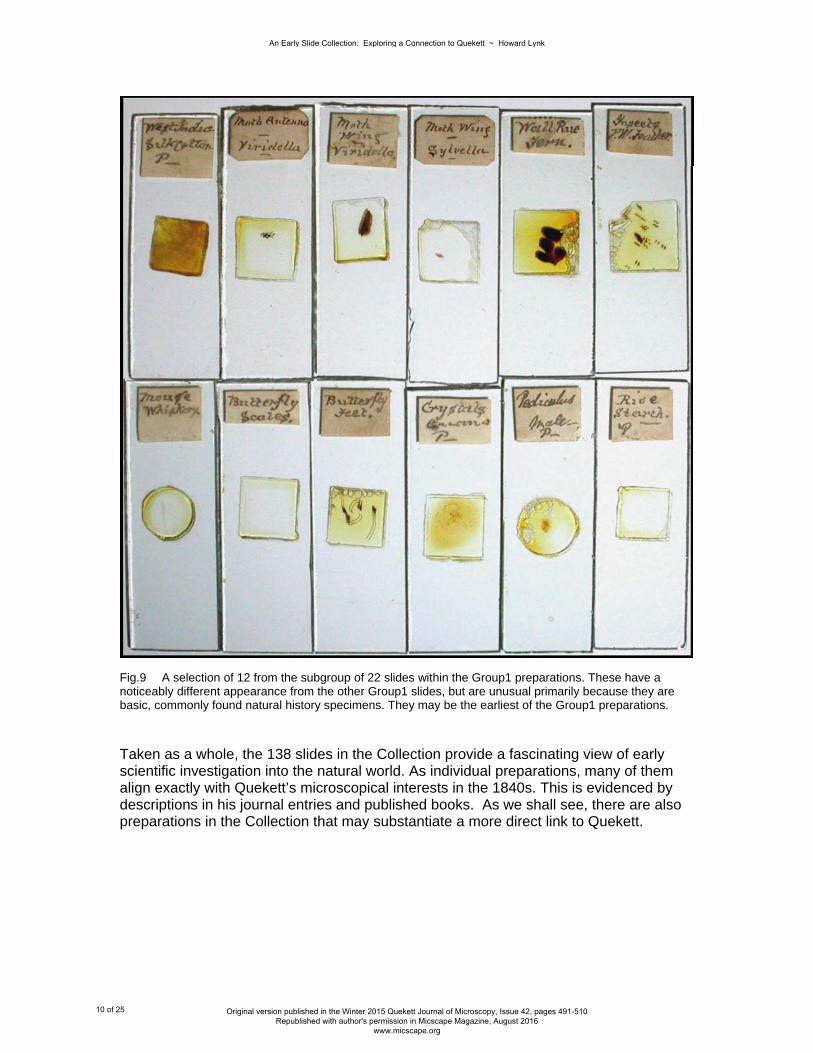
Fig.9 A selection of 12 from the subgroup of 22 slides within the Group1 preparations. These have a noticeably different appearance from the other Group1 slides, but are unusual primarily because they are basic, commonly found natural history specimens. They may be the earliest of the Group1 preparations. Taken as a whole, the 138 slides in the Collection provide a fascinating view of early scientific investigation into the natural world. As individual preparations, many of them align exactly with Quekett’s microscopical interests in the 1840s. This is evidenced by descriptions in his journal entries and published books. As we shall see, there are also preparations in the Collection that may substantiate a more direct link to Quekett.
An Early Slide Collection: Exploring a Connection to Quekett ~ Howard Lynk
10 of 25 Original version published in the Winter 2015 Quekett Journal of Microscopy, Issue 42, pages 491-510 Republished with author's permission in Micscape Magazine, August 2016
www.micscape.org
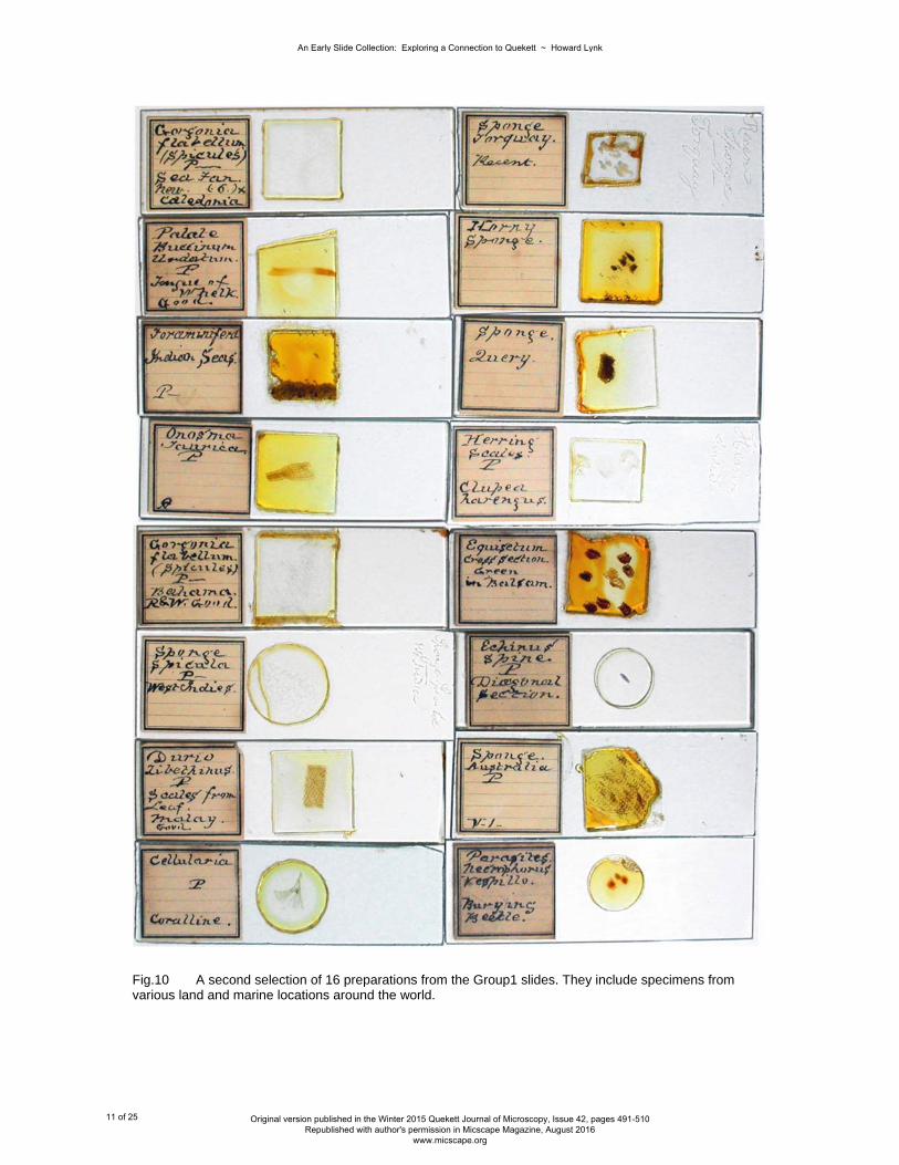
Fig.10 A second selection of 16 preparations from the Group1 slides. They include specimens from various land and marine locations around the world.
An Early Slide Collection: Exploring a Connection to Quekett ~ Howard Lynk
11 of 25 Original version published in the Winter 2015 Quekett Journal of Microscopy, Issue 42, pages 491-510 Republished with author's permission in Micscape Magazine, August 2016
www.micscape.org
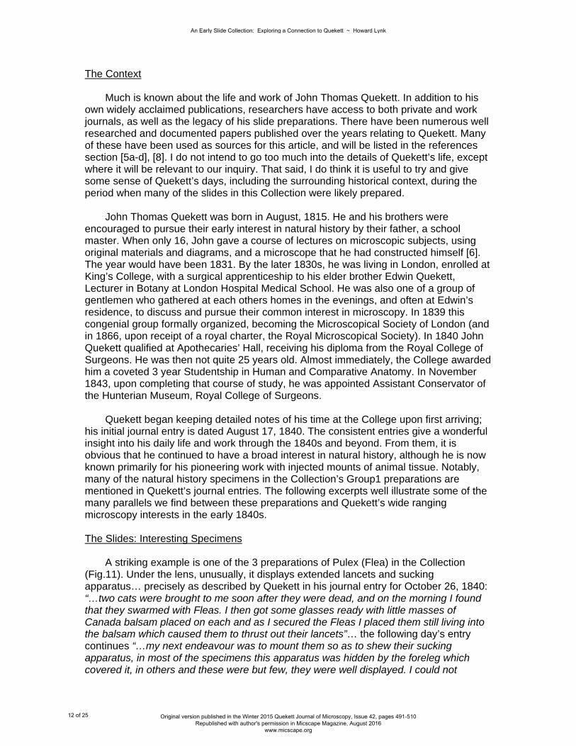
The Context Much is known about the life and work of John Thomas Quekett. In addition to his own widely acclaimed publications, researchers have access to both private and work journals, as well as the legacy of his slide preparations. There have been numerous well researched and documented papers published over the years relating to Quekett. Many of these have been used as sources for this article, and will be listed in the references section [5a-d], [8]. I do not intend to go too much into the details of Quekett’s life, except where it will be relevant to our inquiry. That said, I do think it is useful to try and give some sense of Quekett’s days, including the surrounding historical context, during the period when many of the slides in this Collection were likely prepared. John Thomas Quekett was born in August, 1815. He and his brothers were encouraged to pursue their early interest in natural history by their father, a school master. When only 16, John gave a course of lectures on microscopic subjects, using original materials and diagrams, and a microscope that he had constructed himself [6]. The year would have been 1831. By the later 1830s, he was living in London, enrolled at King’s College, with a surgical apprenticeship to his elder brother Edwin Quekett, Lecturer in Botany at London Hospital Medical School. He was also one of a group of gentlemen who gathered at each others homes in the evenings, and often at Edwin’s residence, to discuss and pursue their common interest in microscopy. In 1839 this congenial group formally organized, becoming the Microscopical Society of London (and in 1866, upon receipt of a royal charter, the Royal Microscopical Society). In 1840 John Quekett qualified at Apothecaries’ Hall, receiving his diploma from the Royal College of Surgeons. He was then not quite 25 years old. Almost immediately, the College awarded him a coveted 3 year Studentship in Human and Comparative Anatomy. In November 1843, upon completing that course of study, he was appointed Assistant Conservator of the Hunterian Museum, Royal College of Surgeons. Quekett began keeping detailed notes of his time at the College upon first arriving; his initial journal entry is dated August 17, 1840. The consistent entries give a wonderful insight into his daily life and work through the 1840s and beyond. From them, it is obvious that he continued to have a broad interest in natural history, although he is now known primarily for his pioneering work with injected mounts of animal tissue. Notably, many of the natural history specimens in the Collection’s Group1 preparations are mentioned in Quekett’s journal entries. The following excerpts well illustrate some of the many parallels we find between these preparations and Quekett’s wide ranging microscopy interests in the early 1840s. The Slides: Interesting Specimens A striking example is one of the 3 preparations of Pulex (Flea) in the Collection (Fig.11). Under the lens, unusually, it displays extended lancets and sucking apparatus… precisely as described by Quekett in his journal entry for October 26, 1840: “…two cats were brought to me soon after they were dead, and on the morning I found that they swarmed with Fleas. I then got some glasses ready with little masses of Canada balsam placed on each and as I secured the Fleas I placed them still living into the balsam which caused them to thrust out their lancets”… the following day’s entry continues “…my next endeavour was to mount them so as to shew their sucking apparatus, in most of the specimens this apparatus was hidden by the foreleg which covered it, in others and these were but few, they were well displayed. I could not
An Early Slide Collection: Exploring a Connection to Quekett ~ Howard Lynk
12 of 25 Original version published in the Winter 2015 Quekett Journal of Microscopy, Issue 42, pages 491-510 Republished with author's permission in Micscape Magazine, August 2016
www.micscape.org
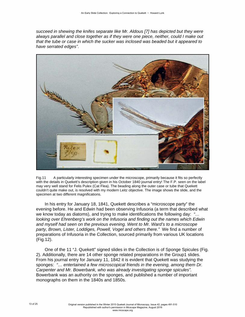
succeed in shewing the knifes separate like Mr. Aldous [7] has depicted but they were always parallel and close together as if they were one piece, neither, could I make out that the tube or case in which the sucker was inclosed was beaded but it appeared to have serrated edges”.
Fig.11 A particularly interesting specimen under the microscope, primarily because it fits so perfectly with the details in Quekett’s description given in his October 1840 journal entry! The F.P. seen on the label may very well stand for Felis Pulex (Cat Flea). The beading along the outer case or tube that Quekett couldn’t quite make out, is resolved with my modern Leitz objective. The image shows the slide, and the specimen at two different magnifications. In his entry for January 18, 1841, Quekett describes a “microscope party” the evening before. He and Edwin had been observing Infusoria (a term that described what we know today as diatoms), and trying to make identifications the following day: “… looking over Ehrenberg’s work on the Infusoria and finding out the names which Edwin and myself had seen on the previous evening. Went to Mr. Ward’s to a microscope party, Brown, Lister, Loddiges, Powell, Vogel and others there.” We find a number of preparations of Infusoria in the Collection, sourced primarily from various UK locations (Fig.12). One of the 11 “J. Quekett” signed slides in the Collection is of Sponge Spicules (Fig. 2). Additionally, there are 14 other sponge related preparations in the Group1 slides. From his journal entry for January 11, 1842 it is evident that Quekett was studying the sponges: “… entertained a few microscopical friends in the evening, among them Dr. Carpenter and Mr. Bowerbank, who was already investigating sponge spicules”. Bowerbank was an authority on the sponges, and published a number of important monographs on them in the 1840s and 1850s.
An Early Slide Collection: Exploring a Connection to Quekett ~ Howard Lynk
13 of 25 Original version published in the Winter 2015 Quekett Journal of Microscopy, Issue 42, pages 491-510 Republished with author's permission in Micscape Magazine, August 2016
www.micscape.org

In his comprehensive paper on Quekett, “Famous Microscopists: John Thomas Quekett, 1815 – 1861” [8], Bracegirdle mentions Quekett’s very early work with Daguerreotype photography. He states: “Interestingly, Quekett was one of the pioneers in the use of the Daguerreotype process, as the following extract reveals…”, he then continues with Quekett’s journal entry for August 14, 1843: “ …call on Ross in my way to the College and arrange about the Iodine and Bromine boxes for the daguerreotype.” Quekett’s early interest in photography may explain a most unusual chemical mount found among the Group1 slides (Fig.12). Unusual because there are only 2 chemical preparations in the entire Collection, but primarily because it is of a rarely seen compound: Iodide of
Lead. Of importance to our investigation, I discovered that for a time in the late 1830s into the 1840s, this compound was used in early experiments investigating practical photographic methods. Iodide of Lead is sensitive to light. Of additional interest, some of that experimental photography was being conducted by J.B. Reade with the chemist Hodgson at Apothecaries’ Hall [9]. This was at the same time Quekett would have been there completing his medical education. Another interesting preparation in the Collection illustrates not only a possible connection to Quekett, but also one of the ways specimens were sourced at the time. The slide is labeled “Sponge Spicules ~ West Indies” with the name “Mr. Ince” engraved on the slide end (Fig.12). Quekett’s journal entry for August 18, 1843 includes: “… carry
Fig.12 This image shows several different preparations from the Group1 slides that parallel some of the many interests that Quekett mentioned in his journals from the early 1840s. The top 2 slides are Infusoria from locations in the UK. The 3rd slide, an unusual chemical mount of Iodide of Lead, could be linked to Quekett’s very early interest in photography. The bottom preparation is of particular interest because of the connection to Mr. Ince, another original member of the MSL along with Quekett. This slide (taken against a dark background) is also shown as the 1st slide, bottom row, Fig.5
An Early Slide Collection: Exploring a Connection to Quekett ~ Howard Lynk
14 of 25 Original version published in the Winter 2015 Quekett Journal of Microscopy, Issue 42, pages 491-510 Republished with author's permission in Micscape Magazine, August 2016
www.micscape.org

with me today some objects for (Mr.) Ince and for Dr. Davies…”. W.H. Ince was an original member of the Microscopical Society of London. His brother, Commander J.M.R. Ince, (later Captain) was in the Royal Navy with service in the Mediterranean, West Indies, East Indies, and Australia. In 1847, W.H. Ince honoured his brother by naming a new insect species from Australia after him [10]. Cmdr. Ince had provided his brother with the unusual specimens, and I think it fairly obvious this was probably not an isolated occurrence. Lastly, we will examine an intriguing slide made by W.H. Darker [2]. It is one of the 7 preparations in Group2. The paper label simply states “Query. Human Bone”, but there is an underlying engraved label in Darker’s hand: “St. Bartholo’ ”. The preparation is an uncovered triple section of what appears to be aged or fossil bone (Fig.5, Fig.17). The mount is prepared in exactly the same way as Darker’s other fossil and mineral slides, for which he was well known. Of course, it is possible this is just a common histology slide of bone from St. Bartholomew’s Hospital, but I have never seen any evidence that Darker prepared such utilitarian mounts. The colouration of the specimen is also quite different from that seen with comparable sections of contemporary mid 19th century bone. My research suggests the history of this unusual specimen may be far more interesting! The tale begins around 1025 when Queen Emma, wife of Canut the Great and mother of Edward the Confessor, purchased a religious relic, purported to be the arm bone of St. Bartholomew the Apostle, from the Italian Bishop of Benevento [11]. She then presented it as a gift to the monks at Christ Church, Canterbury. Thereafter, the relic can be traced, through the occasional inventories of Canterbury Cathedral done by the church, over the next 800 years. One of many religiously significant pieces there, it survived multiple fires, periods of neglect, and episodes of building and reconstruction. It was forgotten and “rediscovered” several times over the centuries. Unfortunately, it was among a number of items that vanished while being stored during the great reconstruction of the Cathedral that was started in 1832 and finally completed in 1840. Those items have never been found. Whether the specimen on the “St. Bartholo’” slide was actually sourced from the same relic that was once at Canterbury Cathedral, or how Darker might have acquired it, we will likely never know. There is little doubt that Quekett would have been interested in such a specimen. He published his monograph “On the Intimate Structure of Bone, As Composing the Skeleton in the Four Great Classes of Animals; Mammals, Birds, Reptiles, and Fishes” in 1846. It was not unusual for Quekett’s expert opinion to be sought in matters where microscopical evidence might prove pivotal. The Torbanehill mineral case (mineral or coal?) [12], and his verification of the material taken from several old Essex Church doors as human skin (Viking pirates!) [13], are well known. In another matter similar to this “St. Bartholo’” bone specimen, it is rumored that Quekett was asked in the early 1850s to verify bone fragments from a crypt at Westminster Abbey. Long thought to be the remains of a legendary historical figure, Quekett determined they were actually of somewhat more humble origin: they were found to be frog bones. There is a sentence in Quekett’s journal entry for August 30, 1843 that makes an unintentional, but important point in regards to his microscopic preparations, and potentially, this Collection of 138 slides: “ … I then started for home and prepared some objects to take with my microscope to Dr. Willis’s with whom I spent the evening…”. It is clear from Quekett’s journal entries over nearly two decades that microscopy (including object preparation) was a passion, practiced not only in the work rooms at the Hunterian Museum, but frequently at home and in social settings with his many friends and associates. One can only come to the conclusion that Quekett made a great many
An Early Slide Collection: Exploring a Connection to Quekett ~ Howard Lynk
15 of 25 Original version published in the Winter 2015 Quekett Journal of Microscopy, Issue 42, pages 491-510 Republished with author's permission in Micscape Magazine, August 2016
www.micscape.org

microscopic preparations over the years, some fair number of which likely remained in his personal collections. The Handwriting Anyone with more than a passing interest in antique microscope preparations has spent time examining and comparing the handwriting on slide labels. In some cases the only way to determine who might have made a slide, is by visually matching the handwriting with preparations by known makers. That said, using handwriting as the only determining factor can easily lead to error and misidentification. Fortunately for this inquiry, we have signed and dated mounts in the Collection that may point to their preparer: John Thomas Quekett. We also have known Quekett preparations to compare them with, along with his journal entries and other publications to help substantiate an identification. For the handwriting comparisons, high resolution images of known Quekett slide preparations were obtained from the Hunterian Museum, Royal College of Surgeons. I also have two original letters written by Quekett, one dated to 1846. His journal entries, notes, personal letters, and other writings were normally written in a longhand cursive script. In contrast, handwriting on Quekett’s slides is generally non~cursive printed lettering, although he sometimes added various words or upper case letters in cursive. To facilitate the comparisons, the illustration figures will usually show examples of writing from two sources: the known Quekett handwriting (either museum slides or personal letters), and the Collection slides, presented together in a side~by~side arrangement. The caption for each illustration will point out or explain pertinent details. All of the 137 paper labeled slides in the Collection have clear and legible descriptions written with ink in the same hand. The Quekett slides in the museum are also labeled with a bold, consistent handwriting. There are important similarities in the handwriting seen on the museum’s Quekett slides, with that on the Collection slides. The labels on both groups are mostly written using printed letter formations rather than a cursive or script style. There are certain upper case letters, such as “J”, “I”, “D”, and “L” where the cursive form is often used in place of the printed letter version (Fig.13). On both group’s labels, the upper case cursive letters match those seen in Quekett’s personal correspondence and journal entries (Fig.14). We find the liberal use of serifs on many letters, both upper and lower case, on both the Museum and Collection slide labels. That said, the serifs are more frequent and exaggerated on the Collection slides (Fig.15). On the museum slides, there are letters where Quekett switches back and forth between using a somewhat unusual letter formation; his lower case “g” is a good example of this. We find this exact same characteristic on the Collection slides (Fig.16). The visual similarity of certain words across both groups is sometimes striking, as one might expect if all were written by Quekett (Fig.17). In general, we find the basic letter formations are nearly identical, and the slope or letter slant consistently the same, as is the overall look and style of the handwriting.
An Early Slide Collection: Exploring a Connection to Quekett ~ Howard Lynk
16 of 25 Original version published in the Winter 2015 Quekett Journal of Microscopy, Issue 42, pages 491-510 Republished with author's permission in Micscape Magazine, August 2016
www.micscape.org

Fig.13 Museum Quekett slides are on the top row, Collection slides the bottom row: in looking at known Quekett preparations, one sees that he used different letter forms of some letters. Sometimes he used the cursive form, and other times the printed… sometimes a cursive word or single letter would be mixed in with printed words. We find this same tendency with the slides in the Collection. Note the “L”, “F”, “T”, “I” and “J”. The 2 forms of “J” seen on the Collection slides is of particular importance because of his signature. In spite of the many similarities, there are some differences between the handwriting on the museum’s Quekett slides and that seen on the preparations in the Collection. In discussing this with several individuals that have long experience with handwriting analysis and comparison, several points were made that may explain the differences. First, an individual’s handwriting naturally changes over time. One only has to look back at their own handwriting for evidence of this. If the Collection slides were prepared and labeled in the early 1840s when Quekett was still in his 20s (as other evidence suggests), we are comparing them to slides labeled many years later. Secondly, a person’s writing can change dramatically depending on the purpose. Preparations originally made and quickly labeled for personal study or “temporary” use would likely look quite different from those labeled for a permanent institutional collection such as the Hunterian Museum. The “institutional” hand will almost always be more consistent, reserved, and conservative… an accurate description for the museum’s Quekett slides
An Early Slide Collection: Exploring a Connection to Quekett ~ Howard Lynk
17 of 25 Original version published in the Winter 2015 Quekett Journal of Microscopy, Issue 42, pages 491-510 Republished with author's permission in Micscape Magazine, August 2016
www.micscape.org

as compared to those in the Collection. Finally, the pen point can make a significant difference in the handwriting. In contrast to those in the Collection, most of the museum Quekett slide labels appear to have been written using broader heavier pen nibs, giving the writing a bold less embellished style.
Fig.14 Here we see several slides from the Collection, one from the museum’s known Quekett mounts, and copies of 2 of Quekett’s letters, arranged for comparison with each other. Take note of the upper case “J” and “I” seen on each of the items. Finally, we have the 14 slides in Group1 that have specimen details engraved on the glass in addition to their paper labels. On most of these the engraving is in a cursive script that is very consistent with the handwriting seen in Quekett’s journals and his personal letters (Fig.18).
An Early Slide Collection: Exploring a Connection to Quekett ~ Howard Lynk
18 of 25 Original version published in the Winter 2015 Quekett Journal of Microscopy, Issue 42, pages 491-510 Republished with author's permission in Micscape Magazine, August 2016
www.micscape.org

Fig.15 A selection of recognized Quekett preparations (top row) for comparison with slides from the Collection on the lower row. One still sees embellishments and vertical serifs on Quekett’s museum mounts, but they are generally held to a minimum, and only on a few characters, such as the “S”. The labels on the slides in the Collection generally show very similar style and placement, but with greater variety and frequency. The Journey: From There to Here Having received and become more familiar with the slides in the Collection, my attention turned towards how best to answer the obvious question: were these actually Quekett’s preparations? In addition to research on the specimens and handwriting, efforts were initiated to discover any historical mention or reference to such slides or the Collection.
An Early Slide Collection: Exploring a Connection to Quekett ~ Howard Lynk
19 of 25 Original version published in the Winter 2015 Quekett Journal of Microscopy, Issue 42, pages 491-510 Republished with author's permission in Micscape Magazine, August 2016
www.micscape.org

Fig.16 Another selection of Quekett slides from the Hunterian, top row, and Collection slides, bottom rows. Again, Quekett sometimes uses different letter formations. In this case, we are especially comparing the lower case “g” anf “f”. Both groups show this somewhat unusual formation of “g”. Most of the Quekett preparations I have seen from the museum use the “g” form seen on the 1st slide top row. That may be what he transitioned to from an earlier form, as seen on his others (top row), and the Collection preparations. It seemed that a reasonable first effort might be an attempt to uncover a Quekett family connection with the slides. Internet based searches provided a trove of genealogical information on the Quekett family. Of interest, I discovered that Prof. Quekett’s great grandson John William Scott “Bill” Quekett (a descendant of his eldest son) had located to Australia with his family in the mid 20th century. Descendants of another son were located, still living in the UK. Using the wonders of modern communication, the “search engine” and email, I sent out several inquiries. An interesting and helpful communication developed with members of both the UK and Australian Queketts. Each basically said the same thing: other than those now in museums, they had no knowledge or memory of any family owned microscope slides that once belonged to their illustrious forebear.
An Early Slide Collection: Exploring a Connection to Quekett ~ Howard Lynk
20 of 25 Original version published in the Winter 2015 Quekett Journal of Microscopy, Issue 42, pages 491-510 Republished with author's permission in Micscape Magazine, August 2016
www.micscape.org

Fig.17 We see a striking similarity in the word “Human”, between the known Quekett on the left, and the Collection slide on the right. I first came across a hint of how the Collection may have made its way into private hands, in Bracegirdle’s previously cited work on Prof. Quekett [8]. His paper mentions the public auction of Quekett’s property after his death, and shows several pages from the auction catalogue. Further online research provided another important piece of information: I was able to discover the published announcement of the auction, including additional new details. Several months after Quekett’s death in August, 1861, an advertisement appeared announcing the upcoming public sale. All of Prof. Quekett’s household effects and personal property were to be sold off, beginning December 2, 1861. The announcement that was in the Athenaeum is shown in (Fig.19). Note what it specifically mentions among the items to be auctioned: “… a large Quantity of Prepared Objects”, those being a part of “… the whole of the Highly Interesting and Important Collections made by the late Prof. Quekett”. The auction sale catalogue was 45 pages, with at least 4 listing various microscopes and related items, including many cabinets and cases with preparations [14]. Although we have no way to know for certain, it may be that our unremarkable cloth covered case of 138 slides is one of the “9 others”. These were listed on page 24, as item #490 [8, 14]: “a mahogany object case, and 9 others”. That would also explain the tray in our case that is numbered 19 instead of 12.
An Early Slide Collection: Exploring a Connection to Quekett ~ Howard Lynk
21 of 25 Original version published in the Winter 2015 Quekett Journal of Microscopy, Issue 42, pages 491-510 Republished with author's permission in Micscape Magazine, August 2016
www.micscape.org

Fig.18 Comparison of a selection of Group1 Collection slides with engraved cursive writing on the glass, and Quekett’s cursive writing as seen in his personal letters. There is a consistency in letter formation, slant, and spacing across the samples, even though one frequently sees differences in hand when using a diamond point stylus on glass as opposed to pen and ink on paper. What is clear is that some significant number of Quekett’s preparations were sold at public auction after his death. This explains how it might be possible for a long forgotten and unrecognized collection of his slides to come to light at a recent estate sale auction. We also know that a great many people emigrated from the UK to Australia in the later 19th and early 20th Century, one of whom must have carried this Collection of slides to their new home. It is tempting to speculate that the pieces from the 1903 London newspaper, placed as separators between the slide trays to prevent damage, might possibly mark the Collection’s date of passage. The Conclusion The question remains: were these John Thomas Quekett’s preparations? Based on the substantial evidence we now have, I think the answer is very likely “yes”. We have the relatively untouched case of preparations, some signed with Quekett’s signature and dated. All of the slides in the Collection are labeled in the same handwriting as the signed examples. The specimens and handwriting can be identified with Quekett’s known preparations and journal entries.
An Early Slide Collection: Exploring a Connection to Quekett ~ Howard Lynk
22 of 25 Original version published in the Winter 2015 Quekett Journal of Microscopy, Issue 42, pages 491-510 Republished with author's permission in Micscape Magazine, August 2016
www.micscape.org

Fig.19 The print announcement of the upcoming auction of Professor John Quekett’s household and personal possessions. This appeared in the November 2nd, 1861 edition of the Athenaeum newspaper. This was one month before the sale was to take place. Of note, the sale included “… a large quantity of prepared objects”. There are preparations in the Collection that are attributable to two other recognized makers from the same time period, helping to verify the dating. In regards to their authenticity, I think it significant that no effort was made to exploit the Quekett name in
An Early Slide Collection: Exploring a Connection to Quekett ~ Howard Lynk
23 of 25 Original version published in the Winter 2015 Quekett Journal of Microscopy, Issue 42, pages 491-510 Republished with author's permission in Micscape Magazine, August 2016
www.micscape.org

the original 2014 auction in Australia. The fact that no attempt was made to market or “sell” the Collection as anything other than a box of old microscope slides makes a powerful argument that they were not forged or fraudulent. With ongoing research, the evidence we have uncovered and brought forth continues to accumulate. Due to limitations in length, our paper has only touched on some of the main highlights; there are numerous other interesting details, especially with the specimens. All point to the same conclusion. Throughout my research on this Collection, I have been fortunate to have the benefit of consultation with various individuals having long experience in the study of the history of science. Several specialize in the Victorian era when Quekett was active. Two have extensive experience with Quekett’s preparations at the Hunterian Museum. Their unanimous opinion is that this group of 138 preparations was indeed once part of John Quekett’s personal collection. There is no way to know with absolute certainty whether the preparations in Group1 were all made by Quekett. That said, based on the specimens and other associated evidence, it is highly likely they were. They would have been prepared during the early 1840s, to support his natural history studies. The small group of 22 basic preparations that are a part of Group1 may be even earlier. Research on the Collection of slides continues. There appears to be a close correlation between many of the specimens and Quekett’s later publications. Perhaps further discoveries or insights concerning the Collection will be presented in a future article or addendum. Acknowledgements My sincere thanks to Emmy Bocaege and the staff at the Royal College of Surgeons, London for assistance in acquiring images of John Quekett’s preparations and journal pages; A grateful thanks to Brian Bracegirdle*, for his encouragement and suggestions, and especially for making his various previously published research on Prof. Quekett accessible, without which this paper would not have been possible; a special thanks to Ruth Richardson for her helpful insights; many thanks to the extended Quekett family, especially to Will Quekett and Nicki Quekett (the Australian connection), to David Quekett, and to Ian Quekett, for communication and kindly sharing of family history; and finally, and finally, my sincere thanks to Quekett Microscopical Club Journal Editor Phil Greaves. * Brian Bracegirdle’s contributions to the research and documentation of the history of microscopy are legendary. This paper would not have been possible without the foundation he provided, and his helpful suggestions. Sadly, he passed away just weeks before it’s publication in the Winter 2015 Quekett Journal. Howard Lynk North Carolina, USA Website: victorianmicroscopeslides.com Email contact: [email protected] Further questions or comments to the author are welcomed.
An Early Slide Collection: Exploring a Connection to Quekett ~ Howard Lynk
24 of 25 Original version published in the Winter 2015 Quekett Journal of Microscopy, Issue 42, pages 491-510 Republished with author's permission in Micscape Magazine, August 2016
www.micscape.org

References 1. Bracegirdle, B – Microscopical Mounts and Mounters, London, Quekett Microcopical Club, 1998. See page 78 2. Davidson, B – Fossils Under the Microscope, Quekett Journal of Microscopy, 41: 731. See pages 735-737 3. Falcon-Lang, H.J., Digrius, D.M. – Paleobotany Under the Microscope, Quekett Journal of Microscopy, 42 : 253. see pages 272-274 4. Quekett, J.T. – A Practical Treatise On the Use of the Microscope, London, Bailliere, 1848 5a. Herlihy, E.P. – Professor John Quekett, Microscopy, 1956, 27 : 331-338 5b. Barron, A.L.E., - Concerning John Thomas Quekett and Some of His Contempories, Microscopy, 1962, 29 : 55-59 5c. Bracegirdle, B. – John Thomas Quekett and his Work on the Healing of Wounds, Microscopy, 1988, 36: 5d. Bracegirdle, B. – John Quekett’s Microscopical Preparations, Microscopy, 1986, 35 : 330-336 6. Lee, S. – Dictionary of National Biography, Vol. XLVII, London, Macmillan & Co., 1896, J.T. Quekett, pages 97-98 7. Lens Aldous, Esq. did many illustrations for Quekett. He was an original member of the Microscopical Society of London, and later the Quekett Microscopical Society. 8. Bracegirdle, B – Famous Microscopist: John Thomas Quekett, 1815 - 1861, Proceedings, Royal Microscopical Society, 1988, 23 : 149-169 9. Wood, R.D. – J.B. Reade, FRS, and the Early History of Photography, Annals of Science, Vol. 27, No.1, 1971 10. Newman, E. – Insects, The Zoologist, Vol4, 1846, see page 1274; Vol. 5, 1847, see pages 1676-1677 11. O’Brien, H. – Queen Emma and the Vikings, A History of Love, Power, and Greed in 11th Century England, Bloomsbury, 2010, Chapter 9 12. Quekett, J.T. – On the Minute Structure of a Peculiar Combustible Mineral, from the Coal Measures of Torbane Hill etc., Transactions, Microscopical Socity of London, 1854, ns2 : 34-65 13. Quekett, J.T. – On the Value of the Microscope in the Determination of Minute Structures of a Doubtful Nature, as Exemplified in the Identification of Human Skin Attached Many Centuries Ago to the Doors of Churches, Transactions, Microscopical Society of London, 1849, 2 :151-157 14. Salmon, M.V. – The Quekett Sale Catalogue, Microscopy, 1962, 29 : 9-11
An Early Slide Collection: Exploring a Connection to Quekett ~ Howard Lynk
25 of 25 Original version published in the Winter 2015 Quekett Journal of Microscopy, Issue 42, pages 491-510 Republished with author's permission in Micscape Magazine, August 2016
www.micscape.org