an analysis of pin track infections - llrs.orgllrs.org/pdfs/annual meeting...
TRANSCRIPT

An Analysis of Pin Track Infections
Christopher Iobst, MD Nemours Children’s Hospital
Orlando, FL

Disclosures
• Speaker’s Bureau for Smith and Nephew

Introduction
• Stability and health of the pin-skin/pin-bone interface critical in external fixation
• Breakdown at the pin-skin/pin-bone interface creates pin track infections
• Pin track infections are an anticipated nuisance of using external fixation

Introduction
• Despite pin track infections being ubiquitous in external fixation the data is limited and poor quality
• Can find references stating range of pin track infections is anywhere between 0% and 100%
• Examine the external fixation literature since 1980 to determine a more refined understanding of pin track infections

Methods
• PubMed search for “external fixation” – 9620 articles found – Search limits created
• Custom range of dates (01/01/1980 – 06/30/2014) • Human only • English only
– 5493 articles found

Methods
• Exclusion criteria – External fixation of axial skeleton (cranium/pelvis/
spine) – Case reports (minimum 5 patients) – Unable to access journal article from Nemours
online library – Results had to report pin track infections as
number of patients affected (not by individual pin/wire)

Methods • Data points extracted: – Year of publication – Number of patients – Average age of patients – Reason for external fixation (trauma, lengthening,
deformity, etc.) – Fixation per segment (two points or more than two points) – Body part (distal radius, femur, humerus, tibia, etc.) – Hydroxy-apatite coated pins versus non-coated pins – Duration of external fixation – Type of fixator (circular versus uni-planar) – Number of patients with documented pin track infections

Results
• Each year (1980-2014) represented by at least one article
• 150 total articles reviewed (6130 patients) – 1980s = 31 articles (1295 patients) – 1990s = 34 articles (1631 patients) – 2000s = 48 articles (1583 patients) – 2010s = 37 articles (1621 patients)

Results
• Total = 6130 patients with 1684 pin track infections
• Overall Pin track infection rate = 27.4%

Results
• Adult patients: – Pin track infection rate = 23% (964/4222)
• Pediatric patients: – Pin track infection rate = 38% (706/1851)

Results
• Age – Under 18 years old = 38% (717/1893) – 18-39 years old = 27% (556/2082) – 40-64 years old = 23% (309/1329) – Over 65 years old = 8% (31/389)

Results
• Patient Etiology – Trauma:
• 24 % Pin track infection rate (986/4161)
– Deformity: • 29% Pin track infection rate (348/1199)
– Lengthening: • 46% Pin track infection rate (239/512)

Results
• Duration of time in frame – 42 days or less = 19.6% (191/972) – 43 to 90 days = 24.2% (475/1956) – 91 to 150 days = 27.2% (498/1828) – More than 150 days = 37.8% (440/1161) – More than 180 days = 47.8% (335/700)

Affect of Duration on Pin Track Infection Rate
19.6 24.2
27.2
37.8
47.8
0
10
20
30
40
50
60
<42 days 43-‐90 days 91-‐150 days > 150 days >180 days
PTI Rate
PTI Rate

Results
• Location of frame pin track infection rate: – Tibia = 33% (754/2278) – Distal radius = 12 % (139/1122) – Femur = 22% (245/1099) – Humerus = 23% (53/228)
– Upper extremity = 14% (213/1511) – Lower extremity = 31% (1285/4155)

Results
• Distal radius: 48% six weeks duration or less
• All distal radius frames removed within 67 days
• Only 3% Femur frames and 12% Tibial frames removed with < 67 days duration

Results
• Pin type – Hydroxyapatite (HA) coated = 29.5% (71/240)
– Non HA coated = 25.9% (1457/5609)

Results
• Frame Type – Circular: 29.5% pin track infection rate
(457/1545)
– Uni-planar: 22.9% pin track infection rate (937/4089)

Results
0
10
20
30
40
50
60
70
80
<90 days 90 to 180 days > 180 days
Circular
Uni-‐planar

Results
0
10
20
30
40
50
60
70
80
90
Trauma Deformity Lengthening
Circular
Uni-‐planar

Results
• 1980s: Pin track infection rate = 23.2% – (1295 patients with 301 infections)
• 1990s: Pin track infection rate = 25.9% – (1631 patients with 424 infections)
• 2000s: Pin track infection rate = 36.1% – (1583 patients with 573 infections)
• 2010s: Pin track infection rate = 23.8% – (1621 patients with 386 infections)

Results
0
10
20
30
40
50
60
Trauma Deformity Lengthening
1980s
1990s
2000s
2010s

Results
0
10
20
30
40
50
60
70
1980s 1990s 2000s 2010s
Distal Radius Femur Tibia
Results
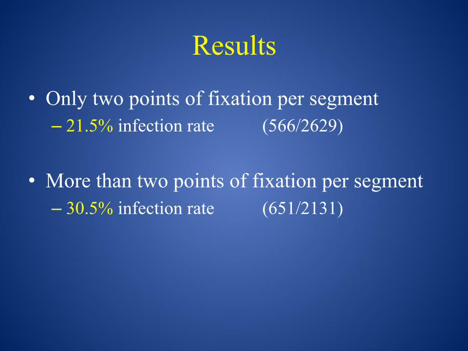
• Only two points of fixation per segment – 21.5% infection rate (566/2629)
• More than two points of fixation per segment – 30.5% infection rate (651/2131)
Results
0
10
20
30
40
50
60
70
80
90
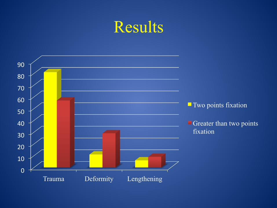
Trauma Deformity Lengthening
Two points fixation
Greater than two points fixation

Results
0
10
20
30
40
50
60
70
Distal Radius
Femur Humerus Tibia
Two points of fixation
More than two points of fixation

Results
0
5
10
15
20
25
30
35
< 42 days > 150 days
Two points of fixation
More than two points of fixation

Discussion
• Overall pin track infection rate since 1980 =
27.4%
(1684/6130 patients)

Discussion
• Pediatric patients 1.7X more likely to develop a pin track infection than an adult patient
• Pin track infection rate appears to steadily decrease with increasing age

Discussion
• Direct correlation between duration of external fixator time and the risk in pin track infection
• As time in frame increases the pin track infection risk increases
• Dramatically starts to increase after 150 days

Discussion
• Underlying etiology for frame affects pin track infection rate
• Lengthening approximately 2X risk compared to deformity correction and trauma patients
• Likely related to duration

Discussion
• Location of frame affects pin track infection rate – Lower extremity 2.2X more likely than upper
extremity to have pin track infection – Tibia>Humerus>Femur>Distal radius – Tibia 2.75X more likely than distal radius

Discussion
• Hydroxy-apatite coated pins did not appear to improve pin track infection rate
• Circular frames have higher pin track infection rate than uni-planar frames – Duration – Location – Etiology

Discussion
• Most at risk patient: Pediatric patient undergoing prolonged tibial lengthening with a circular frame using more than 3 points of fixation per segment

Conclusion
27.4%

References 1980-1989 • 1981 Edge External Fixation for complicated tibial fractures • 1982 Court-Brown Experience with the Sukhtian-Hughes external fixation system • 1983 Coppola Use of the Hoffman external fixator in the treatment of femoral fractures • 1983 Velazco Hoffman fixation for tibial fractures • 1983 Cooney External fixation of distal radius fractures • 1983 Larsson Open tibial shaft fractures • 1983 Karlstrom External fixation of severe open tibial fractures with the Hoffman frame • 1983 Stephens Femoral and tibial lengthening • 1983 Tolo External skeletal fixation in children's fractures • 1983 Hedley External fixation as a secondary procedure • 1984 Dabezies Fractures of the femoral shaft treated by external fixation with the Wagner device • 1984 Green External fixation for the uninfected angulated nonunion of the tibia • 1985 Vaughan Treatment of unstable fractures of the distal radius by external fixation • 1985 Court-Brown Hughes external fixator in treatment of tibial fractures • 1986 Behrens External fixation of the tibia • 1986 Foster Update on external fixators in the treatment of wrist fractures • 1987 Kristiansen External fixation of displaced fractures of the proximal humerus • 1987 Jenkins External fixation of Colles' fractures • 1987 Clyburn Dynamic external fixation for comminuted intra-articular fractures of the distal end of the radius • 1987 McCoy External fixation in contemporary fracture management • 1987 Alonso Use of the AO/ASIF external fixator in children • 1987 Rand Failed total knee arthroplasty treated by arthrodesis of the knee using the Ace-Fischer apparatus • 1988 Edwards Severe open tibial fractures 1989 Paterson Lower limb lengthening by a modified Wagner technique • 1989 Maurer Infection after intramedullary nailing of severe open tibial fractures initially treated with external fixation • 1989 Kongsholm Plaster cast versus external fixation for unstable intraarticular Colles' fractures • 1989 Holbrook Treatment of open fractures of the tibial shart: Ender nailing versus external fixation • 1989 Grill Correction of complicated extremity deformities by external fixation • 1989 Howard External fixation or plaster for severely displaced comminuted Colles' fractures • 1989 Bach Tibia fractures: Plates versus external fixation • 1989 Nagano Shoulder arthrodesis by external fixation

References 1990-1999 • 1990 Cattaneo Lengthening of the humerus using the Ilizarov technique • 1991 Thakur Open tibial fractures • 1991 Jakim External fixation for intra-articular fractures of the distal radius • 1991 Roumen Unstable Colles' fractures in elderly patients • 1991 Dhal External fixation of intertrochanteric fractures of the femur • 1992 Tucker Management of unstable open and closed tibial fractures using the Ilizarov method • 1992 Proubasta Rolando's fracture of the first metacarpal • 1992 Bell The use of the Ilizarov technique in the correction of limb deformities associated with skeletal dysplasia • 1993 Velazquez Complications of use of the Ilizarov technique in the correction of limb deformities in children • 1993 Bonnard Limb lengthening in children using the Ilizarov method • 1994 Marsh Chronic infected tibial nonunions with bone loss • 1994 Tornetta Treatment of grade-IIIb open tibial fractures • 1994 Sommerkamp Dynamic external fixation of unstable fractures of the distal part of the radius • 1995 Davis External fixation of pediatric femoral fractures • 1995 Kanel Unilateral external fixation for corrective osteotomies in patients with hypophosphatemic rickets • 1995 Marsh External fixation and limited internal fixation for complex fractures of the tibial plateau • 1995 Price Dynamic axial external fixation in the surgical treatment of tibia vara • 1995 Stanitski Results of femoral lengthening using the Ilizarov technique • 1995 Marsh Use of an articulated external fixator for fractures of the tibial plafond • 1995 Noordeen Cyclical micromovement and fracture healing • 1995 Pritchett External fixation or closed medullary pinning for unstable Colles’ fractures • 1995 Moens Femoral derotation for increased hip anteversion • 1996 Gaudinez Use of Orthofix T-Garche fixator in late onset tibia vara • 1996 Stanitski Results of tibial lengthenings with the Ilizarov technique • 1997 Blasier External fixation of pediatric femur fractures • 1997 Hull External fixation of children's fractures • 1997 Stanitski Management of late onset tibia vara in the obese patient by using circular external fixation • 1997 Bar-On External fixation or flexible intramedullary nails for femoral shaft fractures in children • 1998 Stanitski Correction of proximal Tibial Deformities in Adolescents with the T-Garches external fixator • 1998 Hosny The treatment of infected non-union of the tibia by compression-distraction techniques using the Ilizarov external fixator • 1998 Hutson Infections in periarticular Fractures of the lower extremity treated with tensioned wire hybrid fixators • 1999 Geiger External fixation in proximal tibial osteotomy • 1999 Marsh External fixation of open humerus fractures • 1999 Skaggs Secondary fractures associated with external fixation in pediatric femur fractures

References 2000-2009 • 2000 Smith Treatment of late onset tibia
vara using Afghan percutaneous osteotomy and Orthofix external fixation
• 2001 Arazi Ilizarov external fixation for severely comminuted supracondylar and intercondylar fractures of the distal femur
• 2002 Kato Callotasis lengthening in patients with brachymetacarpia • 2002
Pommer Hydroxyapatite-coated schanz pins in external fixators used for distraction osteogenesis • 2002 Domb Comparison of dynamic versus static external fixation for
pediatric femur fractures • 2003 Feldman Correction of tibial malunion and nonunion with six axis analysis deformity correction using the Taylor spatial frame • 2004
Handelsman The role of AO external fixation in proximal femoral osteotomies in the pediatric neuromuscular population • 2004 El Hayek External fixators in the treatment of fractures in children
2004 Wong Gait patterns after fracture of the femoral shaft in children managed by external fixation or early hip spica cast
• 2005 Catagni
Cosmetic bilateral leg lengthening • 2005 Carmichael Rates of refracture associated with external fixation in
pediatric femur fractures • 2005 Ring Hinged elbow external fixation for severe elbow contracture 2005 Kubiak Operative treatment of tibial fractures in children • 2005
Moroni Dynamic hip screw compared with external fixation for
treatment of osteoporotic pertrochanteric fractures • 2005 Handelsman The role of the small AO external fixator in
supracondylar rotational femoral osteotomies • 2006 Gausepohl Mechanical distraction for the treatment of posttraumatic stiffness of the elbow in children and adolescents • 2006 Freedman The Ilizarov method for the treatment of resistant clubfoot
2006 Martin Treatment of knee flexion contracture due to central nervous system disorders in adults • 2006 Handelsman
Corrective supracondylar humeral osteotomies using the small AO external fixator
• 2006 Kocaoglu
Reconstruction of segmental bone defects due to chronic osteomyelitis with use of an external fixator and an intramedullary nail • 2006 Egol
Treatment of external fixation pins about the wrist • 2007 Norrish Pin-track infection in HIV-positive and HIV-negative patients with open fractures treated by external fixation Reconstruction of segmental bone defects due to chronic osteomyelitis with use of an external fixator and an intramedullary nail
• 2006 Egol Treatment of external fixation pins about the wrist • 2007 Norrish Pin-track infection in HIV-positive and HIV-negative patients
with open fractures treated by external fixation
• 2007 Myers External fixation of high-energy tibia fractures • 2007 Moroni Alendronate improves screw fixation in osteoporotic bone
• 2007 Zhang Reconstruction with callus distraction for nonunion with bone loss and leg shortening caused by suppurative osteomyelitis of the femur
• 2007 Erdem Lengthening of short bones by distraction osteogenesis
• 2008 Eidelman The use of the Taylor Spatial Frame in adolescent Blount's disease: is fibular osteotomy necessary • 2008 Marangoz Femoral deformity correction in children and young adults
using Taylor Spatial Frame • 2008 Naqui Correction of simple and complex pediatric deformities using
the Taylor spatial frame • 2008 Kiss The Humerus is the best place for bone Lengthening • 2008 McCarthy Pediatric Deformity Correction using a MAC fixator • 2008
Slongo Lateral external fixation - a new surgical technique for displaced unreducible
supracondylar humeral fractures in children • 2008 Sung Reuse of external fixation components • 2008 W-Dahl No clinical benefits using a new design of pins for external fixation • 2008
Antoci Pin tract infection during limb lengthening using external fixation • 2009 Clarke Treatment of Blount Disease • 2009
fixation Pandya Correction of Blount's disease by a multi-axial external fixation system
• 2009 Kim Tibial lengthening using a reamed type intramedullary nail and an Ilizarov external fixator • 2009 Kocaoglu Fixator-assisted acute femoral deformity correction and
consecutive lengthening over an intramedullary nail • 2009
Monga Closed reduction and external fixation for displaced proximal humerual fractures
• 2009 Abramo Open reduction and internal fixation compared to closed reduction and external fixation in distal radial fractures
• 2009 McCarthy External fixation and centralization versus external fixation and ulnar osteotomy • 2009 Schmelzer-Schmied Comparison of external fixation, locking and non-
locking palmar plating for unstable distal radius fractures in the elderly reduction and external fixation in distal radial fractures • 2009 McCarthy External fixation and centralization versus external fixation
and ulnar osteotomy • 2009 Schmelzer-Schmied Comparison of external fixation, locking and non-
locking palmar plating for unstable distal radius fractures in the elderly

References 2010-2014 • 2010 Haddad External fixation for the treatment of intra-articular fractures of the distal radius • 2010 Giannicola Open reduction and internal fixation combined with hinged external fixator in capitellum and trochlea fractures • 2010 Kapoor Capsuloligamentotaxis and definitive fixation by an ankle spanning Ilizarov fixator in high energy pilon fractures • 2010 Belloti Treatment of reducible unstable fractures of the distal radius in adults • 2010 Blondel Hexapodal external fixation in the management of children tibial fractures • 2010 Hove Dynamic compared with static external fixation of unstable fractures of the distal part of the radius • 2010 Ramseier Femoral fractures in adolescents: a comparison of four methods of fixation • 2010 Eidelman Treatment of posttraumatic deformities in children and adolescents using the Taylor spatial frame • 2010 Pieske Clinical benefit of hydroxyapatite-coated pins compared with stainless steel pins in external fixation at the wrist • 2011 Raju Loss of correction in unstable comminuted distal radius fractures with external fixation and bone grafting • 2011 Vekris Proximal screws placement in intertrochanteric fractures treated with external fixation • 2011 Petsatodis External fixation for stable and unstable intertrochanteric fractures in patients older than 75 years of age • 2011 Wani Role of early Ilizarov ring fixator in the definitive management of type II, IIIA, and IIIB open tibial shaft fractures • 2011 Babis High energy tibial plateau fractures treated with hybrid external fixation • 2011 Pieske Hydroxyapatite-coated pins versus titanium alloy pins in external fixation at the wrist • 2011 Kocaoglu Combined technique for the correction of lower limb deformities resulting from metabolic bone disease • 2012 Guo Tibial lengthening over an intramedullary nail in patients with short stature or leg length discrepancy • 2012 Monsell High energy open tibial fractures in children • 2012 Refai Does short term application of an Ilizarov Frame with transfixion pins correct relapsed clubfoot in children • 2012 Al-Sayyad Taylor Spatial Frame in the treatment of upper extremity conditions • 2012 Basbozkurt Ilizarov external fixation without removal of plate or screws • 2012 Marsland Static monolateral external fixation for the Rolando fracture • 2012 El-Sayed Management of simple closed tibial shaft fractures using percutaneous lag screw fixation and Ilizarov external fixation in adults • 2012 Foster The treatment of complex tibial shaft fractures by the Ilizarov method • 2012 Eidelman Correction of residual clubfoot deformities in older children using the Taylor Spatial butt frame and midfoot Gigli saw osteotomy • 2012 Hassan The management of the neglected congenital foot deformity in the older child wit the Taylor Spatial frame • 2013 Pawar Does humeral lengthening with a monolateral frame improve function • 2013 Wani External fixation of pediatric femoral shaft fractures • 2013 Raskolnikov The use of a multiplanar, multi-asix external fixator to achieve knee arthrodesis in a worst case scenario • 2013 Ruette Humeral lengthening by distraction osteogenesis • 2013 Erturk Do additional intramedullary elastic nails improve the results of definitive treatment with external fixation of open tibia fractures • 2013 Ramos The Ilizarov external fixator - a useful alternative for the treatment of proximal tibial fractures • 2013 Ramos Treatment of distal tibial fractures with the Ilizarov external fixator • 2013 Malot Role of hybrid monolateral fixators in managing humeral length and deformity correction • 2013 Kitoh A comparative study of blade plate fixation and external fixation in osteotomies for slipped capital femoral epiphysis • 2013 Chen Will the untreated ulnar styloid fracture influence the outcome of unstable distal radial fracture treated with external fixation • 2014 Tafazal Management of paediatric tibial fractures using two types of circular external fixator