an alternatively spliced muc10 glycoprotein ligand for putative l-selectin binding during mouse...
TRANSCRIPT

Archives of Oral Biology 46 (2001) 745–757
An alternatively spliced Muc10 glycoprotein ligand forputative L-selectin binding during mouse embryonic
submandibular gland morphogenesis
Michael Melnick *, Haiming Chen, Yan-Min Zhou, Tina JaskollLaboratory for De�elopmental Genetics, Uni�ersity of Southern California, 925 W 34th Street, DEN-4266, Los Angeles,
CA 90089-0641, USA
Accepted 13 February 2001
Abstract
Late-gestation (embryonic day 18; E18) mouse submandibular glands (SMG) comprise a network of large andsmall ducts that terminate in lumen-containing, presumptive acini (terminal buds) expressing unique, cell membrane-associated embryonic mucin. The objective here was to clone and sequence embryonic low molecular-weight SMGmucin, predict its secondary structure, and begin to investigate its possible role in SMG development. Evidence wasfound that: (1) embryonic low molecular-weight mucin is an alternatively spliced Muc10 gene product, 220 aminoacids in size (approximately 25 kDa), rich in potential O-glycosylation sites, and variably glycosylated (approximately40 and 68 kDa); (2) consensus secondary-structure prediction for embryonic low molecular-weight mucin is consistentwith a molecule that is anchored to the plasma membrane, directly or indirectly (via a glycolipid), and has a proteincore that serves as a scaffold for carbohydrate presentation; (3) embryonic L-selectin is immunolocalized to theplasma membrane region of terminal-bud epithelial cells in a pattern similar to that seen for embryonic mucin; (4)embryonic, but not adult, mucin is able to bind L-selectin and does so endogenously in E18 SMG. As the primaryrole of L-selectin is to mediate cell adhesion and its ligands are mucin-like glycoproteins, it is suggested that thisembryonic low molecular-weight mucin be termed MucCAM. © 2001 Elsevier Science Ltd. All rights reserved.
Keywords: Submandibular salivary gland; Embryo; Mouse; Mucin sequence; L-selectin binding
www.elsevier.com/locate/archoralbio
1. Introduction
The mouse submandibular gland begins as an out-growth of the oral epithelium into the underlyingmandibular mesenchyme on E11–11.5. The initial bud
of the gland elongates to form a solid epithelial cordwith a bulb at its end. Clefts subdivide the initially solidepithelial bulb to begin a process of repetitive, self-sim-ilar furcations that serve as branch points for newepithelial outgrowths and result in the gland’s ultimatebush-like morphology (Spooner et al., 1989). As theembryonic development of the submandibular glanddiffers between gland regions, depending on the time ofbranch formation, it is often more informative to speakof developmental stage than of gestational age (i.e.initial bud, pseudoglandular, canalicular, and terminalbud) (Jaskoll and Melnick, 1999). Late-gestation sub-mandibular glands (mostly late terminal bud stage)
Abbre�iations: BSA, bovine serum albumin; E, embryonicday; PBS, phosphate-buffered saline; PCR, polymerase chainreaction; SDS-PAGE, sodium dodecyl sulphate–polyacry-lamide gel electrophoresis.
* Corresponding author. Tel.: +1-213-7401417; fax: +1-213-7407560.
E-mail address: [email protected] (M. Melnick).
0003-9969/01/$ - see front matter © 2001 Elsevier Science Ltd. All rights reserved.
PII: S0003-9969(01)00027-9

M. Melnick et al. / Archi�es of Oral Biology 46 (2001) 745–757746
comprise a network of large and small ducts thatterminate in lumen-containing, presumptive acini ex-pressing a unique, cell membrane-associated embryonicmucin (Jaskoll et al., 1998).
Our objective now was to clone and sequence embry-onic low molecular-weight submandibular gland mucin,predict its secondary structure, and begin an investiga-tion of its possible role in gland development.
2. Materials and methods
2.1. Tissue collection
Female B10A/SnSg mice, obtained from JacksonLaboratories (Bar Harbor, ME), were maintained andmated as described by Jaskoll et al. (1994); plug day=day 0 of gestation. Newborn, 21-day old postnatal, andadult non-pregnant and pregnant females were anaes-thetized with methoxyflurane (Metafane) and killed bycervical dislocation. Embryos were dissected in coldPBS containing 0.02% diethyl pyrocarbonate andstaged according to Theiler (1989). Embryonic, neona-tal and adult submandibular glands were dissected,processed for histology, or stored at −70°C. For thecDNA library, five litters of E18 submandibular glandswere dissected, snap-frozen in liquid nitrogen, andstored at −70°C.
2.2. cDNA cloning and sequence analysis
Poly(A)+ RNA was isolated from five litters of 90pooled E18 submandibular glands using the Message-Maker mRNA Isolation System (Life Technologies,Inc., Gaithersburg, MD). Total RNA was isolated us-ing Trizol Reagent, which is a modification of theChomzynski and Sacchi (1987) method. Then,poly(A)+ was isolated from total RNA usingoligo(dT) cellulose in a filter syringe with a double-purification method. cDNA was synthesized using Su-perscript II reverse transcriptase (Life Technologies)and oligo(dT).
An E18 cDNA library was constructed usingpCMVSPORT6 vector plasmid and the SuperScriptSystem (Life Technologies). Approximately 1.5×10⁸
colonies, with an average insert size of 1.9 kb, wereisolated and screened according to the GeneTrappercDNA Positive Selection System (Life Technologies)using PCR primer pairs GGTTTCATTC-CAAGCTCTCC (forward primer) and TTAGGA-GAACGGCGACTGAT (reverse primer) for libraryand colony screening. The sequences of the primers wasbased on a 160 bp region of the adult Muc10 sequence(bp 212–372; Denny et al., 1996) shown by BLAST tobe unique to Muc10. In brief, the oligonucleotide wasbiotinylated at the 3� end with biotin-14-dCTP using
terminal deoxynucleotidyl. Simultaneously, a complexpopulation of double-stranded phagemid DNA con-taining cDNA inserts was converted to single-strandDNA using Gene II (phage F1 endonuclease) andEscherichia coli exonuclease III. Hybrids between thebiotinylated oligonucleotide and single-strand DNAwere formed in solution and then captured on strep-tavidin-coated paramagnetic beads. A magnet was usedto retrieve the beads from the solution and the capturedsingle-strand DNA target was released from the bi-otinylated oligonucleotide by sequentially incubatingbeads in 1× elution buffer and removing them with amagnet; the captured cDNA remains in the superna-tant. After release, the cDNA clone was further en-riched by using a non-biotinylated targetoligonucleotide to prime conversion of the recoveredsingle-strand DNA target to double-strand DNA. Therepaired DNA was transformed into E. coli DH5 �-competent cells and plated. The colonies were screenedby PCR using primer pairs GGTTTCATTC-CAAGCTCTCC (forward primer) and TTAGGA-GAACGGCGACTGAT. (reverse primer). One positiveclone of approximately 1.1 kb was identified and itsnucleotide sequence was determined by sequencing bothstrands by the dideoxynucleotide chain-terminationmethod (Sanger et al., 1977) using an ABI 317 DNASequencer (Perkins Elmer) and primers M13F andM13R synthesized on an ABI 392 DNA/RNA Synthe-sizer. Sequence assembly and analysis was performedusing the Sequencer 4.0.2 (Gene Codes).
2.3. Submandibular gland mucin antibody and Westernblot
A polyclonal antibody to Muc10 protein isoformswas generated by Zymed (South San Francisco, CA)using standard methods. The antigen used was a 22-amino acid peptide [amino acids 22–42:(C)SHHKSRSSQFPVRKYLEDPRY–COOH] fromthe 5�, non-repeat region of the protein (which lacksglycosylation sites) (Denny et al., 1996; present study);this peptide was shown by PSI-BLAST (Altschul et al.,1997) to be unique to Muc10 protein. The antibody wasaffinity-purified with the antigen peptide and its peptidespecificity was determined by ELISA.
To characterize age-specific differences in mucinprotein, Western blot analyses of E18, 1-day neonatal,21-day-old postnatal and adult submandibular glandswere conducted as described by Jaskoll et al. (1998).Each independent sample consisted of E18 (18–20glands from one litter), 1-day-old neonate (four glands/two animals), 21-day-old postnatal (four glands/twoanimals) and adult (two glands/one animal) material.Equivalent amounts of total protein (20 �g) were re-solved on 8% SDS–PAGE gels, transferred to Im-munobilon membranes (Pierce Chemical Co.,
M. Melnick et al. / Archi�es of Oral Biology 46 (2001) 745–757 747
Rockford, IL), and immunoblotted (Jaskoll et al.,1998). Blots were blocked overnight at 4°C with Blottoand then incubated overnight at 4°C with antimucinantibodies (1:100 in Tris-buffered saline). After severalwashes in Tris-buffered saline containing 0.05% Tween20, the blots were then incubated in antirabbit IgG–al-kaline phosphatase conjugate (Pierce Chemical Co.).Immune complexes were visualized with the Pierce 1-step alkaline-phosphatase substrate kit or EnhancedChemiluminescent Plus (Amersham Pharmacia Biotech,Arlington Heights, IL) and autoradiography. Controlsconsisted of blots incubated in preimmune rabbit serumor in the absence of primary antibodies; controls wereroutinely negative. Six independent experiments wereconducted. To demonstrate the specificity of the mucinantibody, 390 �g/100 �l rabbit antimucin IgG wasneutralized by preabsorption with 20 �g of the 22-amino acid peptide for 30 min at room temperaturebefore membrane incubation; no mucin bands wereimmunodetected using the preabsorbed antibody (datanot shown).
2.4. Immunohistochemistry
E18 and adult submandibular glands were fixed inCarnoy’s fixative, processed, embedded in low melting-point paraplast, and immunostained as described byJaskoll et al. (1998). In brief, the sections were incu-bated overnight with the primary antibody [goat anti-L-selectin (1:10) (Santa Cruz)]. Sections were thenincubated with fluoroscein isothiocyanate-labelled,antigoat IgG (1:10; ICN). In all experiments, negativecontrols were incubated with preimmune serum or inthe absence of primary antibody; controls were rou-tinely negative. A minimum of six submandibularglands was evaluated for each age.
2.5. Immunoprecipitation and L-selectin-binding studies
To determine if L-selectin binds to embryonicmucin, mucin protein was immunoprecipitated fromE18 or adult submandibular glands lysated with NP-40 lysate buffer (1% SP-40, 1 mM phenylmethyl-sulphonyl fluoride, 1 �M leupeptin and 0.05 MTris–HCl) at room temperature for 2 h. Each inde-pendent E18 sample consisted of four litters of ap-proximately 80 submandibular glands or one adultsubmandibular gland; three independent samples wereanalysed. The lysate was incubated with 3.9 mg/100 �lantimucin antibody overnight at 4°C with gentle mix-ing and then dialysed against ImmunoPure IgG Bind-ing Bugger (Pierce). The immunocomplexes werecollected using carbohydrate-free, protein A - Sep-
harose beads (Pierce). The beads were collected bycentrifugation three to five times in binding buffer.After each centrifugation, the beads were collected andwashed several times in buffer, transferred to newtubes, and immune complexes eluted by boiling in SDSsample buffer.
The immunoprecipitated sample was cleared by high-speed centrifugation and both pellet and supernatantwere collected. After resuspending the pellet in SDSsample buffer, equivalent amounts of both fractionswere resolved on 8% SDS–PAGE gels, transferred toImmunobilon membranes (Pierce) and immunoblotted(Jaskoll et al., 1998). The presence of mucin proteinwas confirmed by incubation with antimucin antibodyfor each sample as described above and the Mr
calculated.The ability of exogenous L-selectin to bind to mouse
E18 and adult submandibular gland mucin was deter-mined according to the method of Prakobphol et al.(1998). In brief, mouse L-selectin/human IgG (4 �g/mlin PBS/3% BSA) was incubated with 1:500 biotinylatedgoat antihuman IgG (Caltag Laboratories, Burlington,CA) and 1:500 streptavidin–alkaline phosphatase (Cal-tag) for 20 min at room temperature, then diluted 1:1 inPBS/3% BSA (experimental) or 20 mM EDTA (con-trol). The mouse L-selectin/human IgG chimera wasgenerously provided by Dr Steven D. Rosen (Universityof California, San Francisco). Immunobilon mem-branes containing immunoprecipitated soluble (cytoso-lic) and membrane fractions were incubated withpreformed L-selectin/human IgG chimeras–biotinylatedgoat antihuman IgG/streptavidin–alkaline phophatasecomplex overnight at 4°C; control blots were incubatedin the presence of 20 mM EDTA to confirm the specifi-city of Ca2+-dependent L-selectin binding. The L-se-lectin-reactive bands were visualized by 1-step alkalinephosphatase substrate kit (Pierce). The Mr of the L-se-lectin-reactive band was calculated and compared to animmunoblot from the identical sample stained withantimucin antibodies. This experiment was conductedthree times, each experiment consisting of an indepen-dent E18 or adult submandibular gland sample.
To determine if endogenous L-selectin binds to E18or adult submandibular gland mucin, E18 and adultgland samples were immunoprecipitated with an-timucin antibodies, electrophoresed on reducing gels,and immunoblotted as described above. Immunobilonmembranes containing immunoprecipitated soluble(cytosolic) and membrane fractions were preincubatedovernight in Blotto and incubated overnight in goatanti-L-selectin (1:50; Santa Cruz) at 4°C. Biotinylatedswine antigoat IgG (1:500; Caltag) and streptavidin–alkaline phosphatase (1:500; Caltag) were preincu-bated for 30 min at room temperature with shaking,
M. Melnick et al. / Archi�es of Oral Biology 46 (2001) 745–757748
and the membranes were then incubated in biotinylatedantigoat IgG/streptavidin–alkaline phosphatase com-plex for 1 h at room temperature. The L-selectin reac-tive band was visualized by 1-step alkaline phosphatasesubstrate kit (Pierce) and the Mr of the L-selectin-reac-tive band was calculated. Controls consisted of blotsincubated with preimmune serum or in the absence ofprimary antibody. Controls were routinely negative.
3. Results
3.1. cDNA sequence analysis of embryonic Muc10
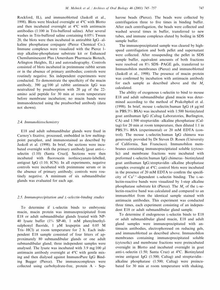
As noted above, we identified a single positive clonefrom our E18 submandibular gland library. The nucle-otide sequence encoded by this full-length cDNA andits deduced amino acid sequence is shown in Fig. 1
Fig. 1. Mouse embryonic submandibular salivary gland mucin sequence: The 5� and 3� non-translated regions are represented bycontinuous sequence. The coding region is presented in codon triplets with their deduced amino acid below in single-letter notation.The most probable initiation codon, stop codon, and polyA signal are underlined. The five repeats in the repeat domain aredesignated by *; the start of the non-repeat C-terminal domain is designated by �.

M. Melnick et al. / Archi�es of Oral Biology 46 (2001) 745–757 749
Fig. 2. Mouse embryonic submandibular salivary gland mucin amino acid sequence: The overlines designate the N-terminal signalsequence (arrow=signal peptide cleavage site) and the C-terminal �-helix. The underlines designate the tandem-repeat domain; *designate putative N-glycosylation sites and � designate putative O-glycosylation sites. Other secondary-structure designations: h,�-helix; c, random coils; e, extended strands.
(GenBank no. AF247816). The cDNA encodes an 885-bp sequence (lacking the polyA tail) that contains amost probable translation start site at bp 104–106,followed by a single open reading frame of 219 aminoacids (approximately 24600 Da). The coding sequencecontains three domains: N-terminal, repeat, and C-ter-minal. The total amino acid composition is rich inserine (approximately 9%), threonine (approximately22%), and proline (approximately 14%); the repeat do-main is particularly rich in threonine (approximately46%) and proline (approximately 18%).
3.2. Signal sequence, glycosylation sites, and putati�esecondary structure
Signal-peptide prediction was performed using theNielsen neural network and hidden Markov modelalgorithms (Nielsen et al., 1997; Nielsen and Krogh,1998). With a probability of 87%, the signal peptidewas predicted to span amino acid position 1–22 (Fig.2). It has long been known that potential N-glycosyla-tion sites are specific to the consensus sequence N–(X)– [S/T]– (X), (X) being any amino acid but prolineand N being the glycosylation site (Marshall, 1972;Gavel and von Heijne, 1990). ScanProsite analysis(www.expasy.ch/cgi-bin/scanprosite) identified potentialN-glycosylation sites at the initial amino acid of each of
four repeats in the repeat domain (Fig. 2). O-glycosyla-tion sites were predicted using the NetOglyc algorithmof Hansen et al. (1995, 1997, 1998); 53 O-glycosylationsites were identified, all in the repeat and C-terminaldomains (Fig. 2).
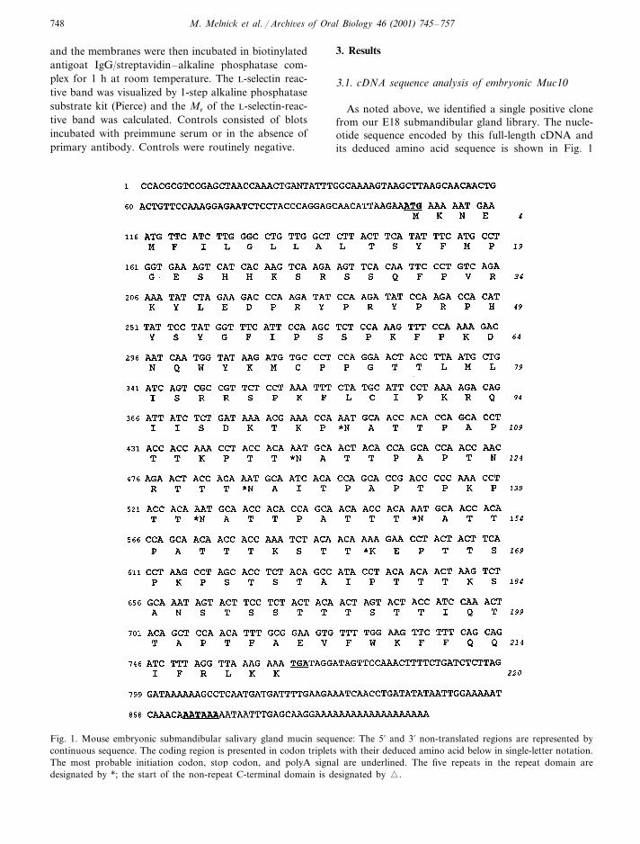
Consensus secondary-structure prediction for thenon-glycosylated peptide was made with the NPS@algorithm (pbil.ibcp.fr/cgi-bin/npsa), which presents ameta-analysis of results from analyses of the querysequence by eight independent prediction algorithms.From the signal sequence cleavage site at amino acid23–203, the secondary structure is essentially randomcoils interspersed by short extended strands (Fig. 2).The C-terminus (amino acid 204–218) is a putative�-helix (Fig. 2). This amino acid sequence was exam-ined with the helical-wheel algorithm of Turcotte(www.bmm.icnet.uk/people/turcotte/Ja�a/HelixWheel/),which is based on Benner et al. (1994) and Naor et al.(1996), and confirmed by the algorithm HELICAL-WHEEL (www.gcg.com). The helical-wheel diagram(Fig. 3) consists of a projection of the side-chain orien-tation on to a plane perpendicular to the long axis ofthe helix. This sequence (amino acid 204–218) appearsto form an amphipathic �-helix, with one face of thehelix containing polar residues and the other containingnon-polar residues (Segrest et al., 1990), a finding ofprobable significance to this membrane-associatedmolecule (discussed below).
M. Melnick et al. / Archi�es of Oral Biology 46 (2001) 745–757750
Fig. 3. The predicted C-terminal �-helix (amino acid 204–218)was analysed using the helical-wheel program of M. Turcotte(www.bmm.icnet.uk/people/turcotte/Ja�a/HW/). This programdisplays a view down the barrel of the �-helix and designatesthe amino acid residues surrounding the helix. Polar aminoacid residues are indicated by *; all other residues are non-po-lar. This is an amphipathic helix. Using the method of Eisen-berg et al. (1984), we calculate the mean hydrophobicity of thenon-polar face to be 1.09/amino acid residue and the polarface to be −1.29/amino acid residue.
(www.labonweb.com ; Compugen, Ltd., Tel Aviv, Israel),which queries a proprietary database of clustered andassembled expressed sequences. The algorithm thatbuilds this database models alternative splicing basedon the alignment characteristics of individual expressedsequences (full-length mRNAs) belonging to the samecluster. The alternatively spliced embryonic Muc10 re-peat domain has approximately 50% of the potential N-and O-glycosylation sites seen in the adult Muc10repeat domain. Additionally, other mucin and mucin-like proteins of high identity and similarity are pre-sented in Table 1.
3.4. Relati�e expression of embryonic and adult Muc10
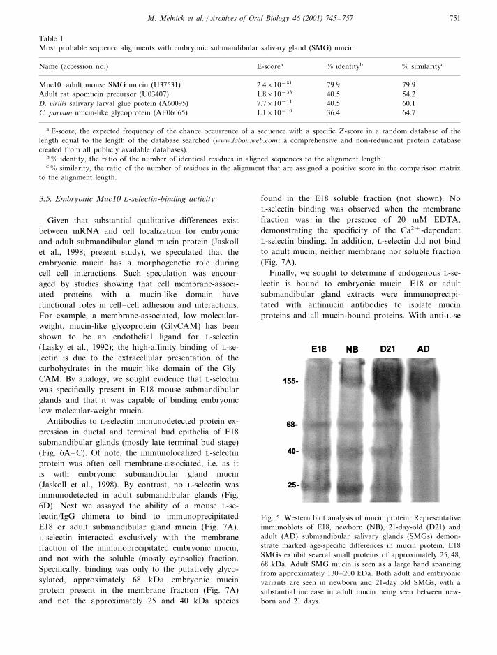
Western blot analysis was used to characterize age-specific differences in submandibular gland mucinprotein using an antibody specifically directed against aunique 21-amino acid sequence (amino acid 22–42)common to both embryonic and adult Muc10. Becauseit lacks potential glycosylation sites, it is not likely to bemasked by carbohydrate chains. As shown in Fig. 5,notable mucin protein differences were seen betweenembryonic, postnatal and adult submandibular glands.
In E18 submandibular glands, several small proteinswere identified with our peptide-specific antibody: ap-proximately 25, 40, and 68 kDa. By contrast, the adultsubmandibular gland exhibits a large band spanningfrom approximately 130–200 kDa, as previously re-ported (Jaskoll et al., 1998). The smaller proteins seenin E18 submandibular glands are absent from adultsubmandibular glands. In newborn and 21-day postna-tal submandibular glands, one sees a transition fromembryonic to adult protein.
3.3. Alignment with adult Muc10
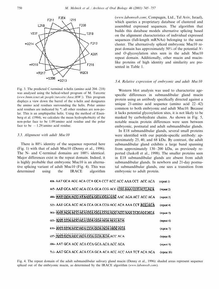
There is 80% identity of the sequence reported here(Fig. 1) with that of adult Muc10 (Denny et al., 1996).The N- and C-terminal domains are 100% identical.Major differences exist in the repeat domain. Indeed, itis highly probable that embryonic Muc10 is an alterna-tive splicing variant of adult Muc10 (Fig. 4). This wasdetermined using the IRACE algorithm
Fig. 4. The repeat domain of the adult submandibular salivary gland mucin (Denny et al., 1996): shaded areas represent sequencespliced out of the embryonic mucin, as determined by the IRACE algorithm (www.labonweb.com).
M. Melnick et al. / Archi�es of Oral Biology 46 (2001) 745–757 751
Table 1Most probable sequence alignments with embryonic submandibular salivary gland (SMG) mucin
% identitybName (accession no.) % similaritycE-scorea
Muc10: adult mouse SMG mucin (U37531) 79.92.4×10−81 79.940.51.8×10−33 54.2Adult rat apomucin precursor (U03407)40.5 60.1D. �irilis salivary larval glue protein (A60095) 7.7×10⁻¹¹
36.4 64.71.1×10−10C. par�um mucin-like glycoprotein (AF06065)
a E-score, the expected frequency of the chance occurrence of a sequence with a specific Z-score in a random database of thelength equal to the length of the database searched (www.labon.web.com : a comprehensive and non-redundant protein databasecreated from all publicly available databases).
b % identity, the ratio of the number of identical residues in aligned sequences to the alignment length.c % similarity, the ratio of the number of residues in the alignment that are assigned a positive score in the comparison matrix
to the alignment length.
3.5. Embryonic Muc10 L-selectin-binding acti�ity
Given that substantial qualitative differences existbetween mRNA and cell localization for embryonicand adult submandibular gland mucin protein (Jaskollet al., 1998; present study), we speculated that theembryonic mucin has a morphogenetic role duringcell–cell interactions. Such speculation was encour-aged by studies showing that cell membrane-associ-ated proteins with a mucin-like domain havefunctional roles in cell–cell adhesion and interactions.For example, a membrane-associated, low molecular-weight, mucin-like glycoprotein (GlyCAM) has beenshown to be an endothelial ligand for L-selectin(Lasky et al., 1992); the high-affinity binding of L-se-lectin is due to the extracellular presentation of thecarbohydrates in the mucin-like domain of the Gly-CAM. By analogy, we sought evidence that L-selectinwas specifically present in E18 mouse submandibularglands and that it was capable of binding embryoniclow molecular-weight mucin.
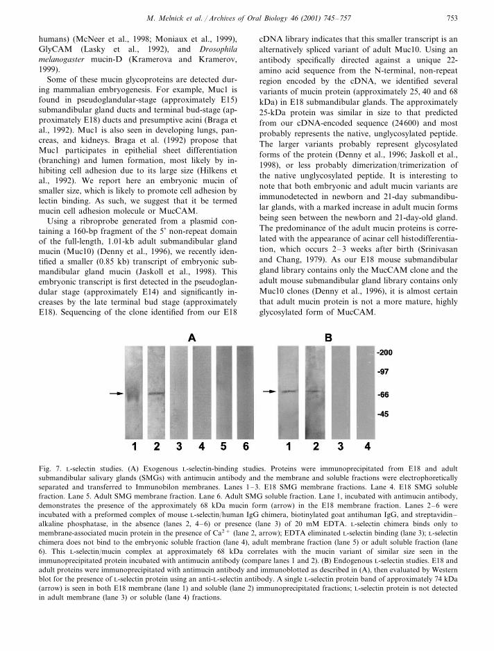
Antibodies to L-selectin immunodetected protein ex-pression in ductal and terminal bud epithelia of E18submandibular glands (mostly late terminal bud stage)(Fig. 6A–C). Of note, the immunolocalized L-selectinprotein was often cell membrane-associated, i.e. as itis with embryonic submandibular gland mucin(Jaskoll et al., 1998). By contrast, no L-selectin wasimmunodetected in adult submandibular glands (Fig.6D). Next we assayed the ability of a mouse L-se-lectin/IgG chimera to bind to immunoprecipitatedE18 or adult submandibular gland mucin (Fig. 7A).L-selectin interacted exclusively with the membranefraction of the immunoprecipitated embryonic mucin,and not with the soluble (mostly cytosolic) fraction.Specifically, binding was only to the putatively glyco-sylated, approximately 68 kDa embryonic mucinprotein present in the membrane fraction (Fig. 7A)and not the approximately 25 and 40 kDa species
found in the E18 soluble fraction (not shown). NoL-selectin binding was observed when the membranefraction was in the presence of 20 mM EDTA,demonstrating the specificity of the Ca2+-dependentL-selectin binding. In addition, L-selectin did not bindto adult mucin, neither membrane nor soluble fraction(Fig. 7A).
Finally, we sought to determine if endogenous L-se-lectin is bound to embryonic mucin. E18 or adultsubmandibular gland extracts were immunoprecipi-tated with antimucin antibodies to isolate mucinproteins and all mucin-bound proteins. With anti-L-se
Fig. 5. Western blot analysis of mucin protein. Representativeimmunoblots of E18, newborn (NB), 21-day-old (D21) andadult (AD) submandibular salivary glands (SMGs) demon-strate marked age-specific differences in mucin protein. E18SMGs exhibit several small proteins of approximately 25, 48,68 kDa. Adult SMG mucin is seen as a large band spanningfrom approximately 130–200 kDa. Both adult and embryonicvariants are seen in newborn and 21-day old SMGs, with asubstantial increase in adult mucin being seen between new-born and 21 days.

M. Melnick et al. / Archi�es of Oral Biology 46 (2001) 745–757752
Fig. 6. Immunolocalization of L-selectin in embryonic submandibular salivary glands (SMGs). (A–C) E18 SMGs. L-selectin isimmunodetected in E18 ductal (d) and terminal bud (t) epithelia in association with cell membranes, as well as in the cytoplasm.Higher-magnification views of E18 SMG ductal; (B) and terminal bud; (C) epithelia demonstrate that L-selectin protein expressionis cell-membrane associated (arrowhead); (D) Adult SMG. L-selectin is not immunodetected in adult SMG ducts (double arrows) oracini (double arrowheads). Bar: (A) 50 �m; (B–D) 20 �m.
lectin antibodies, Western blot analysis of immunopre-cipitated proteins showed a single, approximately 74kDa band in the membrane fraction of E18 immuno-precipitated proteins (Fig. 7B). This was consistent withL-selectin protein (Nicholson et al., 1998). There was asimilar, but weaker, band in the soluble fraction [unlikethe preceding experiment (Fig. 7A)] because some ofthe endogenous L-selectin is released from mucinprotein into the buffer. Demonstration of L-selectinprotein among proteins immunoprecipitated with anti-mucin antibodies is probative of endogenous embryonicmucin/L-selectin complexes in E18 submandibularglands. By contrast, Western blot analysis of adultimmunoprecipitated proteins demonstrated no L-se-lectin protein in either the membrane or soluble frac-tion, indicating that L-selectin is not bound to adultsubmandibular gland mucin (Fig. 7B).
4. Discussion
Mucins have typically been thought of as the majororganic component of saliva or mucus, viscous sub-stances that serve as a protective barrier between ep-ithelial cells and the extracellular milieu of many organsystems (mouth, lungs, reproductive tract, etc.)(Gendler and Spicer, 1995). These secretory mucins arelarge molecules (�200 kDa) that are highly O-glycosy-lated and form oligomers via disulphide bonds. Thereis, also, another class of mucins, and other glyco-proteins with mucin-like domains, that are membrane-associated and serve to modulate cell adhesion (Gendlerand Spicer, 1995). These include Muc1 (MUC1 inhumans) (Braga et al., 1992; Hilkens et al., 1992), CD43(leukosialin) (Ardman et al., 1992; Park et al., 1991;Rosenstein et al., 1991); sialomucin complex (MUC4 in
M. Melnick et al. / Archi�es of Oral Biology 46 (2001) 745–757 753
humans) (McNeer et al., 1998; Moniaux et al., 1999),GlyCAM (Lasky et al., 1992), and Drosophilamelanogaster mucin-D (Kramerova and Kramerov,1999).
Some of these mucin glycoproteins are detected dur-ing mammalian embryogenesis. For example, Muc1 isfound in pseudoglandular-stage (approximately E15)submandibular gland ducts and terminal bud-stage (ap-proximately E18) ducts and presumptive acini (Braga etal., 1992). Muc1 is also seen in developing lungs, pan-creas, and kidneys. Braga et al. (1992) propose thatMuc1 participates in epithelial sheet differentiation(branching) and lumen formation, most likely by in-hibiting cell adhesion due to its large size (Hilkens etal., 1992). We report here an embryonic mucin ofsmaller size, which is likely to promote cell adhesion bylectin binding. As such, we suggest that it be termedmucin cell adhesion molecule or MucCAM.
Using a ribroprobe generated from a plasmid con-taining a 160-bp fragment of the 5’ non-repeat domainof the full-length, 1.01-kb adult submandibular glandmucin (Muc10) (Denny et al., 1996), we recently iden-tified a smaller (0.85 kb) transcript of embryonic sub-mandibular gland mucin (Jaskoll et al., 1998). Thisembryonic transcript is first detected in the pseudoglan-dular stage (approximately E14) and significantly in-creases by the late terminal bud stage (approximatelyE18). Sequencing of the clone identified from our E18
cDNA library indicates that this smaller transcript is analternatively spliced variant of adult Muc10. Using anantibody specifically directed against a unique 22-amino acid sequence from the N-terminal, non-repeatregion encoded by the cDNA, we identified severalvariants of mucin protein (approximately 25, 40 and 68kDa) in E18 submandibular glands. The approximately25-kDa protein was similar in size to that predictedfrom our cDNA-encoded sequence (24600) and mostprobably represents the native, unglycosylated peptide.The larger variants probably represent glycosylatedforms of the protein (Denny et al., 1996; Jaskoll et al.,1998), or less probably dimerization/trimerization ofthe native unglycosylated peptide. It is interesting tonote that both embryonic and adult mucin variants areimmunodetected in newborn and 21-day submandibu-lar glands, with a marked increase in adult mucin formsbeing seen between the newborn and 21-day-old gland.The predominance of the adult mucin proteins is corre-lated with the appearance of acinar cell histodifferentia-tion, which occurs 2–3 weeks after birth (Srinivasanand Chang, 1979). As our E18 mouse submandibulargland library contains only the MucCAM clone and theadult mouse submandibular gland library contains onlyMuc10 clones (Denny et al., 1996), it is almost certainthat adult mucin protein is not a more mature, highlyglycosylated form of MucCAM.
Fig. 7. L-selectin studies. (A) Exogenous L-selectin-binding studies. Proteins were immunoprecipitated from E18 and adultsubmandibular salivary glands (SMGs) with antimucin antibody and the membrane and soluble fractions were electrophoreticallyseparated and transferred to Immunobilon membranes. Lanes 1–3. E18 SMG membrane fractions. Lane 4. E18 SMG solublefraction. Lane 5. Adult SMG membrane fraction. Lane 6. Adult SMG soluble fraction. Lane 1, incubated with antimucin antibody,demonstrates the presence of the approximately 68 kDa mucin form (arrow) in the E18 membrane fraction. Lanes 2–6 wereincubated with a preformed complex of mouse L-selectin/human IgG chimera, biotinylated goat antihuman IgG, and streptavidin–alkaline phosphatase, in the absence (lanes 2, 4–6) or presence (lane 3) of 20 mM EDTA. L-selectin chimera binds only tomembrane-associated mucin protein in the presence of Ca2+ (lane 2, arrow); EDTA eliminated L-selectin binding (lane 3); L-selectinchimera does not bind to the embryonic soluble fraction (lane 4), adult membrane fraction (lane 5) or adult soluble fraction (lane6). This L-selectin/mucin complex at approximately 68 kDa correlates with the mucin variant of similar size seen in theimmunoprecipitated protein incubated with antimucin antibody (compare lanes 1 and 2). (B) Endogenous L-selectin studies. E18 andadult proteins were immunoprecipitated with antimucin antibody and immunoblotted as described in (A), then evaluated by Westernblot for the presence of L-selectin protein using an anti-L-selectin antibody. A single L-selectin protein band of approximately 74 kDa(arrow) is seen in both E18 membrane (lane 1) and soluble (lane 2) immunoprecipitated fractions; L-selectin protein is not detectedin adult membrane (lane 3) or soluble (lane 4) fractions.
M. Melnick et al. / Archi�es of Oral Biology 46 (2001) 745–757754
Although we have previously reported mucinproteins of approximately 110 and 152 kDa in E17submandibular glands (Jaskoll et al., 1998), theseprotein forms were not seen here. It is unclear why theembryonic mucin proteins detected by Western blotdiffer notably between our two studies while the im-munohistology is identical. The most likely explanationis the difference between the antibodies used. In ourpresent study, we employed a peptide-specific antibodymade against a 22-amino acid peptide from the 5�,non-repeat region of the protein. This peptide waschosen because it lacks potential glycosylation sites.Thus, it would be less likely for this region of the mucinprotein antigen to be masked by carbohydrate chains.It is for this reason that we were now able to detect thenative mucin protein (approximately 25 kDa). By con-trast, our previous experimentation utilized polyclonalantibodies directed against the adult-secreted, glycosy-lated submandibular gland mucin protein. This anti-body most probably identified highly glycosylatedproteins and/or their associated carbohydrate chains,including some glycoproteins other than mucin.4.1. L-selectin binding
The need for cells to adhere to one another or to theextracellular matrix is fundamental to organogenesis(Edelman, 1988). It has long been recognized thatlectins and their cognate carbohydrates play an impor-tant part in this process (Mann and Waterman, 1998).We present evidence that at least one possible functionof the embryonic, low molecular-weight mucin is tobind the L-selectin expressed in terminal bud epithe-lium. The primary role of L-selectin is to mediate celladhesion; its ligands are mucin-like glycoproteins (Kalt-ner and Stierstorfer, 1998; Shimizu and Shaw, 1993);hence the suggestion that this embryonic Muc10 geneproduct be termed mucin cell adhesion molecule orMucCAM.
L-selectin has an N-terminal, C-type lectin domainthat recognizes carbohydrate ligands on apposing cells(Bird et al., 1997). C-type lectins are characterized bytheir dependence on Ca2+ ions for carbohydrate bind-ing and their carbohydrate ligands include the tetrasac-charide sialyl Lewisx for selectin binding (Kaltner andStierstorfer, 1998). At present, all the known ligands forL-selectin are mucins or mucin-like molecules, e.g. Gly-CAM (Lasky et al., 1992), PSGL-1 (Aeed et al., 1998),MUC7 (Prakobphol et al., 1998, 1999), and MucCAM(present study). During morphodifferentiation there is aclose relation between the expression of cell-adhesionmolecules, cell adhesion and boundary formation be-tween cell collectives (Edelman, 1988). Interestingly,although L-selectin has been shown to bind to adulthuman submandibular gland MUC7 gene products, wedemonstrate that L-selectin does not bind to adultmouse Muc10 protein.
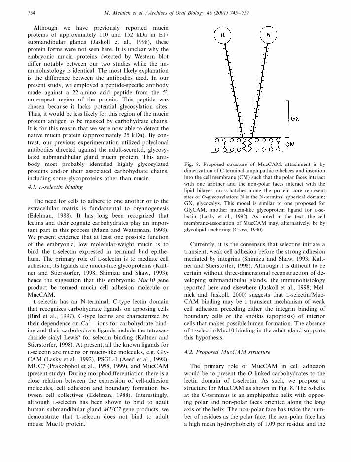
Fig. 8. Proposed structure of MucCAM: attachment is bydimerization of C-terminal amphipathic �-helices and insertioninto the cell membrane (CM) such that the polar faces interactwith one another and the non-polar faces interact with thelipid bilayer; cross-hatches along the protein core representsites of O-glycosylation; N is the N-terminal spherical domain;GX, glycocalyx. This model is similar to one proposed forGlyCAM, another mucin-like glycoprotein ligand for L-se-lectin (Lasky et al., 1992). As noted in the text, the cellmembrane-association of MucCAM may, alternatively, be byglycolipid anchoring (Cross, 1990).
Currently, it is the consensus that selectins initiate atransient, weak cell adhesion before the strong adhesionmediated by integrins (Shimizu and Shaw, 1993; Kalt-ner and Stierstorfer, 1998). Although it is difficult to becertain without three-dimensional reconstruction of de-veloping submandibular glands, the immunohistologyreported here and elsewhere (Jaskoll et al., 1998; Mel-nick and Jaskoll, 2000) suggests that L-selectin/Muc-CAM binding may be a transient mechanism of weakcell adhesion preceding either the integrin binding ofboundary cells or the anoikis (apoptosis) of interiorcells that makes possible lumen formation. The absenceof L-selectin/Muc10 binding in the adult gland supportsthis hypothesis.
4.2. Proposed MucCAM structure
The primary role of MucCAM in cell adhesionwould be to present the O-linked carbohydrates to thelectin domain of L-selectin. As such, we propose astructure for MucCAM as shown in Fig. 8. The �-helixat the C-terminus is an amphipathic helix with oppos-ing polar and non-polar faces oriented along the longaxis of the helix. The non-polar face has twice the num-ber of residues as the polar face; the non-polar face hasa high mean hydrophobicity of 1.09 per residue and the
M. Melnick et al. / Archi�es of Oral Biology 46 (2001) 745–757 755
polar face has a low mean hydrophobicity of −1.29per residue. This C-terminal, amphipathic �-helix has alength of about four turns (Eisenberg et al., 1984).Given the above, we propose that MucCAM is plasma-membrane-bound such that the polar faces interactwith each other to form a dimer and the non-polarfaces interact with the lipid bilayer. This is similar tothat proposed for GlyCAM, another mucin-like glyco-protein ligand for L-selectin (Lasky et al., 1992).
Alternatively, MucCAM may be membrane-associ-ated by covalent linkage to the glycolipid anchor, gly-cosyl-phosphatidylinositol. Many glycoproteins,including cell-adhesion molecules, appear to attach tocell surfaces via glycosyl-phosphatidylinositol anchorstructures that are preassembled and added to nascentproteins by C-terminal cleavage and attachment in theendoplasmic reticulum (Cross, 1990). Coyne et al.(1993) have defined a core consensus signal in theC-terminal domain for glycosyl-phosphatidylinositolanchoring: (1) a C-terminus of at least 11 residues withweak to moderate hydrophobicity, and without a cyto-plasmic tail; (2) a cleavage/attachment tripeptide do-main with small amino acids such as Ser, Ala, or Glypreferred at the first (attachment) and third positions;(3) a spacer domain between the tripeptide and thehydrophobic C-terminus of more than seven and lessthan 20 residues in size. MucCAM presents all charac-teristics of the core consensus signal for glycosyl-phos-phatidylinositol anchoring: (1) there is a 15-residue,hydrophobic C-terminus without a cytoplasmic tail(amino acid 204–218); (2) probably cleavage/attach-ment tripeptides exist at residues 184–186 (SAN), 185–187 (ANS), and 187–189 (STS); (3) there is a spacerdomain of 14–17 residues, depending on the cleavagesite. Given the characteristics of greater anchoring effi-ciency (Coyne et al., 1993), the most likely structurewould be cleavage between amino acids 187 and 189and attachment of the glycosyl-phosphatidylinositol tothe Ser residue at amino acid 187.
Mucin glycoproteins are relatively inflexible becauseof steric interactions between the peptide-linked Gal-Nac residue (O-glycosylation) and adjacent amino acidsin the peptide core (Jentoft, 1990). The repeat domainof MucCAM has 29 putative O-glycosylation sites andthe C-terminal domain has 24. In all, more than 50% ofamino acid residues 103–203 are possible O-glycosyla-tion sites. At 0.25 nm per amino acid residue (Jentoft,1990), this glycosylated region would be expected to bea rather rigid rod with a persistence length of approxi-mately 25 nm. In effect, this extends the MucCAMmolecule well above the approx imately 10 nm glycoca-lyx (extracellular surface), allowing this glycoprotein tointeract with extracellular macromolecules (such as L-selectin) that might otherwise be unable to penetratethe relatively crowded glycocalyx [in a manner similarto other membrane glycoproteins such as decay-acceler-
ating factor and LDL receptor (Jentoft, 1990)]. Further,it is reasonable to expect that the close clustering ofcarbohydrate epitopes will result in rapid on-rates andenhanced avidity of mucin/selectin binding (Lee, 1989;Williams, 1991).
The 102-amino acid, N-terminal domain of Muc-CAM (exclusive of the signal sequence) has a predictedsecondary structure of random coils interrupted byshort extended strands and is without putative glycosy-lation sites. Unlike tightly folded globular peptides,which have a relatively fixed and compact tertiarystructure, those with a random coil conformation forma spherical domain with variant configuration (Jentoft,1990).
Each region of the MucCAM protein, then, has aproposed function. The amphiphathic �-helix anchorsthe molecule to the plasma membrane. The protein coreof the glycosylated region serves as a scaffold forcarbohydrate presentation to L-selectin and, perhaps,other cognate extracellular molecules. The function ofthe spherical domain, however, is obscure.
4.3. E�olution and class considerations
Many investigators have utilized specific carbohy-drate/lectin staining patterns in defining the dynamicsof morphodifferentiation in various embryonic modelsof diverse species and tissues (see Mann and Waterman,1998). It is thought that the sugar/lectin complex ‘infor-mation management system’ is evolutionarily primitiverelative to more specific peptide ligand/receptor mecha-nisms (Mann and Waterman, 1998). In this context, itis not rare to see common genetic and epigenetic mech-anisms at work in completely disparate developmentalprocesses, a kind of co-option of developmental geneticmechanisms (Raff, 1996). This often gives rise to anevolutionarily related class of genes across widely diver-gent species (Hutter et al., 2000).
Of particular interest here is the relation betweenMucCAM and the salivary glue proteins of Drosophila.In the third instar larvae of Drosophila, the majorfunction of the salivary glands is the production of amucoprotein glue (Swida et al., 1990). This glue isextruded shortly after puparium formation and servesto attach the pupal case to the substrate. Multiplerelated salivary glue proteins are expressed by D.melanogaster and D. �irilis. It is more than interestingthat there is a 60% similarity (E=7.7×10⁻¹¹) betweenthe salivary glue protein lpg-1 (Swida et al., 1990) andMucCAM. As large portions of these two sequencescontain close residues, they may belong to the sameprotein family. It is reasonable to speculate, then, thatadditional MucCAMs in diverse species and disparatedeveloping organ systems await discovery.

M. Melnick et al. / Archi�es of Oral Biology 46 (2001) 745–757756
Acknowledgements
We thank Dr Steven D. Rosen (UC San Francisco)for his generous gift of mouse L-selectin/human IgGchimera. The work presented here was supported byNIH/NIDCR Grant no. DE-11942.
References
Aeed, P.A., Geng, J.G., Asa, D., Raycroft, L., Ma, L., Elham-mer, A.P., 1998. Characterization of the O-linked oligosac-charide structures on P-selectin glycoprotein ligand-1(PSGL-1). Glycoconjugate J. 15, 975–985.
Altschul, S.F., Madden, T.L., Schaffer, A.A., Zhang, J.,Zhang, Z., Miller, W., Lipman, D.J., 1997. GappedBLAST and PSI-BLAST. Nucleic Acid Res. 25, 3389–3402.
Ardman, B., Sikorski, M.A., Staunton, D.E., 1992. CD43interferes with T-lymphocyte adhesion. Proc. Natl. Acad.Sci. USA 89, 5001–5005.
Benner, S.A., Badcoe, I., Cohen, M.A., Gerloff, D., 1994.Bone fide prediction of aspects of protein conformation. J.Mol. Biol. 235, 926–958.
Bird, M.I., Foster, M.R., Priest, R., Malhorta, R., 1997.Selectins: physiological and pathophysiological roles.Biochem. Soc. Trans. 25, 1199–1206.
Braga, V.M.M., Pemberton, L.F., Duhig, T., Gendler, S.J.,1992. Spatial and temporal expression of an epithelialmucin, Muc-1, during mouse development. Development115, 427–437.
Chomzynski, P., Sacchi, N., 1987. Single-step method of RNAisolation by acid guanidinuime thyocyanate-phenol-chloro-form extraction. Anal. Biochem. 162, 156–159.
Coyne, K.E., Crisci, A., Lublin, D.M., 1993. Construction ofsynthetic signals for glycosyl-phosphatidylinositol anchorattachment: analysis of amino acid sequence requirementsfor anchoring. J. Biol. Chem. 268, 6689–6693.
Cross, G.A.M., 1990. Glycolipid anchoring of plasma mem-brane proteins. Annu. Rev. Cell. Biol. 6, 1–39.
Denny, P.C., Mirels, L., Denny, P.A., 1996. Mouse sub-mandibular gland salivary apomucin contains repeatedN-glycosylation sites. Glycobiology 6, 43–50.
Edelman, G.M., 1988. Topobiology. Basic Books, New York.Eisenberg, D., Schwarz, E., Komaromy, M., Wall, R., 1984.
Analysis of membrane and surface protein sequences withthe hydrophobic moment plot. J. Mol. Biol. 179, 125–142.
Gavel, Y., von Heijne, G., 1990. Sequence differences betweenglycosylated and non-glycosylated Asn-X-Thr/Ser acceptorsites: implications for protein engineering. Protein Eng. 3,433–442.
Gendler, S.J., Spicer, A.P., 1995. Epithelial mucin genes.Annu. Rev. Physiol. 57, 607–634.
Hansen, J.E., Lund, O., Engelbrecht, J., Bohr, H., Nielsen,J.O., Hansen, J.-E.S., Brunak, S., 1995. Prediction of O-glycosylation of mammalian proteins: specificity patternsof UDP-GalNAC: polypeptide N-acetylgalactosaminyl-transferase. Biochem. J. 308, 801–813.
Hansen, J.E., Lund, O., Rapacki, K., Brunak, S., 1997. O-GLYCBASE VERSION 2.0-A revised database of O-
GLYCOSYLATED Proteins. Nucleic Acids Res. 25,278–282.
Hansen, J.E., Lund, O., Tolstrup, N., Gooley, A.A., Williams,K.L., Brunak, S., 1998. NetOglyc: prediction of mucin typeO-glycosylation sites based on sequence context and sur-face accessibility. Glycoconjugate J. 15, 115–130.
Hilkens, J., Ligtenberg, M.L.L., Vos, H.L., Litvinov, S.V.,1992. Cell membrane-associated mucins and their adhe-sion-modulating property. Trends. Biochem. Sci. 17, 359–363.
Hutter, H., Vogel, B.E., Plenefisch, J.D., Norris, C.R.,Proenca, R.B., Spieth, J., Guo, C., Mastwal, S., Zhu, X.,Scheel, J., Hedgecock, E.M., 2000. Conservation and nov-elty in the evolution of cell adhesion and extracellularmatrix genes. Science 287, 989–994.
Jaskoll, T., Chen, H., Denny, P.C., Denny, P.A., Melnick, M.,1998. Mouse submandibular gland mucin: embryo-specificmRNA and protein species. Mech. Dev. 74, 179–183.
Jaskoll, T., Melnick, M., 1999. Submandibular gland morpho-genesis: stage-specific expression of TGF-a/EGF, IGF,TGF-�. TNF, and IL-6 signal transduction in normalembryonic mice and the phenotypic effects of TGF-�2,TGF-�3, and EGF-R null mutations. Anat. Rec. 256,252–268.
Jaskoll, T., Choy, H.A., Melnick, M., 1994. Glucolcorticoids,TGF-Beta, and embryonic mouse salivary gland morpho-genesis. J. Craniof. Gent. Dev. Biol. 14, 217–230.
Jentoft, N., 1990. Why are proteins O-glycosylated? Trends.Biochem. Sci. 15, 291–294.
Kaltner, H., Stierstorfer, B., 1998. Animal lectins as celladhesion molecules. Acta. Anat. 161, 162–179.
Kramerova, I.A., Kramerov, A.A., 1999. Mucinprotein is auniversal constituent of stable intercellular bridges inDrosophila melanogaster germ line and somatic cells. Dev.Dyn. 216, 349–360.
Lasky, L.A., Singer, M.S., Dowbenko, D., Imai, Y., Henzel,W.J., Grimley, C., Fennie, C., Gillett, N., Watson, S.R.,Rosen, S.D., 1992. An endothelial ligand for L-selectin is anovel mucin-like molecule. Cell 69, 927–938.
Lee, Y.C., 1989. Binding modes of mammalian hepatic Gal/GalNAc receptors. In: Bock, G., Harnett, S. (Eds.), Sym-posium on Carbohydrate Recognition in CellularFunction. Wiley, New York, pp. 80–95.
Mann, P.L., Waterman, R.E., 1998. Glycocoding as an infor-mation management system in embryonic development.Acta Anat. 161, 153–161.
Marshall, R.D., 1972. Glycoproteins. Annu. Rev. Biochem.41, 673–702.
McNeer, R.R., Huang, D., Fregien, N.L., Carraway, K.L.,1998. Sialomucin complex in the rat respiratory tract: amodel for its role in epithelial protection. Biochem. J. 330,737–744.
Melnick, M., Jaskoll, T., 2000. Mouse submandibular glandmorphogenesis: a paradigm for embryonic signal process-ing. Crit. Rev. Oral Biol. Med. 11, 199–215.
Moniaux, N., Nollet, S., Porchet, N., Degand, P., Laine, A.,Aubert, J.-P., 1999. Complete sequence of the humanmucin MUC4: a putative cell membrane-associated mucin.Biochem. J. 338, 325–333.
Naor, D., Fischer, D., Jernigan, R.L., Wolfson, H.J., Nussi-nov, R., 1996. Amino acid pair interchanges at spatiallyconserved locations. J. Mol. Biol. 256, 924–938.
M. Melnick et al. / Archi�es of Oral Biology 46 (2001) 745–757 757
Nicholson, M.W., Barclay, A.N., Singer, M.S., Rosen, S.D.,van der Merwe, P.A., 1998. Affinity and kinetic analysis ofL-selectin (CD62L) binding to glycosylation-dependentcell-adhesion molecule-1. J. Biol. Chem. 273, 763–770.
Nielsen, H., Krogh, A., 1998. Prediction of signal peptides andsignal anchors by a hidden Markov model. In: Glasgow, J.(Ed.), Proceedings of the Sixth International Conferenceon Intelligent Systems for Molecular Biology, AAAI,Menlo Park, California, pp. 122–130.
Nielsen, H., Engelbrecht, J., Brunak, S., von Heijne, G., 1997.Identification of prokaryotic and eukaryotic signal peptidesand prediction of their cleavage sites. Protein Eng. 10, 1–6.
Park, J.K., Rosenstein, Y.J., Remold-O’Donnell, E., Bierer,B.E., Rosen, F.S., Burakoff, S.J., 1991. Enhancement ofT-cell activation by the CD43 molecule whose expression isdefective in Wiskott–Aldrich syndrome. Nature 350, 706–709.
Prakobphol, A., Thomsson, K.A., Hansson, G.C., Rosen,S.D., Singer, M.S., Phillips, N.J., Medzihradszky, K.F.,Burlingame, A.L., Leffler, H., Fisher, S.J., 1998. Humanlow-molecular-weight salivary mucin expresses sialylLewisx determinant and has L-selectin ligand activity. Bio-chemistry 37, 4916–4927.
Prakobphol, A., Tangemann, K., Rosen, S.D., Hoover, C.I.,Leffler, H., Fisher, S.J., 1999. Separate oligosaccharidedeterminants mediate interactions of the low-molecular-weight salivary mucin with neutrophils and bacteria. Bio-chemistry 38, 6817–6825.
Raff, R.A., 1996. The Shape of Life. Genes, Development, andthe Evolution of Animal Form. University of ChicagoPress, Chicago, p. 431.
Rosenstein, Y., Park, J.K., Hahn, W.C., Rosen, F.S., Bierer,B.E., Burakoff, S.J., 1991. CD43, a molecule defective inWiskott-Aldrich syndrome, binds ICAM-1. Nature 354,233–235.
Sanger, F., Wicklen, S., Coulson, A.R., 1977. DNA sequenc-ing with chain-terminating inhibitors. Proc. Natl. Acad.Sci. USA 74, 5463–5467.
Segrest, J.P., De Loof, H., Dohlman, J.G., Brouillette, C.G.,Anatharamaiah, G.M., 1990. Amphipathic helix motif:classes and properties. Proteins 8, 103–117.
Shimizu, Y., Shaw, S., 1993. Mucins in the mainstream. Na-ture 336, 630–631.
Spooner, B.S., Bassett, K.E., Spooner, B.S. Jr., 1989. Embry-onic salivary gland epithelial branching activity is experi-mentally independent of epithelial expansion activity. Dev.Biol. 133, 569–575.
Srinivasan, R., Chang, W.W.L., 1979. The postnatal develop-ment of the submandibular gland. Cell. Tissue Res. 198,363–371.
Swida, U., Lucka, L., Kress, H., 1990. Glue protein genes inDrosophila virilis: their organization, developmental con-trol of transcription and specific mRNA degradation. De-velopment 108, 269–280.
Theiler, K., 1989. The House Mouse. Atlas of EmbryonicDevelopment. Springer-Verlag, New York.
Williams, A.F., 1991. Out of equilibrium. Nature 352, 473–474
.