ampicillin enhances daptomycin- and cationic host...
TRANSCRIPT

Ampicillin Enhances Daptomycin- and Cationic Host Defense Peptide-Mediated Killing of Ampicillin- and Vancomycin-ResistantEnterococcus faecium
George Sakoulas,a Arnold S. Bayer,b,c Joseph Pogliano,d Brian T. Tsuji,e Soo-Jin Yang,b Nagendra N. Mishra,b Victor Nizet,a
Michael R. Yeaman,b,c and Pamela A. Moisef
University of California San Diego School of Medicine, La Jolla, California, USAa; Los Angeles Biomedical Research Institute at Harbor-UCLA Medical Center, Torrance,California, USAb; Geffen School of Medicine, University of California, Los Angeles, Los Angeles, California, USAc; Section of Molecular Biology, University of California SanDiego School of Medicine, La Jolla, California, USAd; University of Buffalo School of Pharmacy and Pharmaceutical Sciences, Amherst, New York, USAe; and CubistPharmaceuticals, Lexington, Massachusetts, USAf
We studied an ampicillin- and vancomycin-resistant Enterococcus faecium (VRE) isolate from a patient with endocarditis andbacteremia refractory to treatment with daptomycin (6 mg/kg of body weight) plus linezolid. Blood cultures cleared within 24 hof changing therapy to daptomycin (12 mg/kg) plus ampicillin. We examined the effects of ampicillin on daptomycin-inducedgrowth inhibition and killing, surface charge, and susceptibility to several prototypical host defense cationic antimicrobial pep-tides. MICs and time-kill curves with daptomycin were assessed in the presence and absence of ampicillin. The impact of ampi-cillin on surface charge was assessed by flow cytometry and a poly-L-lysine binding assay. The effects of ampicillin preexposuresupon VRE killing by five distinct cationic peptides of different structure, charge, origin, and mechanism of action were analyzedusing the epidermal cathelicidin LL-37, thrombin-induced platelet microbicidal proteins (tPMPs), and a synthetic congenermodeled after tPMP microbicidal domains (RP-1), human neutrophil peptide-1 (hNP-1), and polymyxin B (bacteria derived).Fluoroscein-Bodipy-labeled daptomycin was used to evaluate daptomycin binding to VRE membranes in the presence or ab-sence of ampicillin. In media containing ampicillin (25 to 100 mg/liter), daptomycin MICs decreased from 1.0 to 0.38 mg/liter.Based on time-kill analysis and an in vitro pharmacodynamic model, ampicillin enhanced daptomycin activity against the studyVRE from a bacteriostatic to a bactericidal profile. VRE grown in ampicillin (25 to 150 mg/liter) demonstrated an incrementalreduction in its relative net positive surface charge. When grown in the presence (versus absence) of ampicillin (25 and 100 mg/liter), the VRE strain (i) was more susceptible to killing by LL-37, tPMPs, hNP-1, and RP-1 but not to polymyxin B and (ii) exhib-ited greater binding to Bodipy-labeled daptomycin. We conclude that ampicillin induces reductions in net positive bacterial sur-face charge of VRE, correlating with enhanced bactericidal effects of cationic calcium-daptomycin and a diverse range of othercationic peptides in vitro. While the mechanism(s) of such �-lactam-mediated shifts in surface charge remains to be defined,these finding suggest a potential for �-lactam-mediated enhancement of activity of both daptomycin and innate host defensepeptides against antibiotic-resistant bacteria.
Daptomycin, a cyclic lipopeptide antibiotic, associates withcalcium to form a cationic complex that targets the bacterial
cytoplasmic membrane, causing rapid membrane depolarizationand subsequent lethality against susceptible Gram-positive organ-isms (21). There have been a number of recent reports of isolationof daptomycin-resistant Staphylococcus aureus (17, 20–23, 27–29,31, 39, 41) and enterococcus strains (24, 26) emerging duringdaptomycin treatment in patients with recalcitrant infections. Al-though the accepted term is “daptomycin nonsusceptibility,” wewill utilize the term “daptomycin resistant” for ease of data pre-sentation and discussion in this paper. The mechanisms leading todaptomycin resistance in S. aureus are complex, although somestudies have implicated mutations and changes in expression ofgenes involved in the modulation of bacterial surface charge, suchas dlt and mprF (17). Of interest, development of daptomycinresistance in S. aureus has commonly been associated with coevo-lution of reduced susceptibility to killing by a variety of host de-fense molecules, such as cationic antimicrobial peptides (22, 34).The mechanisms of enterococcal resistance to daptomycin remainlargely undefined, but in short appear to parallel the phenotypicchanges of daptomycin resistance in S. aureus with different geno-typic changes (2, 3, 32, 36).
Previously, we identified a reciprocal relationship in the in vitrosusceptibility of enterococci to conventional cationic antibiotics(e.g., gentamicin, streptomycin) or cationic host defense peptides(e.g., platelet microbicidal proteins [PMPs]) versus noncationicantibiotics of different classes (e.g., cell wall-active agents, DNAgyrase inhibitors, protein synthesis inhibitors) (5). Similarly, sub-sequent studies demonstrated that comparative in vitro entero-coccal susceptibility profiles for vancomycin versus cationic hostdefense peptides exhibited reciprocal phenotypes among clinicalisolates of Enterococcus faecium (6, 7). These data emphasized twoimportant themes regarding enterococci: (i) reduced in vitro sus-ceptibility to cationic host defense peptides tracked with reducedsusceptibility to conventional cationic antibiotics; (ii) reduced in
Received 17 August 2011 Returned for modification 1 November 2011Accepted 18 November 2011
Published ahead of print 28 November 2011
Address correspondence to George Sakoulas, [email protected].
Copyright © 2012, American Society for Microbiology. All Rights Reserved.
doi:10.1128/AAC.05551-11
838 aac.asm.org 0066-4804/12/$12.00 Antimicrobial Agents and Chemotherapy p. 838–844
on January 20, 2012 by UN
IV O
F C
ALIF
SA
N D
IEG
Ohttp://aac.asm
.org/D
ownloaded from
vitro susceptibility to such cationic agents correlated inverselywith susceptibility to noncationic antibiotics, particularly ampi-cillin and vancomycin. Finally, we demonstrated additive interac-tions in vitro between cationic host defense peptide congeners andnoncationic antibiotics against selected enterococcal strains (47).
In the present study, we analyzed an ampicillin- andvancomycin-resistant E. faecium (VRE) strain from a case of aorticvalve endocarditis in a hemodialysis patient with bacteremia re-fractory to 7 days of therapy with daptomycin (6 mg/kg of bodyweight every 48 h) plus linezolid (600 mg intravenously [i.v.] every12 h). Based on prior studies showing in vitro synergy betweendaptomycin and ampicillin against Enterococcus spp. (3, 8, 13, 30,37), a combined daptomycin-ampicillin therapeutic regimen wasemployed for this patient. Of interest, the patient’s persistent bac-teremia was rapidly cleared within 24 h of beginning a combina-tion regimen of high-dose daptomycin (12 mg/kg every 48 h) plusampicillin (1 g every 6 h).
In light of this dramatic microbiological response in our pa-tient, combined with the prior in vitro reports cited above dem-onstrating potential antienterococcal “synergy” with daptomycinplus ampicillin, we investigated selected effects of ampicillin onour ampicillin-resistant VRE and demonstrated that ampicillinexposures induced a notable reduction in net positive surfacecharge that was associated with increased surface binding of dap-tomycin.
MATERIALS AND METHODSAntimicrobial susceptibility testing. For the initial (pretherapy) blood-stream VRE strain, MICs of vancomycin, ampicillin, linezolid, and dap-tomycin were determined by standard Etest (AB Biodisk, Solna, Sweden)using CLSI methods (9). MICs were also determined for daptomycin andpolymyxin B by using the standard Etest on Mueller-Hinton agar (MHA)containing 0, 5, 10, 25, 50, or 100 mg/liter of ampicillin. For all experi-ments, a minimum of two assays were performed on different days. Thecomplete antibiotic susceptibility profile report from the Clinical Micro-biology Laboratory for the final bloodstream isolate on day 7 was identicalto that of the original isolate. However, only the original bloodstream VREwas stored and available for further study.
Time-kill assays were performed in duplicate with an initial bacterialinoculum of 106 CFU/ml (to reflect a high-inoculum infection, such asendocarditis) in Mueller-Hinton broth (MHB) supplemented to a cal-cium concentration of 50 mg/liter containing no antibiotic (growth con-trol), ampicillin (20 mg/liter) alone, daptomycin (4 mg/liter; 4� theMIC]) with or without ampicillin (20 mg/liter), or daptomycin (10 mg/liter; 10� the MIC]) with or without ampicillin (20 mg/liter). These an-tibiotic concentrations were chosen to encompass readily achievable freeserum concentrations of each agent during clinical treatment regimens(11, 14, 18). Quantitative bacterial counts were determined by sampling,serially diluting 1:100 to 1:107 in fresh MHB, and plating 20 �l in duplicateafter 0, 6, and 24 h of incubation at 37°C. In order to eliminate antibioticcarryover, the undiluted sample (1 ml) was microcentrifuged in an Ep-pendorf tube at 13,000 rpm for 5 min, and the pellet was resuspended in100 �l fresh MHB, allowing for a limit of detection of 0.7 log10 CFU/ml.
In vitro pharmacodynamic model. To simulate daptomycin plus am-picillin combination regimens, an in vitro pharmacodynamic model wasused, consisting of a one-compartment 500-ml glass chamber (workingmodel volume, 250 ml of broth) with multiple ports for the removal ofbrain heart infusion (BHI) broth, delivery of antibiotic, and collection ofbacterial and antimicrobial samples (19). Briefly, overnight cultures of theVRE isolate were diluted in fresh BHI broth and adjusted to a 1.0 McFar-land turbidity. The suspension was added to BHI broth in the chambermodel, yielding a final volume of 270 ml and a starting inoculum of �108
CFU/ml. Serial samples were taken at 0 (predose), 1, 2, 4, 6, 8, 24, 28, 32,
and 48 h to quantify viable counts. The following regimens for daptomy-cin were simulated using a mean 8-h terminal half-life: 4 mg/kg every 24 h(q24h; free-drug area under the concentration-time curve at 24 h[ƒAUC24] of 154 mg · h/liter), 6 mg/kg q24h (251 mg · h/liter), 8 mg/kg(329 mg · h/liter), and 10 mg/kg (377 mg · h/liter) alone and in combina-tion with ampicillin at 2 g q4h (maximum concentration of drug in serum[Cmax], 70 mg/liter), based on a 1-h mean terminal half-life (11, 14, 18).For combination regimen experiments, the elimination rate was set forthe drug with the shortest half-life, and the drug with the longer half-lifewas supplemented (4, 40).
FITC-labeled PLL binding. Assays were performed using a flow cyto-metric method as previously described (22, 33). Poly-L-lysine (PLL) is apolycationic molecule used to study the interactions of cationic peptideswith charged bacterial envelopes. In this analysis, the extent of bacteria-bound fluorescein isothiocyanate (FITC)-labeled PLL inversely reflectedthe relative surface positive charge. A total of 10,000 events were countedand analyzed using a BD FACSCalibur system (Becton Dickinson Lab-ware, San Jose, CA). Data are expressed as means relative fluorescent units(� the standard deviation [SD]). At least two independent experiments oftriplicate samples were performed.
Cationic antimicrobial peptides. We studied a panel of cationic hostdefense antimicrobial peptides differing in anatomic and host source,molecular mass, net charge at pH 7.5, and proposed mechanism(s) ofaction. Human LL-37 (net charge, �6 at pH 7.5), a cationic cathelicidinantimicrobial peptide prevalent in skin and neutrophils, was purchasedfrom AnaSpec, Inc. (Fremont, CA); the human neutrophil �-defensinhNP-1 (net charge, �3 at pH 7.5) was purchased from Peptide Interna-tional (Louisville, KY); tPMPs were prepared from freshly collected rabbitplatelets, and their bioactive equivalencies were determined as previouslydescribed (42, 43). RP-1 (a synthetic 18-amino-acid congener modeledafter the �-helical microbicidal domain of the tPMP family of plateletpeptides; net charge, �8 at pH 7.5) was synthesized and authenticated aspreviously detailed (45). As a negative-control peptide, a bacteria-derivedand membrane-targeting cyclic cationic molecule, polymyxin B, was used(Sigma, St. Louis, MO); the latter peptide has limited activity againstenterococci due to its poor capacity to penetrate the Gram-positive cellwall.
Antimicrobial peptide microbicidal assays. The VRE strain wasgrown to stationary phase (16 to 20 h) in LB in either the absence ofampicillin or presence of various ampicillin concentrations, pelleted, andthen washed in assay buffer (RPMI plus 5% LB for LL-37; phosphate-buffered saline for polymyxin B, RP-1, and tPMP; 10 mM KH2PO4, pH7.4, for hNP-1 assays). Initial bacterial inocula of 103 CFU/ml were used intPMP, polymyxin B, RP-1, and hNP-1 assays as previously described (42–44). For LL-37 assays, a starting inoculum of 105 CFU/ml was used, as itwas more discriminatory of dose-dependent differences in killing of thestudy isolate by this peptide in extensive preliminary investigations. Pilotstudies were used to determine cationic peptide concentrations thatcaused a �50% reduction in enterococcal counts in the 2-h survival assay.Thus, we utilized the following peptide concentrations: LL-37, 36 mg/liter; RP-1, 5 mg/liter; hNP-1, 2 mg/liter; tPMPs, 2.5 mg/liter equivalent;polymyxin B, 640 mg/liter. The percentage of surviving bacteria (� SD)after 2 h of incubation at 37°C was calculated by plating on blood agarplates. Results represent three separate experiments performed in dupli-cate.
Daptomycin binding assays. To determine if ampicillin was able toimpact the ability of daptomycin to bind to the VRE membrane, the or-ganism was grown in LB broth overnight (14 to 16 h) at 37°C in thepresence or absence of ampicillin (50 to 100 mg/liter) and then incubatedfor 10 min with Bodipy-fluorescein-labeled daptomycin (16 mg/liter;courtesy of Cubist Pharmaceuticals, Lexington, MA). This concentrationof labeled daptomycin was established by pilot studies as the optimalconcentration for fluorescence microscopy. Excess unincorporated labelwas removed by washing the cells three times in LB broth. The cells werecounterstained with FM 4-64 to visualize the membrane and 4=,6-
Ampicillin Enhances Cationic Peptide Killing of VRE
February 2012 Volume 56 Number 2 aac.asm.org 839
on January 20, 2012 by UN
IV O
F C
ALIF
SA
N D
IEG
Ohttp://aac.asm
.org/D
ownloaded from
diamidino-2-phenylindole (DAPI) to visualize the nucleoid and then im-aged using a Delta Vision deconvolution microscope (Applied Precision,Inc. Issaquah, WA) as previously described (36).
Statistics. Statistical evaluations of the differences in survival in thepresence of various cationic peptides and differences in PLL binding wereperformed with the Mann-Whitney U test (Prism 5.0; GraphPad Soft-ware, Inc., San Diego, CA). P values of �0.05 were considered statisticallysignificant.
RESULTSIn vitro susceptibility to antibiotics: MICs. By Etest, the VREstrain was resistant to both vancomycin and ampicillin (MICs of�128 mg/liter for both drugs) but susceptible to linezolid (MIC, 2mg/liter) and daptomycin (MIC, 1 mg/liter). The polymyxin MICwas 160 mg/liter. The daptomycin MIC (1 mg/liter) did notchange when tested in MHA containing ampicillin concentrationsof 5, 10, or 25 mg/liter, but the MIC was reduced approximately3-fold (0.38 mg/liter) in MHA containing 50 or 100 mg/liter am-picillin. No changes were noted for the MIC of polymyxin B inMHA containing the same range of ampicillin concentrations.
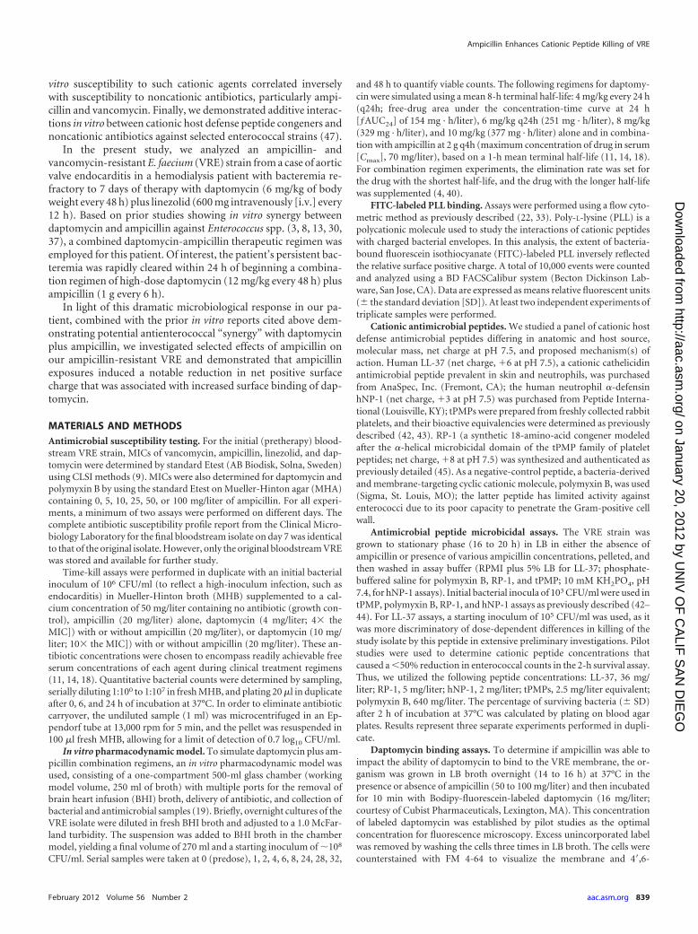
Microbicidal assays. Figure 1 demonstrates the 24-h time-killassay results for the VRE isolate in different combinations of am-picillin and daptomycin. The most relevant finding was that ex-posure of the VRE isolate to either daptomycin at 10 mg/liter(10� the MIC) alone or daptomycin at 10 mg/liter plus ampicillinat 20 mg/liter yielded the same degree of killing (�3 log10 CFU/ml) after 6 h of incubation but significantly more killing thanlower-dose daptomycin (4 mg/liter) with or without ampicilllin.However, at 24 h, VRE exposed to daptomycin at 10 mg/liter aloneshowed regrowth to a net 2 log10 CFU/ml reduction compared totime zero; in contrast, the combination of daptomycin at 10 mg/liter plus ampicillin at 20 mg/liter demonstrated continued bacte-rial killing, with reductions of VRE counts to below the limit ofdetection (�5 log 10 CFU/ml versus the time zero baseline value).As expected, growth curves in ampicillin alone (20 mg/liter) par-alleled those seen in antibiotic-free medium.
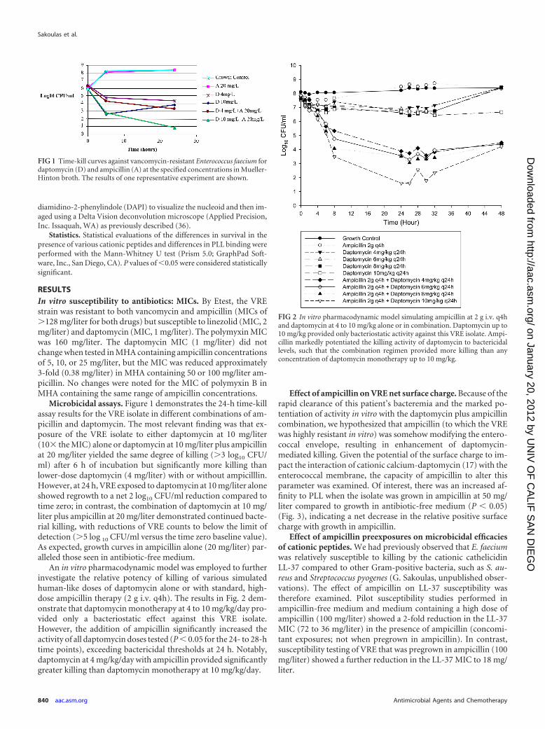
An in vitro pharmacodynamic model was employed to furtherinvestigate the relative potency of killing of various simulatedhuman-like doses of daptomycin alone or with standard, high-dose ampicillin therapy (2 g i.v. q4h). The results in Fig. 2 dem-onstrate that daptomycin monotherapy at 4 to 10 mg/kg/day pro-vided only a bacteriostatic effect against this VRE isolate.However, the addition of ampicillin significantly increased theactivity of all daptomycin doses tested (P � 0.05 for the 24- to 28-htime points), exceeding bactericidal thresholds at 24 h. Notably,daptomycin at 4 mg/kg/day with ampicillin provided significantlygreater killing than daptomycin monotherapy at 10 mg/kg/day.
Effect of ampicillin on VRE net surface charge. Because of therapid clearance of this patient’s bacteremia and the marked po-tentiation of activity in vitro with the daptomycin plus ampicillincombination, we hypothesized that ampicillin (to which the VREwas highly resistant in vitro) was somehow modifying the entero-coccal envelope, resulting in enhancement of daptomycin-mediated killing. Given the potential of the surface charge to im-pact the interaction of cationic calcium-daptomycin (17) with theenterococcal membrane, the capacity of ampicillin to alter thisparameter was examined. Of interest, there was an increased af-finity to PLL when the isolate was grown in ampicillin at 50 mg/liter compared to growth in antibiotic-free medium (P � 0.05)(Fig. 3), indicating a net decrease in the relative positive surfacecharge with growth in ampicillin.
Effect of ampicillin preexposures on microbicidal efficaciesof cationic peptides. We had previously observed that E. faeciumwas relatively susceptible to killing by the cationic cathelicidinLL-37 compared to other Gram-positive bacteria, such as S. au-reus and Streptococcus pyogenes (G. Sakoulas, unpublished obser-vations). The effect of ampicillin on LL-37 susceptibility wastherefore examined. Pilot susceptibility studies performed inampicillin-free medium and medium containing a high dose ofampicillin (100 mg/liter) showed a 2-fold reduction in the LL-37MIC (72 to 36 mg/liter) in the presence of ampicillin (concomi-tant exposures; not when pregrown in ampicillin). In contrast,susceptibility testing of VRE that was pregrown in ampicillin (100mg/liter) showed a further reduction in the LL-37 MIC to 18 mg/liter.
FIG 1 Time-kill curves against vancomycin-resistant Enterococcus faecium fordaptomycin (D) and ampicillin (A) at the specified concentrations in Mueller-Hinton broth. The results of one representative experiment are shown.
FIG 2 In vitro pharmacodynamic model simulating ampicillin at 2 g i.v. q4hand daptomycin at 4 to 10 mg/kg alone or in combination. Daptomycin up to10 mg/kg provided only bacteriostatic activity against this VRE isolate. Ampi-cillin markedly potentiated the killing activity of daptomycin to bactericidallevels, such that the combination regimen provided more killing than anyconcentration of daptomycin monotherapy up to 10 mg/kg.
Sakoulas et al.
840 aac.asm.org Antimicrobial Agents and Chemotherapy
on January 20, 2012 by UN
IV O
F C
ALIF
SA
N D
IEG
Ohttp://aac.asm
.org/D
ownloaded from

The bactericidal activity of LL-37 (36 mg/liter) in the concom-itant presence or the absence of ampicillin (100 mg/liter) was as-sessed, and we found a slight increase in killing (Fig. 4). However,when the VRE was pregrown overnight in LB containing ampicil-lin (100 mg/liter), a marked increase in LL-37 killing was observed(P � 0.027) (Fig. 4).
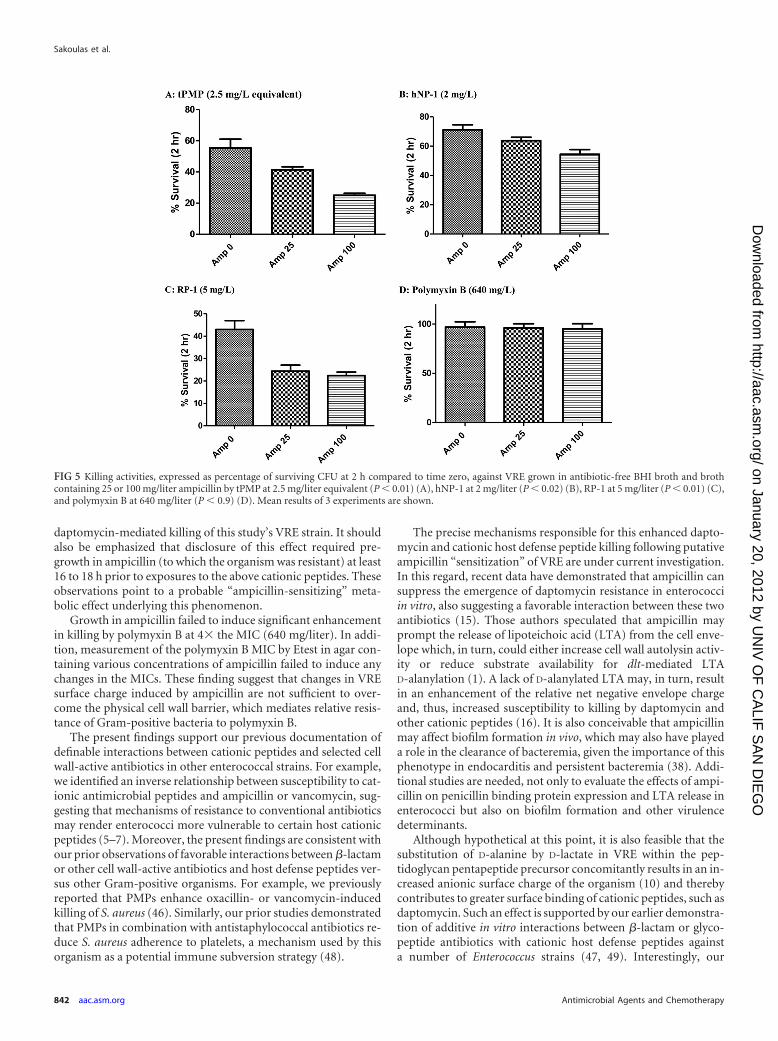
The microbicidal effects of 4 additional cationic peptides weredetermined against the VRE strain after pregrowth in eitherantibiotic-free broth or in broth containing ampicillin at variousconcentrations. Results were as follows: tPMP, 2.5 mg/liter equiv-alent (Fig. 5A); hNP-1, 2 mg/liter (Fig. 5B); RP-1, 5 mg/liter (Fig.5C); polymyxin B, 640 mg/liter (Fig. 5D). Paralleling the dataabove but employing daptomycin plus ampicillin versus the LL-37cationic peptide, pregrowth in ampicillin (100 mg/liter) prior topeptide exposures was associated with significantly greater entero-coccal killing by 3 of the 4 cationic molecules (P � 0.05). As ex-pected for Gram-positive bacteria, the VRE showed relative resis-tance to polymyxin B killing at 4� the MIC, without anysignificant enhancement in killing induced by pregrowth in am-picillin. Among the cationic peptides that did demonstrate in-creased killing of the VRE isolate when grown in ampicillin, onlyRP-1 exhibited enhanced killing following pregrowth in ampicil-lin at both 25 mg/liter and 100 mg/liter compared to growth inampicillin-free medium.
Effect of ampicillin on daptomycin membrane binding. As acomplement to the microbicidal assays described above, dapto-mycin membrane binding studies were performed using Bodipy-fluorescein-labeled daptomycin with VRE pregrown with or with-out ampicillin (50 mg/liter). VRE pregrown in ampicillindemonstrated increased binding to labeled daptomycin comparedto VRE pregrown in antibiotic-free medium when viewed by flu-orescence microscopy and measured by pixelgrams (Fig. 6).
DISCUSSION
In the current study, we identified interesting relationships be-tween ampicillin and daptomycin on both in vitro and in vivo
bases by utilizing a daptomycin-susceptible but ampicillin- andvancomycin-resistant E. faecium clinical isolate. Our studiestranslated observations from “bedside to bench,” following therapid clearance of this strain from the bloodstream after adminis-tration of high-dose daptomycin plus ampicillin in a patient withrefractory VRE bacteremia. Relevant to this striking clinical out-come, we confirmed in vitro that ampicillin at clinically relevantbut sub-MIC levels enhanced the activity of daptomycin from abacteriostatic to a bactericidal potency, even though ampicillinalone had no measureable effect on bacterial growth. These find-ings were further validated in an in vitro pharmacodynamic modelthat demonstrated that simulated coexposures of low-dose dapto-mycin (4 mg/kg/day) plus ampicillin (2 g i.v. q4h) yielded muchgreater killing potency than simulated high-dose daptomycinalone (10 mg/kg/day). Given the apparent increase in the numberof cases of daptomycin-resistant enterococcal infections (24, 26)and the relatively high cost of the drug, the latter finding connotesnot only clinical efficacy impacts but also potentially significantpharmaco-economic implications. Thus, daptomycin adminis-tered at standard approved doses (4 to 6 mg/kg/day) plus ampi-cillin would be significantly less costly than high-dose daptomycin(8 to 12 mg/kg/day).
Of interest, in terms of the salutary influence of ampicillinupon daptomycin-induced killing, ampicillin appeared to sub-stantially reduce the net positive surface charge on our VRE strain,in a concentration-dependent manner. Consistent with this effect,we found that ampicillin preexposure led to increased susceptibil-ity to killing by a number of other cationic peptides, includingthose of diverse structures, charges, sources (i.e., skin, polymor-phonuclear leukocytes, and platelets), and mechanism(s) of ac-tion (e.g., pore formers and non-pore formers). Furthermore,VRE grown in the presence of ampicillin showed significantly in-creased binding to labeled daptomycin compared to VRE grownin medium without ampicillin. These observations strongly sug-gest a charge-based mechanism for the impact of ampicillin on
FIG 4 Killing activity of cathelicidin LL-37 at 36 mg/liter (8 �M), ex-pressed as the percentage of surviving CFU at 2 h compared to time zeroagainst VRE grown to stationary phase in antibiotic-free LB broth (far left)and LB containing ampicillin at 100 mg/liter (middle) (P � 0.027). Thecolumn on the far right demonstrates the killing activity of LL-37 at 36mg/liter coincubated in the presence of ampicillin at 100 mg/liter againstVRE grown in antibiotic-free LB.
FIG 3 FITC-labeled PLL binding assay using flow cytometry results. The de-gree of FITC-labeled PLL inversely reflects the relative surface positive charge.The VRE isolate demonstrated an increased affinity to PLL when it was grownin ampicillin at 50 mg/liter compared to growth in antibiotic-free medium(P � 0.05), indicating a net decrease in relative positive surface charge withgrowth in ampicillin. Data are expressed as mean relative fluorescent units (�SD). Two independent experiments with triplicate samples were performed.
Ampicillin Enhances Cationic Peptide Killing of VRE
February 2012 Volume 56 Number 2 aac.asm.org 841
on January 20, 2012 by UN
IV O
F C
ALIF
SA
N D
IEG
Ohttp://aac.asm
.org/D
ownloaded from
daptomycin-mediated killing of this study’s VRE strain. It shouldalso be emphasized that disclosure of this effect required pre-growth in ampicillin (to which the organism was resistant) at least16 to 18 h prior to exposures to the above cationic peptides. Theseobservations point to a probable “ampicillin-sensitizing” meta-bolic effect underlying this phenomenon.
Growth in ampicillin failed to induce significant enhancementin killing by polymyxin B at 4� the MIC (640 mg/liter). In addi-tion, measurement of the polymyxin B MIC by Etest in agar con-taining various concentrations of ampicillin failed to induce anychanges in the MICs. These finding suggest that changes in VREsurface charge induced by ampicillin are not sufficient to over-come the physical cell wall barrier, which mediates relative resis-tance of Gram-positive bacteria to polymyxin B.
The present findings support our previous documentation ofdefinable interactions between cationic peptides and selected cellwall-active antibiotics in other enterococcal strains. For example,we identified an inverse relationship between susceptibility to cat-ionic antimicrobial peptides and ampicillin or vancomycin, sug-gesting that mechanisms of resistance to conventional antibioticsmay render enterococci more vulnerable to certain host cationicpeptides (5–7). Moreover, the present findings are consistent withour prior observations of favorable interactions between �-lactamor other cell wall-active antibiotics and host defense peptides ver-sus other Gram-positive organisms. For example, we previouslyreported that PMPs enhance oxacillin- or vancomycin-inducedkilling of S. aureus (46). Similarly, our prior studies demonstratedthat PMPs in combination with antistaphylococcal antibiotics re-duce S. aureus adherence to platelets, a mechanism used by thisorganism as a potential immune subversion strategy (48).
The precise mechanisms responsible for this enhanced dapto-mycin and cationic host defense peptide killing following putativeampicillin “sensitization” of VRE are under current investigation.In this regard, recent data have demonstrated that ampicillin cansuppress the emergence of daptomycin resistance in enterococciin vitro, also suggesting a favorable interaction between these twoantibiotics (15). Those authors speculated that ampicillin mayprompt the release of lipoteichoic acid (LTA) from the cell enve-lope which, in turn, could either increase cell wall autolysin activ-ity or reduce substrate availability for dlt-mediated LTAD-alanylation (1). A lack of D-alanylated LTA may, in turn, resultin an enhancement of the relative net negative envelope chargeand, thus, increased susceptibility to killing by daptomycin andother cationic peptides (16). It is also conceivable that ampicillinmay affect biofilm formation in vivo, which may also have playeda role in the clearance of bacteremia, given the importance of thisphenotype in endocarditis and persistent bacteremia (38). Addi-tional studies are needed, not only to evaluate the effects of ampi-cillin on penicillin binding protein expression and LTA release inenterococci but also on biofilm formation and other virulencedeterminants.
Although hypothetical at this point, it is also feasible that thesubstitution of D-alanine by D-lactate in VRE within the pep-tidoglycan pentapeptide precursor concomitantly results in an in-creased anionic surface charge of the organism (10) and therebycontributes to greater surface binding of cationic peptides, such asdaptomycin. Such an effect is supported by our earlier demonstra-tion of additive in vitro interactions between �-lactam or glyco-peptide antibiotics with cationic host defense peptides againsta number of Enterococcus strains (47, 49). Interestingly, our
FIG 5 Killing activities, expressed as percentage of surviving CFU at 2 h compared to time zero, against VRE grown in antibiotic-free BHI broth and brothcontaining 25 or 100 mg/liter ampicillin by tPMP at 2.5 mg/liter equivalent (P � 0.01) (A), hNP-1 at 2 mg/liter (P � 0.02) (B), RP-1 at 5 mg/liter (P � 0.01) (C),and polymyxin B at 640 mg/liter (P � 0.9) (D). Mean results of 3 experiments are shown.
Sakoulas et al.
842 aac.asm.org Antimicrobial Agents and Chemotherapy
on January 20, 2012 by UN
IV O
F C
ALIF
SA
N D
IEG
Ohttp://aac.asm
.org/D
ownloaded from

groups have recently confirmed the enhancement of daptomycinactivity by antistaphylococcal �-lactams against mecA-positivemethicillin-resistant S. aureus (MRSA) strains in vitro, as well as inanimal models of endocarditis (44) and in several cases of refrac-tory MRSA bacteremia (12). In addition, we previously demon-strated the potentiation of killing of S. aureus strains by combina-tions of cell wall-active antibiotics (oxacillin) plus cationic tPMPs(42, 46). Collectively, these findings underscore both similaritiesas well as probable differences in the mechanism(s) betweenstaphylococci and enterococci, vis á vis �-lactam interactions withdaptomycin. For example, although potentiation of killing andreductions in the relative positive surface charge by �-lactamswere observed in both S. aureus and VRE, the enhancement ofdaptomycin staphylocidal activity was seen with either preexpo-sure or concomitant exposure strategies. In contrast, the�-lactam-associated “sensitization” of VRE to subsequent killingby cationic antimicrobial peptides required long-duration pre-growth in ampicillin and was not observed with concomitant ex-posures.
Although limited to a single isolate, the findings from thisstudy may have much broader clinical implications. First, theseresults provide a potential therapeutic option for patients withdaptomycin-refractory VRE bacteremia. Second, these data sug-gest that �-lactam antimicrobials may be beneficial beyond theirdirect antimicrobial properties, in terms of enhancing bacterialclearance by the innate host defense system, in particular as relatedto cationic host defense peptides. These two issues justify an in-depth examination of the frequencies and mechanisms of suchfavorable interactions of various �-lactams with a range of cat-ionic antimicrobial molecules in additional VRE strains. Lastly, itwill be important to examine the potential salutary interactions ofampicillin-daptomycin combinations against other daptomycin-resistant VRE strains.
ACKNOWLEDGMENTS
P.A.M. is employed by Cubist Pharmaceuticals. G.S. has received researchgrants from Cubist and Pfizer Pharmaceuticals, speaking honoraria fromCubist, Pfizer, and Astellas Pharmaceuticals, and consulting fees fromCubist, Astellas, and Ortho-McNeil Pharmaceuticals. A.S.B. has currentresearch grants from Cubist Pharmaceuticals, Morphotek Pharmaceuti-cals, and Astellas Pharmaceuticals and has received speaking honorariafrom Cubist Pharmaceuticals. M.R.Y. is a founder of NovaDigm Thera-peutics, Inc., and has participated in research programs supported in partby grants from Cubist Pharmaceuticals and Pfizer, Inc. M.R.Y. was sup-ported in part by grant R01 AI-48031 from the National Institutes ofHealth, NIAID. A.S.B. was supported in part by grant RO1 AI-39018 fromthe National Institutes of Health. J.P. has received research grants andconsulting fees from Cubist Pharmaceuticals.
REFERENCES1. al-Obeid S, Gutmann L, Williamson R. 1990. Correlation of penicillin-
induced lysis of Enterococcus faecium with saturation of essentialpenicillin-binding proteins and release of lipoteichoic acid. Antimicrob.Agents Chemother. 34:1901–1907.
2. Arias CA, et al. 2011. Genetic bases for in vivo daptomycin resistance inenterococci. N. Engl. J. Med. 365:892–900.
3. Bingen E, Lambert-Zechovsky N, Leclercq R, Doit C, Mariani-Kurkdjian P.1990.Bactericidalactivityofvancomycin,daptomycin,ampicillin,andaminogly-cosides against vancomycin-resistant Enterococcus faecium. J. Antimicrob. Che-mother. 26:619–626.
4. Blaser J. 1985. In-vitro model for simultaneous simulation of the serumkinetics of two drugs with different half-lives. J. Antimicrob. Chemother.15(Suppl. A):125–130.
5. Cashman KA, Bayer AS, Yeaman MR. 1998. Diversity in susceptibility toantibiotics and cationic peptides among Enterococcus faecalis or Enterococ-cus faecium isolates of diverse clinical or geographic origin, abstr. A-26.Abstr. 98th Gen. Meet. Am. Soc. Microbiol., Atlanta, GA. American Soci-ety for Microbiology, Washington, DC.
6. Cashman KA, Hooper DC, Bayer AS, Koo SP, Yeaman MR. 1999. Invitro resistance to vancomycin and endogenous cationic antimicrobialpeptides are independent phenotypes in clinical isolates of Enterococcus
FIG 6 Incorporation of fluorescently labeled daptomycin by VRE grown in the presence or absence of ampicillin. The strain was labeled with 16 mg/literBodipy-labeled daptomycin (green) (A, B, D, and E) for 10 min at 37°C after growth to log phase in either antibiotic-free LB broth (A to C) or in LB brothcontaining ampicillin at 50 mg/ml (D to F). Cells were stained with FM 4-64 (red) (A, B, D, and E) or DAPI (blue) (B and E). Bar, 1 �m. (C and F) Comparisonof daptomycin-Bodipy incorporation, demonstrating higher pixel intensities for daptomycin-Bodipy fluorescence for cells grown with (F) or without (C) 50mg/ml ampicillin. The yellow arrows highlight Bodipy-daptomycin staining (D and E).
Ampicillin Enhances Cationic Peptide Killing of VRE
February 2012 Volume 56 Number 2 aac.asm.org 843
on January 20, 2012 by UN
IV O
F C
ALIF
SA
N D
IEG
Ohttp://aac.asm
.org/D
ownloaded from

faecium, abstr. A-93.Abstr. 99th Gen. Meet. Am. Soc. Microbiol., Chicago,IL. American Society for Microbiology, Washington, DC.
7. Cashman KA, Bayer AS, Yeaman MR. 1999. Diversity of platelet micro-bicidal protein susceptibility in Enterococcus. California State UniversityPress, Long Beach, CA.
8. Cilli F, Aydemir S, Tunger A. 2006. In vitro activity of alone and incombination with various antimicrobials against Gram-positive cocci. J.Chemother. 18:27–32.
9. Clinical and Laboratory Standards Institute. 2006. Methods for dilutionantimicrobial susceptibility tests for bacteria that grow aerobically. Doc-ument M7-A7. Clinical and Laboratory Standards Institute, Wayne, PA.
10. Courvalin P. 2006. Vancomycin resistance in Gram-positive cocci. Clin.Infect. Dis. 42(Suppl. 1):S25–S34.
11. Cubist Pharmaceuticals. 2010. Daptomycin for injection, package insert.Cubist Pharmaceuticals, Lexington, MA.
12. Dhand A, et al. 2011. Use of anti-staphylococcal beta-lactams to increasedaptomycin activity in eradicating persistent bacteremia due tomethicillin-resistant Staphylococcus aureus (MRSA): role of enhanceddaptomycin binding. Clin. Infect. Dis. 53:158 –163.
13. Duez JM, Pechinot A, Siebor E, Cordin X, Kazmierczak A. 1989.Bactericidal activity of daptomycin and vancomycin alone or in combina-tion with tobramycin, netilmicin or ampicillin against Enterococcus.Pathol. Biol. (Paris) 37:263–268.
14. Dvorchik BH, Brazier D, DeBruin MF, Arbeit RD. 2003. Daptomycinpharmacokinetics and safety following administration of escalating dosesonce daily to healthy subjects. Antimicrob. Agents Chemother. 47:1318 –1323.
15. Entenza JM, Giddey M, Vouillamoz J, Moreillon PP. 2010. In vitroprevention of the emergence of daptomycin resistance in Staphylococcusaureus and enterococci following combination with amoxicillin/clavulanic acid or ampicillin. Int. J. Antimicrob. Agents 35:451– 456.
16. Fabretti F, et al. 2006. Alanine esters of enterococcal lipoteichoic acid playa role in biofilm formation and resistance to antimicrobial peptides. In-fect. Immun. 74:4164 – 4171.
17. Friedman L, Alder JD, Silverman JA. 2006. Genetic changes that corre-late with reduced susceptibility to daptomycin in Staphylococcus aureus.Antimicrob. Agents Chemother. 50:2137–2145.
18. Foulds G. 1986. Pharmacokinetics of sulbactam/ampicillin in humans: areview. Rev. Infect. Dis. 8(Suppl. 5):S503–S511.
19. Harigaya Y, et al. 2009. Pharmacodynamics of vancomycin at simulatedepithelial lining fluid concentrations against methicillin-resistant Staphy-lococcus aureus (MRSA): implications for dosing in MRSA pneumonia.Antimicrob. Agents Chemother. 53:3894 –3901.
20. Hayden MK, et al. 2005. Development of daptomycin resistance in vivo inmethicillin-resistant Staphyloccus aureus. J. Clin. Microbiol. 43:5285–5287.
21. Hirschwerk D, Ginocchio CC, Bythrow M, Condon S. 2006. Diminishedsusceptibility to daptomycin accompanied by clinical failure in a patientwith methicillin-resistant Staphylococcus aureus bacteremia. Infect. Con-trol Hosp. Epidemiol. 27:315–317.
22. Jones T, et al. 2008. Failures in clinical treatment of Staphylococcus aureusinfection with daptomycin are associated with alterations in surfacecharge, membrane phospholipid asymmetry, and drug binding. Antimi-crob. Agents Chemother. 52:269 –278.
23. Julian K, et al. 2007. Characterization of a daptomycin-nonsusceptible,vancomycin-intermediate Staphylococcus aureus strain in a patient withendocarditis. Antimicrob. Agents Chemother. 51:3445–3448.
24. Kelesidis T, Humphries R, Uslan DZ, Pegues DA. 2011. Daptomycinnonsusceptible enterococci: an emerging challenge for clinicians. Clin.Infect. Dis. 52:228 –234.
25. Reference deleted.26. Lewis JS, et al. 2005. Emergence of daptomycin resistance in Enterococcus
faecium during daptomycin therapy. Antimicrob. Agents Chemother. 41:1664 –1665.
27. Mangili A, Bica I, Snydman DR, Hamer DH. 2005. Daptomycin-resistant, methicillin-resistant Staphylococcus aureus bacteremia. Clin. In-fect. Dis. 40:1058 –1060.
28. Mariani PG, Sader HS, Jones RN. 2006. Development of decreasedsusceptibility to daptomycin and vancomycin in a Staphylococcus aureusstrain during prolonged therapy. J. Antimicrob. Chemother. 58:481– 483.
29. Marty FM, et al. 2006. Emergence of a clinical daptomycin-resistantStaphylococcus aureus isolate during treatment of methicillin-resistantStaphylococcus aureus bacteremia and osteomyelitis. J. Clin. Microbiol.44:595–597.
30. Mobarakai N, Quale JM, Landman D. 1994. Bactericidal activities ofpeptide antibiotics against multidrug-resistant Enterococcus faecium. An-timicrob. Agents Chemother. 38:385–387.
31. Moise PA, North D, Steenbergen JN, Sakoulas G. 2009. Susceptibilityrelationship between vancomycin and daptomycin in Staphylococcusaureus: facts and assumptions. Lancet Infect. Dis. 9:617– 624.
32. Montero CI, Stock F, Murray PR. 2008. Mechanisms of resistance todaptomycin in Enterococcus faecium. Antimicrob. Agents Chemother. 52:1167–1170.
33. Mukhopadhyay K, et al. 2007. In vitro susceptibility of Staphylococcusaureus to thrombin-induced platelet microbicidal protein-1 (tPMP-1) isinfluenced by cell membrane phospholipid composition and asymmetry.Microbiology 153:1187–1197.
34. Palmer KL, Daniel A, Hardy C, Silverman J, Gilmore MS. 2011. Geneticbases for daptomycin resistance in enterococci. Antimicrob. Agents Che-mother. 55:3345–3356.
35. Reference deleted.36. Pogliano J, et al. 1999. A vital stain for studying membrane dynamics in
bacteria: a novel mechanism controlling septation during Bacillus subtilissporulation. Mol. Microbiol. 31:1149 –1159.
37. Rand KH, Houck H. 2004. Daptomycin synergy with rifampicin andampicillin against vancomycin-resistant enterococci. J. Antimicrob. Che-mother. 53:530 –532.
38. Seidl K, et al. 2011. Combinatorial phenotypic signatures distinguishpersistent from resolving methicillin-resistant Staphylococcus aureus bac-teremia isolates. Antimicrob. Agents Chemother. 55:575–582.
39. Skiest DJ. 2006. Treatment failure resulting from resistance of Staphylo-coccus aureus to daptomycin. J. Clin. Microbiol. 44:655– 656.
40. Tsuji BT, Rybak MJ. 2005. Short-course gentamicin in combination withdaptomycin or vancomycin against Staphylococcus aureus in an in vitropharmacodynamic model with simulated endocardial vegetations. Anti-microb. Agents Chemother. 49:2735–2745.
41. Vikram HR, Havill NL, Koeth M, Boyce JM. 2005. Clinical progressionof methicillin-resistant Staphylococcus aureus vertebral osteomyelitis asso-ciated with reduced susceptibility to daptomycin. J. Clin. Microbiol. 43:5384 –5387.
42. Xiong YQ, Yeaman MR, and Bayer AS. 1999. In vitro microbicidalactivities of thrombin-induced platelet microbicidal protein-1 and humanneutrophil defensin-1 on Staphylococcus aureus are substantially influ-enced by pretreatment with antibiotics differing in mechanisms of action.Antimicrob. Agents Chemother. 43:1111–1117.
43. Xiong YQ, Mukhopadhyay K, Yeaman MR, Adler-Moore J, Bayer AS.2005. Functional interrelationships between cell membrane and cell wallin antimicrobial peptide-mediated killing of Staphylococcus aureus. Anti-microb. Agents Chemother. 49:3114 –3121.
44. Yang SJ, et al. 2010. Daptomycin-oxacillin combinations in experimentalmethicillin-resistant Staphylococcus aureus (MRSA) endocarditis causedby daptomycin-nonsusceptible strains with evolving oxacillin susceptibil-ity (“see-saw effect”). Antimicrob. Agents Chemother. 54:3161–3169.
45. Yeaman MR, Gank KD, Bayer AS, Brass EP. 2002. Synthetic peptidesthat exert antimicrobial activities in whole blood and blood-derived ma-trices. Antimicrob. Agents Chemother. 46:3883–3891.
46. Yeaman MR, Norman DC, Bayer AS. 1992. Platelet microbicidal proteinenhances antibiotic-induced killing of and postantibiotic effect in Staph-ylococcus aureus. Antimicrob. Agents Chemother. 36:1665–1670.
47. Yeaman MR. 2002. Code among chaos in antimicrobial resistance:lessons from host defense proteins, p 10. Keystone Symposium onMolecular and Cellular Biololgy, Santa Fe, NM. Keystone Symposia,Silverthorne, CO.
48. Yeaman MR, Sullam PM, Dazin PF, Bayer AS. 1994. Platelet microbi-cidal protein alone and in combination with antibiotics reduces Staphylo-coccus aureus adherence to platelets in vitro. Infect. Immun. 62:3416 –3423.
49. Yeaman MR, Yount NY. 2003. Mechanisms of antimicrobial peptideaction and resistance. Pharmacol. Rev. 55:27–55.
Sakoulas et al.
844 aac.asm.org Antimicrobial Agents and Chemotherapy
on January 20, 2012 by UN
IV O
F C
ALIF
SA
N D
IEG
Ohttp://aac.asm
.org/D
ownloaded from