amniotic fluid embolism dr. s. parthasarathy md., da., dnb, md (acu), dip. diab.dca, dip. software...
TRANSCRIPT

Amniotic fluid embolismDr. S. Parthasarathy
MD., DA., DNB, MD (Acu), Dip. Diab.DCA, Dip. Software
statistics, Phd
Mahatma Gandhi Medical college and research institute , puducherry , India

What is it ??
• Amniotic fluid embolism (AFE) is a catastrophic obstetric emergency
• sudden, profound, and unexpected maternal collapse
• hypotension, hypoxaemia, and disseminated intravascular coagulation (DIC).
• Altered mental status ??

Precipitating factors
• labor, cesarean delivery, or dilation and evacuation or within 30 minutes postpartum with no other explanation for the clinical findings.
• Rarely after abdominal trauma in pregnant

History
• The entry of the amniotic fluid was first described by Ricardo Meyer in 1926
• In 1941, Steiner and Luschbaugh described histopathologic
findings in the pulmonary vasculature in 8 multiparous women
dying of sudden shock during labor.
• Findings included mucin, amorphous eosinophilic material , and
in some cases squamous cells.
.

Incidence
• Unknown • 1 in 8000 deliveries to 1 in 80000 deliveries• 1980 s - mortality – 80 % • 2002 --- - mortality- 25 % • 75 % autopsy evidence • Neonatal mortality – 70 %

A TRIUMPH- pneumonic
• advanced maternal age; ?? • multiparity;• meconium stained liquor;• intrauterine fetal death; • Poly hydramnios; • frequent or tetanic uterine contractions;• maternal history of allergy or atopy; • microsomia; • uterine rupture; and• placenta accreta• Infection .

The process
• Amniotic fluid enters maternal circulation • ruptured membranes • ruptured uterine or cervical vessels • down a pressure gradient from the uterus to
veins.• Portals• placenta, endocervical, -- tears

What happens
• Physical
• Immunological

Physical theory
Pulmonary obstruction, pulmonary vasospasm
Hypoxemia Cardiac arrest

Clarke theory
Phase 1 - pulmonary artery vasospasm followed by
pulmonary hypertension.Hypoxemia – myocardial
dysfunction
Release by amniotic fluid

Phase 2
• Left ventricular failure and pulmonary edema occurs.
• Biochemical mediators trigger DIC leading to massive haemorrhage and uterine atony.

One more theory
• A more recent hypothesis is that amniotic fluid contains a direct myocardial depressant
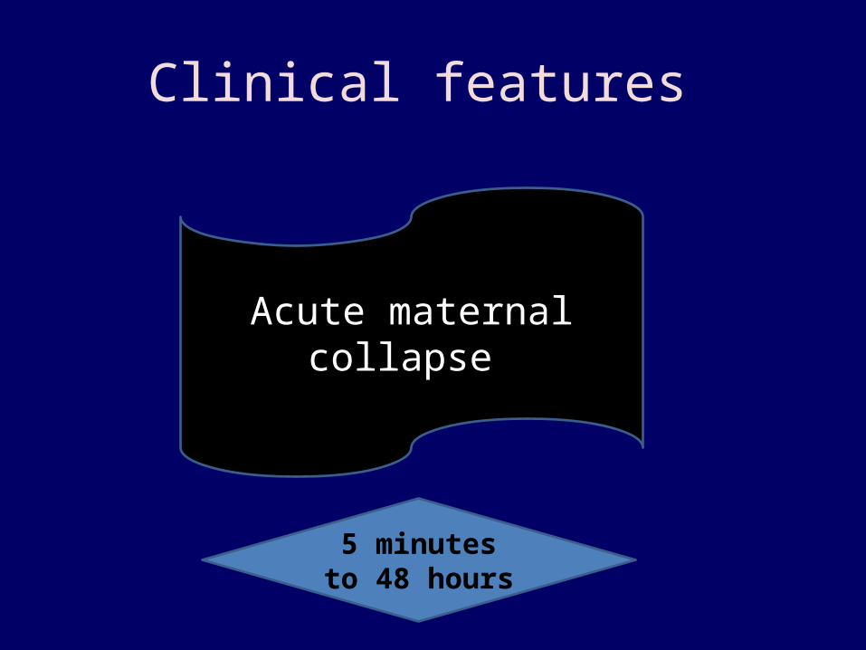
Clinical features
Acute maternal collapse
5 minutes to 48 hours

Premonitory symptoms ??
• Breathlessness• Chest pain• Lightheadedness• Distress or panic• “Pins and needles” in the fingers• Nausea and vomiting• Feeling cold


To be simple
1) Respiratory distress
(2) Cyanosis
(3) Cardiovascular collapse cardiogenic shock
(4) Hemorrhage
(5) Coma.
Breathing, bleeding and three C s

Pathophysiology of DIC
• procoagulant is sloughed fetal skin and respiratory,
gastrointestinal, and genitourinary epithelia.
• Tissue factor is responsible for activating the extrinsic
pathway by binding with factor VII. This complex in
turn triggers clotting by activating factor X.
• DIC


Clinical scenario
• A sudden drop in O2 saturation can be the initial indication of AFE during LSCS.
• More than 1/2 of patients die within the first hour.• Of the survivors 50 % will develop DIC which may
manifest as persistent bleeding from incision or venipuncture sites.
The coagulopathy typically occurs 0.5 to 4 hours after phase 1
10 % have seizures
Mortality – 25 to 60 %

Diagnosis • ABG – hypoxemia • CXR may be normal or show effusions, enlarged
heart, or pulmonary edema. • ECG may show a right strain pattern with ST-T
changes and tachycardia. • Fetal squamous cells in PA catheter • The Sialyl Tn antigen test• TKAH 2 antibody test • Antibody to fetal mucin

• SERUM TRYTASE LEVEL
• MAST CELL DEGRANULATION AND ANAPHYLACTOID FEATURES

Diagnosis
• Measurement of the maternal plasma
concentration of zinc coproporphyrin, a
component of meconium, also has been
proposed as a sensitive test for the diagnosis
of amniotic fluid embolism
• Coagulopathy screening

TEE
• First 30 minute -- Transesophageal 4-chamber images
showed severe pulmonary hypertension, acute right
ventricular failure with a leftward deviation of the
inter atrial and inter ventricular septum, and severe
tricuspid regurgitation.

Differential diagnosis
• Anaphylaxis (Collapse)
• Pulmonary embolus (Collapse)
• Aspiration (Hypoxaemia)
• Pre-eclampsia or eclampsia (Fits, Coagulopathy)
• Haemorrhage (APH ; PPH)
• Septic shock• Drug toxicity
(MgSO4, total spinal,
LA toxicity)• Aortic dissection

• Diagnosis of exclusion and suspicion
• Usually at autopsy

Management

Principles
• Oxygenation • Cardiac output maintenance • Coagulopathy correction
• Uterine tone and delivery of the baby

Oxygenation
• Maintaining oxygenation may necessitate intubation and ventilation.
• CPAP or PEEP may be indicated.

Haemodynamic stability
• Rapid infusion of crystalloids, colloids, • Inotropes
• PAWP catheter ??? • COAGULOPATHY

Coagulopathy • Plasma, cryoprecipitate, and platelets are
frequently required.
• Recombinant factor VII has been used to treat uncontrollable massive obstetric haemorrhage if haemorrhage becomes difficult to control.
• Hematologists consultation

Cryoprecipitate is useful
• it can replenish clotting factors
• cryoprecipitate contains fibronectin
• it could facilitate the removal of cellular and
particulate matter, such as amniotic fluid
debris, from the blood via the monocyte
/macrophage system

Uterine tone
• Oxytocin • Methergin • PGs • Bimanual massage

Baby delivery
• The baby should be delivered as quickly as possible.
• If the mother is undergoing CPR, surgical delivery should be performed within 5 min for improved maternal outcome.

Possible treatment on survival
• Steroids, prostacyclin, nitric oxide as well as
plasma exchange, haemo filtration, and
cardiopulmonary bypass

All these remain true ??
• Catastrophic• Unpredictable• Unpreventable• ? Untreatable

SUMMARY
• Definition • Risk factors -- a triumph• Pathophysiology • Clinical features • Diagnosis • Treatment

The term AFE now appears to be a misnomer.
• Proposed new names include • “sudden obstetric collapse syndrome” and • “anaphylactoid syndrome of pregnancy”.

Thank you