amines by john18 lecture presentation
TRANSCRIPT

© 2012 Pearson Education, Inc.
18 The Nervous System: General and Special Senses
PowerPoint® Lecture Presentations prepared by
Steven Bassett
Southeast Community College
Lincoln, Nebraska

© 2012 Pearson Education, Inc.
Introduction
• Sensory information arrives at the CNS
• Information is “picked up” by sensory
receptors
• Sensory receptors are the interface between
the nervous system and the internal and
external environment
• General senses
• Refers to temperature, pain, touch, pressure,
vibration, and proprioception
• Special senses
• Refers to smell, taste, balance, hearing, and vision

© 2012 Pearson Education, Inc.
Receptors
• Receptors and Receptive Fields
• Free nerve endings are the simplest receptors
• These respond to a variety of stimuli
• Receptors of the retina (for example) are very
specific and only respond to light
• Receptive fields
• Large receptive fields have receptors spread far
apart, which makes it difficult to localize a stimulus
• Small receptive fields have receptors close
together, which makes it easy to localize a stimulus.

© 2012 Pearson Education, Inc.
Figure 18.1 Receptors and Receptive Fields
Receptive fields
Receptive
field 1 Receptive
field 2

© 2012 Pearson Education, Inc.
Receptors
• Interpretation of Sensory Information
• Information is relayed from the receptor to
a specific neuron in the CNS
• The connection between a receptor and a neuron is
called a labeled line
• Each labeled line transmits its own specific
sensation

© 2012 Pearson Education, Inc.
Interpretation of Sensory Information
• Classification of Receptors
• Tonic receptors
• Always active
• Photoreceptors of the eye constantly monitor body
position
• Phasic receptors
• Normally inactive but become active when
necessary (for short periods of time)
• Touch and pressure receptors of the skin (for
example)
© 2012 Pearson Education, Inc.
Receptors
• Central Processing and Adaptation
• Adaptation
• Reduction in sensitivity due to a constant stimulus
• Peripheral adaptation
• Receptors respond strongly at first and then decline
• Central adaptation
• Adaptation within the CNS
• Consciously aware of a stimulus, which quickly
disappears

© 2012 Pearson Education, Inc.
The General Senses
• Classification of the General Senses
• One classification scheme:
• Exteroceptors: provide information about the
external environment
• Proprioceptors: provide information about the
position of the body
• Interoceptors: provide information about the inside
of the body

© 2012 Pearson Education, Inc.
The General Senses
• Classification of the General Senses
• Another classification scheme:
• Nociceptors: respond to the sensation of pain
• Thermoreceptors: respond to changes in
temperature
• Mechanoreceptors: activated by physical
distortion of cell membranes
• Chemoreceptors: monitor the chemical
composition of body fluids

© 2012 Pearson Education, Inc.
The General Senses
• Nociceptors
• Known as pain receptors
• Associated with free nerve endings and large
receptor fields. This makes it difficult to
“pinpoint” the location of the origin of the pain
• Three types
• Receptors sensitive to extreme temperatures
• Receptors sensitive to mechanical damage
• Receptors sensitive to chemicals

© 2012 Pearson Education, Inc.
The General Senses
• Nociceptors
• Fast pain:
• Sensations reach the CNS fast
• Associated with pricking pain or cuts
• Slow pain:
• Sensations reach the CNS slowly
• Associated with burns or aching pains
• Referred pain:
• Sensations reach the spinal cord via the dorsal roots
• Some visceral organ pain sensations may reach the
spinal cord via the same dorsal root

© 2012 Pearson Education, Inc.
Figure 18.2 Referred Pain
Heart
Liver and
gallbladder
Stomach
Small
intestine
Appendix
Colon
Ureters
© 2012 Pearson Education, Inc.
The General Senses
• Thermoreceptors
• Found in the dermis, skeletal muscles, liver,
and hypothalamus
• Cold receptors are more numerous than hot
receptors
• Exist as free nerve endings
• These are phasic receptors
• Information is transmitted along the same
pathway as pain information

© 2012 Pearson Education, Inc.
The General Senses
• Mechanoreceptors
• Receptors that are sensitive to stretch,
compression, twisting, or distortion of the
plasmalemmae
• There are three types
• Tactile receptors
• Baroreceptors
• Proprioceptors
© 2012 Pearson Education, Inc.
The General Senses
• Mechanoreceptors
• Tactile receptors
• Provide sensations of touch, pressure, and
vibrations
• Unencapsulated tactile receptors: free nerve
endings, tactile disc, and root hair plexus
• Encapsulated tactile receptors: tactile corpuscle,
Ruffini corpuscle, and lamellated corpuscle

© 2012 Pearson Education, Inc.
The General Senses
• Mechanoreceptors
• Unencapsulated tactile receptors
• Free nerve endings are common in the dermis
• Tactile discs are in the stratum basale layer
• Root hair plexus monitors distortions and
movements of the body surface

© 2012 Pearson Education, Inc.
Figure 18.3a Tactile Receptors in the Skin
Free nerve endings
Hair
Root hair plexus
Lamellated corpuscle
Ruffini corpuscle
Merkel cells and
tactile discs Tactile
corpuscle
Free nerve
ending
Sensory
nerves

© 2012 Pearson Education, Inc.
Figure 18.3b Tactile Receptors in the Skin
Hair
Root hair plexus
Lamellated corpuscle
Ruffini corpuscle
Merkel cells and
tactile discs
Tactile
corpuscle
Free nerve
ending
Sensory
nerves
Merkel cells
Tactile disc
Merkel cells and tactile discs

© 2012 Pearson Education, Inc.
Figure 18.3c Tactile Receptors in the Skin
Hair
Root hair plexus
Lamellated corpuscle
Ruffini corpuscle
Merkel cells and
tactile discs
Tactile
corpuscle
Free nerve
ending
Sensory
nerves
Free nerve endings
of root hair plexus

© 2012 Pearson Education, Inc.
The General Senses
• Mechanoreceptors
• Encapsulated tactile receptors
• Tactile corpuscle: common on eyelids, lips,
fingertips, nipples, and genitalia
• Ruffini corpuscle: in the dermis, sensitive to
pressure and distortion
• Lamellated corpuscle: consists of concentric
cellular layers / sensitive to vibrations

© 2012 Pearson Education, Inc.
Figure 18.3d Tactile Receptors in the Skin
Tactile corpuscle; the capsule
boundary in the micrograph is
indicated by a dashed line.
Tactile
corpuscle Epidermis
Dermis
Tactile corpuscle LM 550
Capsule
Accessory
cells
Dendrites
Sensory
nerve fiber
Hair
Root hair plexus
Lamellated corpuscle
Ruffini corpuscle
Merkel cells and
tactile discs
Tactile
corpuscle
Free nerve
ending
Sensory
nerves

© 2012 Pearson Education, Inc.
Figure 18.3e Tactile Receptors in the Skin
Capsule
Dendrites
Ruffini corpuscle
Sensory
nerve fiber
Collagen
fibers
Hair
Root hair plexus
Lamellated corpuscle
Ruffini corpuscle
Merkel cells and
tactile discs
Tactile
corpuscle
Free nerve
ending
Sensory
nerves

© 2012 Pearson Education, Inc.
Figure 18.3f Tactile Receptors in the Skin
Lamellated corpuscle
Dendritic
process
Concentric layers (lamellae)
of collagen fibers
separated by fluid
Concentric layers (lamellae)
of collagen fibers
separated by fluid
Accessory cells
(specialized fibrocytes)
Dendritic process
Dermis
LM 125 Lamellated corpuscle
Hair
Root hair plexus
Lamellated corpuscle
Ruffini corpuscle
Merkel cells and
tactile discs
Tactile
corpuscle
Free nerve
ending
Sensory
nerves
© 2012 Pearson Education, Inc.
The General Senses
• Mechanoreceptors
• Baroreceptors
• Stretch receptors that monitor changes in the
stretch of organs
• Found in the stomach, small intestine, urinary
bladder, carotid artery, lungs, and large intestine

© 2012 Pearson Education, Inc.
Figure 18.4 Baroreceptors and the Regulation of Autonomic Functions
Provide information on volume of
tract segments, trigger reflex
movement of materials along tract
Provide information on volume of
urinary bladder, trigger urinary reflex
Baroreceptors of Bladder Wall
Baroreceptors of Digestive Tract
Baroreceptors of Carotid Sinus and Aortic Sinus
Baroreceptors of Lung
Baroreceptors of Colon
Provide information on blood
pressure to cardiovascular and
respiratory control centers
Provide information on lung
stretching to respiratory
rhythmicity centers for
control of respiratory rate
Provide information on volume
of fecal material in colon,
trigger defecation reflex

© 2012 Pearson Education, Inc.
The General Senses
• Mechanoreceptors
• Proprioceptors
• Monitor the position of joints, tension in the tendons
and ligaments, and the length of muscle fibers upon
contraction
• Muscle spindles are receptors in the muscles
• Golgi tendon organs are the receptors in the
tendons
© 2012 Pearson Education, Inc.
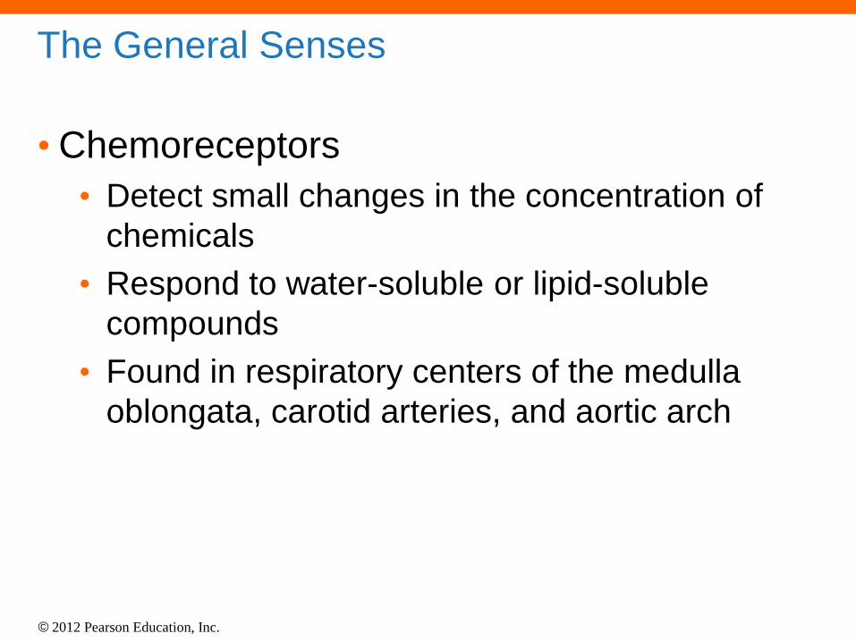
The General Senses
• Chemoreceptors
• Detect small changes in the concentration of
chemicals
• Respond to water-soluble or lipid-soluble
compounds
• Found in respiratory centers of the medulla
oblongata, carotid arteries, and aortic arch
© 2012 Pearson Education, Inc.
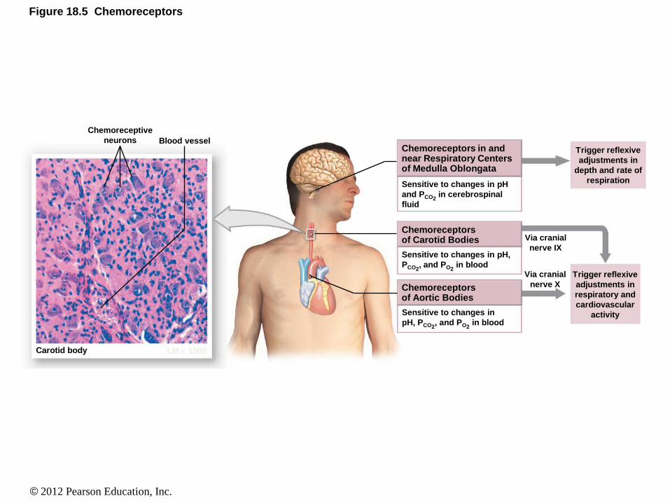
Figure 18.5 Chemoreceptors
Carotid body LM 1500
Blood vessel
Chemoreceptive
neurons
Trigger reflexive
adjustments in
depth and rate of
respiration
Trigger reflexive
adjustments in
respiratory and
cardiovascular
activity
Via cranial
nerve IX
Via cranial
nerve X
Sensitive to changes in pH
and PCO2 in cerebrospinal
fluid
Sensitive to changes in pH,
PCO2, and PO2
in blood
Sensitive to changes in
pH, PCO2, and PO2
in blood
Chemoreceptors in and near Respiratory Centers of Medulla Oblongata
Chemoreceptors of Carotid Bodies
Chemoreceptors of Aortic Bodies

© 2012 Pearson Education, Inc.
The Special Senses
• The special senses include:
• Olfaction (smell)
• Gustation (taste)
• Equilibrium
• Hearing
• Vision

© 2012 Pearson Education, Inc.
Olfaction (Smell)
• Olfaction
• The olfactory epithelium consists of:
• Olfactory receptors
• Supporting cells
• Basal cells

© 2012 Pearson Education, Inc.
Olfaction (Smell)
• Olfactory Pathways
• Axons leave the olfactory epithelium
• Pass through the cribriform foramina
• Synapse on neurons in the olfactory bulbs
• Impulses travel to the brain via CN I
• Arrive at the cerebral cortex, hypothalamus,
and limbic system

© 2012 Pearson Education, Inc.
Figure 18.6a The Olfactory Organs
The distribution of the olfactory receptors
on the left side of the nasal septum is
shown by the shading.
Olfactory
bulb
Olfactory nerve
fibers (N I)
Olfactory
tract
Cribriform plate
of ethmoid
Olfactory
epithelium

© 2012 Pearson Education, Inc.
Figure 18.6b The Olfactory Organs
A detailed view of the olfactory epithelium
Substance being smelled
Olfactory
epithelium
Lamina
propria
Cribriform
plate
Knob
Olfactory cilia:
surfaces contain
receptor proteins
Mucous layer
Supporting cell
Olfactory
receptor cell
Developing olfactory
receptor cell
Olfactory
nerve fibers
To olfactory
bulb
Olfactory
(Bowman’s)
gland
Regenerative basal cell:
divides to replace worn-out
olfactory receptor cells

© 2012 Pearson Education, Inc.
Olfaction (Smell)
• Olfactory Discrimination
• The epithelial receptors have different
sensitivities and we therefore “detect” different
smells
• Olfactory receptors can be replaced
• The replacement activity declines with age

© 2012 Pearson Education, Inc.
Gustation (Taste)
• Gustation
• The tongue consists of papillae
• Papillae consist of taste buds
• Taste buds consist of gustatory cells
• Each gustatory cell has a slender microvilli
that extends through the taste pore into the
surrounding fluid
© 2012 Pearson Education, Inc.
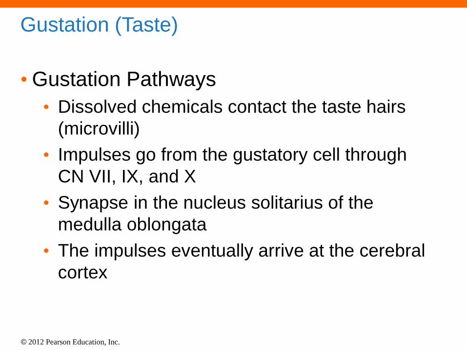
Gustation (Taste)
• Gustation Pathways
• Dissolved chemicals contact the taste hairs
(microvilli)
• Impulses go from the gustatory cell through
CN VII, IX, and X
• Synapse in the nucleus solitarius of the
medulla oblongata
• The impulses eventually arrive at the cerebral
cortex

© 2012 Pearson Education, Inc.
Figure 18.8 Gustatory Pathways
Gustatory
cortex
Thalamic
nucleus
Medial
lemniscus
Nucleus
solitarius
Vagus nerve
(N X)
Facial nerve
(N VII)
Glossopharyngeal
nerve (N IX)

© 2012 Pearson Education, Inc.
Gustation (Taste)
• Gustation Discrimination
• We begin life with more than 10,000 taste
buds
• The number declines rapidly by age 50
• Threshold level is low for gustatory cells
responsible for unpleasant stimuli
• Threshold level is high for gustatory cells
responsible for pleasant stimuli

© 2012 Pearson Education, Inc.
Equilibrium and Hearing
• Equilibrium and Hearing
• Structures of the ear are involved in balance
and hearing
• The ear is subdivided into three regions
• External ear
• Middle ear
• Inner ear
ANIMATION The Ear: Ear Anatomy

© 2012 Pearson Education, Inc.
Equilibrium and Hearing
• The External Ear
• Consists of:
• Auricle
• External acoustic meatus
• Tympanic membrane
• Ceruminous glands

© 2012 Pearson Education, Inc.
Figure 18.9 Anatomy of the Ear
EXTERNAL EAR MIDDLE EAR INNER EAR
Auricle
Auditory ossicles Semicircular canals
Petrous part of temporal
bone
Facial nerve (N VII)
External acoustic meatus
Elastic cartilage
Tympanic membrane
Tympanic cavity
Oval window
Round window
Vestibule
Auditory tube
Cochlea
To nasopharynx
Bony labyrinth of inner ear
Vestibulocochlear nerve (N VIII)

© 2012 Pearson Education, Inc.
Equilibrium and Hearing
• The Middle Ear
• Consists of:
• Tympanic cavity
• Auditory ossicles
• Malleus, incus, and stapes
• Auditory tube (pharyngotympanic tube)

© 2012 Pearson Education, Inc.
Figure 18.9 Anatomy of the Ear
EXTERNAL EAR MIDDLE EAR INNER EAR
Auricle
Auditory ossicles Semicircular canals
Petrous part of temporal
bone
Facial nerve (N VII)
External acoustic meatus
Elastic cartilage
Tympanic membrane
Tympanic cavity
Oval window
Round window
Vestibule
Auditory tube
Cochlea
To nasopharynx
Bony labyrinth of inner ear
Vestibulocochlear nerve (N VIII)

© 2012 Pearson Education, Inc.
Figure 18.10a The Middle Ear
Inferior view of the right temporal
bone drawn, as if transparent, to
show the location of the middle
and inner ear
Inner ear
Tympanic cavity
(middle ear)
External acoustic
meatus
Tympanic membrane
Auditory ossicles
Auditory tube

© 2012 Pearson Education, Inc.
Figure 18.10b The Middle Ear
Structures within the middle ear cavity
Temporal bone
(petrous part)
Stabilizing
ligament
Chorda tympani
nerve (cut), a
branch of N VII
External acoustic
meatus
Tympanic cavity
(middle ear)
Tympanic membrane
(tympanum)
Malleus
Incus
Base of stapes
at oval window
Tensor tympani
muscle
Stapes
Round window
Stapedius
muscle
Auditory tube

© 2012 Pearson Education, Inc.
Figure 18.10c The Middle Ear
The isolated auditory ossicles
Malleus
Incus
Points of
attachment
to tympanic
membrane
Stapes
Base
of stapes
© 2012 Pearson Education, Inc.
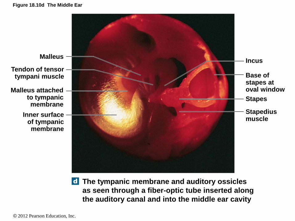
Figure 18.10d The Middle Ear
The tympanic membrane and auditory ossicles
as seen through a fiber-optic tube inserted along
the auditory canal and into the middle ear cavity
Incus
Base of stapes at oval window
Stapes
Stapedius muscle
Malleus
Tendon of tensor tympani muscle
Malleus attached to tympanic membrane
Inner surface of tympanic membrane
© 2012 Pearson Education, Inc.
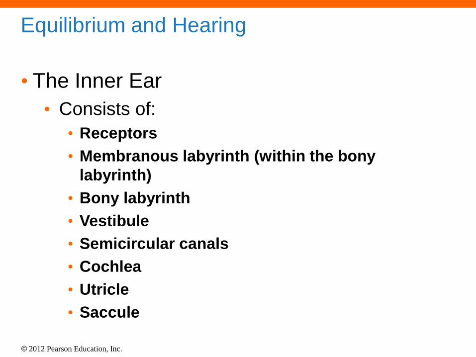
Equilibrium and Hearing
• The Inner Ear
• Consists of:
• Receptors
• Membranous labyrinth (within the bony
labyrinth)
• Bony labyrinth
• Vestibule
• Semicircular canals
• Cochlea
• Utricle
• Saccule

© 2012 Pearson Education, Inc.
Figure 18.9 Anatomy of the Ear
EXTERNAL EAR MIDDLE EAR INNER EAR
Auricle
Auditory ossicles Semicircular canals
Petrous part of temporal
bone
Facial nerve (N VII)
External acoustic meatus
Elastic cartilage
Tympanic membrane
Tympanic cavity
Oval window
Round window
Vestibule
Auditory tube
Cochlea
To nasopharynx
Bony labyrinth of inner ear
Vestibulocochlear nerve (N VIII)

© 2012 Pearson Education, Inc.
Figure 18.12a Semicircular Canals and Ducts
Anterior view of the bony
labyrinth cut away to show the
semicircular canals and the
enclosed semicircular ducts of
the membranous labyrinth
Cochlear duct
Vestibular duct
Saccule
Utricle
Tympanic
duct
Organ of
Corti
Cochlea
Endolymphatic sac
Maculae
Cristae within ampullae
Bony labyrinth
Membranous
labyrinth
KEY
Vestibule
Anterior
Lateral
Posterior
Semicircular
canal
Semicircular
ducts

© 2012 Pearson Education, Inc.
Figure 18.12b Semicircular Canals and Ducts
Cross section of a semicircular canal to
show the orientation of the bony
labyrinth, perilymph, membranous
labyrinth, and endolymph
Perilymph
Bony labyrinth
Endolymph
Membranous
labyrinth

© 2012 Pearson Education, Inc.
Equilibrium and Hearing
• The Inner Ear
• The vestibular complex and equilibrium
• Part of inner ear that provides equilibrium
sensations by detecting rotation, gravity,
and acceleration
• Consists of:
• Semicircular canals
• Utricle
• Saccule

© 2012 Pearson Education, Inc.
Equilibrium and Hearing
• The Vestibular Complex and Equilibrium
• The semicircular canals
• Each semicircular canal encases a duct
• The beginning of each duct is the ampulla
• Within each ampulla is a cristae with hair cells
• Each hair cell contains a kinocilium and stereocilia
• These are embedded in gelatinous material called
the cupula
• The movement of the body causes movement of
fluid in the canal, which in turn causes movement of
the cupula and hair cells, which the brain detects

© 2012 Pearson Education, Inc.
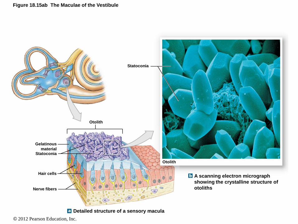
Equilibrium and Hearing
• The Vestibular Complex and Equilibrium
• The utricle and saccule
• The utricle and saccule are connected to the ampulla
and to each other and to the fluid within the cochlea
• Hair cells of the utricle and saccule are in clusters
called maculae
• Hair cells are embedded in gelatinous material
consisting of statoconia (calcium carbonate crystals)
• Gelatinous material and statoconia collectively are
called an otolith
ANIMATION The Ear: Ear Balance

© 2012 Pearson Education, Inc.
Equilibrium and Hearing
• Equilibrium Process
• When you rotate your head:
• The endolymph in the semicircular canals begins to move
• This causes the bending of the kinocilium and stereocilia
• This bending causes depolarization of the associated
sensory nerve
• When you rotate your head to the right, the hair cells are
bending to the left (due to movement of the endolymph)
• When you move in a circle and then stop abruptly, the
endolymph moves back and forth causing the hair cells to
bend back and forth resulting in confusing signals, thus
dizziness

© 2012 Pearson Education, Inc.
Figure 18.13 The Function of the Semicircular Ducts, Part I
Anterior view of
the maculae and
semicircular ducts
of the right side
A section through the ampulla of a
semicircular duct
Endolymph movement along the
length of the duct moves the cupula
and stimulates the hair cells.
Structure of a typical hair cell showing details
revealed by electron microscopy. Bending the
stereocilia toward the kinocilium depolarizes the cell
and stimulates the sensory neuron. Displacement in
the opposite direction inhibits the sensory neuron.
Supporting cell
Sensory nerve
ending
Hair cell
Stereocilia Kinocilium
Displacement in
this direction
inhibits hair cell
Displacement in
this direction
stimulates hair cell
At rest
Ampulla Semicircular duct
Direction of
duct rotation
Direction of relative
endolymph movement
Direction of
duct rotation
Crista
Hair cells
Ampulla
filled with
endolymph Cupula
Supporting cells
Sensory nerve
Saccule Maculae
Utricle
Ampulla Anterior
Posterior
Lateral
Semicircular
ducts
Vestibular branch (N VIII)
Cochlea
Endolymphatic sac
Endolymphatic duct

© 2012 Pearson Education, Inc.
Figure 18.14 The Function of the Semicircular Ducts, Part II
Location and orientation of the membranous
labyrinth within the petrous parts of the
temporal bones
A superior view showing the planes of
sensitivity for the semicircular ducts
Posterior semicircular
duct for ―tilting head‖
Lateral
semicircular
duct for ―no‖
Anterior semicircular
duct for ―yes‖

© 2012 Pearson Education, Inc.
Equilibrium and Hearing
• Equilibrium Process (cont.)
• When you move up or down (elevator
movement):
• Otoliths rest on top of the maculae
• When moving upward, the otoliths press down on
the macular surface
• When moving downward, the otoliths lift off the
macular surface
• When you tilt side to side:
• When tilting to one side, the otoliths shift to one
side of the macular surface
© 2012 Pearson Education, Inc.
Figure 18.15ab The Maculae of the Vestibule
A scanning electron micrograph
showing the crystalline structure of
otoliths
Detailed structure of a sensory macula
Otolith
Gelatinous
material Statoconia
Hair cells
Nerve fibers
Statoconia
Otolith

© 2012 Pearson Education, Inc.
Figure 18.15c The Maculae of the Vestibule
Diagrammatic view of changes in otolith position during tilting of the head
Head in Neutral Position Head Tilted Posteriorly
Gravity Gravity
Receptor
output
increases
Otolith moves
―downhill,‖
distorting hair
cell processes

© 2012 Pearson Education, Inc.
Figure 18.16 Neural Pathways for Equilibrium Sensations
Semicircular
canals
Vestibular
ganglion
Vestibular
branch
Vestibule
Cochlear
branch
Vestibulocochlear nerve
(N VIII)
Vestibulospinal
tracts
To
cerebellum
Vestibular nucleus
To superior colliculus and
relay to cerebral cortex
Red nucleus
N III
N IV
N VI
N XI

© 2012 Pearson Education, Inc.
Equilibrium and Hearing
• The Cochlea
• Consists of “snail-shaped” spirals
• Spirals coil around a central area called the
modiolus
• Within the modiolus are sensory neurons
• The sensory neurons are associated with CN
VIII
• Organ of Corti
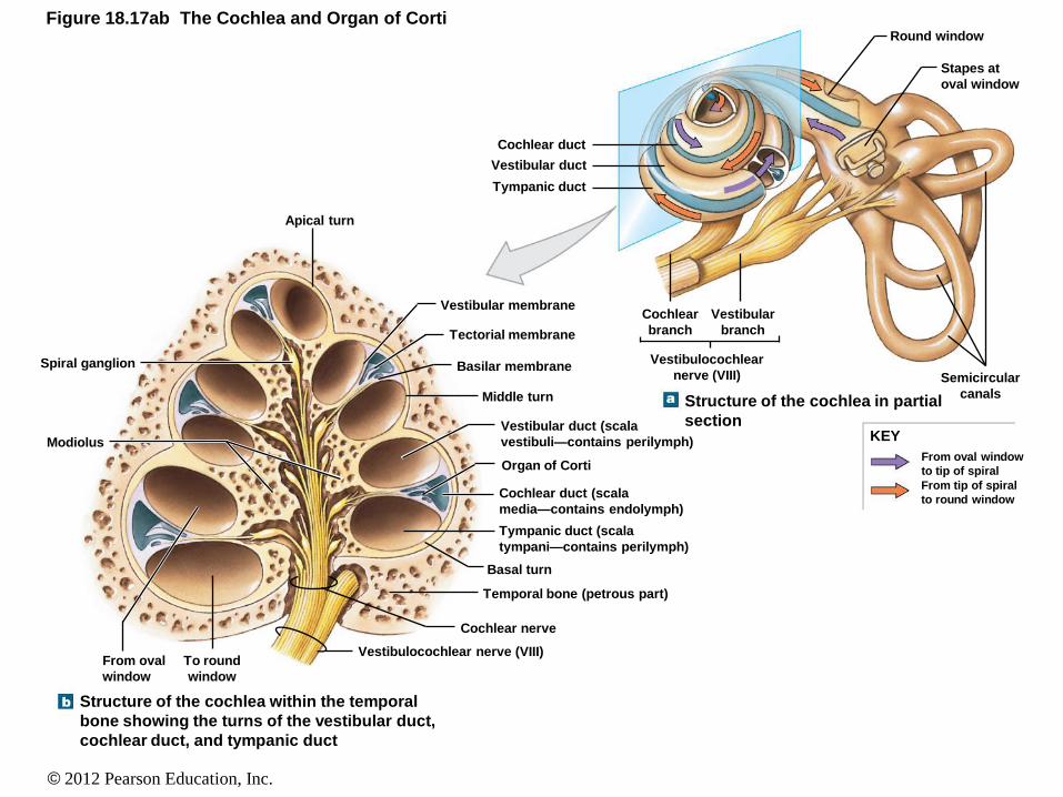
© 2012 Pearson Education, Inc.
Figure 18.17ab The Cochlea and Organ of Corti
Structure of the cochlea within the temporal
bone showing the turns of the vestibular duct,
cochlear duct, and tympanic duct
Structure of the cochlea in partial
section KEY
From tip of spiral
to round window
From oval window
to tip of spiral
Round window
Stapes at
oval window
Semicircular
canals
Vestibulocochlear
nerve (VIII)
Cochlear
branch
Vestibular
branch
Tympanic duct
Vestibular duct
Cochlear duct
Apical turn
Spiral ganglion
Modiolus
Vestibular membrane
Tectorial membrane
Basilar membrane
Middle turn
Vestibular duct (scala
vestibuli—contains perilymph)
Organ of Corti
Cochlear duct (scala
media—contains endolymph)
Tympanic duct (scala
tympani—contains perilymph)
Basal turn
Temporal bone (petrous part)
Cochlear nerve
Vestibulocochlear nerve (VIII) From oval
window
To round
window

© 2012 Pearson Education, Inc.
Equilibrium and Hearing
• The Cochlea (cont.)
• Each spiral consists of three layers • Scala vestibuli (vestibular duct): consists of perilymph
• Scala tympani (tympanic duct): consists of perilymph
• Scala media (cochlear duct): consists of endolymph / this layer is between the scala vestibuli and scala tympani
• There is a basilar membrane between each layer
• The scala vestibuli and scala tympani are connected at the apical end of the cochlea
• Sense organs rest on the basilar membrane within the scala media

© 2012 Pearson Education, Inc.
Equilibrium and Hearing
• The Cochlea
• The Organ of Corti
• Also known as the spiral organ
• Rests on the basilar membrane between the scala
media and the scala tympani
• Hair cells are in contact with an overlying tectorial
membrane
• This membrane is attached to the lining of the
scala media
• Sound waves ultimately cause a distortion of the
tectorial membrane, thus stimulating the organ
of Corti

© 2012 Pearson Education, Inc.
Equilibrium and Hearing
• Auditory Pathways
• Sound waves enter the external acoustic
meatus
• The tympanic membrane vibrates
• Causes the vibration of the ossicles
• The stapes vibrates against the oval window of
the scala tympani
• Perilymph begins to move
© 2012 Pearson Education, Inc.
Figure 18.9 Anatomy of the Ear
EXTERNAL EAR MIDDLE EAR INNER EAR
Auricle
Auditory ossicles Semicircular canals
Petrous part of temporal
bone
Facial nerve (N VII)
External acoustic meatus
Elastic cartilage
Tympanic membrane
Tympanic cavity
Oval window
Round window
Vestibule
Auditory tube
Cochlea
To nasopharynx
Bony labyrinth of inner ear
Vestibulocochlear nerve (N VIII)

© 2012 Pearson Education, Inc.
Figure 18.17a–c The Cochlea and Organ of Corti
Structure of the cochlea within the temporal
bone showing the turns of the vestibular duct,
cochlear duct, and tympanic duct
Structure of the cochlea in partial
section
Histology of the cochlea showing many of the structures
in part (b)
KEY
From tip of spiral
to round window
From oval window
to tip of spiral
Round window
Stapes at
oval window
Semicircular
canals
Vestibulocochlear
nerve (VIII)
Cochlear
branch
Vestibular
branch
Tympanic duct
Vestibular duct
Cochlear duct
Apical turn
Spiral ganglion
Modiolus
Vestibular membrane
Tectorial membrane
Basilar membrane
Middle turn
Vestibular duct (scala
vestibuli—contains perilymph)
Organ of Corti
Cochlear duct (scala
media—contains endolymph)
Tympanic duct (scala
tympani—contains perilymph)
Basal turn
Temporal bone (petrous part)
Cochlear nerve
Vestibulocochlear nerve (VIII) From oval
window
To round
window
Vestibular duct
(from oval window)
Vestibular membrane
Organ of Corti
Basal turn
Basilar membrane
Tympanic duct
(to round window)
Sectional view of cochlear spiral LM 60
Apical turn
Middle turn
Vestibular duct
(scala vestibuli)
Cochlear duct
(scala media)
Tympanic duct
(scala tympani)
Cochlear branch
Spiral ganglion

© 2012 Pearson Education, Inc.
Figure 18.17d–f The Cochlea and Organ of Corti
A color-enhanced SEM
showing a portion of the
receptor surface of the
organ of Corti
Diagrammatic and histological sections through the
receptor hair cell complex of the organ of Corti
Three-dimensional section
showing the detail of the cochlear
chambers, tectorial membrane,
and organ of Corti
Bony cochlear wall
Vestibular duct
Vestibular membrane
Cochlear duct
Tectorial membrane
Basilar membrane
Tympanic duct
Organ of Corti
Spiral
ganglion
Cochlear branch
of N VIII
Cochlear duct (scala media)
Vestibular membrane
Tectorial membrane
Organ of Corti LM 125
Tympanic duct
(scala tympani)
Basilar
membrane
Hair cells
of organ
of Corti
Spiral ganglion
cells of
cochlear nerve
Tectorial membrane
Outer
hair cell
Basilar membrane Inner hair cell Nerve fibers
Stereocilia of inner hair cells
Stereocilia of
outer hair cells
Surface of the organ of Corti SEM 1320
© 2012 Pearson Education, Inc.
Equilibrium and Hearing
• Auditory Pathways (continued)
• As the perilymph moves:
• Pressure is put on the scala media
• This pressure distorts the hair cells of the organ of
Corti
• This distortion depolarizes the neurons
• Nerve signals are sent to the brain via CN VIII
ANIMATION The Ear: Receptor Complexes

© 2012 Pearson Education, Inc.
Figure 18.17de The Cochlea and Organ of Corti
Diagrammatic and histological sections through the
receptor hair cell complex of the organ of Corti
Three-dimensional section
showing the detail of the cochlear
chambers, tectorial membrane,
and organ of Corti
Bony cochlear wall
Vestibular duct
Vestibular membrane
Cochlear duct
Tectorial membrane
Basilar membrane
Tympanic duct
Organ of Corti
Spiral
ganglion
Cochlear branch
of N VIII
Cochlear duct (scala media)
Vestibular membrane
Tectorial membrane
Organ of Corti LM 125
Tympanic duct
(scala tympani)
Basilar
membrane
Hair cells
of organ
of Corti
Spiral ganglion
cells of
cochlear nerve
Tectorial membrane
Outer
hair cell
Basilar membrane Inner hair cell Nerve fibers

© 2012 Pearson Education, Inc.
Figure 18.18 Pathways for Auditory Sensations
KEY
First-order neuron
Second-order neuron
Third-order neuron
Fourth-order neuron
High-frequency
sounds
Low-frequency
sounds
Cochlea
Cochlear branch
Vestibulocochlear
nerve (N VIII)
Cochlear nuclei
Vestibular
branch
To ipsilateral
auditory cortex
Superior olivary nucleus
Motor output to spinal
cord through the
tectospinal tracts
Motor output
to cranial
nerve nuclei
Inferior colliculus
(mesencephalon)
Medial geniculate
nucleus (thalamus)
Low-frequency
sounds
Auditory cortex
(temporal lobe) High-
frequency
sounds
Thalamus

© 2012 Pearson Education, Inc.
Vision
• Vision
• Accessory structures of the eye
• Palpebrae (eyelids)
• Medial and lateral canthus (connect the eyelids at
the corners of the eye)
• Palpebral fissure (area between the eyelids)
• Eyelashes (contain root hair plexus, which triggers
the blinking reflex)
• Conjunctiva (epithelial lining of the eyelids)
• Glands: glands of Zeis, tarsal glands, lacrimal
gland, lacrimal caruncle

© 2012 Pearson Education, Inc.
Figure 18.19a Accessory Structures of the Eye, Part I
Superficial anatomy of the right eye and its
accessory structures
Pupil
Corneal limbus
Lateral canthus
Sclera
Eyelashes
Palpebra
Palpebral fissure
Medial canthus
Lacrimal caruncle
© 2012 Pearson Education, Inc.
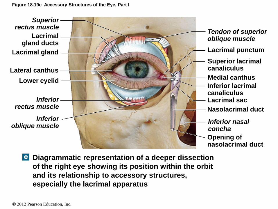
Figure 18.19c Accessory Structures of the Eye, Part I
Diagrammatic representation of a deeper dissection
of the right eye showing its position within the orbit
and its relationship to accessory structures,
especially the lacrimal apparatus
Opening of nasolacrimal duct
Inferior nasal concha
Nasolacrimal duct
Lacrimal sac
Inferior lacrimal canaliculus
Medial canthus
Superior lacrimal canaliculus
Lacrimal punctum
Tendon of superior oblique muscle
Inferior oblique muscle
Inferior rectus muscle
Superior rectus muscle
Lacrimal gland ducts
Lower eyelid
Lateral canthus
Lacrimal gland

© 2012 Pearson Education, Inc.
Vision
• Accessory Structures of the Eye
• Conjunctiva
• Covers the inside lining of the
eyelids and the outside lining of the eye
• Fluid production helps prevent these layers from
becoming dry
• Palpebral conjunctiva
• Inner lining of the eyelids
• Ocular conjunctiva
• Outer lining of the eye

© 2012 Pearson Education, Inc.
Vision
• Accessory Structures
• Glands
• All of the glands are for protection or lubrication
• Glands of Zeis: sebaceous glands / associated with
eyelashes
• Tarsal glands: secrete a lipid-rich product / keeps the
eyelids from sticking together / located along the inner
margin of the eyelids
• Lacrimal glands: produce tears / located at the
superior, lateral portion of the eye
• Lacrimal caruncle glands: produce thick secretions /
located within the canthus areas

© 2012 Pearson Education, Inc.
Vision
• Accessory Structures
• Glands
• An infection of the tarsal gland may result in a cyst
• An infection of any of the other glands may result in
a sty

© 2012 Pearson Education, Inc.
Vision
• Accessory Structures
• Lacrimal glands
• Part of the lacrimal apparatus
• The lacrimal apparatus consists of:
• Lacrimal glands (produce tears)
• Lacrimal canaliculi
• Lacrimal sac
• Nasolacrimal duct

© 2012 Pearson Education, Inc.
Vision
• Accessory Structures
• Lacrimal glands (continued)
• Tears are produced by the lacrimal glands
• Flow over the ocular surface
• Flow into the nasolacrimal canal (foramen)
• This foramen enters into the nasal cavity
• Therefore, when you sob heavily, tears flow
across your eye and down your face and also
through the nasolacrimal canal into your nose
and out, resulting in a “runny” nose
ANIMATION The Eye: Accessory Structures

© 2012 Pearson Education, Inc.
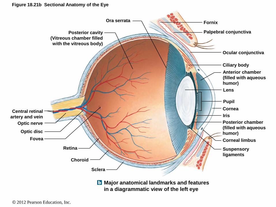
Vision
• The Eyes • Consist of:
• Sclera
• Cornea
• Pupil
• Iris
• Lens
• Anterior cavity
• Posterior cavity
• Three tunics: • (1) fibrous tunic, (2) vascular tunic, and (3) neural
tunic
• Retina
© 2012 Pearson Education, Inc.
Figure 18.21b Sectional Anatomy of the Eye
Major anatomical landmarks and features
in a diagrammatic view of the left eye
Central retinal
artery and vein
Optic nerve
Optic disc
Fovea
Retina
Choroid
Sclera
Posterior cavity
(Vitreous chamber filled
with the vitreous body)
Ora serrata Fornix
Palpebral conjunctiva
Ocular conjunctiva
Ciliary body
Anterior chamber
(filled with aqueous
humor)
Lens
Pupil
Cornea
Iris
Posterior chamber
(filled with aqueous
humor)
Corneal limbus
Suspensory
ligaments

© 2012 Pearson Education, Inc.
Vision
• The Eyes
• The Fibrous Tunic (outer layer)
• Makes up the sclera and cornea
• Provides some degree of protection
• Provides attachment sites for extra-ocular muscles
• The cornea is modified sclera

© 2012 Pearson Education, Inc.
Vision
• The Eyes
• The Vascular Tunic (middle layer)
• Consists of blood vessels, lymphatics, and intrinsic
eye muscles
• Regulates the amount of light entering the eye
• Secretes and reabsorbs aqueous fluid (aqueous
humor)
• Controls the shape of the lens
• Includes the iris, ciliary body, and the choroid
ANIMATION The Eye: Uvea Parts

© 2012 Pearson Education, Inc.
Vision
• The Vascular Tunic
• The iris
• Consists of blood vessels, pigment, and smooth
muscles
• The pigment creates the color of the eye
• The smooth muscles contract to change the
diameter of the pupil

© 2012 Pearson Education, Inc.
Vision
• The Vascular Tunic
• The ciliary body
• The ciliary bodies consist of ciliary muscles
connected to suspensory ligaments, which are
connected to the lens
• The choroid
• Highly vascularized
• The innermost portion of the choroid attaches to the
outermost portion of the retina
ANIMATION The Eye: Ciliary Muscles

© 2012 Pearson Education, Inc.
Vision
• The Eyes
• The Neural Tunic (inner layer)
• Also called the retina
• Made of two layers: (pigmented layer – outer layer)
/ (neural layer – inner layer)
• Retina cells: rods (night vision) and cones (color
vision)

© 2012 Pearson Education, Inc.
Figure 18.22a The Lens and Chambers of the Eye
The lens is suspended between the posterior cavity
and the posterior chamber of the anterior cavity.
Pigmented part
Neural part Neural
tunic
(retina)
Posterior
cavity
Choroid
Ciliary body
Iris
Vascular
tunic
(uvea)
Anterior
cavity
Cornea
Sclera Fibrous
tunic

© 2012 Pearson Education, Inc.
Figure 18.21ab Sectional Anatomy of the Eye
The three layers, or
tunics, of the eye
Fibrous
tunic
(sclera)
Vascular
tunic
(choroid)
Neural
tunic
(retina)
Major anatomical landmarks and features
in a diagrammatic view of the left eye
Central retinal
artery and vein
Optic nerve
Optic disc
Fovea
Retina
Choroid
Sclera
Posterior cavity
(Vitreous chamber filled
with the vitreous body)
Ora serrata Fornix
Palpebral conjunctiva
Ocular conjunctiva
Ciliary body
Anterior chamber
(filled with aqueous
humor)
Lens
Pupil
Cornea
Iris
Posterior chamber
(filled with aqueous
humor)
Corneal limbus
Suspensory
ligaments

© 2012 Pearson Education, Inc.
Figure 18.23a Retinal Organization
Histological organization of the retina. Note that the
photoreceptors are located closest to the choroid
rather than near the vitreous chamber. LIGHT
Amacrine cell
Horizontal cell Cone Rod Choroid
Pigmented part of retina
Rods and cones
Bipolar cells
Ganglion cells
Nuclei of ganglion cells
Nuclei of rods and cones
Nuclei of bipolar cells
The retina LM 70

© 2012 Pearson Education, Inc.
Vision
• Cavities and Chambers of the Eye
• Anterior cavity
• Anterior chamber
• Posterior chamber
• Filled with fluid called aqueous fluid
• Posterior cavity
• Vitreous chamber
• Filled with fluid called vitreous fluid
ANIMATION The Eye: Posterior Cavity

© 2012 Pearson Education, Inc.
Vision
• Cavities and Chambers of the Eye
• Aqueous fluid
• Sometimes called aqueous humor
• Secreted by cells at the ciliary body area
• Enters the posterior chamber (posterior of the iris)
• Flows through the pupil area
• Enters the anterior chamber
• Flows through the canal of Schlemm
• Enters into venous circulation

© 2012 Pearson Education, Inc.
Figure 18.24
Pigmented epithelium
Suspensory
ligaments
Posterior cavity
(vitreous chamber)
Lens
Ciliary process
Choroid
Retina
Sclera
Conjunctiva
Ciliary body
Body of iris
Canal of Schlemm
Posterior chamber
Anterior chamber
Anterior cavity
Cornea
Pupil

© 2012 Pearson Education, Inc.
Vision
• Cavities and Chambers of the Eye
• Vitreous fluid
• Gelatinous material in the posterior chamber
• Sometimes called vitreous humor
• Supports the shape of the eye
• Supports the position of the lens
• Supports the position of the retina
• Aqueous humor can flow across the vitreous fluid
and over the retina
© 2012 Pearson Education, Inc.
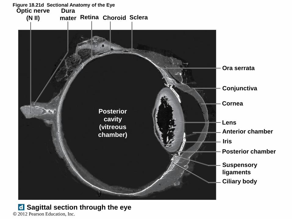
Figure 18.21d Sectional Anatomy of the Eye
Sagittal section through the eye
Ora serrata
Conjunctiva
Cornea
Lens
Anterior chamber
Iris
Posterior chamber
Suspensory
ligaments
Ciliary body
Posterior
cavity
(vitreous
chamber)
Dura
mater Retina Choroid Sclera Optic nerve
(N II)

© 2012 Pearson Education, Inc.
Vision
• Aqueous fluid
• If this fluid cannot drain through the canal of
Schlemm, pressure builds up
• This is glaucoma
• Vitreous fluid
• If this fluid is not of the right consistency, the
pressure is reduced against the retina
• The retina may detach from the posterior wall
(detached retina)

© 2012 Pearson Education, Inc.
Vision
• Visual Pathways
• Light waves pass through the cornea
• Pass through the anterior chamber
• Pass through the pupil
• Pass through the posterior chamber
• Pass through the lens
• The lens focuses the image on some part of the retina • This creates a depolarization of the neural cells
• Signal is transmitted to the brain via CN II
ANIMATION The Eye: Interior Parts of the Eye

© 2012 Pearson Education, Inc.
Figure 18.21e Sectional Anatomy of the Eye
Sagittal section through the eye
Orbital fat Central artery
and vein
Medial rectus
muscle
Ethmoidal
labyrinth
Optic nerve
Optic disc
Fovea
Ora serrata
Ciliary body
Lens Ciliary
processes
Medial canthus
Lacrimal caruncle
Lacrimal punctum
Nose
Anterior cavity
Posterior
chamber
Anterior
chamber
Edge of
pupil
Visual
axis
Cornea
Iris
Suspensory ligament of lens
Corneal limbus
Conjunctiva
Lower eyelid
Lateral canthus
Sclera
Choroid
Retina
Posterior cavity
Lateral rectus muscle

© 2012 Pearson Education, Inc.
Figure 18.26 Anatomy of the Visual Pathways, Part II LEFT SIDE RIGHT SIDE
Left eye
only Right eye
only
Binocular vision
Optic nerve (N II)
Optic chiasm
Optic tract Other hypothalamic
nuclei, pineal gland,
and reticular
formation
Suprachiasmatic
nucleus
Superior
colliculus
Lateral
geniculate
nucleus
Projection
fibers (optic
radiation)
Lateral
geniculate
nucleus
RIGHT CEREBRAL
HEMISPHERE
LEFT CEREBRAL
HEMISPHERE
Visual cortex of
cerebral hemispheres

© 2012 Pearson Education, Inc.
Vision
• Visual Pathways
• The retina
• There are rods and cones all over the retina
• 100% cones in the fovea centralis area
• The best color vision is when an object is
focused on the fovea centralis
• 0% rods or cones in the optic disc area
• If an object is focused on this area, vision does
not occur
• Also known as the “blind spot”
ANIMATION The Eye: Blind Spot

© 2012 Pearson Education, Inc.
Vision
• Visual Pathways
• The retina (cont.) • The cones require light to be stimulated (that’s why
we see color)
• At night (still has to be at least a small amount of light), the cones deactivate and the rods begin to be activated (that’s why we can see at night but we can’t determine color at night)
ANIMATION The Eye: The Retina
ANIMATION The Eye: Light Path
ANIMATION The Eye: Lens and Retina