amgen biotech experience complete genetic engineering ... · introduction complete genetic...
TRANSCRIPT


AMGEN BIOTECH EXPERIENCE | COMPLETE GENETIC ENGINEERING SEQUENCE | STUDENT GUIDE© 2015 Amgen Foundation. All rights reserved.
1
TABLE OF CONTENTSABOUT THE AMGEN BIOTECH EXPERIENCE 3
PROGRAM INTRODUCTION 5
Reading: What Is Genetic Engineering? 7
Your Challenge 15
Program Introduction Glossary 16
CHAPTER 1: SOME TOOLS OF THE TRADE 17
Introduction 19
Laboratory 1.1: How to Use a Micropipette 20
Reading: The Genetic Engineering Process 24
Laboratory 1.2: Gel Electrophoresis 26
Chapter 1 Questions 32
Chapter 1 Glossary 33
CHAPTER 2: HOW DO YOU BEGIN TO CLONE A GENE? 35
Introduction 37
Reading: Plasmids and Restriction Enzymes 38
Activity: Clone that Gene 43
Laboratory 2: Preparing to Clone the rfp Gene: Digesting the pKAN-R and pARA 46
Chapter 2 Questions 51
Chapter 2 Glossary 52
AMGEN BIOTECH EXPERIENCE | COMPLETE GENETIC ENGINEERING SEQUENCE | STUDENT GUIDE© 2015 Amgen Foundation. All rights reserved.
2
CHAPTER 3: BUILDING A RECOMBINANT PLASMID 55
Introduction 57
Reading: Ligases 58
Laboratory 3: Building the pARA-R Plasmid 61
Chapter 3 Questions 64
Chapter 3 Glossary 65
CHAPTER 4: MAKING SURE YOU’VE CREATED A RECOMBINANT PLASMID 67
Introduction 69
Reading: Verification 70
Laboratory 4: Verification of Restriction and Ligation Using Gel Electrophoresis 75
Chapter 4 Questions 80
Chapter 4 Glossary 81
CHAPTER 5: GETTING RECOMBINANT PLASMIDS IN BACTERIA 83
Introduction 85
Reading: Transforming Bacteria with Recombinant Plasmids 86
Laboratory 5: Transforming Bacteria with the Ligation Products 90
Chapter 5 Questions 98
Chapter 5 Glossary 99
CHAPTER 6: GETTING WHAT WE NEED 101
Introduction 103
Reading: Bacteria Multiplication and Protein Purification 104
Laboratory 6: Purifying the Fluorescent Protein 109
Chapter 6 Questions 115
Chapter 6 Glossary 116

3INTRODUCTION | COMPLETE GENETIC ENGINEERING SEQUENCE | STUDENT GUIDE© 2015 Amgen Foundation. All rights reserved.
ABOUT THE AMGEN BIOTECH EXPERIENCEGenetic engineering is a branch of biotechnology that uses special procedures
and techniques to change an organism’s DNA. This ability has had a huge impact
on the field of medicine, as genetically modified bacteria can make human
insulin (the hormone responsible for regulating glucose levels in the blood) and
other life-saving products. It’s rare for high school students to have the chance
to learn about and actually practice the procedures and techniques that are
the foundation of the biotechnology industry—but in this program, you will
have just that opportunity. As you work in the laboratory and carry out the very
experiments that led to breakthroughs in biotechnology, you will gain hands-on
experience with producing genetically modified bacteria.
The procedures in this program were developed through a series of discoveries
that led to important breakthroughs in biotechnology. Some of the pioneering
scientists who made these discoveries received Nobel Prizes in Physiology or
Medicine in 1978 and Chemistry in 1980 and 1993. (The Nobel Prize is the highest
distinction awarded to scientists in these fields from around the world.) The
work that you are about to do is based on this Nobel Prize-winning science—
science that is significant and will continue to play an important role in the
development of biotechnology and medicine. You will follow in the footsteps
of the many scientists who have pushed and continue to push the boundaries
of biotechnology. There are many advances still to be made—and students who
decide to continue studying this field may contribute to those advances.
In science, the ability to keep track of what you are doing and communicate your
work is extremely important. In order to demonstrate that you performed an
experiment, either so that it can be duplicated and verified by others or if you
want to apply for a patent—you need to have a very accurate record of what
you’ve done. As you carry out this program, carefully record your notes, ideas,
observations, results, and answers to questions in a science notebook, in pen.
(For scientific purposes, it is important to keep a record—even of your mistakes.)
If possible, use a separate bound composition notebook and organize the labs
with a table of contents at the front. Since you will use a pen to write with,
you’ll need to cross out any mistakes you make—and it is good practice to simply
“X” out the section you want to change (so that it can still be read) and to note

4 INTRODUCTION | COMPLETE GENETIC ENGINEERING SEQUENCE | STUDENT GUIDE© 2015 Amgen Foundation. All rights reserved.
why you’ve done so. Following these best practices will make this program even
better preparation for you!
The Amgen Biotech Experience (formerly Amgen-Bruce Wallace Biotechnology
Lab Program) had humble beginnings almost 25 years ago with visionary
scientists and teachers who shared passion and energy for imparting their
knowledge with students. Bruce Wallace, one of Amgen’s first staff members,
wanted all students to experience the joy of discovery and the excitement of
having science at their fingertips. A desire for more robust science education
at schools near Amgen’s global headquarters led to involving area high school
teachers and, later, a college professor, in developing curriculum and educator
training in biotechnology. The program grew through word of mouth and
teacher interest, and expanded over time to other states and countries.
Visit the ABE website at www.amgenbiotechexperience.com.
5INTRODUCTION | COMPLETE GENETIC ENGINEERING SEQUENCE | STUDENT GUIDE© 2015 Amgen Foundation. All rights reserved.
PROGRAM INTRODUCTIONAMGEN BIOTECH EXPERIENCE

7INTRODUCTION | COMPLETE GENETIC ENGINEERING SEQUENCE | STUDENT GUIDE© 2015 Amgen Foundation. All rights reserved.
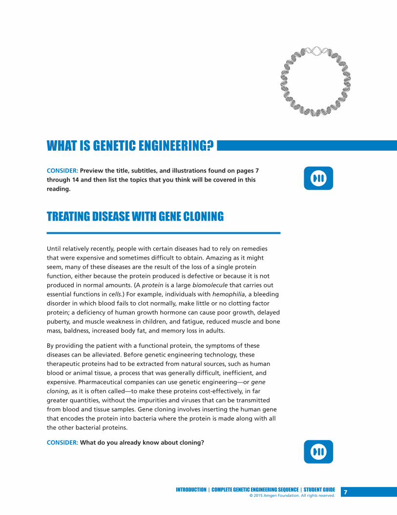
WHAT IS GENETIC ENGINEERING?CONSIDER: Preview the title, subtitles, and illustrations found on pages 7
through 14 and then list the topics that you think will be covered in this
reading.
TREATING DISEASE WITH GENE CLONING
Until relatively recently, people with certain diseases had to rely on remedies
that were expensive and sometimes difficult to obtain. Amazing as it might
seem, many of these diseases are the result of the loss of a single protein
function, either because the protein produced is defective or because it is not
produced in normal amounts. (A protein is a large biomolecule that carries out
essential functions in cells.) For example, individuals with hemophilia, a bleeding
disorder in which blood fails to clot normally, make little or no clotting factor
protein; a deficiency of human growth hormone can cause poor growth, delayed
puberty, and muscle weakness in children, and fatigue, reduced muscle and bone
mass, baldness, increased body fat, and memory loss in adults.
By providing the patient with a functional protein, the symptoms of these
diseases can be alleviated. Before genetic engineering technology, these
therapeutic proteins had to be extracted from natural sources, such as human
blood or animal tissue, a process that was generally difficult, inefficient, and
expensive. Pharmaceutical companies can use genetic engineering—or gene
cloning, as it is often called—to make these proteins cost-effectively, in far
greater quantities, without the impurities and viruses that can be transmitted
from blood and tissue samples. Gene cloning involves inserting the human gene
that encodes the protein into bacteria where the protein is made along with all
the other bacterial proteins.
CONSIDER: What do you already know about cloning?

8 INTRODUCTION | COMPLETE GENETIC ENGINEERING SEQUENCE | STUDENT GUIDE© 2015 Amgen Foundation. All rights reserved.
The ability to make enough of the proteins to treat diseases is the result of two
key discoveries about bacteria made by scientists in the 1970s and ’80s. The first
discovery was that bacteria contain tiny circles of DNA, called plasmids, that
sometimes contain genes that can make them resistant to antibiotics. The second
discovery was that bacteria also contain proteins called restriction enzymes that
can cut DNA at very specific places.
The findings made by basic research often lead to fundamental understandings
about the nature of life. In some instances these findings can also lead to new
technologies that can improve life. With the discovery of plasmids and restriction
enzymes, a whole new era of genetic engineering was launched. Scientists now
have the ability to generate products that can improve health in ways never
before imagined.
One of the first pharmaceutical products produced using these tools was insulin,
which is used to treat diabetes, a debilitating and sometimes fatal disease. To
generate large quantities of human insulin, the sequences of DNA that contain
the codes of human insulin are inserted into a plasmid that is introduced into the
common intestinal bacterium Escherichia coli (E. coli), where the new protein is
synthesized along with all the other bacterial proteins. The genetically modified
bacteria are then grown in large batches, and the insulin is purified for use in
the treatment of diabetes.
CONSIDER: Do you think that treating diabetes with insulin can be considered a
cure? What is the difference between a treatment and a cure?
Why is the ability to produce large quantities of insulin so important, and how
exactly is this done? In the following readings you will learn about diabetes:
what it is and why insulin is in such demand. Then you will carry out some
of the very same procedures that scientists use to produce human insulin in
bacteria. But instead of producing insulin, you will engineer E. coli to produce
a red fluorescent protein. This protein is made by a sea anemone gene that has
undergone a mutation that makes the protein brighter in color. You will give E.
coli a new protein and a trait it did not have before: the ability to glow.

9INTRODUCTION | COMPLETE GENETIC ENGINEERING SEQUENCE | STUDENT GUIDE© 2015 Amgen Foundation. All rights reserved.
DATELINE: AMERICA MAY 2012
TEENAGE DIABETES ON THE RISEThe occurrence of type 2 diabetes in teenagers, once a dis-ease found primarily in adults, has increased dramatically over the past 10 years. Nearly one in four teens between the ages of 12 and 19 is prediabetic (i.e., shows early signs of diabetes) or already has the disease. To make matters even worse, research suggests that the disease progresses more rapidly in children than in adults. In diabetes, the levels of glucose (a type of sugar) in the blood can become danger-ously high, causing complications such as loss of vision, kidney failure, and nerve and blood vessel damage. The onset of diabetes early in life could mean serious health issues, such as heart disease, blindness, and amputation, for individuals in their 30s and 40s, far younger than such complications have been seen in the past.
Why this sudden rise in teenage diabetes? Although being overweight or obese can contribute significantly to devel-oping diabetes, weight is not the only factor; 35 percent of teens of normal weight have glucose levels that are higher than normal, which is one indicator of being prediabetic. Factors such as lack of exercise in this age of increased computer and mobile device use may be part of the prob-lem. While many prediabetics go on to develop full-blown type 2 diabetes, studies indicate that eating less fat and fewer calories and exercising a mere 20 minutes a day can reduce the risk of developing type 2 diabetes by 60 percent.
Diabetes can result from the body’s inability to make sufficient insulin (type 1) or to effectively use the insulin that it does produce (type 2). Many patients with diabetes must take in insulin as an injection. More diabetes in the population will mean a greater demand for insulin.
What is it like for a teen to learn she has diabetes? Read the following story about one teenager’s struggle with the disease.
JENNIFER’S STORY
Jennifer felt hungry all the time, but despite eating what-ever she felt like whenever she wanted to, she was losing weight. She was also very thirsty and was constantly drink-
ing water and then needing to pee. Initially, she just thought it was typical for a 15 year old; she was in a growth spurt and very active with soccer and track at school. Of course she was hungry and thirsty! She was also pleased by her weight loss, since she’d been a bit overweight for a while. Jennifer also felt unusually tired and draggy, especially in the afternoon. But again, who wouldn’t be, since school started at the ungodly hour of 7:30? When she began to have trouble seeing the board in class, she thought, Drat! Do I really need glasses? But it was the cut on her leg that refused to heal and became infected that finally got her to talk to her parents and ultimately go to the doctor’s office. There Jennifer was diagnosed with diabetes.
Jennifer had heard about a disease called diabetes but never gave it much thought. Now she really needed to pay atten-tion. Diabetes is the result of too much of a sugar called glucose in the blood and not enough of it getting into cells, where it provides the energy to construct the biological molecules the body needs to survive. Jennifer learned that in order for glucose to get into cells, the body makes a hor-mone called insulin, which binds to the cells and enables glucose to enter them. For some reason, Jennifer’s body no longer produced normal amounts of insulin, resulting in Jennifer having very high levels of glucose in her blood and not enough glucose getting into her cells. Jennifer hoped she could control her diabetes by eating more fruits and vegetables and getting more exercise. But although this change in habits helped some, it was not sufficient, and Jennifer had to begin injecting herself daily with insulin.
Jennifer now is very aware of what she eats, monitoring ex-actly how much sugar and other carbohydrates she ingests. She checks the level of her blood glucose several times a day by pricking her finger and testing her blood. She also injects her insulin faithfully. She knows that she can’t cure her diabetes and that if the disease progresses further she could suffer very serious complications.

10 INTRODUCTION | COMPLETE GENETIC ENGINEERING SEQUENCE | STUDENT GUIDE© 2015 Amgen Foundation. All rights reserved.
DIABETES TYPES 1 AND 2 DIABETES: TOO MUCH OF A GOOD THINGWhat is diabetes? Diabetes is the result of elevated levels of glucose in the
blood. Glucose is a major source of energy and is used to construct biological
molecules in the body. What you ate for breakfast or lunch today is rapidly
being converted to glucose, which in turn will be used to generate energy, to
synthesize new cells and tissues, and to carry out processes required to sustain
life. The starch in your bread or potato is made up of long chains of glucose
molecules (Figure P.1a). As food passes through your mouth, esophagus, and
stomach (Figure P.1b), these chains are broken down to release glucose. The
glucose is then absorbed through the intestinal wall and enters the bloodstream,
where it is carried to all the cells in the body (Figure P.1c).
Figure P.1: How glucose gets to cells
Figure P.1a: Starch is made up of subunits of glucose bonded together.
Figure P.1b: Nutrients such as starch are broken down into smaller molecules
during digestion in the mouth, esophagus, and stomach.
Glucose units (monomers)
Starch (a polymer of glucose)
Mouth
Pancreas
Esophagus
Stomach
Small Intestine

11INTRODUCTION | COMPLETE GENETIC ENGINEERING SEQUENCE | STUDENT GUIDE© 2015 Amgen Foundation. All rights reserved.
Figure P.1c: Glucose molecules pass through the small intestinal wall
into the bloodstream, which delivers the glucose to cells in the body.
CROSSING THE CELLULAR DIVIDEIn order to get inside a cell, glucose must cross the cell membrane that separates
the inside of the cell from its environment. Insulin, which is made by beta
cells found in the pancreas (see Figure P.1b), binds to a special protein called a
receptor, which causes an opening in the cell membrane and allows glucose to
enter the cell (see Figure P.2). Without insulin, glucose cannot penetrate this
cellular barrier.
Figure P.2: How glucose crosses the cell membrane
Insulin in the blood binds to specific receptors on the cell. This binding alters the conformation of the
cell membrane, resulting in the formation of a glucose channel. Glucose in the blood can now enter
the cell through these channels.
Glucose
Insulinreceptors
Body cell
Nucleus
Glucose inside of cell
Glucose outside of cell
Glucosechannel
Insulin on receptorsopens glucose channel
Glucose enterscell throughglucose channel
Insulin

12 INTRODUCTION | COMPLETE GENETIC ENGINEERING SEQUENCE | STUDENT GUIDE© 2015 Amgen Foundation. All rights reserved.
In both type 1 and type 2 diabetes, glucose is unable to enter the cells, resulting
in elevated levels of glucose in the blood. In type 1 diabetes, the beta cells in
the pancreas are unable to produce insulin. Without insulin to create glucose
channels, the glucose remains in the blood (Figure P.3a). Type 2 diabetes is the
result of a combination of two factors: (1) Cells become resistant to insulin, and
the receptors can no longer bind the hormone (Figure P.3b). As the blood sugar
levels rise, the beta cells pump out more and more insulin to no avail, since the
cells cannot use it. (2) Eventually the beta cells are exhausted and can no longer
produce insulin, and insulin levels in the blood drop while the sugar levels
continue to increase.
Figure P.3: Reduced uptake of glucose by cells
THE PROBLEMS OF TOO LITTLE INTRACELLULAR GLUCOSEWhen cells cannot get glucose, they cannot get the energy and biological
molecules they need. The body responds by breaking down fats and proteins
to obtain its needed energy. Loss of proteins and fats can cause serious damage
to tissues and organs, leading to the symptoms of diabetes that patients like
Jennifer experience, such as blindness and nerve damage (which can result in
amputation).
Figure P.3a: In type 1 diabetes, there is
a lack of insulin in the body.
Figure P.3b: In type 2 diabetes, cells
cannot bind the insulin.
Type 1 Diabetes: Insufficient Insulin Type 2 Diabetes: Insulin Resistance
Diminished glucose uptake
Glucose
Insulinreceptors
Glucosechannel
Diminishedinsulin
Insulinreceptors
Glucosechannel
Diminished glucose uptake
GlucoseInsulin

13INTRODUCTION | COMPLETE GENETIC ENGINEERING SEQUENCE | STUDENT GUIDE© 2015 Amgen Foundation. All rights reserved.
TREATING DIABETESIndividuals with type 1 diabetes can regulate their sugar levels by monitoring
their blood and injecting insulin as needed. Those with type 2 diabetes
can sometimes regulate their blood sugar levels by changing their diet and
increasing the amount that they exercise. However, in many cases, medications
that reduce insulin resistance in cells and increase the levels of insulin in the
blood are required to maintain normal blood sugar levels.
Currently, there is no cure for either type of diabetes.
E. COLI WITH A HUMAN GENEWith the rise in diabetes in the population, the need for insulin for treatment is
also on the rise. Originally isolated from the pancreases of pigs and cows, most
of the insulin used today is genetically engineered human insulin, manufactured
by bacteria. DNA sequences encoding human insulin in plasmids are taken up
by bacteria, which make the hormone along with all of its bacterial proteins.
Insulin is then isolated from the bacteria. In 1982, human insulin was the
first commercially successful product made by recombinant DNA technology.
(Recombinant DNA refers to DNA that contains sequences or genes from two or
more sources.)
CONSIDER: Why might diabetes be on the rise, especially in teenagers?
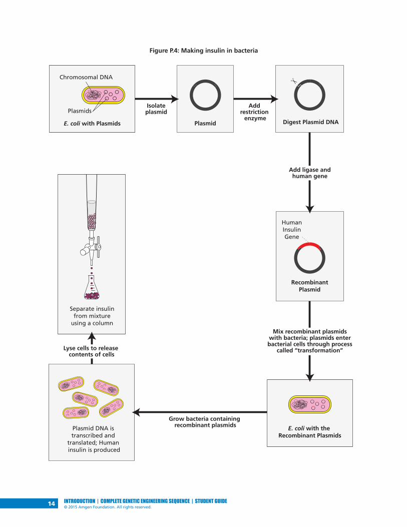
MAKING NEW PROTEINS IN BACTERIAFigure P.4 shows how a human protein—in this case, insulin—can be made
in bacteria. The insulin is then purified so that it can be used by people with
diabetes.
14 INTRODUCTION | COMPLETE GENETIC ENGINEERING SEQUENCE | STUDENT GUIDE© 2015 Amgen Foundation. All rights reserved.
Figure P.4: Making insulin in bacteria
Plasmid DNA is transcribed and
translated; Human insulin is produced
Grow bacteria containing recombinant plasmids E. coli with the
Recombinant Plasmids
Add restriction
enzyme
Separate insulinfrom mixture
using a column
Plasmid
RecombinantPlasmid
HumanInsulinGene
Digest Plasmid DNA
Isolateplasmid
E. coli with Plasmids
Plasmids
Chromosomal DNA
Lyse cells to release contents of cells
Mix recombinant plasmids with bacteria; plasmids enter bacterial cells through process
called “transformation”
Add ligase andhuman gene

15INTRODUCTION | COMPLETE GENETIC ENGINEERING SEQUENCE | STUDENT GUIDE© 2015 Amgen Foundation. All rights reserved.
YOUR CHALLENGE
Your challenge in the Amgen Biotech Experience is to successfully carry out the
steps of the genetic engineering process that is used to make insulin and other
genetically engineered products. You will learn and practice the techniques
and procedures that are part of this process. If you carry out all the steps in the
program, you will create your own genetically modified bacteria.
Note: The number of steps will vary depending on how much time your class
has available.
Instead of cloning insulin or another human gene, you will work with a gene
from a sea anemone, a soft-bodied animal related to coral and jellyfish. (The
gene is called rfp and the protein made by this gene is called red fluorescent
protein.) How will you know if you are successful? The bacteria you create will
have a new and highly visible trait: It will now produce red fluorescent protein!
DID YOU KNOW?Red Fluorescent Protein in Sea Anemones
Red fluorescent protein is derived from a protein found in sea
anemones. While sea anemones are sedentary, remaining attached to
rocks, they are also predatory animals, using their stinging tentacles
to catch their prey. The protein glows because it can absorb one color
of light and then emit light of a different color—a process known as
fluorescence. But why is it important for sea anemones to fluoresce?
Our best guess is that fluorescent proteins somehow help sea anemones
survive, but the role these proteins play is not yet well understood.
Fluorescent molecules may serve as a sunblock, turning harmful UV
light into light that is
less damaging to the
anemone’s tissues.
Another possibility is that
while humans can’t detect
the fluorescence in bright
sunlight, some animals
may be able to, causing
prey to be attracted to
the glow.

16 INTRODUCTION | COMPLETE GENETIC ENGINEERING SEQUENCE | STUDENT GUIDE© 2015 Amgen Foundation. All rights reserved.
PROGRAM INTRODUCTION GLOSSARY Biomolecule: A molecule produced by living cells. Examples include proteins,
carbohydrates, lipids, and nucleic acids.
Cells: The basic units of any living organism that carry on the biochemical
processes of life.
Diabetes: A disease that occurs when the body doesn’t produce or properly use
insulin.
DNA (deoxyribonucleic acid): A double-stranded biomolecule that encodes
genetic information.
Escherichia coli (E. coli): E. coli is a common bacterium found in the gut of warm
blooded animals. Most strains are harmless, including the strain used in these lab
protocols.
Fluorescence: The production of light by a molecule (e.g., red fluorescent protein
will release red light when exposed to ultraviolet light).
Gene cloning: Using genetic engineering techniques to create exact copies, or
clones, of a gene or DNA sequence of interest.
Genetic engineering: A branch of biotechnology that uses specific procedures
and techniques to change an organism’s DNA.
Glucose: A sugar that is a major source of energy and biomolecules to sustain life
processes. Glucose is absorbed through the intestine and travels in the blood to
cells, where it is transported through the cell membrane to be used as energy, to
synthesize cells and tissues, and to carry out other essential processes.
Hemophilia: A disease that occurs when the ability of blood to clot is reduced
due to lack of one or more blood clotting factors.
Insulin: A hormone produced in the pancreas that controls the amount of
glucose in the blood. Insulin is a protein.
Plasmid: A circular molecule of DNA.
Protein: A large biomolecule. Proteins carry out essential functions in cells, from
forming cellular structures to enabling chemical reactions to take place.
Receptor: A protein that receives signals from outside the cell. When a signal
substance binds to a receptor, it directs the cell to do something, for example to
allow biomolecules to enter the cell.
Recombinant DNA: DNA that contains sequences or genes from two or more
sources.
Restriction enzyme: A protein that can cut DNA at a specific sequence.

17CHAPTER 1 | COMPLETE GENETIC ENGINEERING SEQUENCE | STUDENT GUIDE© 2015 Amgen Foundation. All rights reserved.
CHAPTER 1SOME TOOLS OF THE TRADE


19CHAPTER 1 | COMPLETE GENETIC ENGINEERING SEQUENCE | STUDENT GUIDE© 2015 Amgen Foundation. All rights reserved.
INTRODUCTIONThe year 1978 marked a major breakthrough in medicine. For the first time ever,
scientists were able to induce bacterial cells to make human insulin by inserting
human DNA into the cells. This new technology, termed genetic engineering, can
be used to make products that treat the symptoms of certain genetic diseases.
To carry out genetic engineering, you need good laboratory skills. In this chapter,
you’ll focus on gaining practice in the use of micropipettes (instruments used
to transfer small volumes of liquid) and gel electrophoresis (a technique for
separating and identifying biomolecules)—two critical skills for biotechnology.
You will complete two labs, using instruments and supplies that are identical
to the ones used in biotechnology research laboratories. These labs are the
first step in building the skills you’ll need to carry out the subsequent labs and
complete your challenge in this program.
CHAPTER 1 GOALS By the end of this chapter, you will be able to do the following:
• Use micropipettes and the technique of gel electrophoresis correctly
• Explain the importance of micropipettes and gel electrophoresis in genetic
engineering
• Describe how gel electrophoresis separates DNA
• Explain how genetic engineering can be used to treat some genetic
diseases
WHAT DO YOU ALREADY KNOW?Discuss the following questions with your partner and write your ideas in your
notebook. Be prepared to discuss your responses with the class. Don’t worry if
you don’t know all the answers. Discussing these questions will help you think
about what you already know about genetic disease and DNA.
1. What does the term genetic disease mean? What examples of genetic
diseases do you know about?
2. Adding human DNA to bacteria makes it possible to make human insulin.
What do you already know about DNA? Be as detailed as possible and
discuss the location of DNA in the cell, DNA structure, the replication of
DNA, and the components of DNA.

LABORATORY
20 CHAPTER 1 | COMPLETE GENETIC ENGINEERING SEQUENCE | STUDENT GUIDE© 2015 Amgen Foundation. All rights reserved.
LABORATORY 1.1: HOW TO USE A MICROPIPETTEThe purpose of this laboratory is to introduce you to an important tool used
in genetic engineering: the micropipette, shown in Figure 1.1. A micropipette
is used to transfer very small and exact volumes of liquids in either milliliters
(mL, thousandths of a liter) or microliters (µL, millionths of a liter), which are
the measurements of volume most often used in genetic engineering. This
laboratory will give you the chance to learn how to use the micropipette and
to see the relative size of different amounts of solution measured by this very
precise tool and how precise the amounts that you can measure with it are.
Figure 1.1: A P-20 micropipette
Plunger button
Tip ejector
Display window
Pipette tip
Barrel

LABORATORY
21CHAPTER 1 | COMPLETE GENETIC ENGINEERING SEQUENCE | STUDENT GUIDE© 2015 Amgen Foundation. All rights reserved.
BEFORE THE LAB Respond to the following questions with your group and be prepared to share
your answers with the class.
1. Why do you think it is necessary to use very small and exact volumes of
reagents in biotechnology?
2. Read through the Methods section on pages 21 through 23 and briefly
outline the steps, using words and a flowchart.
MATERIALS Reagents
• A plastic microfuge tube rack with a microfuge tube of red dye solution (RD)
Equipment and Supplies
• P-20 micropipette (measures 2.0–20.0 µL)
• Tip box of disposable pipette tips
• Laminated micropipette practice sheet
• Waste container for used tips and microfuge tubes (will be shared among
groups)
SAFETY:
• All appropriate safety precautions and attire required for a science
laboratory should be used, including safety goggles. Please refer to your
teacher’s instructions.
• Wash your hands well with soap after completing the lab.
METHODS1. Check your rack to make sure that you have the reagent listed.
2. Review the parts of the micropipette shown in (see Figure 1.1 on page 20).
3. Find the display window on the handle of the micropipette.
4. Turn the plunger button on the top of the micropipette clockwise—to the
right—to decrease the volume, or counterclockwise—to the left—to increase
the volume.

LABORATORY
22 CHAPTER 1 | COMPLETE GENETIC ENGINEERING SEQUENCE | STUDENT GUIDE© 2015 Amgen Foundation. All rights reserved.
5. Figure 1.2 shows four micropipette volumes. Practice setting the
micropipette to these volumes.
LAB TECHNIQUE: Never set the P-20 micropipette lower than 2.0 µL or
higher than 20.0 µL or you could damage the equipment.
Figure 1.2: Four micropipette volumes
The display window of a micropipette shows how
much fluid it will load and dispense. Four examples of
displays and the corresponding amounts are shown.
6. Review the laminated micropipette practice sheet. Each group member will
pipette five drops of different volumes onto the sheet. Pipetting consists of
two parts: loading the liquid into the micropipette, and dispensing the liquid
from the micropipette.
7. Load the micropipette with 20.0 µL of RD by doing the following:
a. Set the micropipette to 20.0 µL.
b. Open the tip box. Lower the micropipette onto a tip and press down
firmly (do not touch the tip with your fingers). Close the box when done.
c. Bring the micropipette and the RD tube to eye level.
d. Use your thumb to press the plunger to the first stop position, which is
your first point of resistance.
LAB TECHNIQUE: When loading the micropipette, only press the plunger
to the first stop or you will draw too much solution into the pipette tip.
e. Put your pipette tip into the RD and slowly release the plunger to draw
up the solution.
LAB TECHNIQUE: Do not lay down a micropipette with fluid in the tip
or hold it with the tip pointed upward. If the disposable tip is not firmly
seated onto the barrel, fluid could leak back into the pipette.
8. Dispense RD onto the laminated sheet by doing the following:
a. Place the pipette tip over the 20.0 µL circle.

LABORATORY
23CHAPTER 1 | COMPLETE GENETIC ENGINEERING SEQUENCE | STUDENT GUIDE© 2015 Amgen Foundation. All rights reserved.
b. Use your thumb to press the plunger to the first stop position and then
press down to the second stop.
LAB TECHNIQUE: When dispensing liquid from the micropipette, press the
plunger to the first stop to dispense most of the liquid and then press the
plunger to the second stop in order to dispense the last little bit.
c. With the plunger still depressed, pull the pipette out of the tube—this
prevents you from accidentally pulling the liquid back into the tip.
9. Without setting down the micropipette, twist the plunger button to set it to
15.0 µL and repeat steps 7b–8c, dispensing over the 15.0 µL circle.
10. Without setting down the micropipette, twist the plunger button to set it
to 10.0 µL and repeat steps 7b–8c, dispensing it over the 10.0 µL circle when
dispensing the liquid.
11. Without setting down the micropipette, twist the plunger button to set it to
5.0 µL and repeat steps 7b–8c, dispensing it over the 5.0 µL circle.
12. Without setting down the micropipette, twist the plunger button to set it to
2.0 µL and repeat steps 7b–8c, dispensing it over the 2.0 µL circle.
13. Use the tip ejector to place your pipette tip into the waste container.
STOP AND THINK:
• When loading or dispensing a solution, why is it important to
actually see the solution enter or leave the pipette tip?
• You were instructed to avoid contact with the pipette tips—for
example, you were asked to put the pipette tip on without using
your hands, to avoid setting down the micropipette, to use the
ejector button to remove the tip, and to keep the tip box closed.
If you were working with plasmids and bacterial cells, why would
these precautions be important?
14. Using the micropipette practice sheet, each person in your group should
have a chance to load and dispense the five drops of different volumes, with
each person using a new pipette tip.
15. When everyone in your group has had a chance to dispense RD onto the
micropipette practice sheet, draw the approximate sizes of each drop in your
notebook (or take a photograph and tape it into your notebook) and label
them with the amounts.
24 CHAPTER 1 | COMPLETE GENETIC ENGINEERING SEQUENCE | STUDENT GUIDE© 2015 Amgen Foundation. All rights reserved.
THE GENETIC ENGINEERING PROCESS
Do you know somebody who takes insulin, or a blood clotting factor, or human
growth hormone? These substances are all proteins manufactured in certain
human cells. If those cells fail to make these particular proteins, the diseases
diabetes, hemophilia, and growth deficiency can result. A patient with one of
these diseases must be treated with the missing protein.
CONSIDER: Prior to genetic engineering, how could people get missing proteins
for a genetic disease?
Before the development of genetic engineering, it was difficult to obtain human
proteins to treat people who needed them. Now, bacteria can make these
proteins because scientists have figured out a way to change bacterial DNA by
adding human DNA. (see Figure 1.3).
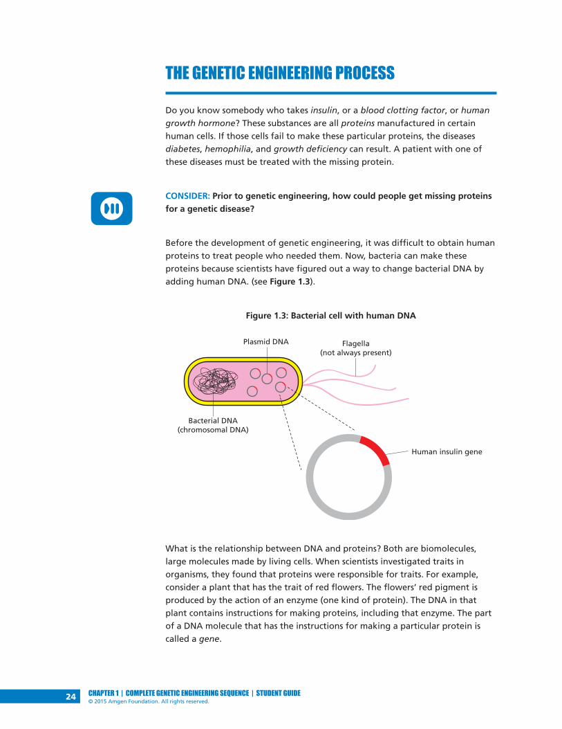
Figure 1.3: Bacterial cell with human DNA
What is the relationship between DNA and proteins? Both are biomolecules,
large molecules made by living cells. When scientists investigated traits in
organisms, they found that proteins were responsible for traits. For example,
consider a plant that has the trait of red flowers. The flowers’ red pigment is
produced by the action of an enzyme (one kind of protein). The DNA in that
plant contains instructions for making proteins, including that enzyme. The part
of a DNA molecule that has the instructions for making a particular protein is
called a gene.
Bacterial DNA(chromosomal DNA)
Plasmid DNA Flagella(not always present)
Human insulin gene

25CHAPTER 1 | COMPLETE GENETIC ENGINEERING SEQUENCE | STUDENT GUIDE© 2015 Amgen Foundation. All rights reserved.
In the genetic engineering process, a human gene is added to a plasmid, a
small circular piece of DNA found in many bacteria. The plasmid is taken up by
bacterial cells, and the cells make the human protein that is encoded by the
human gene along with their own proteins. During this process, biotechnologists
use a combination of tools, some human-made and some biological. Among the
human-made tools are two that you’ll work with in this chapter: micropipettes
and gel electrophoresis.
DID YOU KNOW?The DNA Code
DNA information is encoded by the arrangement of nucleotides, small
molecules that join together to form the DNA molecule. A DNA molecule
has millions of nucleotides. There are four different kinds of nucleotides,
and they are arranged in a specific sequence (order). A specific sequence
of nucleotides in the DNA (i.e., a gene) is a code for how to make a
specific protein. Think of a sequence of nucleotides as similar to a
sequence of written musical notes—the code for how to play music.
Just as different sequences of notes encode different songs, different
sequences of nucleotides encode different proteins.

LABORATORY
26 CHAPTER 1 | COMPLETE GENETIC ENGINEERING SEQUENCE | STUDENT GUIDE© 2015 Amgen Foundation. All rights reserved.
LABORATORY 1.2: GEL ELECTROPHORESISThe purpose of this laboratory is to give you experience with gel electrophoresis,
which is used to separate and identify a mixture of biomolecules including
DNA; the components of each mixture can then be identified by their location
in the gel. Gel electrophoresis works based on the fact that biomolecules
have a negative charge, which means that they will move in response to an
electric charge. The biomolecules move through a gel, and their speed varies
primarily according to their weight, although molecular shape and degree of
charge also influence their movement. In the genetic engineering process, gel
electrophoresis is used to separate and identify plasmids and short linear pieces
of DNA.
The electrophoresis setup consists of a box containing an agarose gel and two
electrodes that create an electric field across the gel when the box is attached
to a power supply. The negative electrode is black, and the positive electrode
is red. Samples of biomolecules are pipetted into wells near the negative
(black) electrode. The samples move through the gel toward the positive (red)
electrode, as shown in Figure 1.4.
Figure 1.4: The gel electrophoresis unit
The gel that the biomolecules move through is composed of agarose, a
polysaccharide (complex sugar) found in seaweed. Its structure is a porous matrix
(like a sponge) with lots of holes through which the solution and biomolecules
flow. See Figure 1.5.
Agarose gelWell
Pipette tip
SB buffer

LABORATORY
27CHAPTER 1 | COMPLETE GENETIC ENGINEERING SEQUENCE | STUDENT GUIDE© 2015 Amgen Foundation. All rights reserved.
Figure 1.5: How biomolecules, including DNA, move through
the agarose gel matrix in gel electrophoresis
BEFORE THE LAB Respond to the following questions with your group, and be prepared to share
your answers with the class.
1. In what circumstances might it be important to use gel electrophoresis to
separate and identify plasmids and short linear pieces of DNA?
2. Read through the Methods section on pages 28 through 31 and briefly
outline the steps for Part A and for Part B, using words and a flowchart.
MATERIALSReagents
• A plastic microfuge tube rack with the following:
• Microfuge tube of red dye solution (RD)
• Microfuge tube of dye solution 1 (S1)
• Microfuge tube of dye solution 2 (S2)
• Microfuge tube of dye solution 3 (S3)
• 50-mL flask containing 1x sodium borate buffer (1x SB) (shared with another
group)
Equipment and Supplies
• P-20 micropipette (measures 2.0–20.0 µL)
• Tip box of disposable pipette tips
Porous gel
Biomolecules
Electrophoresis
Porous gel
Biomolecules

LABORATORY
28 CHAPTER 1 | COMPLETE GENETIC ENGINEERING SEQUENCE | STUDENT GUIDE© 2015 Amgen Foundation. All rights reserved.
• 2 pipetting practice plates loaded with 0.8% agarose gel
• Electrophoresis box loaded with 0.8% agarose gel (will be shared among
groups)
• Microcentrifuge (will be shared among all groups)
• Waste container for used tips and microfuge tubes (will be shared among
groups)
SAFETY:
• All appropriate safety precautions and attire required for a science
laboratory should be used, including safety goggles. Please refer to your
teacher’s instructions.
• Wash your hands well with soap after completing the lab.
METHODSPART A: PIPETTING INTO WELLS
You will practice pipetting RD into preformed wells in an agarose gel.
1. Check your rack to make sure that you have the RD tube.
2. Fill the two pipetting practice plates with 1x SB to a level that just covers the
entire surface of the gel. If you see any “dimples” over the wells, add more
buffer.
3. Set the P-20 micropipette to 10.0 µL and put on a pipette tip.
4. Load the RD into the pipette with 10.0 µL of RD.
LAB TECHNIQUE: Do not lay down a micropipette with fluid in the tip or
hold it with the tip pointed upward.
5. Dispense RD into a well in one of the practice plates by doing the following:
a. Place your elbow on the table to steady your pipette hand. If needed,
also use your other hand to support your pipette hand.
b. Lower the pipette tip until it is under the buffer but just above the well.
LAB TECHNIQUE: Be careful not to place your pipette tip into the well or
you might puncture the gel, which will make the well unusable.
c. Gently press the plunger to slowly dispense the sample. To avoid getting
air into the buffer, do not go past the first stop. The sample will sink into
the well.

LABORATORY
29CHAPTER 1 | COMPLETE GENETIC ENGINEERING SEQUENCE | STUDENT GUIDE© 2015 Amgen Foundation. All rights reserved.
6. Repeat steps 4 and 5 until all the practice plate wells have been filled.
Everyone in your group should get an opportunity to practice pipetting into
the wells.
7. Eject the pipette tip.
PART B: SEPARATING DYES WITH GEL ELECTROPHORESIS
Now you will use gel electrophoresis to separate different dyes. First you will add
dyes into wells in the gel electrophoresis unit. You will then turn the unit on in
order to move the negatively charged dyes through the gel. (You will share the
electrophoresis boxes with one other group; your teacher will tell you which
wells your group should use.)
1. Check your rack to make sure that you have the three dye solutions (S1, S2,
and S3).
2. Review Figure 1.4 on page 26. Check to make sure that the wells in the gel
are located near the negative (black) electrode.
3. Fill the box with 1x SB to a level that just covers the entire surface of the gel.
If you see any “dimples” over the wells, add more buffer.
4. Centrifuge the S1, S2, and S3 tubes.
LAB TECHNIQUE: Distribute the tubes evenly in the microcentrifuge so that
their weight is balanced.
S1S2
S2
S3
S3
S1

LABORATORY
30 CHAPTER 1 | COMPLETE GENETIC ENGINEERING SEQUENCE | STUDENT GUIDE© 2015 Amgen Foundation. All rights reserved.
5. Make a drawing in your notebook that shows the location of the wells in the
electrophoresis box. Record which solution you will place in each well.
6. Set the P-20 micropipette to 10.0 µL and put on a pipette tip.
7. Load 10.0 µL of S1 into the pipette.
8. Dispense the S1 into the well you’ve designated for that solution by doing
the following:
a. Place your elbow on the table to steady your pipette hand. If needed,
also use your other hand to support your pipette hand.
b. Lower the pipette tip until it is under the buffer but just above the well.
LAB TECHNIQUE: Do not puncture the gel or it will become unusable.
c. Gently press the plunger to slowly dispense the sample. To avoid getting
air into the buffer, do not go past the first stop. The sample will sink into
the well.
LAB TECHNIQUE:
• While the plunger is still depressed, pull the tip out of the
buffer so that you don’t aspirate the solution or buffer.
• Use a fresh pipette tip for each sample.
9. Repeat steps 7 and 8 for S2 and S3, using a new pipette tip with each
solution.
10. When all the samples have been loaded, close the cover tightly over the
electrophoresis box. (Carefully close the cover in a horizontal motion, so that
samples don’t spill.)
11. Connect the electrical leads to the power supply. Connect both leads to the
same channel, with cathode (–) to cathode (black to black) and anode (+) to
anode (red to red). See Figure 1.6.
Figure 1.6: Leads from electrophoresis box
connected to correct channel in power supply

LABORATORY
31CHAPTER 1 | COMPLETE GENETIC ENGINEERING SEQUENCE | STUDENT GUIDE© 2015 Amgen Foundation. All rights reserved.
12. Turn on the power supply and set the voltage to 130–135 V. (You will see
bubbles form in the buffer at the red (+) end of the electrophoresis unit.)
13. After two or three minutes, check to see if the dyes are moving toward
the positive (red) electrode. You should begin to see the purple dye
(bromophenol blue) beginning to separate from the blue dye (xylene
cyanole).
STOP AND THINK:
• Study your gel electrophoresis results. Which solution sample
contained a single dye: S1, S2, or S3? How do you know?
• What electrical charge do the dyes have? Explain your reasoning.
• The dyes that you are separating are orange G (yellow),
bromophenol blue (purple), and xylene cyanole (blue). If the
molecular shape and electric charge of all three dyes are similar,
what is the order of the dyes from heaviest to lightest molecules,
based on your initial results? Why do you think this is the correct
order?
14. In approximately 10 minutes, or when you can distinguish all three dyes, turn
off the power switch and unplug the electrodes from the power supply. Do
this by grasping the electrode at the plastic plug, NOT the cord.
15. Carefully remove the cover from the gel box and observe the dyes in the gel.
16. In your notebook, draw the relative location of the bands and their colors in
each of the lanes containing your samples.
17. Leave the gels in the gel box.

32 CHAPTER 1 | COMPLETE GENETIC ENGINEERING SEQUENCE | STUDENT GUIDE© 2015 Amgen Foundation. All rights reserved.
DID YOU KNOW?Gel Electrophoresis in DNA Fingerprinting
DNA fingerprinting uses gel electrophoresis to distinguish between
samples of genetic material. In DNA fingerprinting, human DNA
molecules are treated with enzymes that chop them at certain
characteristic points, thereby reducing the DNA to a collection of smaller
and more manageable pieces. The DNA fragments are loaded into a
gel and placed in an electrical field, which electrophoretically sorts
the DNA fragments into various bands. These bands can be colored
with a radioactive dye to make them visible to imaging techniques.
Methods of DNA identification have been applied to many branches
of science and technology, including medicine (prenatal tests, genetic
screening), conservation biology (guiding captive breeding programs
for endangered species), and forensic science. In the latter discipline,
analysis of the pattern of DNA fragments that results from the action of
restriction enzymes enables us to discriminate between suspects accused
of a crime, or potential fathers in a paternity suit.
CHAPTER 1 QUESTIONS 1. What is the importance of micropipettes and gel electrophoresis in genetic
engineering?
2. How are recombinant plasmids used to treat genetic diseases?

33CHAPTER 1 | COMPLETE GENETIC ENGINEERING SEQUENCE | STUDENT GUIDE© 2015 Amgen Foundation. All rights reserved.
CHAPTER 1 GLOSSARY Agarose: A polymer made up of sugar molecules that is used as the matrix in gel
electrophoresis procedures.
Biomolecule: A molecule produced by living cells. Most biomolecules are long
polymers—made of repeating subunits called monomers—and include proteins,
carbohydrates, lipids, and nucleic acids.
Blood clotting factor: A variety of proteins in blood plasma that participate in
the clotting process.
Cell: The basic unit of any living organism that carries on the biochemical
processes of life.
Diabetes: A disease that occurs when the body doesn’t produce or properly use
insulin.
DNA (deoxyribonucleic acid): A double-stranded molecule made up of
nucleotide subunits that encodes genetic information.
Gel electrophoresis: The movement of charged molecules toward an electrode
of the opposite charge; used to separate nucleic acids and proteins. When used
to separate DNA fragments, electrophoresis will separate the fragments by size,
with smaller fragments moving faster than larger fragments.
Gene: The fundamental physical and functional unit of heredity; an ordered
sequence of nucleotides located in a specific place on the DNA that encode for a
specific functional product.
Genetic engineering: The process of altering the genetic material of cells or
organisms to enable them to make new substances or perform new functions.
Genetic disease: A disease caused by a change in DNA. Genetic diseases are often
inherited from parents.
Growth deficiency: A disease that occurs when the body doesn’t produce enough
human growth hormone.
Hemophilia: A disease that occurs when the ability of blood to clot is reduced
due to lack of one or more blood clotting factors.
Human growth hormone: A hormone secreted by the pituitary gland that
stimulates growth. Human growth hormone is a protein.
Insulin: A hormone produced in the pancreas that controls the amount of
glucose in the blood. Insulin is a protein.
Micropipette: A laboratory instrument used to measure, dispense, and transfer
very small amounts of liquid.

34 CHAPTER 1 | COMPLETE GENETIC ENGINEERING SEQUENCE | STUDENT GUIDE© 2015 Amgen Foundation. All rights reserved.
Nucleotide: A set of molecules that link together to form DNA or RNA. DNA and
RNA each contain four types of nucleotides.
Plasmid: A circular molecule of DNA.
Protein: A large biomolecule. Proteins carry out essential functions in cells,
from forming cellular structures to enabling chemical reactions to take place.
Examples of proteins are enzymes, red fluorescent protein, cell receptors, and
some hormones.
Sequence: A set of related events, movements, or items (such as nucleotides) that
follow each other in a particular order.

35CHAPTER 2 | COMPLETE GENETIC ENGINEERING SEQUENCE | STUDENT GUIDE© 2015 Amgen Foundation. All rights reserved.
CHAPTER 2HOW DO YOU BEGIN TO CLONE A GENE?


37CHAPTER 2 | COMPLETE GENETIC ENGINEERING SEQUENCE | STUDENT GUIDE© 2015 Amgen Foundation. All rights reserved.
INTRODUCTIONIn the Program Introduction, you learned that the increase in diabetes in the
United States has resulted in a great demand for its treatment, insulin. You also
learned that the best way to meet this demand is to insert the human insulin
gene into bacteria, enabling the bacteria to produce the insulin protein in
quantities large enough to meet the demand. Chapter 1 gave you a chance to
work with two physical tools and techniques of genetic engineering that are
used to clone a gene: the micropipette and gel electrophoresis. In this chapter
you will work with two other important genetic engineering tools—plasmids
and restriction enzymes. These “tools” are actually biomolecules found in many
bacteria, and their discovery was crucial to genetic engineering. With these
tools, scientists can modify microorganisms to make human insulin and other
medicines. You will now learn more about these tools and carry out the first
steps in your quest to clone a gene.
CHAPTER 2 GOALS By the end of this chapter, you will be able to do the following:
• Describe the characteristics of plasmids
• Explain how plasmids are used in cloning a gene
• Describe the function of restriction enzymes
• Explain how to use restriction enzymes to create a recombinant plasmid
WHAT DO YOU ALREADY KNOW?Discuss the following questions with your partner and write your ideas in your
notebook. Be prepared to discuss your responses with the class. Don’t worry if
you don’t know all the answers. Discussing these questions will help you think
about what you already know about DNA, plasmids, and restriction enzymes.
1. What is the structure and function of DNA? Describe in words or a drawing
the structure of a DNA molecule. Be as detailed as possible.
2. All living organisms contain DNA. In what ways is DNA from different
organisms the same, and in what ways does it vary?
3. Using your understanding of genes and how they are expressed, explain
why it is possible for a bacterial cell to make a human protein from the
instructions encoded in a human gene.
4. As described in the Program Introduction, scientists use two biological
tools to engineer organisms to make new proteins: plasmids and restriction
enzymes. What do you remember about how these tools are used?

38 CHAPTER 2 | COMPLETE GENETIC ENGINEERING SEQUENCE | STUDENT GUIDE© 2015 Amgen Foundation. All rights reserved.
PLASMIDS AND RESTRICTION ENZYMES
The discovery of plasmids and restriction enzymes in bacteria is a classic
example of how findings from basic research can revolutionize a field. Without
the discovery of these biomolecules, major breakthroughs in understanding
fundamental processes of life and in developing life-saving products might never
have occurred.
PLASMIDSMany different types of bacteria carry two forms of DNA: (1) a single
chromosome made up of a large DNA molecule that contains all the information
needed by the organism to survive and reproduce, and (2) plasmids, which are
small circular DNA molecules, ranging in size from 1,000 to 200,000 base pairs—
two nitrogenous bases joined to connect complementary strands of DNA—that
are present in multiple copies separate from the chromosomal DNA (see Figure
2.1). Some bacteria carry as many as 500 plasmids in each cell.
Figure 2.1: DNA in bacterial cells
Several characteristics of plasmids make them ideal vectors (vehicles for carrying
DNA sequences from one organism to another) for genetic engineering, for
example:
• The ability to replicate, that is, to make copies of itself independently of the
bacterial chromosome. In order to do this, plasmids have a specific sequence
where the host cell DNA synthesis enzymes bind and initiate DNA replication
(a biological process that occurs in all living organisms to make copies of
their DNA). This sequence is called the ori (“origin of replication”) site.
• The ability to initiate transcription (the process by which information
encoded in DNA is transferred to messenger RNA using the host cell RNA
polymerase). This ability requires another specific sequence, called the
promoter sequence. The promoter sequence binds RNA polymerase; this is
where transcription is initiated. All genes have promoter sequences located
Bacterial DNA(chromosomal DNA)
Plasmid DNA Flagella(not always present)

39CHAPTER 2 | COMPLETE GENETIC ENGINEERING SEQUENCE | STUDENT GUIDE© 2015 Amgen Foundation. All rights reserved.
next to them in the DNA. In order for genes such as the insulin gene to
be expressed in bacteria, they must be inserted in the plasmid next to the
promoter sequence.
• A gene or genes that code for resistance to antibiotics, a class of compounds
that kill or inhibit the growth of microorganisms. These genes code for
proteins that inhibit the action of antibiotics secreted by microorganisms and
can confer a selective advantage in nature to plasmid-containing bacteria in
a microbial population in which bacteria compete for survival.
Figure 2.2 illustrates some of the characteristics of plasmids that make them ideal
vectors for genetic engineering.
Figure 2.2: A plasmid vector
The basic components of a plasmid are the ori site for initiation
of DNA replication, a promoter for the initiation of transcription,
and a gene for antibiotic resistance (the state in which bacteria
are no longer sensitive to an antibiotic and will continue to grow
and divide in the presence of the antibiotic).
The plasmids you will work with in this and subsequent labs contain the genes
for resistance to the antibiotics ampicillin and kanamycin. These genes produce
proteins that inactivate the target antibiotic by chemically modifying its
structure.
CONSIDER: Use what you know about natural selection and evolution to
describe how plasmids might confer a selective advantage to their host bacteria.
Antibioticresistance
gene
ori
Promoter

40 CHAPTER 2 | COMPLETE GENETIC ENGINEERING SEQUENCE | STUDENT GUIDE© 2015 Amgen Foundation. All rights reserved.
A fourth feature of plasmids that is critical for genetic engineering is that
they can be passed on from one bacterial strain to another in a process called
bacterial conjugation, which enables bacteria to share and exchange genetic
information. When a plasmid with a gene for antibiotic resistance is taken in
by bacteria lacking that plasmid, the bacteria will then become resistant to
that specific antibiotic. In nature, conjugation occurs with a very low efficiency;
that is, only a small percentage of bacteria in a population can take in plasmid
DNA at any point in time. The presence of an antibiotic resistance gene on the
plasmid vector allows us to identify the small percentage of bacteria that took
in the plasmid. Bacteria that did not take in the plasmid will be killed by the
antibiotic. Those that have the plasmid with the gene of interest will survive and
grow.
In developing techniques for cloning genes in bacteria, scientists had a powerful
tool in plasmids—a vector that can be taken in by bacteria, that replicates
in bacteria to produce many copies of itself, that has a promoter sequence
for transcription of an inserted gene, and that carries a gene for antibiotic
resistance. If you carry out the lab in Chapter 5, you will take advantage of these
features of plasmids when you transfer your recombinant plasmid into bacteria.
Once scientists recognized the power of plasmids as a potential vector, the next
challenge was to determine how to incorporate a foreign gene of interest, such
as the insulin gene, into the plasmid DNA.
RESTRICTION ENZYMESIn the early 1950s, scientists observed that certain strains of E. coli, a
common bacterium found in the human gut, were resistant to infection by
bacteriophage (viruses that infect bacteria by injecting their DNA into the
cell and commandeering the host cell’s molecular processes to make more
bacteriophage). Investigation of this primitive “immune system” led to
the discovery of restriction enzymes, proteins that restricted the growth of
bacteriophage by recognizing and destroying the phage DNA without damaging
the host (bacterial) DNA. Subsequent studies demonstrated that restriction
enzymes from different strains of bacteria cut DNA at specific sequences. These
sequences are called recognition sites.
CONSIDER: How do bacteria that carry a restriction enzyme avoid cutting up
their own DNA?
Table 2.1 provides examples of restriction enzymes isolated from different
strains of bacteria and the DNA sequences they cut. In the examples shown,
the enzymes cut asymmetrically on the strands of DNA, leaving single-stranded
overhanging sequences at the site of the cut. For example, a cut (or digestion)
with EcoRI will leave an AATT overhang (or “sticky end”) on one strand and a
TTAA sticky end on the other strand.
41CHAPTER 2 | COMPLETE GENETIC ENGINEERING SEQUENCE | STUDENT GUIDE© 2015 Amgen Foundation. All rights reserved.
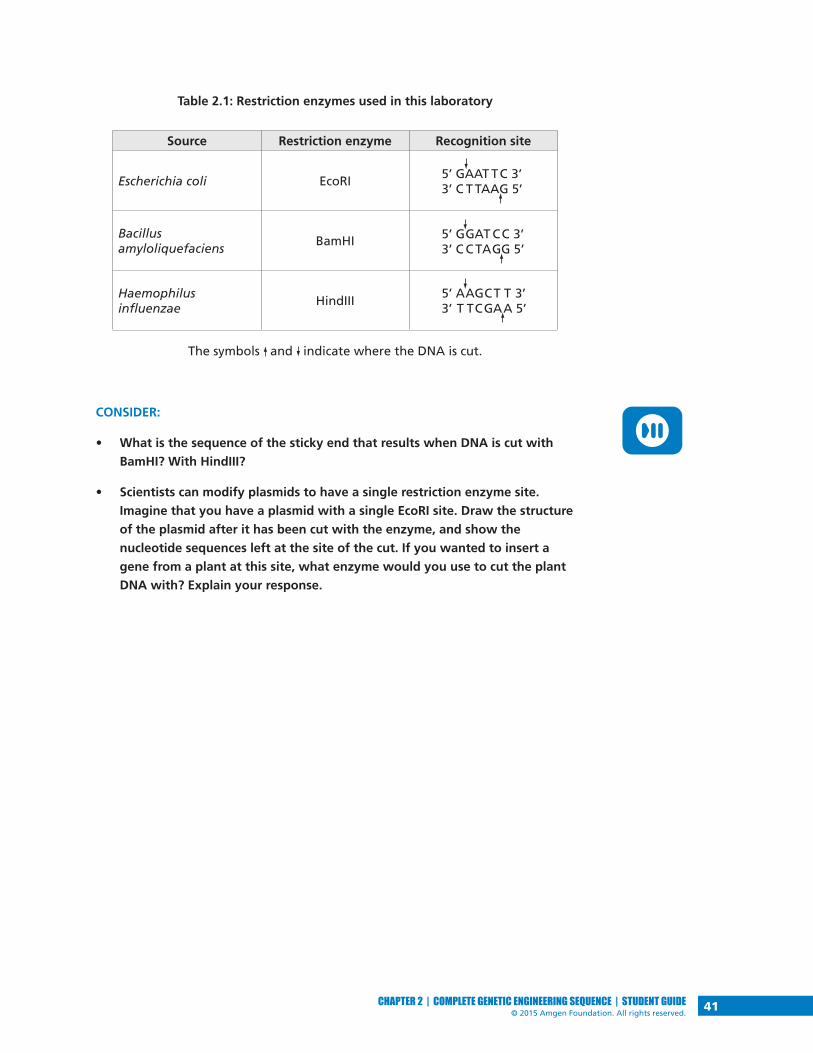
Table 2.1: Restriction enzymes used in this laboratory
The symbols and indicate where the DNA is cut.
CONSIDER:
• What is the sequence of the sticky end that results when DNA is cut with
BamHI? With HindIII?
• Scientists can modify plasmids to have a single restriction enzyme site.
Imagine that you have a plasmid with a single EcoRI site. Draw the structure
of the plasmid after it has been cut with the enzyme, and show the
nucleotide sequences left at the site of the cut. If you wanted to insert a
gene from a plant at this site, what enzyme would you use to cut the plant
DNA with? Explain your response.
Source Restriction enzyme Recognition site
Escherichia coli EcoRI
Bacillusamyloliquefaciens
BamHI
Haemophilusinfluenzae
HindIII
5’ GAAT TC 3’3’ C T TAAG 5’
5’ GGAT CC 3’3’ C C TAGG 5’
5’ AAGCT T 3’3’ T TCGAA 5’

42 CHAPTER 2 | COMPLETE GENETIC ENGINEERING SEQUENCE | STUDENT GUIDE© 2015 Amgen Foundation. All rights reserved.
DID YOU KNOW?The Rise of Antibiotic-Resistant Bacteria
Antibiotics and similar drugs have been used for the last 70 years to
treat patients who have infectious diseases. When prescribed and taken
correctly, antibiotics are enormously valuable in patient care. However,
these drugs have been used so widely and for so long that the infectious
organisms the antibiotics are designed to kill have adapted to them,
making the drugs less effective. Antibiotic resistance occurs when some
bacteria in a population are able to survive when exposed to one or more
antibiotics. These species that have become resistant cause infections
that cannot be treated with the usual antibiotic drugs at the usual
dosages and concentrations. Some have developed resistance to multiple
antibiotics and are dubbed multidrug-resistant bacteria or “superbugs.”
Antibiotic resistance is
a serious and growing
phenomenon and
has emerged as one
of the major public
health concerns of the
21st century. Drug-
resistant organisms
may have acquired
resistance to first-line
antibiotics, requiring
the use of second-line
agents. Typically, the
first-line agent is selected on the basis of several advantages, including
safety, availability, and cost; the second-line agent is usually broader in
spectrum, may be less beneficial in relation to the associated risks, and
may be more expensive or less widely available.

43CHAPTER 2 | COMPLETE GENETIC ENGINEERING SEQUENCE | STUDENT GUIDE© 2015 Amgen Foundation. All rights reserved.
CLONE THAT GENE
You now know about two biological tools for cloning a gene:
1. A plasmid that has several important features:
• A restriction enzyme site or sites that opens the plasmid circle and
enables insertion of the gene of interest into the plasmid DNA
• A sequence for the initiation of DNA replication, called the ori site,
that allows the plasmid to replicate in the bacteria using the host
DNA synthesis enzymes
• A promoter sequence for initiating transcription of the inserted gene
• A gene encoding a protein for antibiotic resistance, which allows for
identification of bacteria that have taken in the plasmid
2. Restriction enzymes for the digestion of both the plasmid and the
human DNA containing the gene of interest (such as insulin) to be
cloned
How do scientists use these two tools to create a recombinant plasmid,
which contains the insulin gene (or any other gene of interest) inserted into
a bacterial plasmid? One important step is choosing a restriction enzyme or
enzymes that cut the plasmid and the human DNA. The restriction enzyme(s)
must do all of the following:
• Cut the plasmid at a site or sites that allow for the insertion of the new
gene.
• Cut the plasmid at an appropriate site to ensure that no important genes
or sequences are disrupted, including the ori site, the promoter, and at
least one of the genes encoding antibiotic resistance.
• Cut the plasmid near the promoter so that the inserted gene can be
expressed.
• Cut the human DNA as close as possible to both ends of the gene of
interest so that it can be inserted into the appropriate site in the plasmid
DNA, without cutting within the gene.
STOP AND THINK: Why is it important that the same enzyme or
enzymes be used to cut both the plasmid and the insulin gene from the
human DNA?

44 CHAPTER 2 | COMPLETE GENETIC ENGINEERING SEQUENCE | STUDENT GUIDE© 2015 Amgen Foundation. All rights reserved.
In this activity, you will make a paper model of a recombinant plasmid that
contains an insulin gene. You have three tasks:
1. Cut the plasmid and the human DNA with the appropriate restriction
enzyme
2. Insert the human insulin gene into the plasmid DNA
3. Determine which antibiotic you would use to identify bacteria that have
taken in the plasmid
HANDOUTS• Plasmid Diagram (RM 2)
• Human DNA Sequence (RM 3)
PROCEDURE1. On the Plasmid Diagram (RM 2):
• Use scissors to cut out the plasmid sequence and tape the ends
together to make a paper model of the plasmid.
• Locate the positions of the ori site, the promoter site, and the genes
for antibiotic resistance.
• Locate the positions of each restriction enzyme recognition site.
2. Choose the restriction enzyme that should be used to cut the plasmid.
Verify that the restriction enzyme meets all the following criteria:
• The ori site on the plasmid is intact
• The promoter site is intact
• At least one of the antibiotic resistance genes is intact
• The enzyme cuts the plasmid only once
• The cut is close to the promoter sequence
3. Review Table 2.1 on page 41 and use scissors to cut the plasmid at the
recognition site exactly as the restriction enzyme would cut it. Write the
sequences of the nucleotides that are left on each end of the plasmid.
4. On the Human DNA Sequence (RM 3), scan the human DNA sequence
and determine where the three restriction enzymes, BamHI, EcoRI, and
HindIII, would cut the DNA.

45CHAPTER 2 | COMPLETE GENETIC ENGINEERING SEQUENCE | STUDENT GUIDE© 2015 Amgen Foundation. All rights reserved.
5. Determine whether the restriction enzyme you chose in step 2 is a good
choice for cutting out the insulin gene from the human DNA by verifying
that it meets all the following criteria:
• It does not cut within the insulin gene
• It cuts very close to the beginning and end of the gene
• It will allow the insulin gene to be inserted into the cut plasmid
6. Review Table 2.1 and use scissors to cut the human DNA at the
recognition site exactly as the restriction enzyme would cut it. Write the
sequences of the nucleotides that are left on each end of the insulin
gene after it is cut from the human DNA.
7. Use tape to insert the insulin gene into the cut plasmid. Verify that the
sticky ends will connect in the correct orientation. (In the lab, a third
biological tool, DNA ligase, is used to permanently connect the sticky
ends together.) This is a paper model of a recombinant plasmid that
contains an insulin gene. Once the plasmid replicates (copies) itself, the
insulin gene is also copied, or cloned!
ACTIVITY QUESTIONS1. Which restriction enzyme did you choose? Why did you choose that one?
2. Where would you insert the insulin gene and why?
3. Which antibiotic would you use to determine if the recombinant DNA
was taken in?

LABORATORY
46 CHAPTER 2 | COMPLETE GENETIC ENGINEERING SEQUENCE | STUDENT GUIDE© 2015 Amgen Foundation. All rights reserved.
LABORATORY 2: PREPARING TO CLONE THE RFP GENE: DIGESTING THE pKAN-R AND pARAThe purpose of this lab is to produce the DNA fragments that will be joined
to make the recombinant plasmid, pARA-R, that can make the red fluorescent
protein in bacteria. To do this you will use restriction enzymes to cut two
plasmids, which will generate DNA fragments. This procedure is called a
restriction digest, and the lengths of the fragments can be determined by gel
electrophoresis (which you may do in Chapter 4).
So far, you’ve learned about using a single plasmid to clone the insulin gene.
Under some circumstances scientists need to use plasmid DNA from different
sources to generate a specific recombinant DNA. In order to clone the red
fluorescent protein (rfp) gene, you will need DNA from two different plasmids.
The plasmid pKAN-R (see Figure 2.3) carries the gene that makes bacteria
resistant to the antibiotic kanamycin, the rfp gene, and a promoter sequence.
The plasmid pARA (see Figure 2.3) contains the gene that makes bacteria
resistant to the antibiotic ampicillin and a DNA sequence that activates the
promoter when the bacteria are grown in the presence of arabinose, a five-
carbon sugar that naturally occurs in various plant and bacterial carbohydrates.
This sequence is called the arabinose activator (araC). If arabinose is present
in the bacteria, the promoter will bind RNA polymerase, and transcription will
occur. If arabinose is not present, the promoter will not bind RNA polymerase,
and transcription will not occur. The plasmid pARA also contains the ori site for
initiating DNA replication.
Figure 2.3: The pKAN-R and pARA plasmids
The relevant components on the plasmids are the rfp gene, the
promoter (pBAD), the ampicillin resistance gene (ampR), and araC.
BamHI
HindIII
pARA4,872 bp
ampR377 bp
araC
ori
pKAN-R5,512 bp
kanR
BamHI
HindIII
rfp
pBAD-rfp807 bp
pBAD

LABORATORY
47CHAPTER 2 | COMPLETE GENETIC ENGINEERING SEQUENCE | STUDENT GUIDE© 2015 Amgen Foundation. All rights reserved.
In addition to showing the relevant components, Figure 2.3 also shows the size
of the plasmid (the number in the center, which indicates the number of base
pairs [bp]) and the sequences where it can be cut by the restriction enzymes that
will be used in the lab. The sites labeled “BamHI” and “HindIII” represent the
recognition sites for these two restriction enzymes. (See Table 2.1 on page 41.)
Figure P.4 in What Is Genetic Engineering? (on page 14) shows the insulin gene
being inserted in a single restriction enzyme site in the plasmid. In the cloning
of the rfp gene, two restriction enzymes (BamHI and HindIII) are used in cutting
the plasmid into which the rfp gene will be inserted and in isolating the rfp gene
from the second plasmid. Using two different restriction enzymes has several
advantages: It allows the inserted gene to be oriented in the correct position for
transcribing the “sense” strand of DNA (the strand that codes for the protein),
and it prevents the plasmid from reforming a circle without the inserted gene.
You’ll learn more about this if you do Chapter 4.
STOP AND THINK: Why does using two different enzymes to cut the
plasmid prevent the plasmid from reforming a circle without the
inserted gene?
BEFORE THE LAB Respond to the following questions with your group and be prepared to share
your answers with the class.
1. Review Figure 2.3. If pKAN-R is digested with BamHI and HindIII, what
fragments are produced? If pARA is digested with BamHI and HindIII, what
fragments are produced? Record the nucleotide sequence of the sticky ends
and the length of each fragment (bp), and indicate the genes and other
important sequences present on each fragment.
2. In order to create a plasmid that can produce the red fluorescent protein in
bacteria, what components are needed in the plasmid?
3. Bacteria can be killed by an antibiotic unless they carry a plasmid that has
the gene for resistance to that antibiotic. Biotechnologists call these genes
selectable markers because only bacteria that carry the gene will survive an
antibiotic. If the uptake of DNA by bacteria is inefficient (as discussed in the
reading), why is a selectable marker critical in cloning a gene in bacteria?
4. Read through the Methods section on pages 49 and 50 and briefly outline
the steps, using words and a flowchart.

LABORATORY
48 CHAPTER 2 | COMPLETE GENETIC ENGINEERING SEQUENCE | STUDENT GUIDE© 2015 Amgen Foundation. All rights reserved.
MATERIALSReagents
• A rack with the following:
• Microfuge tube of 2.5x restriction buffer (2.5xB)
• Microfuge tube of pKAN-R plasmid (K)
• Microfuge tube of pARA plasmid (A)
• Microfuge tube of restriction enzymes BamHI and HindIII (RE)
• Microfuge tube of distilled water (dH2O)
Equipment and Supplies
• P-20 micropipette
• Tip box of disposable tips
• 4 1.5-mL microfuge tubes
• Permanent marker
• Microcentrifuge (will be shared among all groups)
• 37°C water bath with floating microfuge tube rack (will be shared among all
groups)
• Waste container for used tips and microfuge tubes (will be shared among
groups)
SAFETY:
• All appropriate safety precautions and attire required for a science
laboratory should be used, including safety goggles. Please refer to your
teacher’s instructions.
• Wash your hands well with soap after completing the lab.

LABORATORY
49CHAPTER 2 | COMPLETE GENETIC ENGINEERING SEQUENCE | STUDENT GUIDE© 2015 Amgen Foundation. All rights reserved.
METHODS1. Check your rack to make sure that you have all the reagents listed.
2. Use a marker to label four clean microfuge tubes as follows: K+, K–, A+, and
A–. (Also include your group number and class period on each tube.)
3. Review Table 2.2, which summarizes the reagents that you will add in step 4.
Table 2.2: Addition of reagents to the K+, K–, A+, and A– tubes
LAB TECHNIQUE: In step 4, be sure to use a new micropipette tip for
each reagent in each tube to avoid contamination.
4. Add the following:
a. 4.0 µL of 2.5xB to the K+, K–, A+, and A– tubes.
b. 4.0 µL of K to the K+ and K– tubes.
c. 4.0 µL of A to the A+ and A– tubes.
d. 2.0 µL of RE to the K+ and A+ tubes. Add the enzymes directly into the
solution at the bottom of the microfuge tube. Gently pump the solution
in and out with the pipette to mix the reagents. Cap the tubes when
done.
e. 2.0 µL of dH2O to the K– and A– tubes. Gently pump the solution in and
out with the pipette to mix the reagents. Cap the tubes when done.
STOP AND THINK: In step 4, you are asked to set up two tubes without
the restriction enzymes, BamHI and HindIII. What is the purpose of this
step, and why is it important?
K+ tube K– tube A+ tube A– tube
Step 4a: Restriction buffer (2.5xB) 4.0 µL 4.0 µL 4.0 µL 4.0 µL
Step 4b: pKAN-R plasmid (K) 4.0 µL 4.0 µL
Step 4c: pARA plasmid (A) 4.0 µL 4.0 µL
Step 4d: BamHI and HindIII (RE) 2.0 µL 2.0 µL
Step 4e: Distilled water (dH2O) 2.0 µL 2.0 µL

LABORATORY
50 CHAPTER 2 | COMPLETE GENETIC ENGINEERING SEQUENCE | STUDENT GUIDE© 2015 Amgen Foundation. All rights reserved.
5. Spin the four microfuge tubes (K+, A+, K–, and A–) in the microcentrifuge
for several seconds to pool the reagents at the bottom of each tube.
LAB TECHNIQUE: Distribute the tubes evenly in the microcentrifuge so that
their weight is balanced.
6. Place all four tubes into the 37°C water bath. (You will place your tubes in
the floating microfuge tube rack; when the rack is full, your teacher will
place it in the water bath.) Incubate for at least one hour but not longer
than two hours. After the incubation is complete, place all four tubes in the
freezer at –20°C. You will use the contents of the tubes in Laboratory 3.
STOP AND THINK: Why might the enzymes work best at 37°C? Why
should the enzymes then be placed in the freezer?

51CHAPTER 2 | COMPLETE GENETIC ENGINEERING SEQUENCE | STUDENT GUIDE© 2015 Amgen Foundation. All rights reserved.
CHAPTER 2 QUESTIONS Discuss the following questions with your partner and be prepared to share your
answers with the class.
1. List in words or indicate in a drawing the important features of a plasmid
vector that are required to clone a gene. Explain the purpose of each
feature.
2. What role do restriction enzymes have in nature?
3. Using your understanding of evolution, why would bacteria retain a gene
that gives them resistance to antibiotics? How is the existence of bacteria
with antibiotic resistance affecting medicine today?
4. Bacteria, sea anemones, and humans seem, on the surface, to be very
different organisms. Explain how a gene from humans or a sea anemone can
be expressed in bacteria to make a product never before made in bacteria.
5. Due to a mishap in the lab, bacteria carrying a plasmid with a kanamycin-
resistant gene and bacteria carrying a plasmid with an ampicillin-resistant
gene were accidentally mixed together. Design an experiment that will allow
you to sort out the two kinds of bacteria. (Hint: Make sure that you do not
kill off one of the kinds of bacteria you are trying to sort out!)

52 CHAPTER 2 | COMPLETE GENETIC ENGINEERING SEQUENCE | STUDENT GUIDE© 2015 Amgen Foundation. All rights reserved.
CHAPTER 2 GLOSSARY Activator: A protein that regulates transcription of a gene by binding to a
sequence near the promoter, thus enabling RNA polymerase to bind to the
promoter and initiate transcription of the gene. The activator protein can also
block the binding of RNA polymerase and thereby inhibit transcription of the
gene.
Antibiotic: A class of compounds that kill or inhibit the growth of
microorganisms.
Antibiotic resistance: The state in which bacteria are no longer sensitive to an
antibiotic and will continue to grow and divide in the presence of the antibiotic.
Arabinose: A five-carbon sugar that naturally occurs in various plant and
bacterial carbohydrates.
Bacterial conjugation: A process by which two bacterial cells join and transfer
genetic material from one to another.
Bacteriophage: A virus that infects a bacterial cell and uses the cell machinery to
replicate itself, eventually destroying the bacterial cell.
Base pair: Two complementary nitrogen-containing molecules paired together
in double-stranded DNA by weak bonds.
Digestion: The cutting of DNA by a restriction enzyme.
DNA ligase: An enzyme that catalyzes the formation of covalent chemical bonds
in the sugar-phosphate backbone, thereby binding fragments of DNA together.
DNA replication: The biological process of making an identical copy of DNA,
which occurs each time a new cell is formed in living organisms. The double helix
is unwound, and each strand of the original molecule serves as a template for
the production of the complementary strand.
Enzyme: A protein that catalyzes a chemical reaction.
Messenger RNA: An RNA molecule transcribed from the DNA of a gene and used
as the template for protein synthesis.
Origin of replication (ori): A sequence of DNA at which replication of the DNA is
initiated.
Plasmid: A circular molecule of DNA.
Promoter: A specific DNA sequence that binds RNA polymerase and initiates
transcription of the gene.
Recognition site: A specific DNA sequence that is cut by a restriction enzyme.
Many restriction sites are palindromes, sequences that are the same when read
forward or backward.

53CHAPTER 2 | COMPLETE GENETIC ENGINEERING SEQUENCE | STUDENT GUIDE© 2015 Amgen Foundation. All rights reserved.
Recombinant DNA: DNA that contains sequences or genes from two or more
sources.
Restriction digest: A technique in which naturally occurring enzymes are used to
cleave DNA at specific sequences.
Restriction enzyme: A protein that can cut DNA at a specific sequence called a
recognition site.
RNA (ribonucleic acid): A single-stranded biomolecule made up of a nitrogenous
base, a ribose sugar, and a phosphate; RNA plays a critical role in protein
synthesis, transmitting genetic information from DNA to the ribosome where
proteins are then made.
RNA polymerase: A protein that makes messenger RNA from DNA.
Selectable marker: A gene on a plasmid that is introduced into a cell along with
a gene of interest that is being cloned. Selectable markers allow scientists to tell
if the plasmid has been taken in by the cell because the marker can be seen or
detected. A common selectable marker is an antibiotic resistance gene—only
bacteria that have the gene will survive the antibiotic.
Sticky ends: Ends of a DNA molecule cut with certain restriction enzymes. These
ends are asymmetrical in that one strand is longer than the other strand and
therefore has unpaired bases. The sticky ends of two different pieces of DNA
that have been cut with the same restriction enzyme(s) can be joined, as the
unpaired bases on their ends are complementary.
Transcription: The process by which information encoded in DNA is transferred
to messenger RNA, a single-stranded ribonucleic acid.
Vector: A vehicle for moving DNA sequences from one organism to another.


55CHAPTER 3 | COMPLETE GENETIC ENGINEERING SEQUENCE | STUDENT GUIDE© 2015 Amgen Foundation. All rights reserved.
CHAPTER 3BUILDING A RECOMBINANT PLASMID


57CHAPTER 3 | COMPLETE GENETIC ENGINEERING SEQUENCE | STUDENT GUIDE© 2015 Amgen Foundation. All rights reserved.
INTRODUCTIONIn Chapters 1 and 2, you learned about four important tools of genetic
engineering that scientists use to clone the insulin gene: the micropipette,
gel electrophoresis, plasmids, and restriction enzymes. In this chapter, you will
use a fifth tool that is needed to clone genes—DNA ligase. DNA ligase is an
enzyme that catalyzes the joining of DNA fragments; it is one of several enzymes
involved in DNA replication in all cells. In Chapter 2, you prepared the DNA
fragments required for cloning the rfp gene. The next step is to ligate (join
together) these fragments to form the recombinant plasmid.
CHAPTER 3 GOALS By the end of this chapter, you will be able to do the following:
• Describe the role of a DNA ligase in replication
• Explain how DNA ligase is used to create a recombinant plasmid
• Describe possible recombinant plasmids that form when ligating a
restriction digest
WHAT DO YOU ALREADY KNOW?Discuss the following questions with your partner and write your ideas in your
notebook. Be prepared to discuss your responses with the class. Don’t worry
if you don’t know all the answers. Discussing these questions will help you
think about what you already know about enzymes, DNA replication, and DNA
ligation.
1. What is the function of enzymes in reactions?
2. How does DNA replication occur?
3. Why is replication of DNA essential in all cells?
4. Describe what happens when two DNA fragments with complementary
sticky ends join, and speculate how the activity of DNA ligase ensures that
the join is permanent.

58 CHAPTER 3 | COMPLETE GENETIC ENGINEERING SEQUENCE | STUDENT GUIDE© 2015 Amgen Foundation. All rights reserved.
LIGASES
The discoveries of restriction enzymes, plasmids, and ligases in cells were
important in the quest to understand how bacteria grow and reproduce
themselves. But it was understanding how these biomolecules could be applied
to manipulate DNA that launched a new era of genetic engineering, one in
which humans could directly engineer organisms at the genetic level. Plasmids
provided the vehicle to clone genes, and restriction enzymes provided the means
to generate the fragments of DNA needed to form recombinant plasmids. The
final essential ingredient was a way to “glue” the fragments together.
THE ROLE OF LIGASES IN DNA REPLICATIONIn the early 1960s, scientists isolated enzymes that had this ability. These
enzymes, called DNA ligases, were shown to be involved in DNA replication of
chromosomes and plasmids. Replication occurs in a number of steps (as shown in
Figure 3.1):
Figure 3.1: Summary of DNA replication
1. The two strands begin to separate at multiple locations on the DNA
molecule. Each location is an origin of replication, or ori site (the same as
you learned about with plasmids in Chapter 2). The two strands are called
parent strands.
2. Once the parent strands are separated and the bases are exposed, an
enzyme called DNA polymerase adds new nucleotides. Each new nucleotide
base forms hydrogen bonds with existing nucleotide bases on the parent
strand. Complementary base pairs are created in this step.
5. Ligase catalyzes the joining of the DNA fragments
1. Two parent DNA strands separate at an ori site
2. DNA polymerase adds complementary nucleotides to each parent strand
3. A new daughter strand is formed continuously on this parent strand
4. A second daughter strand is formed discontinuously as DNA fragments on this parent strand
DNA polymerase
DNA polymerase
Fragment being made
DNA molecule
3’
3’
5’
5’
DNA ligase
3’5’

59CHAPTER 3 | COMPLETE GENETIC ENGINEERING SEQUENCE | STUDENT GUIDE© 2015 Amgen Foundation. All rights reserved.
3. On one parent strand, DNA polymerase adds nucleotides continuously to
form a new complementary strand, a daughter strand.
4. On the other parent strand, DNA polymerase adds nucleotides
discontinuously, resulting in small fragments of DNA. (To learn about why
one strand is replicated continuously and one discontinuously, refer to Did
You Know? DNA Directionality.)
5. The DNA fragments must be joined by the enzyme DNA ligase in order
to form the second daughter strand. The ligase joins the fragments by
catalyzing the formation of a covalent bond between adjacent nucleotides.
Once the replication steps are complete, two new DNA molecules, each
consisting of one parent strand and one daughter strand, have been made. Both
molecules are exact copies, or replicas, of the original DNA molecule.
CONSIDER:
• What are the roles of hydrogen bonds and covalent bonds in the structure
of DNA?
• Based on what you just learned about DNA replication, what is the possible
role of ligases in joining the sticky ends of DNA fragments?
ROLE OF LIGASES IN GENE CLONINGThe role of ligase in gene cloning is similar to its role in replication in that it
binds DNA fragments together. In gene cloning, recombinant DNA is the result
of ligation of fragments from a restriction digest (as shown in Figure 3.2). Any
two fragments that have ends cut with the same restriction enzyme can be
ligated together. First the unpaired bases at the sticky ends form hydrogen
bonds to each other, and then the ligase catalyzes the formation of covalent
bonds between adjacent nucleotides. If you have multiple fragments, the
ligation procedure can lead to a number of possible products.
Figure 3.2: Ligation of DNA fragments in genetic cloning
Plasmid DNA
Gene of interest
GG
G
G G G
A
A
AA
A
T
TT
T
T
C
CC C
C C
1. Complementary nucleotides in the sticky ends pair up
2. Ligase catalyzes the joining of the DNA fragments together
3’
5’3’
5’

60 CHAPTER 3 | COMPLETE GENETIC ENGINEERING SEQUENCE | STUDENT GUIDE© 2015 Amgen Foundation. All rights reserved.
DID YOU KNOW?DNA Directionality
DNA is a double helix of two intertwined strands that are hydrogen-
bonded together through the bases of the nucleotides. Each strand
has a different directionality, meaning that each strand has a chemical
orientation based on the chemical group that it ends with. The 5’ (5
“prime”) end of the DNA strand is the end that has the fifth carbon in
the deoxyribose sugar ring. The 3’ (3 “prime”) end of the DNA strand
is the end that has the hydroxyl (OH) group on the third carbon in the
deoxyribose sugar ring (see Figure 3.3).
This naming of the ends of the strands is useful in understanding how
DNA polymerase works. DNA polymerase can only assemble new DNA
strands in the 5’ to 3’ direction,
adding new nucleotides to the
3’-hydroxyl group. Therefore,
on one strand replication occurs
continuously as polymerase
adds nucleotides in the 5’ to 3’
direction to form the daughter
strand. On the other strand,
the DNA polymerase also adds
nucleotides in the 5’ to 3’
direction, which results in short
fragments of DNA that must be
ligated together to form the
other daughter strand.
G C
A T
A T
T A
C G
T A
5’ end
5’ endOH
OH
3’ end
3’ end
Figure 3.3: DNA structure

LABORATORY
61CHAPTER 3 | COMPLETE GENETIC ENGINEERING SEQUENCE | STUDENT GUIDE© 2015 Amgen Foundation. All rights reserved.
LABORATORY 3: BUILDING THE pARA-R PLASMIDIn this laboratory you will ligate the DNA fragments you produced during
Laboratory 2, using DNA ligase to make new recombinant plasmids. These
recombinant plasmids will contain the four restriction fragments from Laboratory
2 recombined in different ways to produce new sets of DNA. The ligation process
will result in several different plasmids, but the plasmid that you are interested in
will contain the gene for ampicillin resistance (ampR), the red fluorescent protein
(rfp) gene, a promoter sequence for initiating transcription (pBAD), the arabinose
activator sequence (araC), and the ori sequence for the initiation of DNA
replication. This desired recombinant DNA plasmid is called the pARA-R plasmid
(see Figure 3.4).
Figure 3.4: The pARA-R plasmid
During the lab, you will mix together the DNA fragments from your restriction
digest in Laboratory 2 and the DNA ligase, but you will not be able to observe
anything until Laboratory 4, when you will have the opportunity to separate and
identify your DNA molecules using gel electrophoresis. However, you will prepare
for what you might observe by determining the possible plasmids that can occur
and drawing diagrams of these plasmids. In this work, you are modeling the
process of ligation.
pARA-R5,302 bp
ampR
ori araC
BamHI
HindIII
rfp
pBAD-rfp807 bp
pBAD
LABORATORY
62 CHAPTER 3 | COMPLETE GENETIC ENGINEERING SEQUENCE | STUDENT GUIDE© 2015 Amgen Foundation. All rights reserved.
BEFORE THE LAB Discuss the following questions with your group, record your answers, and be
prepared to share your answers with the class.
1. Review your answer to question 1 in Before the Lab for Laboratory 2 (page
47), in which you described the fragments that formed from the digestion
of pKAN-R and pARA with BamHI and HindIII. Using this information, draw
three possible recombinant plasmids resulting from the joining of two pARA
and pKAN-R fragments. For each plasmid, identify the genes, other important
sequences, and the number of base pairs each has.
2. Read through the Methods section on page 63 and briefly outline the steps,
using words and a flowchart.
MATERIALSReagents
• A rack with the following:
• Microfuge tube of digested pKAN-R from Laboratory 2 (K+)
• Microfuge tube of digested pARA from Laboratory 2 (A+)
• Microfuge tube of 5x ligation buffer (5xB)
• Microfuge tube of DNA ligase (LIG)
• Microfuge tube of distilled water (dH2O)
Equipment and Supplies
• 70°C water bath with floating microfuge tube rack (will be shared among
all groups)
• P-20 micropipette
• Tip box of disposable pipette tips
• Permanent marker
• Microcentrifuge (will be shared among all groups)
• Waste container for used tips and microfuge tubes (may be shared
with another group)
SAFETY:
• All appropriate safety precautions and attire required for a science
laboratory should be used, including safety goggles. Please refer to your
teacher’s instructions.
• Wash your hands well with soap after completing the lab.
LABORATORY
63CHAPTER 3 | COMPLETE GENETIC ENGINEERING SEQUENCE | STUDENT GUIDE© 2015 Amgen Foundation. All rights reserved.
METHODS1. Check your rack to make sure that you have all the reagents listed.
2. Place K+ and A+ tubes from Laboratory 2 in the 70°C water bath (you will
place your tubes in the floating microfuge tube rack; once each group’s
samples are loaded, your teacher will place the rack in the water bath) for 30
minutes. This heat exposure will denature (inactivate) the restriction enzymes.
NOTE: During the 30-minute incubation, share and discuss your answers
to question 1 in Before the Lab. Also share and discuss your answer to the
STOP AND THINK question that follows and begin answering the Chapter 3
Questions on page 64.
STOP AND THINK: Why is it important to inactivate the BamHI and HindIII
restriction enzymes before ligating the fragments? What might happen if
you did not perform this step?
3. Label the LIG tube with your group number and class period.
4. After 30 minutes, remove the K+ and A+ tubes from the water bath and place
them in your rack.
5. Add the following solutions directly into the solution at the bottom of the
LIG tube:
a. 4.0 µL of A+
b. 4.0 µL of K+
c. 3.0 µL of 5xB
d. 2.0 µL of dH2O
LAB TECHNIQUE: In steps 5a–d, be sure to use a new micropipette tip
for each reagent to avoid contamination.
6. After adding the dH2O, gently pump the solution in and out with the pipette
to mix the reagents. Cap the tube when done.
7. Spin the LIG tube in the microcentrifuge for several seconds to pool the
reagents at the bottom of the tube.
LAB TECHNIQUE: Distribute the tubes evenly in the microcentrifuge so that
their weight is balanced.
8. Place your LIG, A+, and K+ tubes in the microfuge racks designated by your
teacher. (Your LIG tube will incubate at room temperature until the next
class. Your A+ and K+ tubes will be returned to the –20°C freezer for use in
Laboratory 4.)

64 CHAPTER 3 | COMPLETE GENETIC ENGINEERING SEQUENCE | STUDENT GUIDE© 2015 Amgen Foundation. All rights reserved.
CHAPTER 3 QUESTIONS Discuss the following questions with your partner and be prepared to share your
answers with the class:
1. What role do DNA ligases have in nature?
2. What role do DNA ligases have in gene cloning?
3. What properties of the DNA restriction fragments produced in Laboratory 2
enable ligation of these fragments?
4. Could two rfp fragments join to form a plasmid during the ligation? If not,
what would prevent that? If so, what would be the result?
5. During ligation, both hydrogen and covalent bonds form. Which bonds form
first? Why do both types of bonds need to form?
DID YOU KNOW?DNA Replication Errors and Evolution
Every time a cell divides and its DNA replicates, it copies and transmits
the exact same sequence of nucleotides to its daughter cells. While
DNA usually replicates with fairly high accuracy, mistakes do happen.
Polymerase enzymes sometimes insert the wrong nucleotide or too many
or too few nucleotides into a sequence. Incorrectly paired nucleotides
that still remain following mismatch repair become permanent
mutations after the next cell division. This is because once such mistakes
are established, the cell no longer recognizes them as errors.
Mutations can be beneficial, harmful, or have no effect at all. If a
mutation gives the organism a selective advantage, the trait may be
passed to future generations as part of the evolutionary process. In fact,
over the course of millions of years, mutations may build up in any given
stretch of DNA at a reliable rate. This allows the gene to be used as a
molecular clock, a tool for estimating how long ago two species split
from each other. This general technique has been used to investigate
several important questions, including the origin of modern humans and
the date of the human-chimpanzee divergence.

65CHAPTER 3 | COMPLETE GENETIC ENGINEERING SEQUENCE | STUDENT GUIDE© 2015 Amgen Foundation. All rights reserved.
CHAPTER 3 GLOSSARY 3’ end: The end of a DNA or RNA strand that has a hydroxyl group connected
to the third carbon of the sugar molecule, which is deoxyribose or ribose. (The
carbon number refers to the position of the carbon in the sugar molecule.)
5’ end: The end of a DNA or RNA strand that terminates at the fifth carbon of the
sugar molecule, which is deoxyribose or ribose. (The carbon number refers to the
position of the carbon in the sugar molecule.)
Catalyze: To increase the rate of a reaction.
Complementary base pair: Nitrogen-containing bases that are found opposite
each other in a double-stranded DNA molecule. Complementarity is the result
of the size and shape of the base and the number of hydrogen bonds between
the adjacent bases in the pair (A and T form two hydrogen bonds, G and C form
three). Adenine is complementary to thymine, and guanine is complementary to
cytosine.
Daughter strand: In DNA replication, a new DNA strand that is created that is a
complement to the original DNA strand.
Denature: To change the shape of a biomolecule and subsequently impact its
function, which is often achieved by heating. For example, a protein folds into
a complex three-dimensional shape that is directly related to its function. Any
process that interferes with protein folding can change its three-dimensional
shape and result in inactivation of the protein.
Directionality: In biochemistry, the orientation of a strand of nucleic acid.
DNA ligase: An enzyme that catalyzes the formation of covalent chemical bonds
in the sugar-phosphate backbone, thereby permanently binding fragments of
DNA together.
DNA polymerase: An enzyme used to replicate DNA molecules.
DNA replication: The biological process of making an identical copy of a section
of DNA, which occurs each time a new cell is formed in living organisms. The
process starts when one double-stranded DNA molecule produces two identical
copies. The double helix is unwound, and each strand of the original molecule
serves as a template for the production of the complementary strand.
Enzyme: A protein that catalyzes a chemical reaction.
Ligation: The reaction that chemically joins two fragments of DNA, resulting in a
recombinant DNA molecule.
Ligate: To join together two DNA ends.

66 CHAPTER 3 | COMPLETE GENETIC ENGINEERING SEQUENCE | STUDENT GUIDE© 2015 Amgen Foundation. All rights reserved.
Parent strand: In DNA replication, the DNA strand that serves as a template for
creating the new complementary strand.
Replica: An exact copy.
Restriction fragment: The piece of DNA that results from the cutting of the DNA
molecule with a restriction enzyme. Fragments are often separated on a gel using
electrophoresis.
Sticky ends: Ends of a DNA molecule cut with certain restriction enzymes. These
ends are asymmetrical in that one strand is longer than the other strand and
therefore has unpaired bases. The sticky ends of two different pieces of DNA that
have been cut with the same restriction enzyme(s) can be joined, as the unpaired
bases on their ends are complementary.

67CHAPTER 4 | COMPLETE GENETIC ENGINEERING SEQUENCE | STUDENT GUIDE© 2015 Amgen Foundation. All rights reserved.
CHAPTER 4MAKING SURE YOU’VE CREATED A RECOMBINANT PLASMID


69CHAPTER 4 | COMPLETE GENETIC ENGINEERING SEQUENCE | STUDENT GUIDE© 2015 Amgen Foundation. All rights reserved.
INTRODUCTIONWhen biologists clone a gene in order to produce human insulin, they create
a recombinant plasmid that has the insulin gene. To do so, they use restriction
enzymes to create DNA fragments that contain the plasmid components and
then use DNA ligase to join those fragments together. As part of the gene
cloning process, biologists have to verify that they have created the recombinant
plasmid they need—that is, the one with the gene of interest as well as all the
necessary components for the protein of interest to be made. In this chapter,
you will continue to work with the tools of genetic engineering as you verify
that you have the recombinant plasmid you need to produce the red fluorescent
protein.
CHAPTER 4 GOALS By the end of this chapter, you will be able to do the following:
• Describe why it is important to verify products created in the genetic
engineering process
• Predict the relative speed of DNA restriction fragments and plasmids
through a gel during gel electrophoresis
• Separate and identify DNA restriction fragments and plasmids using gel
electrophoresis
WHAT DO YOU ALREADY KNOW?Discuss the following questions with your partner and write your ideas in your
notebook. Be prepared to discuss your responses with the class. Don’t worry if
you don’t know all the answers. Discussing these questions will help you think
about what you already know about gel electrophoresis, verification in the lab,
and ligation.
1. Why do DNA restriction fragments and plasmids separate when analyzed by
gel electrophoresis?
2. Why is it important to identify and verify a recombinant plasmid?
3. When DNA fragments are joined with a DNA ligase, an array of products is
created. How does this happen?

70 CHAPTER 4 | COMPLETE GENETIC ENGINEERING SEQUENCE | STUDENT GUIDE© 2015 Amgen Foundation. All rights reserved.
VERIFICATION
It is important to verify work in the lab—there are many sources of potential
error in any procedure, including the procedures used in cloning a gene. In gene
cloning, there is also the problem that some procedures are not selective. For
example, when a DNA ligase is used to ligate—bind together—DNA fragments,
many different combinations result from the ligation process. Unless you verify
your work, you do not know if you have made the recombinant plasmid that is
needed.
HOW TO VERIFY THE RECOMBINANT PLASMID Figure 4.1 shows the method used for verifying your results when making
a recombinant plasmid. You verify that the restriction digest and ligation
procedures worked by comparing the products of both procedures with each
other and with what you started with.

71CHAPTER 4 | COMPLETE GENETIC ENGINEERING SEQUENCE | STUDENT GUIDE© 2015 Amgen Foundation. All rights reserved.
Figure 4.1: Verification method when making a recombinant plasmid
Antibioticresistance
gene
Insulin gene
ori
Starting DNA
DNA fragments
Ligated DNA products
Add mix of ligated products to bacteria
Restriction enzymes
Ligase
Verification
The red sectionrepresents theinsulin gene
Verify that the restriction digest and ligation procedure was
successful by comparing the products of the restriction digest, the
products of the ligation procedure, and the starting materials

72 CHAPTER 4 | COMPLETE GENETIC ENGINEERING SEQUENCE | STUDENT GUIDE© 2015 Amgen Foundation. All rights reserved.
GEL ELECTROPHORESISGel electrophoresis is used extensively in DNA verification and purification. In
order to identify or purify DNA restriction fragments, it is necessary to separate
the various-sized DNA molecules. Gel electrophoresis separates biomolecules
primarily according to their molecular size, which for DNA is measured by
the number of base pairs. The backbone of a DNA molecule, because of its
phosphate groups, is negatively charged and therefore will move away from the
negative (black) electrode and toward the positive (red) electrode. Because it is
easier for small DNA molecules to move through the agarose matrix, they will
migrate faster than larger DNA molecules. See Figure 4.2.
Figure 4.2: Separation of DNA by size using gel electrophoresis
CONSIDER: After different DNA fragments and plasmids have been separated
by gel electrophoresis, the gel is stained to show bands that indicate the
location of each kind of fragment and plasmid. The drawing of a stained
gel below shows a series of bands that have been labeled with letters. The
locations of the wells are also shown. What is the order of the fragments, from
smallest to largest?
Porous gel
DNA
Electrophoresis
Porous gel
DNA
A
B
C
D
E
F
G

73CHAPTER 4 | COMPLETE GENETIC ENGINEERING SEQUENCE | STUDENT GUIDE© 2015 Amgen Foundation. All rights reserved.
PLASMID CONFIGURATIONSWhile short, linear pieces of DNA move as expected when run on gel
electrophoresis, the movement of plasmids is not as straightforward. This is
because a plasmid can exist in different configurations that move at different
rates through the gel. There are three plasmid configurations:
• The most common plasmid configuration is supercoiled. You can visualize
this configuration by thinking of a circular piece of plastic tubing that is
twisted. This twisting or supercoiling results in a very compact molecule, one
that will move through the gel very quickly for its size. This configuration
is only seen in plasmids that have been replicated in bacteria because
supercoiling of a plasmid requires an enzyme that is found in the bacterial
cell. It is the default natural plasmid configuration found in bacteria.
• The second plasmid configuration is a nicked circle. You can visualize this
configuration as a large floppy circle. This plasmid has a break in one of the
covalent bonds located in its sugar-phosphate backbone along one of the
two nucleotide strands. This circular plasmid configuration will not move
through the agarose gel as easily as the supercoiled configuration. Although
it is the same size, in terms of base pairs, it will be located closer to the well
than the supercoiled form.
• The third plasmid configuration is a multimer. You can visualize this
configuration by thinking of two or more plasmids that are connected like
links in a chain. This configuration is only seen in plasmids that have been
replicated in bacteria, because multimers form when plasmids are replicated
so fast that they end up linked together. If two plasmids are linked, the
multimer will be twice as large as a single plasmid and will migrate very
slowly through the gel. In fact, it will move slower than the nicked circle.
The possible plasmid configurations are shown in Figure 4.3.
Figure 4.3: Plasmid configurations
Nicked circleSupercoiled Multimer
74 CHAPTER 4 | COMPLETE GENETIC ENGINEERING SEQUENCE | STUDENT GUIDE© 2015 Amgen Foundation. All rights reserved.
CONSIDER: If you used gel electrophoresis to separate the same plasmid that
has all three configurations, the supercoiled plasmid would move the fastest,
while the multimer would move the slowest. Why do the different plasmid
configurations move the way they do through the gel? Explain in words or a
drawing.
DID YOU KNOW?History of Genetic Engineering
Genetic engineering is not a new phenomenon—it has been done for
centuries in plant and animal breeding. Throughout history, humans
have used selective breeding to produce organisms with desirable traits.
The science of agriculture began with the selection of wild grasses and
subsequent breeding to form the precursors of modern staples such as
wheat, rice, and maize. In selective breeding, two members of the same
species are paired as breeding partners in order to encourage desirable
characteristics in the offspring. For example, cows that have been
observed producing large volumes of milk may be bred to pass that trait
on to future generations.
Selective breeding is one way for humans to nurture desirable traits
in plants and animals, but it is much older and less predictable than
genetic modification. Genetic modification is more precise than
classical breeding, and, in many cases, it is much quicker. Our current
understanding of genetics and heredity allows for the manipulation
of genes and the development of new combinations of traits and
new varieties of organisms. Actually going into a cell and changing its
genome by inserting or removing DNA is a very new technology. But
keep in mind that genetic modification isn’t some “unnatural” process
to create monsters! Genetic modification and traditional selective
breeding are limited by the same constraints, and natural-occurring
mutations may (but not always) have a negative outcome as well.

LABORATORY
75CHAPTER 4 | COMPLETE GENETIC ENGINEERING SEQUENCE | STUDENT GUIDE© 2015 Amgen Foundation. All rights reserved.
LABORATORY 4: VERIFICATION OF RESTRICTION AND LIGATION USING GEL ELECTROPHORESISIn this laboratory, you will use gel electrophoresis to examine the products from
the restriction digest of the pKAN-R and pARA plasmids (Laboratory 2) and the
products from the ligation (Laboratory 3). The sizes of the DNA fragments can be
determined by comparing them to a DNA ladder—a mixture of DNA fragments
with known sizes. (When the DNA ladder is run on gel electrophoresis and
stained, the bands that show the fragments look like the rungs of a ladder.) The
DNA ladder is loaded adjacent to other DNA samples in order to make it easy to
compare the bands in the samples with the bands in the ladder. The results from
the gel electrophoresis will provide evidence that your restriction and ligation
procedures were successful and that you have created the pARA-R recombinant
plasmid that contains the rfp gene.
BEFORE THE LAB Discuss the following questions with your group and be prepared to share your
answers with the class:
1. The pKAN-R and the pARA plasmids you digested in Laboratory 2 were
replicated in a bacterial cell. What configurations—supercoiled, nicked circle,
and multimer—might these two plasmids have before digestion?
2. The ligation you carried out in Laboratory 3 can result in a number of
plasmids, but none of these have been replicated in a bacterial cell. What
configurations—supercoiled, nicked circle, and multimer—might the ligated
plasmids have?
3. You need to catalog all the products you might see, including the different
plasmid configurations. Review your work in Laboratories 2 and 3. What
products might you expect to see in the K–, K+, A–, A+, and LIG tubes? Create
a table that shows all the possible fragments and plasmids by tube. Include
the length (bp size) of each fragment or plasmid, and arrange the products
found in each microfuge tube by size, from smallest to largest. Include any
possible plasmid configurations, and arrange them first by size and next by
speed through the gel, from fastest to slowest.

LABORATORY
76 CHAPTER 4 | COMPLETE GENETIC ENGINEERING SEQUENCE | STUDENT GUIDE© 2015 Amgen Foundation. All rights reserved.
4. Read through the Methods section on pages 77 through 79 and briefly
outline the steps, using words and a flowchart.
MATERIALSReagents
• A rack with the following:
• Microfuge tube of nondigested pKAN-R from Laboratory 2 (K–)
• Microfuge tube of digested pKAN-R from Laboratory 2 (K+)
• Microfuge tube of nondigested pARA from Laboratory 2 (A–)
• Microfuge tube of digested pARA from Laboratory 2 (A+)
• Microfuge tube of ligated plasmid from Laboratory 3 (LIG)
• Microfuge tube of loading dye (LD)
• Microfuge tube of distilled water (dH2O)
• Microfuge tube of DNA ladder (M)
• 50-mL flask containing 1x sodium borate buffer (1x SB) (shared with another
group)
Equipment and Supplies
• 5 1.5-mL microfuge tubes
• Permanent marker
• P-20 micropipette
• Tip box of disposable pipette tips
• Microcentrifuge (will be shared among all groups)
• Electrophoresis box loaded with 0.8% agarose gel (will be shared among
groups)
• Waste container for used tips and microfuge tubes (will be shared among
groups)
• DNA Ladder Diagram (RM 4)
SAFETY:
• All appropriate safety precautions and attire required for a science
laboratory should be used, including safety goggles. Please refer to your
teacher’s instructions.
• Wash your hands well with soap after completing the lab.

LABORATORY
77CHAPTER 4 | COMPLETE GENETIC ENGINEERING SEQUENCE | STUDENT GUIDE© 2015 Amgen Foundation. All rights reserved.
METHODS1. Check your rack to make sure that you have all the reagents listed.
2. Use the marker to label five clean microfuge tubes “geA–,” “geA+,” “geK–,”
“geK+,” and “geLIG.” Include your group number and class period on each
tube.
Note: The “ge” prefix signals that these tubes contain the gel
electrophoresis samples.
3. Read Table 4.1, which summarizes the reagents you will add in steps 4–8 to
verify the restriction and ligation procedures.
Table 4.1: Addition of reagents to the geK–, geK+, geA–, geA+, and geLIG tubes
LAB TECHNIQUE: In steps 4–8, be sure to use a new micropipette tip for each
reagent to avoid contamination.
4. Add 4.0 µL of dH2O to the geK–, geK+, geA–, and geA+ tubes.
5. Add 3.0 µL of dH2O to the geLIG tube.
6. Add 2.0 µL of LD to the geK–, geK+, geA–, geA+, and geLIG tubes.
STOP AND THINK: The DNA is not visible as it moves through the gel.
The loading dye contains the three dyes that you separated in
Laboratory 1.2. Why is it useful to use the loading dye in this lab?
SequencegeK–
tube
geK+
tube
geA–
tube
geA+
tube
geLIG
tube
Steps 4 and 5: Distilled water (dH2O) 4.0 µL 4.0 µL 4.0 µL 4.0 µL 3.0 µL
Step 6: Loading dye (LD) 2.0 µL 2.0 µL 2.0 µL 2.0 µL 2.0 µL
Step 7: Nondigested pKAN-R (K–) 4.0 µL
Step 7: Digested pKAN-R (K+) 4.0 µL
Step 7: Nondigested pARA (A–) 4.0 µL
Step 7: Digested pARA (A+) 4.0 µL
Step 8: Ligated plasmid (LIG) 5.0 µL

LABORATORY
78 CHAPTER 4 | COMPLETE GENETIC ENGINEERING SEQUENCE | STUDENT GUIDE© 2015 Amgen Foundation. All rights reserved.
7. Add the following (remember to use new micropipette tips):
a. 4.0 µL of K– to the geK– tube.
b. 4.0 µL of K+ to the geK+ tube.
c. 4.0 µL of A– to the geA– tube.
d. 4.0 µL of A+ to the geA+ tube.
8. Add 5.0 µL of LIG to the geLIG tube. Return the “LIG” tube to your teacher
for use in the next lab.
9. Spin the five microfuge tubes (geK–, geK+, geA–, geA+, and geLIG) in the
microcentrifuge for several seconds to pool the reagents at the bottom of
each tube.
LAB TECHNIQUE: Distribute the tubes evenly in the microcentrifuge so that
their weight is balanced, making sure that tubes of the same volume are
directly opposite one another.
10. Make sure that the wells in your gel electrophoresis unit are located near the
negative (black) electrode.
11. Fill the box with 1x SB to a level that just covers the entire surface of the gel.
If you see any “dimples” over the wells, add more buffer.
LAB TECHNIQUE: If there are “dimples,” add very small amounts of buffer
to the electrophoresis box. While the gel needs to be completely under the
buffer, you don’t want too much buffer in the box, as this will allow the
electrical current to run through the buffer and not the gel.
12. Make a drawing in your notebook that shows the location of the wells in the
electrophoresis box. The order of the samples in each well is as follows:
13. Using a fresh pipette tip for each sample, dispense 10.0 µL of the DNA ladder
(M), geK–, geK+, geA–, geA+, and geLIG into their designated wells. For each
sample, do the following:
a. Place your elbow on the table to steady your pipette hand. If needed,
also use your other hand to support your pipette hand.
b. Lower the pipette tip until it is under the buffer but just above the well.
LAB TECHNIQUE: Do not puncture the gel, or it will become unusable.
Gently press the plunger to slowly dispense the sample. To avoid getting
air into the buffer, do not go past the first stop. The sample will sink into
the well.
14. When all the samples have been loaded, close the cover tightly over the
electrophoresis box.
M geK+ geA+geK– geA– geLIG

LABORATORY
79CHAPTER 4 | COMPLETE GENETIC ENGINEERING SEQUENCE | STUDENT GUIDE© 2015 Amgen Foundation. All rights reserved.
15. Connect the electrical leads to the power supply. Connect both leads to the
same channel, with cathode (–) to cathode (black to black) and anode (+) to
anode (red to red). See Figure 4.4.
Figure 4.4: Leads from electrophoresis box connected to
correct channel in power supply
16. Turn on the power supply and set the voltage to 130–135 V.
17. After two or three minutes, check to see if the purple loading dye
(bromophenol blue) is moving toward the positive (red) electrode. If it’s
moving in the other direction—toward the negative (black) electrode—check
the electrical leads to see whether they are plugged in to the power supply
correctly.
STOP AND THINK:
• The DNA ladder serves as a standard because it contains a mixture
of DNA molecules of known sizes. By running your samples and the
DNA ladder side by side in your gel, you can estimate the actual
size in base pairs of unknown DNA molecules. The DNA Ladder
Diagram (RM 4) shows 10 DNA bands of known sizes. Using this
information, can you predict the positions of DNA bands produced
by the possible products found in the K–, K+, A–, A+, and LIG tubes
by indicating their position on the DNA Ladder Diagram?
• The DNA samples and the DNA ladder are not visible on the gel.
How might the DNA be made visible once the gel electrophoresis is
complete?
18. Your teacher will explain what to do with your gel. You may not have
sufficient time to complete the electrophoresis. The yellow loading dye will
need to run just near the end of the gel, about 40–50 minutes. After the
gel has finished running, it will need to be stained to show the location of
the DNA fragments and plasmids and your teacher will provide you with a
photograph of the stained gel to analyze.

80 CHAPTER 4 | COMPLETE GENETIC ENGINEERING SEQUENCE | STUDENT GUIDE© 2015 Amgen Foundation. All rights reserved.
CHAPTER 4 QUESTIONS Analyze your gel photograph and discuss the following questions with your
partner. Be prepared to share your answers with the class.
1. Why is it important to verify that you have the correct recombinant plasmid?
2. How did your actual gel results compare to your gel predictions?
3. Do you see any bands that are not expected? What could explain the origin
of these unexpected bands?
4. Does the gel show that your restriction digest and ligation procedures were
successful? Describe the evidence you used to make this assessment.
5. In the geK– and geA– lanes, do you see evidence of multiple configurations
of plasmids? Explain your answer.
6. In the geK+ and geA+ lanes, do you see evidence of complete digestion?
Explain your answer.
7. In which lane would you expect to find the rfp gene and the ampR gene in
the gel photograph? Are you able to locate these two genes? Explain your
answer.
8. Compare the lanes that have linear fragments with the lanes that have
plasmids. Is there a difference in the shape of the bands between these two
DNA forms?
9. In Laboratory 3, you described all the possible plasmids that you could make
by ligating the digested fragments of the pKAN-R and the pARA plasmids.
Two of the rfp gene fragments (807 bp each) may form a circularized
fragment because each end of the fragments terminates in BamHI and HindIII
sticky ends. Is there evidence of a circularized 1,614 bp fragment in the geLIG
tube lane? Explain your answer.

81CHAPTER 4 | COMPLETE GENETIC ENGINEERING SEQUENCE | STUDENT GUIDE© 2015 Amgen Foundation. All rights reserved.
CHAPTER 4 GLOSSARY DNA ladder: A set of known DNA fragments with different sizes in base pairs
(bp) or kilo bases (kb). These DNA fragments are separated and visualized as DNA
bands on a gel. Together, the separated DNA bands look like a ladder on the gel.
DNA ladders are used in gel electrophoresis to determine the size and quantity of
DNA fragments.
Ligate: To join together two DNA ends.
Ligation: The reaction that chemically joins two or more fragments of DNA,
resulting in a recombinant DNA molecule.
Loading dye: A set of dyes that are added to biomolecules such as DNA for gel
electrophoresis. One dye moves farther than the sample, which indicates that it is
time to stop running the gel.
Multimer: A plasmid configuration consisting of multiple plasmids that have
interlocked during formation so they are like links in a chain.
Nicked circle: A plasmid configuration that consists of a single plasmid that has a
break in one of its two strands. The shape of this plasmid is circular.
Supercoiled: A plasmid configuration consisting of a single plasmid that has been
twisted. The shape of this plasmid is more compact (takes up less space) than the
circular form.
Verify: Establish that something is true, accurate, or able to be defended.


83CHAPTER 5 | COMPLETE GENETIC ENGINEERING SEQUENCE | STUDENT GUIDE© 2015 Amgen Foundation. All rights reserved.
CHAPTER 5GETTING RECOMBINANT PLASMIDS IN BACTERIA

85CHAPTER 5 | COMPLETE GENETIC ENGINEERING SEQUENCE | STUDENT GUIDE© 2015 Amgen Foundation. All rights reserved.
INTRODUCTIONInserting a gene into a plasmid vector is an important first step in the gene
cloning process. However, if the ultimate goal is to produce a large amount of a
particular protein, what is your next step? The plasmid must replicate to make
sure that there are many copies of the gene, and the gene must be expressed—
and both activities can only occur inside a cell. Therefore, your next step in the
gene cloning process is to put the recombinant plasmid into E. coli bacteria
through a process that is called transformation, because it changes the DNA
content of the bacteria. In this chapter, you will carry out the transformation of
E. coli bacteria using the recombinant plasmid that contains the rfp gene.
CHAPTER 5 GOALS By the end of this chapter, you will be able to do the following:
• Describe the role of transformation in the gene cloning process
• Explain the purpose of each control in the transformation experiment
• Explain how the information encoded in a gene is expressed as a trait
WHAT DO YOU ALREADY KNOW?Discuss the following questions with your partner and write your ideas in your
notebook. Be prepared to discuss your responses with the class. Don’t worry if
you don’t know all the answers. Discussing these questions will help you think
about what you already know about plasmid uptake and gene expression in
bacteria.
1. Do you think that bacterial uptake of a plasmid from the environment is a
common event? Why or why not?
2. What are the steps involved in transcription and translation of a gene?
3. What is the relationship among genes, proteins, and traits (or observable
characteristics)?
4. What do bacteria and humans have in common that makes it possible for a
human gene to be expressed in bacteria?

86 CHAPTER 5 | COMPLETE GENETIC ENGINEERING SEQUENCE | STUDENT GUIDE© 2015 Amgen Foundation. All rights reserved.
TRANSFORMING BACTERIA WITH RECOMBINANT PLASMIDS
A plasmid is an ideal vector for carrying DNA sequences from one organism
to another. The plasmid is equipped with (1) a promoter that enables gene
transcription, (2) a sequence for the initiation of DNA replication, and (3) an
antibiotic resistance gene. The plasmid can be taken up by bacteria where it
replicates, and its genes are expressed using the bacterial cellular machinery. If
a gene of interest has been inserted into the vector, the bacteria produces the
product encoded by that gene.
CONSIDER: Once a gene has been inserted into a vector, what do you think
is required to make the product encoded by the inserted gene?
BACTERIA TRANSFORMATION Once a recombinant plasmid is made that contains a gene of interest, such as
insulin, the plasmid can enter bacterial cells by a process called transformation.
Figure 5.1 illustrates transformation.
Figure 5.1: Bacterial transformation
The uptake of DNA from the environment of a bacterial cell occurs with a very
low efficiency in nature. E. coli bacteria have complex plasma membranes that
separate the external environment from the internal environment of the cell and
carefully regulate which substances can enter and exit the cell. In addition, the
cell wall is negatively charged and repels negatively charged DNA molecules.
Genomic DNA
Plasmids
Plasmids
Plasmids
Plasmamembrane
Cell wall

87CHAPTER 5 | COMPLETE GENETIC ENGINEERING SEQUENCE | STUDENT GUIDE© 2015 Amgen Foundation. All rights reserved.
CONSIDER: Why is it important that the membranes of E. coli bacteria
carefully regulate which substances can enter and exit the cell?
In order to increase the efficiency of DNA uptake, bacteria are treated in
two ways. First, the E. coli bacteria are placed in a solution that contains
positive calcium ions, which neutralize the negative charge on the cells’ outer
membranes, enabling DNA molecules to cross the plasma membranes and enter
the cell. Next, the bacteria are subjected to a heat shock, a sudden increase in
temperature, which causes the pressure outside the cell to increase. This pressure
difference enables the plasmid DNA to enter the bacterial cell from the outside.
Cells treated with calcium and heat are considered competent to take up DNA
more efficiently, but even with this treatment only about 1 in 10,000 bacterial
cells takes up a plasmid in its environment. So how can the bacteria that have
taken up the recombinant plasmid be identified? Recall that an important
component of a recombinant plasmid is a gene for antibiotic resistance. If you
place bacterial cells in the presence of the antibiotic, only those cells that have
the recombinant plasmid will grow.
DID YOU KNOW?Natural Uptake of Plasmids
Some strains of bacteria naturally exchange plasmids, and those plasmids
can provide a gene that gives a selective advantage, such as antibiotic
resistance, to the cell. One mechanism for efficient transfer of DNA
between bacterial species is bacterial conjugation, in which a plasmid
is shared between two bacterial cells that are in contact. The other
mechanism is transformation, in which bacteria take up DNA, including
plasmids, directly from the external environment. In nature, the usual
source of this DNA is cells that have died and released their contents into
the environment.
While plasmids can be advantageous for bacteria cells, they are not
always advantageous for people. For example, vancomycin was the only
effective antibiotic against Staphylococcus aureus infections. Resistance
to vancomycin is associated with the VanA gene, which is carried on a
plasmid in other species of bacteria (such as the Enterococcus faecium
[BM4147] strain). Exchange of this plasmid made vancomycin less
effective against staph infections.
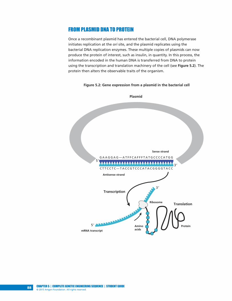
88 CHAPTER 5 | COMPLETE GENETIC ENGINEERING SEQUENCE | STUDENT GUIDE© 2015 Amgen Foundation. All rights reserved.
FROM PLASMID DNA TO PROTEIN Once a recombinant plasmid has entered the bacterial cell, DNA polymerase
initiates replication at the ori site, and the plasmid replicates using the
bacterial DNA replication enzymes. These multiple copies of plasmids can now
produce the protein of interest, such as insulin, in quantity. In this process, the
information encoded in the human DNA is transferred from DNA to protein
using the transcription and translation machinery of the cell (see Figure 5.2). The
protein then alters the observable traits of the organism.
Figure 5.2: Gene expression from a plasmid in the bacterial cell
G A A G G A G — A T F F C A F F F T A T G C C C C A T G G
C T T C C T C — T A C C G T C C C A T A C G G G G T A C C3’
3’
Sense strand
Antisense strand
mRNA transcript
Protein
Ribosome
Transcription
Translation
Plasmid
5’
5’ Aminoacids

89CHAPTER 5 | COMPLETE GENETIC ENGINEERING SEQUENCE | STUDENT GUIDE© 2015 Amgen Foundation. All rights reserved.
Genetic engineering is only possible because genes from different organisms
can be expressed in bacteria. On Earth, all life is related, and the way that
information is encoded in DNA is universal. As you may already know, proteins
are made up of smaller subunits called amino acids, and a sequence of three
nucleotides in DNA code for a single amino acid. These three-nucleotide
sequences are called codons. For example, the codon TTG codes for the amino
acid tryptophan, whereas the codon AAG codes for the amino acid lysine. In
many cases, more than one codon can encode the same amino acid. For example,
AAA is also a codon for lysine. In addition, there are informational codons, such
as the start codon (ATG) and the stop codon (TTA), which show where in the
DNA sequence the code for the protein begins and ends.
DID YOU KNOW?Making DNA from RNA
Even though the DNA code is the same in all life forms, the transcription
and translation of genes in eukaryotes and prokaryotes use different
enzymes and structures. (Human cells are eukaryotes, and bacterial cells
are prokaryotes.) One important difference between these two kinds of
cells is that the genes in eukaryotes contain noncoding sequences called
introns. The RNA polymerase transcribes the gene, producing a large
precursor messenger RNA containing both introns and exons, which are
the coding sequences. The precursor RNA is then spliced, which removes
the introns and joins the exons into the mature messenger RNA.
Prokaryotes are unable to carry out the splicing of the introns. To solve
this problem, scientists use an enzyme, reverse transcriptase, which can
copy RNA into DNA, to make complementary DNA (cDNA) from the
messenger RNA for a particular protein. The cDNA, which has only the
exon sequences, is then inserted into the plasmid vector. The cloned
human insulin gene used to make insulin is prepared in this way.

LABORATORY
90 CHAPTER 5 | COMPLETE GENETIC ENGINEERING SEQUENCE | STUDENT GUIDE© 2015 Amgen Foundation. All rights reserved.
LABORATORY 5: TRANSFORMING BACTERIA WITH THE LIGATION PRODUCTS So far in your quest to clone a gene you have produced recombinant plasmids
and verified that you made the pARA-R plasmid containing the rfp gene that can
make the red fluorescent protein. In this laboratory you will carry out another
step of the gene cloning process, which is to transform E. coli bacteria with this
plasmid. Using E. coli bacteria that have been pretreated with calcium chloride,
you will divide the bacteria into two groups: a control group to which no plasmid
is added, and a treatment group to which you add the ligation products. After
heat-shocking both groups of cells, you will grow them under several different
conditions:
• The control group is grown in the presence of Luria Broth (a medium that
supports bacterial growth).
• The control group is grown in the presence of Luria Broth and the antibiotic
ampicillin.
• The treatment group is grown in the presence of Luria Broth.
• The treatment group is grown in the presence of Luria Broth and the
antibiotic ampicillin.
• The treatment group is grown in the presence of Luria Broth, ampicillin, and
the sugar arabinose.
By examining the growth of bacteria under these conditions, you can verify that
your procedure worked, and you can identify the bacteria transformed with
the pARA-R plasmid that you created in Laboratory 3. How will you know if you
are successful? The bacteria will have a new and highly visible trait: It will now
produce red fluorescent protein, which makes the cells red or bright pink!
The pARA-R plasmid, which you reviewed in Chapter 3, is shown again in
Figure 5.3.

LABORATORY
91CHAPTER 5 | COMPLETE GENETIC ENGINEERING SEQUENCE | STUDENT GUIDE© 2015 Amgen Foundation. All rights reserved.
Figure 5.3: The pARA-R plasmid
The relevant components of this plasmid are the rfp gene, the promoter (pBAD),
the ampicillin resistance gene (ampR), and the arabinose activator protein
gene (araC). The ampR gene confers resistance to the antibiotic ampicillin.
(Biotechnologists call these genes selectable markers because only bacteria having
the gene will survive in the presence of an antibiotic.) The araC gene controls the
promoter. If arabinose, a simple sugar, is present in the bacteria, the activator
protein made by the araC gene turns on the promoter, which then binds RNA
polymerase, and transcription of the rfp gene occurs. Activator proteins are used
in some recombinant plasmids to control production of the protein of interest.
HANDOUTS• Bacterial Growth Predictions (RM 5)
BEFORE THE LAB Discuss the following questions with your group, and be prepared to share your
answers with the class.
1. Ampicillin is an antibiotic that kills bacterial cells by disrupting the formation
of cell walls. However, the pARA-R plasmid has the ampicillin resistance gene,
which produces a protein that breaks down ampicillin. What is the purpose of
growing bacteria that have been transformed in the presence of ampicillin?
2. What will happen when bacterial cells that contain the pARA-R plasmid are
not given arabinose?
pARA-R5,302 bp
ampR
ori araC
BamHI
HindIII
rfp
pBAD-rfp807 bp
pBAD

LABORATORY
92 CHAPTER 5 | COMPLETE GENETIC ENGINEERING SEQUENCE | STUDENT GUIDE© 2015 Amgen Foundation. All rights reserved.
3. In the lab, you will add samples of the control group P– and the treatment
group P+ to plates that contain various combinations of Luria Broth (LB),
ampicillin, and the sugar arabinose. The plates will be arranged as follows:
Using the key on Bacterial Growth Predictions (RM 5), show your predictions
for the growth you would expect for each combination. Then fill in Table 1
and Table 2 in the handout by describing the conclusions that can be drawn if
the predicted growth occurs or does not occur.
4. Read through the Methods section on pages 93 through 97 and briefly
outline the steps, using words and a flowchart.
SAFETY: All appropriate safety precautions and attire required for a science
laboratory should be used. Please refer to your teacher’s instructions.
SAFETY: Use caution when handling E. coli bacteria and use aseptic technique.
Aseptic technique is a set of procedures that ensure protection of the lab worker
and protection of a bacterial sample, which is necessary for the experiment to be
successful. Specifically:
• Do not touch anything that has been or will be in contact with E. coli
bacteria. Students handling equipment that comes into contact with bacteria
should wear gloves.
• Try to avoid spills or contamination of surfaces with anything that has been
in contact with E. coli bacteria. Immediately inform your teacher if a spill or
contamination occurs.
• When you have finished using microfuge tubes, pipette tips, and cell
spreaders, place them immediately into the biohazard bag or waste
container, as directed by your teacher.
• When directed to do so, place your Petri plates back into the original sleeves
and in the biohazard bag.
• Wash your hands well with soap after completing the lab.
MATERIALSReagents
• A rack with the following:
• Microfuge tube of ligated plasmid from Laboratory 3 (LIG)
• Microfuge tube of Luria Broth (LB)
P– P–
LB plate
P+
LB/amp plate
P+ P+
LB/amp/ara plate
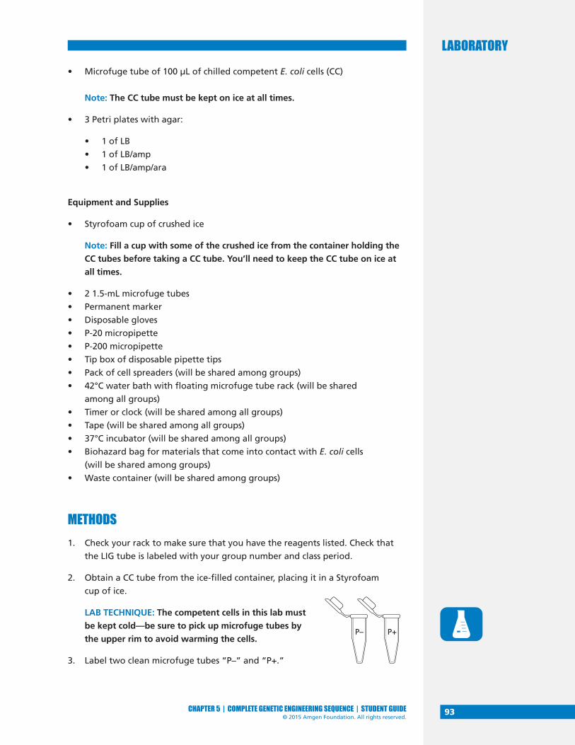
LABORATORY
93CHAPTER 5 | COMPLETE GENETIC ENGINEERING SEQUENCE | STUDENT GUIDE© 2015 Amgen Foundation. All rights reserved.
• Microfuge tube of 100 µL of chilled competent E. coli cells (CC)
Note: The CC tube must be kept on ice at all times.
• 3 Petri plates with agar:
• 1 of LB
• 1 of LB/amp
• 1 of LB/amp/ara
Equipment and Supplies
• Styrofoam cup of crushed ice
Note: Fill a cup with some of the crushed ice from the container holding the
CC tubes before taking a CC tube. You’ll need to keep the CC tube on ice at
all times.
• 2 1.5-mL microfuge tubes
• Permanent marker
• Disposable gloves
• P-20 micropipette
• P-200 micropipette
• Tip box of disposable pipette tips
• Pack of cell spreaders (will be shared among groups)
• 42°C water bath with floating microfuge tube rack (will be shared
among all groups)
• Timer or clock (will be shared among all groups)
• Tape (will be shared among all groups)
• 37°C incubator (will be shared among all groups)
• Biohazard bag for materials that come into contact with E. coli cells
(will be shared among groups)
• Waste container (will be shared among groups)
METHODS1. Check your rack to make sure that you have the reagents listed. Check that
the LIG tube is labeled with your group number and class period.
2. Obtain a CC tube from the ice-filled container, placing it in a Styrofoam
cup of ice.
LAB TECHNIQUE: The competent cells in this lab must
be kept cold—be sure to pick up microfuge tubes by
the upper rim to avoid warming the cells.
3. Label two clean microfuge tubes “P–” and “P+.”
P– P+
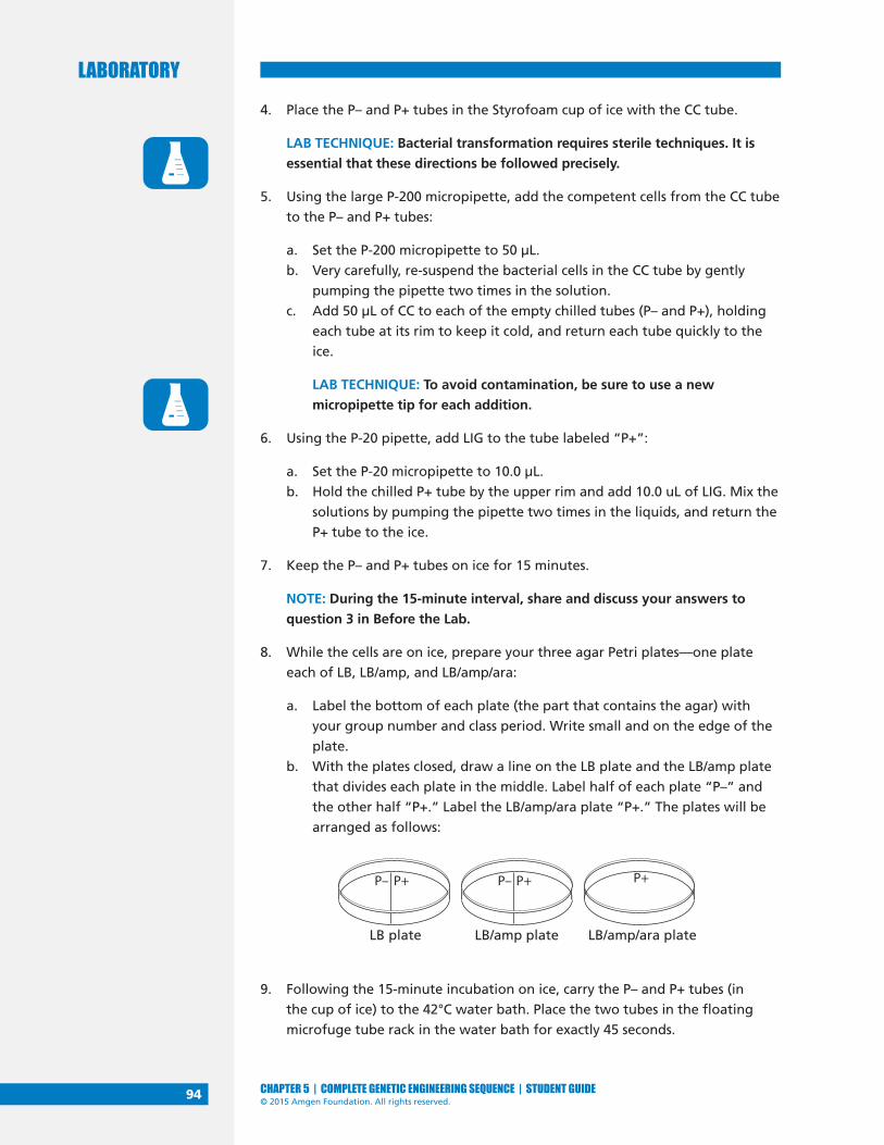
LABORATORY
94 CHAPTER 5 | COMPLETE GENETIC ENGINEERING SEQUENCE | STUDENT GUIDE© 2015 Amgen Foundation. All rights reserved.
4. Place the P– and P+ tubes in the Styrofoam cup of ice with the CC tube.
LAB TECHNIQUE: Bacterial transformation requires sterile techniques. It is
essential that these directions be followed precisely.
5. Using the large P-200 micropipette, add the competent cells from the CC tube
to the P– and P+ tubes:
a. Set the P-200 micropipette to 50 µL.
b. Very carefully, re-suspend the bacterial cells in the CC tube by gently
pumping the pipette two times in the solution.
c. Add 50 µL of CC to each of the empty chilled tubes (P– and P+), holding
each tube at its rim to keep it cold, and return each tube quickly to the
ice.
LAB TECHNIQUE: To avoid contamination, be sure to use a new
micropipette tip for each addition.
6. Using the P-20 pipette, add LIG to the tube labeled “P+”:
a. Set the P-20 micropipette to 10.0 µL.
b. Hold the chilled P+ tube by the upper rim and add 10.0 uL of LIG. Mix the
solutions by pumping the pipette two times in the liquids, and return the
P+ tube to the ice.
7. Keep the P– and P+ tubes on ice for 15 minutes.
NOTE: During the 15-minute interval, share and discuss your answers to
question 3 in Before the Lab.
8. While the cells are on ice, prepare your three agar Petri plates—one plate
each of LB, LB/amp, and LB/amp/ara:
a. Label the bottom of each plate (the part that contains the agar) with
your group number and class period. Write small and on the edge of the
plate.
b. With the plates closed, draw a line on the LB plate and the LB/amp plate
that divides each plate in the middle. Label half of each plate “P–” and
the other half “P+.” Label the LB/amp/ara plate “P+.” The plates will be
arranged as follows:
9. Following the 15-minute incubation on ice, carry the P– and P+ tubes (in
the cup of ice) to the 42°C water bath. Place the two tubes in the floating
microfuge tube rack in the water bath for exactly 45 seconds.
P– P–
LB plate
P+
LB/amp plate
P+ P+
LB/amp/ara plate

LABORATORY
95CHAPTER 5 | COMPLETE GENETIC ENGINEERING SEQUENCE | STUDENT GUIDE© 2015 Amgen Foundation. All rights reserved.
10. After the 45-second heat shock, immediately place the tubes back on ice and
leave them there for at least a minute.
11. Using the large P-200 micropipette, add LB to the P– and P+ tubes:
a. Set the P-200 micropipette to 150 µL.
b. Add 150 µL of LB to the P– tube. Cap the tube and gently flick it two or
three times to mix.
LAB TECHNIQUE: To avoid contamination, be sure to use a new
micropipette tip for each solution.
c. Add 150 µL of LB to the P+ tube. Cap the tube and gently flick it two or
three times to mix.
12. If time permits, allow the cells in the P– and P+ tubes to incubate at room
temperature for 15 minutes.
STOP AND THINK:
• How is the P+ bacteria culture treated differently from the P–
bacteria culture? (A culture is an isolated population of cells.) What
is the purpose of the P– bacteria culture?
• Why do the cells need time to recover after the heat shock?
• Why are the cells incubated at 37°C?
• You used aseptic technique in this lab. Why is this important?
13. Add cells from the P– tube onto your LB and LB/amp plates:
a. Set the P-200 micropipette to 50 µL.
LAB TECHNIQUE: To avoid contamination, be sure to use a new
micropipette tip for each solution.
b. Gently pump the pipette two or three
times in the P– tube to suspend the cells,
and load 50 uL of the P- cells.
c. Open the lid of the LB plate, like a
“clamshell,” and add 50 µL of cells
from the P– tube to the section marked
“P–.” Close the lid.
d. Again, gently pump the pipette two
or three times in the P– tube to suspend
the cells, and load 50 uL of the P– cells.
P–
LB plate
50 µL 50 µL
P+ P–
LB/amp plate
P+
P–

LABORATORY
96 CHAPTER 5 | COMPLETE GENETIC ENGINEERING SEQUENCE | STUDENT GUIDE© 2015 Amgen Foundation. All rights reserved.
e. Open the lid of the LB/amp plate, like a clamshell, and add 50 µL of cells
from the P– tube to the section marked “P–.” Close the lid.
14. Spread the cells from the P– tube on your LB and LB/amp plates:
a. Open the package of sterile cell spreaders
at the end closest to the spreader handles.
Remove only one spreader, and close the
package to keep the others sterile.
b. Open the lid to the LB plate, like a clamshell,
and spread the cells evenly across the entire
P– side of the plate by gently moving the
spreader across the agar surface. (Keep the
cells on the P– side of the plate.) Close the lid.
c. Carefully spread the P– cells on the LB/amp plate, using the same
spreader and technique.
LAB TECHNIQUE: Hold the spreader by the handle and do not allow the
bent end to touch any surface, as this will contaminate the spreader.
Place the used spreader in the biohazard bag.
15. Add cells from the P+ tube to your LB, LB/amp, and LB/amp/ara plates:
a. Make sure that the P-200 micropipette is set to 50 µL.
LAB TECHNIQUE: To avoid contamination, be sure to use a new
micropipette tip for each solution.
b. Gently pump the pipette two or three times in the P+ tube to suspend
the cells, and load 50 uL of the P+ cells.
c. Open the lid of the LB plate, like a clamshell, and add 50.0 µL of cells
from the P+ tube to the section marked “P+.” Close the lid.
d. Again, gently pump the pipette two or three times in the P+ tube to
suspend the cells, and load 50 uL of the P+ cells.
e. Open the lid of the LB/amp plate, like a clamshell, and add 50.0 µL of cells
from the P+ tube to the section marked “P+.” Close the lid.
f. Set the P-200 micropipette to 100 µL, gently pump the pipette two or
three times in the P+ tube, and load 100 µL of the P+ cells.
HandleSpreadingsurface
P–
LB plate
50 µL
P+ P–
LB/amp plate
P+
P+100 µL
LB/amp/ara plate
P+
50 µL

LABORATORY
97CHAPTER 5 | COMPLETE GENETIC ENGINEERING SEQUENCE | STUDENT GUIDE© 2015 Amgen Foundation. All rights reserved.
g. Open the lid of the LB/amp/ara plate, like a clamshell, and add 100.0 µL
of P+ cells to various areas across the surface—not just a single spot. Close
the lid.
16. Spread the cells from the P+ tube on your LB, LB/amp, and LB/amp/ara plates:
a. Open the package of sterile cell spreaders
at the end closest to the spreader handles.
Remove only one spreader, and close the
package to keep the others sterile.
b. Open the lid to the LB plate, like a clamshell,
and evenly spread the cells on the P+ side of
the plate (and only on this side) by gently
moving the spreader across the agar surface.
Close the lid.
c. Carefully spread the P+ cells on the LB/amp plate using the same spreader
and technique.
d. Carefully spread the P+ cells on the LB/amp/ara plate using the same
spreader. Then gently rotate the plate beneath the P+ spreader so that
the cells can be spread over the entire surface of this plate. Close the lid.
LAB TECHNIQUE: Hold the spreader by the handle and do not allow the
bent end to touch any surface, as this will contaminate the spreader.
Place the used spreader in the biohazard bag.
17. Allow all three plates to sit right side up for five minutes.
18. Using provided tape, tape all three plates together and label tape with your
group number and class period.
19. Place the plates in the 37°C incubator upside down to prevent condensation
from dripping onto the gels.
20. Place all microfuge tubes, pipette tips, and cell spreaders in the biohazard
bag.
21. Incubate the plates for 24–36 hours at 37°C.
22. Examine the plates and in your notebook record the amount of growth on
each half.
23. Discard the Petri plates in the biohazard bag when directed to do so.
HandleSpreadingsurface

98 CHAPTER 5 | COMPLETE GENETIC ENGINEERING SEQUENCE | STUDENT GUIDE© 2015 Amgen Foundation. All rights reserved.
CHAPTER 5 QUESTIONS1. Look at the results of your transformation. Do your actual results match your
predicted results? If not, what differences do you see, and what are some
explanations for these differences?
2. How many red colonies were present on your LB/amp/ara plate?
3. Why did the red colonies only appear on the LB/amp/ara plate and not the
LB/amp plate?
4. Recombinant plasmids are engineered so that they can replicate in the cell
independently of the chromosome replication. Why is it important to have
multiple copies of a recombinant plasmid within a cell?
5. How is the information encoded in the rfp gene expressed as a trait? Be sure
to use what you have previously learned about gene expression and the
relationship between DNA, RNA, protein, and traits.
6. Why is it possible for bacteria to make a human protein, such as insulin, or a
sea anemone protein, such as the red fluorescent protein?
7. The only bacteria that could produce the red fluorescent protein in
Laboratory 5 were bacteria that were transformed with the pARA-R plasmid.
Why?
DID YOU KNOW?Making the Connection Between Genes and Proteins
How were scientists able to show that a gene codes for a protein? In
1941, George Beadle and Edward Tatum carried out an experiment in
which they exposed bread mold to UV irradiation, a procedure known to
cause mutations (changes) in genes. Beadle and Tatum created mutant
strains of molds that had lost the ability to synthesize a necessary vitamin.
By feeding the precursors of the vitamin one at a time to the mutants,
Beadle and Tatum were able to determine that the mutants only lacked a
single enzyme catalyzing one reaction.
Beadle and Tatum then investigated whether a single gene caused the
loss of the single enzyme by genetic crosses between the mutants and a
wild-type strain. After culturing the progeny, they found that half had
the same defect as the parent mutant strain and half did not, confirming
that a single gene had been mutated. From these results, Beadle and
Tatum proposed that genes were responsible for coding the proteins of
an organism and that a change in a gene could result in the production of
a defective protein, which in turn could affect the traits of that organism.
In 1958, Beadle and Tatum received the Nobel Prize for this work.

99CHAPTER 5 | COMPLETE GENETIC ENGINEERING SEQUENCE | STUDENT GUIDE© 2015 Amgen Foundation. All rights reserved.
CHAPTER 5 GLOSSARYAmino acid: The building block of proteins. There are 20 amino acids. Each
amino acid is an organic substance that has two groups attached to it—an amino
group (NH2) and a carboxylic acid group (COOH).
Aseptic technique: A set of procedures and carefully controlled conditions to
prevent contamination by pathogens.
Bacterial conjugation: A process by which two bacterial cells join and transfer
genetic material from one to another.
Codon: A group of three mRNA bases that encode a single amino acid.
Competent: A cell that has the ability to be transformed genetically by taking up
DNA from the environment.
Culture: An isolated population of cells that have been grown in a specially
prepared nutrient medium.
Eukaryote: An organism that shelters its genes inside a nucleus and has several
linear chromosomes.
Exon: The segment of a gene that codes for a protein. Exons are both
transcribed and translated.
Expressed: When information encoded in a gene has been converted first into
messenger RNA and then to a protein. This process is called expression.
Intron: The segment of a gene that does not code for a protein. Introns are
transcribed into mRNA but are removed before the exons (the rest of the gene)
are translated into a protein.
Medium: A solution that contains substances that support the growth of
microorganisms. The medium may be solidified by the addition of agar. Luria
Broth is an example of a medium.
Mutation: Change or damage occurring in a section of DNA that alters the
products or processes associated with that section.
Prokaryote: Cell or organism with a single chromosome and no nuclear
membrane; bacteria are prokaryotes.
Promoter: A specific DNA sequence that binds RNA polymerase and initiates
transcription of the gene.
Red fluorescent protein: The protein encoded by the rfp gene. Mutant
fluorescent protein is a molecule that is about 238 amino acids in size. When it is
expressed in bacterial cells, the cells appear red or bright pink.
Reverse transcriptase: An enzyme that catalyzes the formation of DNA from an
RNA template in reverse transcription.

100 CHAPTER 5 | COMPLETE GENETIC ENGINEERING SEQUENCE | STUDENT GUIDE© 2015 Amgen Foundation. All rights reserved.
Selectable marker: A gene on a plasmid that is introduced into a cell along with
a gene of interest that is being cloned. Selectable markers allow scientists to tell
if the plasmid has been taken in by the cell because the marker can be seen or
detected. A common selectable marker is an antibiotic resistance gene—only
bacteria that have the gene will survive the antibiotic.
Splice: To modify messenger RNA for translation by removing introns and joining
exons.
Start codon: The first codon of mRNA translated by a ribosome; typically AUG or
GUG.
Stop codon: A nucleotide triplet within mRNA that signals a termination of
translation.
Trait: A genetically determined characteristic. DNA codes for proteins, which
determine traits.
Transformation: A process that places foreign DNA, such as a plasmid, into a cell.
Translation: The process by which information encoded in messenger RNA is
decoded and transformed into protein.

101CHAPTER 6 | COMPLETE GENETIC ENGINEERING SEQUENCE | STUDENT GUIDE© 2015 Amgen Foundation. All rights reserved.
CHAPTER 6GETTING WHAT WE NEED


103CHAPTER 6 | COMPLETE GENETIC ENGINEERING SEQUENCE | STUDENT GUIDE© 2015 Amgen Foundation. All rights reserved.
INTRODUCTIONGenetic engineering is used to produce therapeutic proteins. To provide a
treatment for diabetes, for example, a recombinant plasmid is engineered to
contain a cloned human insulin gene. Bacteria take up the recombinant plasmid
and express the gene, producing insulin. To date, you have carried out all or
some of these steps using the cloned rfp gene.
The final step in the process is to obtain the protein. To do this, bacteria are
treated with a reagent that lyses them (breaks open their cell walls), and
the protein is separated from the cell contents by a method called column
chromatography. (Chromatography is a method for separating similar substances
by dissolving them and then flowing the solution over a material that binds the
substances to different degrees. Column chromatography uses a column packed
with beads coated with the binding material.)
In this chapter, you will complete this final step. You will lyse the bacteria you
transformed in Chapter 5 and use a column that separates proteins based on
their solubility in water to obtain the red fluorescent protein made by the cloned
rfp gene.
CHAPTER 6 GOALS By the end of this chapter, you will be able to do the following:
• Describe the conditions that are favorable to bacterial growth
• Explain how a protein’s conformation (three-dimensional shape) is related
to its function
• Explain how protein folding occurs
• Explain how column chromatography separates proteins
WHAT DO YOU ALREADY KNOW?Discuss the following questions with your partner, and write your ideas in your
notebook. Be prepared to discuss your responses with the class. Don’t worry if
you don’t know all the answers. Discussing these questions will help you think
about what you already know about bacterial growth and proteins.
1. How do bacteria reproduce?
2. Why are proteins sometimes called workhorse molecules?
3. How might the conformation (shape or folding) of a protein be important
for its function? Focus on one of the following protein functions: acting as
an enzyme (speeding up reaction rates), transporting molecules, signaling, or
forming structures.

104 CHAPTER 6 | COMPLETE GENETIC ENGINEERING SEQUENCE | STUDENT GUIDE© 2015 Amgen Foundation. All rights reserved.
4. A protein is a long linear molecule when it is made, but it immediately folds
into a specific three-dimensional conformation. What properties of the
amino acids in a protein control the folding process?
BACTERIA MULTIPLICATION AND PROTEIN PURIFICATION
Some bacteria can make you very sick. That’s why in both the health care
industry and food preparation, people try to prevent bacteria from growing. But
when scientists use bacteria as hosts for recombinant plasmids, their goal is to
encourage bacteria to grow and to produce the protein of interest.
BACTERIAL GROWTH What factors affect bacterial growth? The last step in Chapter 5 was to place the
bacteria that were transformed with the pARA-R plasmid in a suspension culture
in a shaker flask. The cells were suspended in a nutrient broth, and the shaking
mixed air into the suspension and prevented the bacteria from settling out of
solution, providing conditions favorable to growth.
CONSIDER: Why might the shaker flask be better at supporting bacterial cell
growth than a plate?
Under optimal conditions, such as those provided by the shaker flask, the growth
of a bacterial population is predictable (see Figure 6.1). The growth occurs in
four distinct phases:
• In the lag phase, there is a zero growth rate. There are no new cells and no
cells dying. The cells adjust to the new conditions, grow larger, and prepare
for cell division.
• In the log phase, there is a logarithmic growth rate. The number of new
cells is greater than the number of cells dying. The cells undergo asexual cell
division and double in number about every 20 minutes (which is the average
doubling time for E. coli; other bacteria have different doubling times). This
phase occurs as long as necessary resources such as food and oxygen are
unlimited and there are no unusual factors that cause cell death.
• In the stationary phase, there is a zero growth rate. The number of new cells
equals the number of cells dying. This phase occurs once resources such as
food and oxygen become limited.
• In the death phase, there is a negative growth rate. The number of new cells
is less than the number of cells dying. This phase occurs when resources are
depleted and when toxic waste products build up.

105CHAPTER 6 | COMPLETE GENETIC ENGINEERING SEQUENCE | STUDENT GUIDE© 2015 Amgen Foundation. All rights reserved.
Figure 6.1: Change in bacterial growth over time in the shaker flask
CONSIDER: If the gene of interest is controlled by an operon, such as the
arabinose operon, when is the best time to turn on the gene? Keep in mind:
• Production of the protein takes energy away from the processes of cell
growth and cell division
• A greater number of cells will produce more protein
• Proteins can degrade over time
PROTEIN PURIFICATIONWhen the transformed bacteria are allowed to grow and multiply numerous
times, they can produce enough therapeutic protein to meet the treatment
needs of patients. However, the therapeutic protein must be purified, which
requires separating it from the other contents of the cell, including other
proteins. (A typical bacterium may contain more than 1,000 different kinds of
protein.) Column chromatography is one method used to separate the proteins.
What physical characteristics of proteins enable them to be separated by column
chromatography? Although all proteins are made up of amino acids, each
kind of protein has a specific function and a specific conformation (shape). The
conformation relates to function because the outside surface of a protein has
specific sites that bind to other molecules. These binding sites allow proteins
to attach to other molecules, which is how proteins can catalyze reactions,
transport molecules, provide a signal, and form structures.
Log
of
nu
mb
ers
of
bac
teri
a
Time (hr)0 5 10
Lagphase
Log, orexponentialgrowth,phase
Stationaryphase
Death, orlogarithmicdecline, phase

106 CHAPTER 6 | COMPLETE GENETIC ENGINEERING SEQUENCE | STUDENT GUIDE© 2015 Amgen Foundation. All rights reserved.
When a protein is first synthesized, it is a long flexible chain of amino acids, but
it immediately attains its three-dimensional conformation by a process called
protein folding (see Figure 6.2). Protein folding is dependent on the following
properties of the amino acids in the protein:
• Formation of weak noncovalent bonds between positively charged and
negatively charged side chains of amino acids
• The tendency for hydrophobic (water-insoluble) amino acids to be buried
on the inside of the protein away from water and for hydrophilic (water-
soluble) amino acids to be found on the outside of the protein exposed to
water
• Formation of covalent bonds, called disulfide bridges, that occur between
sulfur-containing amino acids
Figure 6.2: Protein folding
CONSIDER: If a mutation changes an amino acid, how might this change affect
protein folding and protein function?
Specific proteins are either hydrophobic or hydrophilic,
depending on the relative amount of hydrophobic and
hydrophilic amino acids they contain. Hydrophobic
proteins and hydrophilic proteins can be separated by
column chromatography. In this method, a column is
packed with small beads that are coated with a material
(a resin) that attracts hydrophobic amino acids, and the
mixture of proteins is dissolved and passed over the
beads. In order for the hydrophobic amino acids to stick
to the resin, the proteins must be unfolded to expose the
hydrophobic amino acids, which tend to be found on the
inside of the protein. Certain salt solutions called buffers
will unfold proteins to varying degrees.
Unfolded Folded
Resin-coatedbeads
Column

107CHAPTER 6 | COMPLETE GENETIC ENGINEERING SEQUENCE | STUDENT GUIDE© 2015 Amgen Foundation. All rights reserved.
A series of buffers of different concentrations of salt can be used to separate
many proteins from each other. Figure 6.3 shows how three buffers are used to
separate the highly hydrophobic protein red fluorescent protein in a column. A
binding buffer is used to unfold all the proteins so that hydrophobic proteins
stick to the resin and hydrophilic proteins pass through the column. A wash
buffer is poured into the column to release moderately hydrophobic proteins
from the resin, and then an elution buffer is poured into the column to release
red fluorescent protein from the resin. Both the wash and the elution buffers
have a lower salt concentration than the binding buffer and thus cause bound
proteins to fold and begin to cover their hydrophobic amino acids. Depending
on the extent of folding, the proteins are released from the beads.
Figure 6.3: Separation of red fluorescent protein by
hydrophobicity using column chromatography
CONSIDER: If you were trying to use column chromatography to separate insulin
from a mixture of proteins, would you use the same binding, wash, and elution
buffers used for the red fluorescent protein, or would you use buffers with
different salt concentrations? Explain the reasoning for your answer.
Column
Red fluorescent protein
Elution buffer
Stop cock
Column
Hydrophilicproteins
Column
Moderatelyhydrophobicproteins
Binding buffer is passed through
the column
Wash buffer is passed through
the column
Elution buffer is passed through
the column
Binding buffer Wash buffer

108 CHAPTER 6 | COMPLETE GENETIC ENGINEERING SEQUENCE | STUDENT GUIDE© 2015 Amgen Foundation. All rights reserved.
DID YOU KNOW?Recombinant Proteins
As you have learned, when recombinant proteins are produced for use
as human therapeutics, host cells must be grown in large quantities
so that enough recombinant protein is produced to meet treatment
demand (the needs of patients). The recombinant protein is isolated,
purified, and analyzed for activity and quality before it goes to market.
Producing a protein with the proper order of amino acids isn’t always
the whole story, however. Sometimes further processing or modification
is required to yield an active or fully functioning protein. Many
human proteins are glycosylated, meaning that they have a particular
pattern of sugar molecules linked to them. If a protein is translated
but not correctly glycosylated, it may not function properly. Another
modification involves the addition of a phosphate group—a process
known as phosphorylation. Phosphorylation of a protein can act as
a kind of switch, allowing the protein to become more or less active
by uncovering or covering its binding sites. In addition to saccharide
(sugar) and phosphate groups, other chemical groups may be added to a
protein in order to change its function.
Recombinant proteins for therapeutic use include vaccines, hormones,
monoclonal antibodies, and hematopoietic growth factors for the
treatment of diseases including cancer, AIDS, allergies, and asthma. The
number of recombinant proteins has increased greatly in recent years as
the technology used for their production and purification has advanced.

LABORATORY
109CHAPTER 6 | COMPLETE GENETIC ENGINEERING SEQUENCE | STUDENT GUIDE© 2015 Amgen Foundation. All rights reserved.
LABORATORY 6: PURIFYING THE FLUORESCENT PROTEIN In the previous chapter you transformed bacteria and then selected the bacteria
that had the plasmid of interest by placing the cells on a plate that contained LB,
ampicillin, and arabinose. One colony was then selected and grown in a shaker
flask to provide a large population of identical cells that all contain one or more
copies of the recombinant plasmid. Near the end of the log phase of bacterial
growth, the cells were given arabinose to turn on the rfp gene so that it would
make red fluorescent protein. In the first part of this laboratory, you will use a
reagent called lysis buffer to lyse, or break open, the cells. In the second part of
this laboratory, you will use column chromatography to purify the protein.
BEFORE THE LAB Discuss the following questions with your group and be prepared to share your
answers with the class.
1. How can solutions of different salt concentrations, which will unfold proteins
to varying degrees, be used to help purify red fluorescent protein using
column chromatography?
2. Read through the Methods sections for Part A (on pages 110 and 111) and for
Part B (on pages 112 through 114) and briefly outline the steps, using words
and a flowchart.
SAFETY: All appropriate safety precautions and attire required for a science
laboratory should be used. Please refer to your teacher’s instructions.
SAFETY: Use caution when handling E. coli bacteria and use aseptic technique.
Aseptic technique is a set of procedures that ensure protection of the lab worker
and protection of the bacterial sample, which is necessary for the experiment to
be successful. Specifically:
• Do not touch anything that has been or will be in contact with E. coli
bacteria. Hold microfuge tubes and Petri plates on the outside, hold only the
handles of cell spreaders, and do not handle pipette tips at all.
• Try to avoid spills or contamination of surfaces with anything that has been
in contact with E. coli bacteria. Immediately inform your teacher if a spill or
contamination occurs.

LABORATORY
110 CHAPTER 6 | COMPLETE GENETIC ENGINEERING SEQUENCE | STUDENT GUIDE© 2015 Amgen Foundation. All rights reserved.
• When you have finished using microfuge tubes, pipette tips, and cell
spreaders, place them immediately into the biohazard bag.
• When directed to do so, place your Petri plates in the biohazard bag.
• Wash your hands well with soap after completing the lab.
PART A: LYSE CELLS GROWN IN THE SHAKER MATERIALS Reagents
• Microfuge tube rack with the following:
• Microfuge tube of the LB/amp/ara culture of E. coli (EC)
• Microfuge tube of elution buffer (EB)
• Microfuge tube of lysis buffer (LyB)
• Additional 1,000 µL (1 mL) of the LB/amp/ara culture of E. coli (obtain from
your teacher in step 6)
Equipment and Supplies
• Microcentrifuge (will be shared among all groups)
• Liquid waste container
• P-200 micropipette
• Tip box of disposable pipette tips
• Permanent marker
• Waste container (will be shared among groups)
• Biohazard bag for materials that come into contact with E. coli cells (will be
shared among groups)
METHODS1. Check your rack to make sure that you have the reagents listed for Part A.
2. Examine the EC tube and record its color in your notebook.
3. Before you can lyse the cells, you will need to separate the cells from the
growth medium. To do this, spin the EC tube in the microcentrifuge for five
minutes.
LAB TECHNIQUE: Distribute the tubes evenly in the microcentrifuge so that
their weight is balanced, making sure that two tubes of the same volume are
directly opposite one another.

LABORATORY
111CHAPTER 6 | COMPLETE GENETIC ENGINEERING SEQUENCE | STUDENT GUIDE© 2015 Amgen Foundation. All rights reserved.
STOP AND THINK: How can you determine where the red fluorescent
protein is in each separation step?
4. Carefully remove the EC tube from the microcentrifuge to avoid disturbing
the solid pellet that contains the bacterial cells.
5. Set the P-200 micropipette to 200 µL, place a new tip on the micropipette,
and carefully remove the supernatant (liquid) from the EC tube without
disturbing the cell pellet. (You can dispense the supernatant into the liquid
waste container.)
6. Bring the EC tube to your teacher, who will add 1,000 µL (1 mL) of the LB/
amp/ara culture of E. coli to your EC tube.
7. Repeat steps 3–5 (spin the tube for 5 minutes and remove the liquid).
STOP AND THINK: What color is the supernatant? The pellet? What are
the contents of each?
8. Invert the EC tube containing the cell pellet, and use the micropipette to
remove as much of the liquid as you can without touching the cell pellet.
LAB TECHNIQUE: Be sure to use a new micropipette tip for each reagent to
avoid contamination.
9. Using the P-200 pipette, add 150 µL of EB to the cell pellet in the EC tube.
10. Close the cap of the EC tube tightly and drag the tube vigorously across
the surface of the microfuge tube rack to resuspend the cells. Examine the
EC tube carefully. If there are visible clumps of cells, again drag the tube
vigorously across the surface of the microfuge tube rack.
11. Using the P-200 pipette, add 150 µL of LyB to the EC tube. Close the cap of
the EC tube tightly and drag the tube vigorously across the surface of the
microfuge tube rack two times to mix.
12. Label the EC tube with your group number and class period and give it
to your teacher. Your teacher will incubate the cells at room temperature
overnight.
13. Place all microfuge tubes and pipette tips in the biohazard bag.
LABORATORY
112 CHAPTER 6 | COMPLETE GENETIC ENGINEERING SEQUENCE | STUDENT GUIDE© 2015 Amgen Foundation. All rights reserved.
PART B: SEPARATE THE RED FLUORESCENT PROTEIN WITH COLUMN CHROMATOGRAPHYMATERIALS Reagents
• Microfuge tube rack with the microfuge tube of lysed cells (EC, from
Part A of this lab)
• Containers with the following:
• Binding buffer (BB)
• Wash buffer (WB)
• Elution buffer (EB)
• Column equilibration buffer (CEB)
Equipment and Supplies
• 2 1.5-mL microfuge tubes
• Liquid waste container
• P-1,000 micropipette
• Tip box of disposable pipette tips
• Chromatography column
• Microcentrifuge (will be shared among all groups)
• Waste container (will be shared among groups)
METHODS1. Assign tasks in your group. Have one person make sure that your materials
are ready (steps 2–3), another person set up the chromatography column
(steps 4–5), and another person spin the lysed cells and remove the
supernatant (step 6–7).
2. Check to make sure that you have all the reagents listed.
3. Label two clean microfuge tubes “SUPER” and “RFP.”
4. Set up your chromatography column as directed by your teacher, being careful
not to dislodge the stopcock attached to the lower portion of the tube.
LAB TECHNIQUE: Do not allow the column to run dry.
5. Prepare the column:
a. Set the liquid waste collection container under the stopcock.
b. Carefully open the column by turning the stopcock valve, and allow the
liquid to drain into the waste collection container.
LABORATORY
113CHAPTER 6 | COMPLETE GENETIC ENGINEERING SEQUENCE | STUDENT GUIDE© 2015 Amgen Foundation. All rights reserved.
c. Close the valve once there is about 1–2 mm of liquid left above the
resin bed.
d. Make sure that the liquid is not draining from the column into the waste
container.
6. Spin the EC tube in the microcentrifuge for five minutes to create a pellet of
the cell debris.
LAB TECHNIQUE: Distribute the tubes evenly in the microcentrifuge so that
their weight is balanced.
STOP AND THINK: Three buffers you will use in this lab are the binding
buffer (BB), the wash buffer (WB), and the elution buffer (EB). What is
the function of each?
7. Examine the tube. You should see a supernatant and a solid pellet.
STOP AND THINK: What color is the supernatant? The pellet? What are
the contents of each?
LAB TECHNIQUE: Be sure to use a new micropipette tip for each reagent to
avoid contamination.
8. Using the P-1,000 pipette, carefully remove 200 µL of EC supernatant without
disturbing the cell debris pellet, and dispense the supernatant into the SUPER
tube. If you dislodge the debris pellet, you will have to centrifuge the tube
again.
9. Using a new tip, add 200 µL of BB to the SUPER tube. Mix by gently pumping
the solution in and out.
10. Using the same tip but changing the volume, add 400 µL of the SUPER tube
mixture to the chromatography column. Carefully dispense the solution down
the side of the column to avoid disturbing the surface of the resin bed.
11. Open the valve and allow the solution in the column to drain into the waste
collection container. Close the valve once there is about 1–2 mm of liquid left
above the resin bed.
12. Examine the column and locate the red fluorescent protein. Is it spread
throughout the resin bed, or does it appear to be restricted to a single band?
Record your observations in your notebook.
LABORATORY
114 CHAPTER 6 | COMPLETE GENETIC ENGINEERING SEQUENCE | STUDENT GUIDE© 2015 Amgen Foundation. All rights reserved.
13. Using a new tip, add 1,000 µL (1 mL) of WB gently down the side of the
chromatography column.
14. Open the valve and allow the solution in the column to drain into the waste
collection container. Close the valve once there is about 1–2 mm of liquid left
above the resin bed.
15. Examine the column and locate the red fluorescent protein. Has the location
of the red fluorescent protein changed in the resin bed?
16. Using a new tip, add 1,000 µL of EB twice (2 mL total), gently, down the side
of the chromatography column.
17. Set the RFP tube under the stopcock. Open the valve and allow the part of
the eluate that is red to drain into the RFP tube. Close the valve and cap the
tube when done.
18. Set the waste collection container back under the stopcock. Open the valve
and allow the rest of the eluate to drain into the waste container. Close the
valve once there is about 1–2 mm of liquid left above the resin bed.
19. Using a new tip, add 1,000 µL of CEB twice (2 mL total) to the chromatography
column to prepare it for the next class. Cap the column tightly.
20. Pour the contents of the waste collection container down the sink drain.
21. Compare your RFP tube with RFP tubes from other groups. Is there a
difference in intensity of color from sample to sample? Record your
observations in your notebook.

115CHAPTER 6 | COMPLETE GENETIC ENGINEERING SEQUENCE | STUDENT GUIDE© 2015 Amgen Foundation. All rights reserved.
CHAPTER 6 QUESTIONS1. Why is a protein’s conformation important for carrying out its function?
2. What properties of the amino acids in a protein relate to protein folding?
3. Does the eluate containing your red fluorescent protein appear less bright
or brighter than it did in the cell lysate following centrifugation? If there is a
noticeable difference in the intensity of the red color, what might account for
that?
4. What characteristic of red fluorescent protein is used as the basis for
separation by column chromatography?
5. How might the column chromatography procedure be adjusted or modified
to increase the purity of the red fluorescent protein sample?
DID YOU KNOW?Chimeric Proteins
When you determined which bacteria had taken up the recombinant
plasmid that had the rfp gene, you were able to see the transformed
colony because the bacteria fluoresced red when exposed to light. The
ability of a fluorescent protein (FP) to “light up” is a powerful tool
that is revolutionizing cell biology and biomedical research. Numerous
FPs from different organisms have been isolated and even mutated
to provide a toolbox of FP “tags.” The FPs can be attached to other
molecules to monitor processes that occur inside cells and in the body as
a whole.
The function of a protein is directly related to its conformation, which
is the result of protein folding. Other important pieces of information
that shed light on a protein’s role are its distribution, movement,
interactions, and association with other proteins. To visualize these
aspects of a protein, scientists create a molecule, known as a fusion
protein or a chimeric protein, that contains both the protein of interest
and an FP fused together. (A chimera is a mythical animal that has parts
from different animals.) Scientists can measure fluorescence from a
single FP, making fusion proteins powerful visualization tools. However,
this procedure only works if the FP does not interfere with the protein’s
function, so tests are run to ensure that a particular protein acts the
same with and without the FP tag.
116 CHAPTER 6 | COMPLETE GENETIC ENGINEERING SEQUENCE | STUDENT GUIDE© 2015 Amgen Foundation. All rights reserved.
CHAPTER 6 GLOSSARYAmino acid: The building block of proteins. There are 20 amino acids. Each amino
acid is an organic substance that has two groups attached to it—an amino group
(NH2) and a carboxylic acid group (COOH).
Binding site: The area of a biomolecule that binds to a specific substance or part
of a substance.
Buffer: A solution that resists changes in pH, that contains either a weak acid and
its salt or a weak base and its salt.
Colony: A group of the same kind of organisms living closely together, usually
for mutual benefit. Within a bacterial colony, all organisms descend from a single
ancestor and are genetically identical (except for mutations and contamination).
Column chromatography: A method of separating substances in which the
substances are dissolved in a liquid that is allowed to flow through a glass column
filled with small beads. The beads are coated with a material that binds the
substances to different degrees.
Conformation: The three-dimensional shape or structure of something.
Death phase: The period of bacterial growth in culture when the bacteria run out
of nutrients and die.
Eluate: The solution that washes out (e.g., solutions that drip from a
chromatography column).
Elution: The process of extracting a substance that is bound to another by
washing it with a solution.
Fluorescence: The production of light by a molecule (e.g., red fluorescent protein
will release red light when exposed to ultraviolet light).
Hematopoietic growth factor: A group of proteins that promotes the growth,
differentiation, and activity of blood cells.
Hormones: Substances that act as chemical messengers in the body.
Hydrophilic: Water loving; dissolves in water; polar. Some examples are sugar
and salt.
Hydrophobic: Water fearing; does not dissolve in water; non-polar. Some
examples are oil, wax, and red fluorescent protein.
Lag phase: The period of bacterial growth in culture when the bacteria adapt
themselves to growth conditions. Individual bacteria are maturing and are not yet
able to divide.

117CHAPTER 6 | COMPLETE GENETIC ENGINEERING SEQUENCE | STUDENT GUIDE© 2015 Amgen Foundation. All rights reserved.
Log phase: The period of bacterial growth in culture when the number of
bacterial cells doubles in a fixed period of time (also known as the logarithmic or
exponential phase).
Lyse: To break open.
Medium: A solution that contains substances that support the growth of
microorganisms. The medium may be solidified by the addition of agar. Luria
Broth is an example of a medium.
Monoclonal antibody: A type of protein that binds to substances in the body and
is made by clones of a cell formed in the laboratory.
Protein folding: The physical process by which a polypeptide folds into its
characteristic three-dimensional structure, which is essential to the protein’s
function.
Resin: The material used in a chromatography column to coat the beads.
Stationary phase: The period of bacterial growth in culture when the population
stays the same because rates of cell division and cell death are equal. This is often
due to a growth-limiting factor, such as the depletion of an essential nutrient.
Supernatant: The clear liquid found on top of a solid precipitate after a mixture
has been centrifuged.
Suspension culture: A method of growing cells in a liquid growth medium that is
moved around by shaking or stirring. The movement mixes air into the suspension
and prevents the bacteria from settling out of the solution.
Vaccine: A mixture that causes a body’s immune system to make antibodies that
will bind to a bacteria or a virus to fight a disease. The mixture contains killed or
weakened bacteria or viruses.


