american college of radiology imaging network acrin …
TRANSCRIPT

AMERICAN COLLEGE OF RADIOLOGY IMAGING NETWORK
ACRIN 6664 NATIONAL CT COLONOGRAPHY TRIAL
Study Chair Gastroenterologists C. Daniel Johnson, M.D. J. Paul Limburg M.D. Dept. of Radiology Mayo Clinic Mayo Clinic Richard J. Farrell, M.D. 200 First St. SW Beth Israel Hospital Rochester, MN 55905 Kenneth McQuaid, M.D. Phone # (507) 284-8550 Univ. Calif. San Francisco Fax # (507) 266-4609 David Rubin, M.D. [email protected] Univ. Chicago Hospital Study Statistician Co-Investigator Mei-Hsiu Chen, Ph.D. Alicia Y. Toledano, Sc.D. Center for Statistical Sciences Biostatistics Consulting, LLC. Brown University, Box G-H Providence, RI 02912 Pathologist Phone # (401) 863-2578 Lawrence Burgart, M.D. Fax # (401) 863-9182 Hospital Pathology Associates, P.A. [email protected] Epidemiologist Radiologists Ilana Gareen, Ph.D. Giovanna Casola, MD Center for Statistical Sciences UCSD Medical Center Brown University, Box G-H Jugesh Cheema, MD Providence, RI 02912 Yale University Phone # (401) 863-1758 Kevin Coakley, MD Fax # (401) 863-9182 Clinical Radiologists, S.C [email protected] Abraham Dachman, M.D. Univ. Chicago Hospital Jeff Fidler, M.D. Cost Effectiveness Mayo Clinic David Vanness, Ph.D. Robert A. Halvorsen, MD Univ. Wis. Med. School Medical College of Virginia Hosp. Amy Hara, M.D Mayo Clinic Karen Horton, MD Johns Hopkins University
Jay Heiken, M.D. Mallinckrodt Institute of Radiology
Revathy Iyer, MD MD Anderson Cancer Center
Mark Kuo, MD Scottsdale Medical Imaging, LTD. Christine Menias, MD Mallinckrodt Institute of Radiology
Martina Morrin, FFRRCSI Richard Obregon, M.D.
Radiology Imaging Associates Ronald Summers, M.D., Ph.D. National Institutes of Health Judy Yee, M.D. San Francisco V.A. Medical Center Michael Zalis, M.D. Mass. General Hospital
Peter Zimmerman, MD UCLA School of Medicine
Original Protocol Date: September 28, 2004
Version Date: July 7, 2006 Including Amendments 1–2
Activation Date: February 1, 2005 Administrative Update: August 4, 2010
This protocol was designed and developed by the American College of Radiology Imaging Network (ACRIN). It is intended to be used only in conjunction with institution-specific IRB approval for study entry. No other use or reproduction is authorized by ACRIN nor does ACRIN assume any responsibility for unauthorized use of this protocol.
ACRIN 6664 2 July 7, 2006
INDEX SCHEMA 4
1.0 ABSTRACT 6
2.0 BACKGROUND AND SIGNIFICANCE 6
3.0 SPECIFIC AIMS 16
4.0 STUDY OVERVIEW 17
5.0 PARTICIPANT SELECTION 18
6.0 SITE SELECTION 20
7.0 ONLINE REGISTRATION SYSTEM 22
8.0 DATA COLLECTION AND MANAGEMENT 23
9.0 DATA COLLECTION FORMS 26
10.0 INSTITUTIONAL AUDITS 31
11.0 IMAGE SUBMISSION 35
12.0 IMAGING METHODOLOGY 36
13.0 REFERENCE STANDARD 38
14.0 COST-EFFECTIVENESS MODELING 39
15.0 STATISTICAL CONSIDERATIONS 45
16.0 GENDER/MINORITY RECRUITMENT 54
17.0 ADVERSE EVENT REPORTING 55
REFERENCES 62
APPENDIX I SAMPLE CONSENT 69
APPENDIX II ELIGIBILITY CHECKLIST 75
APPENDIX III PARTICIPATING INSTITUTIONS AND SITE PIS 78
APPENDIX IV PROTOCOL-SPECIFIC APPLICATION INFORMATION 79
APPENDIX V EVALUATION OF LARGE LESIONS 80
ACRIN 6664 3 July 7, 2006
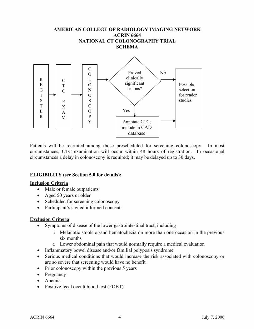
AMERICAN COLLEGE OF RADIOLOGY IMAGING NETWORK ACRIN 6664
NATIONAL CT COLONOGRAPHY TRIAL SCHEMA
C O L O N O S C O P Y
Proved clinically significant lesions?
Annotate CTC; include in CAD
database
Possible selection for reader studies
No
Yes
C T C
E X A M
R E G I S T E R
Patients will be recruited among those prescheduled for screening colonoscopy. In most circumstances, CTC examination will occur within 48 hours of registration. In occasional circumstances a delay in colonoscopy is required; it may be delayed up to 30 days. ELIGIBILITY (see Section 5.0 for details):
Inclusion Criteria • Male or female outpatients • Aged 50 years or older • Scheduled for screening colonoscopy • Participant’s signed informed consent.
Exclusion Criteria
• Symptoms of disease of the lower gastrointestinal tract, including o Melanotic stools or/and hematochezia on more than one occasion in the previous
six months o Lower abdominal pain that would normally require a medical evaluation
• Inflammatory bowel disease and/or familial polyposis syndrome • Serious medical conditions that would increase the risk associated with colonoscopy or
are so severe that screening would have no benefit • Prior colonoscopy within the previous 5 years • Pregnancy • Anemia • Positive fecal occult blood test (FOBT)
ACRIN 6664 4 July 7, 2006

Required Sample Size: 15 institutions. Based on recommendations by the ACRIN Biostatistics Center and the ACRIN DSMC and in accordance with the trial's accrual monitoring plan, the accrual strategy has been modified such that each institution will accrue patients until either 1) the overall trial accrual reaches 2607 participants or 2) December 31, 2006, whichever occurs first. The total number of participants accrued at each institution will vary according to local institutional accrual rates and thus cannot be predetermined.
ACRIN 6664 5 July 7, 2006
1.0 ABSTRACT
Computerized tomographic colonography (CTC), a revolutionary new tool, employs virtual reality technology to produce two- and three-dimensional images that permit a thorough and minimally invasive evaluation of the entire colorectal structure. This nascent imaging tool holds promise in screening colorectal neoplasia because its sensitivity, specificity, safety, cost-effectiveness, and patient acceptability, theoretically, may approach the ideal. Given the societal importance of colorectal cancer control and the limitations of currently used screening approaches, there exists a strong rationale to aggressively investigate CTC for a potential screening application. Extensive preliminary work on this technology has been performed and published. The objective is to clinically validate CTC for detecting colorectal neoplasia in a multicenter trial. Although similar trials are ongoing in a single center, validation of the technique at several centers by multiple radiologists is key to widespread national implementation. This protocol addresses issues of central importance to the clinical application of CTC, in inter-related parts that will be conducted in parallel. Data generated should provide for a balanced appraisal of the value and practicality of this potentially powerful new screening tool.
Our overall hypothesis is that CTC can be performed in a multi-institutional setting at a level of performance comparable to other full structural colorectal screening tests. Compelling advantages of this nascent technique include minimal invasiveness, visualization of the entire colorectum from an endoluminal perspective, multi-dimensional inspection of the colon wall and extracolonic tissues without superimposed anatomic structures, and improved patient acceptance. Our objective is to clinically validate widespread use of CTC in a screening population for the detection of colorectal neoplasia. In Part I, the clinical performance of the CTC examination will be evaluated prospectively, using colonoscopy as the reference standard. In Part II, additional information that is obtained as part of the CTC will be analyzed. In Part III, image archives for further research and cost-effectiveness implications of observed performance outcomes will be addressed.
2.0 BACKGROUND AND SIGNIFICANCE 2.1 Public Health Concerns
Colorectal cancer (CRC) exacts significant morbidity and mortality, especially in industrialized nations. It is the third most common cancer and second leading cause of malignant death in the United States with an estimated 134,000 new CRC cases and 55,000 CRC deaths in 1996.1 The average lifetime incidence of CRC is 6% and is even higher in persons with a family history of colorectal neoplasia or with other well-established CRC risk factors.2 As the natural history of CRC permits the recognition and curative treatment of both precursor adenomas and localized cancers, there is an enormous opportunity to save lives with early detection programs broadly applied to a general population. Indeed, evidence now exists from prospective trials, 2-4 case-control studies, 5-10 and predictive models11-13 to support a benefit by various screening interventions in reducing CRC-specific mortality. However, the potential efficacy and
ACRIN 6664 6 July 7, 2006
practicality of such a screening effort are compromised by limitations in the performance, comfort, and expense of available screening tests. Better tools are needed to more effectively screen for colorectal neoplasia.
2.2 Currently Available Screening Tests Each screening tool in the current armamentarium has limitations that interfere with optimal outcomes. Fecal occult blood testing is noninvasive but compromised by insensitivity and nonspecificity. While conventional structural approaches are more accurate for neoplasm detection, all are invasive and require cathartic bowel cleansing – both disincentives to compliance. 2.2.1 Fecal Occult Blood Tests
Fecal occult blood tests (FOBTs) have been used for nearly three decades to screen CRC. Several FOBTs are available that target different blood analytes.14,
15 The most widely used is the guaiac-based Hemoccult test. All FOBTs have the advantages of relatively low unit cost, noninvasiveness, and portability. Yet fecal blood has proven an ambiguous marker for colorectal neoplasia. Most asymptomatic cancers and the vast majority of premalignant adenomas do not bleed, and most bleeding arises from trivial non-neoplastic sources.14-18 Consequently, both sensitivity and specificity are compromised. When rigorously compared against structural reference standards like colonoscopy, Hemoccult sensitivity has averaged less than 30% for asymptomatic CRC and less than 12% for larger adenomas.18-25 Evidence exists for mortality benefit from FOBT screening.26, 30, 31 CRC mortality reduction appears to be due largely to the detection of early stage cancers rather than to adenomas, and this narrows the window of opportunity for an effective intervention. Case-control studies on FOBT screening have yielded conflicting results, with some showing a small reduction8, 10 and others no effect9,
32 on CRC mortality. Subject compliance rates have averaged 50-70% in formal trials but less than 30% in most community programs.14, 15 Furthermore, most studies have shown that compliance with FOBT screening falls progressively with repeated cycles.3, 33, 34
2.2.2 Proctosigmoidoscopy Case-control studies suggest a marked reduction in distal CRC mortality with sigmoidoscopic screening.5-7, 9 Furthermore, in contrast to FOBT screening, sigmoidoscopy detects precursor adenomas. As a consequence, the incidence of CRC should be reduced with sigmoidoscopic screening, 6 and the benefit on CRC mortality may be preserved at screening frequencies as low as every ten years.7 However, sigmoidoscopic inspection is limited to the left colorectum, and most right-sided cancers are not associated with synchronous rectosigmoid polyps that would trigger a more proximal examination.35-37
Thus, sigmoidoscopic screening is inherently flawed and will fail to detect half of all CRCs. Indeed, case-control studies have suggested no benefit on mortality from right-sided cancers with this screening approach.7, 9 Finally, many refuse to undergo this uncomfortable and typically unsedated procedure. Most community-
ACRIN 6664 7 July 7, 2006
based studies have shown that adherence to sigmoidoscopic screening is low, and some surveys indicate that fewer than half of screenees are willing to return after an initial sigmoidoscopy.38-40
2.2.3 Barium Enema Radiographic examination with barium enema has the advantage of displaying the entire colorectum. However, images are limited to two-dimensional planes with potential distraction and obscuration caused by superimposed radiodense shadows. While sensitivity of barium enema for colorectal neoplasia has varied in referral settings,41-43 recent prospective blinded comparisons against colonoscopy suggest that detection rates may be lower than previously assumed. Based on a preliminary report of the National Polyp Study in which more than 3,000 adults received both air-contrast barium enema and colonoscopy,44 barium enema detected only 44% of clinically important neoplasms (defined as lesions ≥ 10 mm) compared with colonoscopy. Similar comparisons in smaller studies have yielded even lower estimates of barium enema sensitivity for such neoplasms.45 In the one case-control study addressing the benefit of screening barium enema on CRC mortality, none was found.32
2.2.4 Colonoscopy Considered by most to be the diagnostic reference standard for colorectal evaluation, colonoscopy has historically not been considered for CRC screening in the general population due to its expense, small risk for morbidity and mortality,46 and perceived discomfort. However, prospective trials in groups at high risk for CRC have demonstrated that colonoscopic screening and polypectomy reduces both CRC mortality and CRC incidence.4, 47 While recent models have suggested acceptable cost-effectiveness with colonoscopic screening on an every ten year basis, 48 a recent study suggested that colonoscopic screening may contribute to overall mortality.49
2.3 CT Colonography (Prepared Colon) 2.3.1 General
CT colonography (virtual colonoscopy), a non-invasive technique requiring only a bowel prep, is a structural examination of the entire colorectum using volumetric data acquired from a CT scanner combined with advanced computer software for image display. CT colonography (CTC) has several potential advantages over other colon screening tests50 including rapid visualization of the entire colorectum and greater comfort and convenience. It is a safe procedure (similar to barium enema) without the need for sedation and with little risk of perforation.51 Introduction of an enema tip for air insufflation of the colon is the only invasive portion of the examination. Current data suggests that it has high sensitivity and specificity for large adenomas.52-62, In these respects, it approaches the performance of an ideal screening test. CTC using 3D images of the colon was first introduced in 1994.63, 64 Both two-dimensional and three-dimensional images of the colon can be displayed. Three-dimensional images can simulate the endoluminal perspective from a
ACRIN 6664 8 July 7, 2006
colonoscope.65-67 Two-dimensional images can be reformatted to simultaneously display colonic anatomy in multiple oblique planes, allowing optimal direct inspection of the bowel wall, the internal characteristics of a lesion, and extracolonic tissues.52, 68 These methods of image display overcome many disadvantages of existing colorectal screening techniques by displaying the mucosal surface of the colon in potentially unlimited projections, visualization of the entire bowel wall and internal features of lesions, and elimination of overlapping and confusing radiodense structures. Many of the early problems associated with CTC have been addressed. CT data acquisition parameters have been tested.60, 69, 70 Novel methods of image display have been developed, 71-78 and their limitations and capabilities defined.79-82 Automated methods for 3D flight path planning have been developed.83, 84 Interpretive pitfalls and causes of errors have been reported.62, 85-87 Nearly all reports to date indicate that CTC in the prepped colon is becoming widely accepted. Methods used for patient preparation, scanning techniques, image display, and interpretation are now nearly standardized.
The recent Navy study using thin collimation slice thickness, stool tagging, and a primary 3D endoluminal fly through demonstrated performance comparable to optical colonoscopy. It is unclear if these technical improvements are responsible for the improved performance. Further evaluation of CTC in a widespread screening study is required to assess its performance nationally.133
2.3.2 Additional Information Obtained as Part of CTC As part of the CTC examination additional information regarding colon preparation, CT scanning parameters, types of image display, and extracolonic findings are often routinely noted. Although key variables associated with CTC have now been standardized, this study represents a unique opportunity to further refine and potentially improve the examination. Opportunities for substantive investigation include mining the planned database in regards to colon preparation, the ability to detect flat lesions, the prevalence of extracolonic findings, assessment of patient acceptance of the examination, and optimal image displays. In addition, a library of proved lesions will be created to facilitate development of computer-aided diagnosis in the future. A cost-effectiveness study will also be undertaken.
2.4 Preliminary Studies 2.4.1 Diagnostic Accuracy of CTC
A preliminary study has been completed by ACRIN assessing the effectiveness of CTC. In this study CTC examinations with colonoscopic proof of lesions were gathered from 8 institutions across the United States. Examinations were reviewed for quality (excessive stool, fluid or collapse bowel segments), and technical adequacy (scanning parameters and complete anatomic coverage). Only those of satisfactory quality were included for analysis. The prevalence of patients with 10 mm or larger polyps in this group was 47%. These 93 examinations were retrospectively reviewed in a blinded fashion to determine the
ACRIN 6664 9 July 7, 2006
sensitivity and specificity of CTC among 18 reviewers using three different workstations. The primary analysis estimated accuracy for identifying persons with at least one proved lesion at least 10 mm in diameter. The average nonparametric area under the receiver operating characteristic curve (AUC), averaging across readers and workstations, was 0.80 (range: 0.58-0.99; 95% lower confidence bound: 0.74). Accuracy was similar across workstations. The average sensitivity across readers and workstations was 75% (range: 50%-100%; 95% lower confidence bound: 68%), with an associated average specificity of 73% (range: 38%-100%; 95% lower confidence bound: 66%).
Part of the variation in AUCs across readers is due to a trend of decreasing AUCs with decreased reader experience: the average of the AUCs for the most experienced readers was 0.82 (95% CI: 0.75 to 0.88), for readers with some experience 0.81 (95% CI: 0.74 to 0.88), and for readers with less experience 0.77 (95% CI: 0.70 to 0.85). Part of the variation in sensitivity and specificity across readers is likewise attributable to differences in degrees of reader experience. Wide variation in sensitivity and specificity for polyp detection has also been reported by others.88-89 This variation exists even when key variables such as observer experience, training, examination quality and software are considered. Double reading (reporting any polyp detected from both independent reviews) has been shown to significantly improve sensitivity (from 32-34% to 63%) with only a mild reduction in specificity (from 98% to 95%).89 Therefore, double reading may be an important tool for dealing with high interobserver variability.
McFarland,90 also confirmed high interobserver variability using a library of colon segments containing negative and proven lesions (22 polyps; 11 polyps = 1 cm), reported the sensitivity of three trained readers using 2D multiplanar reformatted views, 3D endoluminal views and 3D multiplanar reformations at 73-86%, 71-100%, and 56-100% respectively. Kappa values among readers at 2D multiplanar reformation varied between 53-68.
Reader fatigue and data overload may have been responsible for many of the interpretive errors. The reading method employed in many studies requires the reader to examine a huge set of data for each patient. In a standard 150 image supine data set, reading methods often require the reader to view all of these images from the rectum to the cecum (150) and reverse (150) using lung windows, and again using soft tissue windows forward and backwards (300). This process is then repeated for the prone images (600). Therefore, a minimum of 1200 images is reviewed for each patient. Problem solving with alternative views adds to this. Since the prevalence of 1 cm polyps (the target lesion for this study) is often only about 8%, and assuming that each polyp is seen well on a single slice, over 13,000 images have to be reviewed to find a single large polyp.
ACRIN 6664 10 July 7, 2006
It is likely that reader fatigue and data overloads are responsible for many of the interpretive errors. Double reading may counter perceptual errors in high-volume CTC settings and appears to be a safeguard that requires time but no additional technology. Double reading has been found to increase sensitivity 19-29% with test specificity remaining high at 95%.89
2.4.2 Additional Information Obtained as Part of CTC 2.4.2.1 Colon Preparation
Bowel preparation for CT colonography currently consists of two parts. The first part consists of limiting oral intake to clear liquids or a low-residue diet starting 24 hours before the test. The second part is ingestion of a cathartic or laxative that promotes evacuation of colonic contents. Saline cathartics such as sodium phosphate and magnesium citrate are highly osmotic agents that contain inorganic ions that remain within the small bowel lumen and cause an increase in intraluminal fluid, which subsequently induces peristalsis and evacuation.51 Electrolyte lavage preparations in a nonabsorbable medium such as polyethylene glycol are administered in large volumes for colonic cleansing. Sodium phosphate laxatives typically leave the colon relatively dry and are known as a dry preparation, particularly in comparison with electrolyte lavage solutions. However, when residual material is adherent to the wall of a relatively “dry” colon it can appear as protrusions into the lumen of the colon and can be mistaken for a polyp on CT colonography. Studies comparing the efficacy of oral sodium phosphate and polyethylene glycol electrolyte solutions prior to fiber optic colonoscopy have found either no significant difference in the quality of bowel cleansing between these two agents91-93 or that sodium phosphate is more effective than the lavage solution.94-96 Patients tolerated sodium phosphate cathartics better than polyethylene glycol solution in all of these studies. It was found that patients were more likely to finish their oral sodium phosphate preparation than the lavage solution. Advantages to using sodium phosphate cathartics for CT colonography include a smaller amount of residual fluid compared to the electrolyte lavage solutions as well as the possibility of better patient compliance. Magnesium citrate is a widely used saline cathartic. Whereas sodium phosphate laxatives have been reported to occasionally result in significant electrolyte abnormalities, magnesium citrate ingestion has not been found to produce clinically significant changes in serum electrolytes. Magnesium citrate has been used in conjunction with a decreased volume (2 liters) of polyethylene glycol lavage solution prior to colonoscopy. This
ACRIN 6664 11 July 7, 2006
has been found to reduce preparation time as well as to improve both patient tolerance and the quality of colonoscopy preparation.97
Polyethylene glycol electrolyte lavage solution is often recommended by gastroenterologists for bowel cleansing prior to colonoscopy. The solution is given in large volumes to induce colon evacuation. Although polyethylene glycol is highly effective at cleansing the bowel, it is known as a “wet prep” because it often leaves excess retained fluid in the colon. Retained fluid inherently limits the diagnostic ability of CT colonography examinations. Almost all published studies evaluating the performance of CT colonography for the detection of colorectal polyps have used fiberoptic colonoscopy as the reference standard. Therefore, results from these studies are based on patients who have received polyethylene glycol solution either alone or in combination with another cathartic. Patients may find the volume of polyethylene glycol solution to drink unacceptable and may also experience abdominal discomfort associated with the use of this solution. In a study of 200 patients who were preoperative for colon surgery, 100 patients received polyethylene glycol and 100 patients received phosphosoda. It was found that there was equivalent colonic cleansing, but patient tolerance was better for phospho-soda (65% stated that they would take the same preparation again, 95% drank all of the solution) compared with polyethylene glycol (25% would take the same preparation again, 37% drank all of the solution).98
In a study evaluating the effects of two different bowel preparations on residual fluid at CT colonography, eleven patients received polyethylene glycol and thirty-one patients received phospho-soda the day prior to CT colonography. Three reviewers independently scored the amount of residual fluid within each of six segments per position per patient with 1 meaning no residual fluid and 4 meaning greater than 50% of the lumen filled with fluid. There was a statistically significant larger amount of residual fluid found in those patients who received polyethylene glycol (mean summed score=26.91) compared with those patients who received phosphosoda (mean summed score=16.30).99
2.4.2.2 Stool Tagging Residual stool can be discriminated from polyps by either the presence of internal air or heterogeneous composition. In some cases stool can appear as homogeneous soft tissue attenuation and be indistinguishable from polyps leading to false positive interpretations and unnecessary colonoscopy (patient inconvenience, risk, cost, and discomfort). Elimination of false positive diagnoses at CTC is highly desirable.
Several investigators have studied the usefulness of administering oral contrast material prior to CTC to tag residual stool (reducing false positives) and residual fluid (improving detection of lesions in retained intracolonic fluid) in the colon. The most recent study by Pickhardt
ACRIN 6664 12 July 7, 2006
utilized a 24-hour prep administering four doses of 2.1% liquid barium and two doses of diatrizoate meglumine and diatrizoate sodium. The results of this study reported the sensitivity and specificity for the detection of polyp’s ≥ 8 mm to be superior to optical colonoscopy.133
2.4.2.3 Flat Lesions Fidler et al. recently reviewed results for detecting flat lesions in the colon with CTC.101 They found that many of these lesions could be visualized; however, because of wide reader variability (13-100% sensitivity) and limited number of cases, true sensitivity and specificity could not be assessed.
2.4.2.4 Extracolonic Findings Several studies have reported on the incidence and retrospective significance of extracolonic findings detected through CTC. Dachman et al reported 26 incidental findings in 44 patients, only 1 of which (a 30 mm adrenal mass) resulted in additional work-up.55 Other significant findings included 4 patients with hepatic steatosis, 4 with gallstones, and 1 patient with an inguinal hernia. In a group of 40 patients with incomplete colonoscopy, Morrin et al59 found a 13% incidence of significant extracolonic findings, such as aortic aneurysm, complex ovarian cyst, partially obstructing ventral hernia, and large fibroid uterus with bowel compression. Hopper et al102 found significant extracolonic findings in 10/100 patients (10%) and insignificant extracolonic findings in an additional 80%. Significant findings included spinal block, 40 mm adrenal mass, questionable abscess around the femoral neck, 40 mm aortic aneurysm, porcelain gallbladder, large herniated disc with edematous nerve root, narrow-neck ventral abdominal wall hernia containing colon, fractured orthopedic hardware with a lumbar subluxation, and severe bladder wall thickening in a woman. Hara et al103 formally studied 264 consecutive virtual colonoscopy examinations using 2 observers and found that 30/264 (11%) had highly important extracolonic findings, which resulted in further examination in 18 patients (7%). Six patients underwent surgery because of these findings. Two patients with findings of moderate or low importance underwent additional imaging. Hara et al also did a cost-analysis and found that evaluation of important extracolonic findings can help detect serious disease with little additional cost. Extracolonic findings may be as important as the finding of polyps in these patients, and deserve further study. These studies suffer from the disadvantage of being retrospective in nature.
2.4.2.5 Interpretation Techniques The performance of CTC has varied widely depending on the evaluation method. The performance when viewing all 3D images and consensus interpretation ranges from a sensitivity of 91-94% and specificity of 96%.57, 104 When using 3D images for problem solving and independent
ACRIN 6664 13 July 7, 2006
interpretation only, the evaluation time is less but the performance is also less with a sensitivity of 75-85% and specificity of 91-93%.62, 105
For a CTC exam technique that utilizes fecal and fluid tagging, colonic features including polyp lesions may be submerged by the tagged, ingested colon contents. As a result, the interpreting radiologist’s assessment of these features will be limited: firstly, no 3D endoluminal evaluation of these features will be possible if they are obscured by tagged (opaque) material, and second, the radiologist will have to mentally subtract the tagged (bright) material from the otherwise soft-tissue density features of the colon—a step associated with eye-fatigue. Electronic subtraction has the potential to address these two important limitations by selectively removing the high density tagged material from the CT source images, leaving soft tissue features, such as polyps, untouched. The feasibility of this approach has been demonstrated in published studies. However, it is essential to further document that the subtraction process does not adversely affect the measured size of lesions identified on CTC, as size remains the most important radiologic criteria for assigning risk to a lesion.
2.4.3 Broader Themes 2.4.3.1 Database for Computer-Aided Diagnosis (CAD)
Published results of preliminary experiments show that CAD for CTC is feasible. In a select patient population, CAD had a sensitivity of 65-70% for detecting clinically significant polyps >10 mm.106 Therefore, the creation of a high quality library of proven annotated cases will greatly assist investigators developing CAD techniques by allowing them to test their software on a substantial database of images.
2.4.3.2 Cost-Effectiveness Please refer to Cost-Effectiveness Modeling Section 14.0.
2.5 Significance 2.5.1 CTC As an Accurate Screening Tool
A sensitive and specific examination of the entire colorectum that is safe, cost-effective, and more acceptable to patients could translate into widespread and more effective CRC screening. CTC represents a promising new approach which is now both technically and clinically feasible. Although preliminary performance data on CTC suggests that it will be highly competitive with other structural screening tests, an unbiased assessment of its sensitivity and specificity in a screening population requires a prospective, blinded comparison with colonoscopy. We propose to examine the performance of CTC examining an asymptomatic diverse population using a multi-institutional approach. Such a prospective comparison will be of critical importance in assessing the diagnostic or screening potential of CTC. Its overall effectiveness in a multicenter trial will determine its performance on a national level.
ACRIN 6664 14 July 7, 2006
2.5.1.1 Interobserver Variability
Formal examination of variability in reader performance will provide ranges of values for measures of accuracy (AUC, sensitivity, specificity) that are likely to be seen in clinical use of CTC as a screening examination for CRC. It will also allow identification of factors contributing to differences in accuracy, knowledge of which may be used to design programs aimed toward increasing accuracy for particular subsets of potential CTC readers. Further evaluation of the benefits and limitations of independent second interpretations when the first interpretation occurs in a clinical setting, rather than a high-volume setting, will provide guidance on whether such an approach should be adopted in practice.
2.5.2 Additional Information Obtained As Part of CTC
2.5.2.1 Colon Preparation Several colon preparations exist and are routinely used at colonoscopy. Identifying the preparation associated with the highest detection of polyps would facilitate continuing improvement in CTC performance.
2.5.2.2 Patient Acceptance Since colorectal cancer screening is usually not a one-time event, determining patient acceptance of the procedure and their willingness to be examined again is important in understanding future compliance rates and potential barriers to subsequent screening.
2.5.2.3 Flat Lesions The real prevalence and distributions of flat lesions at CTC are unknown. Description of their size, location, and appearance at CTC will likely assist in better future detection of these lesions.
2.5.2.4 Extracolonic Findings CTC has the unique capability to display colon and extracolonic anatomy – but the real benefits of this added information are unknown. We seek to describe the prevalence and clinical significance of extracolonic abnormalities detected at CTC in a screening population.
2.5.2.5 Interpretation Techniques High interobserver variability was present at the initial ACRIN CTC trial (A6656) and two main methods for primary image review have emerged: 2D with 3D problem solving, and 3D with 2D problem solving. Observer variability may be related to difference in image display preferences and subtle review methodology not previously identified. Differences in the effectiveness of the primary reading paradigm will be determined. In addition, differences in user preferences and image displays will be correlated with polyp detection metrics to better understand these differences. The purpose of evaluating data without and with electronic fluid subtraction is to assess the consequence of adding electronic
ACRIN 6664 15 July 7, 2006
subtraction cleansing to the interpretive methods utilized in CT Colonography (CTC).
2.5.3 Broader Themes 2.5.3.1 Database for Computer-Aided Diagnosis
If the sensitivity of CAD can be improved, the cost of CTC could be lowered and its availability increased, which would benefit patients by improving colon cancer screening. Improvements in CAD will come slowly until well-annotated CTC case material becomes more widely available. Therefore, it is desirable to create a pool of such case material that could be used by imaging processing scientists who do not have local access to high quality CTC case material. In addition, since the ability to test CAD programs depends on the availability of data and because of the inherent variability in size (due to such factors as measurement error, shrinkage, removal in pieces, etc.) the usefulness of the database to detect lesions ≥ 10 mm will be enhanced by collecting data on any meaningful lesions. Therefore, the database will include data on any proved lesion ≥ 7 mm in size.
2.5.3.2 Cost-Effectiveness Please refer to Cost-Effectiveness Modeling Section 14.0.
3.0 SPECIFIC AIMS
3.1 Evaluation of Clinical Performance 3.1.1 Primary Aim of ACRIN 6664
To evaluate the sensitivity of CT colonography for detecting participants with at least one proved clinically significant large lesion (at least 10 mm in diameter), using colonoscopy as the reference standard. In addition to the primary endpoint of sensitivity, secondary endpoints include specificity, area under the ROC curve, and predictive values for detecting clinically significant colorectal neoplasia. Secondary analyses will be performed for 1) proved polyps that are either at least 10 mm in diameter or at least 5 mm in diameter and containing high grade dysplasia, invasive carcinoma, and/or villous features; and for 2) proved polyps at least 5 mm but less than 10mm in diameter. The primary unit of analysis is the participant; secondary units of analysis include anatomical segments of the colon and individual proved polyps.
3.1.2 Secondary Aim To evaluate interobserver variation in accuracy of interpreting CTC examinations, including any benefits of 1) a primary 3D read and/or 2) independent second interpretations.
3.2 Additional Information Obtained as Part of CTC The following secondary aims will be addressed through descriptive statistical analyses of data that are routinely collected as part of the CTC process:
ACRIN 6664 16 July 7, 2006
3.2.1 To describe the effects of different colon preparations, as ordered by the referring gastroenterologist, on accuracy of CTC.
3.2.2 To describe patient acceptance of CT colonography and their willingness to have a repeat examination in comparison to optical colonoscopy.
3.2.3 To describe the various morphologic features, distribution, and frequency of flat colonic lesions, and to estimate the accuracy of CTC in detecting flat lesions in the colon.
3.2.4 To describe the prevalence and clinical significance of extracolonic abnormalities detected in the course of a CTC examination.
3.2.5 To describe the various methods of CTC evaluation and assess differences in software platforms by evaluating user preferences and performance differences, including evaluation times. To analyze the effect of electronic subtraction on: 1) sensitivity to polyps at least 10 mm in diameter, 2) sensitivity to polyps at least 5 mm in diameter, 3) aspects of reading including reader confidence of polyp findings, reported ease of interpretation, stability of polyp size, and time required for interpretation.
3.3 Broader Themes The following secondary aims related to ACRIN’s mission will also be addressed: 3.3.1 To develop a well-annotated database of CTC case materials for future study.
Data appropriate for computed-aided diagnosis development will be collected for this purpose. This data, subject to ACRIN Image Archive policies, will be made available to the image processing and clinical community. Availability of CTC case materials may be via the internet.
3.3.2 To assess the cost-effectiveness of CTC compared to other CRC screening tests.
4.0 STUDY OVERVIEW Outpatients prescheduled for colonoscopy will undergo CTC prior to structural reference standard evaluation by colonoscopy. In most circumstances this will occur on the same day. In occasional circumstances where a delay is required for colonoscopy, the procedure may be delayed up to 30 days. The expected sample size is 2607 participants at 15 institutions (see Section 15.5). The CT scanning technique is described in Section 12.0, and image review methods are described in Section 12.0.
4.1 Evaluation of Clinical Performance The location, estimated size, and proposed clinical significance of all findings identified during image review will be noted, as will global evaluations of whether the participant has large polyps (> 10 mm) and whether the participant has moderate-sized polyps (5 - 10 mm). This information, along with pathology and colonoscopy reports, will be used to address the primary aim of the study. Interobserver variation, including any benefits of primary 3D reads and/or of independent second interpretations, will be addressed in a concurrent and/or subsequent reader study.
ACRIN 6664 17 July 7, 2006

4.2 Additional Information Obtained as Part of CTC A series of descriptive reports will be generated using routinely acquired data in this large patient cohort. These reports will include: 4.2.1 A description of the types of bowel preparations used nationally and their effect
on CTC performance (Aim 3.2.1). 4.2.2 Measures of patient acceptance and willingness to have a repeat examination as
opposed to repeat optical colonoscopy (Aim 3.2.2). 4.2.3 The distribution, size, and detectability of flat polyps (Aim 3.2.3). 4.2.4 The prevalence and significance of extracolonic abnormalities detected at CTC
(Aim 3.2.4). 4.2.5 Differences in user preferences of image displays and their relationship to polyp
detection. In addition, the effects of electronic labeled fluid subtraction will be explored through a rereading study (Aim 3.2.5).
4.3 Broader Themes 4.3.1 CAD Database (Aim 3.3.1)
All CTC cases with proved clinically significant colorectal neoplasia (proved by colonoscopy) and lesions ≥ 7mm will be contributed to the CAD database. Selected risk factors will also be included in the database. Additional information that will be collected as part of the CAD database will include interpretation times, date of examination, date of colonoscopy exam, patient age, risk factors, manual interpretation findings, type of scanner, quality assessment scores, number of clinically significant findings, confidence in manual detections, matching results (including pathology and colonoscopy reports), type of colon preparation and amount consumed by the patient, use of glucagon, amount of oral contrast material consumed, measurements of radiation dose (mAs), and CT technical parameters (slice thickness, reconstruction intervals, kernel, field of view). Participant-identifying information will not be included in the database.
4.3.2 Cost Effectiveness (Aim 3.3.2) We will develop a model that compares the cost-effectiveness of CTC with colonoscopy.
5.0 PARTICIPANT SELECTION
The sample size for this study is expected to be 2607 outpatients at 15 institutions (see Section 15.5). The ACRIN PI and RA will develop a process with referring clinicians to identify potential study participants. Once a participant is determined to be eligible for the study, the ACRIN investigator, or a representative, will explain the study goals and requirements and obtain informed consent. The proportions of participants of each gender, and in minority groups, are expected to roughly match national proportions.
ACRIN 6664 18 July 7, 2006
5.1 Inclusion Criteria • Male or female outpatients
• Aged 50 years or older
• Scheduled for screening colonoscopy
• Participant’s signed informed consent
5.2 Exclusion Criteria • Symptoms of disease of the lower gastrointestinal tract, including
o Melanotic stools or/and hematochezia on more than one occasion in the previous six months
o Lower abdominal pain that would normally require a medical evaluation
• Inflammatory bowel disease and/or familial polyposis syndrome
• Serious medical conditions that would increase the risk associated with colonoscopy or are so severe that screening would have no benefit
• Pregnancy
• Previous colonoscopy within the past five years
• Anemia (hemoglobin less than 10 gm/dl)
• Positive fecal occult blood test (FOBT)
5.3 Enrollment of Study Participant Once eligibility has been determined for participation in the study and a signed IRB approved informed consent form has been obtained, the study participant will be asked to complete the Contact Information Form. This form is completed at the Enrollment Visit. The form collects information used to maintain contact with the participant over the course of the trial as well as the name of a primary (or other) physician to whom results can be communicated.
This form is retained in the study participant’s chart at the site and is not submitted to the ACRIN master database. The completed form is faxed to the ACRIN Biostatistics Center (BC) at Brown University at (401) 863-9182, so that the participants can be contacted for Patient Cost and Acceptance portion of study. The contact information is stored in a Biostatistics Center (BC) database and IS NOT linked to the master ACRIN database. BC personnel will monitor the main database and record the participant ID numbers of each participant accrued. These ID numbers will be provided to the BC RA assigned to administer the Patient Cost and Acceptance questionnaire (PQ form). The BC RA will not have access to the main ACRIN database that contains screening results.
5.3.1 Administration of the Patient Cost and Acceptance Questionnaire The BC RA will mail the Patient Cost and Acceptance (PQ) questionnaire to study participants, along with pre-addressed, stamped envelopes for return mailing to BC, two weeks after the CT Colonography and Colonoscopy
ACRIN 6664 19 July 7, 2006
procedures have been completed. The BC will establish a database to monitor questionnaire completion. If the questionnaires are not received at the BC within 10 days of the date of the mailing, a BC RA will telephone the participant to determine whether the questionnaires were received. Participants who did not receive the questionnaires will have additional questionnaires sent by mail after confirming the correct mailing address. If the questionnaire was received by the participant, but never completed, the BC RA will urge the study participant to complete and return the questionnaire. If the questionnaire is not received at the BC within 20 days of the date of the mailing, the BC RA will telephone the participant and volunteer to assist with questionnaire completion. If necessary, the forms will be administered by telephone; the mode of administration of all such questionnaires will be documented in the trial database using the CS form.
6.0 SITE SELECTION
6.1 Institution Requirements All participating institutions must have a 16-slice helical CT scanner capable of acquiring volumetric data, and a workstation for local interpretations of CTC examinations. All participating institutions must submit or have on file an ACRIN General Qualifying Application (GQA) and submit a Protocol-Specific Application (PSA; both are on the ACRIN web site at www.acrin.org/6664_protocol.aspx). The PSA provides detailed information to allow determination of whether the institution has equipment capable of performing CTC examinations as described in Section 12.0 (including subsections) on an appropriate workstation (see Section 12.5), and whether the institution is likely to be able to recruit at least 150 participants per year (based on colonoscopy volumes from the past 12 months). The radiologist must also show evident of appropriate qualifications and training (see Section 12.4). This Protocol-Specific Application must be approved by the ACRIN Institutional Participants Committee (IPC) and the study PI before the institution is permitted to enroll participants onto the trial.
6.2 IRB Approval and Informed Consent All institutions must have study-specific IRB approval for this protocol. RAs must follow OHRP-approved consent procedures, as well as those set by the Institutional Review Board (IRB) at the institution. A copy of IRB approval letter and a copy of IRB approved institutional study-specific consent form must be on file at ACRIN Headquarters (fax 215-717-0936) prior to registering your first participant.
6.3 Participant Accrual Issues 6.3.1 Potential Risks to Participants
The CTC examination is a low dose radiographic examination.44 Oral contrast agents have an exceedingly high safety profile and have been used for routine clinical CT examination for decades.
6.3.2 Potential Benefits to Participants There is potential benefit to subjects participating in this study. The CTC findings will be reported to the participant’s physician. Occasionally, lesions are discovered that were missed at colonoscopy. In addition, detected extracolonic
ACRIN 6664 20 July 7, 2006
findings will also be made available to the participant’s physician. These recommendations are within standard care practices.
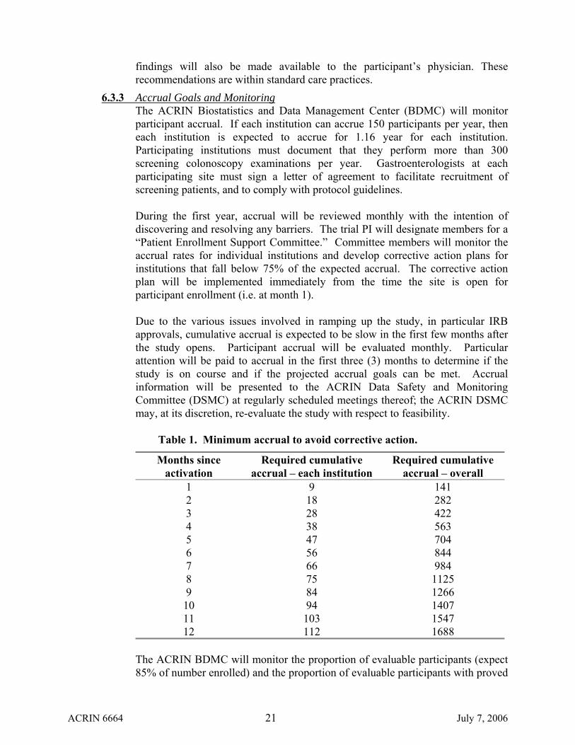
6.3.3 Accrual Goals and Monitoring The ACRIN Biostatistics and Data Management Center (BDMC) will monitor participant accrual. If each institution can accrue 150 participants per year, then each institution is expected to accrue for 1.16 year for each institution. Participating institutions must document that they perform more than 300 screening colonoscopy examinations per year. Gastroenterologists at each participating site must sign a letter of agreement to facilitate recruitment of screening patients, and to comply with protocol guidelines. During the first year, accrual will be reviewed monthly with the intention of discovering and resolving any barriers. The trial PI will designate members for a “Patient Enrollment Support Committee.” Committee members will monitor the accrual rates for individual institutions and develop corrective action plans for institutions that fall below 75% of the expected accrual. The corrective action plan will be implemented immediately from the time the site is open for participant enrollment (i.e. at month 1). Due to the various issues involved in ramping up the study, in particular IRB approvals, cumulative accrual is expected to be slow in the first few months after the study opens. Participant accrual will be evaluated monthly. Particular attention will be paid to accrual in the first three (3) months to determine if the study is on course and if the projected accrual goals can be met. Accrual information will be presented to the ACRIN Data Safety and Monitoring Committee (DSMC) at regularly scheduled meetings thereof; the ACRIN DSMC may, at its discretion, re-evaluate the study with respect to feasibility.
Table 1. Minimum accrual to avoid corrective action.
Months since activation
Required cumulative accrual – each institution
Required cumulative accrual – overall
1 9 141 2 18 282 3 28 422 4 38 563 5 47 704 6 56 844 7 66 984 8 75 1125 9 84 1266 10 94 1407 11 103 1547 12 112 1688
The ACRIN BDMC will monitor the proportion of evaluable participants (expect 85% of number enrolled) and the proportion of evaluable participants with proved
ACRIN 6664 21 July 7, 2006
clinically significant colorectal neoplasia (expect 6-10% of evaluable participants within institution, overall approximately 8%) on an ongoing basis. If a 95% confidence interval for the proportion of evaluable participants excludes 85%, reasons for this will be discussed with the trial team, and possible remedies will be considered. The study may be re-evaluated, in light of the expected results of any proposed remedies, for necessary sample size. If a 95% confidence interval for the proportion of evaluable participants with proved clinically significant colorectal neoplasia excludes 8%, a revised sample size will be calculated. The ACRIN DSMC will be notified of these results at its next regularly scheduled meeting and may at their discretion re-evaluate the study sample size and/or feasibility. Note that this is not an interim analysis, as measures of accuracy (e.g., sensitivity, area under the ROC curve) will not be evaluated. The ACRIN Data Safety and Monitoring Committee (DSMC) will monitor this protocol. At a regularly scheduled meeting of the ACRIN DSMC following trial activation, the ACRIN Biostatistics Center will provide an analysis including projections of sample size and accrual duration under the original accrual plan (12 positives per institution), and under an alternative accrual plan (limiting the trial accrual period to the shorter of December 31, 2006 and/or the time when the total accrual reaches the budgetary limit of 2607 participants). The impact of each plan on the ability of the trial to achieve its primary aim of estimating average sensitivity across radiologists with desired precision will be described. Various combinations of the average sensitivity across radiologists and the variance in sensitivity across radiologists, average prevalence across institutions and variance in prevalence across institutions, and models for accrual rates across institutions will be considered, as will methods of estimating average sensitivity other than taking a simple average of estimates across radiologists. The impact of these closure-to-accrual rules on the ability to estimate area under the ROC curve with desired precision will also be considered. As part of this report, the ACRIN Biostatistics Center will include a recommended rule for closure to accrual from a statistical perspective.
7.0 ONLINE REGISTRATION SYSTEM
7.1 Using the Online Registration System 7.1.1 Once a participant has completed the eligibility checklist (Appendix II) and been
found to be eligible, the participant may be consented. The RA will register the participant within 48 hours (two [2] business days) of imaging by logging onto the ACRIN web site (www.acrin.org) and selecting the link for Data Center Login, then choosing the ACRIN protocols link. The system triggers a program to verify that all regulatory requirements (such as OHRP assurance and IRB approval) have been met by the institution. The registration screens begin by asking for the date on which the eligibility checklist was completed, the identification of the person who completed the checklist, whether the participant was found to be eligible on the basis of the checklist, and the date the study-
ACRIN 6664 22 July 7, 2006
specific informed consent form was signed. Additional questions record participant demographics and the study-specific eligibility questions.
7.1.2 Once the system has verified that the participant is eligible and that the institution
has met regulatory requirements, it assigns a participant-specific case number. The system then moves to a screen, which confirms that the participant has been successfully enrolled. This screen can be printed so that the registering site will have a copy of the registration for the participant’s record. Two e-mails are computer generated and sent to the registering site: the confirmation of eligibility and the participant specific-calendar. The system creates a case file in the study’s database at the DMC and generates a data submission calendar listing all data forms, images, and reports and the dates on which they are due.
7.2 Unsuccessful Registrations 7.2.1 If the institution has not met the regulatory requirements, the system switches to a
screen that includes a brief explanation for the failure to gain access to the registration screens. If during the completion of the eligibility questions a participant is deemed ineligible based on a response, a message box appears instructing the RA to contact the Data Management Center. Either screen may be printed.
7.2.2 In the unlikely event that the ACR web registration site is not accessible, participating sites may still register a participant by faxing the completed eligibility checklist to the DMC at the ACR (215-717-0936, ATTN: PARTICIPANT REGISTRATION). ACR staff will fax a response to the registering site with the confirmation of registration and participant case number and randomization as soon as possible.
8.0 DATA COLLECTION AND MANAGEMENT 8.1 General
8.1.1 The ACRIN web address is www.acrin.org.
8.1.2 Data collection and management will be performed by the Biostatistics and Data Management Center (BDMC) of ACRIN under the direction of Dr. Constantine Gatsonis. The Biostatistics Center (BC) is located at Center for Statistical Sciences at Brown University in Providence, RI, and the Data Management Center (DMC) is located at the American College of Radiology’s Data Management Department in Philadelphia.
8.1.3 The BDMC uses screens on the ACRIN web site to register participants, collect participant data, and maintain calendars of data submissions for each participant. By using the World Wide Web, ACRIN has made participant registration, data entry, and updated calendar information available to clinical sites 24 hours a day.
ACRIN 6664 23 July 7, 2006

8.2 Clinical Data Submission 8.2.1 As soon as a participant has been registered, the RA may download the
participant’s data submission calendar, which lists all forms and/or designated reports required by protocol, along with the date that each form is due at the DMC. These calendars will be updated as the study proceeds to reflect data that have been received, reply deadlines for queries about unclear data, deadlines for follow-up reports of adverse events, or changes in the protocol that might change the data being collected, or their timing. Updated calendars for each participant can be obtained 24 hours a day from the ACRIN website.
8.2.2 An investigator is obliged to submit data according to protocol as detailed on each participant’s calendar as long as the participant is alive and the case status is designated as open or until the study is terminated. The case is closed when all data have been received and reviewed and no outstanding query exists for the case.
8.2.3 To submit data via the ACRIN website, the RA or investigator logs onto the web site and supplies the pre-assigned user name and password. Case report forms will be available on the web site through a series of links. The user selects the link to the appropriate form and enters data directly into the web-based form. As information is entered into the case report form, various logic checks will be performed. These logic checks look for missing data, data that is out of range, and data that is in the wrong format (e.g. character data in a field requiring numeric responses). Such errors will be detected as soon as the user attempts to either submit the form or move to the next page. They must be corrected before the form is transmitted to the DMC. The user will not be able to finalize form transmission to the DMC until all data entered pass these logic checks. Forms that are not completed in one sitting can still be submitted and completed at a later date.
8.2.4 Once a form is complete, the RA or investigator presses the SUBMIT button on the participant calendar and the data are transferred into the clinical database. No further direct revision of the submitted data is allowed after this point. An e-mail is generated and sent to the site listing all of the data completed and just submitted. Should a problem occur during transmission, this automated response supplies an explanation and instructions for resubmitting the data.
8.2.5 If a temporary problem prevents access to the Internet, investigators should wait until access is restored to submit data. The site RA or investigator should notify the DMC of the problem, and the DMC will give an estimated time when access is expected to be restored. If access will be unavailable for an extended period, sites must seek another Internet Service Provider (ISP). On a short-term basis, the ACR can serve as an ISP.
8.3 Data Security The registration system has built-in security features that encrypt all data for transmission in both directions, preventing unauthorized access to confidential
ACRIN 6664 24 July 7, 2006
participant information. Access to the system will be controlled by a sequence of identification codes and passwords.
8.4 Electronic Data Management 8.4.1 Data received from the web-based forms are electronically stamped with the date
and time of receipt by the ACRIN server. A validation program is used to perform more extensive data checks for accuracy and completeness. The logic checks performed on the data at this point are more comprehensive than those built into the web-based data entry screens. They include checking that answers are logical, based on data entered earlier in the current form and the more thorough checks. This validation program produces a log of errors, which is sent to the research associate for resolution. This program is frequently updated to incorporate exceptions to rules so that subsequent, correctly entered data pass validity checks, minimizing the time the DMC RA needs to spend resolving problems. Additional data review will take place once the data is transferred to the BC. The BC will run thorough cross-form validations, frequency distributions to look for unexpected patterns in data, and other summaries needed for study monitoring. Any errors found at the BC will be reported to the DMC RA for resolution.
8.4.2 If the program detects missing or problematic data, the DMC RA will send a
Request for Information (query letter) to the site RA or investigator specifying the problem and requesting clarification. The DMC RA then updates the participant’s data submission calendar with the due date for the site RA or investigator’s response.
8.5 Missing and Delinquent Data Submission In addition to providing the investigator a data collection calendar for each case, institutions are periodically prompted for timely submission of data through the use of a Forms Due Report. Distributed at intervals via the electronic mail system directly to both the RA and the investigator at each site, this report lists data items that are delinquent and those that will come due before the next report date. In addition to prompting clinicians to submit overdue data, the Forms Due Report helps to reconcile the DMC’s case file with that of the RA or investigator.
8.6 Data Quality Monitoring 8.6.1 The BC at Brown University will maintain a study database at its site for
monitoring data quality and for performing interim analyses. These data will be drawn directly from the DMC’s permanent database using a PowerBuilder utility that allows BC staff to log onto the DMC computer and select needed data. This analysis database will be maintained in permanent SAS (Statistical Analysis System software) format on the BC’s ACRIN server and updated on a scheduled basis, usually monthly once the study is in its steady state. Any discrepancies and other data quality issues will be referred to DMC for resolution, since only the DMC can correct the data file. No changes to the data will be made at the BC.
8.6.2 A major goal of the monitoring of data in the BC is to assess compliance with the protocol and to look for unforeseen trends that may be indicative of procedural
ACRIN 6664 25 July 7, 2006

differences among clinical sites. If patterns are discovered in the data that appear to arise from causes specific to an institution, the BDMC will apprise the site of the problem and work with the site until the problem has been resolved. If the BDMC cannot find a solution, the problem will be brought to the Steering Committee for further discussion and resolution.
8.6.3 The BC, in conjunction with the DMC, will prepare frequent summaries of the accrued data to be presented to investigators. These summaries will report accrual rates (overall and by sub-groups of interest to the investigators), assess the completeness and accuracy of the data, and discuss any trends that may impact the outcomes of the trial. These intermittent summaries will not include analyses of the study’s endpoints.
8.6.4 In addition, the ACRIN Quality Assurance staff will review case report forms and source documents on several initial study participants enrolled at each site, including a few cases defined as positive. This educational process is to provide clarification in completion of the case report forms in order to minimize any inconsistencies or misunderstandings.
9.0 DATA COLLECTION FORMS
Anatomic, morphologic, and histologic details on relevant colorectal lesions will be abstracted from the medical, endoscopic, surgical and pathology records. A0 – Registration/Eligibility Checklist (Appendix II): This form collects general demographic characteristics (including age, gender, and race), inclusion/exclusion criteria checks, and receipt of written informed consent. I1 – On-Study Evaluation/Medical History Data: This form is used to record study-specific characteristics indicative of risk (including prior history of colorectal neoplasia, family history of colorectal neoplasia, history of prior colorectal surgery, iron deficiency anemia), indication for colonoscopy and bowel prep information. TA – Local CTC Acquisition: This form is used to collect the following technical parameters: CO2, room air, or both, manual or mechanical insufflator, glucagon: route of administration (subcutaneous, unless contraindicated) and dose (mg), slice thickness, reconstruction interval, mAs, # of images per acquisition, reconstruction algorithm; personnel present for CT acquisition and percent time each personnel member was present. C2 – Local CTC Interpretation: This form is used to rate quality of prep: residual stool, distention, residual fluid – per segment, both supine and prone. Provide characteristics of findings on CTC interpretation (number of findings, colonic segment, location coordinates, size, and confidence for each finding). XYZ coordinates of each lesion > 5 mm in diameter. Global assessment of the likelihood that the patient has at least one polyp greater than or equal to 10mm in diameter; machine type (software), interpretation time.
ACRIN 6664 26 July 7, 2006
FX – Extracolonic Findings Form (Aim 3.2.4): This form is used to record extracolonic abnormalities detected in the course of a CTC examination. Information recorded will include number of abnormalities, location of abnormality, size, diagnosis, need for follow-up evaluation, and need for follow-up treatment. P4 – Central Review Pathology/Colonoscopy Form: This form is used to record the colonoscopy evaluation: extent, complications, assessment of quality of preparation, lesion location, size, morphology, tissue removed vs. lesion fulgurated. Pathological evaluation: Colorectal adenomas: size, site, degree of dysplasia. Other lesion types: hyperplastic, inflammatory, vascular, ulcerative, as well as site and size. For cancers: stage and type, as well as site and size. Copies of the colonoscopy and pathology reports are submitted with this form to the central review pathologist. PL- Local Pathology/Colonoscopy Form: This form is used to record the Colonoscopy evaluation: extent, complications, assessment of quality of preparation, lesion location, size, morphology, tissue removed vs. lesion fulgurated. Pathological evaluation: Colorectal adenomas: size, site, degree of dysplasia. Other lesion types: hyperplastic, inflammatory, vascular, ulcerative, as well as site and size. For cancers: stage and type, as well as site and size. Copies of the colonoscopy and pathology reports are submitted with this form to ACRIN DM. Colonoscopy must take place within 30 days after CTC. B1- Lesion Photograph Transmittal Form: This form is used to affix photographs of all lesions removed as well as a photograph to document the complete colon examination (either the appendiceal orifice or ileocecal valve) for submission to data management. SX –CTC Software Questionnaire (Aim 3.2.5): Assesses differences in software platforms by evaluating user preferences and performance differences, including evaluation times. Information will be recorded about the reviewer (name, site, experience – approximate number of CTC exams evaluated), hardware (CT scanner type, workstation type), software (CTC software type – Vital Images, Navigator, etc.), monitor size (17, 20, or 25 inch), number of monitors, monitor set up, method of examination review, image display used for initial review, image display used for abnormality analysis. In addition, this form will collect the effect of subtraction on: 1) reader confidence of polyp findings 2) ease of interpretation as reported by readers 3) stability of polyp size and 4) the time required for interpretation utilizing subtraction.
C1 – CTC Report
C3 – Colonoscopy Report S2 – Surgical Report P1 – Pathology Report PC- Pathology Specimen Transmittal Form: This form is used to track specimens submitted to the central pathology laboratory: number of slides, accession number, tissue block, etc. The transmittal form and the pathology report are sent to the central pathology lab. A copy of the tracking form should be faxed to the data management center as notification of slide submission.
ACRIN 6664 27 July 7, 2006
CS - PQ Form Cover Sheet: This form accompanies the Patient Cost and Acceptance Questionnaire (PQ) form. It serves as the last page of the questionnaire, which documents the completion time of the questionnaire, and whether the questionnaire was completed by the participant or with assistance. If the PQ is returned by mail, the form is to be completed by the participant. If PQ is completed over the phone, the BC RA will complete the form. The information collected will be submitted by the BC RA to ACRIN headquarters for data entry. PQ-Patient Cost and Acceptance Questionnaire: This questionnaire will be administered to all patients (reference Section 14.3.2). It is self-administered, and asks for travel time away from usual activities, child care, travel expenses, and other out-of-pocket expenses incurred on the day(s) they had the exams for the trial. It will also assess for discomfort, inconvenience, and willingness to return for a repeat examination for CTC and OC. TM – Time Motion Form: The time-motion data constitutes a special sub-study to do micro-cost analysis for this comparatively new procedure. This form will consist of two modules designed to supplement and validate the procedural personnel and interpretation time data collected in forms TA and C2. The first module will be used during direct observation of 18 CTC procedures at each of 3 sites by on-site RAs specifically trained for this purpose. The second module will be used during direct observation of 18 CTC interpretations of each type (primary 2D read and primary 3D read) at each of the 3 sites. The form will collect times spent by all personnel during the CTC procedure and interpretation, all consumables used, and other resources used such as room and machine times devoted to the procedure and interpretation. The form will be created for use by 3 selected RAs only, and only for a limited number of uses at each of the 3 sites represented by the 3 RAs (please see Section 14.3.1). CX - Reader Study Interpretation: This form is similar to the local interpretation form (lesion site, size, confidence), with tick boxes for slice thickness of axial reconstructed images, number of supine axial images (as a redundant check of slice thickness), number of prone axial images (as a redundant check of slice thickness), reader confidence of polyp findings, ease of interpretation as reported by readers, polyp size before and after electronic subtraction, lesion detection with subtraction, and interpretation times without and with electronic subtraction. DP - Imaging Transmittal Form Worksheet: This worksheet is to be completed by the CT Technologist at the completion of the CTC scan. It must be faxed to the Imaging Management Center (IMC) at 215-923-1737 at the same time the images are being sent from the ACRIN PC to ACRIN HQ. QA - CT Quality Assessment Form: This form is completed by the Quality Control Reviewer.
9.1 For Aim 3.3.1 (Database for Computer-Aided Diagnosis) The following information will be abstracted from the data collection forms:
ACRIN 6664 28 July 7, 2006
Interpretation times, date of examination, date of colonoscopy exam, patient age and gender, risk factors, manual interpretation findings, type of scanner, quality assessment scores, number of clinically significant findings, confidence in manual detections, matching results (including pathology and colonoscopy reports), type of colon preparation and amount consumed by the patient, use of glucagon, measurements of radiation dose (mAs), and CT technical parameters (slice thickness, reconstruction intervals, kernel, field of view). In addition to DICOM CTC images, a copy of the pathology report and colonoscopy report and a colonoscopy photograph of each polyp submitted to the database are required.
The submitting institution must also submit coordinates of the polyp (form C2). For each proven lesion, XYZ coordinates must be submitted.
The following information from cases with optical colonoscopy polyp findings, de-identified of patient and institutional information will be abstracted from the data collection forms and transferred to the Cancer Imaging Program, National Cancer Institute (CIP/NCI) at the same interval that data transfer occurs to the Clinical Center, National Institutes of Health or at one year intervals, whichever is shorter.
• Participant age • Participant gender • DICOM CTC 2D slice images with headers that include CT technical parameters
(e.g.: type of scanner, kVp, mAs slice thickness, scan speed, reconstruction interval, kernel, field of view)
• Coordinates (XYZ coordinates for each polyp (C2 form) • Copy of the pathology report and colonoscopy photograph of each polyp
submitted 9.2 For Aim 3.3.2 (Cost-Effectiveness)
Please refer to Cost-Effectiveness Modeling, Section 14.0.
ACRIN 6664 29 July 7, 2006
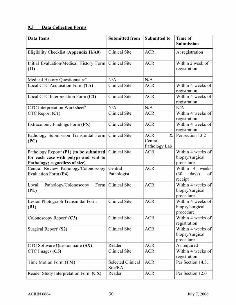
9.3 Data Collection Forms Data Items Submitted from Submitted to Time of
Submission
Eligibility Checklist (Appendix II/A0)
Clinical Site ACR At registration
Initial Evaluation/Medical History Form (I1)
Clinical Site ACR Within 2 week of registration
Medical History Questionnaireb N/A N/A Local CTC Acquisition Form (TA) Clinical Site ACR Within 4 weeks of
registration Local CTC Interpretation Form (C2) Clinical Site ACR Within 4 weeks of
registration CTC Interpretation Worksheetb N/A N/A N/A CTC Report (C1) Clinical Site ACR Within 4 weeks of
registration Extracolonic Findings Form (FX) Clinical Site ACR Within 4 weeks of
registration Pathology Submission Transmittal Form (PC)
Clinical Site ACR & Central Pathology Lab
Per section 13.2
Pathology Reporta (P1) (to be submitted for each case with polyps and sent to Pathology; regardless of size)
Clinical Site ACR Within 4 weeks of biopsy/surgical procedure
Central Review Pathology/Colonoscopy Evaluation Form (P4)
Central Pathologist
ACR Within 4 weeks (30 days) of receipt
Local Pathology/Colonoscopy Form (PL)
Clinical Site ACR Within 4 weeks of biopsy/surgical procedure
Lesion Photograph Transmittal Form (B1)
Clinical Site ACR Within 4 weeks of biopsy/surgical procedure
Colonoscopy Reporta (C3) Clinical Site ACR Within 4 weeks of registration
Surgical Reporta (S2) Clinical Site ACR Within 4 weeks of biopsy/surgical procedure
CTC Software Questionnaire (SX) Reader ACR As required CTC Images (C5) Clinical Site ACR Within 4 weeks of
registration Time Motion Form (TM) Selected Clinical
Site/RA ACR Per Section 14.3.1
Reader Study Interpretation Form (CX) Reader ACR Per Section 12.0
ACRIN 6664 30 July 7, 2006
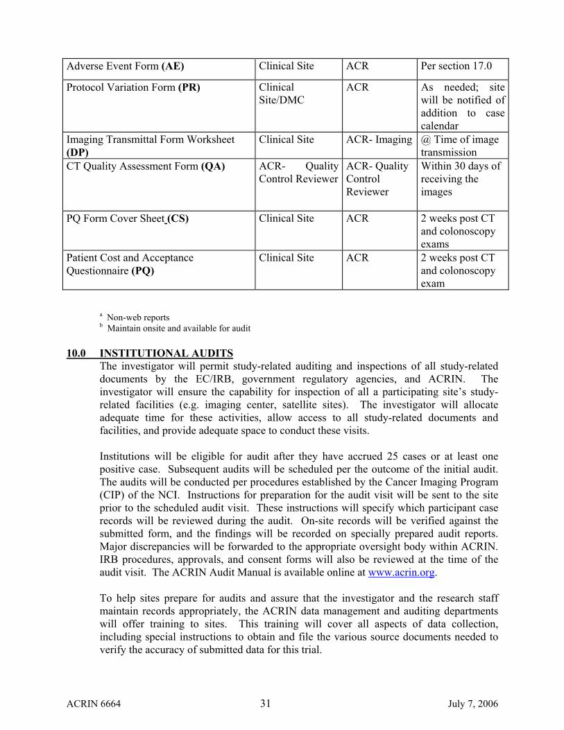
Adverse Event Form (AE) Clinical Site ACR Per section 17.0
Protocol Variation Form (PR) Clinical Site/DMC
ACR As needed; site will be notified of addition to case calendar
Imaging Transmittal Form Worksheet (DP)
Clinical Site ACR- Imaging
@ Time of image transmission
CT Quality Assessment Form (QA) ACR- Quality Control Reviewer
ACR- Quality Control Reviewer
Within 30 days of receiving the images
PQ Form Cover Sheet (CS) Clinical Site ACR 2 weeks post CT and colonoscopy exams
Patient Cost and Acceptance Questionnaire (PQ)
Clinical Site ACR 2 weeks post CT and colonoscopy exam
a Non-web reports b Maintain onsite and available for audit 10.0 INSTITUTIONAL AUDITS
The investigator will permit study-related auditing and inspections of all study-related documents by the EC/IRB, government regulatory agencies, and ACRIN. The investigator will ensure the capability for inspection of all a participating site’s study-related facilities (e.g. imaging center, satellite sites). The investigator will allocate adequate time for these activities, allow access to all study-related documents and facilities, and provide adequate space to conduct these visits.
Institutions will be eligible for audit after they have accrued 25 cases or at least one positive case. Subsequent audits will be scheduled per the outcome of the initial audit. The audits will be conducted per procedures established by the Cancer Imaging Program (CIP) of the NCI. Instructions for preparation for the audit visit will be sent to the site prior to the scheduled audit visit. These instructions will specify which participant case records will be reviewed during the audit. On-site records will be verified against the submitted form, and the findings will be recorded on specially prepared audit reports. Major discrepancies will be forwarded to the appropriate oversight body within ACRIN. IRB procedures, approvals, and consent forms will also be reviewed at the time of the audit visit. The ACRIN Audit Manual is available online at www.acrin.org. To help sites prepare for audits and assure that the investigator and the research staff maintain records appropriately, the ACRIN data management and auditing departments will offer training to sites. This training will cover all aspects of data collection, including special instructions to obtain and file the various source documents needed to verify the accuracy of submitted data for this trial.
ACRIN 6664 31 July 7, 2006
10.1 Source Documents Source data are found in all information, original records of findings, observations, or other activities in a clinical trial necessary for the reconstruction and evaluation of the trial. Source data are contained in source documents. Source documents are the first recording of any observations made or data generated about a study participant while he or she is enrolled in a clinical trial. Source documents for each study participant substantiate the data that are submitted to ACRIN. Source documents must verify the eligibility criteria and data submitted on all case report forms (CRFs). If an item is not mentioned (e.g., history and physical with no mention of a psychological condition), it will be assumed it is not present.
Research records for each case should contain copies of the source documents for the data reported to ACRIN. If information is abstracted from medical charts that are not filed at the investigative sites (e.g. hospital charts), copies of these records should be filed in the research chart. However, every attempt must be made to obtain all records/charts that were used to abstract any study data for this protocol at the time of the audit visit. This will prevent any discrepancies and the inability to verify the document and the data reported.
10.2 Case Report Forms Case report forms (CRFs) are the primary data collection instruments for the study. All data requested on the CRFs must be recorded, and any missing data must be explained. If a space is left blank because the procedure was not done or the question was not asked, “N/D” must be noted. If the item is not applicable to the individual case “N/A” must be noted. All entries must be printed legibly in black ink on the paper case report forms. In the event of any entry errors, corrections must be made by drawing a single straight line through the incorrect entry, writing the initials of the person making the correction, recording the date when the correction is being made, and entering the correct data above the strike through. Do not use white out or an eraser.
Data elements that are extracted from the medical record (such as participant history or official clinical interpretations of images, pathology, or surgery results) and recorded on the case report forms (CRFs) will be audited against the appropriate component of the medical record. Data elements gathered from signed participant questionnaires may be documented on the CRF. The image interpretation required by the study that is a more detailed extraction of information from the image and is not typically documented in the standard radiology report may be recorded on the CRF and is accepted as source documentation if signed and dated by the Investigator. At the time of audit, the auditor will verify the occurrence of the imaging examination, the reader, and the date on which the exam took place from the medical record. Any use of an approved CRF as source documentation requires that the CRF be signed and dated and refer to the source of the information (participant questionnaire, CT, MR, etc.).
Any use of CRFs as source documentation when the protocol has designated the source data will be medical record documentation will be considered a deficiency.
ACRIN 6664 32 July 7, 2006
10.3 Institutional Review Board Sites must obtain local IRB initial approval. Prior to subject registration, a copy of the IRB approval letter for the protocol and the informed consent form must be sent to ACRIN, along with a copy of the IRB approved informed consent form. Investigator will provide copies of IRB approval letters for any amendments, and copies of annual renewals.
10.4 Research Records Maintain source documentation for each case that substantiates the data reported to ACRIN. Source documentation includes the following: • hospital chart or legible copies • clinic chart or legible copies • pathology reports or legible copies • surgical report or legible copies • forms signed and dated by the subject (per Section 10.6) • ACRIN case report forms signed by the physician (per Section 10.6) • worksheets signed by the physician which are used by research staff to submit the
data on case report form(s) • confirmation of submitted case report forms (mailed or emailed from ACRIN to site) • CTC report • Colonoscopy report
Source documentation must verify the eligibility criteria and data submitted on all case reporting forms. If an item is not mentioned (e.g., history and physical with no mention of a psychological condition) it will be assumed it is not present.
It is suggested that the research record for each case contain copies of the source documentation for the data reported to ACRIN. Copy the source documentation as you abstract the data from the primary record. This will prevent a discrepancy and inability to document the data reported when reviewed by auditors.
Anatomic, morphologic, and histology details on relevant colorectal lesions and medical history will be abstracted from the medical, endoscopy, surgical, and pathology records and maintained as part of the participant study file.
10.5 Audit Source Documentation Chart
Case Reporting Form Data Collection Source Documentation Consent Form
ACRIN 6664 33 July 7, 2006
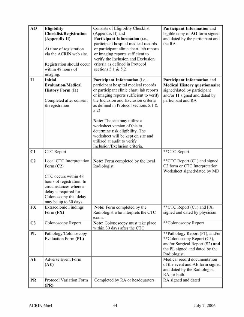
AO Eligibility Checklist/Registration (Appendix II) At time of registration via the ACRIN web site. Registration should occur within 48 hours of imaging.
Consists of Eligibility Checklist (Appendix II) and Participant Information (i.e., participant hospital medical records or participant clinic chart, lab reports or imaging reports sufficient to verify the Inclusion and Exclusion criteria as defined in Protocol sections 5.1 & 5.2)
Participant Information and legible copy of AO form signed and dated by the participant and the RA
I1 Initial Evaluation/Medical History Form (I1) Completed after consent & registration
Participant Information (i.e., participant hospital medical records or participant clinic chart, lab reports or imaging reports sufficient to verify the Inclusion and Exclusion criteria as defined in Protocol sections 5.1 & 5.2) Note: The site may utilize a worksheet version of this to determine risk eligibility. The worksheet will be kept on site and utilized at audit to verify Inclusion/Exclusion criteria.
Participant Information and Medical History questionnaire signed/dated by participant and/or I1 signed and dated by participant and RA
C1 CTC Report **CTC Report
C2 Local CTC Interpretation Form (C2) CTC occurs within 48 hours of registration. In circumstances where a delay is required for Colonoscopy that delay may be up to 30 days.
Note: Form completed by the local Radiologist.
**CTC Report (C1) and signed C2 form or CTC Interpretation Worksheet signed/dated by MD
FX Extracolonic Findings Form (FX)
Note: Form completed by the Radiologist who interprets the CTC exam.
**CTC Report (C1) and FX, signed and dated by physician
C3 Colonoscopy Report Note: Colonoscopy must take place within 30 days after the CTC
**Colonoscopy Report
PL Pathology/Colonoscopy Evaluation Form (PL)
**Pathology Report (P1), and/or **Colonoscopy Report (C3), and/or Surgical Report (S2) and the PL signed and dated by the Radiologist.
AE Adverse Event Form (AE)
Medical record documentation of the event and AE form signed and dated by the Radiologist, RA, or both.
PR Protocol Variation Form (PR)
Completed by RA or headquarters RA signed and dated
ACRIN 6664 34 July 7, 2006
** Clinical reports identified as source documentation must include patient’s name, date of imaging or procedure, the clinical information, and the signature of the examiner/reader.
11.0 IMAGE SUBMISSION
All images for this protocol are requested to be provided in digital format. ACRIN has developed software that allows for electronic transmission to the IMC image archive of images that have been scrubbed of all patient identifiers. Individual PC computers with this software installed will be supplied to each participating site. ACRIN will be contacting each site individually to determine their readiness and ability to work with this system. If you have preliminary questions, you may contact either Rex Welsh or Fraser Wilton (215-574-3215) for information about this system. Once readiness has been determined, imaging personnel from ACRIN will coordinate the shipment and installation of the PC computers and train all operating staff on use of the system. Images stored on the ACRIN DMC image archive will then be routed to other sites involved using either FTP or CD-ROM where appropriate for purposes of secondary interpretation. CT cases will consist of supine and prone data sets. The entire CTC study will be submitted, including the digital scout.
11.1 For Aim 3.3.1 (Database for Computer-Aided Diagnosis) The data (images and annotation) will be checked by ACRIN for completeness. A subset of the data from each institution will be double-checked by the principal investigator to ensure, for example, that polyp coordinates are properly annotated, that the technical parameters were followed, and full data sets were submitted. The data will be checked to ensure that they are readable by standard DICOM file readers.
11.2 Image Quality Review
A review of a sampling of the CT Colonoscopy (CTC) cases will be performed in order to ascertain the quality of images obtained at each institution to assure that the exam is of adequate quality. The CT Colonoscopy (CTC) image sets from the initial three (3) CTC exams performed will be reviewed by the protocol Quality Control reviewer, designate by the Study Principle Investigator at the ACRIN Image Laboratory in Philadelphia, or they will be sent to the protocol Quality Control reviewer via the Internet or on media for quality review. After which, a Radiologist will review a random sample of all CTC exams for quality assurance purposes. An ongoing review will be performed by the Imaging Specialist at ACRIN to ensure images meet the study specific parameters. An additional three (3) CTC exams will be reviewed in the same fashion when the CT scanner is upgraded or a new CT scanner is installed.
ACRIN 6664 35 July 7, 2006
12.0 IMAGING METHODOLOGY 12.1 Patient Preparation
12.1.1 Laxation Each patient will be required to have a fully prepared colon using one of three standardized bowel preparations widely used throughout the United States: 1) Lavage preparation using polyethylene gycol solution, 2) magnesium citrate or 3) phosphosoda. All three preparations will also include the use of bisacodyl tablets (10 mg or current institutional standard of care) that will act to remove any residual fluid and stool in the colon.
12.1.2 Stool/fluid Tagging
Twenty four hours before beginning preparation patients will begin to ingest barium sulfate, given in up to three doses. Following cathartic cleansing, patients will ingest at least 5 ml of water soluble oral contrast material.
12.2 CT Technique
Only 16 slice or higher CT scanners will be used for the study. Slice thickness will be set at 1-2 mm, with 1-1.25 mm reconstruction intervals. The specific CT protocols are listed in Appendix 5.
12.3 CT Colonography Participants will be assigned one of three colon preparations (Go-Lyte lavage, magnesium citrate, or Phosphosoda) dependent upon preference of referring clinician and participant. With either preparation participants will also receive bisacodyl tablets (10 mg or current institutional standard of care). Glucagon, 1 mg (subcutaneous, unless contraindicated), will be given usually from 7 to 15 minutes prior to the procedure unless contraindicated. Participants will be placed in either decubitus (left or right side) position for enema tip insertion and air insufflation (until the participant verbally indicates air administration has reached maximal tolerance). Insufflation will be performed mechanically, with an automated CO2 insufflator unless full colon distention cannot be obtained. In those cases, CO2 will be added manually to achieve full distention. All participants will be imaged in both the supine and prone positions. After insufflation, a standard CT scout scan will be obtained prior to both supine and prone acquisitions to assess the degree of colonic distention. Additional CO2 will be added (as tolerated by the participant) prior to scanning to maintain a fully distended colon. In the event of poor bowel prep (the radiologist is unable to reliably exclude a polyp 1 cm or larger in more than a single segment) the patient will be asked to repeat preparation and reschedule for a repeat examination. All examinations are performed using a multislice helical CT scanner (minimum 16 slice) capable of acquiring volumetric CT data. Images are acquired according to Appendix V. Additional views may be obtained at the discretion of the radiologist after both the prone and supine images have been obtained. Interpretation will be based on the supine and prone views only. Interpretation forms will be completed a second time to include the findings from additional views to note any changes in interpretation.
ACRIN 6664 36 July 7, 2006
12.4 Radiologist Qualifications and Training Reader certification will be required for each radiologist participating in the study. To be considered a reader, the radiologist must ordinarily have read at least 30 cases from a polyp-enriched cohort with colonoscopic correlation. If the radiologist has read over 500 cases, he or she must pass a certifying examination consisting of known cases. 90% accuracy will be considered passing. Readers having read less than 500 cases will be required to attend a one-day training course in CTC interpretation at the GE training center in Milwaukee, WI. Upon completion of the training course, a reader must pass the certifying examination. If the reader fails the certifying exam, he or she will be sent 30 additional cases to review and have the results assessed for a passing score.
12.5 Workstation Requirements CTC data will be transferred onto a workstation equipped with CTC-specific software. The workstation must be able to accommodate at least 1000 loadable images, and be able to scroll seamlessly and load 300 images in 20 seconds. The software must provide axial, MPR, and 3D views, with the ability to interact among them and readily switch between them. The switches between prone and supine, and between MPR and 3D, must be accomplished in less than 15 seconds each. An accurate measurement tool must be part of the software. The 3D view must be interactive; either volume or surface rendering is permitted. For all views, the ability to display supine and prone images side-by-side is highly recommended. A complete 3D endoluminal fly through must be available that can be performed and interpreted within 12 minutes (without problem solving).
12.6 Diagnostic Review Process An experienced radiologist (see Section 12.4) will perform diagnostic review of the images blinded to other examination results, using a workstation meeting the requirements in Section 12.5. Diagnostic review will begin with quality review for presence of fluid and stool.
The colon will be divided into 6 anatomic segments: rectum, sigmoid colon, descending colon, transverse colon (including splenic and hepatic flexures), ascending colon, and cecum. The degree of colonic distention in each segment will be scored using a 5-point scale (1 = collapsed; 2 = poorly visualized; 3 = entire segment visualized but under-distended; 4 = entire segment visualized and well-distended, and 5 = over-distended). A distention score of ≥ 3 represents adequate distention. Each CTC examination will be read by two different radiologists using two different image paradigms to eliminate potential recall bias: 1) Conventional approach - magnified axial images will be viewed in rapid cine sequence and 3-dimensional endoluminal images and multiplanar reformatted images will be generated in areas of bowel that could not confidently be evaluated using the magnified axial sequences alone. Images will be viewed using both soft tissue windows (Level, 70H; width, 500H) and lung windows (insensitive to IV contrast enhancement) (Level, -750H; width, 500H). 2) Review using an endoluminal fly through, viewing the lumen both antegrade and retrograde in both the prone and supine data sets. The 3D images will be used to detect an abnormality, and 2D images will be utilized to problem solve and improve observer confidence of these detected lesions.
ACRIN 6664 37 July 7, 2006
Each participant’s CTC examination will be randomly assigned to a local image review paradigm (conventional approach or endoluminal fly-through, as described in the preceding paragraph). In order to mimic clinical practice, depending on the average practice size of participating institutions at the outset of the trial each primary reader will be assigned either 2 or 4 hypothetical practice partners from among the remaining 14 sites’ primary readers, to make up a practice of 3 or 5 partners, in a counterbalanced fashion. The practice partners will interpret the CTC examination using the other of the two image review paradigms: if the local read is made with the conventional approach, then the practice partner will read using an endoluminal fly-through, and vice versa. At regular intervals, the Biostatistics Center will instruct the ACRIN imaging specialist to transmit groups of 10 consecutive eligible and evaluable cases with known reference standard to appropriate practice partners. The order of partners used will be determined using randomized blocks (thus, if there are 4 practice partners, each partner will see one of the first four groups of 10 such cases, one of the next four groups, etc.). There are sufficient funds in the study budget for each reader to have 4 hypothetical practice partners.
Recording of findings will include lesion location, size, slice number, xyz coordinates, and confidence. In addition, the presence of extracolonic abnormalities will be noted.
All measurements should be made on 2D images. The benefits of electronic subtraction when reviewing using an endoluminal fly-through will be explored by Dr. Michael Zalis, Massachusetts General Hospital in a re-read study.
12.6.1 For Aim 3.3.1 (Database for Computer-Aided Diagnosis)
The coordinates of the polyps found at CTC will be determined by the submitting institution. Polyps determined at lesion matching (13.2) to be true positives and >7 mm in diameter will be included in the database.
13.0 REFERENCE STANDARD
Colonoscopy will be considered the “reference standard,” or truth, with respect to presence or absence of large polyps. The original colonoscopy will be taken as the reference standard in all cases unless, in the opinion of the colonoscopist, the bowel prep was so poor as to warrant a repeat colonoscopy, in which case, the better prepared colonoscopy would become the reference standard. See Section 13.3 for exceptions.
13.1 Colonoscopy
All participating institutions will have a signed letter of agreement from a supervising gastroenterologist that regularly performs colonoscopy. This letter will indicate familiarity with the study protocol requirements as well as a willingness to assist with subject recruitment. These colonoscopists would be encouraged to attend the organizational meeting at study initiation. Only staff endoscopists will perform the colonoscopic examinations. Photographs of all lesions removed as well as a photograph
ACRIN 6664 38 July 7, 2006

to document complete colon examination (either the appendiceal orifice or ileocecal valve) should be submitted with case materials. All lesions will be measured by comparison to the open forceps or by calibrated guidewire. Each biopsied specimen will be retrieved and placed in a separate container for pathologic examination. Flat lesions will refer to polyps > 5 mm in diameter with < 2 mm elevation from flush.
13.2 Pathology of Resected Colorectal Neoplasms
All pathology specimens (polyps > 5 mm) will be sent to a central pathology laboratory at the Mayo Clinic within 15 working days of case submission to ACRIN. Each polyp will be placed in a separate container and labeled to distinguish the location of each polyp. If the polyp is removed in pieces, all pieces will be included in the specimen bottle for that lesion. Local pathologist will provide the size of the lesion. Slides will be prepared according to the standard histology procedure (i.e. hematoxylin and eosin stained). Two glass sides (usually 6-8 serial section cut) and the tissue block will be sent to the central laboratory. The histologic material will be categorized histologically as hyperplastic, lipomatous, adenomatous, mixed, carcinoma, normal mucosa or other. All adenomas will be subcategorized if they contain villous features or dysplasia. Mixed lesions will be considered adenomatous for the purposes of statistical analysis. For colorectal cancers, the size, site, stage, and type will be provided. All specimens will be returned within 30 days to the facility where the participant underwent biopsy after review by the central pathologists. A copy of the pathology report will be attached to the data collection form. Lesion size reported pathologically will be used, unless the lesion was removed in pieces. In the latter instance, size reported colonoscopically will be used.
13.3 Lesion Matching
An independent radiologist will manually review the colonoscopy and pathology reports and match these lesions with findings from CTC using the algorithm shown in Appendix VI. To be considered a match the lesion must be reported at colonoscopy and CTC to be within the same or adjacent segment. If the lesion matches by location then it will be assessed by size. If the lesion is reported to be within 50% in diameter of the size at colonoscopy and CTC it would be considered to be a match. If the lesion does not match by location but is within two colon segments—the colonoscopic photograph will be compared with its CTC image. Matching will be determined by consensus using a review committee. Lesions that match by morphology and by their position on a haustral fold or colon wall will be considered to be a match. Lesions matching by location but not by size will be reviewed in an analogous manner. The review committee will consist of a radiologist and a gastroenterologist. If there is a disagreement such that a consensus decision cannot be made, a third individual (gastroenterologist) will be used to decide. An automated computer matching algorithm using the same size and location matching criteria outlined in appendix V (but without the aid of any written reports, morphology, or images) will also be compared to the consensus manual matching. Any discrepancies between the automated matching and the manual matching will be adjudicated by the first unused member of the review committee.
14.0 COST-EFFECTIVENESS MODELING
ACRIN 6664 39 July 7, 2006
In order to better understand the impact to society of adding CTC to the list of currently-recommended screening approaches, we intend to conduct cost-effectiveness analysis using simulation methods. Our primary method will be to incorporate estimates of CTC sensitivity, specificity and cost into already existing, validated simulations developed under the CISNET initiative. We believe that it is important to create a “virtual” head-to-head evaluation using well-known, peer-reviewed simulations that have already been used to assess current screening technology.
14.1 Background A previously developed colorectal cancer simulation model will be employed to evaluate the potential cost-effectiveness of screening for colorectal cancer (CRC) with CTC. This cost-effectiveness modeling study will be conducted using data elements obtained in the trial as inputs to the models. In the face of rapidly escalating health care spending, policy makers are often forced to make decisions regarding resource allocation. Cost-effectiveness analysis has become the accepted approach to aid such decision making.108 Informed decision making requires knowledge of both the costs and effectiveness of tests or procedures under consideration. The model is described in section 14.2.
The cost-effectiveness model referred to here is an analytical calculation of the incremental costs and incremental benefits of one screening protocol compared to another.109 Because the outcomes and costs of screening for colorectal cancer are the results of stochastic processes, underlying the analytic calculations of the CEA model is a micro-simulation model of screening for colorectal cancer. This simulation model is being developed under direction of Dr. Karen Kuntz at the Harvard School of Public Health as part of the NCI Cancer Intervention and Surveillance Network (CISNET) consortium to evaluate the cost-effectiveness of screening for colorectal cancer and carefully calibrated to SEER data. CTC is currently not included as a screening option in this model.
We will develop a CTC screening strategy and incorporate it into the existing CRC screening model. If different screening intervals for CTC are considered relevant (e.g., 3, 5 or 10 years), these may be separately modeled and included in the analysis. Data derived from the primary aim will be used to inform model parameters, particularly those relating to test performance characteristics. Additional data concerning compliance with repeat screening studies, and possibly also extracolonic findings and their significance (if available from the currently proposed trial), may also be incorporated. In order to estimate the costs associated with performing CTC, resource utilization data will be collected at three of the sites enrolling participants into the CRC trial. Analysis will proceed by comparing one or more strategies involving CTC to the other screening strategies already present in the model. Strategies will be compared with respect to their effectiveness, cost, and cost-effectiveness, using the incremental cost-effectiveness ratio as the principal metric for comparison.
14.2 Model Description A simulation model was developed and used to evaluate a number of screening strategies for colorectal cancer and has been calibrated to SEER data. The model compares the
ACRIN 6664 40 July 7, 2006
lifetime costs and benefits of various screening programs for the general U.S. population. The model is based on the natural history of colorectal cancer in that it models the transitions from normal colonic epithelium to low-risk polyp (defined as <10 mm and non-villous pathology), from low-risk to high-risk polyp (defined as either ≥10 mm or villous pathology), from high-risk polyp to undetected cancer (localized, regional, or distant), and from undetected to symptom-detected cancer. Superimposed on this natural history model is a screening mechanism that can either identify and remove polyps or can identify and stage colorectal cancer. Persons who have a polypectomy return to a polyp-free state; however, their transition probability to a low-risk polyp state is different from polyp-free persons who do not have a polyp history, and also depends on whether their previously diagnosed polyp was of low or high risk. Persons who are screened positive with colorectal cancer are moved to the relevant “detected cancer” state. Survival for persons with either detected or undetected colorectal cancer is modeled using the SEER database. The primary difference between detected and undetected colorectal cancer is that persons with detected cancer are no longer candidates for presentation at a later disease stage. A developmental version of this model has been used to evaluate the cost-effectiveness of FOBT, flexible sigmoidoscopy, barium enema, and colonoscopy.109 For these analyses, it was assumed that, once a person is screened positive with a high-risk polyp, he or she is put onto a surveillance schedule that involves being checked every 3 to 5 years with colonoscopy. The value of combinations of tests (e.g., annual FOBT plus sigmoidoscopy every 5 years) and the effect of varying screening intervals (including “one-time screen” strategies) were evaluated. In addition, the effect of starting to screen at different ages and the effect of varying levels of compliance was also studied. Compliance is especially important for tests such as FOBT and barium enema because they are strictly diagnostic in nature and a positive test requires a person to return for a colonoscopy. Alternatively, flexible sigmoidoscopy has the ability to both diagnose and remove polyps, although only in the distal portion of the colon. In order to adequately evaluate sigmoidoscopy, the distal and proximal areas of the colon are modeled separately. For undetected cancers that are in the proximal colon, polyp status is modeled for the distal colon. In the protocol simulation, a "screened positive" refers to any individual who has one or more lesions detected by screening (with CTC or comparator methods such as FOBT) and confirmed by colonoscopy and biopsy to be adenomatous and greater than or equal to 5 mm in diameter.
14.2.1 Model’s Test Characteristics and Mortality Sensitivities and specificities were estimated for each test for the identification of low-risk polyps, high-risk polyps, and cancer.110-113 In addition, there is a risk of death associated with flexible sigmoidoscopy and colonoscopy among those individuals who experience a perforation and thus require surgery.110, 114
14.2.2 First Screen At the first screen, the initial cohort of individuals in the model is distributed among the health states depending on their underlying colon status and the result of the initial screening test, if performed. To distribute individuals into colon-descriptive states requires estimates of polyp and cancer prevalence among the
ACRIN 6664 41 July 7, 2006
screening population. The prevalence of polyps by age was estimated from various autopsy studies.115-117 Approximately 21% of 50-year-old individuals in the general U.S. population have polyps, of which 2% are high risk. In addition, these polyps are distributed relatively evenly between the proximal and distal colon. The prevalence of cancer was estimated from SEER, adjusted for age, sex, and race.
14.2.3 Disease Progression The annual probabilities of transitioning from a normal colonic epithelium to low-risk polyp were estimated from the age- and sex-specific polyp prevalence from autopsy studies and increases with age. In addition, the annual transition probability from polyp to cancer was estimated from the literature.118, 119
14.2.4 Costs Costs were obtained from the cost-accounting system at Group Health Cooperative in the state of Washington.120 Costs included the cost of screening, the cost of polypectomy, and the costs of cancer, by stage. Costs from prior years will be converted to 2003 dollars using the medical care component of the Consumer Price Index.
14.2.5 Colorectal Cancer Screening Model Calibration The natural history model (i.e., no screening strategy) was calibrated to match incidence of colorectal cancer among those cancer-free at age 50, and stage distribution at diagnosis, as reported by SEER. This was accomplished using SEER data up to 1991, assuming that it reflected an unscreened population. Modeling the annual FOBT screening strategy, it was possible to reproduce the colorectal cancer mortality reduction reported in a randomized clinical trial.121 Modeling the sigmoidoscopy screening strategy it was possible to reproduce the approximate risk reduction reported in a retrospective trial of sigmoidoscopy benefit.122
14.2.6 Colorectal Cancer Screening Model Results In the base-case analysis of 50-year-old white men, annual FOBT plus sigmoidoscopy every 5 years was found to be the most effective strategy considered (note that colonoscopy every 10 years was the only colonoscopy strategy considered), with an incremental cost-effectiveness ratio of $52,900 per life year gained compared to the next least expensive strategy. Annual FOBT alone cost $15,400 per life year gained compared to sigmoidoscopy every 10 years. One-time screening at age 50 resulted in only 20% to 40% of the benefit of repeated screening between 50 and 85 years of age with annual FOBT plus sigmoidoscopy every 5 years.
14.3 Data To Be Collected in the Proposed Study for Use in the CEA Modeling 14.3.1 CTC Cost Estimation
In practice, many different approaches are currently used to assess costs associated with the performance of medical procedures. The purpose of the proposed study is to assess the costs of performing screening CTC in a hospital
ACRIN 6664 42 July 7, 2006
setting. Costs will be analyzed from the societal perspective, in keeping with the recommendations of the U.S. Panel on Cost-Effectiveness in Health and Medicine.108 Costs will be estimated using a micro-costing approach on a random sample of 18 CTC procedures (3 being test cases) performed at each of 3 participating study sites (54 procedures total). Research Associates will be trained to observe time and motion and to extract relevant cost data from hospital accounting systems to do the micro-costing. Costs attributable to personnel, materials, equipment and facilities (i.e., the cost of the room housing the CT equipment) will be included. To calculate personnel costs, the mean time spent performing and interpreting the procedure by individuals of specific training/skill levels will be multiplied by representative salary costs for these individuals and summed for the procedure. The costs of materials used will be summed per procedure. Equipment costs will be calculated taking into account the initial acquisition costs, any additional installation or upgrade costs (e.g., software required for CTC), expected lifetime, depreciation, average number of hours used, number of procedures performed, and typical procedure duration.
Professional time costs are likely to drive both the overall magnitude of and variation in CTC costs. Therefore, we wish to focus our data collection efforts on professional time. Specifically, time motion data collected from the TM form will be used to supplement and validate estimates of personnel-time required to perform the CTC procedure and interpretation as recorded in forms TA and C2. Validation will be conducted using standard concordance measures. As the cost analysis is a secondary analysis, statistical power is a secondary concern to the limitations of the study budget.
14.3.2 Participant Costs and Acceptance Participant costs include time costs, travel expenses, and out of pocket expenses; these will be estimated from a questionnaire administered to participants upon completion of their CTC study. The patient cost and acceptance questionnaire will be administered to all patients. It is self-administered, and asks for travel time away from usual activities, child-care, travel expenses, and other out-of-pocket expenses incurred on the day(s) they had the exams for the trial. It will also assess for discomfort, inconvenience, and willingness to return for a repeat examination for CTC and OC, if it is necessary or recommended.
14.4 Modeling Process The cost-effectiveness modeling will be completed following completion of data collection in the main study. In parallel with data collection in the main study, the colorectal cancer screening simulation model programming will be augmented to include a CTC option for screening. Data concerning sensitivity and specificity of CTC for detection of polyps and colorectal cancers will be used as inputs to the simulation model, and this model will be used to generate simulated screening outcomes for a general reference population. The cost data (Section 14.3) collected in the present trial will be
ACRIN 6664 43 July 7, 2006
used in part to determine screening costs. Quality of life data will be obtained from the prior CEA model and from other published literature.123, 124 The CEA model will be used to compare screening with CTC to screening with colonoscopy under various protocols for starting age and screening intervals.
While incorporation of CTC data into the CISNET model is our primary objective, we also believe that several factors relating to CTC, which are potentially important to policy-makers and patients, will not be easy to incorporate in the CISNET models. Hence, we propose a secondary cost-effectiveness analysis in which we will modify a simulation currently under development by two of the co-investigators131 in order to address the following issues.
First, as the results of the Navy study particularly demonstrate, the size threshold for which an abnormal CTC finding becomes “positive” may have a large impact on CTC sensitivity and specificity. Existing models typically dichotomize adenoma size into small (<10mm) and large (>= 10mm) states with a fixed annual probability of progressing from large to small. We intend to modify the simulation to model adenoma size and dysplasia continuously, thus allowing greater insight into the impact on cost and outcomes of varying the size threshold for a positive CTC. In this context, a "positive" CTC exam refers to any detected lesion of a diameter or volume exceeding a pre-determined threshold warranting follow-up colonoscopy. The optimal size threshold for colonoscopy referral has yet to be determined. The current version of our simulation uses a sophisticated empirical calibration method, which uses numerical optimization to set disease progression parameters such that simulated incidence and prevalence match most closely the observed rates.132 The same calibration method can be used for a continuous size-dysplasia model. Second, existing models consider only surveillance colonoscopy (every 3-5 years, depending on detected adenoma size). The Navy study speculates that CTC may be an effective means of surveillance for patients with detected adenomas. The implications of this suggestion for cost and outcomes are unknown. We will modify our simulation to consider post-polypectomy surveillance using CTC at varying time intervals, depending upon size of the initially-detected adenoma.
Third, existing models have not yet considered the “macro-economic” effects of adopting a new population screening tool. Specifically, current models do not consider the existence of health service capacity constraints—particularly in the face of a rapidly aging population. For example, the increasing use of colonoscopy as a screening tool may be contributing to the current shortage of colonoscopists and colonoscopy-capable facilities. CTC has the potential to either alleviate (by supplanting screening colonoscopy) or exacerbate (by requiring more follow-up colonoscopies) this shortage. CTC may also strain existing imaging capacity (both technological, and human), with implications for future costs of those resources. The implications of capacity constraints may be that CTC is desirable on a small scale, yet prohibitive on a population scale (or vice-versa). We intend to develop a “macro-simulation” module to wrap-around our existing simulation in order to consider these effects.
ACRIN 6664 44 July 7, 2006
15.0 STATISTICAL CONSIDERATIONS
15.1 Statistical Overview This is a multicenter, prospective study in which all participants will undergo CTC examination and colonoscopy. The total sample size is expected to be 2607 outpatients at 15 institutions (see Section 15.5). Aspects of colon preparation, characteristics of flat lesions, prevalence and significance of extracolonic findings, and interpretation techniques will be recorded. A database of annotated positive cases will be constructed. Reader studies concurrent and/or subsequent to the accrual period will be used to evaluate interobserver variability in performance at interpreting CTC (including any benefit of primary 3D and/or independent second interpretations), and any benefit of electronic subtraction in primary 3D reads. Accuracy will generally be evaluated in terms of sensitivity, receiver operating characteristic (ROC) curves, specificity, and predictive values. Estimates will be obtained for each participating radiologist, and overall as an average of the individual estimates. The variances of both individual and overall estimates will include contributions due to different prevalences across institutions and varying performance abilities across radiologists. The study is designed to provide at least 80% probability of estimating the average sensitivity within the population of potential radiologists interpreting CTC exams with a standard deviation of at most 0.05.
For information on cost-effectiveness and its evaluation, please refer to Section 14.
15.2 Study Design 15.2.1 For Primary Aim (Accuracy of CTC)
The design and sample size of ACRIN 6664 are chosen to enable evaluation of the accuracy of CTC for detecting clinically significant colorectal neoplasia, using colonoscopy as the reference standard (Aim 3.1.1). A single radiologist will interpret all of the CTC examinations obtained at a particular institution. The results of these interpretations will be compared with the results of colonoscopy.
15.2.2 For Aim 3.1.2 (Interobserver Variability) The benefits of primary 3D reads and/or independent second interpretations will be evaluated throughout participant accrual, to mimic clinical practice. Depending on the average practice size of participating institutions at the outset of the trial, each primary reader will be assigned either 2 or 4 hypothetical practice partners from among the remaining 14 sites’ primary readers, to make up a practice of 3 or 5 partners, in a counterbalanced fashion. At regular intervals, the Biostatistics Center will instruct the ACRIN imaging specialist to transmit groups of 10 consecutive eligible and evaluable cases with known reference standard to appropriate practice partners. The order of partners used will be determined using randomized blocks (thus, if there are 4 practice partners, each partner will see one of the first four groups of 10 such cases, one of the next four groups, etc.). There are sufficient funds in the study budget for each reader to have 4 hypothetical practice partners.
ACRIN 6664 45 July 7, 2006
A reader study will be performed to complete the evaluation of interobserver variability. Multiple radiologists will independently interpret each case in a selected enriched case sample, to allow separation of contributions to variability in reader accuracy observed across institutions into components due to readers and due to participants. These contributions are not separable based on local reads. The numbers of readers and cases on the reader study will depend upon reader accuracy and its variability across radiologists as estimated from the local reads.
15.2.3 For Aim 3.2.1 (Colon Preparation) Colon preparation and its relationship to accuracy will be evaluated descriptively, using observational data from all participants enrolled on the protocol.
15.2.4 For Aim 3.2.2 (Patient Preference) A self-administered questionnaire will be used to obtain reports of inconvenience, discomfort, and/or willingness to have a repeat examination, for both CTC and optical colonoscopy.
15.2.5 For Aim 3.2.3 (Flat Lesions) Characteristics of flat lesions will be described using observational data from all participants enrolled on the protocol.
15.2.6 For Aim 3.2.4 (Extracolonic Findings) The prevalence and clinical significance of extracolonic abnormalities detected through CTC will be described using observational data from all participants enrolled on the protocol.
15.2.7 For Aim 3.2.5 (Interpretation Techniques) Methods of interpretation will be evaluated descriptively, using observational data from all readers participating in local readings on the protocol, and possibly observational data from additional readers participating in subsequent readings. In addition, the added benefit of electronic stool subtraction to the 3D endoluminal fly-through will be assessed through a reader study.
15.2.8 For Aim 3.3.1 (Database for Computer-Aided Diagnosis) CTC examinations from all participants with confirmed lesions at least 7 mm in diameter will be annotated locally, and the annotated exams will be submitted to the CAD archive.
15.2.9 For Aim 3.3.2 (Cost-Effectiveness) Please refer to Cost-Effectiveness Modeling Section, 14.0.
15.3 Study Endpoints 15.3.1 For Primary Aim (Accuracy of CTC)
The primary endpoint for this study is sensitivity for detection of participants with at least one proved clinically significant colorectal neoplasia (>10 mm). Secondary endpoints include specificity, area under the ROC curve, and predictive values for detecting clinically significant colorectal neoplasia. Secondary analyses will be performed for 1) proved polyps that are either at least
ACRIN 6664 46 July 7, 2006
10 mm in diameter or at least 5 mm in diameter and containing high grade dysplasia, invasive carcinoma, and/or villous features; and for 2) proved polyps at least 5 mm but less than 10mm in diameter. The primary unit of analysis is the participant; secondary units of analysis include anatomical segments of the colon and individual proved polyps.
15.3.2 For Aim 3.2.2 (Patient Acceptance) The primary endpoints for this aim are differences between CTC and optical colonoscopy in inconvenience, discomfort, and willingness to have a repeat exam.
15.3.3 For Aim 3.2.1 (Colon Preparation) Endpoints for this aim include types of preparation used, participant compliance with preparation amounts, and their correlation with accuracy of CTC.
15.3.4 For Aim 3.2.3 (Flat Lesions) The primary endpoints for this aim are prevalence, sensitivity, and specificity. Reasons for false negative exams and added benefit of certain techniques will be assessed.
15.3.5 For Aim 3.2.4 (Extracolonic Findings) Endpoints for this aim are the type and clinical importance of the extracolonic finding.
15.3.6 For Aim 3.2.5 (Interpretation Techniques) Endpoints for this aim include type of technique used for interpretation, evaluation time per exam (average and interobserver variability), performance for polyp detection (average and interobserver variability), and sensitivities from interpretations made on 3D endoluminal views with and without electronic fluid subtraction.
15.3.7 For Aim 3.3.1 (Database for Computer-Aided Diagnosis) The primary endpoint for this aim is successful establishment of an annotated database. Secondary endpoints include sensitivity and number of false positives obtained with CAD software.
15.4 Statistical Analyses Measures of accuracy (primary: sensitivity, secondary: ROC curve and area under it, specificity, predictive values) will be estimated for each radiologist based on his/her local interpretations. Overall estimates will be obtained as an average of the radiologist-specific estimates (possibly weighted with respect to number of participants – total or with at least one proved clinically significant colorectal neoplasia – at each institution). This is preferred to pooling, which can lead to underestimates of certain measures of accuracy.125 Sensitivity, specificity, and predictive values will be estimated based on responses to summary interpretation questions. The truth for each participant is determined by colonoscopy, pathology, and follow-up, as in Section 13.0.
Let the subscript j = 1, . . ., 15 denote institution, J=15; n+j and n-j the number of participants at institution j with (“positives”) and without (“negatives”) at least one
ACRIN 6664 47 July 7, 2006
clinically significant colorectal neoplasia (> 10 mm), respectively; and rj the number of the n+j positives receiving a positive test result from the radiologist at institution j. For the remainder of this section, the unit of analysis is the participant; "positive" is used to describe a participant with at least one clinically significant colorectal neoplasia (>10 mm); and "negative" is used to describe a participant without any clinically significant colorectal neoplasia (> 10 mm). Let the subscript j = 1, . . ., 15 denote institution, J=15; let n+j and n-j denote the numbers of positives and negatives at institution j, respectively; and let rj denote the number of the n+j positives receiving a positive test result from the radiologist at institution j. A positive test result is defined as a CTC interpretation that indicates strong suspicion that the participant has at least one clinically significant colorectal neoplasia (> 10 mm).Under this trial design, n+j is fixed and known. The number of eligible and evaluable positives, n*
+j, will be some large proportion of n+j; eligibility and evaluability will occur independent of sensitivity. Based on experience, sensitivity is considered to vary across the population of potential radiologists, with mean s and variance �2
s. Each rj is a realization of a random variable Rj which, given the jth radiologist’s sensitivity sj, has conditional mean n+jsj and conditional variance n+jsj(1-sj). Estimates of sensitivity for each radiologist are . The overall estimate of
sensitivity is
*/ˆ jjj nrs +=
∑= jsJ
s ˆ1ˆ (secondary analyses may consider weighting this average with
respect to n*+j if that number varies across institutions), with
( ) ( ) ( ){∑ −+−= ++
.1111ˆvar 2**2 ssn
nJs sj
j
σ } This variance is estimated by
( ) (∑ −−
= .ˆˆ)1(
1ˆrav 2ssJJ
s j ) The primary goal of this study is to achieve estimated
. ( ) 205.0ˆvar ≤s
Estimates of specificity for each radiologist will be obtained similarly, based on the number of the n*
-j eligible and evaluable negatives receiving a negative test result from the radiologist at institution j. Here, n-j is a realization of a random variable. Throughout, we allow institutions to have individual prevalences, pj; each n-j will depend on the corresponding pj. Estimation of predictive values will also incorporate features of the study design.
ROC curves and the areas beneath them will be estimated for each radiologist. The test result is each radiologist’s degree of suspicion that the participant has at least one clinically significant colorectal neoplasia (> 10 mm). When possible, smooth ROC curves and the areas beneath them will be estimated; if not possible (e.g., due to small number of positives at each institution), nonparametric methods will be used.126 Similar to estimation for specificity, the estimation of ROC curves and the areas beneath them will account for the fact that n-j is a realization of a random variable. The average area and its standard error will then be computed (secondary analyses may consider weighting this average with respect to n*
-j and/or n*+j). Computing individual measures and then
taking the average eliminates downward bias due to pooling.125
ACRIN 6664 48 July 7, 2006
A similar set of secondary analyses will be performed for 1) proved polyps that are either at least 10 mm in diameter or at least 5 mm in diameter and containing high grade dysplasia, invasive carcinoma, and/or villous features; and for 2) proved polyps at least 5 mm but less than 10mm in diameter. Secondary analyses may also include analyses at the level of individual polyps (e.g., per-polyp sensitivity), and possibly at the level of anatomical segment. 15.4.1 For Aim 3.1.2 (Interobserver Variability)
The observed variation in performance across radiologists in the main study is due to both differences in their diagnostic accuracies and differences in participant populations. Analysis of data from a reader study using the conventional design, wherein each of several readers interprets the same cases, will be used to estimate interobserver variability for different accuracy measures (area under ROC curve, sensitivity, specificity, predictive value). The numbers of readers and cases will be determined based on observed performance among the radiologists on interpretations made at their home institutions. Standard methods for analysis of data from multi-reader, multi-case ROC studies comparing diagnostic modalities will be employed.127-129 For the second interpretation study, within each primary reader a participant will be considered to test positive if either the primary reader or the assigned second reader provided a positive test result, and negative if neither did so. Because there are multiple second readers for each primary reader, allowing accuracy to vary across readers implies that the number of participants with positive test results no longer follows a binomial distribution (the same is true for the number of participants with negative test results). Standard errors will thus be obtained via the bootstrap.
15.4.2 For Aim 3.2.1 (Colon Preparation) Counts of preparation types will be tabulated, and percent of assigned volume drunk will be summarized (e.g., mean and standard deviation), within institution and overall. Frequencies and their standard deviations will be computed, using parametric or exact methods as appropriate for the number of preparations of each type, within institution and overall. Relationships of accuracy of CTC to factors including preparation type, compliance, and quality of preparation.
15.4.3 For Aim 3.2.2 (Patient Acceptance) We will evaluate whether there are differences between CTC and optical colonoscopy in inconvenience, discomfort, and willingness to have a repeat exam.
15.4.4 For Aim 3.2.3 (Flat Lesions) Sensitivity and specificity will be calculated for the subgroup of participants with flat lesions. If subgroup sizes are sufficient, these will be assessed by polyp size (5-10 mm, 10-20 mm, and >20 mm). Reasons for false negative exams will be tabulated, including location and size, quality of bowel preparation, and quality of bowel distention. Added benefit of certain techniques may be explored, including
ACRIN 6664 49 July 7, 2006
conspicuity on lung and soft tissue window settings and conspicuity following contrast administration.
15.4.5 For Aim 3.2.4 (Extracolonic Findings) Counts of finding types and degree of clinical significance will be tabulated and cross-tabulated, within institution and overall. Frequencies and their standard deviations will be computed, using parametric or exact methods as appropriate for the number of extracolonic findings observed, within institution and overall.
15.4.6 For Aim 3.2.5 (Interpretation Techniques) Different evaluation methods will be defined a priori in terms of possible responses to questions regarding interpretation technique. Reviewers will then be grouped by the evaluation method that most closely matches their responses. The evaluation time per exam and performance for polyp detection (sensitivity and specificity, polyps > 10 mm) will be computed per reviewer and per method. Relationships between evaluation times and performance, evaluation times and methods, and methods and performance, will be evaluated. Interobserver variability in evaluation times and performance will be quantified for each method. The findings of the 3D endoluminal fly-through without fluid subtraction will be compared with the interpretation of 3D endoluminal fly-through with fluid subtraction.
15.4.7 For Aim 3.3.1 (Database for Computer-Aided Diagnosis) The number of annotated cases placed into the database will be determined; this number will also be expressed as a proportion of cases eligible for entry into the database. Demographic and disease-specific characteristics of cases with CTC exams in the database will be described.
15.4.8 For Aim 3.3.2 (Cost-Effectiveness) Please refer to Cost-Effectiveness Modeling Section 14.0
15.5 Sample Size Considerations The primary goal of this study is to estimate overall sensitivity with a standard deviation of at most 0.05. The expected prevalence of participants with clinically significant colorectal neoplasia (“positives”) is on the order of 8%; this proportion is expected to vary across institutions. For the remainder of this section, the unit of analysis is the participant; "positive" is used to describe a participant with at least one clinically significant colorectal neoplasia (>10 mm). The expected prevalence of participants with clinically significant colorectal neoplasia is on the order of 8%; this proportion is expected to vary across institutions. Further, measures of accuracy (e.g., sensitivity, area under the ROC curve) are expected to vary across radiologists. This latter variation has the consequence that pooled estimates of certain measures of accuracy are biased;125 thus, estimates must be obtained
ACRIN 6664 50 July 7, 2006
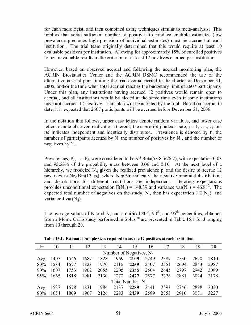
for each radiologist, and then combined using techniques similar to meta-analysis. This implies that some sufficient number of positives to produce credible estimates (low prevalence precludes high precision of individual estimates) must be accrued at each institution. The trial team originally determined that this would require at least 10 evaluable positives per institution. Allowing for approximately 15% of enrolled positives to be unevaluable results in the criterion of at least 12 positives accrued per institution. However, based on observed accrual and following the accrual monitoring plan, the ACRIN Biostatistics Center and the ACRIN DSMC recommended the use of the alternative accrual plan limiting the trial accrual period to the shorter of December 31, 2006, and/or the time when total accrual reaches the budgetary limit of 2607 participants. Under this plan, any institutions having accrued 12 positives would remain open to accrual, and all institutions would be closed at the same time even if some institutions have not accrued 12 positives. This plan will be adopted by the trial. Based on accrual to date, it is expected that 2607 participants will be accrued before December 31, 2006. In the notation that follows, upper case letters denote random variables, and lower case letters denote observed realizations thereof; the subscript j indexes site, j = 1, . . ., J; and iid indicates independent and identically distributed. Prevalence is denoted by P, the number of participants accrued by N, the number of positives by N+, and the number of negatives by N-. Prevalences, P1, . . . PJ, were considered to be iid Beta(58.8, 676.2), with expectation 0.08 and 95.53% of the probability mass between 0.06 and 0.10. At the next level of a hierarchy, we modeled N-j given the realized prevalence pj and the desire to accrue 12 positives as NegBin(12, pj), where NegBin indicates the negative binomial distribution, and distributions for different institutions are independent. Iterating expectations provides unconditional expectation E(N-j) = 140.39 and variance var(N-j) = 46.812. The expected total number of negatives on the study, N-, then has expectation J E(N-j) and variance J var(N-j). The average values of N- and N, and empirical 80th, 90th, and 95th percentiles, obtained from a Monte Carlo study performed in Splus130 are presented in Table 15.1 for J ranging from 10 through 20.
Table 15.1. Estimated sample sizes required to accrue 12 positives at each institution
J= 10 11 12 13 14 15 16 17 18 19 20 Number of Negatives, N-
Avg 1407 1546 1687 1828 1969 2109 2249 2389 2530 2670 2810 80% 1534 1677 1823 1970 2115 2259 2407 2551 2694 2843 2987 90% 1607 1753 1902 2055 2205 2355 2504 2645 2797 2942 3089 95% 1665 1818 1981 2130 2272 2427 2577 2726 2881 3024 3178
Total Number, N Avg 1527 1678 1831 1984 2137 2289 2441 2593 2746 2898 3050 80% 1654 1809 1967 2126 2283 2439 2599 2755 2910 3071 3227
ACRIN 6664 51 July 7, 2006

90% 1727 1885 2046 2211 2373 2535 2696 2849 3013 3170 3329 95% 1785 1950 2125 2286 2440 2607 2769 2930 3097 3252 3418
We know from experience that sensitivity will also vary across radiologists due to variability across participants and to variability in performance abilities across radiologists. In the current study, a single radiologist will interpret all of the CTC exams obtained within an institution. The true individual sensitivities, Sj, are considered to be iid Beta(� �), with parameters chosen to provide expected sensitivity s and variance �2
s. We expect that if 12 positives are accrued at each institution, there will be 10 evaluable positives per institution (standard 85% attrition for clinical trials).
Given the realized individual sensitivity, sj, and common n*
+j=10, we modeled the number of positives detected by each radiologist, Rj, as Bin(10, sj), where Bin indicates the binomial distribution and distributions for different radiologists are independent. Individual estimates of sensitivity are , with unconditional expectation */ˆ jjj nrs +=
( ) sSE j =ˆ and variance ( ) ( ) ( ){ }.111ˆvar 2** ssn
nS sj
jj −+−= +
+
σ . Sample size calculations
focused on the simple average ∑= jsJ
s ˆ1ˆ . This estimator is unbiased for s (the central
tendency of sensitivity in the population of potential radiologists interpreting CTC
exams); its variance is ( ) ( ) ( ){ }∑ −+−= ++
.1111ˆvar 2**2 ssn
nJS sj
j
σ This variance is
estimated by ( ) ( )∑ −−
= .ˆˆ)1(
1ˆrav 2ssJJ
s j
The primary goal of this study is to achieve estimated ( ) 205.0ˆvar ≤s . We performed a Monte Carlo study in Splus, using s and �2
s based on the data from ACRIN 6656. In that trial, subjects were divided into 3 reading groups, and each of 18 readers interpreted CTC examinations for 2 groups in a counterbalanced fashion. The analog to the proposed trial is a triplet of 3 different readers interpreting 3 different reading groups; there were a total of 1512 such unique triplets. For each triplet we computed and an unbiased estimator
of �2s obtained as
s
( ) ( )
⎪⎪⎭
⎪⎪⎬
⎫
⎪⎪⎩
⎪⎪⎨
⎧
−−
−−−
⎟⎟⎠
⎞⎜⎜⎝
⎛−
−
=
∑
∑∑
∑
∑
+
+
+
+ ss
nJ
nJ
ssJ
nJJ
nJ
j
jj
j
js ˆ1ˆ
1
1
ˆˆ1
1
1
1
ˆ
*2
*2
*
*2
2σ . The
mean and median of were 0.7492 and 0.7500, respectively. The mean and median of the distribution of over the triplets were 0.09962 and 0.07242, respectively. Based on the previous studies, we were also interested in s=0.65. We thus explored precision for each combination of s=(0.65, 0.70, 0.75, 0.80) and �2
s = (0.102, 0.072, 0.052). For each combination, we obtained the appropriate (� �) and performed 3000 simulations, as follows: 1) draw s1, . . ., s20 from Beta(��); 2) given s1, . . ., s20, draw r1, . . ., r20 from
s2ˆ sσ
ACRIN 6664 52 July 7, 2006
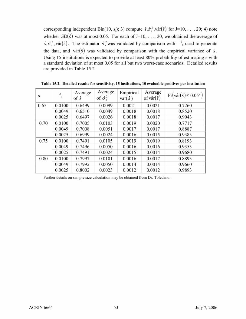
corresponding independent Bin(10, sj); 3) compute ( )ss s ˆrav,ˆ,ˆ 2σ for J=10, . . ., 20; 4) note whether was at most 0.05. For each of J=10, . . ., 20, we obtained the average of
. The estimator was validated by comparison with �2s used to generate
the data, and was validated by comparison with the empirical variance of s . Using 15 institutions is expected to provide at least 80% probability of estimating s with a standard deviation of at most 0.05 for all but two worst-case scenarios. Detailed results are provided in Table 15.2.
( )sSD ˆ( )ss s ˆrav,ˆ,ˆ 2σ 2ˆ sσ
( )srav ˆ
Table 15.2. Detailed results for sensitivity, 15 institutions, 10 evaluable positives per institution
s �2s
Average of s
Average of 2ˆ sσ
Empirical var( ) s
Average of ( )srav ( )( )205.0ˆravPr ≤s
0.0100 0.6499 0.0099 0.0021 0.0021 0.7260 0.0049 0.6510 0.0049 0.0018 0.0018 0.8520
0.65
0.0025 0.6497 0.0026 0.0018 0.0017 0.9043 0.0100 0.7005 0.0103 0.0019 0.0020 0.7717 0.0049 0.7008 0.0051 0.0017 0.0017 0.8887
0.70 0.0025 0.6999 0.0024 0.0016 0.0015 0.9383
0.0100 0.7491 0.0105 0.0019 0.0019 0.8193 0.0049 0.7496 0.0050 0.0016 0.0016 0.9353
0.75 0.0025 0.7491 0.0024 0.0015 0.0014 0.9680
0.0100 0.7997 0.0101 0.0016 0.0017 0.8893 0.0049 0.7992 0.0050 0.0014 0.0014 0.9660
0.80 0.0025 0.8002 0.0023 0.0012 0.0012 0.9893
Further details on sample size calculation may be obtained from Dr. Toledano.
ACRIN 6664 53 July 7, 2006
16.0 GENDER/MINORITY RECRUITMENT
16.1 Participating Site Recruitment Institutions that wish to join the National CT Colonography Trial are required to complete an ACRIN Protocol Specific Application (PSA) as found on the ACRIN web site (www.acrin.org/).ACRIN’s Institutional Participants Committee (IPC) reviews all PSAs to ensure the 15 participating sites can perform the protocol requirements that relate to the study’s primary aims. However, sites also will be required to answer questions in the PSA about the institution’s ability to enroll minority participants. If, as anticipated, more PSAs are submitted to ACRIN than can be accepted to participate in the trial, an institution’s ability to recruit minority and female participants will be an important criterion for site selection.
Demographic Criterion: The PSA application will request documentation of minority and female patient encounters for the most recent two years for the institution and, if available, specifically for the gastroenterology department. These data will be used in the site selection process and in determining realistic minority and women recruitment goals for each institution.
Clinician Diversity Criterion: It is well documented that people are much more likely to trust and want to be a part of programs in which the representatives look like them and come from their culture. As such, institutions will record information about minority representation within the study team as well as about minority clinicians at their institution who could potentially refer patients into the study including primary care physicians, gynecologists, and gastroenterologists.
Minority Recruitment Interest Level Criterion: Institutions will be asked to rate their level of commitment for enrolling minority participants into the trial. An example rating scale could include:
• High: Will develop an extensive program to recruit patients and provide program
outline. • Medium: Will implement several minority recruitment activities and provide
examples. • Low: Is not interested in implementing minority recruitment activities.
16.2 Participant Recruitment Tools
ACRIN Brochure: ACRIN will produce a patient brochure that includes a graphical representation of an ethnically diverse population for the sites to distribute throughout their institution and network.
Brochure Translation: ACRIN will investigate funding for the Spanish translation of the patient brochure as well as consider translation into other languages when provided documentation of the need.
ACRIN 6664 54 July 7, 2006
Coordination with NCI Partnership Program: ACRIN will work with the selected trial sites to coordinate minority recruitment efforts with the National Cancer Center’s Partnership Program when and if appropriate.
17.0 ADVERSE EVENT REPORTING
17.1 Definition of Adverse Event An Adverse Event (AE) is any untoward medical occurrence in a participant that does not necessarily have a causal relationship with the study intervention. An AE can therefore be any unfavorable and unintended sign (including an abnormal laboratory finding), symptom or disease temporally associated with the use of a medical treatment or procedure regardless of whether it is considered related to the medical treatment or procedure (attribution of unrelated, unlikely, possible, probable, or definite).
17.2 Definition of Serious Adverse Effect A Serious Adverse Event (SAE) is defined as any untoward medical occurrence that:
Results in death or Is life-threatening (at the time of the event) or Requires inpatient hospitalization or prolongation of an existing
hospitalization or Results in persistent or significant disability or incapacity or Results in congential anomaly/birth defects.
17.3 Adverse Event Grading Grade is used to denote the severity of the adverse event. An AE is graded using the following categories (provided the term does NOT appear in the current version of the Common Toxicity Criteria for Adverse Events [CTCAE 3.0]):
1 – Mild 2 – Moderate 3 – Severe 4 – Life-threatening or disabling 5 – Fatal
(For terms listed in the CTCAE, the grade is still recorded as 1, 2, 3, 4, or 5; however, the definition of the various grades will be specific to the term being used.)
17.4 Regulatory and Reporting Requirements of Adverse Events Routine reporting is defined as documentation of adverse events on source documents and AE CRF, and submission to ACRIN for preparation of a report for Data and Safety Monitoring Committee (DSMC) review, quarterly reports to CDUS, and the final study report.
ACRIN 6664 55 July 7, 2006
Expedited reporting is defined as immediate notification of NCI and ACRIN within the specified timeframe outlined in the protocol and the ACRIN Adverse Event Reporting Manual. Routine reporting requirements also apply. The adverse event reporting period for this protocol is defined as the period from the initiation of any study procedures and up to 30 days after the last primary intervention. Serious Adverse Events meeting the criteria for expedited reporting, as specified in the protocol, require (a) telephone notification to both NCI and ACRIN within 24 hours of first knowledge of death (ONLY), (b) completed AdEERS report faxed to both NCI and ACRIN within 10 days of knowledge and a hard copy to NCI only via mail, and (c) documentation of event on AE case report form for scheduled submission to ACRIN. Adverse Events meeting the criteria for routine reporting, as specified in the protocol, should be reported using the Adverse Event Case Report Form and submitted to ACRIN.
17.4.1 Adverse events will be reported for the period of the protocol in which
participants undergo primary interventions (screening CTC and colonoscopy; ingestion of bowel preparation solutions, bisacodyl tablets, and glucagon; insertion of enema tip; and balloon insufflation and colorectal insufflation). The reporting of AEs in this protocol will conform to the following: 1. Grade 3 Expected and Unexpected AEs with attribution of possible, probable,
or definite will be reported by routine reporting procedures. 2. Grade 4 Expected AEs with attribution of possible, probable, or definite will
be reported by routine reporting procedures. 3. Grade 4 Unexpected AEs with attribution of possible, probable, or definite
will be reported within ten (10) days of first knowledge of the event by Expedited Written Report.
4. Grade 5 AEs, or Deaths with attribution of possible, probable, or definite will be reported within 24 hours of first knowledge of the event by Telephone Report to ACRIN and NCI-CIP and followed by Expedited Written Report within ten (10) days of first knowledge of the event.
The following table summarizes the reporting requirements for AEs:
Type of Report AEs occurring within 2 hours of the primary trial interventions (except for
wound infection: occurring within 1 week)
Routine Reporting
Expedited Written Report in 10
days of first knowledge of AE
Telephonic Report within 24 hours of
first knowledge of AE to NCI-CIP and
ACRIN
ACRIN 6664 56 July 7, 2006
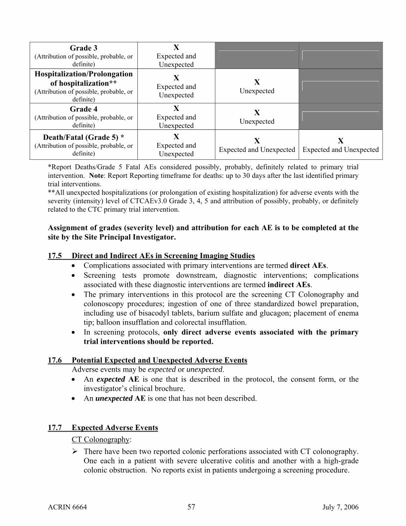
Grade 3 (Attribution of possible, probable, or
definite)
X Expected and Unexpected
Hospitalization/Prolongation of hospitalization**
(Attribution of possible, probable, or definite)
X Expected and Unexpected
X Unexpected
Grade 4 (Attribution of possible, probable, or
definite)
X Expected and Unexpected
X Unexpected
Death/Fatal (Grade 5) * (Attribution of possible, probable, or
definite)
X Expected and Unexpected
X X Expected and Unexpected Expected and Unexpected
*Report Deaths/Grade 5 Fatal AEs considered possibly, probably, definitely related to primary trial intervention. Note: Report Reporting timeframe for deaths: up to 30 days after the last identified primary trial interventions. **All unexpected hospitalizations (or prolongation of existing hospitalization) for adverse events with the severity (intensity) level of CTCAEv3.0 Grade 3, 4, 5 and attribution of possibly, probably, or definitely related to the CTC primary trial intervention. Assignment of grades (severity level) and attribution for each AE is to be completed at the site by the Site Principal Investigator.
17.5 Direct and Indirect AEs in Screening Imaging Studies
• Complications associated with primary interventions are termed direct AEs. • Screening tests promote downstream, diagnostic interventions; complications
associated with these diagnostic interventions are termed indirect AEs. • The primary interventions in this protocol are the screening CT Colonography and
colonoscopy procedures; ingestion of one of three standardized bowel preparation, including use of bisacodyl tablets, barium sulfate and glucagon; placement of enema tip; balloon insufflation and colorectal insufflation.
• In screening protocols, only direct adverse events associated with the primary trial interventions should be reported.
17.6 Potential Expected and Unexpected Adverse Events
Adverse events may be expected or unexpected. • An expected AE is one that is described in the protocol, the consent form, or the
investigator’s clinical brochure. • An unexpected AE is one that has not been described.
17.7 Expected Adverse Events CT Colonography:
There have been two reported colonic perforations associated with CT colonography. One each in a patient with severe ulcerative colitis and another with a high-grade colonic obstruction. No reports exist in patients undergoing a screening procedure.
ACRIN 6664 57 July 7, 2006
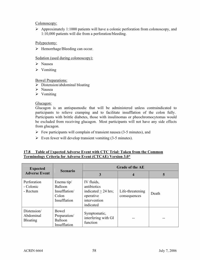
Colonoscopy: Approximately 1:1000 patients will have a colonic perforation from colonoscopy, and
1:10,000 patients will die from a perforation/bleeding. Polypectomy:
Hemorrhage/Bleeding can occur.
Sedation (used during colonoscopy): Nausea Vomiting
Bowel Preparations:
Distension/abdominal bloating Nausea Vomiting
Glucagon: Glucagon is an antispasmodic that will be administered unless contraindicated to participants to relieve cramping and to facilitate insufflation of the colon fully. Participants with brittle diabetes, those with insulinomas or pheochromocytomas would be excluded from receiving glucagon. Most participants will not have any side effects from glucagon.
Few participants will complain of transient nausea (3-5 minutes), and Even fewer will develop transient vomiting (3-5 minutes).
17.8 Table of Expected Adverse Event with CTC Trial: Taken from the Common Terminology Criteria for Adverse Event (CTCAE) Version 3.0*
Grade of the AE Expected
Adverse Event Scenario 3 4 5
Perforation - Colonic - Rectum
Enema tip/ Balloon Insufflation/ Colon Insufflation
IV fluids, antibiotics indicated > 24 hrs; operative intervention indicated
Life-threatening consequences Death
Distension/ Abdominal Bloating
Bowel Preparation/ Balloon Insufflation
Symptomatic, interfering with GI function
-- --
ACRIN 6664 58 July 7, 2006

Nausea Glucagon/ Bowel Preparation
Inadequate oral caloric or fluid intake; IV fluids, tube feedings, or TPN indicated > 24 hrs.
Life-threatening consequences Death
Vomiting Glucagon/ Bowel Preparation
> 6 episodes in 24 hrs; IV fluids, or TPN indicated > 24 hrs.
Life-threatening consequences Death
Hemorrhage/ Bleeding**
Polypectomy/ Enema tip/ Balloon Insufflation/ Colon Insufflation
Transfusion, post-operative interventional radiology, endoscopic or operative intervention is indicated.
Life-threatening; disabling Death
*Note: The table of expected events contains many, but not all, adverse events that could occur within the CTC Trial. **Associated with surgery, intra-operative or postoperative: postoperative period is defined as < 72 hours after surgical procedure.
17.9 When to Report Prompt reporting of adverse events is the responsibility of each investigator, clinical research associate, and nurse engaged in clinical research. Anyone uncertain about whether a particular serious adverse event should be reported need to contact the ACRIN headquarters at 215-574-3150 for assistance. Any adverse event considered NOT directly related to the treatment or procedure should NOT be reported as a serious adverse event in this trial. General guidance can also be found in the ACRIN Adverse Event Reporting Manual.
All deaths/Grade 5 fatal events occurring up to 30 days after the last identified primary trial intervention, with attribution of possible, probable, or definite and regardless of whether the event was expected or unexpected must be reported following the expedited AE reporting requirements.
All deaths/fatal (Grade 5) adverse events associated with the primary trial interventions should also be reported by telephone to NCI and ACRIN within 24 hours of first knowledge of the event, followed by Expedited Written Report within ten (10) days of first knowledge of the event.
ACRIN 6664 59 July 7, 2006
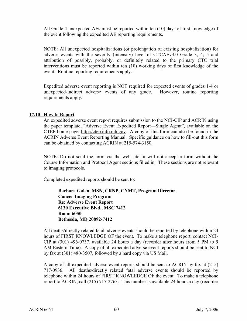
All Grade 4 unexpected AEs must be reported within ten (10) days of first knowledge of the event following the expedited AE reporting requirements. NOTE: All unexpected hospitalizations (or prolongation of existing hospitalization) for adverse events with the severity (intensity) level of CTCAEv3.0 Grade 3, 4, 5 and attribution of possibly, probably, or definitely related to the primary CTC trial interventions must be reported within ten (10) working days of first knowledge of the event. Routine reporting requirements apply. Expedited adverse event reporting is NOT required for expected events of grades 1-4 or unexpected-indirect adverse events of any grade. However, routine reporting requirements apply.
17.10 How to Report
An expedited adverse event report requires submission to the NCI-CIP and ACRIN using the paper template, “Adverse Event Expedited Report—Single Agent”, available on the CTEP home page, http://ctep.info.nih.gov. A copy of this form can also be found in the ACRIN Adverse Event Reporting Manual. Specific guidance on how to fill-out this form can be obtained by contacting ACRIN at 215-574-3150.
NOTE: Do not send the form via the web site; it will not accept a form without the Course Information and Protocol Agent sections filled in. These sections are not relevant to imaging protocols. Completed expedited reports should be sent to:
Barbara Galen, MSN, CRNP, CNMT, Program Director Cancer Imaging Program Re: Adverse Event Report 6130 Executive Blvd., MSC 7412 Room 6050 Bethesda, MD 20892-7412
All deaths/directly related fatal adverse events should be reported by telephone within 24 hours of FIRST KNOWLEDGE OF the event. To make a telephone report, contact NCI-CIP at (301) 496-0737, available 24 hours a day (recorder after hours from 5 PM to 9 AM Eastern Time). A copy of all expedited adverse event reports should be sent to NCI by fax at (301) 480-3507, followed by a hard copy via US Mail.
A copy of all expedited adverse event reports should be sent to ACRIN by fax at (215) 717-0936. All deaths/directly related fatal adverse events should be reported by telephone within 24 hours of FIRST KNOWLEDGE OF the event. To make a telephone report to ACRIN, call (215) 717-2763. This number is available 24 hours a day (recorder
ACRIN 6664 60 July 7, 2006

after hours from 5 PM to 8:30 AM Eastern Time). During business hours, ACRIN Data Managers for the protocol will be available. All expedited adverse event reports should be sent to your local Institutional Review Board (IRB). Adverse events not requiring expedited reporting are normally reported to the local IRB in an annual report and/or continuing review. Please refer to your local institution’s IRB policies regarding adverse events, serious adverse events, and safety reports.
ACRIN 6664 61 July 7, 2006
REFERENCES 1. Parker S, Torry T, Bolden S, Windigo P. Cancer Statistics 1996. CA - Cancer J Clin 1996;65:5-27. 2. Bowland C, Malignant tumors of the colon, in Textbook of Gastroenterology, T. Yamada, Editor.
1995, J.B. Lippincott, Co.: New York. p. 1967-2026. 3. Winawer S, Flehinger B, Schottenfeld D, et al. Screening for colorectal cancer with fecal blood
testing and sigmoidoscopy. J Natl Cancer Inst 1993;85:1311. 4. Winawer S, Zauber A, Ho M, et al. Prevention of colorectal cancer by colonoscopic polypectomy.
N Engl J Med 1993;329:1977-81. 5. Muller A, Sonnenberg A. Protection by endoscopy against death from colorectal cancer. A case-
control study among veterans. Arch Int Med 1995;155:1741-48. 6. Muller A, Sonnenberg A. Prevention of colorectal cancer be flexible endoscopy and
polypectomy—a case control study of 32,702 veterans. Ann Int Med 1995;123:904-10. 7. Selby J, Friedman G, Quesenberry C, et al. A case-control study of screening sigmoidoscopy and
mortality from colorectal cancer. N Engl J Med 1992;326:653-57. 8. Selby J, Friedman G, Quesenberry C, Weiss N. Effect of fecal occult blood testing on morality
from colorectal cancer: a case control study. Ann Int Med 1993;118:1-6. 9. Newcomb P, Norfleet R, Storer B, et al. Screening sigmoidoscopy and colorectal cancer mortality.
J Natl Cancer Inst 1992;84:1572-75. 10. Lazovich D, Weiss N, Stevens N, et al. A case-control study to evaluate the effectiveness of
screening for fecal occult blood. J Med Screen 1995;2:84-89. 11. Eddy D, Nugent T, Eddy J, et al. Screening for colorectal cancer in a high-risk population: results
of a mathematical model. Gastroenterology 1987;92:682-92. 12. Eddy D. Screening for colorectal cancer. Ann Int Med 1990;113:373-84. 13. Wagner J, Tunis S, Brown M, Ching A, Almeda R, The cost-effectiveness of colorectal cancer
screening in average risk adults, in Prevention and Early Detection of Colorectal Cancer, G. Young and B. Levin, Editors. 1996, W.B. Saunders: Philadelphia.
14. Ahlquist D, Occult gastrointestinal bleeding, in Textbook of Gastroenterology, T. Yamada, Editor. 1995, J.B. Lippincott, Co.: New York. p. 699-717.
15. Simon J. Occult blood screening for colorectal carcinoma: a critical review. Gastroenterology 1985;88:820-37.
16. Ahlquist D, McGill D, Schwartz S, et al. Fecal blood levels in health and disease: a study using HemoQuant. N Engl J Med 1985;312:1422-28.
17. Ahlquist D, McGill D, Fleming J, et al. Patterns of occult bleeding in asymptomatic colorectal cancer. Cancer 1989;63:1826-30.
18. Ahlquist D, Wieand H, Moertel C, McGill D, et al. Accuracy of fecal occult blood screening for colorectal neoplasia: a prospective study using hemoccult and hemoquant tests. JAMA 1993;269:1262-67.
19. Bang K, Tillett S, Noar S, Blair A, McDougall V. Sensitivity of fecal hemoccult testing and flexible sigmoidoscopy for colorectal cancer screening. J Occup Med 1986;28:709-13.
20. Demers R, Stawick L, Demers P. Relative sensitivity of the fecal occult blood test and flexible sigmoidoscopy in detecting polyps. Preventive Med 1985;14:55-62.
21. Jahn H, Joergensen O, Kronborg O, Fenger C. Can hemoccult II replace colonoscopy after radical surgery for colorectal cancer or after polypectomy? Dis Colon Rectum 1992;35:253-56.
22. Ribet A, Frexinos J, Escourrou J, Delpu J. Occult blood tests and colorectal tumors. Lancet 1980;1:417-21.
ACRIN 6664 62 July 7, 2006
23. Robinson M, Kronborg O, Williams C, Bostock K, et al. Faecal occult blood testing and colonoscopy in the surveillance of subjects at high risk of colorectal neoplasia. Br J of Surg 1995;82:318-20.
24. Rozen P. Screening for colorectal neoplasia in the Tel Aviv area: cumulative data 1979-89 and initial conclusions. Is J Med Sci 1992;28:8-20.
25. Williams C, Macrae F, Bartram C. A prospective study of diagnostic methods in adenoma follow-up. Endoscopy 1982;14:74-78.
26. Mandel J, Bond J, Church T, et al. Reducing mortality from colorectal cancer by screening for fecal occult blood. N Engl J Med 1993;328:1365-71.
27. Ahlquist D. Fecal occult blood testing for colorectal cancer. Can we afford to do this? (Review). Gastroenterol Clin N Am 1997;26(1):41-55.
28. Frank J. Occult blood screening for colorectal cancer: the yield and costs. Am J Prev Med 1985;1:18-24.
29. Ahlquist D, Moertel C, McGill D. Screening for colorectal cancer (Letter-to-Editor). N Engl J Med 1993;329:1351.
30. Hardcastle J, Chamberlain J, Robinson M, Moss S, Amar S, et al. Randomised controlled trial of faecal occult blood screening for colorectal cancer. Lancet 1996;348:1472-77.
31. Kronberg O, Fenger C, Olson J, Jorgensen O, Sondergaard O. Randomised study of screening for colorectal cancer with faecal occult blood test. Lancet 1996;348:1467-71.
32. Scheitel S, Ahlquist D, Wollan P, Hagen P, Silverstein M. Colorectal cancer screening: a case control community study. J GIM 1995;10(4):5103.
33. Neale A, Demers R, Herman S. Compliance with colorectal cancer screening in a high-risk occupational group. J Occup Med 1989;31:1007-12.
34. Robra S, Schwartz F, Experiences with a nation-wide screening program for colorectal cancer in the Federal Republic of West Germany, in Hemoccult screening for the early detection of colorectal cancer, J. Hardcastle, Editor. 1986, Schattaurer: Stuttgart. p. 13-20.
35. Lemmel G, Hageman J, Rex D, Rahmani E. Neoplasia distal to the splenic flexure in patients with proximal colon cancer. Gastrointest Endosc 1996;44:109-11.
36. Chodash H, Ahlquist D. Are rectosigmoid polyps a sensitive marker for proximal colon cancers? Gastrointest Endosc 1990;36:213.
37. Dinning J, Hixson L, Clark L. Prevalence of distal colonic neoplasia associated with proximal colon cancer. Arch Int Med 1994;154(8):853-56.
38. Maule W. Screening for colorectal cancer by nurse endoscopists. N Engl J Med 1994;330:183-87. 39. Berry D, Clarke P, Hardcastle J, Vellacott K. Randomized trial of the addition of flexible
sigmoidoscopy to faecal occult blood testing for colorectal neoplasia population screening. Br J Surg 1999;84:1274-76.
40. Pignone M, Bucholtz D, Harris R. Patient preferences for colorectal cancer screening. J Gen Int Med 1999;14:432-37.
41. Fork F. Double contrast enema and colonoscopy in polyp detection. Gut 1981;22:971-77. 42. Steine S, Stordahl A, Lunde O, et al. Double-contrast barium enema versus colonoscopy in the
diagnosis of neoplastic disorders: aspects of decision-making in general practice. Fam Prac 1993;10:288-91.
43. Johnson C, Carlson H, WF T, Weiland L. Barium enemas in carcinoma of the colon: sensitivity of double- and single-contrast studies. AJR 1983;140:1143-49.
44. Ott D, Analysis of accuracy, complications, and cost of barium enema for CRC diagnosis. 1994, NIH: Bethesda, MD.
ACRIN 6664 63 July 7, 2006
45. Norfleet R, Ryan M, Wyman J, et al. Barium enema versus colonoscopy for patients with polyps found during flexible sigmoidoscopy. Gastrointest Endosc 1991;37:531-34.
46. Rankin G, Indications, contraindications, and complications of colonoscopy, in Gastroenterologic Endoscopy, M. Sivak, Editor. 1987, W.B. Saunders, Co. p. 868-80.
47. Jarvinen H, Mecklin J, Sistonen P. Screening reduces colorectal cancer rate in families with hereditary nonpolyposis colorectal cancer. Gastroenterol 1995;108:1405-11.
48. Lieberman D. Cost-effectiveness model for colon cancer screening. Gastroenterology 1995;109:1781-90.
49. Thiss-Evenson E, Hoff G, Sauar J, Langmark F, Majak B, Vatn M. Population-based surveillance by colonoscopy: effect on the incidence of colorectal cancer. Telemark Polyp Study I. Scand J Gastroenterol 1999;34:414-20.
50. Vining D. Virtual endoscopy: is it reality? Radiology 1996;200:30-31. 51. Gelfand D, Chen M, Ott D. Preparing the colon for the barium enema. Radiology 1991;178:609-
13. 52. Hara A, Johnson C, Reed J, et al. Detection of colorectal polyps by computed tomographic
colography: feasibility of a novel technique. Gastroenterology 1996;110:284-90. 53. Hara A, Johnson C, Reed J, Ehman R, Ilstrup D. Colorectal polyp detection using computed
tomographic colography: two- versus three-dimensional techniques. Radiology 1996;200:49-54. 54. Royster A, Fenlon H, Clarke P, Nunes D, Ferrucci J. CT colonoscopy of colorectal neoplasms:
two-dimensional and three-dimensional virtual-reality techniques with colonoscopic correlation. AJR 1997;169:1237-42.
55. Dachman A, Kuniyoshi J, Boyle C, Samara Y, et al. CT colonography with three-dimensional problem solving for detection of colonic polyps. AJR 1998;171:989-95.
56. Macari M, Berman P, Dicker M, Milano A, Megibow A. Usefulness of CT colonography in patients with incomplete colonoscopy. AJR 1999;173:561-64.
57. Fenlon H, Nunes D, Schroy PI, Barish M, Clarke P, Ferrucci J. A comparison of virtual and conventional colonoscopy for the detection of colorectal polyps. NEJM 1999;341(20):1496-1503.
58. Fenlon H, McAneny D, Nunes D, Clarke P, Ferrucci J. Occlusive colon carcinoma: virtual colonoscopy in the preoperative evaluation of the proximal colon. Radiology 1999;210:423-428.
59. Morrin M, Kruskal J, Farrell R, Goldberg S, McGee J, Raptopoulos V. Endoluminal CT colonography after an incomplete endoscopic colonoscopy. AJR 1999;172:913-18.
60. Chen S, Lu D, Hecht J, Kadell B. CT colonography: value of scanning in both the supine and prone positions. AJR 1999;172:595-99.
61. Yee J, Akerkar G, Hung R, Sreinauer-Gebauer A, McQuaid K, Wall S. Performance characteristics of CT colonography for colorectal polyp and cancer detection (abstract). AJR 2000;174(3 Suppl):55.
62. Fletcher J, Johnson C, Welch T, MacCarty R, Ahlquist D, Reed J. Optimization of CT colonography technique: a prospective trial in 180 patients. Radiology 2000;216:704-11.
63. Vining D, Gelfand D. Noninvasive colonoscopy using helical CT scanning, 3D reconstruction, and virtual reality. in Presented at the 1994 meeting of the Society of Gastrointestinal Radiologists. February 13-18, 1994. Maui, Hawaii.
64. Vining D, Gelfand D, Bechtold R, et al. Technical feasibility of colon imaging with helical CT and virtual reality (abstract). Am J Roentgenol 1994;162(Suppl):104.
65. Magnusson M, Lenz R, Danielsson P. Evaluation of methods for shaded suface display of CT volumes. Comput Med Imaging Graph 1991;15:247-56.
66. McAdams H, Shahidi R, Argiro V, Chotas H, Kurylo W, Tapson V, Clinical evaluation of perspective surface and volume rendering techniques for performing virtual bronchoscopy, in
ACRIN 6664 64 July 7, 2006
Computer Assisted Radiology (Proceedings of the International Symposium on Computer and Communication Systems for Image Guided Diagnosis and Therapy), H. Lemke, et al., Editors. 1996, Elsevier Science B.V.: New York. p. 73-77.
67. Rubin G, Beaulieu C, Argiro V, Ringl H, Norbash A, et al. Perspective volume rendering of CT and MR images: applications for endoscopic imaging. Radiology 1996;199:321-30.
68. Reed J, Hara A, Johnson C. Interpretation of image sets containing convoluted tubular structures via transluminal sections and steerable intraluminal perspective views. Medical Imaging 1996: Image Processing, Loew MH, Hanson KM, Eds. Proc SPIE 1996;2710:1009-19.
69. Beaulieu C, Napel S, Daniel B, Ch’en I, Rubin G, Johnstone I, Jeffrey R, Jr. Detection of colonic polyps in a phantom model: implications for virtual colonoscopy data acquisition. J Comput Assist Tomogr 1998;22(4):656-663.
70. Yee J, Hung R, Akerkar G, Wall S. The usefulness of glucagon hydrochloride for colonic distention in CT colonography. AJR 1999;173:169-72.
71. Reed J, Johnson C. Image display system for synchronous interpretation of supine and prone computed tomographic colography. Medical Imaging 1998: Image Display, Kim Y and Mun SK, Eds. Proc SPIE 1998;3335:306-15.
72. Reed J, Johnson C. Virtual pathology: a new paradigm for interpretation of computed tomographic colonography. Medical Imaging 1998: Image Display, Kim Y and Mun SK, Eds. Proc SPIE 1998;3335:439-49.
73. Karadi C, Beaulieu C, Jeffrey RJ, Paik D, Napel S. Display modes for CT colonography. Part I: Synthesis and insertion of polyps into patient CT data. Radiology 1999;212:195-201.
74. Beaulieu C, Jeffrey RJ, Karadi C, Paik D, Napel S. Display modes for CT colonography. Part II. Blinded comparison of axial CT and virtual endoscopic and panoramic endoscopic volume-rendered studies. Radiology 1999;212:203-212.
75. McFarland E, Wang G, Brink J, Balfe D, Heiken J, Vannier M. Spiral computed tomographic colonography: determination of the central axis and digital unraveling of the colon. Acad Radiol 1997;4:367-73.
76. McFarland H, Brink J, Loh J, Wang G, Argiro, V, Balfe D, Heiken J, Vannier M. Visualization of colorectal polyps with spiral CT colography: evaluation of processing parameters with perspective volume rendering. Radiology 1997;205:701-07.
77. Reed J, Johnson C. Automatic segmentation, tissue characterization, and rapid diagnosis enhancements to the computed tomographic colonography analysis workstation. Proc SPIE 1997;3033:433-40.
78. Wang G, Dave S, Brown B, Zhang Z, McFarland E, Haller J, Vannier M. Colon unraveling based on electrical field - recent progress and further work (abstract). in Presented at the SPIE Annual Meeting, February 19-251999. San Diego, CA.
79. McFarland E, Brink J. Helical CT colonography (virtual colonoscopy): the challenge that exists between advancing technology and generalizability. AJR 1999;173:549-559.
80. Hopper K, Iyriboz A, Wise S, Neuman J, Mauger D, Kasales C. Mucosal detail at CT virtual reality: surface versus volume rendering. Radiology 2000;214:517-22.
81. Whiting B, McFarland E, Dellabarca C, Brunsden B. Study of CT artifacts in virtual colonoscopy (abstract). in Presented at the SPIE Annual Meeting, February 19-251999. San Diego, CA.
82. Stoll E, Stern C, Stucki P, Wildermuth S. A new filtering algorithm for medical magnetic resonance and computer tomography images. J Digital Imaging 1999;12(1):23-28.
83. Paik D, Beaulieu C, Jeffrey R, Rubin G, Napel S. Automated flight path planning for virtual endoscopy. Med Phys 1998;25(5):629-37.
84. Wyatt C, Ge Y, Vining D. Automatic segmentation of the colon (abstract). in Presented at the SPIE Annual Meeting, February 19-251999. San Diego, CA.
ACRIN 6664 65 July 7, 2006
85. Fenlon H, Clarke P, Ferrucci J. Virtual colonoscopy: imaging features with colonoscopic correlation. AJR 1998;170:1303-09.
86. Fletcher J, Johnson C, MacCarty R, Welch T, Reed J, Hara A. CT colonography: potential pitfalls and problem-solving techniques. AJR 1999;172:1271-78.
87. Hara A, Johnson C, Reed J. Colorectal lesions: evaluation with CT colography. RadioGraphics 1997;17:1157-67.
88. Johnson CD, Toledano AY, Herman BA et al. Computerized Tomographic Colonography: Performance Evaluation in a retrospective multi-center setting. Gastroenterology. 2003 Sep;125(3):688-95.
89. Johnson CD, Harmsen SW, MacCarty RL et. al. Prospective Blinded Evaluation of CT Colonography for Screen-Detection of Colorectal Polyps. Accepted in Gastroenterology.
90. McFarland HG, Brink JA, Pilgram TK, et al. Spiral CT colonography: reader agreement and diagnostic performance with two- and three-dimensional image-display techniques. Radiology 2001; 218:375-383.
91. Golub RW, Kerner BA, Wise WE, Meesig DM, Hartmann RF, Khanduja KS, Aguilar PS. Colonoscopic bowel preparations- which one? A blinded , prospective,randomized trial. Dis Colon Rectum 1995;38:594-599.
92. Afridi SA, Barthel JS, King PD, Pineda JJ, Marshall JB. Prospective, randomized trial comparing a new sodium phosophate-bisacodyl regimen with conventional PEG-ES lavage for outpatient colonoscopy preparation. Gastrointest Endosc 1995;41:485-489.
93. Marshall JB, Pineda JJ, Barthel JS, King PD. Prospective, randomized trail comparing sodium phosphate solution with polyethylene glycol-electrolyte lavage for colonoscopy preparation. Gastrointest Endosc 1993;39:631-634.
94. Cohen SM, Wexner SD, Binderow SR, Nogueras JJ, Daniel N, Ehrenpreis ED, Jensen J, Bonner GF, Ruderman WB. Prospective, randomized, endoscopic-blinded trial comparing precolonoscopy bowel cleansing methods. Dis Colon Rectum 1994;37:689-696.
95. Kolts BE, Lyles WE, Achem SR, Burton L, Geller AJ, MacMath T. A comparison of the effectiveness and patient tolerance of oral sodium phosphate, castor oil, and standard electrolyte lavage for colonoscopy or sigmoidoscoopy preparation. Am J Gastroenterol 1993;88:1218-1223.
96. Vanner SJ, MacDonald PH, Paterson WG, Prentice RS, DaCosta LR, Beck IT. A randomized prospective trial comparing oral sodium phosphate with standard polyethylene glycol-based lavage solution (Golytely) in the preparation of patients for colonoscopy. Am J Gastroenterol 1990;4:422-427.
97. Sharma VK, Chockalingham SK, Ugheoke EA, Kapur A, Ling PH, Vasudevaa R, Howden CW. Prospective, randomized, controlled comparison of the use of polyethylene glycol electrolyte lavage solution in four-liter versus two-liter volumes and pretreatment with either magnesium citrate or bisacodyl for colonoscopy preparation. Gastrointest Endosc 1998;47:167-171.
98. Oliviera L, Wexner SD, Daniel N, DeMarta D, Weiss EG, Nogueras JJ, Bernstein M. Mechanical bowel preparation for elective colorectal surgery- a prospective, randomized, surgeon-blinded trail comparing sodium phosphate and polyethylene glycol-based oral lavage solutions. Dis Colon Rectum 1997;40:585-591.
99. Macari M, Lavelle M, Pedrosa I, Milano A, Dicker M, Megibow AJ, Xue X Effect of different bowel preparations on residual fluid at CT colonography. Radiology 2001;218:274-277.
100. Morrin MM, Farrell RJ, Kruskal JB, Reynolds K, McGee JB, Raptopoulos V. Utility of Intravenous Contrast for CT colonography. Radiology 2000; 217(3): 765-771.
101. Fidler, JL, Johnson, CD, MacCarty, RL, Welch, TJ, Hara, AK, Harmsen, WS. Detection of flat lesions in the colon with CT colonography. Abdom Imaging 2002; 27:292-300.
ACRIN 6664 66 July 7, 2006
102. Hopper KD, Khandelwal M, Thompson C. CT colonoscopy: experience of 100 cases using volumetric rendering. Proceedings of SPIE 2001;12:489-494
103. Hara AK, Johnson CD, MacCarty RL, Welch TJ. Incidental extracolonic findings at CT colonography. Radiology 2000;215:353-357
104. Yee J, Akerkar GA, Hung RK, Steinauer-Gebauer AM, Wall SD, McQuaid KR. Colorectal neoplasia: performance characteristics of CT Colonography for detection in 300 patients. Radiology 2001;219:685-692.
105. Hara AK, Johnson CD, Reed JE et al. Colorectal polyp detection using CT Colography: Initial assessment of sensitivity and specificity. Radiology 1997; 205:59-65.
106. Summers RM, Johnson CD, Pusanik LM, Malley JD, Youssef AM, Reed JE. Automated Polyp Detection at CT Colonography: Feasibility Assessment in a Human Population. Radiology 2001; 219:51-59.
107. Cohan, R. H., Matsumoto, J. S., Quagliano, P. V. (1998) Manual on contrast media, American College of Radiology, Reston, VA
108. Gold MR, Siegel JE, Russel LB, Weinstein MC (eds). Cost-effectiveness in health and medicine. Oxford University Press 1996
109. Frazier AL, Colditz GA, Fuchs CS, Kuntz KM. Cost-effectiveness of screening for colorectal cancer in the general population. JAMA 2000;284:1954-1961.
110. Winawer SJ, Fletcher RH, Miller L, Godlee F, Stolar MH, Mulrow CD, Woolf SH, Glick SN, Ganiats TG, Bond JH, Rosen L, Zapka JG, Olsen SJ, Giardiello FM, Sisk JE, Van Antwerp R, Brown-Davis C, Marciniak DA, Mayer RJ. Colorectal cancer screening: clinical guidelines and rationale. Gastroenterology 1997;112:594-642.
111. Allison J, Tekewa I, Ransom L, Adrain A. A comparison of fecal occult-blood tests for colorectal-cancer screening. N Engl J Med 1996;334:155-159.
112. Hixson L, Fennerty M, Sampliner R, Garewal H. Prospective blinded trial of the colonoscopic miss-rate of large colorectal polyps. Gastrointest Endosc 1991;37:127-127.
113. Jentschura D, Raute M, Winter J, Hendel T, Kraus M, Manegold BC. Complications in endoscopy of the lower gastrointestinal tract. Therapy and prognosis. Surg Endosc 1994;8:672-676.
114. Lo AY, Beaton HL. Selective management of colonoscopic perforations. J Am Coll Surg 1994;179:333-337.
115. Arminski TC, McLean DW. Incidence and distribution of adenomatous polyps of the colon and rectum based on 1,000 autopys examinations. Dis Colon Rectum 1964;7:249-261.
116. Rickert RR, Auerback O, Garfinkel L, Hammond EC, Frasca JM. Adenomatous lesions of the large bowel: an autopsy survey. Cancer 1979;43:1847-1857.
117. Blatt LJ. Polyps of the colon and rectum: incidence and distribution. Dis Colon Rectum 1961;4:277-282.
118. Morson B. The polyp-cancer sequence in the large bowel. Proc R Soc Med 1974;67:451-457. 119. Fenoglio CM, Lane N. The anatomical precursor of colorectal carcinoma. Cancer 1974;34:819-823. 120. Taplin SH, Barlow W, Urban N, Mandelson MT, Timlin DJ, Ichikawa L, Nefcy P. Stage, age,
comorbidity, and direct costs of colon, prostate, and breast cancer care. J Natl Cancer Inst 1995;87:417-426.
121. Mandel JS, Bond JH, Church TR, Snover DC, Bradley GM, Schuman LM, Ederer F. Reducing mortality from colorectal cancer by screening for fecal occult blood. Minnesota Colon Cancer Control Study. N Engl J Med 1993;328:1365-1371.
122. Selby J, Friedman G, Quesenberry C, Weiss N. A case-control study of screening sigmoidoscopy and mortality from colorectal cancer. N Engl J Med 1992;326:653-657.
ACRIN 6664 67 July 7, 2006
123. Ness RM, Holmes A, Klein R, Greene J, Dittus R. Outcome states of colorectal caner: identification and description using patient focus groups. Am J Gastroenterology 1998; 93:1491-1497.
124. Ness RM, Holmes A, Klein R, Dittus R. Utility valuations for outcome states of colorectal cancer. Am J Gastroenterology 1999; 94:1650-1657.
125. Hanley JA. Receiver operating characteristic (ROC) methodology: the state of the art. Crit Rev Diagn Imaging. 1989; 29 (3) : 307-35.
126. Hanley JA, McNeil BJ. The meaning and use of the area under a receiver operating characteristic (ROC) curve. Radiology. 1982 Apr;143(1):29-36.
127. Dorfman DD, Berbaum KS, Metz CE.Receiver operating characteristic rating analysis. Generalization to the population of readers and patients with the jackknife method. Invest Radiol. 1992 Sep;27(9):723-31.
128. Obuchowski NA.Multireader, multimodality receiver operating characteristic curve studies: hypothesis testing and sample size estimation using an analysis of variance approach with dependent observations. Acad Radiol. 1995 Mar;2 Suppl 1:S22-9.
129. Toledano AY, Gatsonis C. Ordinal regression methodology for ROC curves derived from correlated data. Stat Med. 1996 Aug 30;15(16):1807-26.
130. S-PLUS. Data Analysis Products Division, MathSoft, Seattle, WA. 131. Vanness DJ and Ahlquist DA. Discrete Event Simulation of the Cost-Effectiveness of Colorectal
Cancer Screening by a DNA-Based Stool Test Relative to Current Screening Practice. Gastroenterology, 2001: 120(5 Suppl 1), A.406.
132. Vanness DJ: “A Minimum Distance Method for Calibrating Cost-Effectiveness Simulations to Observed Epidemiological Data: The Case of Colorectal Cancer Screening” Fourth World Congress of the International Health Economics Association, San Francisco, June 15-18, 2003.
133. Pickhardt PJ, Choi JR, Hwang I, et al. Computed tomographic virtual colonoscopy to screen for colorectal neoplasia in asymptomatic adults. N Engl J Med. 2003 Dec 4;349(23):2191-200.
ACRIN 6664 68 July 7, 2006
APPENDIX I: Sample Consent
ACRIN 6664 NATIONAL CT COLONOGRAPHY TRIAL
SAMPLE CONSENT FORM FOR RESEARCH STUDY
[Note: ACRIN does not monitor compliance with the Health Insurance Portability and Accountability Act (HIPAA); that is the responsibility of local IRBs. Local IRBs may choose to combine the authorization elements in the informed consent. Information on ACRIN’s HIPAA policy, as well as a template for HIPAA authorization, can be found at www.acrin.org.] You are being asked to participate in a clinical trial (a type of research study). You are being asked to be in this research study because you are 50 years or older and are scheduled for a screening colonoscopy. You should not have had a colonoscopy examination in the past 5 years. Clinical trials include only participants who choose to take part. You are being asked to volunteer because you meet the requirements to enroll into this study. Your participation is voluntary, which means you can choose whether or not you want to be in this study. Before you make a decision you will need to know what the study is about, the possible risks and benefits of being in this study, and what you will be asked to do in this study. The research team is going to talk to you about the study, and you will be given this consent form to read. You may find some of the medical language difficult to understand. Please ask the study doctor and/or research staff about this form or if you have any questions. If you decide to do this study, you will be asked to sign and date this form. Please take your time to make your decision. Discuss it with your family doctor, friends and family. The National Cancer Institute (NCI) booklet, “Taking Part in Clinical Trials: What Cancer Patients Need to Know,” is available from your doctor. WHY IS THIS STUDY BEING DONE? The purpose of this study is to determine if CT (computed tomography) scan images are as useful in detecting colon polyps as a colonoscopy. HOW MANY PEOPLE WILL TAKE PART IN THE STUDY? About 2607 people will take part in this study. Patients will be asked to participate from at least 15 medical centers across the United States. WHAT WILL HAPPEN IF I TAKE PART IN THIS STUDY? If you take part in this study, you will have the following tests and procedures: Before you begin the study: You will drink a liquid laxative and contrast material solution the night before the exam and will fast (not eat) overnight starting 24 hours before the test. You will be allowed to sip water before the exam. During the study: You will have one of three different bowel preparations depending on your study doctor’s or your preference. You will also be given several pills (bisacodyl tablets as per your institution’s
ACRIN 6664 69 July 7, 2006
standard of care) as part of the bowel preparation. You may be given an injection of a medication (glucagon, 1 mg) to help relax your colon and prevent cramping usually from 7 to 15 minutes prior to the colonoscopy. However, if your study doctor determines that it is in your best interest for your health not to receive glucagon, you will not be given the injection. A research staff may be present for the procedure to write down the time and the medications used during the examination. For the CT colonography, you will be taken into the CT room and asked to lie on your side. An enema tip will be placed in your rectum and your colon will be slowly inflated with air (carbon dioxide) until you feel full. The CT scan will require you to hold your breath for 20 seconds. You may be asked to turn onto your stomach for additional CT scan, which will require you to hold your breath for another 20 seconds. Following the scan, as much air as possible will be removed from the colon by opening the enema tubing to room air. You will then have an endoscopic colonoscopy. In the event of poor bowel prep and your study doctor is unable to obtain clear images, you will be asked to repeat the preparation and reschedule for a repeat examination. Rarely, a polyp is seen at CT Colonography that is not seen at colonoscopy. If the polyp is large (1 cm in diameter or larger), you will be asked to have a repeat colonoscopy examination to find this polyp. This would require you to repeat bowel preparations for the repeat colonoscopy examination. After the Colonoscopy Procedures: Approximately 2 weeks after completing the CT scan and colonoscopy procedures, a questionnaire will be sent to you to ask about any discomfort you may have had, inconvenience you have experienced, and, if it is recommended or necessary, would be willing to return to have the same examination. Your images and some information about them (such as age, gender, and symptoms) will be sent to and stored at ACRIN Headquarters in Philadelphia and at NCI in Bethesda, Maryland. If you have a biopsy as a result of your screening CT and colonoscopy, samples of biopsied tissue will be sent to the Mayo Clinic in Rochester, Minnesota for review and then returned to your study doctor. Then your study doctor will make sure the samples are handled per your study doctor’s institutional policy regarding tissue samples. Some participants will have their images and information added to an ACRIN/NCI Computer-Aided Diagnosis (CAD) database. Future researchers will be able to access the images and information, but those researchers will not be given the your name or any other personal information that will identify you. HOW LONG WILL I BE IN THE STUDY? Time for informed consent review, colon preparation, the CT examination, and the colonoscopy should take less than 24 hours. The study doctor may decide to take you off this study if it is in your best interest, if you do not follow the study rules, or if the study is stopped. You can stop participating at any time. However, if you decide to stop participating in the study, we encourage you to talk to the study doctor and your regular doctor first.
ACRIN 6664 70 July 7, 2006
WHAT ARE THE RISKS OF THE STUDY? Side effects related to the CT examination are rare and often minor when they occur. CT Colonography/Colonoscopy:
There is a very small risk of a tear (hole) in the colon, which may cause bleeding. Bowel Preparation:
Bloating Nausea Vomiting
Sedation, used in colonoscopy:
Nausea Vomiting
Biopsies, if necessary:
May cause bleeding Placing of the Enema Tip:
Injection of air into the colon can be uncomfortable, or induce bloating—but usually does not last for more than two hours.
There is a very small risk of a tear (hole) in the colon. Glucagon:
Can cause nausea or vomiting. Not given to participants with certain growths of the adrenal gland or uncontrolled
diabetes mellitus. Radiation:
You will be exposed to radiation in this research study. The amount of radiation you will receive has a low risk of harmful effects. The risk from radiation exposure is equivalent or slightly more than the exposure from a
colon x-ray. Reproductive Risk: This study may be harmful to an unborn child. There is not enough medical information to know what the risks might be to an unborn child in a woman who takes part in this study. Women who can become pregnant must have a negative pregnancy test before taking part in this study. For the pregnancy test, blood will be drawn from a vein in your arm by a needle one day before the study.
Likely: Discomfort when the needle is placed in your vein Less Likely: Bruising or bleeding at the site of the blood draw Rare: Infection at the site of the blood draw
You will be told the results of the pregnancy test. If the pregnancy test shows that you are pregnant, you will not be able to take part in the study.
ACRIN 6664 71 July 7, 2006
ARE THERE POTENTIAL BENEFITS TO TAKING PART IN THE STUDY? If you agree to take part in this study, there may or may not be direct medical benefit to you. There is a potential benefit to participating in this study. CT scan can find polyps that were missed at colonoscopy. In addition, CT can inspect the other organs in the abdomen and pelvis for abnormalities. The results of the CT exam will be made available to your regular doctor. We hope the information learned from this study will benefit other patients in the future. WHAT OTHER OPTIONS ARE THERE? You may choose to not participate in this study.
You can choose to have a CT scan without participating in this study. A screening colonoscopy to detect colon polyps will be given to you whether or not you take part in this study. Your doctor can tell you more and the possible benefits of the different available options. WHAT ABOUT CONFIDENTIALITY? You understand that every attempt will be made by the research doctors to keep all the information collected in this study strictly confidential, including your personal information. We cannot guarantee absolute confidentiality. Records of your progress while on the study will be kept in a confidential form at this institution and in a computer file at the headquarters of the American College of Radiology Imaging Network (ACRIN). All data sent to ACRIN over the Internet will be coded so that other people cannot read it. Your personal information may be disclosed if required by law. You further understand that authorized representatives of ACRIN, the Food and Drug Administration (FDA), the National Cancer Institute (NCI), the local Institutional Review Board (IRB), the Statistical Center at Brown University, and other groups or organizations that have a role in this study will have access to and may copy both your medical and research records due to your participation in this study. This access is necessary to ensure the accuracy of the findings and your safety and welfare. If any publication or presentations result form this study, you will not be identified by name. Results will be reported in a summarized manner in which you cannot be identified.
Your images from the colon examination and some physical information about you (such as your age, gender, and possibly symptoms) will be sent to an electronic database to be kept permanently on file at ACRIN headquarters in Philadelphia, Clinical Center, National Institutes of Health, and NCI in Bethesda, MD for use in future research. Your name and other information that could be used to identify you personally will not be included. In addition, if you have a biopsy, some of your tissue will be sent to a central location for review. Also, if colonoscopy shows that you have clinically significant colorectal neoplasia and you have a lesion that is equal to or greater than 7mm, your images and some information about you (such as your age, gender, and symptoms) will be added to a ACRIN/NCI Computer-Aided Diagnosis database for use in future research. This database will be kept at ACRIN Headquarters and NCI, and the information will be shared with other researchers in the future. Your name and other information that could be used to identify you personally will not be included.
ACRIN 6664 72 July 7, 2006

WHAT ARE THE COSTS? You will not need to pay for the CT colonography, which is done just for this research study. However, you and your health plan will need to pay for all other tests and procedures that you would normally have as part of your regular medical care. Tests and procedures that you may otherwise receive as part of your regular medical care may include colonoscopy, flexible sigmoidoscopy, endorectal ultrasound, and/or surgery. These tests are not covered by this research study. In the case of injury or illness resulting from this study, emergency medical treatment is available but will be provided at the usual charge. No funds have been set aside to compensate you in the event of injury. You or your insurance company will be charged for continuing medical care and/or hospitalization. You will receive no payment for taking part in this study.
WHAT ARE MY RIGHTS IF I TAKE PARIT IN THIS STUDY? Taking part in this study is voluntary. You may choose not to take part or may leave the study at any time. Leaving the study will not result in any penalty or loss of benefits to which you are entitled. During the study, we may find out more information that could be important to you. A Data Safety and Monitoring Board, an independent group of experts, may be reviewing the data from this research throughout the study. This includes information that might cause you to change your mind about being in the study. We will tell you about the new information from this or other studies that may affect your health, welfare, or willingness to stay in this study. WHOM DO I CALL IF I HAVE QUESTIONS OR PROBLEMS? (This section must be completed) For additional information about your health, you may contact: Name Telephone Number For information about this study, you may contact: Name Telephone Number For information about your rights as a research subject, you may contact: (OHRP suggests that this person not be the investigator or anyone else directly involved with the research)
ACRIN 6664 73 July 7, 2006
Name Telephone Number WHERE CAN I GET MORE INFORMATION? You may call the NCI’s Cancer Information Service at 1–800–4–CANCER (1–800–422–6237) or TTY: 1–800–332–8615 Visit the NCI’s Web sites for comprehensive clinical trials information http://cancertrials.nci.nih.gov or the American College of Radiology Imaging Network’s website www.acrin.org. SIGNATURE You are voluntarily making a decision whether or not to participate in this study. Your signature below means that you have read the above information, you understand the information presented, and you have decided to participate in the study. Your signature also means that the information on this consent form has been fully explained to you and all your questions have been answered to your satisfaction. You will be given a copy of this signed agreement for you to keep. You may also request a copy of the protocol (full study plan). Participant (or Legal Representative) Signature Date
ACRIN 6664 74 July 7, 2006
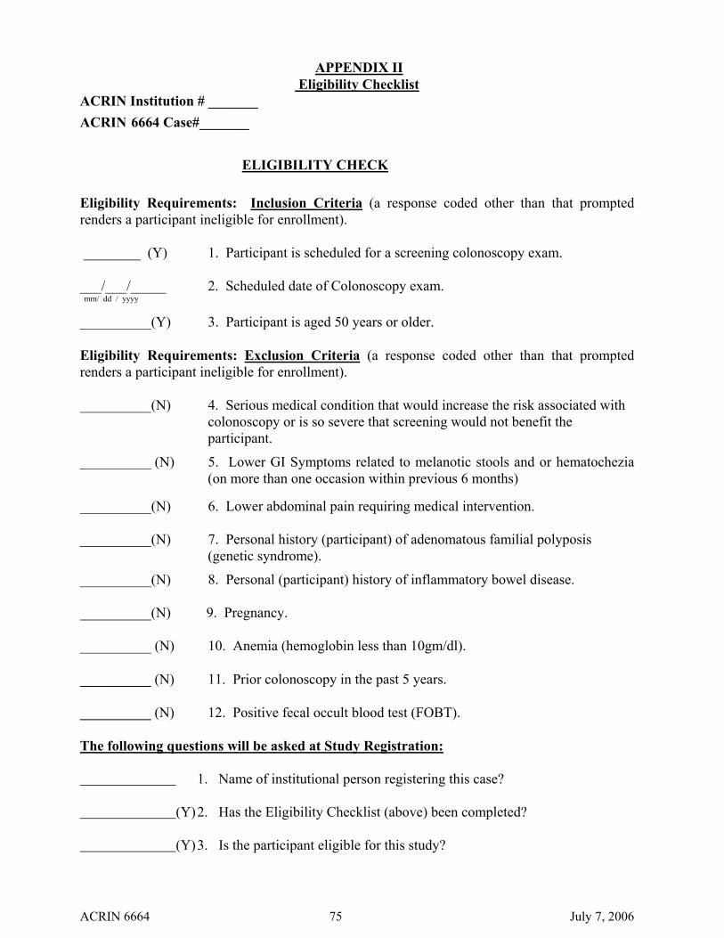
APPENDIX II Eligibility Checklist
ACRIN Institution # _______ ACRIN 6664 Case#_______ ELIGIBILITY CHECK Eligibility Requirements: Inclusion Criteria (a response coded other than that prompted renders a participant ineligible for enrollment). ________ (Y) 1. Participant is scheduled for a screening colonoscopy exam. ___/___/_____ 2. Scheduled date of Colonoscopy exam. mm/ dd / yyyy
__________(Y) 3. Participant is aged 50 years or older. Eligibility Requirements: Exclusion Criteria (a response coded other than that prompted renders a participant ineligible for enrollment). __________(N) 4. Serious medical condition that would increase the risk associated with
colonoscopy or is so severe that screening would not benefit the participant.
__________ (N) 5. Lower GI Symptoms related to melanotic stools and or hematochezia (on more than one occasion within previous 6 months)
__________(N) 6. Lower abdominal pain requiring medical intervention. __________(N) 7. Personal history (participant) of adenomatous familial polyposis
(genetic syndrome).
__________(N) 8. Personal (participant) history of inflammatory bowel disease. __________(N) 9. Pregnancy. __________ (N) 10. Anemia (hemoglobin less than 10gm/dl). __________ (N) 11. Prior colonoscopy in the past 5 years. __________ (N) 12. Positive fecal occult blood test (FOBT). The following questions will be asked at Study Registration: 1. Name of institutional person registering this case? (Y) 2. Has the Eligibility Checklist (above) been completed? (Y) 3. Is the participant eligible for this study?
ACRIN 6664 75 July 7, 2006
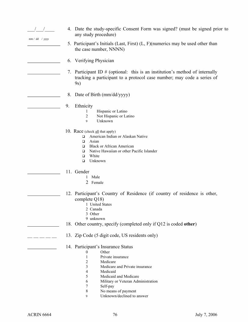
___/___/____ 4. Date the study-specific Consent Form was signed? (must be signed prior to
any study procedure) mm / dd / yyyy
5. Participant’s Initials (Last, First) (L, F)(numerics may be used other than the case number, NNNN)
6. Verifying Physician 7. Participant ID # (optional: this is an institution’s method of internally
tracking a participant to a protocol case number; may code a series of 9s)
8. Date of Birth (mm/dd/yyyy) 9. Ethnicity
1 Hispanic or Latino 2 Not Hispanic or Latino 9 Unknown
10. Race (check all that apply) American Indian or Alaskan Native Asian Black or African American Native Hawaiian or other Pacific Islander White Unknown
11. Gender
1 Male 2 Female
12. Participant’s Country of Residence (if country of residence is other,
complete Q18) 1 United States 2 Canada 3 Other 9 unknown
_____________ 18. Other country, specify (completed only if Q12 is coded other) __ __ __ __ __ 13. Zip Code (5 digit code, US residents only) 14. Participant’s Insurance Status
0 Other 1 Private insurance 2 Medicare 3 Medicare and Private insurance 4 Medicaid 5 Medicaid and Medicare 6 Military or Veteran Administration 7 Self-pay 8 No means of payment 9 Unknown/declined to answer
ACRIN 6664 76 July 7, 2006
15. Will any component of the participant’s care be given at a military or
VA facility? 1 No 2 Yes 9 Unknown
____/____/_____ 16. Scheduled date of CTC exam (mm/dd/yyyy) _____________ 17. Registration Date
Completed by Date form completed: __/___/____ Participant signature: ________________________________________________________ (If information is obtained through direct interview with the participant, participant signature and date MUST appear on document) __________________________________________________ Signature of person entering data onto the web
ACRIN 6664 77 July 7, 2006

APPENDIX III PARTICIPATING INSTITUTIONS AND SITE PIs
Total of 15 participating institutions, Site Principal Investigators and lead Gastroenterologists will be identified upon review and approval of completed ACRIN Protocol Specific Application (PSA).
ACRIN 6664 78 July 7, 2006
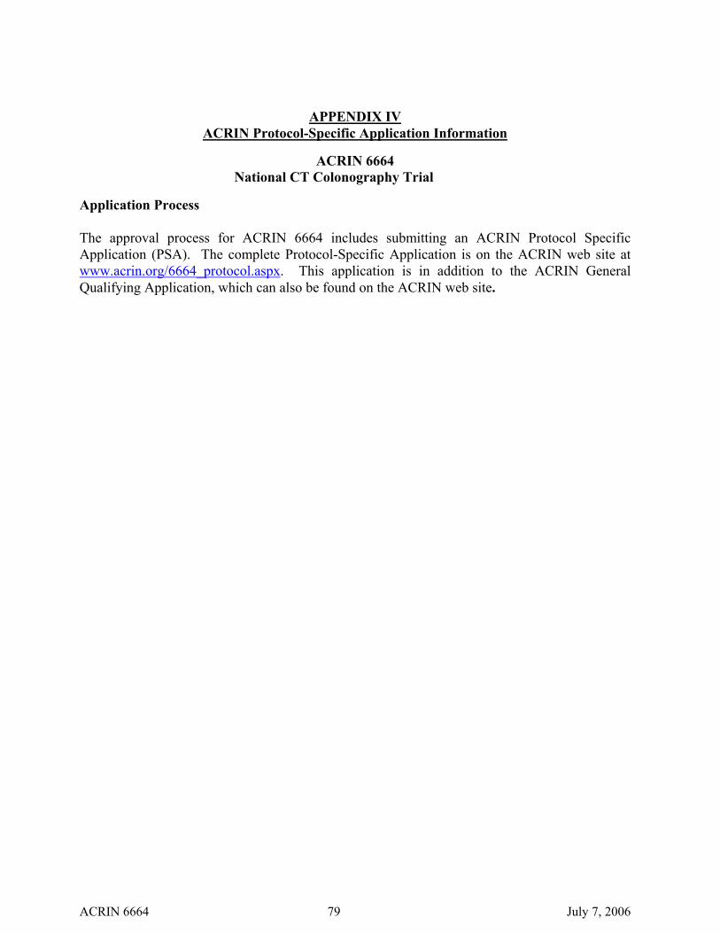
ACRIN 6664 79 July 7, 2006
APPENDIX IV ACRIN Protocol-Specific Application Information
ACRIN 6664
National CT Colonography Trial
Application Process The approval process for ACRIN 6664 includes submitting an ACRIN Protocol Specific Application (PSA). The complete Protocol-Specific Application is on the ACRIN web site at www.acrin.org/6664_protocol.aspx. This application is in addition to the ACRIN General Qualifying Application, which can also be found on the ACRIN web site.
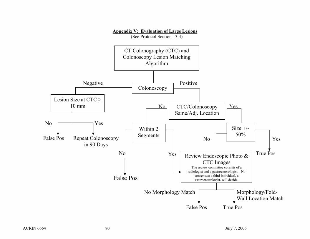
Appendix V: Evaluation of Large Lesions (See Protocol Section 13.3)
False Pos
CT Colonography (CTC) and Colonoscopy Lesion Matching
Algorithm
Colonoscopy
Review Endoscopic Photo & CTC Images
The review committee consists of a radiologist and a gastroenterologist. No
consensus: a third individual, a gastroenterologist, will decide.
Positive Negative
Lesion Size at CTC > 10 mm CTC/Colonoscopy
Same/Adj. Location
Size +/- 50%
Within 2 Segments
Yes No
True Pos False Pos
No Morphology Match
No Yes
No Yes
True Pos
Morphology/Fold-Wall Location Match
False Pos Repeat Colonoscopy in 90 Days
No Yes
ACRIN 6664 80 July 7, 2006