alternative conformational model of a seed protein … conformational model of a seed protein dek1...
TRANSCRIPT
Alternative Conformational Model of a Seed Protein DeK1 forBetter Understanding of Structure-Function Relationship
Surya Bhushan Kumar*, 1, Kavya Venkateswaran**, 1, and Suman Kundu*, **, #
Abstract: Understanding of the cell development and differentiation processes in plant seeds in general is poor. One gene, among others, thatpredominantly regulates the aleurone cell formation, differentiation and specification in seeds is called defective kernel (DeK1) and severalcell biology and genetic experiments have unequivocally established this fact. However, the mechanism behind such processes is still unclearand understanding the protein functionality of DeK1 is vital to elucidating its role in endosperm cell development. Only preliminaryinvestigations have been performed for just one domain of the protein in vitro and its functional implications have been highlighted lately.An initial attempt at in silico modeling of the protein has shown promise and necessitated thorough investigation of the protein to helpunderstand structure-function relationship in details thus corroborating experimental findings and laying foundation for further studies.DeK1 sequences in public databases were used as raw material for elaborate computational analysis of the protein. DeK1 is a multi-passmembrane protein with interesting structural features and the present analysis provides an alternative model for DeK1 structure that can helppan both in vitro and in vivo studies. The transmembrane helices were shown to have a number of conserved charged and polar residues thatcan form salt bridges and help in ligand binding or transmitting an external signal in addition to maintaining structural integrity. Theprotein possesses a big loop of about 280-300 amino acid residues on the cytoplasmic side of the membrane. It has a number of putativephosphorylation sites, multiple cysteine residues and a high density of charged residues, all of which could be important for protein-proteininteraction and signaling pathways. The loop has a nuclear localization propensity as well. The long C-terminal tail of DeK1 with homologyto calpain domain may be activated by the big loop, or conversely the big loop could be a substrate for the calpain protease in addition toits demonstrated autocatalytic property. Any or all of these features could be important in signaling events and several hypotheses have beenforwarded for the structure-function relationship of the novel protein. The results provide a platform for deciphering the biochemicalcharacteristics of DeK1.
Keywords: DeK1, defective kernel, seed protein, aleurone layer, big loop, transmembrane topology.
*1 School of Biotechnology, Banaras Hindu University, Varanasi 221005, India.**1 Department of Biochemistry, University of Delhi South Campus, Benito Juarez Road, New Delhi-110021, India,# Corresponding author: E-mail: [email protected].
I INTRODUCTION
A typical plant seed contains an outer layer called thepericarp. The inner endosperm represents the storagetissue of the seed, providing nourishment to the growingseedling at germination. The endosperm is a simple tissuecomposed of three major cell types - the starchyendosperm which makes up most of the bulk of the seed,the basal transfer layer and the aleurone layer sandwichedbetween the pericarp and the starchy endosperm (Becraftet al, 2001; Olsen at al, 2008). The transfer layer functionsin nutrient uptake from the mother plant during seeddevelopment. The starchy endosperm is the major storagesite for starch and proteins. While performing somestorage function, the primary role of the aleurone is as adigestive tissue. At germination, it secretes amylase intothe starchy endosperm causing the breakdown of thestored starch, providing the growing seedlings with sugarsfor energy and growth. The aleurone layer is also a natural
JOURNAL OF PROTEINS AND PROTEOMICSVol. 1, No. 2, July-December 2010, pp. 77 – 90
© International Science Press
storehouse for oil and fat bodies (Ritchie and Gilroy,1998). The apparent simplicity of the cellular arrangement,however, is deceptive and endosperm development is ahighly specialized process with many unique features.How the endosperm cells specificate into aleurone cellsor starchy endosperm cells or transfer cells is yet largelyunknown (Olsen, 2001; Berger, 1999; Johnson et al,2008) and needs to be investigated.
One protein that seems to play crucial role in aleuroneand endosperm differentiation is DeK1 (Becraft, 2001;Becraft et al, 2002; Lid et al, 2002; Olsen et al, 2008;Johnson et al, 2008). DeK1 is an acronym for defectivekernel (Sheridan and Neuffer, 1980; Lid et al, 2002).Absence or deletion of this gene in seeds has resulted inabnormal or defective seed kernels and hence the name(Lid et al, 2005). Though most seeds have a single layerof aleurone cells the presence of multiple layers havebeen reported in Coroico seeds (Galiant, 1990; Nelson
78 Journal of Proteins and Proteomics
and Chang, 1974). This indicates scope for engineeringmultiple layers of aleurone cells in seeds provided weunderstand the genes that control aleurone cell fatespecification. Investigating DeK1 in detail could providean initiation in this direction.
DeK1 is distributed all over the endosperm cells.However, the greatest effect of this gene in cellspecification is observed in the aleurone cells (Lid et al,2002; Becraft et al, 2002). Mutants that have disruptedaleurone development have been identified. Such mutantslack an aleurone layer (Becraft and Asuncion-Crabb,2000; Lid et al, 2005). This is further exemplified in again-of-function experiment where overexpression of thegene results in an extra layer of aleurone (Becraft andAsuncion-Crabb, 2000; Lid et al, 2005). It is thus evidentthat aleurone cell formation is a highly specialized eventand that endosperm cells, which are only on the surface,get differentiated into aleurone cells (Gifford et al, 2003).DeK1 plays a critical role in this developmental process.Further investigation into such plant proteins and genescould help improve our poor understanding of celldevelopment and differentiation in general.
While genetic analyses and cell biology studies havehighlighted the importance of DeK1 gene (Becraft andAsuncian-Crabb, 2000; Becraft et al, 2002; Lid et al, 2002;Lid et al, 2005; Ahn et al, 2004; Johnson et al, 2005),there is minimal information at the protein level. A portionof the protein was found to have homology to animalcalpains and the protease activity of this domain inisolation was demonstrated experimentally (Wang et al,2003). It was recently shown that this protease domaincan complement DeK1 function in mutants throughautocatalysis (Johnson et al, 2008). Successfulunderstanding of the regulation of aleurone cell formationby DeK1 needs complimentary knowledge about the totalDeK1 protein as well. It is vital to know how this proteinis synthesized, degraded and transported, which proteinsit interacts with, how it triggers the signaling events, howit senses the presence of endosperm cells on the surfacedirecting aleurone formation, what ligands and substrateit would bind to, what is the nature and biochemicalcharacteristics of the protein, what kind of secondarystructure and three-dimensional structure it forms, howare structural elements related to its biological functionand finally how the protein can be engineered to manipulateits function. The fact that DeK1 is linked to distinctphenotype (defective kernel, absence of aleurone layer)makes it a viable candidate for such investigation. Theonly attempt at visualizing the protein structure (Lidet al., 2002) predicted one model for DeK1 protein.However, it is unable to answer several questions and
the topology of membrane helices and the orientation androle of a big loop is in doubt. Alternative topologies andpossibilities need to be explored.
As a preliminary initiative to our objective, the presentinvestigation deals with “in silico” modelling of DeK1protein. We have analysed Dek1 sequences from threeplant species namely Rice (Oryza sativa), Maize (Zeamays), and Arabidopsis (Arabidopsis thaliana), theformer two being monocotyledonous and the latter beingdicotyledonous. The results help us to visualize a tentativesecondary structure of the protein and help us to formulatehypothesis regarding the structure-function relationshipof the protein. The studies build a basis for detailedresearch into the DeK1 system.
II MATERIALSAND METHODS
All results were obtained computationally. PubMed(http://www.ncbi.nlm.nih.gov/entrez/query.fcgi) andpopular Internet search engines were used for literaturesurvey. The DNA and amino acid sequences of the targetprotein DeK1 from different plant sources, as well as allother proteins used as controls or for reference, wereobtained either from NCBI (http://www.ncbi.nih.gov) orEMBL-EBI (http://www.ebi.ac.uk/). These sequenceswere downloaded and analyzed in detail. The individualamino acid sequence file, in each case, was converted toBLAST or FASTA format.
The theoretical physical properties of the DeK1proteins were calculated using Proteomics tools fromthe Expasy home page (www.expasy.org). CLUSTALW(http://www.ebi.ac.uk/clustalw/) from Expasy homepage was used for sequence alignment (Thompsonet al, 1994; Jeanmougin et al, 1998). PSORT(http://www.psort.org/) provided links to the PSORTfamily of programs for subcellular localization predictionas well as other datasets and resources relevant tolocalization prediction. The following PSORT programsfor localization prediction were used: WoLF PSORT is arecently updated version of PSORT II for the predictionof eukaryotic sequences; PSORT II (Nakai and Horton,1999; Horton and Nakai, 1997) for eukaryotic sequences;PSORT (Nakai and Kanehisa, 1991) for plant sequences.SIGNALP 3.0 server (www.cbs.dtu.dk/services/SignalP)was used to predict the presence and location of signalpeptide cleavage sites in amino acid sequences fromdifferent organisms (Bendtsen et al, 2004).
The following algorithms were used to predict themembrane topology of DeK1 proteins:
TMHMM (www.cbs.dtu.dk/service/TMHMM)(Krogh et al, 2001)
Alternative Conformational Model of a Seed Protein DeK1 for Better Understanding of Structure-Function Relationship 79
SOSUI (http://sosui.proteome.bio.tuat.ac.jp/sosui_submit.html) (Hirokawa et al, 1998; Mitakuand Hirokawa, 1999; Mitaku et al, 1998)
HMMTOP (http://www.enzim.hu/hmmtop/)(Tusnady and Simon, 1998)
TMPRED (http://www.ch.embnet.org/software/TMPRED_form.html) (Hofmann and Stoffel, 1993;Arai et al, 2004)
TOPPRED (www.pasteure.fr/seqanal/interfaces/toppred) (Stoffel et al, 1993)
TMAP (http://bioinfo.limbo.ifm.liu.se/tmap/) (Davidet al, 2002; Persson and Argos, 1996; Persson andArgos, 1994)
PHOBIUS (http://phobius.cgb.ki.se/and http://phobius.binf.ku.dk/) (Kall et al, 2004).
Of these TMHMM had earlier been reported to bethe best (Melen et al, 2003; Moller et al, 2001). However,PHOBIUS is the newest algorithm available (2004) andseems to have certain advantages over the other as seenin the Results section.
The NetPhos 2.0 server (http://www.cbs.dtu.dk/services/NetPhos/) was used to predict serine, threonineand tyrosine phosphorylation sites in eukaryotic proteins(Blom et al, 1999). Nuclear localization signal (NLS) waspredicted using a software from the website mentioned(http://cubic.bioc.columbia.edu/PredictNLS) (Cokolet al, 2000).
III RESULTS AND DISCUSSION
DeK1 Sequences
A search of public databases for DeK1 amino acidsequences revealed that such sequences from four plantspecies are available. For the present investigation, DeK1sequences of Rice (Oryza sativa), Maize (Zea mays) andArabidopsis (Arabidopsis thaliana) plants were used, withNCBI accession numbers Q8RYA5, Q8RUQ1 (Q8RVL1)and Q8RVL2, respectively. While rice and maize aremonocotyledonous plants, Arabidopsis is a dicotyledo-nous plant. Hence an analysis of these three sequenceswould be interesting for comparative purposes.Evolutionarily conserved and functionally importantresidues can be extracted from such analysis. Theconsistency of in silico analysis across species and acrossmonocots and dicots would make the predictions andmodels more reliable. Arab DeK1 has 2151 amino acidside chains, while maize DeK1 has 2159 and rice DeK1has 2162 amino acid side chains. As a prelude toexperimental studies we have resorted to computational
methods here to investigate DeK1 structure. The threesequences were thus subjected to various predictionalgorithms.
Predicted Physical Parameters of DeK1 Proteins
There is minimal information about the biochemicalcharacteristics of DeK1 protein, since the full lengthprotein has not been studied yet. Since biochemical andphysical properties define a protein, it is interesting toknow some of these properties and whether they differbetween DeK1 proteins from different species of plants.Thus, the molecular mass (M
w) of these proteins are
predicted to be 240, 751 Da for Rice DeK1, 238, 865 Dafor Arabidopsis DeK1 and 239, 945 Da for Maize DeK1.Their isoelectric points (pI) are 5.85, 6.15 and 5.69,respectively. These results indicate that the DeK1 proteinswould have similar physical properties.
Sequence Alignment of DeK1’sSequences of the three DeK1’s were aligned byCLUSTALW to check the overall identity and similarityof the sequences (data not shown). Rice and Maize DeK1have 90% identity, while each of these sequences areonly 70% similar to Arabidopsis DeK1. This is expectedsince while rice and maize are monocotyledonous plants,Arabidopsis is dicotyledonous. The high similarity insequences indicates that DeK1 performs an importantfunction and has been conserved evolutionarily. It alsoindicates that DeK1 proteins probably have a similarprotein fold, three-dimensional structure and consensusfunctional conformation.
Subcellular Localization
The probability of finding a protein in a particular locationof the cell, or a particular organelle of the cell can bepredicted using its sequence information. For all the threeDeK1 sequences the prediction algorithms (PSORT)produced the same results:
56.0%: extracellular, including cell wall; 12.0%:cytoplasmic; 12.0%: endoplasmic reticulum; 8.0%:vesicles of secretory system; 8.0%: nuclear; 4.0%:mitochondrial.
The prediction indicates that DeK1 is majorly anextracellular protein and this has already been observedexperimentally (Johnson et al, 2008), raising confidencein in silico analysis of DeK1 sequences. So DeK1 mighteither be a membrane protein and docked in the cell wallor secreted out of the cytoplasm as a solubleextracytoplasmic protein. In either case, DeK1 wouldhave a signal peptide or a stretch of sequence that leadsthe protein to its specific location of the cell.
80 Journal of Proteins and Proteomics
Hemoglobin from Pseudoterranova decipiens(codworm), which has been investigated experimentally(Dixon et al, 1991; Gibson et al, 1993), was used as acontrol protein to test the accuracy of PSORT algorithm.Keeping with experimental observations that the nematodehemoglobin is an extracellular protein, the following resultswere obtained: 48.0%: extracellular, including cell wall;16.0%: cytoplasmic; 12.0%: nuclear; 8.0%: vesicles ofsecretory system; 8.0%: endoplasmic reticulum; 4.0%:vacuolar; 4.0%: mitochondrial.
Signal Peptide Presence and Location Prediction
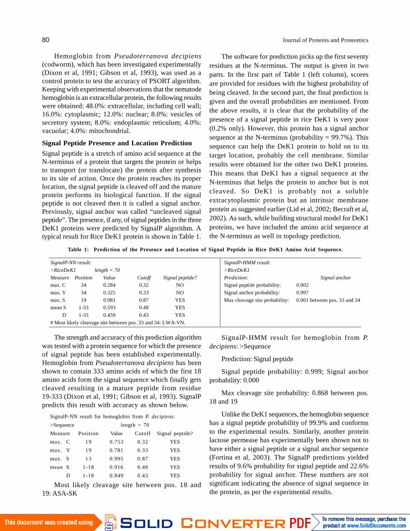
Signal peptide is a stretch of amino acid sequence at theN-terminus of a protein that targets the protein or helpsto transport (or translocate) the protein after synthesisto its site of action. Once the protein reaches its properlocation, the signal peptide is cleaved off and the matureprotein performs its biological function. If the signalpeptide is not cleaved then it is called a signal anchor.Previously, signal anchor was called “uncleaved signalpeptide”. The presence, if any, of signal peptides in the threeDeK1 proteins were predicted by SignalP algorithm. Atypical result for Rice DeK1 protein is shown in Table 1.
The software for prediction picks up the first seventyresidues at the N-terminus. The output is given in twoparts. In the first part of Table 1 (left column), scoresare provided for residues with the highest probability ofbeing cleaved. In the second part, the final prediction isgiven and the overall probabilities are mentioned. Fromthe above results, it is clear that the probability of thepresence of a signal peptide in rice DeK1 is very poor(0.2% only). However, this protein has a signal anchorsequence at the N-terminus (probability = 99.7%). Thissequence can help the DeK1 protein to hold on to itstarget location, probably the cell membrane. Similarresults were obtained for the other two DeK1 proteins.This means that DeK1 has a signal sequence at theN-terminus that helps the protein to anchor but is notcleaved. So DeK1 is probably not a solubleextracytoplasmic protein but an intrinsic membraneprotein as suggested earlier (Lid et al, 2002; Becraft et al,2002). As such, while building structural model for DeK1proteins, we have included the amino acid sequence atthe N-terminus as well in topology prediction.
Table 1: Prediction of the Presence and Location of Signal Peptide in Rice DeK1 Amino Acid Sequence.
SignalP-NN result:
>RiceDeK1 length = 70
Measure Position Value Cutoff Signal peptide?
max. C 34 0.284 0.32 NO
max. Y 34 0.325 0.33 NO
max. S 19 0.981 0.87 YESmean S 1-33 0.593 0.48 YES
D 1-33 0.459 0.43 YES
# Most likely cleavage site between pos. 33 and 34: LWA-VN.
SignalP-HMM result:
>RiceDeK1
Prediction: Signal anchor
Signal peptide probability: 0.002
Signal anchor probability: 0.997
Max cleavage site probability: 0.001 between pos. 33 and 34
The strength and accuracy of this prediction algorithmwas tested with a protein sequence for which the presenceof signal peptide has been established experimentally.Hemoglobin from Pseudoterranova decipiens has beenshown to contain 333 amino acids of which the first 18amino acids form the signal sequence which finally getscleaved resulting in a mature peptide from residue19-333 (Dixon et al, 1991; Gibson et al, 1993). SignalPpredicts this result with accuracy as shown below.
SignalP-NN result for hemoglobin from P. decipiens:
>Sequence length = 70
Measure Position Value Cutoff Signal peptide?
max. C 19 0.753 0.32 YES
max. Y 19 0.781 0.33 YES
max. S 13 0.995 0.87 YES
mean S 1-18 0.916 0.48 YES
D 1-18 0.849 0.43 YES
Most likely cleavage site between pos. 18 and19: ASA-SK
SignalP-HMM result for hemoglobin from P.decipiens: >Sequence
Prediction: Signal peptide
Signal peptide probability: 0.999; Signal anchorprobability: 0.000
Max cleavage site probability: 0.868 between pos.18 and 19
Unlike the DeK1 sequences, the hemoglobin sequencehas a signal peptide probability of 99.9% and conformsto the experimental results. Similarly, another proteinlactose permease has experimentally been shown not tohave either a signal peptide or a signal anchor sequence(Fortina et al, 2003). The SignalP predictions yieldedresults of 9.6% probability for signal peptide and 22.6%probability for signal anchor. These numbers are notsignificant indicating the absence of signal sequence inthe protein, as per the experimental results.

Alternative Conformational Model of a Seed Protein DeK1 for Better Understanding of Structure-Function Relationship 81
Membrane Topology Prediction
The subcellular localization prediction of DeK1 andprevious literature indicates that DeK1 is a membraneprotein (Lid et al, 2002; Wang et al, 2003). To understandthe structure-function relationship in membrane proteinsit is important to know how the protein spans themembrane. The number of transmembrane domains, theirtopology or orientation, the number and orientation ofloops, the orientation of the N-terminal and C-terminaldomains and other related structural information regardingmembrane proteins are important. Lid et al. had modeledDeK1 transmembrane domain based on only one topologyprediction algorithm, TMHMM (Lid et al, 2002). In silicopredictions should be based on multiple algorithms sinceeach algorithm has its own limitations. Using multiplealgorithms enhance the confidence in prediction, especiallyif the sequence information is robust and producesconsensus across different algorithms. Moreover, suchan extensive exercise allows prediction of alternatepossibilities for structural models and corroboration ofexperimental findings become easier. Lid et al’s modelalso fails to explain facts like the presence of a big loopon the extracytoplasmic side while it has a large numberof charged residues and probability for phosphorylationand therefore more likely to be cytoplasmic. Further, themodel was not analyzed thoroughly for structure-functionrelationship in relation to its role in aleurone specification.Hence, we have revisited DeK1 model and investigatedalternative conformation for DeK1 to provide a boost toin solution studies. A number of membrane topologyprediction algorithms, as mentioned in the Materials andMethods section, were used to predict the transmembranedomains (TM). These topology prediction algorithmspredict the number and position of transmembrane helices(TMH).
A typical prediction result for rice DeK1 usingPhobius as the prediction algorithm is shown in Table 2.The predictor identifies the regions which can formtransmembrane helices, e.g. amino acid residues 6-34,68-90, 96-119, etc (denoted as TRANSMEM in the aboveresults). This then delineates the non-membrane portionsof the protein. The non-membrane portions called loopsare predicted to be on the cytoplasmic or non-cytoplasmicside. The N-terminus, which for rice DeK1, is fromamino acid 1 to amino acid 5 (above) is on the “outside”(non-cytoplasmic) of the membrane, while the longC-terminus tail from amino acid side chain number 1108to 2162 is on the “inside” of the membrane. Thus riceDeK1 has 23 transmembrane helices, 11 loops on theoutside and 11 loops on the inside. One of the loops, wecall “Big Loop” is exceptionally long in length (~280
residues) and is on the cytoplasmic side or inside of themembrane, unlike proposed by Lid et al (2002).
Table 2: Topology Prediction of RiceDeK1 using PHOBIUS.
ID RiceDEK1
F T DOMAIN 1 5 NON CYTOPLASMIC.
F T TRANSMEM 6 34
F T DOMAIN 35 67 CYTOPLASMIC.
F T TRANSMEM 68 90
F T DOMAIN 91 95 NON CYTOPLASMIC.
F T TRANSMEM 96 119
F T DOMAIN 120 130 CYTOPLASMIC.
F T TRANSMEM 131 152
F T DOMAIN 153 163 NON CYTOPLASMIC.
F T TRANSMEM 164 186
F T DOMAIN 187 233 CYTOPLASMIC.
F T TRANSMEM 234 258
F T DOMAIN 259 263 NON CYTOPLASMIC.
F T TRANSMEM 264 282
F T DOMAIN 283 293 CYTOPLASMIC.
F T TRANSMEM 294 315
F T DOMAIN 316 320 NON CYTOPLASMIC.
F T TRANSMEM 321 341
F T DOMAIN 342 626 CYTOPLASMIC.
F T TRANSMEM 627 646
F T DOMAIN 647 665 NON CYTOPLASMIC.
F T TRANSMEM 666 687
F T DOMAIN 688 698 CYTOPLASMIC.
F T TRANSMEM 699 717
F T DOMAIN 718 722 NON CYTOPLASMIC.
F T TRANSMEM 723 744
F T DOMAIN 745 773 CYTOPLASMIC.
F T TRANSMEM 774 794
F T DOMAIN 795 822 NON CYTOPLASMIC.
F T TRANSMEM 823 849
F T DOMAIN 850 855 CYTOPLASMIC.
F T TRANSMEM 856 877
F T DOMAIN 878 888 NON CYTOPLASMIC.
F T TRANSMEM 889 912
F T DOMAIN 913 923 CYTOPLASMIC.
F T TRANSMEM 924 944
F T DOMAIN 945 949 NON CYTOPLASMIC.
F T TRANSMEM 950 973
F T DOMAIN 974 984 CYTOPLASMIC.
F T TRANSMEM 985 1005
F T DOMAIN 1006 1016 NON CYTOPLASMIC.
F T TRANSMEM 1017 1039
F T DOMAIN 1040 1059 CYTOPLASMIC.
F T TRANSMEM 1060 1083
F T DOMAIN 1084 1088 NON CYTOPLASMIC.
F T TRANSMEM 1089 1107
F T DOMAIN 1108 2162 CYTOPLASMIC.
82 Journal of Proteins and Proteomics
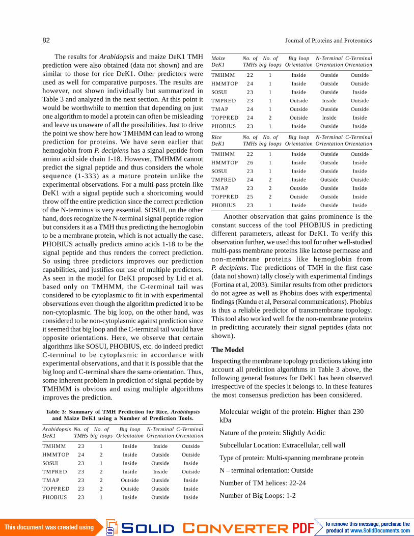
The results for Arabidopsis and maize DeK1 TMHprediction were also obtained (data not shown) and aresimilar to those for rice DeK1. Other predictors wereused as well for comparative purposes. The results arehowever, not shown individually but summarized inTable 3 and analyzed in the next section. At this point itwould be worthwhile to mention that depending on justone algorithm to model a protein can often be misleadingand leave us unaware of all the possibilities. Just to drivethe point we show here how TMHMM can lead to wrongprediction for proteins. We have seen earlier thathemoglobin from P. decipiens has a signal peptide fromamino acid side chain 1-18. However, TMHMM cannotpredict the signal peptide and thus considers the wholesequence (1-333) as a mature protein unlike theexperimental observations. For a multi-pass protein likeDeK1 with a signal peptide such a shortcoming wouldthrow off the entire prediction since the correct predictionof the N-terminus is very essential. SOSUI, on the otherhand, does recognize the N-terminal signal peptide regionbut considers it as a TMH thus predicting the hemoglobinto be a membrane protein, which is not actually the case.PHOBIUS actually predicts amino acids 1-18 to be thesignal peptide and thus renders the correct prediction.So using three predictors improves our predictioncapabilities, and justifies our use of multiple predictors.As seen in the model for DeK1 proposed by Lid et al.based only on TMHMM, the C-terminal tail wasconsidered to be cytoplasmic to fit in with experimentalobservations even though the algorithm predicted it to benon-cytoplasmic. The big loop, on the other hand, wasconsidered to be non-cytoplasmic against prediction sinceit seemed that big loop and the C-terminal tail would haveopposite orientations. Here, we observe that certainalgorithms like SOSUI, PHOBIUS, etc. do indeed predictC-terminal to be cytoplasmic in accordance withexperimental observations, and that it is possible that thebig loop and C-terminal share the same orientation. Thus,some inherent problem in prediction of signal peptide byTMHMM is obvious and using multiple algorithmsimproves the prediction.
Table 3: Summary of TMH Prediction for Rice, Arabidopsisand Maize DeK1 using a Number of Prediction Tools.
Arabidopsis No. of No. of Big loop N-Terminal C-TerminalDeK1 TMHs big loops Orientation Orientation Orientation
TMHMM 23 1 Inside Inside Outside
HMMTOP 24 2 Inside Outside Outside
SOSUI 23 1 Inside Outside Inside
TMPRED 23 2 Inside Inside Outside
TM AP 23 2 Outside Outside Inside
TOPPRED 23 2 Outside Outside Inside
PHOBIUS 23 1 Inside Outside Inside
Maize No. of No. of Big loop N-Terminal C-TerminalDeK1 TMHs big loops Orientation Orientation Orientation
TMHMM 22 1 Inside Outside Outside
HMMTOP 24 1 Inside Outside Outside
SOSUI 23 1 Inside Outside Inside
TMPRED 23 1 Outside Inside Outside
TM AP 24 1 Outside Outside Outside
TOPPRED 24 2 Outside Inside Inside
PHOBIUS 23 1 Inside Outside Inside
Rice No. of No. of Big loop N-Terminal C-TerminalDeK1 TMHs big loops Orientation Orientation Orientation
TMHMM 22 1 Inside Outside Outside
HMMTOP 26 1 Inside Outside Inside
SOSUI 23 1 Inside Outside Inside
TMPRED 24 2 Inside Outside Outside
TM AP 23 2 Outside Outside Inside
TOPPRED 25 2 Outside Outside Inside
PHOBIUS 23 1 Inside Outside Inside
Another observation that gains prominence is theconstant success of the tool PHOBIUS in predictingdifferent parameters, atleast for DeK1. To verify thisobservation further, we used this tool for other well-studiedmulti-pass membrane proteins like lactose permease andnon-membrane proteins like hemoglobin fromP. decipiens. The predictions of TMH in the first case(data not shown) tally closely with experimental findings(Fortina et al, 2003). Similar results from other predictorsdo not agree as well as Phobius does with experimentalfindings (Kundu et al, Personal communications). Phobiusis thus a reliable predictor of transmembrane topology.This tool also worked well for the non-membrane proteinsin predicting accurately their signal peptides (data notshown).
The Model
Inspecting the membrane topology predictions taking intoaccount all prediction algorithms in Table 3 above, thefollowing general features for DeK1 has been observedirrespective of the species it belongs to. In these featuresthe most consensus prediction has been considered.
Molecular weight of the protein: Higher than 230kDa
Nature of the protein: Slightly Acidic
Subcellular Location: Extracellular, cell wall
Type of protein: Multi-spanning membrane protein
N – terminal orientation: Outside
Number of TM helices: 22-24
Number of Big Loops: 1-2
Alternative Conformational Model of a Seed Protein DeK1 for Better Understanding of Structure-Function Relationship 83
The big loop predicted by all softwares = ~aminoacid position 330-630
Big loop orientation: Inside
C-terminal length: ~1000 amino acids
C-terminal orientation: Inside
Signal peptide at the N-terminus: Signal anchor
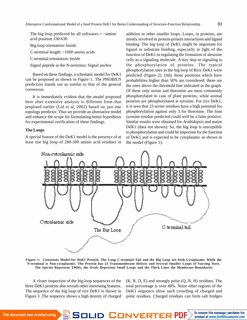
Based on these findings, a schematic model for DeK1can be proposed as shown in Figure 1. The PHOBIUSprediction stands out as similar to that of the generalconsensus.
It is immediately evident that the model proposedhere after extensive analysis is different from thatproposed earlier (Lid et al, 2002) based on just onetopology predictor. Thus we provide an alternative modeland enhance the scope for formulating better hypothesisfor experimental verification of these findings.
The Loops
A special feature of the DeK1 model is the presence of atleast one big loop of 280-300 amino acid residues in
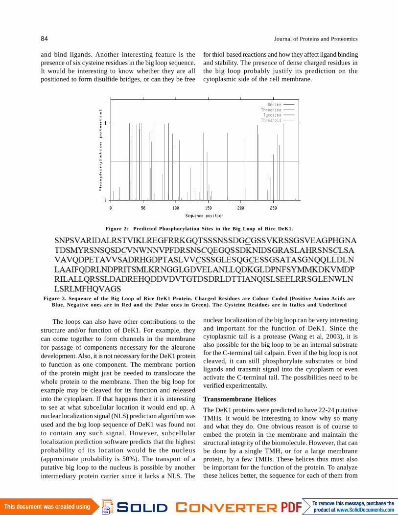
addition to other smaller loops. Loops, in proteins, aremostly involved in protein-protein interactions and ligandbinding. The big loop of DeK1 might be important forligand or substrate binding, especially in light of thefunction of DeK1 in regulating the formation of aleuronecells as a signaling molecule. A key step in signaling isthe phosphorylation of proteins. The typicalphosphorylation sites in the big loop of Rice DeK1 werepredicted (Figure 2). Only those positions which haveprobabilities higher than 50% are considered; these arethe ones above the threshold line indicated in the graph.Of these only serine and threonine are most commonlyphosphorylated in case of plant proteins, while animalproteins are phosphorylated at tyrosine. For rice DeK1,it is seen that 23 serine residues have a high potential forphosphorylation against only 3 for threonine. The lonetyrosine residue predicted could well be a false positive.Similar results were obtained for Arabidopsis and maizeDeK1 (data not shown). So, the big loop is susceptibleto phosphorylation and could be important for the functionof DeK1 and is expected to be cytoplasmic as shown inthe model (Figure 1).
Figure 1: Consensus Model for DeK1 Protein. The Long C-terminal Tail and the Big Loop are both Cytoplasmic While theN-terminal is Non-cytoplasmic. The Protein has 23 Transmembrane Helices and Several Smaller Loops of Varying Sizes.
The Spirals Represent TMHs, the Ovals Represent Small Loops and the Thick Lines the Membrane Boundaries
A closer inspection of the big loop sequences of thethree DeK1 proteins also reveals other interesting features.The sequence of the big loop of rice DeK1 is shown inFigure 3. The sequence shows a high density of charged
(K, R, D, E) and strongly polar (Q, N, H) residues. Thetotal percentage is over 40%. None other regions of theDeK1 sequence show such crowding of charged andpolar residues. Charged residues can form salt bridges
84 Journal of Proteins and Proteomics
and bind ligands. Another interesting feature is thepresence of six cysteine residues in the big loop sequence.It would be interesting to know whether they are allpositioned to form disulfide bridges, or can they be free
Figure 2: Predicted Phosphorylation Sites in the Big Loop of Rice DeK1.
The loops can also have other contributions to thestructure and/or function of DeK1. For example, theycan come together to form channels in the membranefor passage of components necessary for the aleuronedevelopment. Also, it is not necessary for the DeK1 proteinto function as one component. The membrane portionof the protein might just be needed to translocate thewhole protein to the membrane. Then the big loop forexample may be cleaved for its function and releasedinto the cytoplasm. If that happens then it is interestingto see at what subcellular location it would end up. Anuclear localization signal (NLS) prediction algorithm wasused and the big loop sequence of DeK1 was found notto contain any such signal. However, subcellularlocalization prediction software predicts that the highestprobability of its location would be the nucleus(approximate probability is 50%). The transport of aputative big loop to the nucleus is possible by anotherintermediary protein carrier since it lacks a NLS. The
nuclear localization of the big loop can be very interestingand important for the function of DeK1. Since thecytoplasmic tail is a protease (Wang et al, 2003), it isalso possible for the big loop to be an internal substratefor the C-terminal tail calpain. Even if the big loop is notcleaved, it can still phosphorylate substrates or bindligands and transmit signal into the cytoplasm or evenactivate the C-terminal tail. The possibilities need to beverified experimentally.
Transmembrane Helices
The DeK1 proteins were predicted to have 22-24 putativeTMHs. It would be interesting to know why so manyand what they do. One obvious reason is of course toembed the protein in the membrane and maintain thestructural integrity of the biomolecule. However, that canbe done by a single TMH, or for a large membraneprotein, by a few TMHs. These helices thus must alsobe important for the function of the protein. To analyzethese helices better, the sequence for each of them from
for thiol-based reactions and how they affect ligand bindingand stability. The presence of dense charged residues inthe big loop probably justify its prediction on thecytoplasmic side of the cell membrane.
Figure 3. Sequence of the Big Loop of Rice DeK1 Protein. Charged Residues are Colour Coded (Positive Amino Acids areBlue, Negative ones are in Red and the Polar ones in Green). The Cysteine Residues are in Italics and Underlined
Alternative Conformational Model of a Seed Protein DeK1 for Better Understanding of Structure-Function Relationship 85
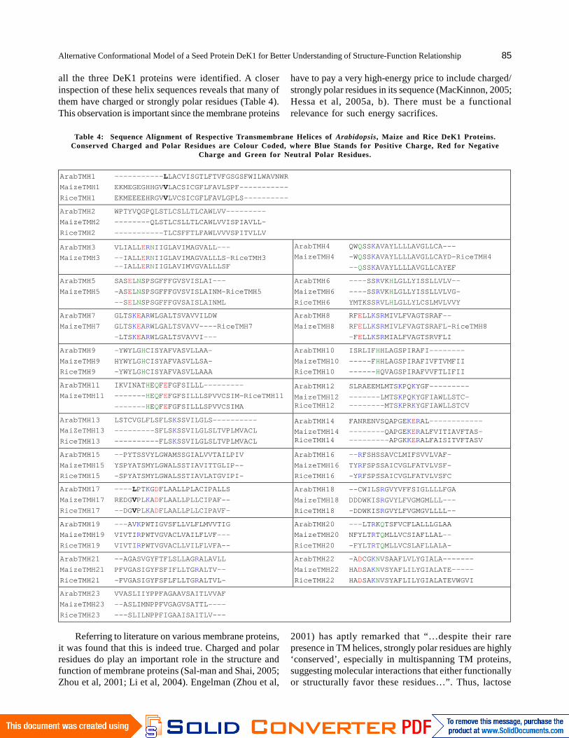
all the three DeK1 proteins were identified. A closerinspection of these helix sequences reveals that many ofthem have charged or strongly polar residues (Table 4).This observation is important since the membrane proteins
have to pay a very high-energy price to include charged/strongly polar residues in its sequence (MacKinnon, 2005;Hessa et al, 2005a, b). There must be a functionalrelevance for such energy sacrifices.
Table 4: Sequence Alignment of Respective Transmembrane Helices of Arabidopsis, Maize and Rice DeK1 Proteins.Conserved Charged and Polar Residues are Colour Coded, where Blue Stands for Positive Charge, Red for Negative
Charge and Green for Neutral Polar Residues.
ArabTMH1 -----------LLACVISGTLFTVFGSGSFWILWAVNWR
MaizeTMH1 EKMEGEGHHGVVLACSICGFLFAVLSPF-----------
RiceTMH1 EKMEEEEHRGVVLVCSICGFLFAVLGPLS----------
ArabTMH2 WPTYVQGPQLSTLCSLLTLCAWLVV---------
MaizeTMH2 --------QLSTLCSLLTLCAWLVVISPIAVLL-
RiceTMH2 -----------TLCSFFTLFAWLVVVSPITVLLV
ArabTMH3 VLIALLERNIIGLAVIMAGVALL---
MaizeTMH3 --IALLERNIIGLAVIMAGVALLLS-RiceTMH3--IALLERNIIGLAVIMVGVALLLSF
ArabTMH4 QWQSSKAVAYLLLLAVGLLCA---
MaizeTMH4 -WQSSKAVAYLLLLAVGLLCAYD-RiceTMH4
--QSSKAVAYLLLLAVGLLCAYEF
ArabTMH5 SASELNSPSGFFFGVSVISLAI---
MaizeTMH5 -ASELNSPSGFFFGVSVISLAINM-RiceTMH5
--SELNSPSGFFFGVSAISLAINML
ArabTMH6 ----SSRVKHLGLLYISSLLVLV--
MaizeTMH6 ----SSRVKHLGLLYISSLLVLVG-
RiceTMH6 YMTKSSRVLHLGLLYLCSLMVLVVY
ArabTMH7 GLTSKEARWLGALTSVAVVILDW
MaizeTMH7 GLTSKEARWLGALTSVAVV----RiceTMH7
-LTSKEARWLGALTSVAVVI---
ArabTMH8 RFELLKSRMIVLFVAGTSRAF--
MaizeTMH8 RFELLKSRMIVLFVAGTSRAFL-RiceTMH8
-FELLKSRMIALFVAGTSRVFLI
ArabTMH9 -YWYLGHCISYAFVASVLLAA-
MaizeTMH9 HYWYLGHCISYAFVASVLLSA-
RiceTMH9 -YWYLGHCISYAFVASVLLAAA
ArabTMH10 ISRLIFHHLAGSPIRAFI--------
MaizeTMH10 -----FHHLAGSPIRAFIVFTVMFII
RiceTMH10 ------HQVAGSPIRAFVVFTLIFII
ArabTMH11 IKVINATHEQFEFGFSILLL---------
MaizeTMH11 -------HEQFEFGFSILLLSPVVCSIM-RiceTMH11
-------HEQFEFGFSILLLSPVVCSIMA
ArabTMH12 SLRAEEMLMTSKPQKYGF---------
MaizeTMH12 -------LMTSKPQKYGFIAWLLSTC-RiceTMH12 --------MTSKPRKYGFIAWLLSTCV
ArabTMH13 LSTCVGLFLSFLSKSSVILGLS----------
MaiZeTMH13 ---------SFLSKSSVILGLSLTVPLMVACL
RiceTMH13 ----------FLSKSSVILGLSLTVPLMVACL
ArabTMH14 FANRENVSQAPGEKERAL------------
MaizeTMH14 --------QAPGEKERALFVITIAVFTAS-RiceTMH14 ---------APGKKERALFAISITVFTASV
ArabTMH15 --PYTSSVYLGWAMSSGIALVVTAILPIV
MaizeTMH15 YSPYATSMYLGWALSSTIAVITTGLIP--
RiceTMH15 -SPYATSMYLGWALSSTIAVLATGVIPI-
ArabTMH16 --RFSHSSAVCLMIFSVVLVAF-
MaizeTMH16 TYRFSPSSAICVGLFATVLVSF-
RiceTMH16 -YRFSPSSAICVGLFATVLVSFC
ArabTMH17 ----LPTKGDFLAALLPLACIPALLS
MaizeTMH17 REDGVPLKADFLAALLPLLCIPAF--
RiceTMH17 --DGVPLKADFLAALLPLLCIPAVF-
ArabTMH18 --CWILSRGVYVFFSIGLLLLFGA
MaizeTMH18 DDDWKISRGVYLFVGMGMLLL---
RiceTMH18 -DDWKISRGVYLFVGMGVLLLL--
ArabTMH19 ---AVKPWTIGVSFLLVLFLMVVTIG
MaizeTMH19 VIVTIRPWTVGVACLVAILFLVF---
RiceTMH19 VIVTIRPWTVGVACLLVILFLVFA--
ArabTMH20 ---LTRKQTSFVCFLALLLGLAA
MaizeTMH20 NFYLTRTQMLLVCSIAFLLAL--
RiceTMH20 -FYLTRTQMLLVCSLAFLLALA-
ArabTMH21 --AGASVGYFTFLSLLAGRALAVLL
MaizeTMH21 PFVGASIGYFSFIFLLTGRALTV--
RiceTMH21 -FVGASIGYFSFLFLLTGRALTVL-
ArabTMH22 -ADCGKNVSAAFLVLYGIALA-------
MaizeTMH22 HADSAKNVSYAFLILYGIALATE-----
RiceTMH22 HADSAKNVSYAFLILYGIALATEVWGVI
ArabTMH23 VVASLIIYPPFAGAAVSAITLVVAF
MaizeTMH23 --ASLIMNPPFVGAGVSATTL----
RiceTMH23 ---SLILNPPFIGAAISAITLV---
Referring to literature on various membrane proteins,it was found that this is indeed true. Charged and polarresidues do play an important role in the structure andfunction of membrane proteins (Sal-man and Shai, 2005;Zhou et al, 2001; Li et al, 2004). Engelman (Zhou et al,
2001) has aptly remarked that “…despite their rarepresence in TM helices, strongly polar residues are highly‘conserved’, especially in multispanning TM proteins,suggesting molecular interactions that either functionallyor structurally favor these residues…”. Thus, lactose
86 Journal of Proteins and Proteomics
permease from E.coli has six amino acid residues thatare absolutely necessary for its function (Zhou et al, 2001;Zhao et al, 1999). All these residues are polar and presentin TM helices forming three pair of salt bridges. Voltage-gated K+, Na+ and Ca2+ channels have “conserved” polarresidues in their TM helices that help in voltage sensingand/or association of helices (Bezanilla, 2000). Amongthe members of G-protein coupled receptor superfamily,family of rhodopsin-like receptors have “conserved” polarresidues extremely important for ligand binding and signaltransduction (Flower, 1999; Gether and Kobilka, 1998).In light-harvesting complex, ion pairs in TM helices helpstabilize the membrane protein structure (Kuhlbrandt etal, 1994). This suggests that the charged/strongly polarresidues in the TMHs of DeK1 must serve a functional/structural role.
The sequences of the corresponding TM helices fromthree DeK1 proteins were aligned as shown in Table 4 inlight of the above knowledge from literature. That is TMhelix number 1 from rice, Arabidopsis and maize DeK1were aligned. Then TM helix number 2 from rice,Arabidopsis and maize DeK1 were aligned, so on and soforth. From the alignment, the “conserved charged andstrongly polar residues” were identified. From these
results it is evident that many of the TM helices haveeither conserved charged residues or strongly polarresidues or both. So, one reason for DeK1 to have somany TMHs may be to load the protein with charged orpolar residues while still being embedded in thehydrophobic environment of the membrane. This wouldargue that the ligand that prompts signaling in DeK1 ischarged in nature. The charged or polar residues in DeK1TMHs would help to bind the ligand and transmit it throughthe membrane.
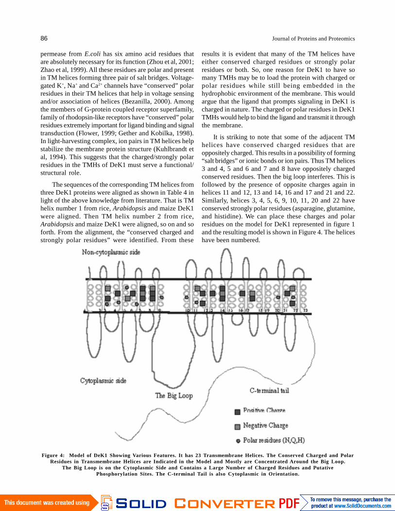
It is striking to note that some of the adjacent TMhelices have conserved charged residues that areoppositely charged. This results in a possibility of forming“salt bridges” or ionic bonds or ion pairs. Thus TM helices3 and 4, 5 and 6 and 7 and 8 have oppositely chargedconserved residues. Then the big loop interferes. This isfollowed by the presence of opposite charges again inhelices 11 and 12, 13 and 14, 16 and 17 and 21 and 22.Similarly, helices 3, 4, 5, 6, 9, 10, 11, 20 and 22 haveconserved strongly polar residues (asparagine, glutamine,and histidine). We can place these charges and polarresidues on the model for DeK1 represented in figure 1and the resulting model is shown in Figure 4. The heliceshave been numbered.
Figure 4: Model of DeK1 Showing Various Features. It has 23 Transmembrane Helices. The Conserved Charged and PolarResidues in Transmembrane Helices are Indicated in the Model and Mostly are Concentrated Around the Big Loop.
The Big Loop is on the Cytoplasmic Side and Contains a Large Number of Charged Residues and PutativePhosphorylation Sites. The C-terminal Tail is also Cytoplasmic in Orientation.

Alternative Conformational Model of a Seed Protein DeK1 for Better Understanding of Structure-Function Relationship 87
It is interesting to see how in the model the conservedcharged and polar residues are crowded mostly near thebig loop. Oppositely charged amino acid residues caninteract to form salt bridges either in the same loop orbetween adjacent loops. Such salt bridges can be dynamicin the sense that they can form and open up reversibly inpresence of a stimuli or a ligand. The conserved stronglypolar residues (spheres in Figure 4) can neutralize charges.Polar residues when present in TM helices can act bothas acids and bases and so can form salt bridgesthemselves. As in lactose permease and some othermembrane proteins, these salt bridges along withconserved charged and polar residues could be importantstructurally and/or functionally for DeK1 protein.
C-terminal Tail
The long C-terminal tail (Figure 4) is the only domain ofDeK1 for which some biochemical information is available(Wang et al, 2003) and functional implications have beenhighlighted (Johnson et al, 2008). Constituting the majorportion of DeK1 (about 1000 amino acids out of 2159amino acid residues), the C-terminal tail codes for acalpain like domain. The maize DeK1 calpain like domainwas shown to have high similarity to animal calpain,including a conserved catalytic site (Lid et al, 2002; Wanget al, 2003). Arabidopsis thaliana and maize DeK1 share70% overall identity; the identity of calpain like domainbeing even higher at 85% (Lid et al, 2002). Calpains arecysteine proteinases that are activated by rise inintracellular calcium concentration and are involved inmediating signal transduction leading to cell differentation,proliferation and cell death in animal system (Sato andKawashima, 2001; Ahn et al, 2004). In vitro studies havedemonstrated that the maize DeK1 calpain has proteinaseactivity similar to animal m-calpain and that the activitydepends on cysteine residue of conserved active site(Wang et al, 2003). In contrast to m-calpain, the DeK1calpain was active in the absence of calcium, the activitybeing stimulated by addition of calcium. However, thespecific activity of the putative proteinase was extremelylow (Wang et al, 2003) and it might be necessary to findthe proper substrate. Lately, it was demonstrated thatthe calpain like proteinase domain undergoes autocatalysisand this domain seems to play a major role in impartingDeK1 its physiological function (Johnson et al, 2008).
The conventional calpains typified by human m-calpains, are cytosolic enzymes activated by a rise inintracellular Ca, as well as by signaling that includes theprotein kinase C-tyrosine kinase or adhesion molecule-derived cascade (Sato and Kawashima, 2001). Uponactivation, the enzyme associates with proteins orphospholipids in the plasma membrane and undergoes
autolysis (Sato and Kawashima, 2001). The substratesof calpains are typically part of signal transductioncascades, such as protein kinase C’s (Kishimoto et al,1989; Temmblay et al, 2000). Through these mechanismsconventional calpain fulfill essential function in multipledevelopmental pathways in animals (Arthur et al, 2000).By analogy, the C-terminus of DeK1, could be involvedin developmental processes in the endosperm cells thatare at the surface thus converting them to aleurone cells.Like animal calpains, the activated C-terminal calpaindomain of DeK1 might associate with the plasmamembrane with the help of charged residues in the TMhelices to undergo autolysis. The big loop, with itscapacity to be heavily phosphorylated, might activate thecalpain domain of DeK1 either directly or through otherintermediary proteins. It is also possible that the big loopis itself a putative substrate for the C-terminal proteinase.All these processes could trigger the downstream signalingthat regulates cell differentiation.
IV SUMMARY
The in silico investigations performed here delineate theDeK1 sequence and help to model the protein. The modelprovides us with domains that individually or incombination can be important for the structure andfunction of the protein. It also indicates various structuralelements that can be studied experimentally to understandthe protein better. The model provides a platform fortesting various hypotheses regarding the protein function.
Previously, during genetic analysis, a preliminaryattempt was made to model DeK1 (Lid et al, 2002).However, only one predictor TMHMM was used. Wehave shown here how one predictor and especiallyTMHMM alone can lead to wrong predictions. The modelthat was obtained (Lid et al, 2002) did not fit into theirobservations and expectations, and it was turned upsidedown to fit into their hypothesis regarding the functionof the C-terminal tail of DeK1. However, our attempt atmodeling DeK1 has yielded a better model that can fitinto their hypothesis without having to turn it upside downor changing features of the theoretical model.
The bioinformatic analysis has supported many ofthe results from genetic analysis. It can be saidunequivocally that DeK1 is a membrane protein and thatit is localized in the cell wall. However, some of theprevious results were contradicted as well. It wassuggested that the C-terminal tail and the big loop areoppositely oriented to the membrane. However, we haveshown that they are on the same side (cytoplasmic side)of the membrane. Our results help hypothesize therelationship of the structure of the protein to its functionbetter. At the least, we have shown an alternative model
88 Journal of Proteins and Proteomics
and the benefits of using multiple topology predictionalgorithms in decoding a membrane protein sequence. Itis also established from our studies that PHOBIUS is abetter TM topology predictor, atleast for DeK1.
Our observations and analysis regarding the featuresof the big loop, its relation to the smaller loops, thepresence of conserved charged and polar residues in theTM helices of DeK1, and their functional implicationsare novel.
We can now visualize a putative DeK1 structureatleast at the secondary structure level, albeit theoretical.We know what to expect when we initiate experimentalinvestigations, what to look at, what to observe closely,what the challenges are and what the various optionsare. The theoretical studies here have given us a solidfooting for experimental approaches.
Genetic analysis and cell biology studies have shownthat DeK1 plays an important role in aleurone celldevelopment (Lid et al, 2005; Lid et al, 2002; Becraft,2001; Becraft and Asuncion-Crabb, 2000; Becraft et al,2002). Endosperm cells that are on the “surface” seemto get differentiated into aleurone cells forming a singlelayer below the pericarp (Gifford et al, 2003). DeK1regulates this process. It must be doing so by a signalingmechanism yet unknown. The model proposed heresupports this role of DeK1. If it has to sense the “surface”it can best be done from the cell membrane than thecytoplasm. The model shows that DeK1 has a numberof loops that protrude out in the non-cytoplasmic sidethat might sense any stimuli. If an external ligandstimulates the signaling process then the loops can cometogether to form a channel to allow the passage of theligand. The conserved charged and strongly polar residuesin the TM helices can help ligand binding. These residuescan form salt bridges as well and in the presence of aputative ligand can open up to help ligand binding. If thisis true then the helices near the big loop would beimportant since the charged and polar residues arecrowded in this region. Such opening and closing of saltbridges can also lead to conformational changes in theprotein structure and the signal can be sent inside as aresult.
The properties of the big loop hint at an importantrole as well. Signaling events ought to involve kinasesand phosphorylation to initiate the signaling cascade. Thebig loop contains a number of probable phosphorylationsites for this to happen. The high charge density on thebig loop may help this protein to interact with othersubstrate proteins as well. That is to say, the big loopcould be a docking site for substrates. The importance
of calpains in animal cells in regulating cell developmenthas been mentioned in the results and discussion section.The C-terminal tail of DeK1 has homology to calpaindomains and a cysteine residue at its active site typical ofa proteinase. The big loop could be a substrate for thecalpain-like protease. The protease could cleave the bigloop off the protein, which can then reach the nucleustriggering a set of reactions that signal the cells to specifyinto aleurone cells. We have shown theoretically that thebig loop by itself has a high probability of nuclearsublocalization.
As discussed in the previous section, the human m-calpains are cytosolic enzymes that are activated bysignaling that includes the protein kinase C-tyrosine kinaseor adhesion molecule-derived cascade. The big loop canprobably serve this purpose as well. In summary, themodel of DeK1 shows that it has many features that canmake the protein capable of signaling events leading toregulation of cell development and differentiation.
V ACKNOWLEDGEMENTS
The financial assistance provided by University of Delhithrough R&D grants, Department of Biotechnology(DBT) through research grant to SK (BT/PR8901/BRB/10/535/2007), and University Grants Commission (UGC)through Special Assistance (SAP) programme to theDepartment of Biochemistry is greatly acknowledged.
REFERENCES
[1] Ahn, J.W., Kim, M., Lim, J.H., Kim, G.T., and Pai, H.S. (2004).“Phytocalpain Controls the Proliferation and DifferentiationF a t e s o f C e l l i n P l a n t O r g a n D e v e l o p m e n t ” . Plant J. 38: 969-981.
[2] Arai, M., Mitsuke, H., Ikeda, M., Xia, J.X., Kikuchi, T., Shatake,M., and Shimizu, T. (2004). “CornPredII: a Consensus PredictionMethod for Obtaining Transmembrane Topology Models withHigh Reliability.” Nucleic Acids Res. 32: W390-393.
[3] Arthur, J.S.C., Elec, J.S., Hegadorn, C., Williams, K., and Greer.P.A. (2000). “Disruption of the Murine Calpain Small SubunitGene, Capn4: Calpain is Essential for Embryonic Developmentbut not for Cell Growth and Division.” Mol. Cell.Biol. 20:4474-4481.
[4] Becraft, P.W. (2001). “Cell Fate Specification in the CerealEndosperm”. Semin. Cell. Dev. Biol. 12: 387-394.
[5] Becraft, P.W., and Asuncion-Crabb,Y. (2000). “Positional CuesSpecify and Maintain Aleurone Cell Fate in Maize EndospermDevelopment”. Development. 127: 4039-4048.
[6] Becraft, P.W., Brown, R.C., Lemmon, B.E.O., Ferstad, H.G.,and Olsen, O.A. (2001). “Endosperm Development. In “CurrentTrends In The Embryology Of Angiosperms.” (S. S. Bhojwani, Ed.)pp. 353-374.
[7] Becraft, P.W., Li, K., Dey, N., and Asuncion-Crabb, Y. (2002).“The Maize Dek1 Gene Functions in Embryonic PatternFormation and in Cell Fate Specification.” Development. 129:5217-5225.
Alternative Conformational Model of a Seed Protein DeK1 for Better Understanding of Structure-Function Relationship 89
[8] Bendtsen, J.D., Nielsen, H., von Heijne G., and Brunak, S. (2004).“Improved Prediction of Signal Peptides: SignalP 3.0.” J. MoI.Biol. 340: 783-795.
[9] Berger, F. (1999). “Endosperm Development.” Curr. Opin. PlantBiol. 2: 28-32.
[10] Bezanilla, F. (2000). “The Voltage Sensor in Voltage-dependentIon Channel.” Physiol. Rev. 80: 555-592.
[11] Blom, N., Gammeltoft, S., and Brunak, S. (1999). “Sequence-and Structure-based Prediction of Eukaryotic ProteinPhosphorylation Sites.” J. Mol. Biol. 294: 1351-1362.
[12] Cokol, M., Nair, R., and Rost, B. (2000). “Finding NuclearLocalization Signals.” EMBO Rep. 2001: 411-415.
[13] David, D., Das, S.J.N., Thomas, U., Chen-Ni, C., and Jan, W.G.(2002). “Rapid Topological Mapping of E. Coli Inner- membraneProtein.” Proc. Natl. Acad. Sci. USA 5: 2690-2695.
[14] Dixon, B., Walker, B. Kimmins, W., and Pohajdak, B. (1991).Isolation and Sequencing of a cDNA for an Unusual Hemoglobinfrom the Parasitic Nematode Pseudoterranova Decipiens.”Proc. Natl Acad. Sci. USA. 88: 5655-5659.
[15] Flower, D.R (1999). “Modeling G-protein-coupled Receptorsfor Drug design.” Biochim. Biophys. Acta. 1422: 207-234.
[16] Fortina, M.G., Ricci, G., Mora, D., Guglielmetti, S., andManachini, P.L. (2003). “Unusual Organization for Lactoseand Galactose Gene Clusters in Lactobacillus Helveticus.”Appl. Environ. Microbiol. 69: 3238-3243.
[17] Galinat, W.C. (1990). “Multi-layered Expression of AleuroneSpecific Genes.” Maize News Lett. 64: 121.
[18] Gether, U., and Kobilka, B.K. (1998). “G. Protein-coupledReceptors. II. Mechanism of Agonist Activation.” J. Biol. Chem.273: 17979-17982.
[19] Gibson, Q.H., Regan, R., Olson, J.S., Carver, T.E., Dixon, B.,Pohajdak, B., Sharma, P.K., and Vinogradov, S.N. (1993).“Kinetics of Ligand Binding to Pseudoterranova Decipiens andAscaris Suum Hemoglobins and to Leu-29 → Tyr sperm whaleMyoglobin Mutant.” J. Biol. Chem. 268: 16993-16998.
[20] Gifford, M.L., Dean, S., and Ingram, G.C. (2003). “TheArabidopsis ACR4 Gene Plays a Role in Cell Layer OrganisationDuring Ovule Integument and Sepal Margin Development.”Development. 130: 4249–4258
[21] Hessa, T., Kim, H., Bihimaier, K., Lundin, C., Boekel, J.,Andersson, H., Nilsson, I., Stephan, H., and von Heijne G.(2005a). “Recognition of Transemembrane Helix byEndoplasmic Reticulum Translocon.” Nature. 433: 377-381.
[22] Hessa, T., White, S.H., and von Heijne, G. (2005b). “MembraneInsertion of a Potassium-channel Voltage Sensor.” Science 307:1427.
[23] Hirokawa, T., Boon-Chieng, S., and Mitaku, S. (1998). “SOSUI:Classification and Secondary Structure Prediction System forMembrane Proteins.” Bioinformatics. 14: 378-379.
[24] Hofmann, K., and Stoffel, W. (1993). “A Database of MembraneSpanning Protein Segments.” Biol.Chem. 374: 166.
[25] Horton, P., and Nakai, K. (1997). “Better Prediction of ProteinCellular Localization Sites with the k Nearest NeighborsClassifier.” Proc. Int. Conf. Intell. Syst. Mol. Biol. 5: 147-152.
[26] Jeanmougin, F., Thompson, J.D., Gouy, M., Higgins, D.G., andGibson, T.J. (1998). “Multiple Sequence Alignment with ClustalX.” Trends Biochem Sci. 23: 403-405.
[27] Johnson, K.L., Degnan, K.A., Walker, J.R., and Ingram, G.C.(2005). “AtDEK1 is Essential for Specification of EmbryonicEpidermal Cell Fate.” Plant J. 44: 114-127.
[28] Johnson, K.L., Faulkner, C., Jeffree C.D., and Ingram, G.C.(2008). “The Phytocalpain Defective Kernel 1 is a NovelArabidopsis Growth Regulator whose Activity is Regulated byProteolytic Processing.” The Plant Cell, 20, 2619–2630.
[29] Kall , L., Krogh, A., and Sonnhammer, E.L. (2004).“A Combined Transmembrane Topology and Signal PeptidePrediction Method.” J. Mol. Biol. 338: 1027–1036.
[30] Kishimoto, A., Mikawa, K., Hasimoto, K., Yasuda, I., Tanaka,S., Tominaga, M., Kurada, T. and Nishizuka, Y. (1989). “LimitedProteolysis of Protein Kinase C subspecies by Calcium-dependentNeutral Protease (calpain).” J. Biol.Chem. 264: 4088-4092.
[31] Krogh, A., Larsson, B., von Heijne G., and Sonnhammer, E.L.(2001). “Predicting Transmembrane Protein Topology with aHidden Markov Model: Application to Complete Genome.”J. Mol. Biol. 305: 567-580.
[32] Kuhlbrandt, W., Wang, D.N., and Fujiyoshi, Y. (1994). “AtomicModel of Plant Light-harvesting Complex by ElectronCrystallography.” Nature. 367: 614-621.
[33] Li, X., Ding, J., Liu, Y., Brix, B.J., and Fliegel, L. (2004).“Functional Analysis of Acidic Amino Acid in Cytosolic Tail ofthe Na+/H+ exchanger.” Biochemistry 43: 16477-16486.
[34] Lid, S. E., Gruis, D., Jung, R., Lorentzen, J.A., Ananiev, E.,Chamberlin, M., Niu, X., Meeley, R., Nichols, S., and Olsen,O-.A. (2002). “The Defective Kernel 1 (dek1) Gene Requiredfor Aleurone Cell Development in the Endosperm of MaizeGrains Encodes a Membrane Protein of the Calpain GeneSuperfamily.” Proc. Natl. Acad. Sci. U.S.A. 99: 5460-5465.
[35] Lid, S.E, Olsen, L., Nestestog, R., Aukerman, M., Brown, R.C.,Lemmon, B., Mucha, M., Opsahl-Sorteberg, H-G, and Olsen,O-A. (2005). “Mutation in the Arabidopsis Thaliana DeK1Calpain Gene Perturbs Endosperm and Embryo DevelopmentWhile Over-expression Affects Organ Development Globally.”Planta, 221, 339-351.
[36] MacKinnon, R. (2005). “Membrane Protein Insertion andStability.” Science. 307: 1425-1426.
[37] Melen, K., Krogh, A., and von Heijne, G. (2003). “ReliabilityMeasures for Membrane Protein Topology PredictionAlgorithms.” J. Mol. Biol. 327: 735-744.
[38] Mitaku, S., and Hirokawa, T. (1999). “Physicochemical Factorsfor Discriminating between Soluble and Membrane Proteins:Hydrophobicity of Helical Segments and Protein Length.”Protein Eng. 11: 953-957.
[39] Mitaku, S., Hirokawa, T., and Ono, M. (1998). “Classificationof Membrane Proteins by Types of Transmembrane HelicesUsing SOSUI System.” Genome Informatics. 9: 367-368.
[40] Moller, S., Croning, M.D.R., and Apweiler, R. (2001). Evaluationof Methods for the Prediction of Membrane Spanning Proteins.”Bioinformatics. 17: 646-653.
[41] Nakai, K. and Kanehisa, M. (1991). “Expert System forPredicting Protein Localization Site in Gram-negative Bacteria.”Proteins, 11, 95-110.
[42] Nakai, K., and Horton, P. (1999). “PSORT: a Program forDetecting Sorting Signals in Proteins and Predicting theirSubcellular Localization.” Trends Biochem. Sci. 24: 34-36.
90 Journal of Proteins and Proteomics
[43] Nelson, O.E., and Chang, M.T. (1974). “Effect of MultipleAleurone Layers on the Protein and Amino Acid Content ofMaize Endosperm.” Crop Sci. 14: 374-376.
[44] Olsen, L.T., Divon, H. H., Al, R., Fosnes, K., Lid S. E., andOpsahl-Sorteberg, H. (2008). “The Defective Seed5 (des5)Mutant: Effects on Barley Seed Development and HvDek1,HvCr4, and HvSal1 Gene Regulation.” J. Exp. Bot. 59:3753-3765.
[45] Olsen, O-.A. (2001). “Endosperm Development: Cellularizationand Cell Fate Specification.” Annu. Rev. Plant Physiol. PlantMol Biol. 52: 233-267.
[46] “Penalties and Weight Matrix Choice.” Nucleic Acid Res. 22:4673-4680.
[47] Persson, B., and Argos, P. (1994). “Prediction of TransmembraneSegments in Proteins Utilising Multiple Sequence Alignments.”J. Mol. Biol. 237: 182-192.
[48] Persson, B., and Argos, P. (1996). “Topology Prediction ofMembrane Proteins.” Prot. Sci.5: 363-371.
[49] Ritchie, S., and Gilroy, S. (1998). “Abscisic Acid SignalTransduction in the Barley Aleurone is Mediated byPhospholipase D. Activity.” Proc. Natl. Acad. Sci. USA. 95:2697-2702.
[50] Sal-man, N. and Shai, Y. (2005). “Arginine Mutations within aTransmembrane Domain of Tar, an Escherichia Coli AspartateReceptor, can Drive Homodimer Dissociation and HeterodimerAssociation in vivo”. Biochem. J. 385: 29-36.
[51] Sato, K. and Kawashima, S. (2001). “Calpain Function in theModulation of Signal Transduction Molecules.” J. Biochem.382: 743-751.
[52] Sheridan, W.F. and Neuffer, M.G. (1980). Defective KernelMutants of Maize. II. Morphology and Embryo Culture Studies.”Genetics. 95: 945-968.
[53] Stoffel, W., Duker, M., and Hofmann, K. (1993). “MolecularCloning and Gene Organization of the Mouse Mitochondrial 3,2-trans-enoyl-CoA Isomerase.” FEBS Lett. 333: 119-122.
[54] Temmblay, R., Chakravarthy, B., Hewitt, K., Tauskela, J., Morly,P., Atkinson, T., and Durkin, J.P. (2000). “Transient NMDAReceptor Inactivation Provides Long-term Protection toCultured Cortical Neurons from a Variety of Death Signals.”J. Neurosci. 20: 7183-7192
[55] Thompson, J.D., Higgins, D.G. and Gibson, T.J. (1994),Clastal W., “Improving the Sensitivity of Progressive MultipleSequence Alignment Through Sequence Weighting, Position-Specific Gap Penalties and Weight Matrix Choice”. Nucleic AcidsRes., 22, 4673 – 4680.
[56] Tusnady, G.E. and Simon, I. (1998). “Principles Governing AminoAcid Composition of Integral Membrane Proteins: Applicationto Topology Prediction.” J. Mol. Biol . 283:489-506.
[57] Wang, C., Barry, J.K., Min, Z., Tordsen, G., Rao, A.G. andOlsen, O-.A. (2003). The Calpain Domain of Maize DeK1Protein Contains the Conserved Catalytic Triad and Functionsas a Cysteine Proteinase.” J Biol. Chem. 278: 34467-34474.
[58] Zhao, M., Zen, K.C., Hubbell, W.L., and Kaback, H.R. (1999).“Proximity between Glu126 and Arg144 in the LactosePermease of Escherichia coli.” Biochemistry. 38: 7407-7412.
[59] Zhou, F.X., Merianos, H.J., Brunger, A.T. and Engelman, D.M.(2001). “Polar Residues Drive Association of PolyleucineTransmembrane Helices.” Proc. Natl. Acad. Sci. USA. 98:2250-2255.