alternate glucocorticoid receptor ligand binding structures influence outcomes in an
TRANSCRIPT
Comparative Biochemistry and Physiology, Part C 156 (2012) 121–129
Contents lists available at SciVerse ScienceDirect
Comparative Biochemistry and Physiology, Part C
j ourna l homepage: www.e lsev ie r .com/ locate /cbpc
Alternate glucocorticoid receptor ligand binding structures influence outcomes in anin vivo tissue regeneration model
Sumitra Sengupta a,1, William H. Bisson b,1, Lijoy K. Mathew a, Siva K. Kolluri a, Robert L. Tanguay a,⁎a Department of Environmental and Molecular Toxicology, the Environmental Health Sciences Center, Oregon State University, Corvallis, OR, USAb Pharmaceutical Biochemistry Group, School of Pharmaceutical Sciences, University of Geneva, Switzerland
Abbreviations: dpf, Days post fertilization; dpa, Daysligand binding domain; DEX, dexamethasone; BDP, BBeclo, beclomethasone; PDB, Protein data bank; RMSDHB, hydrogen bonding.⁎ Corresponding author at: Department of Environme
Oregon State University, Sinnhuber Aquatic Research LaCorvallis, OR 97333, USA. Tel.: +1 541 737 6514; fax: +
E-mail address: [email protected] (R1 These authors contributed equally to this manuscri
ered as first authors.
1532-0456/$ – see front matter © 2012 Elsevier Inc. Alldoi:10.1016/j.cbpc.2012.05.003
a b s t r a c t
a r t i c l e i n f oArticle history:Received 1 April 2012Received in revised form 19 May 2012Accepted 19 May 2012Available online 24 May 2012
Keywords:DockingDynamicsGlucocorticoidsIn vivoRegenerationSARTissueZebrafish
Since their characterization, glucocorticoids (GCs), the most commonly prescribed immunomodulatorydrugs, have undergone numerous structural modifications designed to enhance their activity. In vivo assess-ment of these corticosteroid analogs is essential to understand the difference in molecular signaling of theligands that share the corticosteroid backbone. Our research identified a novel function of GCs as modulatorsof tissue regeneration and demonstrated that GCs activate the glucocorticoid receptor (GR) to inhibit earlystages of tissue regeneration in zebrafish (Danio rerio). We utilized this phenomenon to assess the effect ofdifferent GC analogs on tissue regeneration and identified that some GCs such as beclomethasone dipropionate(BDP) possess inhibitory properties, while others, such as dexamethasone and hydrocortisone have no effect onregeneration. We performed in silico molecular docking and dynamic studies and demonstrated that type andsize of substitution at the C17 position of the cortisol backbone confer a unique stable conformation to GR on li-gand binding that is critical for inhibitory activity. In the field of tissue regeneration, our study is one of the firstStructure Activity Relationship (SAR) investigations performed in vertebrates demonstrating that the in vivo tis-sue regeneration model is a powerful tool to probe structure function relationships, to understand regenerativebiology, and to assist in rational drug design.
© 2012 Elsevier Inc. All rights reserved.
1. Introduction
Glucocorticoids are the most commonly used anti-inflammatoryand immunosuppressive agents highly efficacious in the treatment ofdisease, but they are also associated with serious side effects. Hence,improvements in the therapeutic profile of these drugs are needed. Todate, the majority of the structural modifications of glucocorticoidreceptor (GR) ligands were designed to eliminate side effects. Thereare approximately 20 topically active anti-inflammatory corticosteroidson the market (http://www.hfs.illinois.gov/pharmacy/topical.html).Development of non-steroidal dissociated ligands such as AL438(Einstein et al., 2004; Schacke et al., 2007; Xu et al., 2009) suggestedthat ligand structures can be manipulated to induce differential andmore desired biological activity. Recent reports of distinct ligand guidedresponses by GR have opened new avenues in the field of drug
post amputation; GR-LBD, GReclomethasone dipropionate;, Root mean square deviation;
ntal and Molecular Toxicology,boratory, 28645 East HWY 34.1 541 737 0497.
.L. Tanguay).pt and should both be consid-
rights reserved.
discovery and development.Whilemost of the studies have been eitherin silico or in cultured cells, an in vivo model to evaluate ligand depen-dent responses of GR is lacking.
In the last few years, the use of in vivo zebrafish (Danio rerio)model in scientific research is rapidly growing. Initially, it was a pop-ular model to study vertebrate development because the zebrafishembryos rapidly develop externally from the mother and are nearlytransparent (Hao et al., 2010). The current use of zebrafish in earlydrug discovery and lead optimization phases covers a wide range ofapplications: screening of lead compounds, target identification,target validation,morpholino oligonucleotide screens, assay developmentfor drug discovery, physiology based drug discovery, quantitative struc-ture–activity relationship (QSAR) and drug toxicity assays (Chakrabortyet al., 2009). The zebrafish model is also useful to identify compoundswith favorable physiochemical properties and excellent drug-likenesswith the aim of speeding up the drug development process.
While GCs are mostly used as anti-inflammatory agents, the newlyidentified role of GCs as modulators of tissue regeneration has openeda new paradigm in the field of regenerative medicine. In our group,we identified GCs as modulators of tissue regeneration utilizing anearly life stage model. A two-day post fertilization (dpf) zebrafishcompletely regenerates its caudal fin three days post amputation(dpa) by a process known as epimorphic tissue regeneration. Sincemammals have limited capacity for epimorphic regeneration of com-plex structures, larval zebrafish offers a unique alternative model to
RegeneratingA
Non-RegeneratingB
Prednisone
Cortisone
TriamcinoloneTriamcinolone Diacetate
Hydrocortisone Acetate
Beclomethasone Dipropionate
Flumethazone Pivalate
Clobetasol Dipropionate
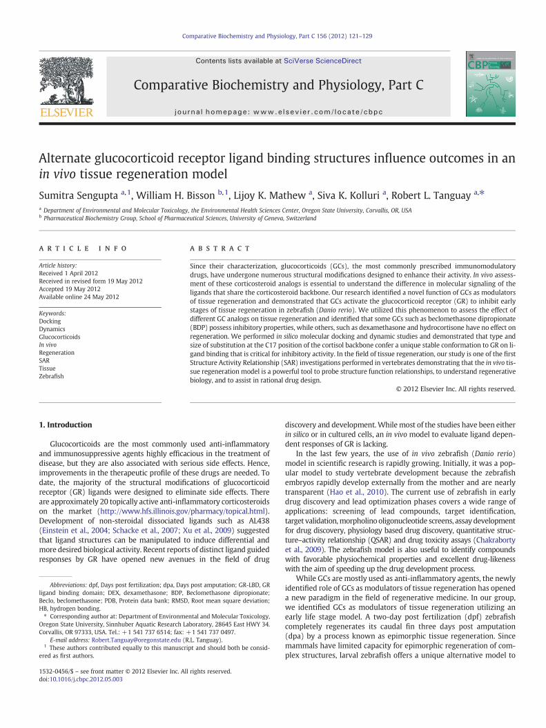
Fig. 1. Structure of selected glucocorticoids. Chemical structure of selected chemicals fromthe 2000member FDA approved library that permitted regeneration (cortisol, prednisone,hydrocortisone acetate, triamcinolone) or inhibited tissue regeneration (beclomethasonedipropionate, clobetasol dipropionate, flumethazone pivalate, triamcinolone diacetate)response.
122 S. Sengupta et al. / Comparative Biochemistry and Physiology, Part C 156 (2012) 121–129
identify therapeutic strategies that can promote optimal healing andreplacement of tissue damaged by trauma, disease, or congenital de-fects. We combined this early life stage regeneration model withchemical genetics to identify modulators of regeneration. The guidinghypothesis is that a chemical that inhibits regenerationmust have im-pacted a molecular target critical for the regenerative process. Identi-fication of such chemical targets will allow a better understanding ofthe regeneration promoting pathways, paving a path for enhancedmammalian regeneration. As a proof of concept, we screened a 2000member library of FDA approved drugs that contained thirty-threeGCs. The GCs that inhibited regeneration rendered characteristic ‘V’shaped architecture to the caudal fin upon exposure (Mathew et al.,2007).We performed further studieswith beclomethasone dipropionate(BDP) as a representative GC and determined that activation of GR isnecessary for the GCs to block the earliest stages of tissue regeneration.The activated GR functions as a ligand dependent transcriptional regu-lator and GCs exert a wide range of physiological effects following bind-ing.We aimed to explore the structure activity relationship (SAR) of theknownGR ligands in the context of tissue regeneration in order to iden-tify a pharmacophore backbone that dictates regenerative response aswell as reveal novel facts about ligand dependent responses of GR inan in vivomodel.
2. Material and methods
2.1. Zebrafish husbandry and imaging
Zebrafish (Danio rerio) embryos (5D strain) (Hillwalker et al.,2010) were obtained from a breeding colony and raised using stan-dard husbandry procedures for all the experiments (Westerfield,1993, 2000). Caudal fins of 2 day post fertilization (2 dpf) larvae wereamputated as previously described (Poss et al., 2002; Andreasen et al.,2006; Mathew et al., 2006) and chemical screening was performedbased on our previously described in vivo larval regeneration assayprotocol (Mathew et al., 2007). All experimental groups consisted ofsample size n=12. Images were captured under bright field using aNikon SMZ1500 microscope at 10× magnification on 2% agarose platesafter anesthetizing the embryos using tricaine.
2.2. Chemical exposures
Amputated larvae (2 dpf) were exposed to 1 μM dexamethasone(DEX) (D1756, Sigma-Aldrich, St Louis, MO, USA), beclomethasonedipropionate (BDP) (B3022, Sigma), beclomethasone (Beclo) (B0385,Sigma), or hydrocortisone (HC) (H4001, Sigma) as shown in Fig. 1.R198897 (21-Cl-9-α-F-17-α-HO-16-β-me-pregna-1,4-diene-3,11,20-trione butanoate), was also purchased from Sigma, and ST075178(2 S,10 S,11 S,13 S,15 S,17 S,1R,14R) -1 -fluoro -17-hydroxy-14-(2-hydroxyacetyl)-2,13, 15-trimethyl-5-oxotetracyclo[8.7.0.0b2,7>.0b11,15>]heptadeca-3,6-dien-14-yl pentanoate, and ST075183(2-((2 S,10 S,11 S,15 S,17 S,1R,13R,14R)-1-fluoro-14,17-dihydroxy-2,13,15-trimethyl-5-oxotetracyclo[8.7.0.0b2,7>.0b11,15>]heptadeca-3,6-dien-14-yl)-2-oxoethyl acetate) were purchased from TimTec (Newark, DE, USA) asshown in Fig. 6. All chemicals were resuspended in DMSO and exposureswere performed in zebrafish embryo buffer. DMSO concentration wasmaintained at less than 1% and controls used for each chemical weremaintained at matched DMSO concentration.
2.3. RNA isolation
The caudal fins of 2 dpf embryos were amputated, and embryoswere placed individually in wells of 96-well plates with exposure so-lutions of dimethyl sulfoxide (DMSO, vehicle control) or chemical.Twelve embryos were pooled for each of the three replicates pertreatment and RNA was isolated from the whole embryos using TriReagent from Sigma, as per manufacturer's instruction.
2.4. Quantitative real time reverse transcriptase polymerase chainreaction (qRT-PCR)
Total RNA was isolated from whole embryos. Each treatment com-prised three replicates with an n=12 embryos per replicate andcDNA was synthesized from 1 μg of total RNA isolated from eachgroup using Superscript II (Life Technologies) with oligo(dT) primersfollowed by RNaseH treatment to eliminate RNA contamination fol-lowing manufacturer's instructions. QRT-PCR was performed on theOpticon 2 real time PCR detection system (MJ Research) using SYBRgreen qPCR detection kits (Finnzymes). Gene specific primers are

123S. Sengupta et al. / Comparative Biochemistry and Physiology, Part C 156 (2012) 121–129
listed in supplemental Table S1. Each sample was normalized to en-dogenous β-actin quantity. Agarose gel electrophoresis and meltcurve analysis confirmed expected PCR product formation. Statisticalsignificance of differences in mRNA abundance was determined byone-way ANOVA on log10 transformed data with a post test usingTukey's method (pb0.05) (Sigmastat Software).
2.4.1. OligonucleotidesThe primers used for qRT-PCR were synthesized by MWG-Biotech
(Alabama, USA). Oligotech and Primer blast programs were used todesign the primers listed in supplemental Table S1. Forward and an-tisense reverse primers are prefixed with F and R accordingly.
2.5. Morpholinos
Fluorescein tagged zebrafish GR (zf GR) morpholino (5′‐CGGAAC-CCTAAAATACATGAAGCAG‐3′) designed to target the splicing ofexons 7 and 8 was used to knockdown GR expression. Standard con-trol morpholino (Gene Tools) (5′ CTCTTACCTCAGTTACAATTTATA 3′)was injected at matching concentration. The morpholinos were dilut-ed to a stock concentration of 3 mM in 1× Danieau's solution (58 mMNaCl, 0.7 mM KCl, 0.4 mM MgSO4, 0.6 mM Ca(NO3)2, 5 mM HEPES,pH 7.6) (Nasevicius and Ekker, 2000) and approximately 2 nl of con-trol and zf GR morpholino was injected in 1–2 cell stage embryos. Theinjected embryos were screened for uniform fluorescence at 24 hpf toconfirm uniform distribution of the morpholino. At 2 dpf, caudal finsof morphants were amputated, followed by exposure to chemical.
2.6. Sequence alignment and homology modeling
The GR ligand binding domain (LBD) sequences in FASTA formatfor human and zebrafish were retrieved from the NCBI database.Sequence alignment was performed online with the LALIGN program[http://www.chembnet.org/software/LALIGN_form.html]. We usedthe X-Ray crystal structure of the human GR-LBD bound to dexameth-asone (DEX) in the agonist conformation available in the Protein DataBank (PDB ID: 1P93) as the 3D coordinate template for the homologymodeling of the zebrafish GR-LBD. The model was energetically re-fined using the internal coordinate space with Molsoft ICM v3.5-1p(Abagyan et al., 1994; Cardozo et al., 1995). Geometrical quality as-sessments of the pdb and homology models, were performed usingPROCHECK (Laskowski et al., 1993).
2.7. Molecular docking
The energy terms were based on the all-atom vacuum force fieldECEPP/3 with appended terms from the Merck Molecular Force Field toaccount for solvation free energy and entropic contribution (Abagyanet al., 1994). Modified intermolecular terms such as soft van der Waalsand hydrogen-bonding as well as a hydrophobic termwere added. Con-formational sampling was based on the biased probability Monte Carlo(BPMC) procedure, which randomly selects a conformation in the inter-nal coordinate space and thenmakes a step to a new randomposition in-dependent of the previous one according to a predefined continuousprobability distribution. It also has been shown that after each randomstep, full localminimization improves the efficiency of the iterative dock-ing procedure. In the ICM-VLS (Molsoft ICM v3.5-1p) screening proce-dure, the ligand scoring was optimized to obtain maximal separationbetween binders and non binders (Totrov and Abagyan, 1997, 2001).
The 3D coordinates of human GR-LBD–DEX complex in the agonistconformation was taken from crystal structures (PDB ID:1P93) (Kauppiet al., 2003). BDPwasmanually inserted into the GR-LBD binding pocketby matching the orientation of the 3-C_O ketone oxygen from theA-ring of DEX in the crystal structure involved in HB interactions withresidues Gln570 (α3) and Arg611 (α7) (Kauppi et al., 2003).
2.8. Molecular dynamic simulations
The prep files of DEX and BDP were performed using the programANTECHAMBER (AMBER 10), (Case et al., 2008). GR-LBD–DEX andGR-LBD–BDP complex models were immersed in a box of watermolecules (TIP3P model) and Na+ counter ions were added to thesolvent bulk of the protein/water complexes to maintain neutralityof the system using program LEAP AMBER10, (Case et al., 2008). Peri-odic boundary conditions were applied. The AMBER force field (Caseet al., 2008) all atom parameters (parm03) were used for the proteinand the Na+ ions. The total number of atoms for the GR-LBD–DEXand GR-LBD–BDP water boxed complexes is 34,926 and 34,942,respectively. The minimization protocol consisted of 1000 cycles ofsteepest descent followed by conjugate gradient method until theroot-mean square deviation (rmsd) of the Cartesian elements of thegradient reached a value smaller than 0.15 Å. The dynamic protocolconsisted of three steps: MD1, MD2 and MD3. The initial temperaturefor MD1, MD2 and MD3 were set at 0, 150, and 300 K respectively.During all dynamic steps the reference temperature of the systemwas fixed at 300 K according to Berendsen's coupling algorithms (DiNola et al., 1994). The initial velocity of the beginning of simulationis taken from Maxwellian distribution set at the desired temperature.The time step for all three dynamic procedures was 0.002 picosec(ps). For minimization and molecular dynamics, the primary cutoffdistance for non bonded interaction was set at 9 Å. Regarding themolecular dynamic protocol used, the first (MD1) aimed the equili-bration of water molecules and ions of the water boxed and chargeneutralizedmodel. An initial velocity was given to the system and tra-jectories were allowed to evolve in time according to Newtonian lawskeeping the model protein fixed. The number of dynamic steps was7500 corresponding to 15 ps. Next, 15 ps of constant volume dynamic(MD2) was performed on the entire system to adjust density to avalue of one (g/cm-3). In the third step, a 1000 ps (GR-LBD–Dex) and1500 ps (GR-LBD–BDP) constant pressure dynamic (MD3) at 1 atmwas applied without any constraint to assess conformational stability.The energy minimization, molecular dynamics and the correspondinganalyseswere performed using programAMBER10. Geometrical qualityassessments of the pdbmodels, were performed at different time pointsusing PROCHECK (Laskowski et al., 1993).
2.9. Statistical analysis
All experiments comprised a sample size of n=12. All statisticalcalculations were performed using Sigmaplot v. 11 (Systat SoftwareInc., San Jose, CA, USA) and p-values of b0.05 were considered statis-tically significant.
3. Results
3.1. Glucocorticoids elicit differential regenerative responses
Previous chemical genetic screening revealed GCs such as BDP asnovel modulators of tissue regeneration in a zebrafish model. Wefollowed this screen with a dose response analysis, and pursuedmechanistic studies with BDP (Mathew et al., 2007). Since BDP in-hibits regeneration even at low nanomolar concentrations, we per-formed further experiments at a screening concentration of 1 μM tounderstand how BDP modulates the regenerative process. Our resultsdemonstrated that activation of GR is necessary for these GCs toinhibit tissue regeneration. Mechanistic data revealed that over-expression of Cripto-1, an inhibitor of Activin signaling, mediatesBDP impaired tissue regeneration (unpublished data). Since all themembers of the glucocorticoid family act as ligands of the GR, wereevaluated the results of our screen that contained a total of thirtythree GCs. While seven GCs of the library inhibited tissue regenera-tion, twenty-one had no effect. The list of these twenty-one GCs

124 S. Sengupta et al. / Comparative Biochemistry and Physiology, Part C 156 (2012) 121–129
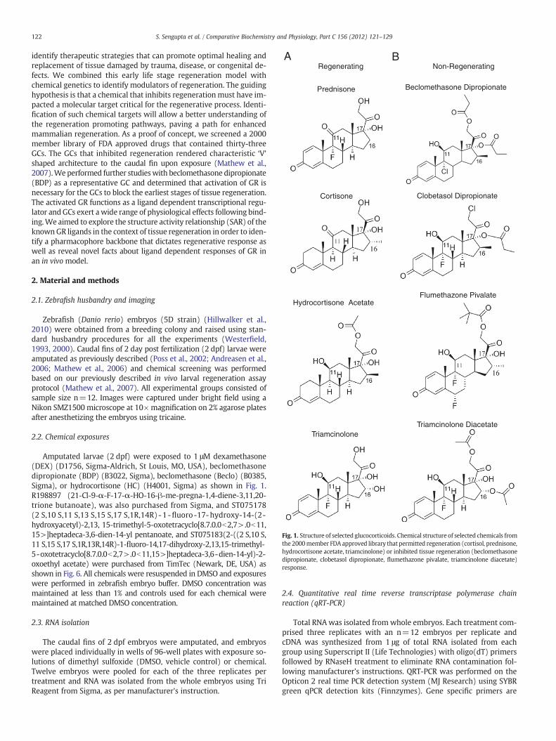
composed of well-known GCs such as Dexamethasone (Dex), Hydro-cortisone (HC) and beclomethasone (beclo). A representative groupof chemical structures is illustrated in Fig. 1. We further validatedthe results of DEX, HC and beclo by purchasing these chemicalsfrom commercial sources and repeating the regeneration assays. Theresults confirmed that the chemicals did not inhibit regeneration atthe screening concentration. We adopted a SAR approach to under-stand this differential response of members of the glucocorticoid fam-ily based primarily on their effects on fin regeneration. There is astrong correlation between GCs that inhibit regeneration and theirability to induce Cripto-1 expression. Unlike BDP, DEX, HC and beclodid not result in elevated Cripto-1 expression (Fig. 2) and they hadno impact on regeneration. We then aimed to understand this differ-ential response of the GC through SAR.
3.2. Inappropriate activation of GR is requisite for inhibiting tissueregeneration
All of the chemicals selected for the study are GR ligands known tomodulate downstream GR target genes such as Annexin a1b andFKBP506. Annexin a1b (anxa1b) is one of the transcripts repressed
A) Beclomethasone dipropionate (BDP
IIIIIIIIII
DMSO BDP
0.0
0.5
1.0
1.5
2.0
2.5
3.0
3.5*
Cripto-1
Rel
ativ
e ab
un
dan
ce o
f
- ac
tin
C) Dexamethasone (Dex)
DMSO Dex
0.0
0.2
0.4
0.6
0.8
1.0
1.2 Cripto-1
Rel
ativ
e ab
un
dan
ce o
f
- ac
tin
Fig. 2. Differential response of glucocorticoids in larval regeneration model. Caudal fin of 2 dexposed to A) beclomethasone dipropionate (BDP), B) beclomethasone (Beclo), C) dexamegeneration assay. Images were acquired at 3 dpa (10×) and RNAwas collected at 1 dpa fromof Cripto-1 transcript at 1 dpa is elevated onBDP exposure. However, therewas nodifference inand the asterisks indicate statistical significance (One way ANOVA, n=3) (pb0.05).
by activated GR at 24 h post amputation following BDP exposure. Inorder to evaluate whether these ligands activated GR in our systemirrespective of their effect on regeneration, we performed qRT-PCRand evaluated anxa1b expression following ligand exposure. DEX,HC, Beclo and BDP exposure suppressed anxa1b expression at 1 μMat 24 h post exposure, indicating activation of GR by exposure tothese ligands (Fig. 3). However, among the above ligands only BDP in-hibits regeneration. The fact that DEX, HC and Beclo activate GR andmodulate anxa1b expression similar to BDP, yet are unable to inhibitregeneration or elevate Cripto-1 expression indicate differentialactivity of GR upon binding by BDP compared to DEX, HC or Beclo.We hypothesized that specific GR conformation triggered by certainligands inhibits tissue regeneration.
3.3. Molecular docking studies revealed a conformational differenceinduced by ligand binding
To understand the difference in activated forms of GR we per-formed molecular docking studies with the human GR-LBD, as thecrystallographic structure of zebrafish homologue is not available.The human and zebrafish GR-LBD share 72% sequence identity and
)
D) Hydrocortisone (HC)
IIIIIIIIIII
DMSO HC0.0
0.1
0.2
0.3
0.4
0.5
Rel
ativ
e ab
un
dan
ce o
f
- ac
tin
Cripto-1
B) Beclomethasone (Beclo)
IIIIIIIIII
DMSO Beclo
0.0
0.2
0.4
0.6
0.8
1.0
Rel
ativ
e ab
un
dan
ce o
f
- ac
tin
Cripto-1
pf larvae was amputated (dotted lines mark the plane of amputation) and continuouslythasone (Dex) and D) hydrocortisone (HC) at 1 μM concentration for three days for re-whole embryos for cDNA synthesis and qRT PCR for Cripto-1 expression. The abundanceexpression on dex, beclo orHC treatment. The respective values represent themean±SEM

0
2
4
6
8
10
12
14
* * *
*
DMSOBeclo Dex HC
BDP
Rel
ativ
e ab
un
dan
ce o
f an
xa1b
Fig. 3. Activation of GR by different ligands irrespective of their effects on regenerativeresponse. 2 dpf larvae were exposed to 1 μM dex, beclo, HC, BDP and DMSO followingamputation. The abundance of anxa1b transcript estimated by qRT PCR at 1 dpa in thewhole embryo indicate significantly reduced expression in the exposed larvae indicatingGR activation. The respective values represent the mean±SEM and the asterisks indicatestatistical significance (One way ANOVA, n=3) (pb0.05).
125S. Sengupta et al. / Comparative Biochemistry and Physiology, Part C 156 (2012) 121–129
majority of the residues directly involved in the binding to DEX suchas Gln 570 (α3), Arg 611 (α7), Gln 642 (α8) and Thr 739 (α11) areconserved between the two species (Supplemental Fig. 1). The ho-mology model of zebrafish GR-LBD was then built using the available3D coordinate of the human GR-LBD bound to DEX (PDB ID: 1P93)and energetically refined as described in the Material and methodssection.
3.4. Molecular docking
Selected steroidal GR agonists that evoked differential impact onregeneration were docked into the human and zebrafish GR-LBDmodels. Docking results were similar for both species. Most of the ‘ac-tive’ GR ligands that inhibit regeneration did not dock into the GR-LBD binding pocket, while most of the ‘inactive’ GR ligands that didnot inhibit regeneration did dock into GR-LBD (Table S2). This sug-gests that docked steroidal GR ligands are stable in GR-LBD–DEXagonist conformation, while steroidal GR ligands like BDP do notstably fit into the binding pocket. In order to bind and stabilizethe agonist 3D tertiary structure of the GR-LBD, active compoundsinduce conformational changes involving either residue sidechains or secondary structure portions of the protein. Indeed, strongsteroidal GR agonists deacylcortivazol (DAC) and fluticasone furoate(FF) were co-crystallized into the human GR-LBD (PDB ID: 3BQD andPDB ID: 3CLD, respectively) (Suino-Powell et al., 2008) (SupplementalFig. 2).
To identify the conformational changes of the GR-LBD upon active li-gand binding, we ran 1.01 ns and 1.51 nsmolecular dynamic (MD) sim-ulationswith the human GR-LBD complexedwith ligands DEX and BDP,respectively. DEX is a known inactive compound (with respect to inhi-bition of tissue regeneration) and for this reason theX-Ray crystal struc-ture (PDB ID: 1P93) was considered as the 3D structure reference forsteroidal GR agonists unable to inhibit regeneration. BDP was insteadmanually inserted into the human GR-LBD binding pocket as describedin the Material and methods section.
3.5. Molecular dynamic simulations
By looking at the Root-Mean-Square-Deviation (RMSD) as a func-tion of time of all models, the GR-LBD–DEX complex reached aplateau during 1.01 ns MD (Fig. 4A), representing stable conforma-tion over time. On the other hand, GR-LBD–BDP complex reached
stability after adding 500 ps for a total of 1.51 ns MD (Fig. 4A). Thisis mainly due to the instability of BDP in the binding pocket duringthe simulation time (Fig. 4B). DEX crystallographic orientation withthe hydrogen bonding (HB) network remained stable over timewith a low RMSD of 0.75 Å, while BDP revealed considerable instabil-ity especially in the range between 0.3 and 0.5 ns MD (Fig. 4B). Over-all, the low RMSD of both GR-LBD complexes at equilibration between2.5 and 2.85 Å and structural comparison along the simulation timeindicates that the starting X-Ray structure represents a stable confor-mation and the MD protocol is suited to assess the stability of themodels.
Distances (Å) between atoms of specific residues were calculatedand analyzed over the simulation time applied. The stability of thecrystallographic orientation of DEX in the GR-LBD over time was con-firmed (Fig. 4C–E). The HB network involving the side chains of resi-dues Gln 570 (Fig. 4C), Arg 611 (Fig. 4D) and Gln 642 (Fig. 4E) iscritical for the stability of DEX in the GR-LBD agonist conformationwith an average calculated distance of 3 Å. This was not the case forBDP. (Fig. 4F–H) The stability of the human GR-LBD–DEX complexduring simulation time is proved by superimposing DEX and theside chains of residues Gln 570, Arg 611, Gln 642 and Thr 739 fromthe pdb structure of the complex at initial (t=0 ns) and final(t=1.01 ns) MD time (Fig. 5A). No 3D significant differences weredetected for the residues and the ligand over time (Fig. 5A). During1.51 ns MD the GR-LBD–BDP complex was instead very unstable(Fig. 4A,B). As a matter of fact, the calculated inter-atomic distancesbetween BDP and key residue side chain atoms showed that confor-mational changes are occurring over time in order to stabilize the li-gand–protein complex in the agonist conformation (Fig. 4F–H).
BDP was inserted manually into the GR-LBD binding pocket by po-sitioning the 3-C_O keton oxygen in the vicinity of the side chains ofGln 570 and Arg 611 to maintain the energetically favorable HB net-work observed with DEX and other GR agonists. During the simula-tion; however, these HB interactions were unstable due to residueside chain conformation changes, which produced significant fluctua-tions in the calculated inter-atomic distances (Fig. 4F–H). For a betterunderstanding of these fluctuations we superimposed BDP and aminoacids Gln 570, Arg 611 and Gln 642 from the PDB structure of thecomplex at initial (t=0 ns) and final (t=1.51 ns) MD time (Fig. 5B,C).During 1.51 ns MD (Figs. 4F, 5B) the side chain of Gln 570 rotated in-creasing the distance between the amidic N\H of the side chain of Gln570 and BDP from 3.3 Å (t=0) to 4.52 Å (t=1.51 ns).and thus exclud-ing the formation of anyHB interaction. In the case of Arg 611, therewasonly a change in the orientation between the twoN\H atoms of the pri-mary amino group of the side chain of Arg 611. Hence, that HB interac-tion with BDP remained stable with an inter-atomic distance betweenthe two functional groups of less than 3 Å for the entire period of simu-lation (Figs. 4G, 5B). The analysis of the inter-atomic distances over timebetween BDP and the side chain of Gln 642 produced themost interest-ing results (Fig. 4H). Significant conformational changes involving thisresidue and the ligand are taking place. We calculated the inter-atomicdistance between the side chain of Gln 642 and the two carbonyl oxygenatoms C-2_O (C-17-endo-propionate ester) and C-4_O (C-17-exo-propionate ester) of BDP (Fig. 1). From the graphic shown in Fig. 4Hwe observed that the distance during simulation time between theside chain of Gln 642 and C-2_O of BDP remains more or less stablearound 3.5 Å, whereas the distance between Gln 642 and C-4_O ofBDP is unstable (some stability is reached after 1 ns MD) provingthe C-17-exo-propionate ester moiety of BDP is moving in a consider-able way. We then analyzed the superimposition of residue Gln 642and BDP from the pdb structure of the complex at initial (t=0 ns)andfinal (t=1.51 ns)MD time (Fig. 5C). The side chain of Gln 642 shiftstowards the binding cavity to stabilize BDP in the binding pocket. As aconsequence, the C-17-exo-propionate ester moiety of BDP (Fig. 1) ismoving towards a hydrophobic pocket surrounded by residues Trp600, Leu 732, Leu 733 and Ile 757 (Fig. 5C).

Fig. 4.Molecular dynamic simulations reveal instability and residue side chain conformational changes in the GR-LBDwhen bound to BDP. RMSD graphics (all atoms plotted) versus time(picoseconds) of A) GR-LBD (pdb 1p93) in complex with dexamethasone, Dex (red) during 1.01 ns MD and beclomethasone di-propionate, BDP (black) during 1.51 ns MD and B) Dex(red) (pdb 1p93) during 1.01 ns MD and BDP (black) during 1.51 ns MD. Evolution of interatomic intramolecular distance during MD in the complex between C, D, E) Dex and F, G,H) and BDP and GR-LBD, respectively. Initial time (t=0 ps) is measured after minimization stage (see Material and methods). Color code: C) black, NH2 R611—O1_C Dex; D) black,NE2 Q570—O1_C Dex; E) black OE1 Q642—HO3-C Dex; F) black, NE2 Q570—O6_C BDP; G) black, NH1 R611—O6_C BDP, red, NH2 R611—O6_C BDP; H) black, OE1 Q642—O4_CBDP, red, OE1 Q642—O2_C BDP.
126 S. Sengupta et al. / Comparative Biochemistry and Physiology, Part C 156 (2012) 121–129
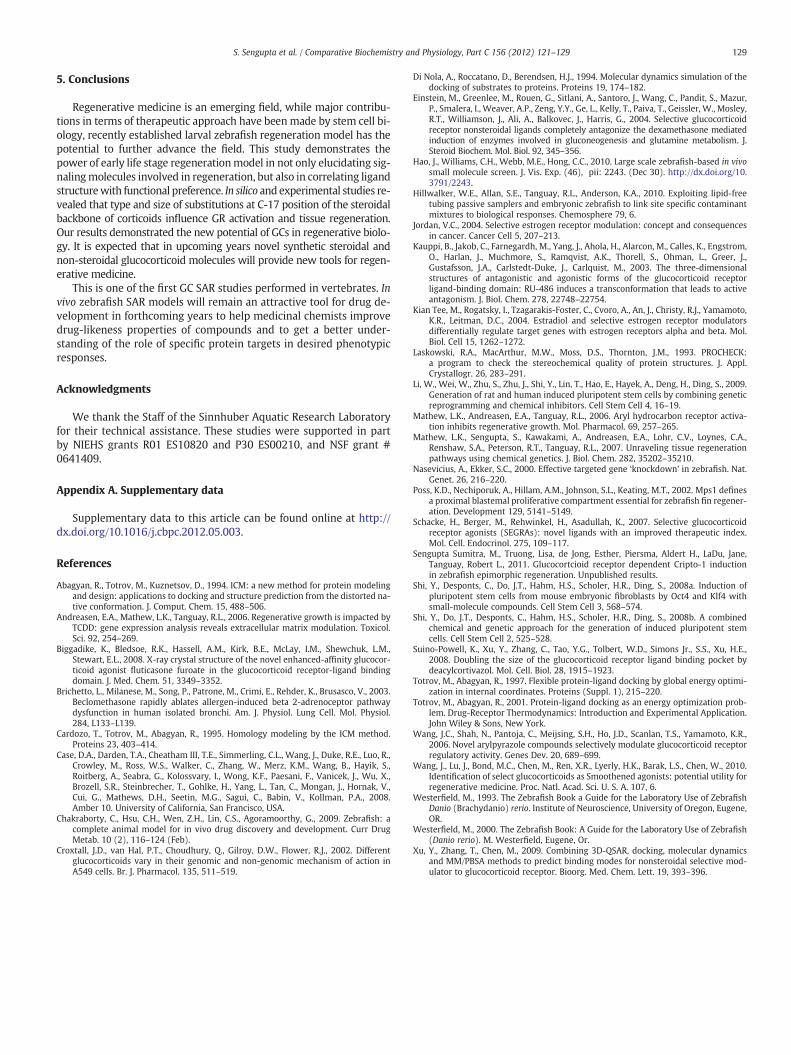
3.6. Novel ligands identified based on molecular docking results confirmthe importance of C17 substitution
To further validate the effect of the C17-substitution on in vivo re-generation inhibition, we selected and acquired several steroidalcompounds commercially available from Sigma (Diflorasone diacetate(DFDA)), and R198897 TimTec (ST 075183 and ST 075178) (Fig. 6). Ini-tially, the compoundswere docked into DEX–GR-LBD and none of them
docked suggesting potential inhibitory properties. Based on the in silicoresults, dose dependent in vivo larval regeneration analysis was per-formed with the new compounds revealing that all of them inhibit re-generation at the 1 μM concentration (Table S3) (Supplemental Fig.3). Thus, we identified novel GR ligands that kept the cortisol backbone,but varied in C17 substitution size with the presence of sterically hin-dered esters or chlorine atoms (Fig. 6). In addition, QRT-PCR studiesdemonstrated induced fkbp5 expression in embryos exposed to the

Fig. 5. GR-LBD residue side chain conformational changes allow binding of bigger size GR agonist BDP for regeneration inhibitory activity. Residual side chain and ligand shift of GR-LBD(pdb 1p93) in the complex between A) Dex during 1.01 ns MD and B,C) BDP during 1.51 ns MD. The ligands are colored by atom type with carbon atoms in white (Dex) and in yellow(BDP) at initial time (t=0 ps) D) and inmagenta (1015 psMD for Dex and 1515 psMD for BDP) and displayed as sticks. Residues are colored in orange (Initial time, t=0 ps) and green(1015 ps MD for Dex and 1515 ps MD for BDP) and displayed as sticks (ICM v3.5-1p).
127S. Sengupta et al. / Comparative Biochemistry and Physiology, Part C 156 (2012) 121–129
novel ligands. In the absence of GR, this induction was diminished.Finally, Cripto −1 expression (Fig. 6) is elevated following exposure tothese inhibitory ligands in a GR dependent manner.
4. Discussion
Ligand dependent response of nuclear receptors has led to struc-ture activity predictions and eventually understanding the biologyof the nuclear receptors. Since the GR is the major target of themost widely used class of drugs, understanding how this receptor re-sponds to varying structures of ligands is crucial for further drug de-velopment. However, majority of the ligands are evaluated in vitro.Numerous lead compounds that demonstrate excellent results invitro are withdrawn from the market due to either acute in vivo tox-icity or the inability to replicate cell based results. This can be avoidedby utilizing in vivo models that are amenable to screening to identifynew GR ligands with differential activities. We previously reportedthat inappropriately activated GR modulates tissue regeneration(Mathew et al., 2007). This has opened avenues for the potentialuse of GR ligands for regenerative medicine. However, further studiesare required not only to understand the role of activated GR in tissueregeneration, but also to explore how GC structure dictates regenera-tive outcome.
So far, chemical genetic approaches have identified numerousmod-ulators of stem cell differentiation and stem cell fate (Shi et al., 2008a,2008b; Li et al., 2009). The recent characterization of fluorinated GCsasmodulators of stem cell activity underlines the requirement for better
understanding of structure function relationship amongst the GR li-gands (Wang et al., 2010). Since there are numerous commerciallyavailable structural analogs of cortisol, we exploited existing drugs.This allowed us to bypass the requirement of synthesizing new analogsto modulate regeneration.
Our previous results demonstrate that GR activation inhibited tis-sue regeneration; however, not all ligands that activate GR inhibit re-generation (Mathew et al., 2007). Previous studies demonstrated thatligand chemistry dictates biological response by activated receptor.The best examples are the estrogen receptor ligands estradiol and ta-moxifen that invoke different gene expression profile as well as dif-ferent function in different cell type (Jordan, 2004; Kian Tee et al.,2004). The striking difference in ligand structures suggests complicat-ed correlation between chemical structures and biological response.
It has been reported that differences in ligand chemistry can give riseto a host of functionally distinct GR-containing regulatory complexes(Wang et al., 2006) and hence impact different set of genes. Since wehave evaluated the role of ligand-dependent GR activation in the invivo regeneration model, the phenotypic assay served as an initial readout of differential response of the ligands. This was further confirmedby GR-dependent activation of Cripto-1, which was required to inducea block in early stages of blastema formation (unpublished data). How-ever, we observed induction of GR target genes such as FKBP506 uponexposure to the GR ligands irrespective of their effect on regeneration.Lack of Cripto-1 induction by DEX, HC or Beclo supports previous reportsthat the hosts of genes affected by these ligands are different andnot crit-ical for inhibition of tissue regeneration (Croxtall et al., 2002; Brichetto et

IIIIIIIIII
IIIIIIIIII
GR MO
Con MO
R198897B
Cripto-1
DMSO R198897 GR MO+ R198897R
elat
ive
abu
nd
ance
of
β -
acti
n
0
20
40
60
80
100
120
***
DMSO R198897 GR MO+ R198897
0
200
400
600
800
1000
1200FKBP506
Rel
ativ
e ab
un
dan
ce o
f β
- ac
tin
ST75183C
IIIIIIIIIIII
IIIIIIIIIGR MO
Con MO
0
20
40
60
80
DMSO ST75183 GR MO+ ST75183R
elat
ive
abu
nd
ance
of
β -
acti
n FKBP506
Cripto-1
Rel
ativ
e ab
un
dan
ce o
f β
- ac
tin
0
20
40
60
80
100
***
DMSO ST75183 GR MO+ ST75183
E ST75178
IIIIIIIII
GR MO
IIIIIIIIICon MO
DMSO ST75178 GR MO -ST75178 R
elat
ive
abu
nd
ance
of
β -
acti
n
0
20
40
60
80
100
120
***
Cripto-1
DMSO ST75178 GR MO-ST75178
0
200
400
600
800
1000
1200
Rel
ativ
e ab
un
dan
ce o
f β
- ac
tin
FKBP506
DFDAD
ΙΙIIIIIIIII
IIIIIIIIIIGR MO
Con MO
DMSO DFDA GR MO+ DFDAR
elat
ive
abu
nd
ance
of
β -
acti
n
0
50
100
150
200
250
300
0
500
1000
1500
2000
2500
3000
Rel
ativ
e ab
un
dan
ce o
f β
- ac
tin
DMSO DFDA GR MO+ DFDA
Cripto-1
FKBP506
IIIIIIIIIGR MO
IIIIIIIIICon MO
BDPA
FKBP506
∗
∗
Rel
ativ
e ab
un
dan
ce o
f β
- ac
tin
0
80
140
200
DMSO BDP GR MO+ BDP
FKBP506
0
40
80
120
160
200
DMSO BDP GR MO+ BDP
***
Rel
ativ
e ab
un
dan
ce o
f β
- ac
tin
Cripto-1
Fig. 6. Novel ligands identified based on C17 substitution inhibit tissue regeneration. Structures of novel GR ligands B) R198897, C) ST75183, D) DFDA and E) ST75178 identifiedbased on cortisol backbone and C17 substitution. GR splice variant MO transiently knocked down GR compared to standard control morpholino injected embryos. GR and controlmorphants were amputated at 2 dpf and exposed to DMSO or the novel GR ligands. Regenerative progression was assessed and images were acquired after three days of exposure.The abundance of FKBP506 and Cripto-1 estimated by qRT PCR at 1 dpa in the whole embryo indicates significantly elevated expression in the control morphants and significantlyreduced expression in the corresponding GRmorphants when exposed to the novel GR ligands. The respective values represent the mean±SEM and the asterisks indicate statisticalsignificance (One way ANOVA, n=3) (pb0.05).
128 S. Sengupta et al. / Comparative Biochemistry and Physiology, Part C 156 (2012) 121–129
al., 2003; Sengupta Sumitra et al., 2011) The differential regulation ofgenes by the GR is likely due to the recruitment of distinct co-regulatorsby the GR upon binding to different ligands.
In order to initiate a SAR studywe performed docking studies againsthuman and zebrafishGR-LBDmodelswith the database of known steroi-dal GR agonists previously tested in the regeneration assay (Table S2).The conformational changes of residues observed with BDP during1.51 ns MD are similar to the reported data (Biggadike et al., 2008;Suino-Powell et al., 2008). These residues in the GR-LBD influence ligandbinding directly and are flexible enough to expand the binding pocketvolume to accommodate large ligands. This allows the helical tertiarystructure of the GR-LBD agonist conformation to stay intact. This alsosuggests that these residues might play a role in the thermodynamicequilibrium between the inactive (no effect on regeneration) and the ac-tive (inhibit regeneration) GR-LBD conformation. Active ligands possesssterically hindered ester moieties or chlorine atoms as substituents atC-17 position and exo-Me-stereochemistry at C-16 position (endo-Me-stereochemistry also works for few non-regenerating compounds like
Triamcinolone Diacetate). From the SAR analysis, substitutions at C-3,C-9 and C-11 positions do not play a role in the inhibitory activity.Molec-ular Docking runs showed that active GR agonists are unable to dock intothe human GR-LBD–DEX binding pocket (inactive conformation) (TableS2) and this is primarily due to the size of ligands.
For our study we have utilized an in vivo system and thus, metabo-lism and uptake might play a role in differential response. Metabolismof BDP involves hydrolysis to beclomethasone monopropionate (17-BMP) and finally beclomethasone (Beclo). Unfortunately, we could notevaluate the effect of 17-BMP in tissue regeneration due to commercialunavailability of the compound. However docking studies revealed com-parable results for 17-BMP and BDP (Table S2). This offers a potentialpossibility that even if BDP is metabolized to 17-BMP in vivo, accordingto our in silico predictions it induces the same conformation as of BDP.Since we are unable to characterize the metabolism of GR ligands inzebrafish, the fact that, exposing all these ligands induces suppressionof anxa1b expression, confirms both the activation of GR upon ligandbinding and the uptake of the ligands.
129S. Sengupta et al. / Comparative Biochemistry and Physiology, Part C 156 (2012) 121–129
5. Conclusions
Regenerative medicine is an emerging field, while major contribu-tions in terms of therapeutic approach have beenmade by stem cell bi-ology, recently established larval zebrafish regeneration model has thepotential to further advance the field. This study demonstrates thepower of early life stage regenerationmodel in not only elucidating sig-nalingmolecules involved in regeneration, but also in correlating ligandstructurewith functional preference. In silico and experimental studies re-vealed that type and size of substitutions at C-17 position of the steroidalbackbone of corticoids influence GR activation and tissue regeneration.Our results demonstrated the new potential of GCs in regenerative biolo-gy. It is expected that in upcoming years novel synthetic steroidal andnon-steroidal glucocorticoid molecules will provide new tools for regen-erative medicine.
This is one of the first GC SAR studies performed in vertebrates. Invivo zebrafish SAR models will remain an attractive tool for drug de-velopment in forthcoming years to help medicinal chemists improvedrug-likeness properties of compounds and to get a better under-standing of the role of specific protein targets in desired phenotypicresponses.
Acknowledgments
We thank the Staff of the Sinnhuber Aquatic Research Laboratoryfor their technical assistance. These studies were supported in partby NIEHS grants R01 ES10820 and P30 ES00210, and NSF grant #0641409.
Appendix A. Supplementary data
Supplementary data to this article can be found online at http://dx.doi.org/10.1016/j.cbpc.2012.05.003.
References
Abagyan, R., Totrov, M., Kuznetsov, D., 1994. ICM: a new method for protein modelingand design: applications to docking and structure prediction from the distorted na-tive conformation. J. Comput. Chem. 15, 488–506.
Andreasen, E.A., Mathew, L.K., Tanguay, R.L., 2006. Regenerative growth is impacted byTCDD: gene expression analysis reveals extracellular matrix modulation. Toxicol.Sci. 92, 254–269.
Biggadike, K., Bledsoe, R.K., Hassell, A.M., Kirk, B.E., McLay, I.M., Shewchuk, L.M.,Stewart, E.L., 2008. X-ray crystal structure of the novel enhanced-affinity glucocor-ticoid agonist fluticasone furoate in the glucocorticoid receptor-ligand bindingdomain. J. Med. Chem. 51, 3349–3352.
Brichetto, L., Milanese, M., Song, P., Patrone, M., Crimi, E., Rehder, K., Brusasco, V., 2003.Beclomethasone rapidly ablates allergen-induced beta 2-adrenoceptor pathwaydysfunction in human isolated bronchi. Am. J. Physiol. Lung Cell. Mol. Physiol.284, L133–L139.
Cardozo, T., Totrov, M., Abagyan, R., 1995. Homology modeling by the ICM method.Proteins 23, 403–414.
Case, D.A., Darden, T.A., Cheatham III, T.E., Simmerling, C.L., Wang, J., Duke, R.E., Luo, R.,Crowley, M., Ross, W.S., Walker, C., Zhang, W., Merz, K.M., Wang, B., Hayik, S.,Roitberg, A., Seabra, G., Kolossvary, I., Wong, K.F., Paesani, F., Vanicek, J., Wu, X.,Brozell, S.R., Steinbrecher, T., Gohlke, H., Yang, L., Tan, C., Mongan, J., Hornak, V.,Cui, G., Mathews, D.H., Seetin, M.G., Sagui, C., Babin, V., Kollman, P.A., 2008.Amber 10. University of California, San Francisco, USA.
Chakraborty, C., Hsu, C.H., Wen, Z.H., Lin, C.S., Agoramoorthy, G., 2009. Zebrafish: acomplete animal model for in vivo drug discovery and development. Curr DrugMetab. 10 (2), 116–124 (Feb).
Croxtall, J.D., van Hal, P.T., Choudhury, Q., Gilroy, D.W., Flower, R.J., 2002. Differentglucocorticoids vary in their genomic and non-genomic mechanism of action inA549 cells. Br. J. Pharmacol. 135, 511–519.
Di Nola, A., Roccatano, D., Berendsen, H.J., 1994. Molecular dynamics simulation of thedocking of substrates to proteins. Proteins 19, 174–182.
Einstein, M., Greenlee, M., Rouen, G., Sitlani, A., Santoro, J., Wang, C., Pandit, S., Mazur,P., Smalera, I., Weaver, A.P., Zeng, Y.Y., Ge, L., Kelly, T., Paiva, T., Geissler, W., Mosley,R.T., Williamson, J., Ali, A., Balkovec, J., Harris, G., 2004. Selective glucocorticoidreceptor nonsteroidal ligands completely antagonize the dexamethasone mediatedinduction of enzymes involved in gluconeogenesis and glutamine metabolism. J.Steroid Biochem. Mol. Biol. 92, 345–356.
Hao, J., Williams, C.H., Webb, M.E., Hong, C.C., 2010. Large scale zebrafish-based in vivosmall molecule screen. J. Vis. Exp. (46), pii: 2243. (Dec 30). http://dx.doi.org/10.3791/2243.
Hillwalker, W.E., Allan, S.E., Tanguay, R.L., Anderson, K.A., 2010. Exploiting lipid-freetubing passive samplers and embryonic zebrafish to link site specific contaminantmixtures to biological responses. Chemosphere 79, 6.
Jordan, V.C., 2004. Selective estrogen receptor modulation: concept and consequencesin cancer. Cancer Cell 5, 207–213.
Kauppi, B., Jakob, C., Farnegardh, M., Yang, J., Ahola, H., Alarcon, M., Calles, K., Engstrom,O., Harlan, J., Muchmore, S., Ramqvist, A.K., Thorell, S., Ohman, L., Greer, J.,Gustafsson, J.A., Carlstedt-Duke, J., Carlquist, M., 2003. The three-dimensionalstructures of antagonistic and agonistic forms of the glucocorticoid receptorligand-binding domain: RU-486 induces a transconformation that leads to activeantagonism. J. Biol. Chem. 278, 22748–22754.
Kian Tee, M., Rogatsky, I., Tzagarakis-Foster, C., Cvoro, A., An, J., Christy, R.J., Yamamoto,K.R., Leitman, D.C., 2004. Estradiol and selective estrogen receptor modulatorsdifferentially regulate target genes with estrogen receptors alpha and beta. Mol.Biol. Cell 15, 1262–1272.
Laskowski, R.A., MacArthur, M.W., Moss, D.S., Thornton, J.M., 1993. PROCHECK:a program to check the stereochemical quality of protein structures. J. Appl.Crystallogr. 26, 283–291.
Li, W., Wei, W., Zhu, S., Zhu, J., Shi, Y., Lin, T., Hao, E., Hayek, A., Deng, H., Ding, S., 2009.Generation of rat and human induced pluripotent stem cells by combining geneticreprogramming and chemical inhibitors. Cell Stem Cell 4, 16–19.
Mathew, L.K., Andreasen, E.A., Tanguay, R.L., 2006. Aryl hydrocarbon receptor activa-tion inhibits regenerative growth. Mol. Pharmacol. 69, 257–265.
Mathew, L.K., Sengupta, S., Kawakami, A., Andreasen, E.A., Lohr, C.V., Loynes, C.A.,Renshaw, S.A., Peterson, R.T., Tanguay, R.L., 2007. Unraveling tissue regenerationpathways using chemical genetics. J. Biol. Chem. 282, 35202–35210.
Nasevicius, A., Ekker, S.C., 2000. Effective targeted gene ‘knockdown’ in zebrafish. Nat.Genet. 26, 216–220.
Poss, K.D., Nechiporuk, A., Hillam, A.M., Johnson, S.L., Keating, M.T., 2002. Mps1 definesa proximal blastemal proliferative compartment essential for zebrafish fin regener-ation. Development 129, 5141–5149.
Schacke, H., Berger, M., Rehwinkel, H., Asadullah, K., 2007. Selective glucocorticoidreceptor agonists (SEGRAs): novel ligands with an improved therapeutic index.Mol. Cell. Endocrinol. 275, 109–117.
Sengupta Sumitra, M., Truong, Lisa, de Jong, Esther, Piersma, Aldert H., LaDu, Jane,Tanguay, Robert L., 2011. Glucocortcioid receptor dependent Cripto-1 inductionin zebrafish epimorphic regeneration. Unpublished results.
Shi, Y., Desponts, C., Do, J.T., Hahm, H.S., Scholer, H.R., Ding, S., 2008a. Induction ofpluripotent stem cells from mouse embryonic fibroblasts by Oct4 and Klf4 withsmall-molecule compounds. Cell Stem Cell 3, 568–574.
Shi, Y., Do, J.T., Desponts, C., Hahm, H.S., Scholer, H.R., Ding, S., 2008b. A combinedchemical and genetic approach for the generation of induced pluripotent stemcells. Cell Stem Cell 2, 525–528.
Suino-Powell, K., Xu, Y., Zhang, C., Tao, Y.G., Tolbert, W.D., Simons Jr., S.S., Xu, H.E.,2008. Doubling the size of the glucocorticoid receptor ligand binding pocket bydeacylcortivazol. Mol. Cell. Biol. 28, 1915–1923.
Totrov, M., Abagyan, R., 1997. Flexible protein-ligand docking by global energy optimi-zation in internal coordinates. Proteins (Suppl. 1), 215–220.
Totrov, M., Abagyan, R., 2001. Protein-ligand docking as an energy optimization prob-lem. Drug-Receptor Thermodynamics: Introduction and Experimental Application.John Wiley & Sons, New York.
Wang, J.C., Shah, N., Pantoja, C., Meijsing, S.H., Ho, J.D., Scanlan, T.S., Yamamoto, K.R.,2006. Novel arylpyrazole compounds selectively modulate glucocorticoid receptorregulatory activity. Genes Dev. 20, 689–699.
Wang, J., Lu, J., Bond, M.C., Chen, M., Ren, X.R., Lyerly, H.K., Barak, L.S., Chen, W., 2010.Identification of select glucocorticoids as Smoothened agonists: potential utility forregenerative medicine. Proc. Natl. Acad. Sci. U. S. A. 107, 6.
Westerfield, M., 1993. The Zebrafish Book a Guide for the Laboratory Use of ZebrafishDanio (Brachydanio) rerio. Institute of Neuroscience, University of Oregon, Eugene,OR.
Westerfield, M., 2000. The Zebrafish Book: A Guide for the Laboratory Use of Zebrafish(Danio rerio). M. Westerfield, Eugene, Or.
Xu, Y., Zhang, T., Chen, M., 2009. Combining 3D-QSAR, docking, molecular dynamicsand MM/PBSA methods to predict binding modes for nonsteroidal selective mod-ulator to glucocorticoid receptor. Bioorg. Med. Chem. Lett. 19, 393–396.