alt-(maleic anhydride) and styrene: polymerization, … 10.0 mg sample of 6 was heated at 250 c for...
TRANSCRIPT
# Supplementary Material (ESI) for Chemical Communications # This journal is © The Royal Society of Chemistry 2005
Supplementary Information for “Shell-crosslinked micelles
from amphiphilic AB and ABA block copolymers of styrene-
alt-(maleic anhydride) and styrene: polymerization, assembly
and stabilization in one pot”
Materials. Styrene (99 %) was purchased from Sigma Aldrich (St Louis, MO); inhibitor
was removed by passing over a column of basic alumina prior to use. Maleic anhydride
(99 %), dodecanethiol (>98 %), tetrabutylammonium bromide (98 %), carbon disulfide (
99.9 %), acetone (99.5 %), chloroform (99.8 %), 2-bromopropionic acid (99 %) and 1,2-
ethylenedioxy bis(2-ethylamine) (98 %) were obtained from Sigma Aldrich and used
without further purification. AIBN was recrystallized from methanol before use. All other
solvents and reagents were obtained from Sigma Aldrich and used without further
purification.
Measurements. 1H NMR (300 MHz) and 13C NMR (75 MHz) spectra were recorded on a
Varian Mercury 300 MHz spectrometer. IR spectra were obtained on a Perkin Elmer BX
FT-IR system equipped with a diffuse reflectance accessory. Elemental analysis was
performed by M-H-W Laboratories (Phoenix, AZ). Glass transition temperatures (Tg) were
measured by differential scanning calorimetry (DSC) at a heating rate of 10 °C/min on a
Mettler Toledo DSC822e using Mettler Toledo Star SW 7.01 software. Tg values were
taken at the midpoint of the inflection tangent on the third heating scan.
Gel Permeation Chromatography. Polymer molecular weight distributions were
determined by gel permeation chromatography, conducted on a Waters Chromatography

# Supplementary Material (ESI) for Chemical Communications # This journal is © The Royal Society of Chemistry 2005
Inc. model 150-CV, equipped with a model 410 DRI detector, a Precision Detectors
PD2040 dual angle (15° and 90°) light scattering detector and a three-column series of
Polymer Laboratories PLgel 10 µm mixed B 300 × 7.5 mm columns. The system was
equilibrated at 35 °C in anhydrous THF, which served as the polymer solvent and eluent
(flow rate 1.00 mL/min). Data collection and analysis was performed using Precision
Detectors software. Interdetector delay volume and the light scattering detector calibration
constant were determined using a nearly monodisperse polystyrene calibrant (Pressure
Chemical Co., Mp = 90 000 g.mol-1, Mw/Mn < 1.04). The differential refractometer was
calibrated with standard polystyrene reference material (SRM 706 NIST) of known specific
refractive index increment (dn/dc = 0.184 mL/g). The dn/dc values of the analyzed
polymers were then determined from the differential refractometer response.
Dynamic Light Scattering. Particle size distributions were determined on a Brookhaven
Instruments Co. dynamic light scattering system, consisting of a model BI-9000T digital
goniometer, a model EMI-9865 photomultiplier, and a model 95-2 Ar laser (Lexel Corp.)
operated at 514.5 nm. Measurements were made at 20 ± 1 °C. Prior to analysis, solutions
were centrifuged in a model 5414 microfuge (Brinkman Inst. Co.) for 4 min to remove dust
particles. Scattered light was collected at a fixed angle of 90 °C. The digital correlator was
operated with 522 channels, an initial delay of 1.4 µs, a final delay of 10 ms, and a duration
time of 15 minutes. A photomultiplier aperture of 400 µm was used. Only measurements
for which the measured and calculated baselines of the intensity autocorrelation function
agreed to within ± 0.1% were used to calculate nanoparticle diameter distributions. All
determinations were made in triplicate. The calculations of the nanoparticle diameter

# Supplementary Material (ESI) for Chemical Communications # This journal is © The Royal Society of Chemistry 2005
distributions were performed by cumulants analysis using the ISDA software package
(Brookhaven Instruments Co.).
Transmission Electron Microscopy. Carbon-coated copper grids were prepared by
oxygen plasma treatment to make the surface hydrophilic. Particle samples were diluted 9:1
in water. A drop of diluted sample was deposited onto a grid; after 1 minute excess solvent
was wicked away. A drop of 1% phosphotungstic acid (PTA) stain was then added, and left
for 1 minute before excess stain was wicked away. Particle diameters and standard
deviations were calculated from measurements of a minimum of 100 particles from three
TEM micrographs from different regions.
Atomic Force Microscopy. Tapping-mode atomic force microscopy measurements were
conducted in air with a Nanoscope III BioScope system (Digital Instruments) operated
under ambient conditions with standard silicon tips (type: OTESPA-70; L: 160 µm; normal
spring constant: 50 N/m; resonance frequency 246-282 kHz). Samples were diluted 99:1 in
water, then a 10 µL drop of the solution was deposited onto a freshly-cleaved mica surface
and allowed to evaporate. The average particle heights were calculated from the section
analysis of at least 50 particles selected from at least 3 different regions. Typical AFM
micrographs for micelles and crosslinked particles are shown in Figures S1 and S2
respectively.
Synthesis of S-1-dodecyl S’-( , -dimethylacetic acid) trithiocarbonate, 1. The
compound was synthesized according to the method of Lai et al.S1 KOH (2.5 g dissolved in
2.5 mL H2O) was added to a solution of dodecanethiol (10.1 g, 0.050 mol) and
tetrabutylammonium bromide (0.64 g, 2 mmol) in 25 mL of acetone. Carbon disulfide (2.6
mL) was added dropwise and the resulting orange-red solution was was stirred 30 min

# Supplementary Material (ESI) for Chemical Communications # This journal is © The Royal Society of Chemistry 2005
before addition of chloroform (5 mL). KOH (12.0 g dissolved in 10 mL H2O) was added
dropwise with vigorous stirring at 0 °C and the mixture was stirred overnight at room
temperature. DI H2O (60 mL) was then added and the mixture was acidified with 10 mL
conc. HCl. Excess acetone was removed in vacuo and the solid product was separated by
filtration and dissolved in hot isopropanol. On cooling, bis(dodecyl) trithiocarbonate
crystallized out and was filtered off. The isopropanol was removed in vacuo and the
resulting oil was recrystallized from hexanes, yielding 9.20 g yellow crystals, m. p. 63 °C
(lit. 62-3 °C).S1 Yield: 60 %.
1H NMR (CDCl3, 300 MHz, 25 °C): 0.89 (t, 3H, J = 6.75 Hz, CH3(CH2)11-), 1.2-1.5 (m,
18H, CH3(CH2)9CH2CH2S-), 1.68 (quint., 2H, J = 7.5 Hz, -CH2CH2S-), 1.73 (s, 6H,
(CH3)2C<), 3.29 (t,2H, J = 7.5 Hz, -CH2S-) ppm. 13C NMR (CDCl3): 14.4 (CH3(CH2)11-),
22.9 (CH3CH2-), 25.4 ((CH3)2C<), 28.0, 29.2, 29.3, 29.6, 29.7, 29.8, 29.9, 32.1 (dodecyl
carbons), 37.3 (-CH2S-), 55.8 (HOOC(CH3)2CS-), 178.8 (-COOH), 221.0 (-SC(=S)S-)
ppm. IR (diffuse reflectance): 2921, 2850, 1714, 1281, 1070, 813 cm-1. MS: 365.164 [M +
H]+. Elemental Analysis: C 54.25 H 8.21 S 25.14. Theoretical: C 56.00 H 8.85 S 26.38.
Synthesis of bis(2-propionic acid) trithiocarbonate, 2.
2-Bromopropionic acid (4.7 mL, 50 mmol) was added slowly to a suspension of CS2 (6.0
mL, 110 mmol) in a solution of 6.6 g KOH in 150 mL H2O. The yellow solution was
stirred 48 h at room temperature, then extracted twice with 20 mL MeCl2 to remove excess
CS2. The yellow aqueous layer was acidified with conc. HCl and extracted with MeCl2 (5 x
20 mL). The combined organic layers were dried over MgSO4 and solvent was removed in
vacuo to give 4.6 g yellow solid. This was recrystallized from acetone/hexanes to give
3.44 g yellow crystals, m. p. 90 °C. Yield: 51.8 %

# Supplementary Material (ESI) for Chemical Communications # This journal is © The Royal Society of Chemistry 2005
1H NMR (CDCl3, 300 MHz, 25 °C): 0.89 (t, 3H, J = 6.75 Hz, CH3(CH2)11-), 1.2-1.5 (m,
18H, CH3(CH2)9CH2CH2S-), 1.68 (quint., 2H, J = 7.5 Hz, -CH2CH2S-), 1.73 (s, 6H,
(CH3)2C<), 3.29 (t,2H, J = 7.5 Hz, -CH2S-) ppm. 13C NMR (CDCl3): 14.4 (CH3(CH2)11-),
22.9 (CH3CH2-), 25.4 ((CH3)2C<), 28.0, 29.2, 29.3, 29.6, 29.7, 29.8, 29.9, 32.1 (dodecyl
carbons), 37.3 (-CH2S-), 55.8 (HOOC(CH3)2CS-), 178.8 (-COOH), 221.0 (-SC(=S)S-)
ppm. IR (diffuse reflectance): 2921, 2850, 1714, 1281, 1070, 813 cm-1.
MS: 254.982 [M + H]+. Elemental Analysis: C 33.14 H 4.20 S 37.59. Theoretical: C 33.06
H 3.96 S 37.82.
Polymerizations. Styrene, maleic anhydride, 1 or 2 and AIBN (20 mol% relative to RAFT
agent) were mixed in appropriate ratio with dioxane (20 mL) and degassed by purging with
N2 for 10 min. In a typical experiment, 15 g STY, 5 g maleic anhydride, 183 mg 1 and 16
mg AIBN were used. The mixtures were heated in an oil bath set to 60 °C for 21 h. A small
sample was taken in order to estimate conversion by NMR spectroscopy, 20 mL of THF
was added and the resulting solutions were precipitated into 1L Et2O in order to isolate the
polymer, which was subsequently dried under vacuum at 120 °C. Molecular weight
distributions were measured by GPC, compositions were calculated by elemental analysis.
Characterization data is given in Table 1 of the main text. Dynamic scanning calorimetry of
the diblock polymers 3-5 revealed two glass transitions for each, at approximately 78 and
170 °C (Table S1). The triblock copolymer 6 also showed two glass transitions, at 115 and
164 °C respectively. Polymers exhibited characteristic IR absorbances at 1856 and 1776
cm-1 (coupled anhydride C=O stretches), 1494 and 1453 cm-1 (aromatic C=C ring stretches)
and 699 cm-1 (aromatic out of plane C-H bending). A typical IR spectrum (for diblock
copolymer 5) is shown in Figure S3A.

# Supplementary Material (ESI) for Chemical Communications # This journal is © The Royal Society of Chemistry 2005
Table S1. Glass transition temperatures for polymers 3-6.
Polymer Tg1 (°C) Tg2 (°C)
3 77.6 172
4 76.5 180
5 76.8 170
6 115 164
Pyrolysis of 6. A 10.0 mg sample of 6 was heated at 250 °C for 30 minutes under a stream
of nitrogen in a Perkin-Elmer TGA/SDTA851e thermogravimetric analysis system. This
treatment resulted in a mass loss of 0.7 mg (7 %). The residue was dissolved in THF and its
molecular weight distribution was analyzed by GPC (Figure S4).
Formation of Micelles. Block copolymer (0.1 g) was dissolved in 50 mL THF, to which
was added one drop (~10 µL) triethylamine. Nanopure (> 18 M .cm-1) water (50 mL) was
added at 10 mL/hour with stirring. A further 50 mL water was then added in one portion,
and the mixture was dialyzed (6000-8000 MWCO tubing) vs. DI water for several days to
remove THF. More water was then added to reach a final volume of 200 mL (0.5 mg/mL).
The final suspensions ranged in appearance from colorless to bluish-white.
Crosslinking. To a micellar solution of block copolymer (0.5 mg/mL) was added 1,2-
ethylenedioxy bis(ethylamine) (10 mol% relative to maleic anhydride units). After stirring
20 minutes, EDC was added (20 mol% relative to maleic anhydride units). The solution
was stirred overnight, then dialyzed for 3 days against DI water to remove unreacted
crosslinker and urea byproducts (6000-8000 MWCO tubing).

# Supplementary Material (ESI) for Chemical Communications # This journal is © The Royal Society of Chemistry 2005
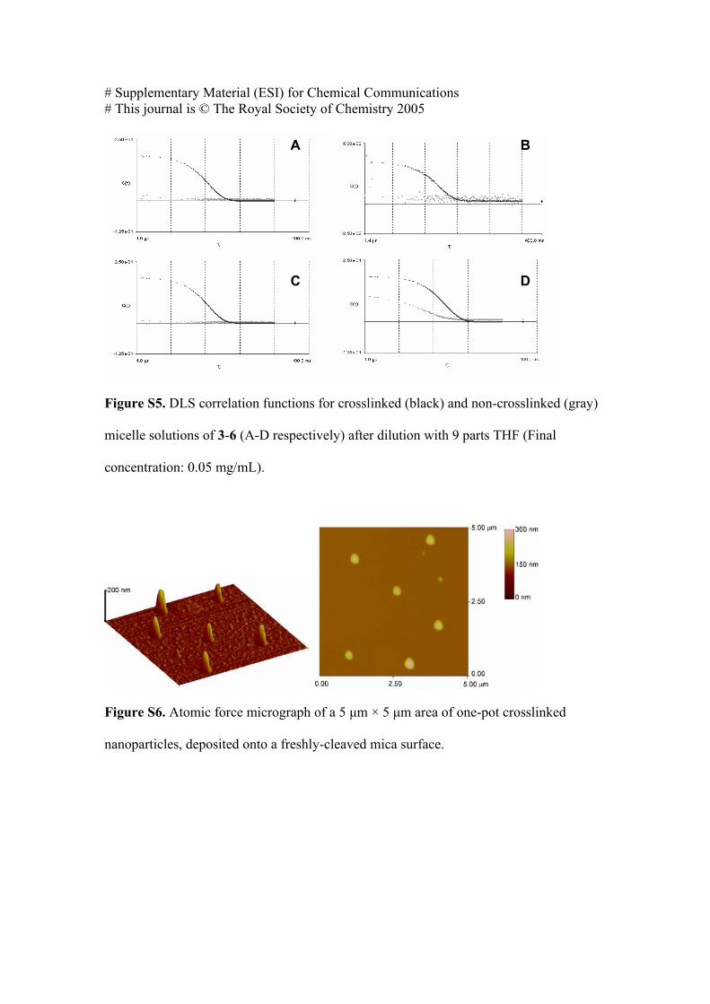
Confirmation of crosslinking. Formation of amide bonds was confirmed by IR
spectroscopy of lyophilized sample (Figure S3C), with the appearance of a new absorption
at 1636 cm-1, corresponding to the amide C=O stretch. To demonstrate the formation of a
crosslinked network, both the crosslinked particle suspensions and their non-crosslinked
precursors were diluted with 9 parts THF. The resulting suspensions were analyzed by DLS
(Figure S5). The absence of scattering for non-crosslinked samples of 3-5 indicated that
these polymers formed a molecular solution in this solvent mixture. Their crosslinked
analogs, however, did not dissociate under this treatment. Dilution with THF was not
sufficient to completely dissociate the triblock copolymer 6, but a clear difference in
scattering intensity between non-crosslinked and crosslinked samples of 6 was observed
(Figure S5D).
One-pot copolymer synthesis, micellization and crosslinking to produce shell-
crosslinked nanoparticles. MA (0.5 g, 5.1 mmol) and STY (1.5 g, 14.4 mmol) were
heated at 60 °C in the presence of 2.0 g dioxane, 1 (18 mg, 49 µmol), and a trace of AIBN,
for a period of 16 hours under N2. The resulting mixture (conversion = 89 % by NMR,
theoretical Mn = 36300 g.mol-1) was dissolved in 1 L of THF. A 250 mL aliquot of the
solution was taken, to which 1,2-ethylenedioxy bis(2-ethylamine) (18.9 mg, 128 µmol,
10% relative to MA) was added with vigorous stirring, followed immediately by 250 mL
nanopure water. The THF was removed by evaporation in vacuo (at RT) and the micellar
solution was diluted to 1 L with nanopure water. The resulting mixture contained
nanoparticles with Dh 164 ± 2 nm, and a polydispersity of 0.08 ± 0.03 (measured by DLS).
AFM analysis of the nanoparticles gave an average height of 118 nm and standard
deviation of 38 nm. The persistence of these particles in 9:1 THF:H2O indicated that

# Supplementary Material (ESI) for Chemical Communications # This journal is © The Royal Society of Chemistry 2005
crosslinking had taken place (Figure S7). As a control, the procedure was repeated with a
second 250 mL aliquot of the polymer solution in THF, using triethylamine (25.9 mg, 256
µmol, 10% relative to MA) in place of 1,2-ethylenedioxy bis(ethylamine). DLS of the
resulting solution revealed the presence of nanoparticles with average diameter 137 ± 5 nm,
and polydispersity of 0.20 ± 0.02 (measured by DLS). On dilution with 9 parts THF, these
particles dissociated to form a molecular solution (Figure S7)

# Supplementary Material (ESI) for Chemical Communications # This journal is © The Royal Society of Chemistry 2005
100 nm
100 nm
100 nm
Figure S1. Atomic force micrographs of 1 µm × 1 µm areas of micelles of 3 (A), 4 (B), 5
(C), and 6 (D) deposited on a freshly cleaved mica surface.
A
B
C
D

# Supplementary Material (ESI) for Chemical Communications # This journal is © The Royal Society of Chemistry 2005
100 nm
Figure S2. Atomic force micrographs of 2.5 µm × 2.5 µm areas of crosslinked micelles
of 3 (A), 4 (B), 5 (C), and 6 (D) deposited on a freshly cleaved mica surface.
A
B
C
D

# Supplementary Material (ESI) for Chemical Communications # This journal is © The Royal Society of Chemistry 2005
4000 3500 3000 2500 2000 1750 1500 1250 1000 750 500
Tra
nsm
issi
on (A
. U.)
Wavenumber (cm-1)
Figure S3. IR spectra of diblock copolymer 5: (A) before hydrolysis; (B) after
micellization and lyophilization; (C) after micellization, crosslinking and lyophilization.
1000 10000 100000
Nor
mal
ized
RI r
espo
nse
before pyrolysisMn = 20600PDI = 1.09
MW (Da)
after pyrolysisMn = 11000PDI = 1.12
Figure S4. Molecular weight distributions of triblock copolymer 6, before and after
pyrolysis at 250 °C for 30 minutes. Distributions are normalized to equal area.
# Supplementary Material (ESI) for Chemical Communications # This journal is © The Royal Society of Chemistry 2005
Figure S5. DLS correlation functions for crosslinked (black) and non-crosslinked (gray)
micelle solutions of 3-6 (A-D respectively) after dilution with 9 parts THF (Final
concentration: 0.05 mg/mL).
Figure S6. Atomic force micrograph of a 5 µm × 5 µm area of one-pot crosslinked
nanoparticles, deposited onto a freshly-cleaved mica surface.
A B
C D

# Supplementary Material (ESI) for Chemical Communications # This journal is © The Royal Society of Chemistry 2005
Figure S7 DLS correlation functions for crosslinked (black) and non-crosslinked (gray)
one-pot micelles and crosslinked nanoparticles after dilution with 9 parts THF (Final
concentration: 0.05 mg/mL).
References.
S1. J. T. Lai, D. Filla and R. Shea, Macromolecules, 2002, 35, 6754.