alma mater studiorum – università di bologna · 1 alma mater studiorum – università di...
TRANSCRIPT

1
Alma Mater Studiorum – Università di Bologna
DOTTORATO DI RICERCA IN
BIOLOGIA CELLULARE, MOLECOLARE E INDUSTRIALE
Progetto 2: Biologia Funzionale e Molecolare
Ciclo XXV
Settore Concorsuale: 05/E2
Settore scientifico-disciplinare: BIO/11
The role of the NadR regulator during infection and its implication
for the coverage of a new Meningococcus B vaccine
Presentata da: Luca Fagnocchi
Coordinatore Dottorato Relatori
Chiar.mo Prof. Chiar.mo Prof. Vincenzo Scarlato Vincenzo Scarlato
Dott.ssa Isabel Delany
Esame finale anno 2013

2

3
ATTIVITÀ DI RICERCA
Durante il Dottorato di ricerca mi sono occupato dello studio della regolazione
dell´espressione genica in Neisseria meningitidis. In particolare ho studiato la regolazione
trascrizionale mediata dal repressore NadR, in risposta a segnali presenti nell’ospite e di
rilevanza fisiologica. Ho caratterizzato il meccanismo molecolare di repressione mediato
da NadR sui suoi geni target e il meccanismo mediante il quale molecole presenti nei siti
d’infezione del meningococco alterano l’attività regolatoria di NadR. Infine, ho studiato
l’impatto della regolazione mediata da NadR su l’antigene NadA, sulla copertura di un
nuovo vaccino meningococcico, chiamato 4CMenB.
In parallelo, ho indagato il ruolo nella regolazione trascrizionale di piccoli RNA non
codificanti indotti in diverse condizioni e/o stress incontrati dal meningococco durante la
sua patogenesi.
Nel periodo del Dottorato di Ricerca sono stato co-autore dei seguenti lavori scientifici:
Fagnocchi L, Pigozzi E, Scarlato V, Delany I. “In the NadR regulon, adhesins and diverse meningococcal functions are regulated in response to signals in human saliva”. J Bacteriol. 2012 Jan;194(2):460-74. doi: 10.1128/JB.06161-11. Epub 2011 Nov 11.
Brier S, Fagnocchi L, Donnarumma D, Scarselli M, Rappuoli R, Nissum M, Delany I, Norais N. “Structural Insight into the Mechanism of DNA-Binding Attenuation of the Neisserial Adhesin Repressor NadR by the Small Natural Ligand 4-Hydroxyphenylacetic Acid”. Biochemistry. 2012 Aug 28;51(34):6738-52. Epub 2012 Aug 15.
Fagnocchi L, Biolchi A, Ferlicca F, Boccadifuoco G, Brunelli B, Brier S, Norais N, Chiarot E, Bensi G, Kroll JS, Pizza M, Donnelly J, Giuliani MM, Delany I. “Transcriptional Regulation of the nadA Gene in Neisseria meningitidis Impacts on the Prediction of Coverage of the 4CMenB Vaccine”. Infect Immun. 2013 Feb;81(2):560-9. doi: 10.1128/IAI.01085-12. Epub 2012 Dec 10.
Fagnocchi L, Bottini S, Fantappiè L, Golfieri G, del Tordello E, Siena E, Serruto D, Scarlato V, Muzzi A, Delany I. “Global identification and characterization of small non-coding RNAs in Neisseria meningitidis in response to multiple stress conditions”. Manuscript in preparation.

4

5
TABLE OF CONTENTS
TITOLO ........................................................................................................................................................ 1
ATTIVITÀ DI RICERCA ................................................................................................................................... 2
TABLE OF CONTENTS ................................................................................................................................... 5
1 INTRODUCTION .................................................................................................................................. 9
1.1 MENINGOCOCCAL DISEASE ......................................................................................................................... 9
1.2 THE PATHOGEN ...................................................................................................................................... 10
1.2.1 Classification and epidemiology ............................................................................................... 11
1.2.2 Pathogenesis ............................................................................................................................ 13
1.3 GENETICS .............................................................................................................................................. 16
1.4 GENE REGULATION AND ADAPTATION TO THE HOST ENVIRONMENT .................................................................. 17
1.4.1 Genome plasticity .................................................................................................................... 18
1.4.2 Transcriptional regulators ........................................................................................................ 20
1.4.3 The MarR family of transcriptional regulators ......................................................................... 24
1.4.4 The Neisserial adhesin Regulator (NadR) ................................................................................. 27
1.5 VIRULENCE FACTORS AND ADHESINS ........................................................................................................... 28
1.5.1 The Neisserial adhesin A (NadA) .............................................................................................. 33
1.6 MENINGOCOCCAL VACCINES ..................................................................................................................... 38
1.6.1 Reverse vaccinology and the 4CMenB vaccine......................................................................... 40
1.6.2 Vaccine coverage prediction .................................................................................................... 42
2 RESULTS ........................................................................................................................................... 45
2.1 IN THE NADR REGULON, ADHESINS AND DIVERSE MENINGOCOCCAL FUNCTIONS ARE REGULATED IN RESPONSE TO
SIGNALS IN HUMAN SALIVA ................................................................................................................................. 45
2.1.1 Global analysis of gene expression in the NadR mutant .......................................................... 45
2.1.2 Functional classification of the NadR-regulated genes ............................................................ 50
2.1.3 Binding of NadR to its targets .................................................................................................. 53
2.1.4 The NadR target genes can be classified in two types regarding their promoter architecture 56

6
2.1.5 Ligand-responsive regulation of NadR target genes expression by 4HPA ............................... 58
2.1.6 The NadR-dependent regulation of NadA, MafA and NadR itself is common among
meningococcal strains ............................................................................................................................ 60
2.1.7 Incubation with human saliva has the same effect on NadA and MafA expression as 4HPA .. 61
2.1.8 4HPA has differential activity on NadR binding to type I and type II promoters in vitro ......... 63
2.1.9 The 4HPA mediated co-repression of mafA is not due to repositioning of NadR on the
promoter ................................................................................................................................................ 66
2.1.10 3C scanning mutagenesis reveals extended NadR binding sequence in the operator of mafA
promoter region ..................................................................................................................................... 68
2.2 STRUCTURAL INSIGHT INTO THE MECHANISM OF DNA-BINDING ATTENUATION OF NADR BY THE SMALL NATURAL
LIGAND 4HPA ................................................................................................................................................. 70
2.2.1 Characterization of the structural model of NadR. .................................................................. 70
2.2.2 Localization of the 4HPA binding pocket.................................................................................. 72
2.2.3 Mutation of key residues in the 4-HPA binding pocket of NadR .............................................. 74
2.2.4 In vivo behaviour of selected site directed NadR mutants ....................................................... 75
2.2.5 In vitro characterization of the DNA- and 4-HPA binding activities of the purified NadR
mutant proteins ...................................................................................................................................... 78
2.2.6 NadR Y115A does not act as a hyper-repressor on the promoter of mafA1 ............................ 81
2.3 TRANSCRIPTIONAL REGULATION OF THE NADA GENE IMPACTS ON THE PREDICTION OF COVERAGE OF THE 4CMENB
VACCINE ........................................................................................................................................................ 83
2.3.1 Strains with MATS RP ≤ PBT express NadA in an immunogenic form during invasive disease 83
2.3.2 All strains carrying the nadA gene can express high levels of the NadA protein and therefore
be killed by vaccine-induced bactericidal antibodies ............................................................................. 87
2.3.3 NadA expression can be induced in vitro by different physiologically relevant signals ........... 92
2.3.4 NadA induction in the selected strain NGP165 ........................................................................ 95
2.3.5 In NGP165, neither 4HPA nor 3Cl-4HPA have any effect on the expression of the other major
antigens of the 4CMenB ......................................................................................................................... 97
2.3.6 hSBA and MATS performed with 3Cl4-HPA predict 4CMenB vaccine coverage of the
NGP165 strain ........................................................................................................................................ 98
7
2.3.7 Sera from 4CMenB-immunized infants protect infant rats from infection with strain NGP165.100
2.3.8 The promoter of nadA is activated in vivo during infection of the infant rat model .............. 102
3 DISCUSSION.................................................................................................................................... 105
3.1 IN THE NADR REGULON, ADHESINS AND DIVERSE MENINGOCOCCAL FUNCTIONS ARE REGULATED IN RESPONSE TO
SIGNALS IN HUMAN SALIVA ............................................................................................................................... 105
3.2 STRUCTURAL INSIGHT INTO THE MECHANISM OF DNA-BINDING ATTENUATION OF NADR BY THE SMALL NATURAL
LIGAND 4HPA ............................................................................................................................................... 110
3.3 TRANSCRIPTIONAL REGULATION OF THE NADA GENE IMPACTS ON THE PREDICTION OF COVERAGE OF THE 4CMENB
VACCINE....................................................................................................................................................... 114
4 MATERIALS AND METHODS ............................................................................................................ 119
4.1 BACTERIAL STRAINS AND CULTURE CONDITIONS .......................................................................................... 119
4.2 CONSTRUCTION OF MUTANT AND COMPLEMENTING STRAINS ........................................................................ 120
4.2.1 Generation of NadR null mutant and NadR complementing strains ..................................... 120
4.2.2 Generation of MC58 strains expressing NadR mutated proteins ........................................... 121
4.2.3 Generation of lux reporter strains .......................................................................................... 122
4.3 WESTERN BLOT ANALYSIS ....................................................................................................................... 123
4.4 PROTEIN EXPRESSION AND PURIFICATION ................................................................................................... 124
4.5 ELECTROMOBILITY SHIFT ASSAYS (EMSA).................................................................................................. 125
4.6 DNASE I FOOTPRINT ............................................................................................................................. 126
4.7 3C MUTATION SCANNING ....................................................................................................................... 127
4.8 RNA SAMPLES PREPARATION .................................................................................................................. 127
4.9 MICROARRAY ANALYSES ......................................................................................................................... 128
4.10 QUANTITATIVE REAL TIME PCR (QRT-PCR) .............................................................................................. 128
4.11 HUMAN SALIVA SAMPLES ....................................................................................................................... 129
4.12 HUMAN SERUM SAMPLES ....................................................................................................................... 129
4.13 IMMUNIZATION OF MICE ........................................................................................................................ 130
4.14 SERUM BACTERICIDAL ASSAY (SBA).......................................................................................................... 131
4.15 MENINGOCOCCAL ANTIGEN TYPING SYSTEM (MATS) ELISA ....................................................................... 131

8
4.16 PASSIVE PROTECTION AND IN VIVO IMAGING IN INFANT RATS. ........................................................................ 132
5 APPENDIX ....................................................................................................................................... 134
5.1 TABLE 1 - STRAINS USED IN THIS STUDY ..................................................................................................... 134
5.2 TABLE 2 - PLASMIDS USED IN THIS STUDY .................................................................................................. 136
5.3 TABLE 3 - OLIGONUCLEOTIDES USED IN THIS STUDY ..................................................................................... 137
6 BIBLIOGRAPHY ............................................................................................................................... 140

1 INTRODUCTION
9
1 INTRODUCTION
1.1 Meningococcal disease
Neisseria meningitidis, otherwise known as meningococcus, is an exclusively human
pathogen, which represents a major cause of meningitis and sepsis, devastating
meningococcal disease, which can kill children and young adults within hours despite the
availability of effective antibiotics.
Meningococcal disease was first reported in 1887 by Anton Weichselbaum, who
described the meningococcal infection of the cerebrospinal fluid of a patient [1]. Each
year there are an estimated 1.2 million cases of invasive meningococcal disease and
135,000 deaths. During epidemics the incidence of meningococcal disease can rise above
1 per 1,000 persons [2, 3]. Despite availability of antibiotic treatment, approximately 10
to 14% of people who contract meningococcal disease die, with a rate between 40-55%
among patients with meningococcal sepsis [4, 5]. Approximately 11 to 19% of individuals
surviving the disease often suffer from permanent sequelae, including neuro-
developmental deficits, hearing loss, seizures, ataxia, hemiplegia as well as amputation of
limbs [4, 6-8].
Most cases of meningococcal disease occur in otherwise healthy individuals without
identified risk factors and for reasons not fully understood. However, certain biological,
environmental and social factors have been associated with an increased risk of disease.
Infants under 1 year of age, with a peak between 0 and 7 months, are the population at
highest risk of infection due to their immature immune systems (6.33-7.08 cases per
100,000). A second peak in incidence is observed in adolescents and young adults (14-24

1 INTRODUCTION
10
years; 0.75 cases per 100,000) largely due to increased carriage in this population [9].
Microbial factors influencing its virulence, environmental condition facilitating exposure
and acquisition, impaired immune system, genetic polymorphisms and naso- and
oropharyngeal irritation caused by smoking and respiratory tract infection, represent
important factors for disease development [4, 10-15].
As the classic signs and symptoms, such as rash, fever, and headache, are unspecific
mainly in the early course, diagnosis of meningococcal disease becomes challenging and
may be mis-diagnosed. Due to the rapid progression of meningococcal disease, if
appropriate treatment is delayed and sometimes despite early antibiotic treatment, it can
lead to death within 24 to 48 hours from the first sign of symptoms [6].
Meningococcal disease occurs mainly as sporadic cases in industrialized countries, even if
small regions suffer from epidemic outbreaks (e.g. New Zealand). On the contrary, it is
largely epidemic in the so-called “meningitidis belt” in the sub-Saharan Africa.
1.2 The pathogen
Neisseria meningitidis is a β-proteobacterium, gram-negative, bean-shaped diplococcus
(Figure 1.1) and member of the bacterial family of Neissiriaceae. As a Gram negative, it
has an outer membrane composed of lipids, outer membrane proteins (OMPs), and
lipooligosaccharide (LOS), the peptidoglycan layer and the inner membrane. A
polysaccharide capsule is ususllay attached to the outer membrane of meningococcus.
Pathogenic strains are almost always encapsulated, however the invasive potential of non
encapsulated disease isolates has recently been reported [16]. It is aerobic and requires

1 INTRODUCTION
11
glucose, pyruvate or lactate as carbon sources [17], optimal growth occurs at 35-37 °C
with 5-10% (v/v) CO2. It is non-motile and non-sporulating and generally piliated.
Figure 1.1 - Neisseria meningitidis diplococci.
(A) Meningococcal diplococci indicated by the black arrow in proximity and within leukocytes in the
cerebrospinal fluid (Gram staining, modified from [18]). (B) Colored scanning electron micrograph (SEM) of
Neisseria meningitidis bacteria on human epithelium (modified from www.sciencephoto.com).
1.2.1 Classification and epidemiology
Meningococci strains are traditionally classified by serologic typing systems depending on
the structural differences and therefore the immunological reactivity of surface exposed
epitopes on the outer membrane or the capsule. 10 serosubtypes and 20 serotypes have
been defined according to antigenic properties of outer membrane PorA and PorB
proteins, respectively [19] as well as 13 immunotypes depending on LOS differences [20,
21]. On the basis of the bacterial polysaccharide capsule, at least 12 different serogroups
have been identified (A, B, C, E-29, H, I, K, L, W , X, Y, Z). Out of these, six are responsible
for more than 90% of meningococcal disease worldwide: A, B, C, X, Y and W [2, 22-24].

1 INTRODUCTION
12
These serogroups are distributed widely and differently from one part of the globe to the
other. In Europe, South America and Australia, serogroups B and C predominate, whereas
in Asia serogroups A and C are the most common. In North America, most meningococcal
disease is caused by serogroups B, C, and Y [25, 26]. In Africa, epidemics occur every 8-10
years and serogroup A, historically the serogroup responsible for the larger epidemics, is
responsible for most cases in the “meningitis belt" region, even if serogroup W also
causes a substantial proportion of cases [2, 27, 28], (Figure 1.2).
Meningococcal immuno-evasion phenomena such as high frequency phase and antigenic
variation of outer-membrane structures as well as horizontal gene transfer enhance the
virulence of meningococci by conferring them genotypic and phenotypic diverstiy and
there is also evidence of capsular switching [29-31]. For this reason serotyping is not
suitable for modern epidemiology. DNA-based approaches have been developed to
characterize meningococcal strains including pulsed-field gel electrophoresis (PFGE), multi
locus enzyme electrophoresis (MLEE) and PCR [32-35]. A genetic typing system based on
polymorphisms in multiple housekeeping genes, called Multi Locus Sequence Typing
(MLST), is now the gold standard for molecular typing and epidemiologic studies. MLST
characterizes isolates on the basis of the nucleotide sequences of internal fragments of
seven housekeeping genes defining their sequence type (ST) [36]. Menigococci can in this
way be classified into lineages, termed clonal complexes (CC). A clonal complex is a group
of STs that share at least four of the seven loci in common with a central ancestral
genotype [37]. Despite huge diversity in meningococcal population, only a minority of
these clonal complexes are associated with invasive disease, known as hyper-invasive
lineages [38]. ST-1, ST-4 and ST-5 complexes are restricted nearly exclusively to serogroup

1 INTRODUCTION
13
A strains, ST-11 is associated both to C and W-135 serogroups, ST8, ST-32 and ST41/44
are associated with serogroup B worldwide together with the more recent ST-269, while
ST-23 complex is associated with serogroup Y[39]. Why hyper-invasive meningococcal
lineages are more pathogenic than others remains still unknown.
Figure 1.2 - Global distribution of invasive meningococcal serogroups.
The image summarizes the serogroup-specific incidence in different geographical areas of the world
(modified from www.meningitisinfo.com).
1.2.2 Pathogenesis
The pathogenesis of Neisseria meningitidis is a complex multi-stage process (Figure 1.3).
Meningococci may be acquired via respiratory droplets or saliva. The asymptomatic
meningococcal colonization of the upper respiratory tract is common and is found in
approximately 10-15% of healthy adults [2]. This carrier state represents a successful

1 INTRODUCTION
14
commensal relationship between the host and the bacterium: it provides the only known
reservoir for the human-adapted meningococcal infection and may also contribute to
establishing host immunity [40]. Meningococcal carriage is very low in the first years of
life (4.5% in infancy), whereas it is highest in adolescents and young adults, peaking at 10-
35% in 20-24-year olds [41, 42] before decreasing to about 8-10% in older than 50 years
of age [41, 43]. Compared with the carriage rate, meningococcal disease is rare; however
in a small subset of cases the colonization represents the initial step of disease.
Figure 1.3 - Stages in the pathogenesis of Neisseria meningitidis.
The image summarizes the steps during meningococcal colonization and infection. Detailed description is
reported in the text. Modified from [44].
The initial contact with nasopharyngeal epithelial cells is mediated by Type IV pili, which
may recognize the host receptor CD46 [45], but this remains controversial. Then,
meningococci proceed to proliferate on the surface of human non-ciliated epithelial cells,
forming small micro-colonies at the site of initial attachment [40]. After the initial
colonization, there is a loss or down regulation of the capsule, which sterically masks the
outer membrane proteins. This event is thought to occur both via cell contact induced

1 INTRODUCTION
15
repression [46], and by selection of low or no-capsule expressing bacteria due to phase
variation [47]. Close adherence of meningococci to the host epithelial cells is mediated by
a variety of possible redundant adhesins, previously masked by the capsule, resulting in
the appearance of cortical plaques [40].
At this stage of colonization meningococci can invade the pharyngeal mucosal epithelium.
One trigger of meningococcal internalization is represented by interaction of the bacterial
opacity proteins, Opa and Opc, with CD66/CEACAMs and integrins, respectively, on the
surface of the epithelial cell [48]. Meningococci can be internalized through the
recruitment of factors leading to the formation and extension of epithelial cell
pseudopodia that engulf the bacteria within intracellular vacuoles [49, 50]. Once
internalized in the epithelial cells meningococcus can evade the host immune response,
find more available nutrients and survive thanks to factors including IgA1 protease, which
degrades lysosome-associated membrane proteins [51]. Meningococci are capable of
intracellular replication partly due to the capacity of the organism to acquire iron through
specialized transport systems, such as the hemoglobin binding receptor (HmbR),
transferring binding protein (TbpAB) and lactoferrin binding protein (LbpAB) [52].
In healthy individuals, bacteria that cross the mucosal epithelium are eliminated through
serum bactericidal activity. However, in susceptible individuals, meningococcus can
occasionally cross the mucosal epithelial barrier through either transcytosis or directly
following damage to the monolayer integrity or through phagocytes in a ‘Trojan horse’
manner [44] and eventually enter the bloodstream surviving the host defense
mechanism. Survival in blood is promoted by mechanisms such as up-regulation of
capsule expression, as it provides resistance to antibiotic- and complement- mediated

1 INTRODUCTION
16
killing and inhibits phagocytosis [53], and recruitment of both negative regulators of the
complement cascade, such us factor H recruited by factor H-binding protein (fHbp) [54]
and NspA [55], and complement regulators, such as C4-binding protein (C4bp), bound by
PorA porins [56]. Once inside the blood, the meningococci face two fates: either the
bacteria multiply slowly in the blood, eventually passing across the brain vascular
endothelium (or the epithelium of the choroid plexus) and seeding local sites, resulting in
infection of the meninges and the cerebrospinal fluid [57], or they undergo rapid
multiplication in the bloodstream, resulting in clinical features of bacterial septicemia or
meningococcemia [4, 58].
Meningococcal disease represents a failed or dysfunctional relationship with the host,
and the factors that trigger menicococcal entrance in the blood are not yet fully
understood but should be dependent on both the host and pathogen and include
impairment of the integrity of the human nasopharyngeal mucosa, the lack of a
protective immune response and microbial factors influencing virulence [2, 3, 59].
1.3 Genetics
The genome sequences of many disease strains and carriage strains have been reported,
showing that the circular meningococcal chromosome is between 2.0 and 2.2 megabases
in size and contains about 2000 genes [60-62]. The meningococcus shares about 90%
nucleotide homology with either Neisseria gonorrhoeae or the commensal Neisseria
lactamica. 70% of the genome encodes for essential metabolic functions. 10% of the
genome is represented by mobile elements, such as IS elements and prophage sequences
[60], leading to DNA transfer between meningococci, gonococci, commensal spp. as well

1 INTRODUCTION
17
as other bacteria (e.g. Haemophilus) [63]. Except for the IHT-A1 capsule locus, no specific
core pathogenome has been identified [64], suggesting that virulence may be dependent
on multiple redundant genes, and therefore polygenic in nature. The acquisition of the
capsule locus by horizontal gene transfer, possibly from Pasteurella multocida or P.
hemolytica, appears to be a major event in the evolution of the pathogenicity of the
meningococcus from an un-encapsulated ancestor [40, 61].
The GC percentage is widely variable along the chromosome with an average of 51.63%,
with defined regions of low GC content that likely have been acquired by a relatively
recent horizontal gene transfer [65]. These events are common in N. meningitidis due to
its natural transformation competence [66]. Another characteristic of the meningococcal
genome is the abundance of repetitive DNA sequences, polymorphic regions and genetic
switches (e.g. slipped-strand mispairing), leading to genetic instability, duplication or
deletion of regions of the genome, as well as genetic recombination [63, 67]. About 20%
of the genome consists of repeated regions of different natures [68]. In summary, a
central characteristic of the genome is its plasticity contributing to the non-clonal
behaviour of meningococcus and phenotypic diversity, which allow the bacteria to
successfully adapt and invade the host.
1.4 Gene regulation and adaptation to the host environment
During infection, N. meningitdis can invade diverse sites within the human host, which
represent different niches with respect to nutrients, environmental factors and
competing microorganisms. Therefore it is subjected to constant selective pressures and
its ability to rapidly adapt its metabolism and cellular composition to environmental

1 INTRODUCTION
18
changes is essential for its survival [69]. Bacteria have two major and complementary
mechanisms for adapting to changes in their environment: changing their genotype
(genome plasticity) or altering gene expression, both leading to phenotypic variations.
1.4.1 Genome plasticity
The high natural competence of meningococci is a leading cause of horizontal gene
transfer and therefore genome variability. In addition, the above mentioned abundance
of repetitive DNA sequences contributes to genome plasticity. The most frequent repeat
sequence element is the Neisserial DNA uptake sequence (DUS). There are nearly 2000
copies of this 12-bp sequence, involved in recognition and uptake of DNA from
environment [70]. The 20 bp long dRS3 elements, comprising 6 bp terminal inverted
repeats, promote both permanent genomic changes, such as insertion and chromosomal
rearrangement [61], and recombination with exogenous DNA [71]. Correia elements,
(CEs), are mobile elements of 100-150 bp, comprising 26 bp inverted repeats, which carry
transcription initiation sequences as well as binding sites for DNA bending protein,
suggesting a role in modulating the expression of nearby genes [72, 73]. Finally the
meningococcal genome is also littered with insertion sequences (IS) [74] and other repeat
sequences with incompletely determined functions such as, AT-rich repeats [60] and REP2
repeats [75].
Phase variation (PV). PV is the adaptive process by which bacteria undergo frequent and
reversible phenotypic changes resulting from genetic alterations in specific loci of their
genomes. Short tandem sequence repeats are the basis for PV, which can occur during
replication through slipped-strand mispairing (SSM, Figure 1.4), altering the unit number

1 INTRODUCTION
19
of these repeats. The presence of repeat units may cause a slippage during replication of
either the synthesis strand, generating addition events, or the template strand, leading to
deletions in the new born filament [63]. When repeats occur in the coding sequence,
close to or in the promoter region, they can change the transcriptional and translational
state of the gene, by introducing frameshift mutations or changing critical promoter
spacing. This results in either an on/off switching or an altered level of the gene
expression and hence function and antigenicity of the encoded proteins [47, 76-78]. In
most loci, known or predicted to be controlled by PV, changes in DNA repeat length
produce reversible translational frameshift mutations in the coding sequence of the gene,
thereby switching the expression of the encoded protein ‘on’ or ‘off’ [79]. On the other
hand, the phase variable repeat tracts in the porA, fetA, and opc genes are located
between the -10 and the -35 promoter elements and are thought to result in altered
sigma-factor binding [77, 78, 80]. The phase regulation of the Neisserial Adhesin A
(NadA), is particular, altering the level of expression of nadA and will be discussed below.
It has been proposed that in N. meningitidis over 100 genes are potentially phase
variable, altering mainly virulence-associated, surface-exposed molecules such as outer-
membrane proteins PorA, Opc, Opa, pili and specific adhesins, as well as LPS and capsule
[63, 81, 82]. It has been demonstrated that meningococcal strains associated with disease
have high frequency of phase variation, indicating a substantial benefit in varying surface
components during invasion and/or transmission between hosts [83, 84].

1 INTRODUCTION
20
Figure 1.4 - Slipped-strand mispairing.
Slipped-strand nucleotide mispairing can generate variation in gene expression. Illegitimate base pairing in
regions of repetitive DNA during replication, coupled with inadequate DNA mismatch repair systems, can
produce deletions or insertions of repeat units. Bulging in the replicated and template strands gives rise to
larger and smaller numbers of repeat units, respectively. The figure shows a strand of DNA (blue) being
carried through two rounds of replication. From [85].
Antigenic variation. Antigenic variation is a mechanism of immune evasion and refers to
the expression of functionally conserved moieties that are antigenically distinct within a
clonal population [86]. Only one variant is expressed at any given time, although the cell
contains the genetic information (or acquires the genetic potential through DNA uptake)
to produce a whole range of antigenic variants. This process is distinct from phase
variation, as the antigen is consistently produced, but in different forms. In the
pathogenic Neisseria species, antigenic variation occurs in several surface components,
including type IV pili, lipooligosaccharides and opa proteins [63, 87, 88].
1.4.2 Transcriptional regulators
Survival under the extreme and rapidly changing conditions of the host requires timely
and appropriate alterations in gene expression and protein activity, which occur in a
bacterial cell in response to stimuli signalling these new environmental conditions. At the
transcriptional level, these alterations could be controlled by global factors, for example

1 INTRODUCTION
21
through changes in associations between different alternative sigma factors and core RNA
polymerase, which essentially reprogram promoter recognition specificities of the
enzyme to allow expression of new sets of target genes [89]. Although extensive
transcriptional regulation is expected to accompany the infection process of
N.meningitidis, only 36 putative transcriptional regulators (according to the
Comprehensive Microbial Resource database, http://cmr.jcvi.org) are encoded by the
meningococcal genome, compared to Escherichia coli, which harbors more than 200
transcriptional regulators. This reveals a striking limitation for transcriptional regulation,
which is possibly related to the restricted ecological niches of the Neisseriacae [90]. Only
few of the predicted regulators have been characterized and the regulons of even fewer
have been deeply studied, involed in the adaptation of meningococcus to iron and oxygen
limitation and response to nitric oxide.
The Ferric Uptake Regulator (Fur). It has been very well established that bacterial
pathogenesis and survival are dependent on the ability to acquire iron within the host
[91, 92]. Cell growth and multiplication, in fact, require essential nutrients such as iron,
which is limiting in the human host being sequestrated by human iron proteins. Although
N. meningitidis does not produce siderophores for iron acquisition, it possesses outer
membrane receptors that have been postulated to scavenge the iron-loaded
siderophores secreted by other bacteria colonizing the nasopharyngeal tract [93] such as
the hemoglobin binding receptor (HmbR), transferring binding protein (TbpAB) and
lactoferrin binding protein (LbpAB) [52]. However, iron overload results in toxicity for the
bacterium; therefore, iron uptake is tightly regulated, as in many bacteria, by Fur [94, 95].
Fur senses internal iron concentration and binds to and represses iron uptake genes using

1 INTRODUCTION
22
ferrous iron as a co-repressor [96-98]. Fur has been also reported to act positively in the
expression of certain genes. The regulon of Fur comprises more than 200 gene [99]
regulated either with a direct mechanism (binding upstream promoter sequences [96]) or
an indirect mechanism which involves a posttranscriptional regulation mediated by a Fur
repressed small regulatory RNA named NrrF [100, 101].
The Fumarate and Nitrate reductase Regulator protein (FNR). During its infection N.
meningitidis is exposed to highly divergent partial pressures of oxygen (high in the upper
respiratory tract and low in the mucus membranes and in the blood [102]. It has been
shown that although N. meningitides fails to grow under strictly anaerobic conditions,
under oxygen limitation the bacterium express a denitrification pathway system that
supplements growth [103, 104]. The transcriptional activator FNR enables the
meningococci to survive under oxygen limitation by inducing sugar fermentation and
denitrification pathways, utilizing nitrite and nitric oxide as electron acceptors [105, 106].
FNR binds to DNA and activates target genes, involved in bacterial anaerobic metabolism
as a dimer containing [4Fe-4S] cluster, during oxygen limitation. This cluster dissociates in
the presence of oxygen, destabilizing the dimer, with loss of FNR activity [106-108]. Only a
total of 9 transcriptional units have been identified as being part of the regulon of FNR
[106]. Interestingly factor H binding protein (fHBP) which enables the bacterium to evade
complement-mediated killing by binding factor H [54, 109], has been shown to be
positively regulated by oxygen limitation through a FNR dedicated promoter [110]. This
result, together with the observation that a knock-out of FNR in N.menigitidis is
attenuated in the mouse and infant rat animal models of infection [106], indicate the

1 INTRODUCTION
23
importance of these responses for the pathogenesis and the survival of meningococcus in
the human host.
The Nitric oxide (NO)-sensitive Repressor (NsrR). N. meningitidis is exposed to the free-
radical gas nitric oxide (NO), generated both internally by its own metabolism and
externally by the human host tissue in its natural habitat, which is rich in macrophages, a
potent source of NO during infection [111, 112]. On one hand, meningococcus
synthesizes NO detoxification proteins that protect the organism from being killed by the
toxicity of extracellular macrophage-generated NO [113]. On the other hand, under
anaerobic growth, it utilizes internally generated NO as part of a bacterial metabolic
pathway. NsrR is the major NO-responsive transcriptional regulator, repressing a small
regulon of 4 genes. As NO concentration increases, it is specifically inactivated, through
the alteration of its iron-sulfur cluster, thus leading to up-regulation of denitrifying genes
[114, 115].
Other transcriptional regulators. The LysR-type regulator CrgA is upregulated upon
contact with human epithelial cells; it acts as a repressor of its own transcription and
reportedly type IV pili subunits [46, 116, 117] and as an activator of the mdaB gene,
coding for a hypothetical NADPH-quinone oxidoreductase [118].
NMB0573 (annotated as AsnC) is a global regulator controlling the response to poor
nutrient conditions, which are perceived by binding of this regulator to leucine and
methionine, two amino acids representing general nutrient abundance [119].
The Zinc uptake regulator (Zur) is a Fur-like regulator that responds specifically to Zn and
controls Zn-uptake by regulating a TonB receptor that functions in high affinity Zn
acquisition [120].

1 INTRODUCTION
24
1.4.3 The MarR family of transcriptional regulators
The Multiple antibiotic resistance Regulator (MarR) family of transcriptional regulators is
widely distributed throughout the bacterial and archaeal domains and it has been
suggested that the MarR family is one of nine families of transcription factors to have
evolved before the divergence of these domains over 3 billion years ago [121, 122].
MarR regulators include proteins critical for control of virulence factor production,
bacterial response to antibiotics and oxidative stresses and catabolism of environmental
aromatic compounds [122-125]. The eponymous protein MarR was originally
characterized as the repressor of the multiple antibiotic resistance operon marAB in E.
coli [126]. Typically, MarR-like regulators appear as homodimers and bind to relatively
short palindromic or pseudopalindromic binding sites on DNA, consistent with their
dimeric structure, although the lengths of the inverted repeats and the spacing between
half-sites are variable. Members of the MarR family share the same core fold, in which
each monomer consists of 6 α-elices and 2 β-strands (Figure 1.5).
The N-terminal a-helix 1 and the C-terminal helices 5 and 6 interdigitate with the
corresponding regions of the other subunit to form a dimerization domain, while the DNA
binding domain is represented by wing helix motifs, spanning a-helix 3 and 4 and
comprising b-strands 2 and 3. The two DNA binding lobes in the dimer contact the DNA in
the major groove [125]. MarR regulators usually act as repressors, even if exceptions are
reported, such as BadR from Rhodopseudomonas palustris which induces transcription of
the badDEFG operon [127]. The majority of the MarR family members are regulated by
the non-covalent binding of low-molecular-weight specific anionic lipophilic (usually
phenolic) compounds.

1 INTRODUCTION
25
Figure 1.5 - Typical structure of MarR transcriptional regulators.
Structure of E.coli MarR dimer, showing the conserved domains of the family of transcriptional regulators.
Modified from [128].
The interaction of ligands typically attenuates the ability of MarR dimers to bind their
cognate DNA sequence, resulting in de-repression of transcription, but the opposite effect
has also been reported [129]. Co-crystal structures of MarR homologs have been
obtained with the molecule salicylate which, although a direct biological relevance may
not be apparent, binds to numerous MarR homologs albeit at very high concentrations
[128, 130-133]. The natural ligands for MarR proteins are often not known. The
mechanism of ligand-mediated allosteric control of DNA binding is unique amongst
prokaryotic transcriptional regulators in that the DNA and ligand binding domains almost
completely overlap in the residues involved [125, 128].

1 INTRODUCTION
26
Multiple mechanisms of repression have been described for MarR. The locations of the
MarR binding sites often overlap the –35 and/or –10 promoter elements of their target
genes, suggesting that repression is achieved by steric inhibition of RNA polymerase
binding to the promoter. However, the homolog HpaR from E. coli likely represses
transcription by blocking promoter escape by RNA polymerase, while SlyA from
Salmonella typhimuriumhas been suggested to prevent open complex formation and the
binding sites of other homologs suggest that they impede transcriptional elongation [134,
135]. Cooperative binding to closely spaced recognition sequences has been
demonstrated for OhrR from Bacillus subtilis and may occur for other MarR homologs, as
well [136, 137].
A MarR homolog has been characterized from N. gonorrhoeae that mediates the
resistance of this organism to antimicrobial hydrophobic agents. The Fatty acids
resistance Repressor (FarR) represses its own transcription and that of the distally located
farAB operon, which encodes an efflux pump that exports host-derived antimicrobial
agents such as long-chain fatty acids, by binding to three binding sites overlapping and
upstream of the farAB promoter [138, 139].
Two of the 36 putative transcriptional regulators in N. meningitidis are members of the
MarR family and are encoded by the genes NMB1585 and NMB1843. The structure of the
NMB1585-encoded protein has been resolved, and it was shown to bind to its own
promoter DNA, but neither its target genes nor the signal to which it responds is known
[140].

1 INTRODUCTION
27
1.4.4 The Neisserial adhesin Regulator (NadR)
The product of the NMB1843 gene is a MarR transcriptional regulator of 16.6 kDa per
monomer and a homologue (with a nucleotide and amino acids sequences identity of
>97% and >99%, respectively) of the gonococcal FarR, which was first described as a
regulator of the farAB efflux pump that mediates gonococcal fatty acid resistance [138,
139]. In contrast, fatty acid resistance is a high intrinsic feature of N. meningitidis, and a
knockout of the NMB1843 gene was unchanged in its sensitivity to fatty acids, but
adhered considerably more to epithelial cells than the wild type. Therefore, the NMB1843
protein has been reported to play no role in regulating fatty acid and was, instead,
demonstrated to repress expression of the meningococcal adhesin NadA [141, 142]. For
this reason it was renamed Neisserial adhesin Regulator (NadR).
The urogenital tracts colonized by gonococci are rich in hydrophobic compounds and
these bacteria need to rely on active efflux systems for structurally diverse hydrophobic
agents [143, 144]. In meningococci, NadR has evolved a different regulatory circuit, since
efflux pumps became constitutively expressed. The nadA gene was acquired by horizontal
gene transfer only by meningococci and, after this event, NadR has gained control over
this meningococcus-specific surface protein involved in host colonization, thus
contributing to divergent niche adaptation in pathogenic Neisseriae.
As introduced above, nadA is phase variable due to a repeat sequence upstream of the
nadA promoter region, which alters its expression by controlling the transcriptional
activity of the promoter [145, 146]. NadR was demonstrated to be the major mediator of
this control [141]. It binds with high affinity to two sequences flanking the variable repeat
region and changes in the number of repeats affect the ability of NadR to repress the

1 INTRODUCTION
28
nadA promoter [141]. As typical for MarR-like proteins, a small molecule ligand, 4-
hydroxyphenylacetic acid (4HPA), was identified which is able to relieve the DNA binding
activity of NadR and derepress/induce nadA expression [141]. 4HPA is a catabolite of
aromatic amino acids and is secreted in human saliva [147]. This metabolite may act as a
relevant niche signal to meningococci present in the oropharynx, which is bathed in
saliva, for the modulation of the activity of NadR. The structural bases of the 4HPA
regulation of the NadR DNA binding activity are not clear and represent a topic of this
thesis.
NadR is highly conserved throughout all the Neisseria spp., while nadA is present only in
few strains (see below). For this reason it is logical that NadR should regulate other target
genes in addition to nadA. It has been described that, even if being a highly specialized
repressor of nadA, NadR can regulate at lower extent other four genes during exponential
growth phase [90]. The identification of genes that may be co-regulated with nadA by
NadR, in response to signals that could have a biological relevance during pathogenesis of
meningococcus, is part of the work of this thesis.
1.5 Virulence factors and adhesins
The virulence of N.meningitidis is influenced by multiple factors, comprising both the
above mentioned genetic mechanisms, allowing the bacteria to vary its phenotype and
adapt to the host, and iron sequestration mechanisms. Additionally, meningococci
express multiple molecules acting as endotoxin, secreted factors or surface proteins,
located in different compartment of the menincococcal cell membrane (Figure 1.6), which
interact with host cellular molecule.

1 INTRODUCTION
29
Capsule. The major virulence factor of N.meningitidis is the polysaccharide capsule, which
plays a crucial role in meningococcal fitness, protecting the bacterium during airborne
transmission between hosts [44, 148] and facilitating colonization and virulence by
protecting the meningococcus from desiccation and the host innate and adaptive immune
effector mechanisms such as phagocytic killing, opsonisation, antimicrobial peptides and
complement-mediated bactericidal killing [149, 150]. Like many other virulence factors its
expression is phase variable [47] and capsule switching between one serogroup to
another provides a selective advantage that allows the bacterium to evade opsonisation
or neutralization by natural or vaccine-induced protective anti-capsular antibodies [31].
Lipooligosaccharides (LOS). LOS are endotoxin and major constituent of the outer leaflet
of the meningococcal outer membrane (OM), responsible for the physical integrity and
proper functioning of the membrane and required for resistance of N. meningitidis to
complement [151]. LOS comprises an inner and outer oligosaccharide core attached to
the lipid A portion, that anchors the LOS into the outer leaflet of OM. Lipid A is
responsible for the toxicity of LOS due to its ability to binds to different receptors on
monocytic and dendritic cells triggering the secretion of various inflammation mediators
and leading to endothelial damage, capillary leakage and septic shock [152, 153]. Phase
and antigenic variations, leading to different saccharide chains, dramatically alter
antigenic properties of LOS, enabling individual meningococci to display a repertoire of
multiple LOS structures simultaneously [154].

1 INTRODUCTION
30
Figure 1.6 - The meningococcal cell membrane.
Cross sectional representation of the different compartments, composing the meningococcal membrane.
Major virulence factors are shown and described in the text. From [4].
Pili. Pili are long filamentous structures consisting of protein subunits that extend from
the bacterial surface beyond the capsule [155, 156]. Meningococcal pili belong to type IV
pilus family, members of which undergo rapid extension and retraction. They represent
the major contributor to adhesive property of the capsule and are involved in the
initiation of the meningococcus-host cell interaction [157, 158]. In addition to adhesion,
pili, are involved in several other functions, such as facilitating the uptake of foreign DNA
from the extracellular environment, therefore increasing transformation frequency, a
property that contributes to the virulence by promoting genetic adaptability [159].

1 INTRODUCTION
31
Twitching motility generated by pilus retraction is important for passage through the
mucosal layer, movement over epithelial surface and micro-colonies formation [160]. The
pilus is composed of identical subunits of pilin, expressed from the pilE locus. The pilE
gene undergoes sequence variation due to homologous recombination with multiple non-
expressed truncated pilS genes, resulting in different adhesive and immunogenic pili
variants [161].
Opacity proteins (Opa and Opc). The opacity proteins (Opa and Opc) are integral outer
membrane proteins that mediate pathogen-host interaction, adhering to and invading of
epithelial and endothelial cells [156]. They are beta-barrel proteins, which vary in three of
the four surface loops they possess [87]. Both bind the heparansulphate proteoglycans
and sialic acids [162, 163] but they also display a degree of receptor specificity for
CEACAM (carcino-embryonic antigen-related cell-adhesion molecule), which are highly
expressed during inflammation [164, 165].
Minor Adhesins. N. meningitidis has evolved a number of other surface structures that
mediate interaction with host cells. Numerous apparently minor adhesins are generally
expressed at low levels during in vitro growth but may be important in in vivo infections.
It has been reported that in conditions that mimic the host infection such as iron [97] and
oxygen limitation [106] or serum [166] and blood [167] the transcriptome of N.
meningitidis is considerably altered and, as a result, some virulence factors may be over-
expressed. Furthermore, several adhesins are subject to antigenic variation and/or phase
variation, which allow bacteria to generate a broad and variable repertoire of surface
structure that facilitates evasion of immune effectors mechanisms and adaptation to
different niche [44].

1 INTRODUCTION
32
Several minor adhesins belong to the family of autotransporter adhesin. Among them,
Neisseria Hia homologue A (NhhA), mediates low levels of adhesion to epithelial cells and
to extracellular matrix components as laminin and vitronectin [168]. More recent studies
also support its contribution in colonization by preventing phagocytosis and complement
attack [169]. Adhesion penetration protein (App), an autotransporter protein with a
highly conserved aminoacid sequence, is present in all Neisseria species. It has been
shown to mediate bacterial interaction to ephitelial cells during the early stages of
colonization, before it is autocleaved. At later stages, App autocleavage may allow
bacterial detachment, therefore facilitating bacterial spread [170]. Meningococcal serine
protease A (MspA) is homologous to App and may also be cleaved and secreted. It is
expressed by several but not all virulent Neisseria strains and mediates binding to both
epithelial and endothelial cells and elicits the production of bactericidal antibodies [171].
The multiple adhesin family (Maf) is a family of glycolipid adhesins, characterised first in
the gonococcus, which may play a role in Opa-independent cell invasion [172]. The
neisserial adhesin A (NadA) is a subject of this thesis and will be discussed below in
details.
Porins. PorA and PorB, the most abundant proteins present in the outer membrane, are
beta-barrel proteins which associate in trimers and function by creating pores for the
passage of small hydrophilic solutes necessary for bacterial metabolism. PorA proteins are
generally present in most meningococcal strains but their expression varies considerably
[78, 173]. While not considered adhesins, they interact with numerous human cell and
proteins [174], possibly having implication in pathogenesis and generation of an effective
immune response. PorA elicits a protective immune response in humans [175, 176], while

1 INTRODUCTION
33
the role of PorB in stimulating immune protection is less clear, being immunogenic [177]
but poorly accessible for antibodies [178].
Immune evading mechanisms. The ability to escape the elaborate machinery of the
human immune system is a key determinant in the virulence of human pathogens. Many
factors contribute to the virulence of N.meningitidis, involving mechanism to face
antimicrobial peptides, reactive nitrogen and oxygen species, complement-mediated
killing and, ultimately, the humoral and cellular components of the immune system. Efflux
pump have been shown to have a critical contribution to antimicrobial peptide resistance
[179]. Enzymes such as catalases (Kat), superoxide dismutase (SodB and SodC), nitrite
reductase (AniA) and nitric oxide reductase (NorB) neutralize the toxic effects of
neutrophilis and macrophages reactive oxygen and nitrogen species (ROS) [102, 113, 180,
181]. Moreover, N. meningitidis uses a variety of mechanisms to survive to the
bactericidal action of the complement system [150], involving the capsule, LOS and other
factors. One such factor is the fHbp, which factor H (fH), the main inhibitor of the
complement alternative pathway. Sequestering fH allows microbes to use this down-
regulator to limit complement activation on their surface. Neisserial Heparin-Binding
Antigen (NHBA) has been described to bind heparin which may increase bacterial serum
resistance due to the potential interactions of heparin with fH [182].
1.5.1 The Neisserial adhesin A (NadA)
The Neisserial adhesin A (NadA) was firstly identified during a bioinformatic analysis of
the genome of a virulent N. meningitidis B strain for finding novel vaccine candidates [62,
183].

1 INTRODUCTION
34
Structure. NadA is a surface-exposed member of the Oligomeric coiled-coil adhesin (Oca)
family of bacterial Trimeric Autotransporter adhesins (TAs), such as YadA of Yersinia spp.
[184-186], UspAs of Moraxella catarrhalis [187, 188], Vomp proteins of Bartonella
quintana [189], BadA of Bartonella henselae [190] and HadA of Haemophilus influenzae
biogroup aegyptius [191]. All the TAs share an obligate trimeric architecture, formed by
three identical inter-winded polypeptides. They are typically organized with a variable N-
terminal globular portion, called the passenger, projected into the external environment
and comprising the binding site(s) for target cell receptors and a conserved C-terminal
domain that drives the self-secretion and anchors the protein onto the bacterial outer
membrane. These two domains are separated by a flexible coiled-coil stalk comprising a
leucine zipper, which have a propensity to form trimers. The C-terminal anchor can form
a barrel-shaped channel inserted into the outer membrane, which the passenger domain
crosses during its delivery to the bacterial surface [192, 193]. NadA, as a member of this
sub-group, is predicted to have a similar structure [194, 195], and the protein is
characterized by a high degree of biochemical stability to heat, detergent and reduction
[196].
Function. It has been shown that the NadA passenger mediates adhesion to human
epithelial cells [197], suggesting a key role of NadA in bacterial adhesion to the naso- and
oro-pharyngeal epithelia during meningococcal colonization of the human upper
respiratory tract. NadA plays also a role in invasion of the mucosal epithelium as it has
been demonstrated to mediate invasion into human epithelial cells [198]. Moreover, this
protein is unique among TAs, being able to bind and activate macrophages [199] and
dendritic cells [200]. As a consequence of this interaction NadA leads to the maturation of

1 INTRODUCTION
35
dendritic cells as well as the activation and differentiation of monocytes into
macrophages [199, 201]. In this sense NadA has a role in modulating the immune
response after the crossing of the epithelial barrier, by targeting immune cells. NadA is
capable of inducing strong cellular immune responses [202], bactericidal antibodies in
animal models [183, 202-204] and in humans [205] as well as the secretion of pro-
inflammatory signals from monocytes and macrophages [199]. Recent studies indicate
that part of the molecule can bind human β1 integrins [206] and extracellular chaperone
human heat shock protein Hsp90 [207, 208]. These observations suggest NadA plays a
multivalent role during the complex process of meningococcal infection which is not
limited to bacterial adherence and invasion of mucosal epithelium, but includes
translocation across the mucosal layer and release of chemokines by host dendritic cells,
monocytes and macrophages recruited at bacterial entry sites.
Classification and molecular epidemiology. The nadA gene is an independent genetic
unit [203] and is the result of an insertion of foreign DNA in the meningococcal genome.
The GC content of the NadA region is lower than the chromosome, which suggests
acquisition by horizontal gene transfer and subsequent limited evolution to generate five
variants, each of them including a number of subvariants [203, 209, 210]. NadA-1, -2, -3
and -5 occur in invasive strains, whereas NadA-4 has only been found in strains associated
with nasopharyngeal carriage.
NadA is a risk factor for the development of meningococcal disease, as it is present in 50%
of the disease-associated strains and overrepresented, almost 100%, in hypervirulent
meningococcal lineages (clonal complexes ST-32, ST-8, ST-11, ST-1157 and ST-213) [210-
213]. All together, the nadA gene is carried by about 30% of pathogenic isolates collected

1 INTRODUCTION
36
from patients in 5 European countries and the US [211-213]. Only 5% of carriage isolates
obtained from healthy individuals harbour the gene [210], furthermore, nadA is absent
from N. gonorrhoeae and the commensal N.lactamica and N. cinerea isolates.
Regulation and expression. The nadA gene shows growth phase dependent expression,
reaching a maximal level in the stationary phase [141, 203, 214]. As mentioned above the
expression of nadA is also subject to phase variation, through the presence of a variable
length tetranucleotide repeat upstream of its promoter [141, 145]. For this reason nadA
expression varies both between different strains, as well as within a single strain, having
variants where changes in the repeats number result in promoters with low, medium or
high activity [141, 145]. However, the major mediator of the phase variable expression of
nadA is NadR, which binds to two high affinity sites flanking the tetranucleotide repeat.
One operator overlaps the -10 region of the promoter and the transcriptional start site,
therefore binding of NadR is consistent with its function as a repressor through sterically
hindering RNA polymerase to access to the promoter. The other high affinity operator is
on the distal upstream side. The DNA bending protein, Integration Host Factor (IHF), also
binds on a single binding site that is located between the two high affinity NadR
operators. The ability of IHF to bend DNA may facilitate the looping of the DNA of the
nadA promoter and bring the distal operator near to the core promoter elements,
possibly leading to interactions of NadR dimers present on spatially proximal operators in
a mechanism similar to the one described for the lac operon [215, 216] and purposed for
the gonococcal FarR on the farAB promoter [138]. The alpha-subunit of RNA polymerase
binds to the distal NadR operator and also immediately upstream of the core promoter
overlapping the TAAA tract which may function as UP-like elements. A model for nadA

1 INTRODUCTION
37
regulation has been proposed in which differential distancing between the NadR
operators and the contact points of RNA polymerase result in optimal or suboptimal
configuration of the protein complexes and, therefore, in efficient or inefficient NadR-
mediated repression and cis-enhancement of RNA polymerase activity on the basal
promoter strength [141], (Figure 1.7).
The 4HPA molecule, a catabolite of aromatic amino acids which is commonly found in
human saliva [147], is able to induce nadA expression by alleviating the NadR-mediated
repression, suggesting that NadA may be induced in the mucosal niche which is bathed in
saliva, during infection [141]. Moreover, anti-NadA antibodies have been found in sera of
young children (age 0.2–4.0 years) convalescing after meningococcal disease, suggesting
that during invasive human infection NadA is expressed to an immunogenic level which is
sufficient to drive a robust humoral response during infection [205].
1 INTRODUCTION
38
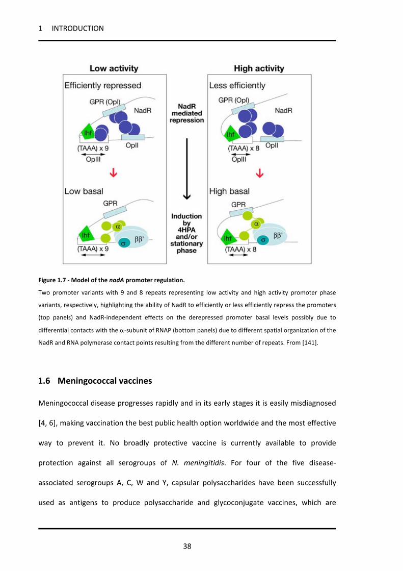
Figure 1.7 - Model of the nadA promoter regulation.
Two promoter variants with 9 and 8 repeats representing low activity and high activity promoter phase
variants, respectively, highlighting the ability of NadR to efficiently or less efficiently repress the promoters
(top panels) and NadR-independent effects on the derepressed promoter basal levels possibly due to
differential contacts with the α-subunit of RNAP (bottom panels) due to different spatial organization of the
NadR and RNA polymerase contact points resulting from the different number of repeats. From [141].
1.6 Meningococcal vaccines
Meningococcal disease progresses rapidly and in its early stages it is easily misdiagnosed
[4, 6], making vaccination the best public health option worldwide and the most effective
way to prevent it. No broadly protective vaccine is currently available to provide
protection against all serogroups of N. meningitidis. For four of the five disease-
associated serogroups A, C, W and Y, capsular polysaccharides have been successfully
used as antigens to produce polysaccharide and glycoconjugate vaccines, which are

1 INTRODUCTION
39
currently in use. Quadrivalent vaccines against serogroups A, C, W and Y include the
conjugate vaccines Menactra (Sanofi Pasteur) and Menveo (Novartis), and the
polysaccharide vaccine Menomune (Sanofi Pasteur), Mencevax (GlaxoSmithKline) and
NmVac4-A/C/Y/W-135 (JN-International Medical Corporation) [217]. A vaccine called
MenAfriVac has been developed through a program called the Meningitis Vaccine Project
to prevent meningitis group A infections in the ‘meningitidis belt’ [218].
The use of capsular polysaccharide as the basis of a vaccine for prevention of serogroup B
strains (MenB) diseases has been problematic. A capsular polysaccharide of MenB is
identical to a widely distributed human carbohydrate (α[2-8]N-acetylneuraminic acid or
polysialic acid), which, being a self-antigen, is a poor immunogen in humans and may
elicit autoantibodies [219, 220]. In addition, as mentioned above, the most represented
surface exposed protein, PorA and Opa, which should be used as antigens, show
sequence and antigenic variability and poor conservation between the diverse strains that
cause endemic MenB disease. As a result of these limitations, the only currently available
MenB vaccines are the “tailor made” outer membrane vesicle (OMV) vaccines, that have
been developed and successfully used to control epidemics that have primarily been
caused by a single strain [176]. Although effective, the limitation of the OMV vaccines is
that they are strain-specific vaccines and can be used against clonal disease outbreaks but
not for prevention of sporadic disease caused by heterologous strains. OMV vaccines
have proved to be effective and have been used in Norway [221], Cuba [222], Chile [223]
and New Zealand [224] to disrupt homologous strain outbreaks.

1 INTRODUCTION
40
1.6.1 Reverse vaccinology and the 4CMenB vaccine
The availability of whole genome sequences in the genomic era has radically changed the
approach to vaccine development. To overcome the problems regarding the production
of a MenB vaccine, an in-silico genome-based approach, called Reverse Vaccinology
(Figure 1.8) has been used, allowing the identification of approximately 600 open reading
frames that were likely to encode potentially surface exposed proteins in the MenB strain
MC58 [183]. The candidate antigens sequences were analysed for their conservation
among a wide range of MenB strains and expressed as recombinant protein in E.coli to
test their surface localization and ability of inducing bactericidal antibodies in mice. This
analyses revealed more than 90 previously unknown surface-located proteins, 29 of
which were able to induce bactericidal antibodies, the favourites of which were selected
for further studies [203, 225-228].
This approach has led to the development of a novel four component recombinant
protein vaccine against MenB named 4CMenB [204, 229]. 4CMenB has progressed
through clinical trials that have demonstrated its safety [230, 231] and its efficacy in
inducing a protective immune response in infants, children, adolescents and adults
against potentially the majority of MenB strains [232, 233]. The 4CMenB vaccine contains
five Genome-derived Neisseria Antigens (GNA), which are formulated as one single
recombinant protein, the Neisserial adhesin A (NadA) [198, 203], and two recombinant
fusion proteins, the factor H binding protein (fHbp) [234, 235] and Neisserial Heparin-
Binding Antigen (NHBA) [182, 226] fused with meningococcal gene products GNA2091
and GNA1030, respectively. NadA, fHbp and NHBA are the major antigens that were
selected through reverse vaccinology, based on their ability to induce broad protection

1 INTRODUCTION
41
inferred by serum bactericidal activity (SBA) assays or observed in passive protection in
the infant rat or mouse protection assays [204]. GNA2091 and GNA1030 are two
accessory antigens, well conserved in N. meningitidis strains but less well functionally
characterized than the major antigens. They are included in 4CMenB formulation as the
fusion proteins induce more potent immune responses in SBA than the individual antigen
[204]. The vaccine also contains Outer Membrane Vesicles (OMVs) from the
meningococcus B strain NZ98/254 in which PorA serosubtype 1.4 represents the major
antigen [236].
Figure 1.8 - Reverse vaccinology.
The process of reverse vaccinology applied to the development of the 4CMenB vaccine is schematized here
and described in details in the text.

1 INTRODUCTION
42
1.6.2 Vaccine coverage prediction
The studies performed by Goldschneider in 1969 demonstrated that the presence of
bactericidal antibodies in the serum, assessed by serum bactericidal assay with human
complement (hSBA), correlates with resistance to meningococcal meningitis. Therefore,
hSBA is widely accepted as a surrogate marker of protection against meningococcal
disease [12, 237-239]. hSBA assay made possible the development of meningococcal
vaccines based initially on the serogroup A, C, Y and W capsular polysaccharides, and
later, on these same polysaccharides covalently conjugated to protein carriers [240-242].
However, using hSBA in measuring the potential effectiveness of the 4CMenB vaccine to
kill circulating serogroup B strains presents two problems: on one hand, it requires
performing the assay against many diverse strains for each geographic region, which is
impractical, especially for infants, where serum volumes are very limited, on the other
hand the hSBA reflects the cumulative effect of all of the antibodies present in a serum
sample in killing the bacteria, making it dificult to understand the contribution of each
individual antigen to the function of the four component 4CMenB vaccine.
In order to overcome these issues and predict the coverage of 4CMenB, and a novel assay
has been developed, the Meningococcal Antigen Typing System (MATS) [243], (Figure
1.9). MATS assesses simultaneously the antigenic cross-reactivity and the level of
expression of the antigens present on the surface of an unknown meningococcal isolate
with respect to reference MenB strains for a specific antigen. MATS uses ELISA assays to
measure the three 4CMenB vaccine recombinant antigens NadA, fHbp, and NHBA. The
output value is the MATS Relative Potency (RP), which correlates with the hSBA assay and
may predict whether a strain would be killed due to antibodies elicited by the 4CMenB

1 INTRODUCTION
43
vaccine. MATS RP threshold values for complement-mediated killing of MenB by
antibodies against NadA, fHbp and NHBA antigens was established and termed the
Positive Bactericidal Threshold (PBT), for each specific antigen. Regarding PorA, the MATS
determines the PorA subtype by serological analysis or by PCR sequencing and strains
that carry the P1.4 PorA subtype are considered killed by vaccine-induced immune sera.
MATS can be easily performed in large panel of strains, making it possible to survey large
collections of MenB isolates in order to determine the potential for strain coverage by the
4CMenB vaccine of a target geographic region [244, 245]. Currently MATS is being used
by several meningococcal reference laboratories to establish the potential coverage of
the 4CMenB vaccine in different countries in Europe, North and South America and
Australia. According to MATS, it has been estimated that 78% of circulating MenB strains
in five European countries would have at least one antigen rated above the PBT and
therefore would be covered by the 4CMenB vaccine. However, the estimated
contribution of the NadA antigen to the vaccine coverage would comprise only about 2%
of the tested strains [246, 247]. Currently, the amount of the 4CMenB antigens expressed
by meningococci during human pathogenesis is unknown, but accurate estimates are
important to evaluate the real effectiveness of the vaccine. The study of NadA expression
during host infection and its implication on vaccine coverage prediction are a focus of this
thesis.

1 INTRODUCTION
44
Figure 1.9 - Schematic of the MATS ELISA method.
(A) MenB bacteria are grown overnight on chocolate agar. (B) A suspension of bacteria taken from the plate
is prepared to a specified OD600. (C) Detergent is added to the suspension to extract the capsule and
expose the antigens. (D) Serial dilutions of extract are tested in the MATS ELISA. A specific capture antibody
(yellow) binds one of the antigens (example: fHbp, blue) from the extract, which is then detected with a
specific biotin-labeled antibody (yellow and purple) and a streptavidin–enzyme conjugate (green and gold).
(E) Plates are read at 490 nm in an ELISA reader. (F) Results are calculated by comparing the curve of OD490
vs. dilution obtained with the serially diluted unknown strain to a serially diluted reference strain tested in
the same ELISA plate. From [243].

2 RESULTS
45
2 RESULTS
2.1 In the NadR regulon, adhesins and diverse meningococcal functions
are regulated in response to signals in human saliva
The meningococcal NadR was shown to repress expression of the NadA adhesin and play
a major role in NadA phase variable expression [141]. However, while nadA is present in
about 30% of circulating strains, NadR is always present and well conserved in all
meningococcal genomes available.
The aim of this part of the thesis is to clarify the global role of NadR during menincoccal
pathogenesis. We wish to elucidate the NadR regulon by finding its gene targets and the
kind of regulation mediated on them. We would also like to understand how the NadR
activity is affected by the 4HPA molecule, that is present in saliva and, therefore, in the
niche of meningococcal colonization, and which was previously shown to induce nadA
expression [141].
2.1.1 Global analysis of gene expression in the NadR mutant
In order to identify NadR-regulated genes in N. meningitidis we used a custom-made
Agilent oligonucleotide microarray to compare the transcriptional profiles of MC58 wild
type and MC-∆1843 NadR mutant strains grown until mid-log phase. Three independent
2-colour microarray experiments were performed comparing pooled RNA from triplicate
cultures of each strain, as well as three dye-swap experiments. The results obtained from
these experiments were averaged after slide normalization and 28 differentially
expressed genes were identified with a log2 ratio >0.9 transcriptional change and a t-test

2 RESULTS
46
statistics p-value ≤ 0.01 (Table 2.1.1). When we reduce the criterion to log2 ratio >0.6 in
the original 3 experiments (excluding the dye-swap) we can select another 9 differentially
up regulated genes in the NadR mutant with a p-value ≤ 0.01 (Table 2.1.1). RT-PCR
analyses were consistent with the microarray data (Table 2.1.1) confirming the putative
NadR-regulated genes identified in this global gene expression analysis. 31 genes were
up-regulated, whereas 6 were down-regulated in the NadR mutant. The contiguous genes
that are in the same orientation and exhibiting similar regulation have been grouped into
likely operons (Table 2.1.1).
In order to confirm that the regulation is NadR dependent, we complemented the MC-
∆1843 mutant by reintroducing the nadR gene under the control of an inducible Ptac
promoter in a different locus in the genome, generating the strain ∆NadR_C. By altering
the concentration of IPTG in the growth medium of the ∆NadR_C strain, we obtained a
level of NadR expression in the complemented strain comparable to that of the wildtype
(data not shown) and prepared total RNA from this strain in the same condition as the
microarrays experiments. We measured the transcriptional levels of the target genes by
RT-PCR in the complemented ∆NadR_C strain and observed that for all of the genes
tested that were up-regulated in MC-∆NadR (MC-∆NadR vs MC58), complementation of
the mutant (NadR_C vs MC58) restored their expression to levels similar or slightly lower
than the wild type level. In contrast, none of the down-regulated genes was restored to
wild type through complementation of the mutant, suggesting that these effects were not
dependent on the activity of the NadR protein. Among the 6 down-regulated genes
NMB1844, NMB1842 and NMB1841 are contiguous and in the same orientation to the
NadR-encoding gene NMB1843 and may be polarly affected by the insertion of the
2 RESULTS
47
antibiotic resistance cassette for the generation of the mutant. The lack of
complementation of deregulation of NMB0119 and NMB0120 is instead likely to be due
to a nadR-independent effect such as phase variation or secondary mutations.
These data suggest that NadR functions solely as a repressor protein, negatively
regulating multiple loci. Through global gene analysis we identify at least 18 hypothetical
operons/transcriptional units coding for 31 genes whose expression was repressed by the
nadR gene (Figure 2.1.1).

2 RESULTS
48
Table 2.1.1. Differentially regulated genes in NadR null mutant
Microarray results ∆1843 vs MC58wt RT-PCR Fold change
Gene Name OriA Function AverageB (Fold Change) Pval ∆1843 vs
MC58 NadR_C vs ∆1843 Gel shift
NMB0119 ↑ hypothetical protein -4.5 1.8E-14 -2.7 1.1 NMB0120 | hypothetical protein -8.4 2.3E-06 -4.4 1.1 nd NMB0207 ↓ glyceraldehyde 3-phosphate dehydrogenase, gapA 4.0 8.5E-07 5.4 -5.9 + NMB0375 | MafA1 adhesin, mafA1 2.1 4.4E-05 2.3 -2.3 + NMB0374D ↓ MafB1 adhesin, mafB1 1.3 4.7E-02 nd nd NMB0401 ↓ proline dehydrogenase, putA 1.9 1.2E-04 2.4 -3.1 + NMB0429 | hypothetical protein, putative regulatory protein 1.9 3.1E-06 nd nd + NMB0430 | putative carboxyphosphonoenolpyruvate
phosphonomutase, prpB 1.9 9.6E-04 nd nd
NMB0431 ↓ methylcitrate synthase/citrate synthase 2, prpC 1.9 1.6E-04 nd nd NMB0652 | MafA2 adhesin, mafA2 2.0 1.4E-04 2.3 -2.3 + NMB0653 | MafB-related protein, mafB2 1.9 6.8E-07 nd nd NMB0654 ↓ hypothetical protein 2.1 3.6E-03 nd nd NMB0865 ↑ hypothetical protein 1.9 4.0E-05 2.8 -5.5 nd NMB0866 | hypothetical protein 2.0 8.9E-03 nd nd NMB0955 | 2-oxoglutarate dehydrogenase, E1 component, sucA 1.9 2.7E-07 2.9 -3.2 + NMB0956D ↓ 2-oxoglutarate dehydrogenase, sucB 1.6 2.7E-02 nd nd NMB1205 ↓ hypothetical protein 2.4 8.4E-04 nd nd + NMB1277 ↑ transporter, BCCT family 1.9 1.2E-06 1.7 -4.5 + NMB1299 ↓ sodium- and chloride-dependent transporter 2.2 3.2E-03 nd nd - NMB1476 ↑ glutamate dehydrogenase, NAD-specific, gluD 4.1 2.3E-11 7.9 -11.1 + NMB1477 | hypothetical protein, comA competence protein 2.3 7.0E-03 nd nd

2 RESULTS
49
NMB1478 | phosphoglycolate phosphatase 2.3 3.6E-05 3.7 -2.6 - NMB1479 ↓ regulatory protein RecX 2.2 2.1E-06 nd nd NMB1841 ↑ mannose-1-phosphate guanyltransferase-related
protein -8.5 1.1E-16 -3.8 1.0
NMB1842 | putative 4-hydroxyphenylacetate 3-hydroxylase -14.1 0.0E+00 -5.6 0.9 NMB1843 | transcriptional regulator -25.5 8.3E-09 -12.3 nd + NMB1844 | hypothetical protein -3.0 4.3E-06 -1.6 1.1 - NMB1994 ↓ putative adhesin/invasin, nadA 59.6 0.0E+00 97.3 -133 + AverageC Pval NMB0535 ↓ glucose/galactose transporter, gluP 1.9 3.6E-04 nd nd - NMB0702 ↓ competence protein ComA 1.8 1.1E-03 1.9 -7.1 - NMB0978 ↑ NAD(P) transhydrogenase, beta subunit, pntB 1.7 6.4E-03 4.3 -2.9 NMB0979* | Hypothetical protein 1.6 1.3E-01 nd nd NMB0980* | NAD(P) transhydrogenase, alpha subunit, pntA 1.6 2.8E-02 1.8 -1.2 + NMB1609 ↑ trans-sulfuration enzyme family protein, metZ O-
succinylhomoserine sulfhydrolase 1.7 2.2E-06 2.3 -4.0 +
NMB2097 ↑ hypothetical protein, pykA pyruvate kinase 1.5 2.0E-04 nd nd NMB2098 | conserved hypothetical protein 1.6 4.1E-03 nd nd NMB2099 | conserved hypothetical protein, putative 5-
formyltetrahydrofolate cyclo-ligase 1.7 4.7E-03 2.4 -4.3 +
A Ori: orientation. Genes are grouped into likely operons as predicted by their similar orientation and proximity. Vertical solid lines represent the first and following genes in the predicted operon. Arrows pointing up represent the last gene in the operon in the reverse strand of the genome. Arrows pointing down represent the last gene in the operon in the forward strand of the genome. B Average values of three separate microarray experiments and three dye-swap experiments. C Average values of three separate microarray experiments. D Genes with values outside the criteria used are included when contiguous and oriented similarly to upstream or downstream co-regulated genes. E ND, not determined.

2 RESULTS
50
2.1.2 Functional classification of the NadR-regulated genes
Among all NadR-regulated genes (Figure 2.1.1), the NadA adhesin shows the most altered
expression profile in the ∆1843 mutant. Interestingly, 2 of the 3 mafA (multiple adhesin
family A) loci were also repressed by NadR. The NadR-regulated MafA1 and MafA2
(encoded by NMB0375 and NMB0652) are expressed in MC58, while mafA3 is a
pseudogene containing a frameshift. The maf loci, similarly to the locus of the Type IV pili,
consist of downstream silent cassettes of the expressed genes and are thought to
undergo antigenic variation through recombination of the coding sequences with the
silent cassettes [71]. The maf genes encode a family of variable lipoproteins originally
identified as glycolipid-binding proteins in pathogenic Neisseria [71, 248, 249], which have
been shown to adhere to glycolipid receptors on human cells [250, 251], and thus
predicted to be adhesins. While only approximately 40% of circulating meningococcal
strains carry the nadA gene, which is thought to have been acquired by horizontal gene
transfer, all meningococcus strains carry multiple loci expressing the Maf adhesins.
In addition to outer membrane adhesins, NadR is able to repress the expression of a
number of genes coding for inner membrane transporters including those involved in
transport of sugars (NMB0535, glucose/galactose transporter), compatible solutes
(NMB1277, encoding a putative glycine betaine transporter), transporters of unknown
substrates (NMB1299, encoding an Na- and Cl-dependent transporter) and even DNA
(NMB0702, encoding the ComA protein). NadR also regulates genes involved in energy
metabolic pathways including the NMB0401 (putA encoding proline dehydrogenase),
NMB1476 (gluD encoding glutamate dehydrogenase) and NMB0955-957 (sucAB-lpdA1
encoding 2-oxoglutarate dehydrogenase) genes involved in sequential steps of L-proline

2 RESULTS
51
and glutamate catabolism, along with other genes that may be involved in amino acid
metabolism (NMB1609, NMB1842), and other energy metabolic processes (NMB0207,
gapA-1 encoding a glyceraldehyde 3-phosphate dehydrogenase; NMB0430-431 coding for
a putative 2-methylcitrate pathway; NMB1478, gph encoding phosphoglycolate
phosphatase; and NMB0978-980, pntAB encoding NAD(P) transhydrogenase) as well as a
number of hypothetical genes (NMB0865/0866, NMB1477, NMB2099-2097) whose
function is unknown. Included also in the list are two possible regulators of gene
expression: NMB1479, coding for a putative transcriptional regulator, and NMB1205,
which synthesizes a small non-coding regulatory RNA recently named AniS [252].

2 RESULTS
52
Figure 2.1.1 – Functional classification of the NadR direct and indirect regulated genes.
(A) The diagram shows the numbers of genes in the NadR regulon divided according to their functional
classification. Percentages of the total genes in the regulon are reported for each functional class. (B)
Schematic representation of the loci controlled by NadR, clustered according to their functional
classification. Directly NadR bound targets have been identified through EMSA analyses and indicated by a
red box at the 5’ of the ORF.

2 RESULTS
53
2.1.3 Binding of NadR to its targets
It has been previously demonstrated that NadR binds to the nadA and farAB promoter
regions [141, 253], although no transcriptional regulation of farAB is thought to occur in
meningococcus [253]. Moreover, it has been recently reported that NadR-regulated
genes were not directly bound by the protein [90], suggesting an indirect NadR
regulation. To determine which of the NadR-repressed genes are under direct regulation
of NadR, we amplified by PCR the upstream promoter regions of identified target genes
and performed gel-shift analysis with purified recombinant NadR. We found that out of
19 target promoter regions tested, 14 are bound by the NadR recombinant protein (Table
2.1.1 and Figure 2.1.1B). Interestingly, NadR binds its own promoter, suggesting a
possible auto-regulation. The five promoter regions not directly bound by NadR were
those of NMB0535, NMB0702, NMB1299, NMB1844 and gph (NMB1478), although we
cannot exclude that the promoters and therefore NadR regulatory sites are further
upstream than the regions tested.
In order to demonstrate the specificity of NadR binding on its targets, a gel shift
experiment on the promoter region of NMB0401/putA was performed in the presence of
increasing concentration of sonicated salmon sperm DNA (non-specific competitor) or
cold putA promoter (specific competitor) (Figure 2.1.2A).
One slow migrating radioactive complex is formed by adding approx 3 nM of NadR
protein to the labeled putA promoter. A second complex appears by increasing the
concentration of NadR to 12.5 nM. 1-fold of cold putA promoter probe is sufficient to
abolish the higher complex and 5-fold completely prevents the binding of NadR to the

2 RESULTS
54
labeled probe. Instead, up to 25-fold of non-specific competitor had no effect on protein-
DNA complex formation, showing that NadR binds specifically to this promoter gene.
A panel of representative gel shift experiments from Table 2.1.1 is shown in Figure 2.1.2B.
All these gel shifts were performed in the presence of more than 10-fold excess of non-
specific competitor DNA in order to avoid non-specific NadR binding. It is worth noting
that with the exception of the promoter regions of NMB0375 (Figure 2.1.2B) and
NMB0652 (data not shown) genes, coding for MafA1 and MafA2 respectively, all other
promoters exhibit multiple DNA-protein complexes, and therefore are likely to contain
multiple binding sites for NadR.
We conclude that NadR can bind to the upstream promoter region in vitro of at least 14
transcriptional units consisting of 26 genes, confirming that these genes are members of
the NadR regulon. NadR regulation of the other genes may occur through indirect
mechanisms such as the regulation of an intermediate regulating factor.

2 RESULTS
55
Figure 2.1.2 - NadR binds to its target genes in a specific way.
(A) Gelshift analysis of NMB0401/putA promoter region. In the left panel 40 fmols of labeled probe was
incubated with increasing amounts of NadR protein corresponding to the indicated concentrations. To test
the specificity of NadR binding at the NMB0401 promoter region, in the right panel the same amount of
probe was incubated either without or with increasing amount of non-specific (sonicated salmon sperm
DNA) or specific competitor DNA (not labeled NMB0401 promoter probe) , in presence of the indicated
concentrations of NadR protein. (B) A representative panel of gelshift experiments reported in Table 1 is
shown. Promoter regions of indicated targets were amplified by PCR and radioactively end-labeled. 40
fmols of labeled probe was incubated with 2 μg of non-specific competitor DNA and increasing amounts of
NadR. Asterisks indicate free probes and arrows indicate DNA-protein complexes.

2 RESULTS
56
2.1.4 The NadR target genes can be classified in two types regarding their
promoter architecture
The previously described promoter region of nadA includes two distally spaced high
affinity binding sites for NadR (named OpI and OpII) and one with lower affinity
interposing the others (named OpIII) ([141] and schematic representation in Figure
2.1.3B). The presence of multiple NadR binding sites was suggested by gelshift analysis on
all NadR identified targets except for NMB0375 and NMB0652. To better understand
possible differences between promoter architectures of NadR regulated genes we
performed DNase I footprint analysis of NadR purified protein on a representative panel
of promoter regions (Figure 2.1.3A).
Both the promoter region of nadR/NMB1843 and putA/NMB0401 display two distally
spaced NadR protected regions, one overlapping the predicted promoter sequences and
the second distally upstream. This promoter architecture is comparable to the well-
known promoter region of NadA (Figure 2.1.3B) where the OpII overlaps the –10 box of
the promoter and the OpI is distally spaced. On the other hand, the promoter region of
mafA1/NMB0375 (Figure 2.1.3A and schematic representation in Figure 2.1.3B)
comprises only one binding site for NadR which overlaps the +1 and downstream of the
promoter.
Taken together these data suggest the existence of two types of promoter architecture
for NadR regulated genes: type I genes, including nadA and the majority of genes of the
regulon, have multiple NadR binding sites and in particular two high affinity sites, one
overlapping the promoter (centered at -4, -19 and -13 position for nadA, nadR and putA,
respectively) and one distally upstream (centered at -129, -103 and -79, respectively);
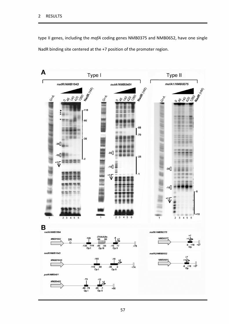
2 RESULTS
57
type II genes, including the mafA coding genes NMB0375 and NMB0652, have one single
NadR binding site centered at the +7 position of the promoter region.

2 RESULTS
58
Figure 2.1.3 - Type I and type II genes have different promoter architectures.
(A) DNase I footprint analyses of NadR on a representative panel of the promoter regions of its target
genes. 40 fmol of each probe were incubated with increasing amount of NadR purified protein as indicated
and then cleaved with DNase I. Regions of protection are indicated with vertical unbroken lines. Regions of
non-specific protection, possibly due to multimerization of NadR purified protein, are indicated with vertical
dotted lines. Hypersensitive bands are marked by a head arrow; protected disappearing bands are indicated
with asterisks. The predicted +1, -10 and -35 of each promoter region were deduced from the DNA
sequences and are properly located with respect to the G+A sequences. The positions of NadR protected
regions are also indicated. Two classes of promoter architectures are indicated as type I and type II. (B)
Schematic representations of type I and type II promoter regions. The structure of the nadA promoter is
reported as previously described [146], [145] and [141]. The low affinity operator III (Op III) at the TAAA
repeats tract is represented by a dark grey box. Regions bound and protected by NadR in DNase I footprint
analysis are represented by black boxes. The putative +1 of putA, nadR mafA1/2 promoters are shown. The
centre of each NadR binding site is reported as well. DR, direct repeat (border of region of horizontal
transfer).
2.1.5 Ligand-responsive regulation of NadR target genes expression by 4HPA
We have previously shown that a small molecule, 4-hydroxyphenylacetic acid (4HPA),
regulates NadR-mediated repression of NadA expression in meningococcus [141].
Therefore, we investigated the 4HPA-responsive regulation of the genes belonging to the
NadR regulon. To understand the in vivo effect of the small molecule on target gene
expression, we quantitatively analyzed the transcriptional level of selected target genes in
the MC58 wild type and NadR null mutant in the presence or absence of 5 mM 4HPA,
using RT-PCR (Figure 2.1.4). Surprisingly, we found that not all targets respond in a similar
fashion to the 4HPA inducer molecule and two classes of gene targets could be defined,
corresponding to the two types of genes identified due to their promoter region
architectures. We found that the expression of most genes was induced by the 4HPA
molecule (type I genes), either partially (i.e. nadA) or fully (i.e. putA, gph, nmb1277) with
respect to the maximal derepression achieved in the NadR mutant. Intriguingly, we found

2 RESULTS
59
that only mafA genes (type II genes) expression responds in the opposite way to 4HPA,
with mafA transcription being repressed in the presence of 4HPA and suggesting that this
small molecule acts as a co-repressor of NadR at the mafA1 and mafA2 promoters. In the
NadR mutant strain 4HPA-dependent regulation is largely absent indicating that NadR is
the mediator of the 4HPA transcriptional responses.
This analysis suggests that while NadR represses all genes in its regulon, 4HPA may act as
an inducer (type I genes) or a co-repressor (type II genes), resulting in alternative NadR-
mediated responses in vivo, which can be at least partially due to the differential
promoter architectures of the two types of NadR targets.
Figure 2.1.4 - Effect of 4HPA on gene expression.
RT-PCR analysis of NadR target transcripts in total RNA prepared from the wildtype (MC58) and MC-∆NadR
mutant strains (∆NadR) after growth in the presence or absence of 5 mM 4HPA. RT-PCR was performed in
duplicate on three independent biological replicates and the average values are shown. The primers used in
qRT-PCR for mafA1 and mafA2 genes cannot distinguish between the NMB0375 and NMB0652 genes as the
sequences are identical. Two types of transcriptional patterns can be detected, type I (4HPA-induced) and
type II (4HPA-repressed).

2 RESULTS
60
2.1.6 The NadR-dependent regulation of NadA, MafA and NadR itself is common
among meningococcal strains
In order to investigate whether NadR-dependent regulation of NadA and MafA protein
expression was exhibited by other strains, we extended our studies to a larger panel of
meningococcal strains. Western blot analysis were carried out on the wild type and their
respective NadR mutant strains grown in the presence or absence of 4HPA (Figure 2.1.5).
Expression of NadA and MafA is variable between strains, while NadR is expressed to
essentially the same level (lanes 1). All NadR null mutants exhibited higher levels of NadA
and MafA than their respective wild types (lanes 3 versus 1) confirming the NadR
repression of both nadA and mafA genes. In all the wild type strains, the 4HPA molecule
results in induction of both NadA and, to a lesser extent, NadR and co-repression of MafA
(lanes 2 versus 1), while it had no effect on NadA or MafA expression in the NadR mutants
(lanes 4 versus 3), indicating that the effects of 4HPA are NadR-dependent.
Taken together, these results suggest that both the NadR-dependent regulation of NadA
and MafA and the 4HPA activity as an inducer of nadA and a co-repressor of mafA are not
restricted to the MC58 strain but are common throughout different meningococci.
Furthermore, it would appear that the level of NadR itself is induced by 4HPA, most
probably through alleviation of autoregulatory repression of its own promoter.

2 RESULTS
61
Figure 2.1.5 - NadR-regulated adhesins responses in a broad panel of meningococcal strains.
Western blot of wildtype (lanes 1 and 2) and corresponding NadR knockout (lanes 3 and 4) cultures of the
indicated strains grown in the presence (lanes 2 and 4) and absence (lanes 1 and 3) of 1 mM 4HPA as
indicated, showing NadA, MafA and NadR expression. The NadA protein migrated at a molecular weight of
98 kDa and corresponds to the trimeric form of the protein [203] and the MafA band migrates at 36 kDa
and corresponds to both MafA1 and MafA2 proteins coded for by the NMB0375 and NMB0652 genes
whose sequences are 100 % identical. This band is absent in Western blots of extracts from the NMB0375
and NMB0652 double mutant [254] (data not shown). A non-specific band indicated with an asterisk is
reported as a loading control.
2.1.7 Incubation with human saliva has the same effect on NadA and MafA
expression as 4HPA
It has been previously demonstrated that the 4HPA molecule is a catabolite of aromatic
amino acids secreted in human saliva [147]. To test whether the effects due to 4HPA in
vitro may have biological relevance we tried to achieve similar results with human saliva.
Mid log cultures of MC58 were incubated for 1 hour either with 4HPA or with increasing
amount of human saliva from three different donors. Total protein extracts were
obtained and the expression levels of NadA and MafA were analyzed by Western blot
(Figure 2.1.6A). The Western blots show the induction of NadA and the repression of

2 RESULTS
62
MafA following incubation with 4HPA (compare lanes 2 to 1 of each blot) and, even if
with much greater experimental variability, the same results are obtained following
incubation with increasing concentration of human saliva from three donors (lanes 3,4,5
compared to 1).
Figure 2.1.6B shows a histogram with average values of quantified signals of Western blot
bands from six independent experiments. While NadA is induced by 4HPA as well as by
90% human saliva, MafA expression is co-repressed by both 4HPA and saliva at 50% and
90% concentrations, in a statistically relevant manner with respect to its basal level in GC
growth.
This analysis suggests that the 4HPA molecule used in our experiments mimics a signal
present in the human saliva, which produces the same regulatory effects on nadA and
mafA expression.

2 RESULTS
63
Figure 2.1.6 - Human saliva has the same activity of 4HPA on NadA and MafA expression.
(A) Western Blot analyses of total protein of mid log cultures of MC58 incubated for 1 hour either with
4HPA (lane 1) or with increasing amount of human saliva (HS) from 3 different donors (lanes 3, 4 and 5), as
indicated. The levels of NadA and MafA proteins are shown. (B) Histogram representing the average
expression levels of NadA (above) and MafA (below). The average values were calculated by quantification
of Western Blots bands signals of 2 independent experiments with human saliva of each donor (6
experiments in total). Expression levels of NadA and MafA of cultures grown solely in GC medium were set
to 1 arbitrary unit. To note that for the quantification, Western Blots bands were normalized for a non-
specific band, indicated with an asterisk in the panel A, in order to avoid non 4HPA- or NadR-dependent
effects due to possible protein degradation in saliva. One asterisk (*) means p value < 0.05, two asterisks
(**) p value < 0.01, both versus the growth in GC.
2.1.8 4HPA has differential activity on NadR binding to type I and type II
promoters in vitro
It was previously reported that the 4HPA molecule can attenuate the binding of NadR to
the nadA promoter in vitro [141], supporting the idea that it interacts with the protein

2 RESULTS
64
and alters its DNA-binding activity. To investigate the role of 4HPA on the in vitro activity
of NadR in binding to both type I and type II promoters, we performed gelshift analysis in
the presence of NadR and increasing amounts of 4HPA on a selected panel of target
promoter regions (Figure 2.1.7A).We demonstrate that for type I (gap-A1, gluD, putA,
nadR) promoters, 4HPA is able to inhibit the NadR-DNA binding in vitro in a dose-
dependent manner , similarly to what we reported for nadA [141]. However, addition of
4HPA had no significant effect on binding of NadR at the type II (mafA1 and mafA2)
promoters. In a parallel and complementary set of experiments we performed gelshift
analysis on the promoters of nadA and mafA1 with increasing amount of NadR purified
protein in the presence or absence of 5 mM 4HPA (Figure 2.1.7B-C). While 4HPA has no
effect on NadR binding activity on the promoter of mafA1 (Figure 2.1.7C), in order to have
the same amount of DNA-protein complexes on nadA promoter, more NadR is required in
presence of 4HPA (Figure 2.1.7B), indicating that 4HPA alleviates the binding of NadR on
nadA. The same results regarding the 4HPA responsive activity of NadR were also
observed on distinct NadR binding sites of the nadA and mafA promoters, as well as using
crude extracts from MC58 strain instead of the recombinant purified NadR protein, to
ensure that any eventual unknown factor required for NadR mediated repression could
be present (data not shown).
This analysis demonstrate that 4HPA affects and decreases the NadR DNA-binding affinity
only to type I promoters that are induced by 4HPA in vivo, while not having any in vitro
effect on NadR binding activity on type II promoters that are co-repressed in vivo.

2 RESULTS
65
Figure 2.1.7 - NadR responds differentially to 4HPA on type I and type II promoter regions.
(A) Gelshift analysis of selected NadR targets in the presence of increasing concentrations of 4HPA. Radio-
actively labeled promoter regions of the indicated genes were incubated with 0 (lane 1) or 36 nM NadR
(lanes 2-4) and 4HPA was added at final concentration of 1 mM (lanes 3) and 5 mM (lanes 4). (B) In vitro
NadR response to 4HPA on the promoter region of nadA and (C) mafA1 studied by gelshift. The nadA and
mafA1 promoter regions where amplified with primers pairs Nad-N1/Nad-B1 and P375-F/P375-652R,
respectively. 40fmol of end labelled probe were incubated with increasing amount of NadR protein (nM),
either with or without 5 mM 4HPA, as indicated. Free probes are indicated by asterisks and DNA bound by
NadR is indicated by arrow heads.

2 RESULTS
66
2.1.9 The 4HPA mediated co-repression of mafA is not due to repositioning of
NadR on the promoter
In order to understand the mechanism by which 4HPA mediates the NadR co-repression
of the mafA promoter, we evaluate the possibility that the 4HPA binding to NadR could
cause a conformational change, leading the repressor to bind a different DNA sequence
than the one previously observed, causing a more efficient repression. Ligand-induced
repositioning of the DNA binding domain is not new and has been described for the
MarR-family of transcriptional repressors [255].
To asses this possibility we performed DNAse I footprint analyses of NadR on the mafA
promoter in absence and in presence of 4HPA (Figure 2.1.8). Ligand binding does not alter
the operator bound by NadR on the promoter of mafA. Moreover, in agreement with
EMSA analyses, the NadR DNA binding activity on the promoter of mafA is not altered by
4HPA.
Taken together with the previous data, this indicates that the differential 4HPA
responsive activity of NadR on different types of targets is not due to the intrinsic binding
activity of the protein and that the nature of the binding site at the type II promoters may
be different to those of the type I promoters and these differences may define the
regulation to which they will be subjected.
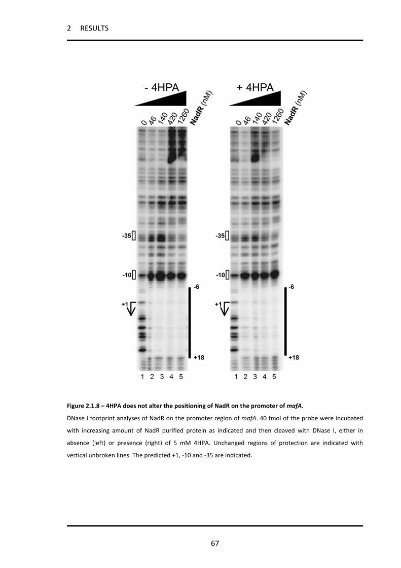
2 RESULTS
67
Figure 2.1.8 – 4HPA does not alter the positioning of NadR on the promoter of mafA.
DNase I footprint analyses of NadR on the promoter region of mafA. 40 fmol of the probe were incubated
with increasing amount of NadR purified protein as indicated and then cleaved with DNase I, either in
absence (left) or presence (right) of 5 mM 4HPA. Unchanged regions of protection are indicated with
vertical unbroken lines. The predicted +1, -10 and -35 are indicated.

2 RESULTS
68
2.1.10 3C scanning mutagenesis reveals extended NadR binding sequence in the
operator of mafA promoter region
In order to elucidate intrinsic differences between single NadR operators at the nadA and
mafA1 promoters and the role of nucleotides on NadR binding activity, we generated
DNA probes corresponding to the sequences of the high affinity binding sites of the nadA
(Op I and OpII) and mafA1 promoters, identified by DNase I footprint. For each probe we
designed a set of mutants, in which three nucleotides were sequentially substituted with
CCC. To detect the results of the 3C scanning mutagenesis on NadR binding activity, the
probes were submitted to EMSA analysis (figure 2.1.9A). By identifying the residues
whose mutations do not alter the binding of NadR, we defined the minimal binding site
(MBS) required for NadR binding on each operator (figure 2.1.9B). Mutation of nucleotide
triplets within the MBS results in either reduced (dark grey residues) or absent (black
residues) NadR binding activity. Interestingly, the definition of MBS clearly shows that the
operator on the promoter of mafA1 is significantly extended with respect to the high
affinity binding sites in promoter of nadA.
These observations suggest a differential mode of NadR binding between the nadA and
mafA1 operator, which could be partially responsible for the alternative 4HPA responsive
activity of NadR at the two promoters.

2 RESULTS
69
Figure 2.1.9 - 3C scanning mutagenesis of nadA and mafA single binding sites. (A) DNA probes corresponding to the wild type NadR-protected sequences from DNAI footprints or to mutated sequences, in which sequential triplicate nucleotides were substituted with CCC, were submitted to gelshift analysis with increasing amount of NadR purified protein (0, 36 and 360 nM, respectively). The mutated nucleotides are in italics and underlined in the sequences above each gelshift. The NadR binding activity to each probe is reported below each experiment as a percentage of the binding activity shown by NadR on the wild type sequence. Black values are associated with mutation causing loss or highly compromised NadR binding activity; dark grey values with less affected NadR binding activity; light grey values with not affected NadR binding activity. (N.A.= not active in binding). (B) The minimal binding sequence (MBS) of each binding site is shown, with nucleotide substitutions essential for binding in black, affecting affinity in dark grey and with no effect in NadR binding activity in light grey.
2 RESULTS
70
2.2 Structural insight into the mechanism of DNA-binding attenuation of
NadR by the small natural ligand 4HPA
NadR is a member of the MarR family of transcriptional regulators. A characteristic
feature of these proteins is their capability to bind and respond to a variety of effector
molecules [124, 125]. Although the crystal structures of several MarR homologs have
been solved in their apo- and ligand-bound states, the molecular mechanisms of
regulation associated with ligand binding is still not known, partly due to the fact that the
natural ligand is often not known.
As seen above, 4HPA is a natural ligand of NadR and is a key factor which determines
NadR activity. The aim of this part of the thesis is to elucidate the mechanism of
regulation of NadR by the 4-HPA molecule. We focused on NadR activity in response to
4HPA on nadA, being the gene majorly affected by NadR. We would like to identify the
4HPA binding pocket of NadR and the key residues involved in this domain.
2.2.1 Characterization of the structural model of NadR.
A structural model of NadR was generated using the crystal structure of the
transcriptional regulator PA4135 from Pseudomonas aeruginosa [256] which shares 42%
sequence identities with NadR (Figure 2.2.1A). According to this model, the NadR dimer
adopts the typical “triangle” shape of the MarR family with each monomer consisted of
the secondary structure elements α1-α2-α3-α4-β1-wing-β2-α5-α6 (Figure 2.2.1B). The
two monomers associate via a dimerization interface created by their N- and C-terminal
helices (α1, α5 and α6) and each monomer contains a winged helix-turn-helix (w-HTH)
DNA-binding motif (α3, α4, β1, wing, β2).

2 RESULTS
71
The quality of the model was assessed by amide hydrogen/deuterium exchange mass
spectrometry (HDX-MS), as reported by Brier and collegues [8]. The rate at which backbone
amide hydrogens (NHs) exchange in solution is directly dependent on the dynamics and
the structure of the protein [257]. Therefore, regions with secondary structures or
occluded from the deuterated buffer will exchange more slowly than regions without any
secondary structures and/or fully exposed to the solvent. As shown by data reported in
[8] the core of the protein, including the dimerization interface, is well protected from
exchange, to generate the well-packed hydrophobic core of the dimerization domain. In
contrast, the w-HTH DNA-binding motif was found to be highly accessible, consistent with
the fact that these regions must remain fully accessible and/or dynamic to interact with
DNA [132]. Altogether, the HDX data correlate well with the homology-based structural
model of NadR.

2 RESULTS
72
Figure 2.2.1 - Homology model and dynamic analysis of NadR. (A) Sequence alignment between NadR and transcriptional regulator PA4135 from Pseudomonas aeruginosa. NadR shares 42% sequence identity with PA4135. Dark and grey shading indicates the position of the conserved and highly similar residues, respectively. The alignment was performed with CLUTALW. (B) Ribbon representation of the proposed homodimeric structure of NadR obtained with SWISS-MODEL and based on the X-ray structure of PA4135. One subunit is colored in gold and the other in light blue. According to this model, each monomer adopts the typical fold of the MarR family members, which consists of 6 α-helices and 2-stranded β-sheets. The dimerization interface and the DNA-binding regions are indicated.
2.2.2 Localization of the 4HPA binding pocket
To localize the 4-HPA biding pocket, the exchange behaviors of the apo- and HPA-bound
forms were monitored and compared by HDX-MS, as reported in [8], resulting in four
regions, including Met1-Leu14 (N-term α1), Met22-Leu29 (C-term α1), Asn54-Cys61 (α3) and

2 RESULTS
73
Tyr104-Glu119 (α5), protected from exchange upon 4-HPA binding. To visualize more precisely
our results, these regions were mapped onto the structural model of the dimer.
Interestingly, with the exception of α3, the reduction of solvent accessibility was
clustered at the interface between the dimerization and the w-HTH DNA-binding domains
(Figure 2.2.2). This interface has already been identified as a common salicylate-binding
pocket in several MarR homologues including MTH313 [133], ST1710 [132], SlyA [258]
and TcaR [130]. Although salicylate does not bind NadR, based on these observations, we
conclude that 4-HPA likely shares the same protein binding region.
Figure 2.2.2 - The 4HPA binding pocket within NadR. Ribbon representation of NadR showing the position of the putative 4-HPA binding region identified by HDX-MS. Regions with altered deuterium uptake are colored in orange (chain A) and red (chain B). The 4-HPA-binding pocket is located at the interface between the dimerization and the DNA-binding domains and is mainly formed by the α1 N-terminal domain of chain A and the helices α2, α5 and the α1 C-terminal domain of chain B (see expanded view).

2 RESULTS
74
2.2.3 Mutation of key residues in the 4-HPA binding pocket of NadR
In an attempt to identify the residues of NadR in contact with 4-HPA in the identified
binding pocket, an in silico docking approach has been used as described by Brier and
collegues [8]. As a result of this analyses, a predicted network of hydrogen bonds
coordinating 4-HPA was observed, which mainly involves residues Arg18, Trp39, Arg40,
Arg43 and Tyr115 (Figure 2.2.3). These residues were selected for mutagenesis to further
determine their in vivo and in vitro functions in 4-HPA binding. Additional residues (His7,
Ser9, Asn11 and Phe25) were identified through the in silico docking approach, and were
also mutagenised to assess whether they play a role in the binding of the small molecule.
In order to obtain meningococcal strains expressing the NadR mutated forms, the
previously described MC58-∆1843 nadR null mutant strain [141] was complemented
either by the wild type NadR protein or by the introduction of the desired alanine
substituted forms of NadR. Note that we failed to introduce the R43A substitution in
NadR and, as a consequence, this residue was not further studied.

2 RESULTS
75
Figure 2.2.3 - Key residues in the 4HPA binding pocket. Tridimensional surface model of the structure of the NadR protein, obtained as described in [8]. The binding pocket for the 4-HPA molecule is illustrated in the main model and in the magnification showing residues putatively involved in 4HPA binding.
2.2.4 In vivo behaviour of selected site directed NadR mutants
Total lysates from MC58 strains expressing different forms of NadR were subjected to
Western Blot analyses of NadA expression to assess the ability of each NadR mutant
protein to repress nadA expression and respond to the 4-HPA inducer. As reported in
Figure 2.2.4A, NadR R18A and R40A are unable to repress nadA (compare lines 5 & 9 to
lines 1 & 3) and mimic the behavior of the ∆1843 strain in the presence and absence of 4-
HPA. These results suggest that both substitutions affect the ability of NadR to repress
the nadA promoter, likely by preventing DNA binding.

2 RESULTS
76
The protein NadR W39A presents a lower expression level compared to wild type NadR
(compare line 7 to lines 1 & 3) which indicates that the stability of the protein is
compromised by the substitution. Possibly as a result, NadR W39A is unable to repress
nadA expression in vivo and exhibits a behavior similar to that of the ∆1843 strain.
Interestingly, the addition of 4-HPA increases the expression level of NadR R39A thus
suggesting that 4-HPA still interacts and stabilizes NadR W39A (compare lines 7 & 8).
In contrast to R18A, W39A and R40A, the substitution of Tyr115 by Ala does not abrogate
the in vivo repressive function of the protein. Interestingly, NadR Y115A represses more
efficiently nadA than NadR WT thus suggesting that NadR Y115A acts as a hyper-repressor
of NadA expression (compare line 11 to lines 1 & 3). Moreover, the addition of 4-HPA does
not affect the repressive activity of NadR Y115A (compare line 11 to lines 2 & 4). Taken
together, these data reveal that the Y115A substitution compromises the response of the
protein in vivo to the 4-HPA inducer without abrogating its ability to repress nadA.
In a second round of mutagenesis, we generated a new set of meningococcal strains
expressing NadR mutated proteins in the additional residues identified by in silico docking
(His7, Ser9, Asn11 and Phe25). We then test their ability to repress nadA and sense 4HPA in
vivo (Figure 2.2.4B). Interestingly, three out of the four new mutations (His7, Ser9 and
Phe25) lead to a phenotype similar to the one presented by the Tyr115, being able to
hyper-repress nadA expression (lines 5, 7 and 11 compared to 1) and almost unable to
respond to 4HPA (compare lines 5 & 6, 7 & 8, 11 & 12, respectively). The mutant in Asn11,
instead, is just slightly affected in its ability to repress nadA and is able to sense 4HPA,
leading to induction of nadA (compare lines 9 & 10 to 1 & 2, respectively).
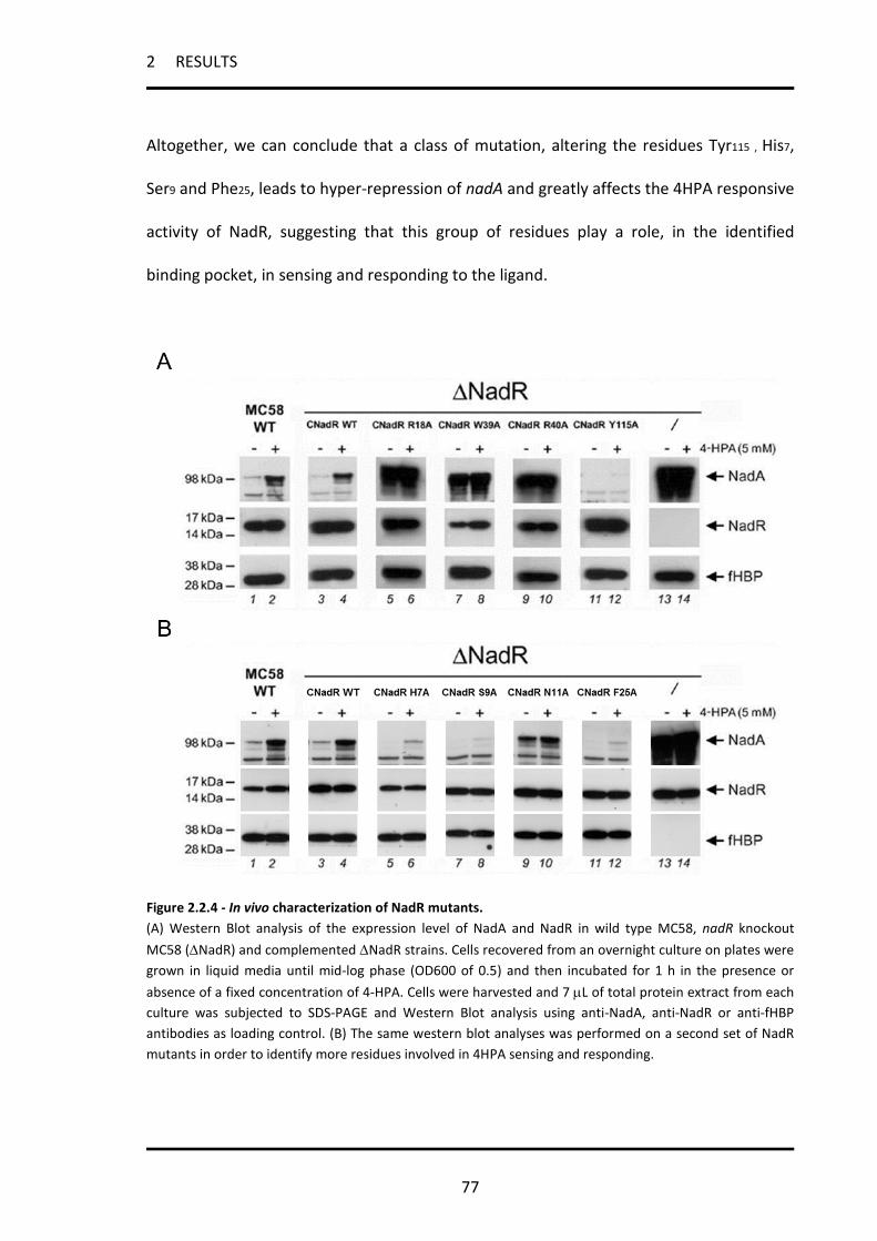
2 RESULTS
77
Altogether, we can conclude that a class of mutation, altering the residues Tyr115 , His7,
Ser9 and Phe25, leads to hyper-repression of nadA and greatly affects the 4HPA responsive
activity of NadR, suggesting that this group of residues play a role, in the identified
binding pocket, in sensing and responding to the ligand.
Figure 2.2.4 - In vivo characterization of NadR mutants. (A) Western Blot analysis of the expression level of NadA and NadR in wild type MC58, nadR knockout MC58 (∆NadR) and complemented ∆NadR strains. Cells recovered from an overnight culture on plates were grown in liquid media until mid-log phase (OD600 of 0.5) and then incubated for 1 h in the presence or absence of a fixed concentration of 4-HPA. Cells were harvested and 7 µL of total protein extract from each culture was subjected to SDS-PAGE and Western Blot analysis using anti-NadA, anti-NadR or anti-fHBP antibodies as loading control. (B) The same western blot analyses was performed on a second set of NadR mutants in order to identify more residues involved in 4HPA sensing and responding.

2 RESULTS
78
2.2.5 In vitro characterization of the DNA- and 4-HPA binding activities of the
purified NadR mutant proteins
To further elucidate the behavior of the NadR mutants R18A, W39A, R40A and Y115A, the
recombinant proteins were expressed and purified from E. coli to assess their in vitro
DNA-binding activity as well as their response to 4-HPA. Further analyses reported in [8]
demonstrated that the solubility, stability and global fold of NadR Y115A and R40A were
comparable to that of wild-type NadR, while both R18A and W39A substitutions
significantly modify the native structural integrity of the protein, precluding their use in in
vitro analyses.
To test the binding activity of the NadR proteins in vitro, we prepared a radiolabeled DNA
probe containing the high affinity binding site OpI from the nadA promoter [141]. The
binding specificity of NadR WT to the OpI labeled probe was first assessed by EMSA in the
presence or absence of increasing amounts of either sonicated salmon sperm DNA (non-
specific competitor) or non-labeled OpI DNA (specific competitor). As shown in Figure
2.2.5A (lines 1-3), a slow migrating complex is formed between NadR WT and the OpI
labeled probe in the presence of 15 ng protein. The addition of increasing amounts of
non-specific competitor does not disrupt the complex (Figure 2.2.5A, lines 4-6), while the
use of cold-OpI DNA prevents the binding of NadR WT to the radiolabeled OpI probe in a
dose dependent manner (Figure 2.2.5A, lines 7-9). This experiment demonstrates that
NadR WT binds specifically to the OpI site of the nadA promoter. Similar results were
obtained using a labeled probe comprising the high affinity binding site OpII [141] from
the nadA promoter (data not shown).

2 RESULTS
79
We next investigated the in vitro ability of NadR Y115A and R40A to bind to the OpI
labeled probe. While NadR WT forms a DNA-protein complex with OpI, no interaction
could be detected with NadR R40A even at high protein concentrations (Figure 2.2.5B).
The DNA-binding activity of NadR R40A is abolished by the substitution indicating that
Arg40 is essential for DNA binding. Therefore, the in vivo defect of this mutant to repress
NadA can be directly linked to its incapability to interact with the nadA promoter. In
contrast to NadR R40A, the Y115A substitution does not abrogate the in vitro DNA-
binding activity of the protein (Figure 2.2.5B). Interestingly, NadR Y115A binds OpI with a
lower affinity compared to NadR WT. The apparent reduction of DNA-binding affinity
contrasts with the in vivo hyper-repressive activity of NadR Y115A and suggests that the
repressive ability of the NadR protein is not exclusively based on its ability to bind the
nadA promoter DNA. Finally, the addition of 4-HPA does not disrupt the NadR Y115A/DNA
complex even at 10 mM concentration (Figure 2.2.5C). In vitro experiments to assess
NadR binding activity and 4HPA response were performed also with the OpII labeled
probe and identical results were obtained (data not shown). Moreover the same results
were also confirmed on the whole promoter of nadA, comprising multiple binding sites,
and using crude extracts instead of the NadR purified protein, to avoid the possibility that
unknown factors required for NadR activity were missing in the reaction (Figure 2.2.5D).
Taken together, these data demonstrate that while NadR R40A has lost DNA-binding
activity and no longer represses nadA, NadR Y115A is a hyper-repressor and is ‘blind’ to
the 4-HPA molecule.

2 RESULTS
80
Figure 2.2.5 - Substitution Y115A completely abolishes the in vitro effects of 4-HPA.
(A) Gel-mobility shift assays performed with a radioactively labeled DNA probe containing the individual OpI
operator in the presence of increasing amounts of purified NadR WT (lines 1-3) and in the presence of
either increasing amount of a non-specific competitor (salmon sperm DNA, lines 4-6) or a specific
competitor (cold OpI DNA, lines 7-9). The positions of the free and complexed DNA are indicated. (B) Gel-
mobility shift assays showing the binding activity of NadR WT, NadR R40A and NadR Y115A on the OpI radio
labeled probe. The R40A substitution completely abolishes the DNA-binding activity of NadR while the
Y115A substitution reduces the DNA-binding affinity. (C) Gel-mobility shift assay reporting the effects of 4-
HPA on the OpI binding activity of NadR WT and NadR Y115A. The presence of increasing concentrations of
4-HPA only induces the release of the DNA probe from NadR WT. (D) Gel-mobility shift assay performed
with crude extracts (Crude Ex.) on the whole promoter of nadA, comprising multiple binding sites. The
effects of substitution Y115A on NadR DNA binding and 4HPA responsive activities are reported (5mM 4HPA
is added were indicated).

2 RESULTS
81
2.2.6 NadR Y115A does not act as a hyper-repressor on the promoter of mafA1
To assess if NadR Y115A also had hyperrepressive activity on other NadR target
promoters, namely type II, we further investigated both the in vivo behavior and the in
vitro binding activity of the NadR Y115A on the single operator structured mafA1
promoter.
Western blot analysis (Figure 2.2.6A) reveals that the MafA1 protein level is not altered in
vivo by the replacement of NadR wild type with the Y115A. EMSA experiments,
performed on the whole promoter of mafA1 with crude extracts (Figure 2.2.6B), show
that NadR Y115A has a reduced binding activity in vitro compared to the wild type
protein, accordingly with what we saw on the promoter of nadA. However, because the
binding mode of NadR on type I and type II promoters could be different as demonstrated
above (Figure 2.1.9), the reduced in vitro binding activity of Y115A results in no
detectable binding on promoter of mafA1 in vitro, even with high amount of crude
extracts. As previously demonstrated (Figure 2.1.7), 4HPA has no effect on NadR binding
activity on the promoter of mafA1
The fact that the hyper-repressive activity of Y115A is only visible on the type I promoter
of nadA comprising multiple NadR binding sites, but not on the type II promoter of mafA1
comprising one single NadR binding site, suggests that this is not an intrinsic property of
the mutant protein, rather it is linked to the promoter architecture of the gene which is
acting on and could be partly due to an altered tetramerization/multimerization capacity
of the mutant.

2 RESULTS
82
Figure 2.2.6 – In vivo and in vitro characterization of NadR Y115A on mafA1.
(A) Western blot analysis showing the level of expression of MafA1 in presence or absence of either the
NadR wild type or the Y115A, as indicated. (B) Gel-mobility shift assay performed with crude extracts (Crude
Ex.) on the whole promoter of mafA1, comprising only one NadR binding site. The effects of substitution
Y115A on NadR DNA binding and 4HPA responsive activities are reported (5mM 4HPA is added were
indicated).

2 RESULTS
83
2.3 Transcriptional Regulation of the nadA Gene Impacts on the
Prediction of Coverage of the 4CMenB Vaccine
NadA is one of the major antigens of the 4CMenB vaccine, therefore the regulation of its
expression impacts on the vaccine coverage prediction. NadA is repressed by NadR under
in vitro growth conditions. As seen in previous chapters, different approaches have been
followed in order to mimic nadA expression during host infection, including the use of
physiologically relevant molecules and the ex vivo model of saliva, under which NadR-
repression is relieved.
The aim of this part of the thesis is to investigate whether the level of expression of nadA
is different between in vitro growth condition and during invasive infection using in vivo
models. We evaluate further how this difference could implicate on current methods for
coverage prediction analyses and whether, when taking this into consideration, we could
better predict the contribution of NadA in vivo to the effectiveness of the 4CMenB
vaccine.
2.3.1 Strains with MATS RP ≤ PBT express NadA in an immunogenic form during
invasive disease
Litt and colleagues [205] observed that many protein antigens, including the NadA
protein, were recognized by antibodies present in sera of children convalescing after
meningococcal disease. Importantly, NadA was significantly more strongly recognized by
convalescent patients infected with strains carrying the nadA gene, than by uninfected
control subjects [205]. We have extended this study by subjecting the 14 nadA positive
isolates, matched to the sera of the Litt study, to Western blot and MATS analysis in order

2 RESULTS
84
to visualize the in vitro levels of NadA expression of the infecting strain. In Figure 2.3.1A,
Western blots reveal that while the regulator NadR is expressed comparably by all strains
tested, the levels of NadA are variable, and some strains (Nm067, Nm100 and Nm188)
failed to express the protein at detectable levels under the in vitro growth condition used.
When analyzed by MATS, the level of NadA expression, calculated as relative potency
compared to a reference strain (5/99), correlated well with Western blots results (Figure
2.3.1B and Table 2.3.1). As shown, only 5 strains have RP values above the PBT of NadA
which is 0.009 (Nm036, Nm037, Nm081, Nm088 and Nm154), while the remaining strains
show RP ≤ PBT. However, sera from children infected either by strains that failed to
express NadA in vitro (MATS RP = 0) or by strains with a NadA RP < PBT, nonetheless are
able to recognize at least one form of the NadA recombinant proteins used in the dot blot
experiments of Litt and colleagues ([205] and Figure 2.3.1C) more efficiently than sera
from subjects infected by nadA- meningococci.
These data show that strains with MATS RP values below the PBT are nevertheless able to
express the NadA protein in an immunogenic form during invasive disease, driving a
robust humoral response. This observation suggests that the levels of expression of NadA
under in vitro growth conditions may differ from, and be lower than, those reached
during infection in the human host.
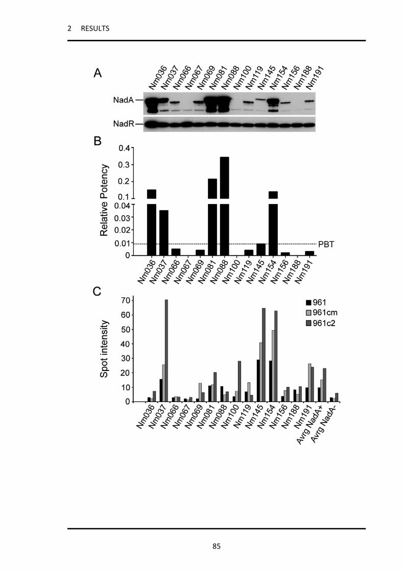
2 RESULTS
85

2 RESULTS
86
Figure 2.3.1 - Strains with MATS RP ≤ PBT express NadA in an immunogenic form during infection.
(A) Western blot analyses of the wild type nadA+ strains from the Litt study isolated from convalescing
children after meningococcal disease [205], showing NadA and NadR expression. The NadA protein migrates
at the molecular weight of about 98 kDa corresponding to the trimeric form of the protein, but varies
slightly according to the NadA allele expressed by each strain, while the NadR band migrates at 16 kDa. (B)
MATS relative potency (RP) of NadA determined by the MATS ELISA for the panel of isolated strains. A black
dashed line represents the positive bactericidal threshold (PBT) for NadA. The RPs of each strain are
reported in supplementary table 1. (C) Spot intensity of dot blot experiments adjusted from the study of Litt
and colleagues [205]. Reactivities of sera from children infected with the reported isolated strains towards
the full length NadA recombinant protein (961) as well as two truncated forms comprising the extracellular
portion of NadA (961cm and 961c2) are reported. The average values of reactivity for the 14 nadA+ strain
and the 17 nadA- strains from the original study were calculated and reported in the graph as references
(Avrg NadA+ and Avrg NadA-, respectively).
Table 2.3.1 - MATS RPs of NadA for nadA+ strains isolated from patients
MATS NadA Strain Low CLa RPb High CLc
NM036 0.131 0.151 0.174 NM037 0.027 0.035 0.045 NM066 0.003 0.005 0.007 NM067 NA NA NA NM069 0.003 0.004 0.005 NM081 0.173 0.214 0.256 NM088 0.245 0.342 0.501 NM100 NA NA NA NM119 0.003 0.004 0.004 NM145 0.008 0.009 0.013 NM154 0.105 0.14 0.186 NM156 0.002 0.002 0.003 NM188 NA NA NA NM191 0.002 0.003 0.004 a = experimental low confidence limit (StatLIA). b = relative potency. c = experimental high confidence limit (StatLIA). NA = not available.

2 RESULTS
87
2.3.2 All strains carrying the nadA gene can express high levels of the NadA
protein and therefore be killed by vaccine-induced bactericidal antibodies
The main mediator of varying expression levels of NadA within and between strains is the
transcriptional regulator NadR, through its ability to differentially repress the phase
variant promoter of nadA. To understand the relevance of NadR repression to the
variable expression levels of NadA observed in different MenB strains under in vitro
conditions, a representative panel of strains covering a range of NadA expression levels
was selected (Table 2.3.2) and the nadR gene was deleted in each of them. We evaluated
the implications of the alleviation of NadR repression under in vitro conditions by
Western blot, MATS and SBA analysis (Figure 2.3.2 and Tables 2.3.3 and 2.3.4).
As previously shown [141], all the nadR- strains expressed considerably more NadA than
their wild type forebears, confirming that deletion of nadR results in strong induction of
NadA. Furthermore, all nadR- strains expressed comparable high levels of the NadA
antigen measured by Western blot (Fig. 2.3.2A). The MATS assay performed on wild type
and nadR- strain pairs demonstrated that the ratio of the RP values for NadA increased
from 3 to up to 100-fold in the mutant strains (Fig. 2.3.2B), indicating that all these strains
can express high levels of immunogenically relevant NadA antigen when NadR repression
is abolished. The MATS assay correlates with the hSBA [243] at values of RP higher than
the PBT. Therefore, we compared the ability of immune sera to kill the nadR- knock out
strains and their related wild types. Table 2.3.3 shows that sera from mice immunized
with NadA alone or with the 4CMenB vaccine have an increased NadA-specific
bactericidal activity on nadR- strains compared with wild type strains. The only exception
is for strain 5/99, in which, as expected, there is no significant difference in SBA titres

2 RESULTS
88
between the wild type and the nadR- strains. In this strain the NadR-mediated repression
of NadA is minimal: NadA is highly expressed in the wild type strain. Sera from
immunizations with NHBA and fHbp (Table 2.3.3) had invariant activity towards wild-type
and nadR- strains, confirming that the knock-out of nadR does not alter the susceptibility
of these strains in the bactericidal assay and suggesting that neither NHBA nor fHbp
expression is regulated in a NadR-dependent way. hSBA assay performed on the mutant
strains demonstrates that sera from clinical trial subjects of different age groups
immunized with the 4CMenB vaccine formulations were efficiently able to kill all nadR-
strains and exhibited extremely high bactericidal titers (Table 2.3.4). Of note, antibodies
present in sera from some age groups and apparently ineffective in killing of certain
strains (e.g. B3937 and NGP165) show the ability to efficiently kill the equivalent
recombinant strains once NadR repression has been relieved.
Taken together these data demonstrate that all the strains carrying nadA can potentially
express NadA to a level which is sufficient to be recognized and to mediate killing by
bactericidal antibody elicited by the 4CMenB vaccine.

2 RESULTS
89
Table 2.3.2 - Selected strains used in this analysis
Strain Clonal complex STa Year Countryb Typing PorA Match OMV_NZc
fHbp IDd NHBA NadA variant
5/99 ST-8 complex/Cluster A4 1349 1999 N B:2b:P1.5,2 N 23 20 2
961-5945 ST-8 complex/Cluster A4 153 1996 AUS B:2b:P1.21,16 N 16 20 2
LNP17094 ST-8 complex/Cluster A4 153 1999 F B:2b:P1.10 N 16 22 2
B3937 ST-18 complex 6344 1995 D B:22:P1.16 N 17 23 3
M10574 ST-32 complex/ET5 complex 803 2003 USA B:NT:P1.7-2,13-1 N 76 3 1
M14933 ST-32 complex/ET5 complex 32 2006 USA B:ND:P1.22-1,14 N 76 3 1
MC58 ST-32 complex/ET5 complex 74 1985 UK B:15:P1-7,16b N 1 3 1
NGP165 ST11 complex/ET-37 complex 11 1974 N B:NT:P1.2 N 29 29 2
a = multilocus sequence type. b = AUS, Austria; D, Denmark; F, France; N, Norway; UK, United Kingdom; USA, United States of America. c = No strains match the PorA P1.4 allele in the OMV_NZ vaccine component. d = The fHbp allele identification numbers (ID) are reported here according to Oxford database nomenclature.
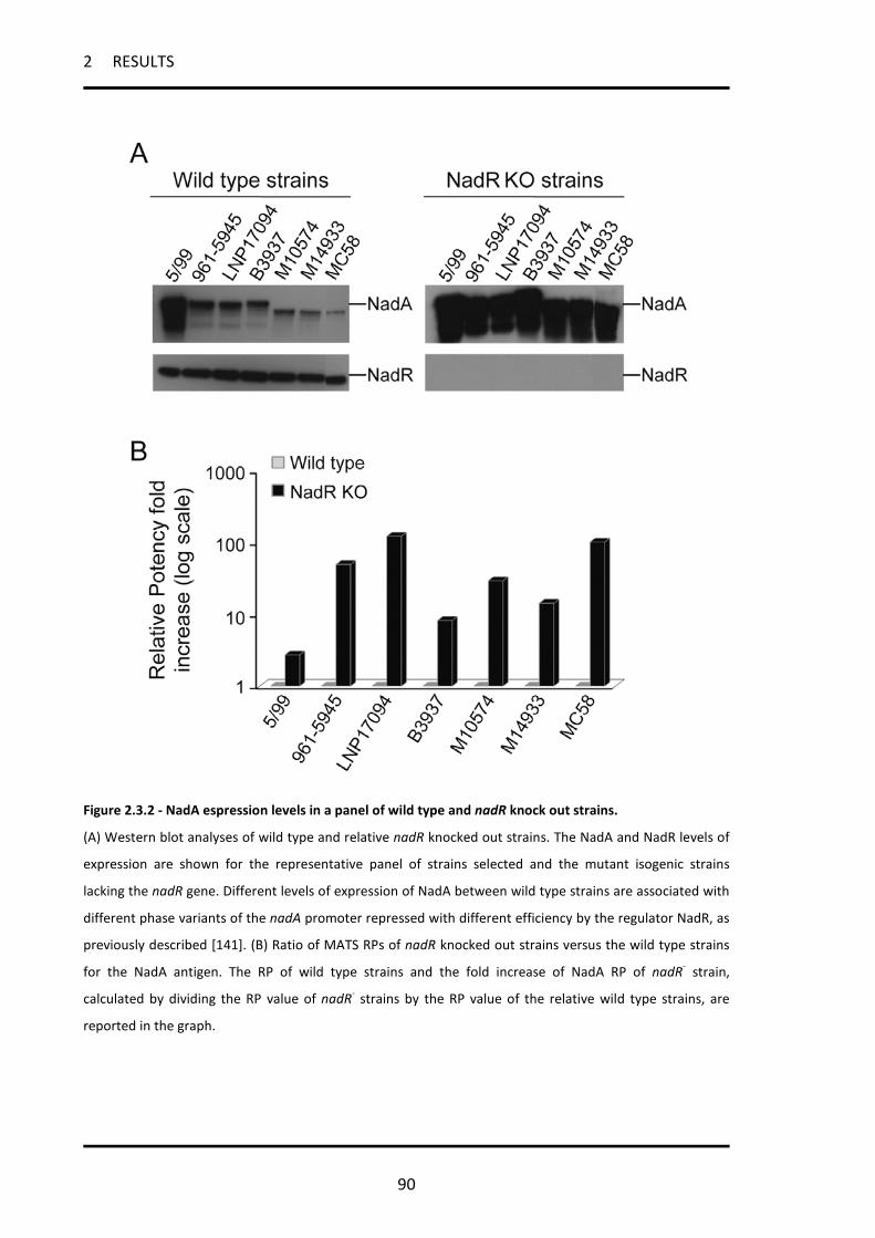
2 RESULTS
90
Figure 2.3.2 - NadA espression levels in a panel of wild type and nadR knock out strains.
(A) Western blot analyses of wild type and relative nadR knocked out strains. The NadA and NadR levels of
expression are shown for the representative panel of strains selected and the mutant isogenic strains
lacking the nadR gene. Different levels of expression of NadA between wild type strains are associated with
different phase variants of the nadA promoter repressed with different efficiency by the regulator NadR, as
previously described [141]. (B) Ratio of MATS RPs of nadR knocked out strains versus the wild type strains
for the NadA antigen. The RP of wild type strains and the fold increase of NadA RP of nadR- strain,
calculated by dividing the RP value of nadR- strains by the RP value of the relative wild type strains, are
reported in the graph.

2 RESULTS
91
Table 2.3.3 - rSBA performed with immunized mice sera and rabbit complement on wild
type and nadR knock out strains
Strain NHBA fHbp NadA 4CMenBa
5/99 512 <16 >65536 >32768
5/99 nadR- 128 <16 >65536 >65536
961-5945 1024 1024 1024 4096
961-5945 nadR- 1024 2048 >65536 >65536
LNP17094 1024 <16 128 4096
LNP17094 nadR- 512 <16 32768 >65536
B3937 <16 1024 512 2048
B3937 nadR- <16 512 >65536 >65536
M10574 4096 128 >8192 >8192
M10574 nadR- 4096 64 >32768 >32768
M14933 4096 <16 512 >8192
M14933 nadR- 4096 <16 >32768 >32768
NGP165 128 <16 128 512
NGP165 nadR- 128 <16 >32768 >32768 a = vaccine formulation with both the three recombinant major antigen and the OMVs, as described in the text. Grey boxes associate with no killing in the rSBA. Light green boxes associate with killing in the rSBA. Dark green boxes associate with significant increased killing in the rSBA, due to NadR knocking out (significant increament = at least 2 dilution titers). Strains were considered killed if pooled mouse sera achieved an rSBA titer ≥128.

2 RESULTS
92
Table 2.3.4 - hSBA on wild type and nadR knock out mutants selected strains
Strain study1 study2 Adult Infant
pre-immune 4CMenB post 3a Routine 4CMenB post 4b 5/99 <4 256 <4 >512
961-5945 <4 16 <2 16 961-5945 nadR- <4 >512 <4 >512
LNP17094 <4 16 <2 16 LNP17094 nadR- <4 >512 <4 >512
B3937 <4 8 <2 <2 B3937 nadR- <4 >512 8 >512
M10574 <4 32 <2 64 M10574 nadR- <4 >512 <4 >512
M14933 <4 16 <2 32 M14933 nadR- <4 >512 8 >512
NGP165 <4 <4 2 4 NGP165 nadR- <4 >256 4 >256
a = 3 doses of the vaccine given at 0, 2 and 6 months. b = 3 doses of the vaccine given at 0, 2 and 6 months + 1 boost between 12 and 24 months. Grey boxes associate with no killing in the hSBA. Light green boxes associate with killing in the hSBA. Dark green boxes associate with significant increased killing in the rSBA, due to NadR knocking out (significant increament = at least 2 dilution titers). Strains were considered killed if pooled sera from different age groups who received the 4CMenB achieved an SBA titer ≥8.
2.3.3 NadA expression can be induced in vitro by different physiologically relevant
signals
It has been previously shown that NadR-mediated repression of the nadA promoter can
be alleviated by 4-HPA, a catabolite of aromatic amino acids which is commonly found in
human saliva [141, 147]. As seen above, human saliva has been shown to induce NadA
expression to the same level as 4-HPA in strain MC58, suggesting that in vivo the nadA
gene might be induced by signals present in saliva [259-261].
We used the substrate binding pocket, involved in the interaction of the 4-HPA ligand and
the NadR repressor, represented in Figure 2.2.2, for in silico docking experiments to

2 RESULTS
93
screen a number of molecules structurally similar to 4-HPA, in order to identify
candidates for other potentially physiologically relevant inducers of NadA expression. Any
molecule that was identified in silico as able to dock in the binding pocket of NadR was
tested for its ability to induce NadA expression in MC58 in in vitro grown cultures (Figure
2.3.3A). Among the molecular species tested, some (2-HPA, 2,4-HPA and the 3,4-HPA)
were unable to induce NadA expression, while others (3Cl4-HPA, NO2-4-HPA, 3Br4-HPA
and, to a lesser extent, 3-HPA) increased the level of expression of NadA to a comparable
level to 4-HPA itself. We then verified which of the newly found inducers might have a
significant role during meningococcal infection. Interestingly, 3Cl4-HPA, which is
structurally similar to 4-HPA (Figure 2.3.4A), has been shown to be produced during
inflammatory processes as a catabolite of chlorinated aromatic amino acids [262], while
under acid condition or in the presence of polymorphonuclear leukocytes (inflammation),
the 4-HPA is nitrated to 4-hydroxy-3-nitrophenylacetic acid (NO2-4-HPA) [262]. Therefore
these molecules represent possible natural ligands that the meningococcus might
encounter during infection of the host.
We also assess the hypothesis that meningococcus itself can produce a signal which is
able to induce nadA expression. In order to do this we recovered the medium of an in
vitro growth (spent medium) and use it to try to induce nadA in MC58 strain. Figure
2.3.3B shows that the spent medium is able to induce the expression of the NadA, even if
to a lesser extent than 5mM 4HPA. This could be probably due to the higher dilution of
the signal molecule in the spent medium.

2 RESULTS
94
Altogether, these results suggest that during infection, multiple signal molecules, either
produced by the host (4HPA, 3Cl4-HPA, NO2-4-HPA or an unknown molecule in saliva) or
by meningococci themselves, could lead to the induction of nadA.
2.3.3 – Identification of novel nadA inducers.
(A) Western blot analyses of the level of expression of NadA in the MC58 strain growth in absence or
presence of small molecules. Mid-log cultures of MC58 were incubated for one hour with 2 or 5 mM of the
indicated small molecules in order to check in vivo the NadA inducing activity of the compounds. (B)
Western blot analyses of nadA expression in the MC58 strain following incubation with 30% spent medium
recovered after 5 or 7 hours liquid cultures. In both experiments the NadR as well as the fHbp levels of
expression are also reported. The fHbp is used as a gel loading control and indicated with an asterisk in the
figure. To note that none of the small molecules have an evident effect on either the NadR or the fHbp
expression levels.

2 RESULTS
95
2.3.4 NadA induction in the selected strain NGP165
The 4CMenB vaccine has been formulated in order to confer protection by targeting
multiple antigens on the surface of as many strains as possible. To evaluate the
contribution of NadA to vaccine coverage and to test the hypothesis that levels of NadA
expression in vitro could underestimate the predicted efficacy of bactericidal antibody in
mediating the killing of NadA positive strains during infection, we selected strain NGP165
for a case study. NGP165 is mismatched to 4CMenB for fHbp and PorA (carrying fHbp
variant 3.29 and PorA serosubtype 1.2, while the vaccine comprises fHbp variant 1.1 and
PorA serosubtype 1.4), and with respect to NHBA, it has MATS RP below the PBT and
almost negative rSBA titers in the preclinical studies (Table 2.3.3). Thus only the NadA
antigen could plausibly contribute to 4CMenB-induced antibody-mediated killing of this
strain.
Due to their physiological relevance we decided to test both the 4-HPA and the 3Cl4-HPA
molecules in in vitro assays using NGP165 selected strain, in order to assess the putative
level of NadA expression in the host.
Figure 2.3.4B shows NadA expression is induced by both 4-HPA and 3Cl4-HPA to a similar
level, with no statistically significant difference seen between the inductions achieved by
the two molecular species in three biological replicates. As previously reported for strain
MC58 [259-261], human saliva from different donors is able to induce NadA expression in
a dose-dependent manner, to the same extent as 4-HPA (or 3Cl4-HPA, data not shown) in
NGP165 (Figure 2.3.4C).
The use of HPA derivatives in in vitro assays achieves levels of NadA expression similar to
ex vivo human saliva and may mimic the predicted levels in the host.
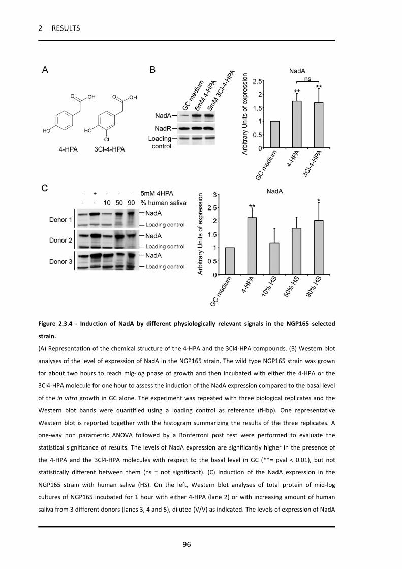
2 RESULTS
96
Figure 2.3.4 - Induction of NadA by different physiologically relevant signals in the NGP165 selected
strain.
(A) Representation of the chemical structure of the 4-HPA and the 3Cl4-HPA compounds. (B) Western blot
analyses of the level of expression of NadA in the NGP165 strain. The wild type NGP165 strain was grown
for about two hours to reach mig-log phase of growth and then incubated with either the 4-HPA or the
3Cl4-HPA molecule for one hour to assess the induction of the NadA expression compared to the basal level
of the in vitro growth in GC alone. The experiment was repeated with three biological replicates and the
Western blot bands were quantified using a loading control as reference (fHbp). One representative
Western blot is reported together with the histogram summarizing the results of the three replicates. A
one-way non parametric ANOVA followed by a Bonferroni post test were performed to evaluate the
statistical significance of results. The levels of NadA expression are significantly higher in the presence of
the 4-HPA and the 3Cl4-HPA molecules with respect to the basal level in GC (**= pval < 0.01), but not
statistically different between them (ns = not significant). (C) Induction of the NadA expression in the
NGP165 strain with human saliva (HS). On the left, Western blot analyses of total protein of mid-log
cultures of NGP165 incubated for 1 hour with either 4-HPA (lane 2) or with increasing amount of human
saliva from 3 different donors (lanes 3, 4 and 5), diluted (V/V) as indicated. The levels of expression of NadA

2 RESULTS
97
are shown. On the right, histogram reporting the average values of NadA expression from 5 independent
experiments, after quantification of the band signals. Western blot bands were normalized for a non-
specific band, indicated as loading control on the right panel, in order to avoid non 4-HPA- or NadR-
dependent effects on NadA expression due to possible protein degradation in saliva. A one-way non
parametric ANOVA followed by a Bonferroni post test were performed. * = pval < 0.05, ** = pval < 0.01 in
the graph, comparing all condition to the basal GC medium level. Comparable level of NadA induction to 4-
HPA and saliva were obtained with the 3Cl4-HPA molecule (data not show).
2.3.5 In NGP165, neither 4HPA nor 3Cl-4HPA have any effect on the expression of
the other major antigens of the 4CMenB
In order to be sure that the effects that we could see in NGP165 following nadA induction
with either 4HPA or 3Cl-4HPA would be only due to the increase of the amount of NadA,
we assess the effect of these molecules on NHBA and fHbp, the other two major antigens
in the 4CMenB vaccine. According to the regulon of NadR that we identified, these two
molecules are not regulated by this repressor. However a NadR-independent, HPA-
dependent effect on their expression cannot be excluded.
Figure 2.3.5 demonstrates that the HPA molecules do not alter the expression of either
NHBA or fHbp.

2 RESULTS
98
Figure 2.3.5 – Expression of NHBA and fHbp in NGP165 in response to nadA inducers
(A) Representative Western blot analyses of the level of expression of NHBA and fHbp in the NGP165 strain,
in response to 4HPA and 3Cl-4HPA, as indicated. The constitutively expressed RNA chaperon Hfq has been
used as a loading control. The experiment was repeated three times with independent biological replicas.
(B) Histogram reporting the average values of NHBA and fHbp expression from 3 independent experiments,
after quantification of the band signals. Western blot bands were normalized on Hfq. Standard deviations
are reported. A one-way non parametric ANOVA followed by a Bonferroni post test were performed, but no
statistically relevant differences were seen neither for NHBA nor fHbp in different conditions.
2.3.6 hSBA and MATS performed with 3Cl4-HPA predict 4CMenB vaccine coverage
of the NGP165 strain
We performed the hSBA and the MATS assays on the NGP165 wild type and the nadR
knock out mutant grown in the absence or presence of 3Cl4-HPA (Table 2.3.5). The
NGP165 wild type strain, expressing low levels of NadA in vitro (Figure 2.3.4B), has a NadA
MATS RP = 0.005, below the PBT for NadA (0.009). Results of the hSBA demonstrated that
NGP165 was resistant to killing by pooled sera from infants who received 4 doses of
4CMenB. When grown in the presence of 3Cl4-HPA, the MATS RP of NGP165 increased to

2 RESULTS
99
0.028 (Table 2.3.5) and the strain was rendered susceptible in the hSBA using the same
infants’ sera. Bactericidal titers increased from 4 with pre-immune sera to 128 with
immunized sera (Table 2.3.5). As seen for other strains tested, a more pronounced
increase in NadA expression was seen in the nadR- mutant, in which the nadA gene is fully
de-repressed (NadA MATS RP = 0.503). This situation correlates with positive bactericidal
titers of >256 in hSBA.
In conclusion, using a modified in vitro growth protocol (with HPA supplementation) that
we consider more accurately reflects the level of NadA expression that occurs in vivo, the
MATS and hSBA assays predicted that NGP165 would be efficiently killed during infection
by anti-NadA antibodies in sera of subjects immunized with the 4CMenB vaccine.
Table 2.3.5 - hSBA and MATS of the NGP165 strain performed with the 3Cl4-HPA
molecule
Strain Inducer NadA MATS RP
hSBA study3 Infant
routine 4CMenB post 4 NGP165 none 0.005 2 4 NGP165 3Cl4-HPA 0.028 4 128
NGP165 nadR- none 0.503 2 >256 Grey boxes associate with either MATS RP below the PBT (which is 0.009 for NadA) or no killing in the hSBA. Green boxes associate with either MATS RP above the PBT or killing in the hSBA. Strains were considered killed if pooled sera from infants who received three immunizations plus one booster of 4CMenB achieved an SBA titer ≥8.
2 RESULTS
100
2.3.7 Sera from 4CMenB-immunized infants protect infant rats from infection with
strain NGP165.
To determine whether NGP165 would be killed in vivo by anti-NadA antibodies, we
performed a passive protection assay in the infant rat model [263]. Figure 2.3.6 reports
the results of these experiments. Groups of infant rats were inoculated i.p. with an
infectious dose (105 CFU) of NGP165 after being treated with control serum, or pre- or
post-immune sera from mice and infants immunized with either NadA or the 4CMenB.
Administration of pre-immune sera from either mice or human infants had no effect on
the ability of NGP165 to infect infant rats: 105 CFU of NGP165 led to sustained infection in
all but one of the 19 animals tested. Sera from mice or human infants immunized with the
4CMenB vaccine formulation conferred protection on the infant rats as well (15 out of 16
and 4 out of 4 rats respectively protected in two experiments). The same human sera
results in protection of infant rats from infection with a NGP165 NHBA KO strain (4 out of
5 rats), demonstrating that killing of the strain is not due to anti-NHBA antibodies present
in the sera. Taken together these data suggest that in this in vivo model NadA is
expressed to a sufficient level to be recognized by specific anti-NadA antibodies elicited
by NadA in the 4CMenB vaccine and to mediate killing of the bacterium.
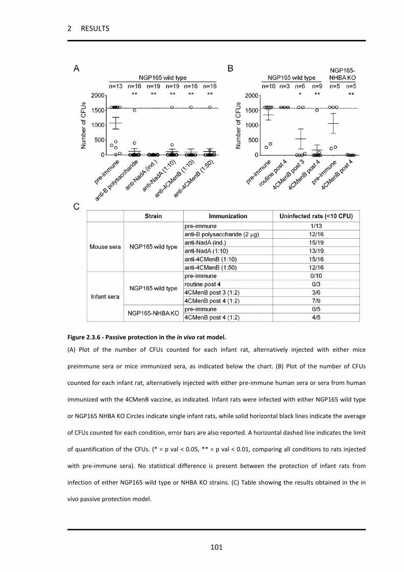
2 RESULTS
101
Figure 2.3.6 - Passive protection in the in vivo rat model.
(A) Plot of the number of CFUs counted for each infant rat, alternatively injected with either mice
preimmune sera or mice immunized sera, as indicated below the chart. (B) Plot of the number of CFUs
counted for each infant rat, alternatively injected with either pre-immune human sera or sera from human
immunized with the 4CMenB vaccine, as indicated. Infant rats were infected with either NGP165 wild type
or NGP165 NHBA KO Circles indicate single infant rats, while solid horizontal black lines indicate the average
of CFUs counted for each condition, error bars are also reported. A horizontal dashed line indicates the limit
of quantification of the CFUs. (* = p val < 0.05, ** = p val < 0.01, comparing all conditions to rats injected
with pre-immune sera). No statistical difference is present between the protection of infant rats from
infection of either NGP165 wild type or NHBA KO strains. (C) Table showing the results obtained in the in
vivo passive protection model.

2 RESULTS
102
2.3.8 The promoter of nadA is activated in vivo during infection of the infant rat model
In order to directly evaluate the expression/induction of nadA during infection, we
generated reporter strains carrying the promoterless luciferase operon (negative control)
or carrying the operon under the control of the nadA promoter (PnadA-lux). The
bioluminescence of the resulting strains was evaluated in in vitro experiments.
Interestingly, the PnadA-lux strain is significantly less bioluminescent than the negative
control (data not shown), indicating that the nadA promoter is efficiently repressed under
in vitro conditions and as expected 4HPA-specific induction of PnadA-lux (10-fold) and the
derepression of PnadA-lux in the NadR KO background (385-fold) was observed (Figure
2.3.7A). The infective dose of 104 CFU of the negative control and the PnadA-lux reporter
strains were used to infect groups of 5 infant rats and images of ventral views of
intraperitoneally infected rats were collected either immediately or 3 and 24 hours after
infection. Immediately after infection, both the negative control and the PnadA reporter
strains are poorly bioluminescent (less than 1.5-fold change from the background) (dark
grey and white circles, respectively in Figure 2.3.7B), however three hours post infection,
the PnadA-lux reporter strain is significantly induced (6-fold on average) while the
negative control maintains low levels of bioluminescence (Figure 2.3.7B-C). It is worth
noting that in Pnad-luxA infected rats a widespread bioluminescence is observed over the
entire rat indicative of bioluminescence from bacteria in a systemic infection. 24 hours
after infection the bioluminescent signals from both the negative control and the reporter
strains are almost indistinguishable from the background (Figure 2.3.7B). These data
produced in the infant rat model of septicemia, indicate that the nadA promoter is

2 RESULTS
103
induced during infection in vivo, suggesting that NadA is expressed during invasive
disease.

2 RESULTS
104
Figure 2.3.7 - Direct visualization of PnadA expression in the in vivo infant rat model.
(A) Images of serial dilution of liquid cultures of wild type and relative NadR KO strains carrying either the
promoterless lux operon (negative control) or the PnadA-lux reporter, grown in absence or presence of
4HPA, were taken with an IVIS 100 system. Bioluminescence values were obtained after analyzing the
images with the Living Image 3.1 software. The basal level of bioluminescence of these strains was obtained
by setting to one the bioluminescence of wild type strains observed during growth in GC medium solely.
The bioluminescence increment from this basal level was calculated for relative NadR KO strains and during
growth in GC in presence of 4HPA and reported in the graph, as indicated (white columns represent the
negative control and black columns represent the PnadA-lux reporter). (B) Histogram representing the
bioluminescence increment from the background (infant rats infected with the 2996 wild type strain) of
groups of 5 infant rats infected with 104 CFU of the 2996 strain carrying an integrated copy of either the
promoterless luxCDABE operon (negative control, dark grey circles) or the 2996 nadA-lux reporter strain
(white circles) as well as median values of bioluminescence increment of each group (solid horizontal red
lines) at time points indicated. T-test analyses were performed to assess statistical significant differences
between the two groups of infant rats at each time point (* = pval <0.05, *** = pval <0.001, ns = not
significant). (C) Panel of ventral views of groups of 5 infant rats infected with 104 CFU of the negative
control or the PnadA-lux reporter, as indicated, taken 3 hours after infection. Red boxes indicate the regions
of interest (ROI) that were taken into consideration by the Live Imaging software to quantify bioluminescent
values. Blood samples were recovered 24 hours after infection and CFU counts confirm that the bacterial
load within infant rats infected with different strains was similar.

3 DISCUSSION
105
3 DISCUSSION
3.1 In the NadR regulon, adhesins and diverse meningococcal functions
are regulated in response to signals in human saliva
In this first part of the thesis we dissect the role of NadR, a member of the MarR family of
transcriptional regulators, in N. meningitidis and define its regulon. We show that NadR
co-ordinately regulates many genes that all respond to the small signalling molecule
4HPA, a metabolite of amino acid catabolism that is secreted in human saliva. Of the
genes with an altered expression profile, NadR had the greatest effect on the nadA gene
which exhibited >60 fold derepression in the NadR mutant. All other genes in the NadR
regulon display much lower expression rate changes, in accordance with the previously
described NadR targets [90].
Interestingly in addition to the NadA adhesin, NadR represses 2 mafA (multiple adhesin
family A) loci of MC58 (NMB0375 and NMB0652), encoding putative adhesins. We have
demonstrated that NadR controls the expression of NadA and the MafA1/A2 adhesins in
opposing ways in response to the 4HPA inducer molecule. While 4HPA induces NadA
expression, it results in co-repression of the MafA1 and MafA2 adhesins in a panel of
meningococcal strains. Interestingly, human saliva has the same differential effect as
4HPA on NadA and MafA expression, suggesting that in vitro 4HPA could mimic
physiologically relevant signaling molecules in vivo. The presence of differential
regulatory responses mediated by one unique regulatory protein suggests that the roles
of the NadA and MafA adhesins are mutually exclusive. This regulatory response may
indicate an important molecular switch enabling adaptation of meningococcus to

3 DISCUSSION
106
changing niches: as a consequence of the external changing niche-specific signals, a
dedicated group of adhesins (i.e. NadA or MafA) relevant for the colonization of that
niche will be expressed.
Moreover, the NadR-repressed glycolytic enzyme encoded by the gapA-1 gene was
recently shown to play a role in adhesion in meniningococcus [264] and its homologues in
many other systems are well known to function as a surface exposed adhesin [265-270].
Therefore, Gap-A1 may represent a third adhesin under the control of NadR that will be
co-expressed with NadA in response to niche signals.
We have demonstrated that the nadA and other 4HPA-induced promoters (type I)
comprise multiple NadR binding sites while the mafA1/A2 promoters (Class II) consist of
only one binding site for NadR, centred downstream of the promoter sequence. In
addition, 4HPA alters the DNA binding activity of NadR at only type I promoters in vitro,
without affecting the NadR binding at type II. Therefore, both the architecture of the
promoter and the nature of the NadR operators at the promoter may direct the response
that NadR will effect to the same signal.
We propose a model (Figure 3.1.1) in which the apo-NadR can bind to and repress nadA
and other 4HPA-induced genes (type I) through a looping mechanism, which may result in
steric hindrance of RNA polymerase access to the promoter, possibly through dimer-
dimer interactions on multiple binding sites. This model has been previously proposed for
both the gonococcal farAB promoter [138] and the meningococcal nadA promoter [141]
and is supported by the binding at these promoters of the Integration Host Factor (IHF), a
histone-like protein which was demonstrated to bend DNA upon binding [271] and could
facilitate the interaction between NadR dimers bound at different operators. Following
3 DISCUSSION
107
the binding of 4HPA or other biologically relevant signals (Figure 3.1.1, lower panel), the
NadR protein can be stabilized in a conformational state which is not able to efficiently
bind to type I operators, thus causing the induction of type I genes.
On the other hand the mafA1 promoter (type II) could be less efficiently repressed by the
apo-NadR. However, the 4HPA bound-NadR can still bind at type II binding sites resulting
in a more effective repression of type II genes in vivo. The interaction of a small molecule
with a MarR regulator is normally associated with disruption of DNA binding, however
ligand-binding can enhances the DNA binding activity of MarR homologues [129]. In the
case of NadR on type II promoters, the 4HPA molecule does not directly increase the
affinity of the protein for its binding site (figure 2.1.7C). Therefore, we suggest that the
4HPA mediated co-repression of type II genes is the result of either a less efficient
promoter clearance due to occupancy of 4HPA bound-NadR at the type II operator or
different binding kinetics at type II operators between the apo- and the 4HPA bound-
NadR forms.
All these considerations suggest that, rather than different signals, it seems to be the
different nature of operators and promoters to decide the kind or regulation performed
by the NadR protein on its different targets.

3 DISCUSSION
108
Figure 3.1.1 - Model of NadR regulation on type I and type II gene promoters.
The promoters of nadA and mafA1 are schematically illustrated and represent the type I and type II
promoters, respectively. The top panels show the different NadR mediated repression of its different
targets. The lower panels show the activity of 4HPA on NadR binding at type I versus type II promoters.
In addition to regulating adhesins, NadR also co-ordinates many other cellular functions in
response to 4HPA or similar signals present in saliva, controlling a generalized adaptation
of the meningococcus to the host niche colonization. Interestingly, it has been
demonstrated that maltodextrins produced in human saliva by α-amylase are crucial for
group A streptococcus to successfully infect the oropharynx [272, 273], suggesting that
signals present in saliva, other than 4HPA, could be used by bacteria to modulate the
gene expression and adapt to the host niche.
NadR is able to repress the expression of various genes implicated in transport of a range
of known and unknown substrates, suggesting that it is involved in controlling the
substrate-uptake machinery of meningococcus during infection. In addition it also

3 DISCUSSION
109
regulates genes involved in energy metabolic pathways, supporting the idea that it can re-
route the metabolism of the cell in response to niche signals. In particular, the ability of
NadR of inducing the pathways for amino acid catabolism, such as proline and glutamate
utilization, in response to the presence of 4HPA, which is itself an amino acid metabolite,
suggests that the environmental availability of amino acid metabolites signals to the cell
that amino acids may be abundantly available and NadR adapts the cell metabolism,
accordingly. Furthermore, NadR links into other regulatory circuits within the cell by
affecting expression levels of transcriptional or post-transcriptional regulators, thus
indirectly regulating other targets.
In identifying the NadR regulon and the signals to which it responds we have gained
insights into a co-ordinated response of the meningococcus which may be relevant during
colonization of the oropharynx. While NadR in meningococcus has evolved away from
regulating fatty acid resistance, it coordinates a number of seemingly unrelated functions
within the cell in response to a niche-specific small molecule signal. It is tempting to
speculate that during colonization of the oropharynx, NadR controls the adhesion
properties of the cell, while also adapting metabolism and other physiologically relevant
processes to the competitive host niche.

3 DISCUSSION
110
3.2 Structural insight into the mechanism of DNA-binding attenuation of
NadR by the small natural ligand 4HPA
After elucidating the global role of NadR, in the second part of the thesis we explored the
mechanism by which the 4HPA molecule regulates the NadR DNA-binding activity, on
nadA, on which, as stated above, NadR had the greatest effect among its target genes.
We found that the 4-HPA ligand binds in a pocket located at the junction between the
dimerization interface and the DNA-binding lobe. This ligand binding site is mainly formed
by structural elements of the two monomers including helix α1 of the first subunit and
helices α1 and α5 of the second subunit. Although the number of salicylate sites on MarR
molecules is still controversial, superimposition of the NadR/4-HPA complex with
available representative MarR/salicylate structures shows that 4-HPA shares a salicylate
binding region common to all available models [130-132]. The position of the ligand
binding pocket appears therefore well conserved among MarR family members. However
the ligand binding site of NadR displays significant variations in amino-acid composition
compared to other representative MarR homologues, which suggests differences in ligand
specificity. Indeed, NadR does not respond to the broad specific effector salicylate in vitro
[141] and is unable to associate with the ligand even at high concentrations (our
unpublished results). These observations suggest that members of this family have
probably evolved separately to respond to distinct signaling molecules thus enabling
bacteria to adapt and respond to changing environmental conditions within their natural
niches.
The presence of this conserved ligand binding pocket has led to the hypothesis of a
shared mechanism of regulation among members of the MarR family. For proteins that

3 DISCUSSION
111
appear to be preconfigured for DNA binding in their apo configuration, the binding of
ligands produces a large conformational change that is transmitted to the DNA binding
lobes [133]. The distance between the two recognition helices that is essential for
association with two consecutive DNA major grooves is altered in the complex thus
precluding DNA binding.
The formation of the 4-HPA/NadR complex results in an overall accessibility of the protein
similar to the one observed in the absence of the ligand, indicating that 4-HPA likely locks
NadR in a conformation incompatible with DNA binding without inducing large
conformational changes in the DNA-binding lobes. The binding of 4-HPA induces minor
conformational changes in the DNA-binding lobes. This is supported by our mutagenesis
experiments, in which the substitution of Tyr115 in the ligand binding site alters the
dynamics of helix α3 (as reported in [8]) suggesting a cross-talk between elements of the
HPA- and the DNA-binding domains. Moreover, the presence of the ligand seems to be
not only transmitted to the DNA-binding domain but also to the dimerization interface.
Indeed, in the Y115A NadR mutant, 4-HPA accelerates the dimer dissociation rate (data
not shown). It is well established that members of the MarR family possess an intrinsic
conformational flexibility at the dimerization interface that is exploited on binding of
either cognate DNA or ligand to modify the position of the DNA-binding domains [124,
125]. Based on these observations, it is tempting to speculate that 4-HPA makes use of
the structural malleability of the dimerization interface to alter the position of the DNA-
binding lobes. It seems plausible that 4-HPA reduces the flexibility of the dimerization
interface to prevent the motions of the DNA-binding lobes required for interaction with

3 DISCUSSION
112
DNA. On the basis of this model, the binding of the ligand may lock the DNA-binding lobes
in place without affecting their global solvent accessibility.
Finally, our mutagenesis data reveal that a class of mutation represented by Tyr115
(including His7, Ser9 and Phe25 mutants) plays a key role in the mechanism of regulation of
NadR by 4-HPA. 4HPA can still bind to NadR Y115A, as reported in [8], suggesting that the
substitution of this single residue is not sufficient to destroy the ligand binding pocket.
However, the Y115A substitution compromises the 4-HPA-mediated stabilization of the
complex and prevents DNA dissociation. Taken together, these data suggest that 4-HPA
makes use of Tyr115 to lock the dimer in a conformation incompatible with DNA binding.
Other residues can be involved in the same process and we demonstrated that the
substitution of other residues (His7, Ser9 and Phe25) leads to a phenotype similar to the
one obtained with NadR Y115A, regarding NadA repression and 4HPA response in vivo,
suggesting that these amino acids can cooperate with the Tyr115.
Taking all these observation into consideration we propose the following model for the 4-
HPA-mediated regulation of NadR (Figure 3.2.1). NadR, as other MaR regulators, has an
intrinsic conformational flexibility and can pass from a configuration incompatible with
DNA binding to a form able to bind DNA, spontaneously and in a reversible way. The
binding of 4-HPA in its binding pocket may lock the position of the DNA-binding lobes in
place by acting as a “glue” between the dimerization and the DNA-binding domains, thus
preventing the cross-talking between the different domains of NadR which is essential to
induce the conformational changes required for DNA interaction. The Tyr115 plays a key
role in this cross-talking mechanism, by being involved in the transduction of the 4HPA
binding to the responsive effects in the dimerization domain and therefore in the DNA-

3 DISCUSSION
113
binding lobes. The substitution of the Tyr115 causes, indeed, the stacking of NadR onto the
DNA and the inability to respond to the 4HPA inducer. The model proposed is based on
the comparison of our data with available structural models of regulation of MarR
homologs. The resolution of the crystal structure of NadR in its free and ligand-bound
state would greatly facilitate our understanding of the mechanism of regulation of NadR
by 4-HPA.
Figure 3.2.1 - Model of NadR DNA binding activity regulation.
The 4-HPA-mediated regulation of NadR on nadA is schematized here and fully described in the discussion.
In the NadR dimer schematic model, blue ellipses, yellow circles and green ellipses represent the
dimerization domain, the ligand binding pocket and the DNA binding lobes, respectively. The altered ligand
pocket in the mutated form Y115A of NadR is represented by orange circles. The small molecule ligand is
represented by a red dot.

3 DISCUSSION
114
We have demonstrated in the first part of the thesis that NadR regulates a regulon of
genes in response to 4-HPA. The MafA1 adhesins are differentially regulated by NadR in
response to 4HPA with respect to nadA. It is tempting to speculate that, while the
conformational modification mediated by 4HPA on NadR is the same as we propose in
our model generated on nadA, the regulatory outcome may differ due to differential
promoter architectures and DNA binding sequences. In fact we demonstrated that,
differentially from nadA, the Tyr115 mutant has no effect on MafA1 protein level
expression, suggesting that the hyper-repressive activity of Y115A on the type I promoter
of nadA comprising multiple NadR binding sites is influenced by promoter architecture
and possibly by an altered tetramerization capacity of the mutant.
3.3 Transcriptional Regulation of the nadA Gene Impacts on the
Prediction of Coverage of the 4CMenB Vaccine
In the third part of this work, we evaluated the role of NadR regulation on nadA
expression during infectious disease and therefore the impact on vaccine coverage
prediction.
In the absence of an efficacious broadly protective vaccine, MenB is the leading cause of
bacterial meningitis and septicemia in many industrialized countries. A novel
multicomponent vaccine, 4CMenB, is able to induce bactericidal antibodies against strains
expressing vaccine antigens, but because MenB clinical isolates are diverse, it is necessary
to evaluate the potential of the vaccine to kill circulating strains and therefore the
potential public health impact of this vaccine. The MATS assay, which assesses the
relative contributions of the 4 major components present in the 4CMenB vaccine,

3 DISCUSSION
115
predicts whether a given isolate can be killed or not [274]. An evaluation using this assay
of more than 1,000 MenB strains from 5 EU countries predicted that 73% to 87% would
be covered by 4CMenB. However, the relative contribution of NadA to this combined
coverage prediction appers to be low (less than 2%) [246].
The role of NadA in eliciting bactericidal antibodies protecting against circulating strains,
has been unclear due to NadR-mediated repression of NadA expression under the in vitro
growth conditions used for MATS and hSBA assays, which are performed in the early
phase of growth, when NadR represses maximally nadA expression [141]. For this reason
it is reasonable to anticipate that nadA expression in vitro could be different than the
level reached in the host. Litt and colleagues [205] previously showed the presence of
antibodies that recognized recombinant NadA in children convalescing after
meningococcal disease. Interestingly, we report here that sera from children infected by
isolates that failed to express NadA in culture (MATS RP = 0), or express low NadA levels
(MATs RP value < PBT) are able to recognize NadA significantly more than sera from
subjects infected by nadA- strains (Figure 2.3.1). Taken together, these observations
suggest that despite the low levels of NadA expression in vitro, all these strains express
NadA in an immunogenic form in the setting of invasive disease. Furthermore, anti-NadA
antibodies are also found in healthy individuals with levels tending to increase with age
[275], suggesting that NadA is expressed in vivo, at a level sufficient to drive the immune
response. NadA expression is also induced in the ex vivo model of human saliva (Figures
2.1.6 and 2.3.4), suggesting again high expression in the niche of meningococcal
colonization, which can be mimicked in vitro by addition of 4-HPA or 3Cl4-HPA
representing natural inducers of NadA present in the host.

3 DISCUSSION
116
Using the recombinant nadR- strains and the HPA inducers as a model for in vivo
expression, we have validated NadA as a potent immunogen in all ages and a valid target
for protective responses. Once repression mediated by NadR is relieved and NadA is
expressed at high levels, strains normally resistant to killing by 4CMenB immune sera are
rendered highly susceptible to killing in SBA. The molecular mechanism of both NadR
repression of nadA and induction mediated by HPA compounds is conserved in a wide
panel of N. meningitidis strains belonging to different clonal complexes (Figures 2.1.5 and
2.3.2). These observations suggest that any strain carrying nadA could potentially be
targeted by bactericidal antibody elicited by the 4CMenB vaccine when NadR repression
is relieved during infection.
The ability of passively administered vaccinees’ sera to protect mice from infection of
NGP165, and to return protective results in the modified MATS and hSBA assays, when
NGP165 is grown in vitro with added HPA, suggests that this treatment mimics the in vivo
status of NGP165 with respect to NadA expression. It is noteworthy that passively
administered sera from mice immunized with NadA recombinant protein alone protect
infant rats from infection with NGP165, demonstrating that the level of expression
reached by NadA alone in vivo is sufficient to promote bacterial killing. Finally, a
bioluminescent nadA-lux fusion demonstrates the induction of the nadA promoter three
hours after infection in the infant rat model, demonstrating the expression of nadA
during bacterimia in vivo.
The expression of NadA in vivo in the host is in line with its putative role during
meningococcal infection. Its adhesive role is important in the course of colonization of the
human upper respiratory tract. Following the passage across the epithelium,

3 DISCUSSION
117
meningococcus invades tissues and blood, where NadA can interact with human blood
leukocytes and lead to enhanced immune stimulation [199, 201, 276, 277]. For these
reasons, NadA is to be considered an important meningococcal virulence factor, involved
in progressively invasive steps of infection. In accordance with this statement we
demonstrated here that different signals are able to modulate the NadR repressive
activity on NadA (Figure 2.3.3). Signals present in saliva, which we can mimic by using HPA
molecules at high concentration during in vitro growth, may mediate NadA induction
during the colonization of the oro-pharynx. However, because NadA could be required
during other steps of the pathogenesis, multiple signals in niches other than the pharynx
could modulate NadA expression as soon as the bacterium passes the epithelial barrier.
We demonstrate indeed in the infant rat model of bacteremia that NadA is expressed
widely three hours post infection and this up-regulation could be due to molecules such
as the 3Cl-4HPA and NO2-4HPA, which are produced by leukocytes during inflammatory
processes [262, 278]. Furthermore, we cannot exclude that other molecules can act on
NadR by increasing its repressive action on its targets.
From our data, current methods used to predict coverage of strains by the 4CMenB
vaccine are underestimating the contribution of the NadA antigen, suggesting that all
nadA+ meningococcal strains may be susceptible to bactericidal anti-NadA antibodies
elicited by vaccination. A more accurate prediction may be obtained by addition of
physiologically relevant inducers to in vitro grown bacteria, resulting in similar NadA
expression levels to those measured in ex vivo and in vivo models of infection. This study
provides new insights into the expression of the NadA antigen during infection, which are
3 DISCUSSION
118
fundamental for the implementation of prophylactic strategies such as vaccines and
evaluation of their impact on public health.

4 MATERIALS AND METHODS
119
4 MATERIALS AND METHODS
4.1 Bacterial strains and culture conditions
The N. meningitidis strains used in this study include the wild type MC58, 5/99, 961-5945,
LNP17094, B3937, M10574, M14933, NGP165 and 2996 strains, derivatives of these wild
type strains and the previously reported clinical isolates NM036, NM037, NM066, NM067,
NM069, NM081, NM088, NM100, NM119, NM145, NM154, NM156, NM188 and NM191
[205]. All strains used are reported in Appendix Table 1.
N. meningitidis strains were routinely cultured, stocked and transformed as previously
described [279]. They were grown on GC (Difco) agar medium plates supplemented with
Kellogg's supplement I at 37°C/5% CO2 overnight (O/N). For liquid cultures, after O/N
grown on solid medium, few colonies were inoculated in 7 ml GC broth supplemented
with Kellogg's supplement I to an initial optical density at 600 nm (OD600) of 0.05.
When required erythromycin and/or chloramphenicol antibiotics were added to culture
media at the final concentration of 5 µg/ml and 20 µg/ml, respectively, isopropyl β-D-1-
thiogalactopyranoside (IPTG) were added to achieve final concentrations of 1 mM. 2-, 3-,
or 4-Hydroxyphenylacetic acid (2,3, or 4-HPA), 3-Chloro-, 3-Fluoro- or 3-Bromo-4-
Hydroxyphenylacetic acid (3Cl4-HPA, 3Fl4-HPA, or 3Br4-HPA), 2,4- or 3,4-
Dihydroxyphenylacetic acid (2,4-HPA, or 3,4-HPA) in aqueous solution was added to
culture media to achieve final concentrations of 1, 2 or 5 mM. All these compounds were
obtained from Sigma-Aldrich with exception of the 3Br4-HPA, which was purchased from
Chemsigma.

4 MATERIALS AND METHODS
120
E. coli DH5-α [280] and BL21(DE3) [281] cultures were grown in Luria–Bertani (LB)
medium, and when required, ampicillin, chloramphenicol and/or IIPTG were added to
achieve final concentrations of 100, 20 µg/ml and 1 mM, respectively.
4.2 Construction of mutant and complementing strains
DNA manipulations were carried out routinely as described for standard laboratory
methods [282]. Total lysates from single colonies of all transformants were used as template
for diagnostic PCR analysis to confirm the correct insertion by a double homologous
recombination event. The nadA promoter was amplified and sequenced in each transformant
to ensure that the same numbers of repeats were present as in the derivative strain. The
NGP165 NHBA null mutant was generated as described previously [182].
4.2.1 Generation of NadR null mutant and NadR complementing strains
The NadR mutants of the MC58, 5/99 and 961-5945 strains were previously reported
[141] and indicated as MC-∆1843, 5/99-∆1843, 961-∆1843, respectively. The NadR
mutants of the LNP17094, B3937, M10574 and M14933 strains were generated by
transformation of the wildtype strains with p∆1843ko::Cm [141] and selection on
chloramphenicol, leading to the generation of LNP-∆1843, B39-∆1843, M10-∆1843 and
M14-∆1843 strains, respectively.
For complementation of the MC-∆1843 NadR null mutant [141], the nadR gene under the
control of the Ptac promoter was reinserted into the chromosome of MC-∆1843 strain
between the converging open reading frames (ORFs) NMB1428 and NMB1429, by
transformation with pComEryPind-nadR. pCompEryPind-nadR is a derivative plasmid of

4 MATERIALS AND METHODS
121
the pSLComCmr [118], in which the nadR gene was amplified from the MC58 genome
with the primer pair 1843-F and 1843-R2 and cloned as a 441-bp NdeI/NsiI fragment
downstream of the Ptac promoter. The chloramphenicol resistance cassette of
pSLComCmr was substituted with an erythromycin resistance cassette, amplified with
primers EryXbaF and EryBamR and cloned into the XbaI-BamHI sites (Appendix Table 3),
generating pComEryPind-nadR. This plasmid was transformed into MC-∆1843 for the
generation of the ∆NadR_C complemented mutant strain and transformants were
selected on erythromycin. The nadR gene in the complemented strains was expressed in an
Isopropyl β-D-1-thiogalactopyranoside (IPTG) inducible manner. A final concentration of 1
mM IPTG resulted in NadR expression levels similar to that of MC58 wild type strain.
4.2.2 Generation of MC58 strains expressing NadR mutated proteins
In order to generate MC58 mutant strains expressing only amino-acid substituted forms
of NadR, plasmids containing the sequence of nadR alternatively mutated in the codons
coding for His7, Ser9, Asn11, Arg18, Phe25, Trp39, Arg40 and Tyr115 were constructed using the
QuikChange II XL Site-Directed Mutagenesis Kit (Stratagene). Briefly, the nadR gene was
mutated in the pComEry-1843 plasmid using 8 couples of mutagenic primers (H7A-F/H7A-
R, S9A-F/S9A-R, N11A-F/N11A-R, R18A-F/R18A-R, F25A-F/F25A-R W39A-F/W39A-R, R40A-
F/R40A-R and Y115A-F/Y115A-R (Appendix Table 3)). The resulting plasmids were named
pComEry-1843H7A, -1843S9A, -1843N11A, -1843R18A, -1843F25A, -1843W39A, -
1843R40A and -1843Y115A, and contain a site-directed mutant allele of the nadR gene, in
which the selected codons were respectively substituted by a GCG alanine codon, and
were used for transformation of the MC-∆1843 strain. The resulting transformed strains

4 MATERIALS AND METHODS
122
were named MC-∆1843_H7A, _S9A, _N11A, _CR18A, _F25A, _CW39A, _CR40A and
_CY115A, respectively. The nadR wild type or mutated genes in the complemented strains
were expressed in an Isopropyl β-D-1-thiogalactopyranoside (IPTG) inducible manner. A
final concentration of 1 mM IPTG resulted in NadR expression levels similar to that of
MC58 wild type strain.
4.2.3 Generation of lux reporter strains
To generate bacterial luciferase transcriptional fusions of the promoters under study at a
chromosomal location between the two converging ORFs NMB1074 and NMB1075,
flanked on both sides with transcriptional terminators, plasmid pSL-LuxFla for allelic
exchange in N. meningitidis strains was constructed. Briefly, the promoterless luxCDABE
operon and cat cassette were subcloned from pSB1075 [283] into pBleuskript II [9] as an
EcoRI-BamHI fragment and then cloned as a 6.5 kb XhoI-BamHI fragment into pSL-furlacZ
[94] replacing 4.7kb containing an erytromycin cassette, and fur-lacZ fusion, generating
pSL-lux-Fla. The nadA promoter was amplified with primers NadluxF2/NadluxR2 and
cloned as a 250 bp XhoI-KpnI fragment upstream of the luxCDABE operon generating pSL-
PnadA-lux. The pSL-lux-Fla and pSL-PnadA-lux plasmids were used for transformation of
the MC58, and MC58-∆1843 strains generating MC58-lux, MC58-PnadA-lux, and MC58-
∆1843-lux and MC58-∆1843-PnadA-lux, respectively, for the in vitro reporter analyses,
and the 2996 strain generating 2996-lux and 2996-PnadA-lux, respectively, for the in vivo
reporter analysis.

4 MATERIALS AND METHODS
123
4.3 Western blot analysis
N. meningitidis colonies from freshly grown overnight plate cultures were resuspended in
GC medium to an optical density at 600 nm (OD600) of 0.05 and grown at 37°C to
logarithmic phase (OD600 of 0.5, ca. 2 hours incubation). Samples of 1 ml were harvested
and resuspended in 1x SDS-PAGE loading buffer (50 mM Tris Cl pH 6.8, 2.5 % SDS, 0.1 %
Bromophenol Blue, 10% glycerol, 5% beta-mercaptoethanol, 50 mM DTT), normalizing
the concentration to a relative OD600 of 5.
To prepare protein extracts from bacteria incubated with either different small molecules
or human saliva, N. meningitidis colonies from overnight GC plate cultures were
resuspended in GC to an OD600 of 0.05. Liquid cultures were grown until mid log phase
(OD600 = 0.4), harvested and resuspended in either GC, GC + 2mM or GC + 5mM of the
indicated molecule or 10%, 50%, 90% (V/V) human saliva in GC containing EDTA-free
Protease Inhibitors Cocktail (Roche). Bacteria were then incubated for 1 hour with
agitation at 37°C. 1 ml of each liquid culture was harvested and resuspended in 100 ml of
SDS-PAGE loading buffer. When required the growth was followed by 1 hour induction at
37°C with 1 mM IPTG.
For Western blot analysis, 10 µg of each total protein sample in 1x SDS-PAGE loading
buffer was separated by SDS-PAGE, and transferred onto nitrocellulose membrane using
an iBlot Dry Blotting System (Invitrogen). Membranes were blocked overnight at 4° C by
agitation in blocking solution (10% skimmed milk, 0.05% Tween-20, in PBS) and incubated
for 90 min at 37°C with primary antibodies (anti-NadA [203], anti-NadR [141], anti-Maf
[254], anti-fHbp [234] and anti-Hfq [284] polyclonal sera) in 3% skim milk blocking
solution. After washing, the membranes were incubated in a 1:2000 dilution of

4 MATERIALS AND METHODS
124
peroxidase-conjugated anti-rabbit immunoglobulin (Biorad) or anti-mouse
immunoglobulin (Dako) in 3% skim milk blocking solution for 1 hour at room temperature
and the resulting signal was detected using either the Supersignal West Pico
chemiluminescent substrate (Pierce) or the Western Lightning-ECL chemiluminescent
substrate (Perkin-Elmer). Quantification of the signals from western blots bands was
performed by using a PhosphorImager and ImageQuant software (Molecular Dynamics).
4.4 Protein expression and purification
The expression and purification of the NadR recombinant protein were carried out as fully
described previously [141]. For expression and purification of NadR mutated proteins, the
sequence of nadR was mutated in the expression plasmid pET15-1843 [141] as described
above using the QuikChange II XL Site-Directed Mutagenesis Kit (Stratagene) and the
mutagenic primers pairs R18A-F/R18A-R, W39A-F/W39A-R, R40A-F/R40A-R and Y115A-
F/Y115A-R (Appendix Table 3). The resulting pET15-1843R18A, -1843W39A, -1843R40A
and -1843Y115A vectors were subsequently transformed into E. coli BL21(DE3) strain for
protein expression and purification. Fractions containing the purified proteins were
analyzed by SDS-PAGE, pooled and dialyzed overnight against 50 mM Tris-HCl pH 8.0, 300
mM NaCl using a 10-kDa molecular weight cutoff dialysis membrane (Slide-A-lyzer dialysis
cassettes, Pierce). The 6xHisTag was cleaved at room temperature using thrombin
agarose resin (Sigma Aldrich) and removed with the Ni-NTA matrix. The purity and the
identity of each protein were verified by SDS-PAGE and mass spectrometry and the
concentrations were measured using the Bradford assay. Purified proteins were
aliquoted, flash frozen in ethanol/dry ice and stored at -80°C until use.
4 MATERIALS AND METHODS
125
4.5 Electromobility shift assays (EMSA).
For gel shift experiments, a probe corresponding to the respective promoter region (150-
280 bp) of the first gene in probable operons containing NadR target genes was amplified
using primers listed in Appendix Table 3 and named with P (for promoter) and a number
according to the NMB annotation. Two probes spanning from -170 to -116 (OpI) and -9 to
+81 (OpII) with respect to the MC58 nadA promoter were amplified using Nad-N2/gpr-R
and Nad-N5/Nad-Sp primer pairs respectively, to perform experiments on single NadR
binding sites of nadA. Two pmoles of each fragment were then radioactively labeled at
their 5’ ends with 30 µCi of (γ-32P)-ATP (6000 Ci/mmol; NEN) using 10 U of T4
polynucleotide kinase (New England Biolabs) and used at the final concentration of 1.6
nM. The unincorporated radioactive nucleotides were removed using TE-10 chromaspin
columns (Clontech).
For each binding reaction, 40 fmoles of labeled probe were incubated with recombinant
NadR wild type or NadR mutant proteins, suspended in Protein Solution (5 mM Tris-HCl
pH 8, 60 mM NaCl), in 25 µl final volume of Gelshift Binding Buffer (40 mM Tris-HCl pH 8,
5 mM MgCl2, 50 mM KCl, 0.05% NP40, 10% glycerol) with 30 nM salmon sperm DNA as
non-specific competitor. for 15 min at room temperature, and run on 6% native
polyacrylamide gels buffered with 0.5 x TBE, at 100 Volts for 90-180 min at 4°C. When
indicated, 1, 2, 5 or 10 mM 4-HPA was added to 100 ng of recombinant NadR WT or NadR
mutant proteins in the binding reaction, to test the proteins responses to 4-HPA. Gels
were dried and exposed to autoradiographic films at –80°C. To assess the specificity of
binding of NadR on the DNA labeled probes, 1.6 nM, 8 nM and 40 nM (1-, 5- or 25-fold

4 MATERIALS AND METHODS
126
respect to the probe) of either salmon sperm DNA or cold DNA probes were added as
non-specific or specific competitors, respectively.
EMSA bands radioactivity was quantified using a phosphorimager and the Image Quant
software (Molecular Dynamics).
4.6 DNAse I footprint
The nadR, putA and mafA1 promoter regions were amplified with the primer couples
P1843-F2/P1843-sR2, P401-F3/P401-R, P375-F/P375-652R, respectively. The PCR products
were purified and cloned into pGEMT vector (Promega) as 258, 280 and 267 bp fragments
generating pGEMT-P1843, pGEMT-P401 and pGEMT-P375. 2 pmols of each plasmid were
5’ end-labeled by T4 polynucleotide kinase with [γ-32P]-ATP after digestion at either the
NcoI or SpeI site of the pGEMT polylinker. Following a second digestion with either SpeI
or NcoI respectively, the labeled probes were separated from the linearised vector by
polyacrylamide gel electrophoresis (PAGE) as described previously [141]. DNA-protein
binding reactions were carried out for 15 minutes at RT in footprint buffer (10 mM Tris
HCl pH 8, 50 mM NaCl, 10 mM KCl, 5 mM MgCl2 10% glycerol and 0.05% NP40) containing
40 fmol of labeled probe, 200 ng of sonicated salmon sperm as the non-specific
competitor and nM concentrations of NadR purified protein as indicated in the figures.
Samples were then treated with 0.03 U of DNase I (Roche) for 1 minute at RT. DNase I
digestions were stopped, samples were purified, loaded and run on 8M urea-6%
polyacrilamide gels as described [285]. A G+A sequence reaction was performed [286] for
each probe and run in parallel to the fooprinting reactions.

4 MATERIALS AND METHODS
127
4.7 3C mutation scanning
To perform the 3C mutation scanning of the single NadR binding sites on the nadA and
mafA1 promoters, DNA oligos corresponding to the forward and reverse strands of either
the wild type NadR-protected sequences from DNAI footprints or to mutated sequences,
in which sequential triplicate nucleotides were substituted with CCC, were ordered from
Sigma. 100 pmols of each oligonucleotide pair were annealed in annealing buffer (10 mM
Tris HCl pH 8, 50 mM NaCl and 1 mM EDTA) by using a standard thermal cycler. The mix
was heated to 95°C and left at this temperature for 5 minutes, followed by ramp cooling
to 25°C over a period of 45 minutes. The annealed oligos were stored at -20°C, 5’ end
labeled and submitted to EMSA analysis as described in the previous section.
4.8 RNA samples preparation
Bacterial cultures were grown in GC liquid medium to an OD600 of 0.5 and then added to
the same volume of frozen media to bring the temperature immediately to 4° C. Total
RNA was isolated using an RNeasy kit (Qiagen) as described by the manufacturer. RNA
pools for microarray experiments were prepared from 3 independent cultures of bacteria.
Total RNA was extracted separately from each bacterial pellet and 15 µg of each
preparation were pooled together. Three independent pools were prepared for each
condition.

4 MATERIALS AND METHODS
128
4.9 Microarray analyses
DNA microarray analysis was performed using an Agilent custom-designed
oligonucleotide array as previously described [252]. cDNA probes were prepared from
RNA pools (5 µg) obtained from MC58 wildtype and MC-∆1843 null mutant cells (see
above) and hybridized as described by Fantappiè and colleagues [252]. Three
hybridizations were performed using cDNA probes from three independent MC58 and
MC-∆1843 pools, respectively, and the corresponding dyeswap experiments (with MC58
and MC-∆1843 pools inversely labeled with the Cy3 and Cy5 fluorophores) were also
performed. Differentially expressed genes were assessed by grouping all log2 ratio of the
Cy5 and Cy3 values corresponding to each gene, within experimental replicas and spot
replicas, and comparing them against the zero value by Student’s t-test statistics (one
tail).
The array layout was submitted to the EBI ArrayExpress and it is available with the
identifier A-MEXP-1967. The entire set of supporting microarray data has been deposited
in the ArrayExpress public database under the accession number E-MTAB-803.
4.10 Quantitative Real Time PCR (qRT-PCR)
Approximately 4 µg of the total RNA preparation was treated with RQ1 RNase-free DNase
(Promega). RNA was then reverse transcribed using random hexamer primers and
Moloney murine leukemia virus reverse transcriptase (Promega) as recommended by the
manufacturer. For negative controls, all RNA samples were also incubated without
reverse transcriptase. Primer pairs for each target gene were designed and optimized and
are listed in Appendix Table 3 named with the number according to their NMB annotation

4 MATERIALS AND METHODS
129
and rt for RT-PCR. Primers 16S_F and 16S_R were used for the 16S rRNA normalization
control (Appendix Table 3). All RT-PCRs were performed in triplicate using a 25 µl mixture
containing cDNA (5 µl of a 1/5 dilution), 1x brilliant SYBR green quantitative PCR master
mixture (Stratagene), and approximately 5 pmol of each primer. Amplification and
detection of specific products were performed with an Mx3000 real-time PCR system
(Stratagene) using the following procedure: 95°C for 10 min, followed by 40 cycles of 95°C
for 30 s, 55°C for 1 min, and 72°C for 30 s and then a dissociation curve analysis. The 16S
rRNA gene was used as the endogenous reference control, and relative gene expression
was determined using the 2-∆∆Ct relative quantification method.
4.11 Human saliva samples
Human saliva was obtained from healthy non-smoking donors in the morning at least ten
hours after eating and after rinsing the mouth with water. In order to minimize the
degradation of the proteins the sample was kept on ice during the collection process.
Immediately after collection, the sample of human saliva was stored at -20°C.
4.12 Human serum samples
Healthy human volunteers were immunized under informed consent with the
experimental serogroup B vaccine containing 50 μg each of GNA2091-fHbp, NHBA-
GNA1030, and NadA and 25 μg of Outer Membrane Vesicles from the New Zealand strain
NZ98/254, adsorbed to aluminium hydroxide. Serum samples before and after
immunization were obtained from the following clinical trials:
4 MATERIALS AND METHODS
130
Study 1 was a clinical trial conducted in healthy adults, laboratory workers. Pooled sera
were derived from 23 subjects before and after 3 doses of 4CMenB at 0, 2 and 6 months.
Study 2 was a clinical study evaluating the safety, immunogenicity and lot consistency of
4CMenB administered to infants at 2, 4 and 6 months of age. Extensions of this clinical
study investigated a fourth (booster) dose at 12 months of age. Pooled sera were derived
from 107 infants at 7 months of age who received the primary series of 3 doses of routine
vaccine at 2, 4 and 6 month of age and from 141 infants who received the primary series
of 3 doses of 4CMenB at 2, 4 and 6 month of age plus a booster in the second year of life.
Study 3 was a clinical study evaluating the safety, tolerability and immunogenicity of
4CMenB administered to infants at 2, 4 and 6 months of age. Extensions of this clinical
study investigated a fourth (booster) dose at 12, 18 or 24 months of age. Pooled sera
were derived from 109 infants at 5 months of age who received the primary series of 3
doses of routine vaccine at 2, 3 and 4 month of age and from 69 infants who received the
primary series of 3 doses of 4CMenB at 2, 4 and 6 months of age plus a booster in the
second year of life.
4.13 Immunization of mice
To prepare antisera, 20 μg of individual antigens NadA, NHBA-GNA1030, GNA2091-fHbp
or a combination of 20 μg of each of NHBA-GNA1030, GNA2091-fHbp and NadA with or
without 10 μg of deoxycholate-extracted OMV derived from the strain NZ98/254 were
used to immunize 6-week-old CD1 female mice (Charles River). Five to ten mice per group
were used. The antigens were administered intraperitoneally (i.p.), together with
aluminium hydroxide (3 mg/ml) on days 0, 21 and 35.

4 MATERIALS AND METHODS
131
4.14 Serum bactericidal assay (SBA)
Serum bactericidal antibody activity against Neisseria meningitidis strains with mice
antisera was evaluated as previously described [287] with pooled baby rabbit serum used
as the complement source (rSBA). Serum bactericidal antibody assays with human
complement (hSBA) were performed as described by Borrow et al. with minor
modifications [287]. MenB bacteria were subcultured overnight on Chocolate Agar and
resuspended in Mueller Hinton medium (MHM) to an OD600 of 0.25 before use in the
assay. Serum bactericidal titers were determined as the last dilution that resulted in at
least a 50% reduction in colony forming units (CFU) relative to the number of CFU present
at the beginning of the bactericidal reaction (i.e. 50% of T0). Human plasma obtained
from volunteer donors after informed consent was selected for use as complement
source with a particular MenB strain only if it did not significantly reduce CFU of that
strain relative to T0 when added to the assay at a final concentration of 50%. The final
assay mixture contained 25% human plasma complement. When required, 4-HPA and
3Cl4-HPA in aqueous solution were added to chocolate agar at a final concentration of 5
mM.
4.15 Meningococcal Antigen Typing System (MATS) ELISA
The MATS assay was been performed as previously described [243]. Briefly, bacteria are
grown overnight on Chocolate Agar plates (Biomerieux) ± 4-HPA and 3Cl4-HPA
supplementation. Bacterial suspensions are prepared from overnight cultures, single
colonies being collected and suspended to an OD600 = 0.4, using Ultrospec 10 Classic
(Amersham Biosciences). Bacterial suspensions are lysed with a detergent (EmpigenBB

4 MATERIALS AND METHODS
132
5%, Sigma) added to 1/10 volume, followed by incubationfor 1 hour at 45°C in a shaking
water bath. Samples are added to ELISA microwell plates, which were previously coated
with polyclonal rabbit antibody against NadA (961c) to capture the antigen from the
lysate, and diluted at 2-fold step dilution. Captured antigen is then detected with a
biotin-labeled rabbit antibody (Biotin-Ab) against the antigen followed by HRP-labeled
streptavidin. The signal is provided by reduction of o-phenylenediamine substrate (OPD,
Sigma). Each plate contains 1 reference strain and 6 sample strains: 5/99 is used as the
reference strain for NadA. Relative Potency is a mathematically-determined quantity
obtained by comparing the serial dilution curves of the test sample and the reference
strain. Raw data reduction and analysis is performed by StatLIA (Brendan Technologies).
4.16 Passive protection and in vivo imaging in infant rats.
The ability of anti-NadA antibodies to confer passive protection against N. meningitidis
bacteremia was tested in infant rats challenged intraperitoneally (i.p.). In vivo imaging of
the bioluminescence of the PnadA-lux reporter and control strains was monitored in
infant rats infected i.p. The day before the challenge/infection, freshly thawed bacteria
were inoculated onto chocolate agar and were grown overnight at 37°C in 5% CO2. On the
morning of the challenge/infection, several colonies were inoculated in MHM
supplemented with 0.25% (wt/vol) glucose to a starting OD600 of∼0.05. After 1.5 h growth
at 37°C in 5% CO2, the bacterial suspension reached an OD600 of ∼0.25 and was diluted in
PBS to obtain 105-107 CFU/ml. 5–7-day-old pups from litters of out-bred Wistar rats
(Charles River) were numbered. The protection and challenge were performed as follows:
Groups of 3–19 animals were treated i.p. at time 0 with 100 µl doses of different dilutions

4 MATERIALS AND METHODS
133
of test or control antisera. Three hours later, the animals were challenged i.p. with a
100µl dose of 105 CFU of N. meningitidis strain NGP165. Eighteen hours after the bacterial
challenge, blood samples were obtained by cheek puncture with a syringe containing 25U
of heparin without preservative (American Pharmaceutical Partnersand CFUs were
measured. Rats were considered infected when >10 CFUs were counted on plates
carrying 100 µl of blood. Counts above the threshold were verified for positivity by
examining plates carrying 10- and 100-fold dilutions of blood. For in vivo imaging of
bioluminescent reporters, groups of 5 animals were inoculated i.p. at time 0 with 100 µl
doses of 104 CFU of the 2996-lux or 2996-Pnad-lux strains. Rats were then anesthetized
using a constant flow of 2.5% isoflurane mixed with oxygen. Bioluminescence
measurements of ventral views of each group of rats were taken of at time 0, 3 and 24
hours, using a an IVIS 100 system (Xenogen Corp., Alameda, CA) according to instructions
from the manufacturer. Analysis and acquisition were performed using Living Image 3.1
software (Xenogen Corp.). Quantifying was performed using the photons per second
emitted by each rat. 2 rats infected with the 2996 wild type strain under the same
conditions of acquisition were used for subtracting the background. Twenty four hours
after infection, blood samples were obtained and CFU counts measured.

5 APPENDIX
134
5 APPENDIX
5.1 Table 1 - Strains used in this study
Strains Relevant characteristics Reference N.meningitidis MC58 Clinical isolate, sequenced strain containing 9 TAAA tetranucleotide repeats in the nadA promoter, CC32 [62] 5/99 Clinical isolate containing 8 TAAA tetranucleotide repeats in the nadA promoter, CC8 NIPH1 961-5945 Clinical isolate containing 12 TAAA tetranucleotide repeats in the nadA promoter, CC8 [203] LNP17094 Clinical isolate containing 12 TAAA tetranucleotide repeats in the nadA promoter, CC32 [243] B3937 Clinical isolate containing 12 TAAA tetranucleotide repeats in the nadA promoter, CC18 [243] M10574 Clinical isolate containing 6 TAAA tetranucleotide repeats in the nadA promoter, CC32 CDC2 M14933 Clinical isolate containing 6 TAAA tetranucleotide repeats in the nadA promoter, CC32 CDC2 NGP165 Clinical isolate containing 9 TAAA tetranucleotide repeats in the nadA promoter, CC11 [243] 2996 Clinical isolate, CC8 [243] NM036 Strain carrying the nadA gene from a collection of 31 clinical isolates from children ≤ 4 years of age, convalescing after meniningococcal disease [205] NM037 Strain carrying the nadA gene from a collection of 31 clinical isolates from children ≤ 4 years of age, convalescing after meniningococcal disease [205] NM066 Strain carrying the nadA gene from a collection of 31 clinical isolates from children ≤ 4 years of age, convalescing after meniningococcal disease [205] NM067 Strain carrying the nadA gene from a collection of 31 clinical isolates from children ≤ 4 years of age, convalescing after meniningococcal disease [205] NM069 Strain carrying the nadA gene from a collection of 31 clinical isolates from children ≤ 4 years of age, convalescing after meniningococcal disease [205] NM081 Strain carrying the nadA gene from a collection of 31 clinical isolates from children ≤ 4 years of age, convalescing after meniningococcal disease [205] NM088 Strain carrying the nadA gene from a collection of 31 clinical isolates from children ≤ 4 years of age, convalescing after meniningococcal disease [205] NM100 Strain carrying the nadA gene from a collection of 31 clinical isolates from children ≤ 4 years of age, convalescing after meniningococcal disease [205] NM119 Strain carrying the nadA gene from a collection of 31 clinical isolates from children ≤ 4 years of age, convalescing after meniningococcal disease [205] NM145 Strain carrying the nadA gene from a collection of 31 clinical isolates from children ≤ 4 years of age, convalescing after meniningococcal disease [205] NM154 Strain carrying the nadA gene from a collection of 31 clinical isolates from children ≤ 4 years of age, convalescing after meniningococcal disease [205] NM156 Strain carrying the nadA gene from a collection of 31 clinical isolates from children ≤ 4 years of age, convalescing after meniningococcal disease [205] NM188 Strain carrying the nadA gene from a collection of 31 clinical isolates from children ≤ 4 years of age, convalescing after meniningococcal disease [205] NM191 Strain carrying the nadA gene from a collection of 31 clinical isolates from children ≤ 4 years of age, convalescing after meniningococcal disease [205] MC-∆1843::CmR NadR null mutant in MC58 strain, generated with the p∆1843ko::CmR plasmid, CmR [141]
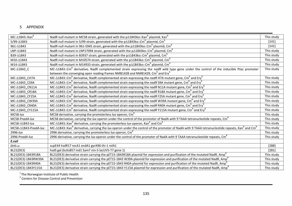
5 APPENDIX
135
MC-∆1843::KanR NadR null mutant in MC58 strain, generated with the p∆1843ko::KanR plasmid, KanR This study 5/99-∆1843 NadR null mutant in 5/99 strain, generated with the p∆1843ko::CmR plasmid, CmR [141] 961-∆1843 NadR null mutant in 961-5945 strain, generated with the p∆1843ko::CmR plasmid, CmR [141] LNP-∆1843 NadR null mutant in LNP17094 strain, generated with the p∆1843ko::CmR plasmid, CmR This study B39-∆1843 NadR null mutant in B3937 strain, generated with the p∆1843ko::CmR plasmid, CmR This study M10-∆1843 NadR null mutant in M10574 strain, generated with the p∆1843ko::CmR plasmid, CmR This study M14-∆1843 NadR null mutant in M14933 strain, generated with the p∆1843ko::CmR plasmid, CmR This study MC-∆1843_C MC-∆1843::CmR derivative, NadR complemented strain expressing the nadR wild type gene under the control of the inducible Ptac promoter
between the converging open reading frames NMB1428 and NMB1429, CmR and EryR This study
MC-∆1843_CH7A MC-∆1843::CmR derivative, NadR complemented strain expressing the nadR H7A mutant gene, CmR and EryR This study MC-∆1843_CS9A MC-∆1843::CmR derivative, NadR complemented strain expressing the nadR S9A mutant gene, CmR and EryR This study MC-∆1843_CN11A MC-∆1843::CmR derivative, NadR complemented strain expressing the nadR N11A mutant gene, CmR and EryR This study MC-∆1843_CR18A MC-∆1843::CmR derivative, NadR complemented strain expressing the nadR R18A mutant gene, CmR and EryR This study MC-∆1843_CF25A MC-∆1843::CmR derivative, NadR complemented strain expressing the nadR F25A mutant gene, CmR and EryR This study MC-∆1843_CW39A MC-∆1843::CmR derivative, NadR complemented strain expressing the nadR W39A mutant gene, CmR and EryR This study MC-∆1843_CR40A MC-∆1843::CmR derivative, NadR complemented strain expressing the nadR R40A mutant gene, CmR and EryR This study MC-∆1843_CY115A MC-∆1843::CmR derivative, NadR complemented strain expressing the nadR Y115A mutant gene, CmR and EryR This study MC58-lux MC58 derivative, carrying the promoterless lux operon, CmR This study MC58-PnadA-lux MC58 derivative, carrying the lux operon under the control of the promoter of NadA with 9 TAAA tetranucleotide repeats, CmR This study MC58-∆1843-lux MC-∆1843::KanR derivative, carrying the promoterless lux operon, KanR and CmR This study MC58-∆1843-PnadA-lux MC-∆1843::KanR derivative, carrying the lux operon under the control of the promoter of NadA with 9 TAAA tetranucleotide repeats, KanR and CmR This study 2996-lux 2996 derivative, carrying the promoterless lux operon, CmR This study 2996-PnadA-lux 2996 derivative, carrying the lux operon under the control of the promoter of NadA with 9 TAAA tetranucleotide repeats, CmR This study E.coli DH5-α supE44 hsdR17 recA1 endA1 gyrA96 thi-1 relA1 [288] BL21(DE3) hsdS gal (λcIts857 ind1 Sam7 nin-5 lacUV5-T7 gene 1) [281] BL21(DE3)-1843R18A BL21(DE3) derivative strain carrying the pET15-1843R18A plasmid for expression and purification of the mutated NadR, AmpR This study BL21(DE3)-1843RW39A BL21(DE3) derivative strain carrying the pET15-1843 W39A plasmid for expression and purification of the mutated NadR, AmpR This study BL21(DE3)-1843R40A BL21(DE3) derivative strain carrying the pET15-1843 R40A plasmid for expression and purification of the mutated NadR, AmpR This study BL21(DE3)-1843Y115A BL21(DE3) derivative strain carrying the pET15-1843 Y115A plasmid for expression and purification of the mutated NadR, AmpR This study
1 The Norwegian Institute of Public Health 2 Centers for Disease Control and Prevention

5 APPENDIX
136
5.2 Table 2 - Plasmids used in this study
Plasmid Relevant characteristics Reference pET15b Expression vector for N-terminal His tagged proteins, AmpR Invitrogen pET15-1843 pET15b derivative for expression of recombinant 1843 protein, AmpR [141] pET15-1843R18A pET15-1843 derivative for expression of recombinant 1843-R18A mutant protein, AmpR This study pET15-1843W39A pET15-1843 derivative for expression of recombinant 1843-W39A mutant protein, AmpR This study pET15-1843R40A pET15-1843 derivative for expression of recombinant 1843-R40A mutant protein, AmpR This study pET15-1843Y115A pET15-1843 derivative for expression of recombinant 1843-Y115A mutant protein, AmpR This study pGEM-T E.coli cloning vector, AmpR Promega p∆1843ko::CmR Construct for generating knockout of the NMB1843 gene, CmR [141] p∆1843ko::KanR Construct for generating knockout of the NMB1843 gene, KanR This study pSLComCmr Plasmid consisting of the chloramphenicol resistance gene flanked by upstream and downstream regions for allelic replacement at a
chromosomal location between ORFs NMB1428 and NMB1429, CmR [118]
pComEryPind-NadR Plasmid for complementation of the NadR null mutant, derivative of pSLComCmr containing a copy of the nadr gene under the control of the Ptac promoter, EryR
This study
pComEry-1843H7A Plasmid for complementation of the NadR null mutant with the H7A mutant form of nadR, derivative of pComEry-NadR, EryR This study pComEry-1843S9A Plasmid for complementation of the NadR null mutant with the S9A mutant form of nadR, derivative of pComEry-NadR, EryR This study pComEry-1843N11A Plasmid for complementation of the NadR null mutant with the N11A mutant form of nadR, derivative of pComEry-NadR, EryR This study pComEry-184 R18A Plasmid for complementation of the NadR null mutant with the R18A mutant form of nadR, derivative of pComEry-NadR, EryR This study pComEry-1843F25A Plasmid for complementation of the NadR null mutant with the F25A mutant form of nadR, derivative of pComEry-NadR, EryR This study pComEry-1843W39A Plasmid for complementation of the NadR null mutant with the W39A mutant form of nadR, derivative of pComEry-NadR, EryR This study pComEry-1843R40A Plasmid for complementation of the NadR null mutant with the R40A mutant form of nadR, derivative of pComEry-NadR, EryR This study pComEry-1843Y115A Plasmid for complementation of the NadR null mutant with the Y115A mutant form of nadR, derivative of pComEry-NadR, EryR This study pSB1075 N-acyl homoserine lactone biosensor based on lasR::lasI’luxCDABE, AmpR [283] pBleuskcript II E. coli cloning and expression vector, AmpR [9] pSL-furlacZ Plasmid containing upstream and downstream regions for allelic replacement at a chromosomal location between ORFs NMB1074 and NMB1075,
EryR [94]
pSL-lux-Fla pSL-furlacZ derivative with the promoterless luxCDABE operon and a chloramphenicol cassette, CmR This study pSL-PnadA-lux pSL-lux-Fla derivative with the luxCDABE operon under the control of the promoter of nadA and a chloramphenicol cassette, CmR This study

5 APPENDIX
137
5.3 Table 3 - Oligonucleotides used in this study
Name Sequence1 Site 1843-F attcacatATG NdeI CCTACCCAATCAAAACATGCG 1843-R2 attcatgcat NsiI CGGCGTATTACGAGTTCAACGCATCCTCG EryXbaF attcgtctaga XbaI GCAAACTTAAGAGTGTGTTGATAG EryBamR atatatggatcc BamHI GGGACCTCTTTAGCTTCTTGG NadluxF2 attcactcgaggctagc XhoI-NheI TAAGACACGACACCGGCAGAATTG NadluxR2 attcaggtacc KpnI TACGCTCATTACCTTTGTGAGTGG 119rt-R AGTGTTGCCCTGTGTAGCAG 120rt-F GCATGGATTATAACCTATCGATCTAA 120rt-R TACGCTTGCTGTTCCCTGT 207rt-F TGACCAAATTCGACACCGT 207rt-R ATCGACACCGAGTTCTTTCC 401rt-L AGGTATCGGTTTCGTTGTCC 401rt-R TTGATTTCGCTGTCCCAATA 652-375rt-F CGAATATTTCGCCGTTGAC 652-375rt-R AAAGGGCGTATTGTTCTTGG 702rt-F GATTTGCTGCTGTCGGACTA 702rt-R GCCTATCCCGTTGGATAATG 865rt-F ACCGCTGGTGGTATGATAGG 865rt-R GCACCATTTGCACGAATAAC 955rt-F TGTGCCGGTCAGGTTTACTA 955rt-R GTTCGGATATTTCGCCAGTT 978rt-F AAACACAAGCTCAACGCACT 978rt-R GGGCAATCAGGGTCATAATC 980rt-F TTTGAAACCGTTGTCGAAAG 980rt-R GGCGCGTTGACCTTATAAAT 1277rt-F AGACGCAGGAGCAGGATATT 1277rt-R CCGTATTCTTCCGAAAGCTC 1476rt-F ACGTTGCCATTTACAACGAA 1476rt-R GTTCGGCGTTGGTAATTTCT 1478rt-F CATCGGCAAACTGGTTCA 1478rt-R CTCAAATGGTCGCGGTAGTA 1609rt-F TACTACACCGGATTGTCCGA 1609rt-R CTTCTTGATCGGCAACTTCA 1841rt-F CCTTTGACCGATACCACTCC 1841rt-R ATGACGATTTCGGTAAACCC 1842rt-F TTTCAAACTTCGTTCCGTGA 1842rt-R CCGTCATTGTAATGCCGTAA 1843rt-F AGGGCGAGAAGCTGTATGAG 1843rt-R CAGGTCTTTAAGCAGCAGCA 1844rt-F ATGCGGTTTATAGCGTATTG 1844rt-R TTAAGTCTCCAAGTTATCG 1994rt-F GCTGGCACAGCTAATACTGC 1994rt-R TCAGCTTTGTTCGTAGCGAT 2099rt-F ACCTGCCTTATATCGAACCG 2099rt-R AGGTTCAAATCATGCACACG 16S_F ACGGAGGGTGCGAGCGTTAATC 16S_R CTGCCTTCGCCTTCGGTATTCCT P207-F2 CTCAAACAATACAAAGCCAAACAGG P207-R GCCCATGGTTTGTTCCTTTGTTGAGGG P375-F CCGCAATGGGTGGAAGCCGCCGC P652-F ACAGTCAAAATGCCGTCTGAAAGCC
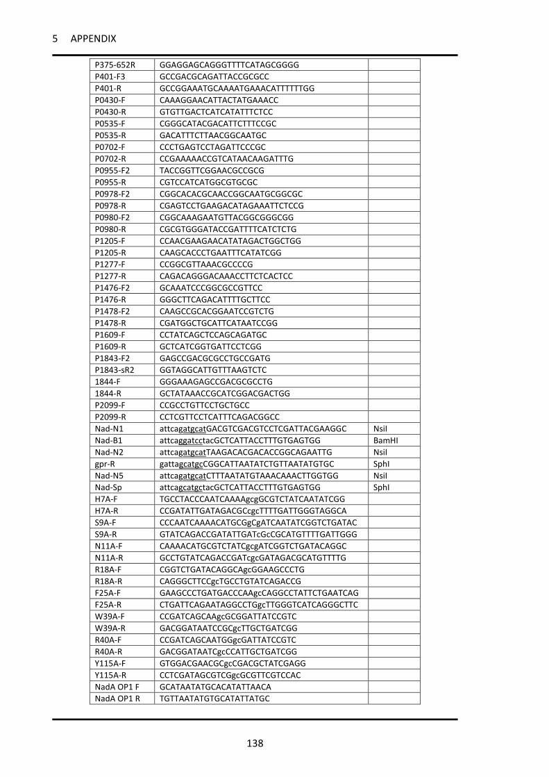
5 APPENDIX
138
P375-652R GGAGGAGCAGGGTTTTCATAGCGGGG P401-F3 GCCGACGCAGATTACCGCGCC P401-R GCCGGAAATGCAAAATGAAACATTTTTTGG P0430-F CAAAGGAACATTACTATGAAACC P0430-R GTGTTGACTCATCATATTTCTCC P0535-F CGGGCATACGACATTCTTTCCGC P0535-R GACATTTCTTAACGGCAATGC P0702-F CCCTGAGTCCTAGATTCCCGC P0702-R CCGAAAAACCGTCATAACAAGATTTG P0955-F2 TACCGGTTCGGAACGCCGCG P0955-R CGTCCATCATGGCGTGCGC P0978-F2 CGGCACACGCAACCGGCAATGCGGCGC P0978-R CGAGTCCTGAAGACATAGAAATTCTCCG P0980-F2 CGGCAAAGAATGTTACGGCGGGCGG P0980-R CGCGTGGGATACCGATTTTCATCTCTG P1205-F CCAACGAAGAACATATAGACTGGCTGG P1205-R CAAGCACCCTGAATTTCATATCGG P1277-F CCGGCGTTAAACGCCCCG P1277-R CAGACAGGGACAAACCTTCTCACTCC P1476-F2 GCAAATCCCGGCGCCGTTCC P1476-R GGGCTTCAGACATTTTGCTTCC P1478-F2 CAAGCCGCACGGAATCCGTCTG P1478-R CGATGGCTGCATTCATAATCCGG P1609-F CCTATCAGCTCCAGCAGATGC P1609-R GCTCATCGGTGATTCCTCGG P1843-F2 GAGCCGACGCGCCTGCCGATG P1843-sR2 GGTAGGCATTGTTTAAGTCTC 1844-F GGGAAAGAGCCGACGCGCCTG 1844-R GCTATAAACCGCATCGGACGACTGG P2099-F CCGCCTGTTCCTGCTGCC P2099-R CCTCGTTCCTCATTTCAGACGGCC Nad-N1 attcagatgcat NsiI GACGTCGACGTCCTCGATTACGAAGGC Nad-B1 attcaggatcc BamHI tacGCTCATTACCTTTGTGAGTGG Nad-N2 attcagatgcat NsiI TAAGACACGACACCGGCAGAATTG gpr-R gattagcatgc SphI CGGCATTAATATCTGTTAATATGTGC Nad-N5 attcagatgcat NsiI CTTTAATATGTAAACAAACTTGGTGG Nad-Sp attcagcatgc SphI tacGCTCATTACCTTTGTGAGTGG H7A-F TGCCTACCCAATCAAAAgcgGCGTCTATCAATATCGG H7A-R CCGATATTGATAGACGCcgcTTTTGATTGGGTAGGCA S9A-F CCCAATCAAAACATGCGgCgATCAATATCGGTCTGATAC S9A-R GTATCAGACCGATATTGATcGcCGCATGTTTTGATTGGG N11A-F CAAAACATGCGTCTATCgcgATCGGTCTGATACAGGC N11A-R GCCTGTATCAGACCGATcgcGATAGACGCATGTTTTG R18A-F CGGTCTGATACAGGCAgcGGAAGCCCTG R18A-R CAGGGCTTCCgcTGCCTGTATCAGACCG F25A-F GAAGCCCTGATGACCCAAgcCAGGCCTATTCTGAATCAG F25A-R CTGATTCAGAATAGGCCTGgcTTGGGTCATCAGGGCTTC W39A-F CCGATCAGCAAgcGCGGATTATCCGTC W39A-R GACGGATAATCCGCgcTTGCTGATCGG R40A-F CCGATCAGCAATGGgcGATTATCCGTC R40A-R GACGGATAATCgcCCATTGCTGATCGG Y115A-F GTGGACGAACGCgcCGACGCTATCGAGG Y115A-R CCTCGATAGCGTCGgcGCGTTCGTCCAC NadA OP1 F GCATAATATGCACATATTAACA NadA OP1 R TGTTAATATGTGCATATTATGC

5 APPENDIX
139
NadA OP2 F TATATACTTTAATATGTAAACA NadA OP2 R TGTTTACATATTAAAGTATATA MafA OP F ACTCAAAATCCGTTCAACATCAAACAAA MafA OP R TTTGTTTGATGTTGAACGGATTTTGAGT NadA1mut1F cccTAATATGCACATATTAACA NadA1mut1R TGTTAATATGTGCATATTAggg NadA1mut2F GCAcccTATGCACATATTAACA NadA1mut2R TGTTAATATGTGCATAgggTGC NadA1mut3F GCATAAcccGCACATATTAACA NadA1mut3R TGTTAATATGTGCgggTTATGC NadA1mut4F GCATAATATcccCATATTAACA NadA1mut4R TGTTAATATGgggATATTATGC NadA1mut5F GCATAATATGCAcccATTAACA NadA1mut5R TGTTAATgggTGCATATTATGC NadA1mut6F GCATAATATGCACATcccAACA NadA1mut6R TGTTgggATGTGCATATTATGC NadA1mut7F GCATAATATGCACATATTcccA NadA1mut7R TgggAATATGTGCATATTATGC MafAmut1F cccCAAAATCCGTTCAACATCAAACAAA MafAmut1R TTTGTTTGATGTTGAACGGATTTTGggg MafAmut2F ACTcccAATCCGTTCAACATCAAACAAA MafAmut2R TTTGTTTGATGTTGAACGGATTgggAGT MafAmut3F ACTCAAcccCCGTTCAACATCAAACAAA MafAmut3R TTTGTTTGATGTTGAACGGgggTTGAGT MafAmut4F ACTCAAAATcccTTCAACATCAAACAAA MafAmut4R TTTGTTTGATGTTGAAgggATTTTGAGT MafAmut5F ACTCAAAATCCGcccAACATCAAACAAA MafAmut5R TTTGTTTGATGTTgggCGGATTTTGAGT MafAmut6F ACTCAAAATCCGTTCcccATCAAACAAA MafAmut6R TTTGTTTGATgggGAACGGATTTTGAGT MafAmut7F ACTCAAAATCCGTTCAACcccAAACAAA MafAmut7R TTTGTTTgggGTTGAACGGATTTTGAGT MafAmut8F ACTCAAAATCCGTTCAACATCcccCAAA MafAmut8R TTTGgggGATGTTGAACGGATTTTGAGT MafAmut9F ACTCAAAATCCGTTCAACATCAAAcccA MafAmut9R TgggTTTGATGTTGAACGGATTTTGAGT NadA2mut1F cccATACTTTAATATGTAAACA NadA2mut1R TGTTTACATATTAAAGTATggg NadA2mut2F TATcccCTTTAATATGTAAACA NadA2mut2R TGTTTACATATTAAAGgggATA NadA2mut3F TATATAcccTAATATGTAAACA NadA2mut3R TGTTTACATATTAgggTATATA NadA2mut4F TATATACTTcccTATGTAAACA NadA2mut4R TGTTTACATAgggAAGTATATA NadA2mut5F TATATACTTTAAcccGTAAACA NadA2mut5R TGTTTACgggTTAAAGTATATA NadA2mut6F TATATACTTTAATATcccAACA NadA2mut6R TGTTgggATATTAAAGTATATA NadA2mut7F TATATACTTTAATATGTAcccA NadA2mut7R TgggTACATATTAAAGTATATA
1 Capital letters indicate N. meningitidis derived sequences, small letters indicate sequences added for cloning purpose, underlined letters indicate the site included in the oligonucleotide.

6 BIBLIOGRAPHY
140
6 BIBLIOGRAPHY
1. Weichselbaum A: Ueber die Aetiologie der akuten meningitis cerebrospinalis. Fortschr Med 1887, 5:573.
2. Stephens DS, Greenwood B, Brandtzaeg P: Epidemic meningitis, meningococcaemia, and Neisseria meningitidis. Lancet 2007, 369(9580):2196-2210.
3. Caugant DA, Maiden MC: Meningococcal carriage and disease--population biology and evolution. Vaccine 2009, 27 Suppl 2:B64-70.
4. Rosenstein NE, Perkins BA, Stephens DS, Popovic T, Hughes JM: Meningococcal disease. The New England journal of medicine 2001, 344(18):1378-1388.
5. Brandtzaeg P, van Deuren M: Meningococcal infections at the start of the 21st century. Advances in pediatrics 2005, 52:129-162.
6. Thompson MJ, Ninis N, Perera R, Mayon-White R, Phillips C, Bailey L, Harnden A, Mant D, Levin M: Clinical recognition of meningococcal disease in children and adolescents. Lancet 2006, 367(9508):397-403.
7. World Health Organization: Meningococcal meningitidis Fact Sheet N°141. 8. Kaplan SL, Schutze GE, Leake JA, Barson WJ, Halasa NB, Byington CL, Woods CR, Tan TQ,
Hoffman JA, Wald ER et al: Multicenter surveillance of invasive meningococcal infections in children. Pediatrics 2006, 118(4):e979-984.
9. Cohn AC, MacNeil JR, Harrison LH, Hatcher C, Theodore J, Schmidt M, Pondo T, Arnold KE, Baumbach J, Bennett N et al: Changes in Neisseria meningitidis disease epidemiology in the United States, 1998-2007: implications for prevention of meningococcal disease. Clinical infectious diseases : an official publication of the Infectious Diseases Society of America 2010, 50(2):184-191.
10. Bilukha OO, Rosenstein N: Prevention and control of meningococcal disease. Recommendations of the Advisory Committee on Immunization Practices (ACIP). MMWR Recommendations and reports : Morbidity and mortality weekly report Recommendations and reports / Centers for Disease Control 2005, 54(RR-7):1-21.
11. Brigham KS, Sandora TJ: Neisseria meningitidis: epidemiology, treatment and prevention in adolescents. Current opinion in pediatrics 2009, 21(4):437-443.
12. Goldschneider I, Gotschlich EC, Artenstein MS: Human immunity to the meningococcus. I. The role of humoral antibodies. The Journal of experimental medicine 1969, 129(6):1307-1326.
13. Harrison LH: Prospects for vaccine prevention of meningococcal infection. Clinical microbiology reviews 2006, 19(1):142-164.
14. Imrey PB, Jackson LA, Ludwinski PH, England AC, 3rd, Fella GA, Fox BC, Isdale LB, Reeves MW, Wenger JD: Meningococcal carriage, alcohol consumption, and campus bar patronage in a serogroup C meningococcal disease outbreak. Journal of clinical microbiology 1995, 33(12):3133-3137.
15. Zuschneid I, Witschi A, Quaback L, Hellenbrand W, Kleinkauf N, Koch D, Krause G: Invasive meningococcal disease with fatal outcome in a Swiss student visiting Berlin. Euro surveillance : bulletin europeen sur les maladies transmissibles = European communicable disease bulletin 2008, 13(45):pii: 19031.
16. Johswich KO, Zhou J, Law DK, St Michael F, McCaw SE, Jamieson FB, Cox AD, Tsang RS, Gray-Owen SD: Invasive Potential of Nonencapsulated Disease Isolates of Neisseria meningitidis. Infection and immunity 2012, 80(7):2346-2353.
17. Frantz ID: Growth Requirements of the Meningococcus. Journal of bacteriology 1942, 43(6):757-761.

6 BIBLIOGRAPHY
141
18. Rouphael NG, Stephens DS: Neisseria meningitidis: biology, microbiology, and epidemiology. Methods Mol Biol 2012, 799:1-20.
19. Frasch CE, Zollinger WD, Poolman JT: Serotype antigens of Neisseria meningitidis and a proposed scheme for designation of serotypes. Reviews of infectious diseases 1985, 7(4):504-510.
20. Mandrell RE, Zollinger WD: Lipopolysaccharide serotyping of Neisseria meningitidis by hemagglutination inhibition. Infection and immunity 1977, 16(2):471-475.
21. Scholten RJ, Kuipers B, Valkenburg HA, Dankert J, Zollinger WD, Poolman JT: Lipo-oligosaccharide immunotyping of Neisseria meningitidis by a whole-cell ELISA with monoclonal antibodies. Journal of medical microbiology 1994, 41(4):236-243.
22. Boisier P, Nicolas P, Djibo S, Taha MK, Jeanne I, Mainassara HB, Tenebray B, Kairo KK, Giorgini D, Chanteau S: Meningococcal meningitis: unprecedented incidence of serogroup X-related cases in 2006 in Niger. Clinical infectious diseases : an official publication of the Infectious Diseases Society of America 2007, 44(5):657-663.
23. Frasch CE: Vaccines for prevention of meningococcal disease. Clinical microbiology reviews 1989, 2 Suppl:S134-138.
24. Jarvis GA, Vedros NA: Sialic acid of group B Neisseria meningitidis regulates alternative complement pathway activation. Infection and immunity 1987, 55(1):174-180.
25. Jackson LA, Schuchat A, Reeves MW, Wenger JD: Serogroup C meningococcal outbreaks in the United States. An emerging threat. JAMA : the journal of the American Medical Association 1995, 273(5):383-389.
26. Rosenstein NE, Perkins BA, Stephens DS, Lefkowitz L, Cartter ML, Danila R, Cieslak P, Shutt KA, Popovic T, Schuchat A et al: The changing epidemiology of meningococcal disease in the United States, 1992-1996. The Journal of infectious diseases 1999, 180(6):1894-1901.
27. Molesworth AM, Thomson MC, Connor SJ, Cresswell MP, Morse AP, Shears P, Hart CA, Cuevas LE: Where is the meningitis belt? Defining an area at risk of epidemic meningitis in Africa. Transactions of the Royal Society of Tropical Medicine and Hygiene 2002, 96(3):242-249.
28. Nicolas P, Norheim G, Garnotel E, Djibo S, Caugant DA: Molecular epidemiology of neisseria meningitidis isolated in the African Meningitis Belt between 1988 and 2003 shows dominance of sequence type 5 (ST-5) and ST-11 complexes. Journal of clinical microbiology 2005, 43(10):5129-5135.
29. Beddek AJ, Li MS, Kroll JS, Jordan TW, Martin DR: Evidence for capsule switching between carried and disease-causing Neisseria meningitidis strains. Infection and immunity 2009, 77(7):2989-2994.
30. Tsang RS, Law DK, Tyler SD, Stephens GS, Bigham M, Zollinger WD: Potential capsule switching from serogroup Y to B: The characterization of three such Neisseria meningitidis isolates causing invasive meningococcal disease in Canada. The Canadian journal of infectious diseases & medical microbiology = Journal canadien des maladies infectieuses et de la microbiologie medicale / AMMI Canada 2005, 16(3):171-174.
31. Swartley JS, Marfin AA, Edupuganti S, Liu LJ, Cieslak P, Perkins B, Wenger JD, Stephens DS: Capsule switching of Neisseria meningitidis. Proceedings of the National Academy of Sciences of the United States of America 1997, 94(1):271-276.
32. Caugant DA, Froholm LO, Bovre K, Holten E, Frasch CE, Mocca LF, Zollinger WD, Selander RK: Intercontinental spread of a genetically distinctive complex of clones of Neisseria meningitidis causing epidemic disease. Proceedings of the National Academy of Sciences of the United States of America 1986, 83(13):4927-4931.
33. Bevanger L, Bergh K, Gisnas G, Caugant DA, Froholm LO: Identification of nasopharyngeal carriage of an outbreak strain of Neisseria meningitidis by pulsed-field gel

6 BIBLIOGRAPHY
142
electrophoresis versus phenotypic methods. Journal of medical microbiology 1998, 47(11):993-998.
34. Weis N, Lind I: Epidemiological markers in Neisseria meningitidis: an estimate of the performance of genotyping vs phenotyping. Scandinavian journal of infectious diseases 1998, 30(1):69-75.
35. Mothershed EA, Sacchi CT, Whitney AM, Barnett GA, Ajello GW, Schmink S, Mayer LW, Phelan M, Taylor TH, Jr., Bernhardt SA et al: Use of real-time PCR to resolve slide agglutination discrepancies in serogroup identification of Neisseria meningitidis. Journal of clinical microbiology 2004, 42(1):320-328.
36. Maiden MC, Bygraves JA, Feil E, Morelli G, Russell JE, Urwin R, Zhang Q, Zhou J, Zurth K, Caugant DA et al: Multilocus sequence typing: a portable approach to the identification of clones within populations of pathogenic microorganisms. Proceedings of the National Academy of Sciences of the United States of America 1998, 95(6):3140-3145.
37. Urwin R, Maiden MC: Multi-locus sequence typing: a tool for global epidemiology. Trends in microbiology 2003, 11(10):479-487.
38. Maiden MC: Population genomics: diversity and virulence in the Neisseria. Current opinion in microbiology 2008, 11(5):467-471.
39. Caugant DA: Genetics and evolution of Neisseria meningitidis: importance for the epidemiology of meningococcal disease. Infect Genet Evol 2008, 8(5):558-565.
40. Stephens DS: Biology and pathogenesis of the evolutionarily successful, obligate human bacterium Neisseria meningitidis. Vaccine 2009, 27 Suppl 2:B71-77.
41. Caugant DA, Tzanakaki G, Kriz P: Lessons from meningococcal carriage studies. FEMS microbiology reviews 2007, 31(1):52-63.
42. Christensen H, May M, Bowen L, Hickman M, Trotter CL: Meningococcal carriage by age: a systematic review and meta-analysis. The Lancet infectious diseases 2010, 10(12):853-861.
43. Claus H, Maiden MC, Wilson DJ, McCarthy ND, Jolley KA, Urwin R, Hessler F, Frosch M, Vogel U: Genetic analysis of meningococci carried by children and young adults. The Journal of infectious diseases 2005, 191(8):1263-1271.
44. Virji M: Pathogenic neisseriae: surface modulation, pathogenesis and infection control. Nature reviews Microbiology 2009, 7(4):274-286.
45. Kallstrom H, Blackmer Gill D, Albiger B, Liszewski MK, Atkinson JP, Jonsson AB: Attachment of Neisseria gonorrhoeae to the cellular pilus receptor CD46: identification of domains important for bacterial adherence. Cellular microbiology 2001, 3(3):133-143.
46. Deghmane AE, Giorgini D, Larribe M, Alonso JM, Taha MK: Down-regulation of pili and capsule of Neisseria meningitidis upon contact with epithelial cells is mediated by CrgA regulatory protein. Molecular microbiology 2002, 43(6):1555-1564.
47. Hammerschmidt S, Muller A, Sillmann H, Muhlenhoff M, Borrow R, Fox A, van Putten J, Zollinger WD, Gerardy-Schahn R, Frosch M: Capsule phase variation in Neisseria meningitidis serogroup B by slipped-strand mispairing in the polysialyltransferase gene (siaD): correlation with bacterial invasion and the outbreak of meningococcal disease. Molecular microbiology 1996, 20(6):1211-1220.
48. Gray-Owen SD, Blumberg RS: CEACAM1: contact-dependent control of immunity. Nat Rev Immunol 2006, 6(6):433-446.
49. Doulet N, Donnadieu E, Laran-Chich MP, Niedergang F, Nassif X, Couraud PO, Bourdoulous S: Neisseria meningitidis infection of human endothelial cells interferes with leukocyte transmigration by preventing the formation of endothelial docking structures. J Cell Biol 2006, 173(4):627-637.
50. Nassif X, Bourdoulous S, Eugene E, Couraud PO: How do extracellular pathogens cross the blood-brain barrier? Trends in microbiology 2002, 10(5):227-232.

6 BIBLIOGRAPHY
143
51. van Ulsen P, Tommassen J: Protein secretion and secreted proteins in pathogenic Neisseriaceae. FEMS microbiology reviews 2006, 30(2):292-319.
52. Perkins-Balding D, Ratliff-Griffin M, Stojiljkovic I: Iron transport systems in Neisseria meningitidis. Microbiology and molecular biology reviews : MMBR 2004, 68(1):154-171.
53. Uria MJ, Zhang Q, Li Y, Chan A, Exley RM, Gollan B, Chan H, Feavers I, Yarwood A, Abad R et al: A generic mechanism in Neisseria meningitidis for enhanced resistance against bactericidal antibodies. The Journal of experimental medicine 2008, 205(6):1423-1434.
54. Madico G, Welsch JA, Lewis LA, McNaughton A, Perlman DH, Costello CE, Ngampasutadol J, Vogel U, Granoff DM, Ram S: The meningococcal vaccine candidate GNA1870 binds the complement regulatory protein factor H and enhances serum resistance. J Immunol 2006, 177(1):501-510.
55. Lewis LA, Ngampasutadol J, Wallace R, Reid JE, Vogel U, Ram S: The meningococcal vaccine candidate neisserial surface protein A (NspA) binds to factor H and enhances meningococcal resistance to complement. PLoS pathogens 2010, 6(7):e1001027.
56. Jarva H, Ram S, Vogel U, Blom AM, Meri S: Binding of the complement inhibitor C4bp to serogroup B Neisseria meningitidis. J Immunol 2005, 174(10):6299-6307.
57. Nassif X: A revolution in the identification of pathogens in clinical laboratories. Clinical infectious diseases : an official publication of the Infectious Diseases Society of America 2009, 49(4):552-553.
58. Tinsley C, Nassif X: Meningococcal pathogenesis: at the boundary between the pre- and post-genomic eras. Current opinion in microbiology 2001, 4(1):47-52.
59. Stephens DS, Hoffman LH, McGee ZA: Interaction of Neisseria meningitidis with human nasopharyngeal mucosa: attachment and entry into columnar epithelial cells. J Infect Dis 1983, 148(3):369-376.
60. Parkhill J, Achtman M, James KD, Bentley SD, Churcher C, Klee SR, Morelli G, Basham D, Brown D, Chillingworth T et al: Complete DNA sequence of a serogroup A strain of Neisseria meningitidis Z2491. Nature 2000, 404(6777):502-506.
61. Schoen C, Blom J, Claus H, Schramm-Gluck A, Brandt P, Muller T, Goesmann A, Joseph B, Konietzny S, Kurzai O et al: Whole-genome comparison of disease and carriage strains provides insights into virulence evolution in Neisseria meningitidis. Proceedings of the National Academy of Sciences of the United States of America 2008, 105(9):3473-3478.
62. Tettelin H, Saunders NJ, Heidelberg J, Jeffries AC, Nelson KE, Eisen JA, Ketchum KA, Hood DW, Peden JF, Dodson RJ et al: Complete genome sequence of Neisseria meningitidis serogroup B strain MC58. Science 2000, 287(5459):1809-1815.
63. Davidsen T, Tonjum T: Meningococcal genome dynamics. Nature reviews Microbiology 2006, 4(1):11-22.
64. Dunning Hotopp JC, Grifantini R, Kumar N, Tzeng YL, Fouts D, Frigimelica E, Draghi M, Giuliani MM, Rappuoli R, Stephens DS et al: Comparative genomics of Neisseria meningitidis: core genome, islands of horizontal transfer and pathogen-specific genes. Microbiology 2006, 152(Pt 12):3733-3749.
65. Schoen C, Tettelin H, Parkhill J, Frosch M: Genome flexibility in Neisseria meningitidis. Vaccine 2009, 27 Suppl 2:B103-111.
66. Maiden MC: Population genetics of a transformable bacterium: the influence of horizontal genetic exchange on the biology of Neisseria meningitidis. FEMS microbiology letters 1993, 112(3):243-250.
67. Hilse R, Hammerschmidt S, Bautsch W, Frosch M: Site-specific insertion of IS1301 and distribution in Neisseria meningitidis strains. Journal of bacteriology 1996, 178(9):2527-2532.
68. Achaz G, Rocha EP, Netter P, Coissac E: Origin and fate of repeats in bacteria. Nucleic acids research 2002, 30(13):2987-2994.

6 BIBLIOGRAPHY
144
69. Hill DJ, Griffiths NJ, Borodina E, Virji M: Cellular and molecular biology of Neisseria meningitidis colonization and invasive disease. Clin Sci (Lond) 2010, 118(9):547-564.
70. Ambur OH, Frye SA, Tonjum T: New functional identity for the DNA uptake sequence in transformation and its presence in transcriptional terminators. Journal of bacteriology 2007, 189(5):2077-2085.
71. Bentley SD, Vernikos GS, Snyder LA, Churcher C, Arrowsmith C, Chillingworth T, Cronin A, Davis PH, Holroyd NE, Jagels K et al: Meningococcal genetic variation mechanisms viewed through comparative analysis of serogroup C strain FAM18. PLoS genetics 2007, 3(2):e23.
72. Buisine N, Tang CM, Chalmers R: Transposon-like Correia elements: structure, distribution and genetic exchange between pathogenic Neisseria sp. FEBS letters 2002, 522(1-3):52-58.
73. Black CG, Fyfe JA, Davies JK: A promoter associated with the neisserial repeat can be used to transcribe the uvrB gene from Neisseria gonorrhoeae. Journal of bacteriology 1995, 177(8):1952-1958.
74. Mahillon J, Chandler M: Insertion sequences. Microbiology and molecular biology reviews : MMBR 1998, 62(3):725-774.
75. Morelle S, Carbonnelle E, Nassif X: The REP2 repeats of the genome of Neisseria meningitidis are associated with genes coordinately regulated during bacterial cell interaction. Journal of bacteriology 2003, 185(8):2618-2627.
76. Moxon R, Bayliss C, Hood D: Bacterial contingency loci: the role of simple sequence DNA repeats in bacterial adaptation. Annual review of genetics 2006, 40:307-333.
77. Sarkari J, Pandit N, Moxon ER, Achtman M: Variable expression of the Opc outer membrane protein in Neisseria meningitidis is caused by size variation of a promoter containing poly-cytidine. Molecular microbiology 1994, 13(2):207-217.
78. van der Ende A, Hopman CT, Zaat S, Essink BB, Berkhout B, Dankert J: Variable expression of class 1 outer membrane protein in Neisseria meningitidis is caused by variation in the spacing between the -10 and -35 regions of the promoter. Journal of bacteriology 1995, 177(9):2475-2480.
79. Moxon ER, Rainey PB, Nowak MA, Lenski RE: Adaptive evolution of highly mutable loci in pathogenic bacteria. Current biology : CB 1994, 4(1):24-33.
80. Carson SD, Stone B, Beucher M, Fu J, Sparling PF: Phase variation of the gonococcal siderophore receptor FetA. Molecular microbiology 2000, 36(3):585-593.
81. Feil EJ, Spratt BG: Recombination and the population structures of bacterial pathogens. Annual review of microbiology 2001, 55:561-590.
82. Moxon ER, Lenski RE, Rainey PB: Adaptive evolution of highly mutable loci in pathogenic bacteria. Perspectives in biology and medicine 1998, 42(1):154-155.
83. Richardson AR, Stojiljkovic I: Mismatch repair and the regulation of phase variation in Neisseria meningitidis. Molecular microbiology 2001, 40(3):645-655.
84. Richardson AR, Yu Z, Popovic T, Stojiljkovic I: Mutator clones of Neisseria meningitidis in epidemic serogroup A disease. Proceedings of the National Academy of Sciences of the United States of America 2002, 99(9):6103-6107.
85. Thomson N, Sebaihia M, Cerdeno-Tarraga A, Bentley S, Crossman L, Parkhill J: The value of comparison. Nature reviews Microbiology 2003, 1(1):11-12.
86. Borst P: Molecular genetics of antigenic variation. Immunology today 1991, 12(3):A29-33.
87. Aho EL, Dempsey JA, Hobbs MM, Klapper DG, Cannon JG: Characterization of the opa (class 5) gene family of Neisseria meningitidis. Molecular microbiology 1991, 5(6):1429-1437.
6 BIBLIOGRAPHY
145
88. Vazquez JA, Berron S, O'Rourke M, Carpenter G, Feil E, Smith NH, Spratt BG: Interspecies recombination in nature: a meningococcus that has acquired a gonococcal PIB porin. Molecular microbiology 1995, 15(6):1001-1007.
89. Kazmierczak MJ, Wiedmann M, Boor KJ: Alternative sigma factors and their roles in bacterial virulence. Microbiology and molecular biology reviews : MMBR 2005, 69(4):527-543.
90. Schielke S, Spatz C, Schwarz RF, Joseph B, Schoen C, Schulz SM, Hubert K, Frosch M, Schubert-Unkmeir A, Kurzai O: Characterization of FarR as a highly specialized, growth phase-dependent transcriptional regulator in Neisseria meningitidis. International journal of medical microbiology : IJMM 2011, 301(4):325-333.
91. Andrews SC, Robinson AK, Rodriguez-Quinones F: Bacterial iron homeostasis. FEMS microbiology reviews 2003, 27(2-3):215-237.
92. DeVoe IW: The meningococcus and mechanisms of pathogenicity. Microbiological reviews 1982, 46(2):162-190.
93. Carson SD, Klebba PE, Newton SM, Sparling PF: Ferric enterobactin binding and utilization by Neisseria gonorrhoeae. Journal of bacteriology 1999, 181(9):2895-2901.
94. Delany I, Ieva R, Alaimo C, Rappuoli R, Scarlato V: The iron-responsive regulator fur is transcriptionally autoregulated and not essential in Neisseria meningitidis. Journal of bacteriology 2003, 185(20):6032-6041.
95. Escolar L, Perez-Martin J, de Lorenzo V: Opening the iron box: transcriptional metalloregulation by the Fur protein. Journal of bacteriology 1999, 181(20):6223-6229.
96. Delany I, Rappuoli R, Scarlato V: Fur functions as an activator and as a repressor of putative virulence genes in Neisseria meningitidis. Molecular microbiology 2004, 52(4):1081-1090.
97. Delany I, Grifantini R, Bartolini E, Rappuoli R, Scarlato V: Effect of Neisseria meningitidis fur mutations on global control of gene transcription. Journal of bacteriology 2006, 188(7):2483-2492.
98. Grifantini R, Sebastian S, Frigimelica E, Draghi M, Bartolini E, Muzzi A, Rappuoli R, Grandi G, Genco CA: Identification of iron-activated and -repressed Fur-dependent genes by transcriptome analysis of Neisseria meningitidis group B. Proc Natl Acad Sci U S A 2003, 100(16):9542-9547.
99. Grifantini R, Frigimelica E, Delany I, Bartolini E, Giovinazzi S, Balloni S, Agarwal S, Galli G, Genco C, Grandi G: Characterization of a novel Neisseria meningitidis Fur and iron-regulated operon required for protection from oxidative stress: utility of DNA microarray in the assignment of the biological role of hypothetical genes. Molecular microbiology 2004, 54(4):962-979.
100. Mellin JR, Goswami S, Grogan S, Tjaden B, Genco CA: A novel fur- and iron-regulated small RNA, NrrF, is required for indirect fur-mediated regulation of the sdhA and sdhC genes in Neisseria meningitidis. Journal of bacteriology 2007, 189(10):3686-3694.
101. Metruccio MM, Fantappie L, Serruto D, Muzzi A, Roncarati D, Donati C, Scarlato V, Delany I: The Hfq-dependent small noncoding RNA NrrF directly mediates Fur-dependent positive regulation of succinate dehydrogenase in Neisseria meningitidis. Journal of bacteriology 2009, 191(4):1330-1342.
102. Archibald FS, Duong MN: Superoxide dismutase and oxygen toxicity defenses in the genus Neisseria. Infection and immunity 1986, 51(2):631-641.
103. Anjum MF, Stevanin TM, Read RC, Moir JW: Nitric oxide metabolism in Neisseria meningitidis. Journal of bacteriology 2002, 184(11):2987-2993.
104. Rock JD, Moir JW: Microaerobic denitrification in Neisseria meningitidis. Biochemical Society transactions 2005, 33(Pt 1):134-136.

6 BIBLIOGRAPHY
146
105. Rock JD, Mahnane MR, Anjum MF, Shaw JG, Read RC, Moir JW: The pathogen Neisseria meningitidis requires oxygen, but supplements growth by denitrification. Nitrite, nitric oxide and oxygen control respiratory flux at genetic and metabolic levels. Molecular microbiology 2005, 58(3):800-809.
106. Bartolini E, Frigimelica E, Giovinazzi S, Galli G, Shaik Y, Genco C, Welsch JA, Granoff DM, Grandi G, Grifantini R: Role of FNR and FNR-regulated, sugar fermentation genes in Neisseria meningitidis infection. Molecular microbiology 2006, 60(4):963-972.
107. Kiley PJ, Beinert H: The role of Fe-S proteins in sensing and regulation in bacteria. Current opinion in microbiology 2003, 6(2):181-185.
108. Edwards J, Cole LJ, Green JB, Thomson MJ, Wood AJ, Whittingham JL, Moir JW: Binding to DNA protects Neisseria meningitidis fumarate and nitrate reductase regulator (FNR) from oxygen. J Biol Chem, 285(2):1105-1112.
109. Schneider MC, Exley RM, Chan H, Feavers I, Kang YH, Sim RB, Tang CM: Functional significance of factor H binding to Neisseria meningitidis. J Immunol 2006, 176(12):7566-7575.
110. Oriente F, Scarlato V, Delany I: Expression of factor H binding protein of meningococcus responds to oxygen limitation through a dedicated FNR-regulated promoter. Journal of bacteriology 2010, 192(3):691-701.
111. Lundberg JO, Weitzberg E: Nasal nitric oxide in man. Thorax 1999, 54(10):947-952. 112. Lundberg JO: Airborne nitric oxide: inflammatory marker and aerocrine messenger in
man. Acta physiologica Scandinavica Supplementum 1996, 633:1-27. 113. Stevanin TM, Moir JW, Read RC: Nitric oxide detoxification systems enhance survival of
Neisseria meningitidis in human macrophages and in nasopharyngeal mucosa. Infection and immunity 2005, 73(6):3322-3329.
114. Rock JD, Thomson MJ, Read RC, Moir JW: Regulation of denitrification genes in Neisseria meningitidis by nitric oxide and the repressor NsrR. Journal of bacteriology 2007, 189(3):1138-1144.
115. Heurlier K, Thomson MJ, Aziz N, Moir JW: The nitric oxide (NO)-sensing repressor NsrR of Neisseria meningitidis has a compact regulon of genes involved in NO synthesis and detoxification. Journal of bacteriology 2008, 190(7):2488-2495.
116. Deghmane AE, Giorgini D, Maigre L, Taha MK: Analysis in vitro and in vivo of the transcriptional regulator CrgA of Neisseria meningitidis upon contact with target cells. Molecular microbiology 2004, 53(3):917-927.
117. Deghmane AE, Petit S, Topilko A, Pereira Y, Giorgini D, Larribe M, Taha MK: Intimate adhesion of Neisseria meningitidis to human epithelial cells is under the control of the crgA gene, a novel LysR-type transcriptional regulator. The EMBO journal 2000, 19(5):1068-1078.
118. Ieva R, Alaimo C, Delany I, Spohn G, Rappuoli R, Scarlato V: CrgA is an inducible LysR-type regulator of Neisseria meningitidis, acting both as a repressor and as an activator of gene transcription. Journal of bacteriology 2005, 187(10):3421-3430.
119. Ren J, Sainsbury S, Combs SE, Capper RG, Jordan PW, Berrow NS, Stammers DK, Saunders NJ, Owens RJ: The structure and transcriptional analysis of a global regulator from Neisseria meningitidis. The Journal of biological chemistry 2007, 282(19):14655-14664.
120. Stork M, Bos MP, Jongerius I, de Kok N, Schilders I, Weynants VE, Poolman JT, Tommassen J: An outer membrane receptor of Neisseria meningitidis involved in zinc acquisition with vaccine potential. PLoS Pathog, 6:e1000969.
121. Perez-Rueda E, Collado-Vides J, Segovia L: Phylogenetic distribution of DNA-binding transcription factors in bacteria and archaea. Computational biology and chemistry 2004, 28(5-6):341-350.

6 BIBLIOGRAPHY
147
122. Perez-Rueda E, Collado-Vides J: Common history at the origin of the position-function correlation in transcriptional regulators in archaea and bacteria. Journal of molecular evolution 2001, 53(3):172-179.
123. Ellison DW, Miller VL: Regulation of virulence by members of the MarR/SlyA family. Current opinion in microbiology 2006, 9(2):153-159.
124. Perera IC, Grove A: Molecular mechanisms of ligand-mediated attenuation of DNA binding by MarR family transcriptional regulators. Journal of molecular cell biology 2010, 2(5):243-254.
125. Wilkinson SP, Grove A: Ligand-responsive transcriptional regulation by members of the MarR family of winged helix proteins. Current issues in molecular biology 2006, 8(1):51-62.
126. Seoane AS, Levy SB: Characterization of MarR, the repressor of the multiple antibiotic resistance (mar) operon in Escherichia coli. Journal of bacteriology 1995, 177(12):3414-3419.
127. Egland PG, Harwood CS: BadR, a new MarR family member, regulates anaerobic benzoate degradation by Rhodopseudomonas palustris in concert with AadR, an Fnr family member. Journal of bacteriology 1999, 181(7):2102-2109.
128. Alekshun MN, Levy SB, Mealy TR, Seaton BA, Head JF: The crystal structure of MarR, a regulator of multiple antibiotic resistance, at 2.3 A resolution. Nature structural biology 2001, 8(8):710-714.
129. Providenti MA, Wyndham RC: Identification and functional characterization of CbaR, a MarR-like modulator of the cbaABC-encoded chlorobenzoate catabolism pathway. Applied and environmental microbiology 2001, 67(8):3530-3541.
130. Chang YM, Jeng WY, Ko TP, Yeh YJ, Chen CK, Wang AH: Structural study of TcaR and its complexes with multiple antibiotics from Staphylococcus epidermidis. Proceedings of the National Academy of Sciences of the United States of America 2010, 107(19):8617-8622.
131. Dolan KT, Duguid EM, He C: Crystal structures of SlyA protein, a master virulence regulator of Salmonella, in free and DNA-bound states. The Journal of biological chemistry 2011, 286(25):22178-22185.
132. Kumarevel T, Tanaka T, Umehara T, Yokoyama S: ST1710-DNA complex crystal structure reveals the DNA binding mechanism of the MarR family of regulators. Nucleic acids research 2009, 37(14):4723-4735.
133. Saridakis V, Shahinas D, Xu X, Christendat D: Structural insight on the mechanism of regulation of the MarR family of proteins: high-resolution crystal structure of a transcriptional repressor from Methanobacterium thermoautotrophicum. Journal of molecular biology 2008, 377(3):655-667.
134. Stapleton MR, Norte VA, Read RC, Green J: Interaction of the Salmonella typhimurium transcription and virulence factor SlyA with target DNA and identification of members of the SlyA regulon. The Journal of biological chemistry 2002, 277(20):17630-17637.
135. Galan B, Kolb A, Sanz JM, Garcia JL, Prieto MA: Molecular determinants of the hpa regulatory system of Escherichia coli: the HpaR repressor. Nucleic acids research 2003, 31(22):6598-6609.
136. Evans K, Adewoye L, Poole K: MexR repressor of the mexAB-oprM multidrug efflux operon of Pseudomonas aeruginosa: identification of MexR binding sites in the mexA-mexR intergenic region. Journal of bacteriology 2001, 183(3):807-812.
137. Fuangthong M, Helmann JD: The OhrR repressor senses organic hydroperoxides by reversible formation of a cysteine-sulfenic acid derivative. Proceedings of the National Academy of Sciences of the United States of America 2002, 99(10):6690-6695.

6 BIBLIOGRAPHY
148
138. Lee EH, Hill SA, Napier R, Shafer WM: Integration Host Factor is required for FarR repression of the farAB-encoded efflux pump of Neisseria gonorrhoeae. Molecular microbiology 2006, 60(6):1381-1400.
139. Lee EH, Rouquette-Loughlin C, Folster JP, Shafer WM: FarR regulates the farAB-encoded efflux pump of Neisseria gonorrhoeae via an MtrR regulatory mechanism. Journal of bacteriology 2003, 185(24):7145-7152.
140. Nichols CE, Sainsbury S, Ren J, Walter TS, Verma A, Stammers DK, Saunders NJ, Owens RJ: The structure of NMB1585, a MarR-family regulator from Neisseria meningitidis. Acta crystallographica Section F, Structural biology and crystallization communications 2009, 65(Pt 3):204-209.
141. Metruccio MM, Pigozzi E, Roncarati D, Berlanda Scorza F, Norais N, Hill SA, Scarlato V, Delany I: A novel phase variation mechanism in the meningococcus driven by a ligand-responsive repressor and differential spacing of distal promoter elements. PLoS pathogens 2009, 5(12):e1000710.
142. Schielke S, Huebner C, Spatz C, Nagele V, Ackermann N, Frosch M, Kurzai O, Schubert-Unkmeir A: Expression of the meningococcal adhesin NadA is controlled by a transcriptional regulator of the MarR family. Molecular microbiology 2009, 72(4):1054-1067.
143. Shafer WM, Veal WL, Lee EH, Zarantonelli L, Balthazar JT, Rouquette C: Genetic organization and regulation of antimicrobial efflux systems possessed by Neisseria gonorrhoeae and Neisseria meningitidis. Journal of molecular microbiology and biotechnology 2001, 3(2):219-224.
144. Jerse AE, Sharma ND, Simms AN, Crow ET, Snyder LA, Shafer WM: A gonococcal efflux pump system enhances bacterial survival in a female mouse model of genital tract infection. Infection and immunity 2003, 71(10):5576-5582.
145. Martin P, van de Ven T, Mouchel N, Jeffries AC, Hood DW, Moxon ER: Experimentally revised repertoire of putative contingency loci in Neisseria meningitidis strain MC58: evidence for a novel mechanism of phase variation. Molecular microbiology 2003, 50(1):245-257.
146. Martin P, Makepeace K, Hill SA, Hood DW, Moxon ER: Microsatellite instability regulates transcription factor binding and gene expression. Proceedings of the National Academy of Sciences of the United States of America 2005, 102(10):3800-3804.
147. Takahama U, Oniki T, Murata H: The presence of 4-hydroxyphenylacetic acid in human saliva and the possibility of its nitration by salivary nitrite in the stomach. FEBS letters 2002, 518(1-3):116-118.
148. Romero JD, Outschoorn IM: The immune response to the capsular polysaccharide of Neisseria meningitidis group B. Zentralblatt fur Bakteriologie : international journal of medical microbiology 1997, 285(3):331-340.
149. Vogel U, Frosch M: Mechanisms of neisserial serum resistance. Molecular microbiology 1999, 32(6):1133-1139.
150. Schneider MC, Exley RM, Ram S, Sim RB, Tang CM: Interactions between Neisseria meningitidis and the complement system. Trends in microbiology 2007, 15(5):233-240.
151. Geoffroy MC, Floquet S, Metais A, Nassif X, Pelicic V: Large-scale analysis of the meningococcus genome by gene disruption: resistance to complement-mediated lysis. Genome research 2003, 13(3):391-398.
152. Brandtzaeg P, Bjerre A, Ovstebo R, Brusletto B, Joo GB, Kierulf P: Neisseria meningitidis lipopolysaccharides in human pathology. Journal of endotoxin research 2001, 7(6):401-420.
153. Braun JM, Blackwell CC, Poxton IR, El Ahmer O, Gordon AE, Madani OM, Weir DM, Giersen S, Beuth J: Proinflammatory responses to lipo-oligosaccharide of Neisseria

6 BIBLIOGRAPHY
149
meningitidis immunotype strains in relation to virulence and disease. The Journal of infectious diseases 2002, 185(10):1431-1438.
154. Jennings MP, Srikhanta YN, Moxon ER, Kramer M, Poolman JT, Kuipers B, van der Ley P: The genetic basis of the phase variation repertoire of lipopolysaccharide immunotypes in Neisseria meningitidis. Microbiology 1999, 145 ( Pt 11):3013-3021.
155. Pinner RW, Spellman PA, Stephens DS: Evidence for functionally distinct pili expressed by Neisseria meningitidis. Infection and immunity 1991, 59(9):3169-3175.
156. Virji M, Alexandrescu C, Ferguson DJ, Saunders JR, Moxon ER: Variations in the expression of pili: the effect on adherence of Neisseria meningitidis to human epithelial and endothelial cells. Molecular microbiology 1992, 6(10):1271-1279.
157. Stephens DS, McGee ZA: Attachment of Neisseria meningitidis to human mucosal surfaces: influence of pili and type of receptor cell. The Journal of infectious diseases 1981, 143(4):525-532.
158. Virji M, Kayhty H, Ferguson DJ, Alexandrescu C, Heckels JE, Moxon ER: The role of pili in the interactions of pathogenic Neisseria with cultured human endothelial cells. Molecular microbiology 1991, 5(8):1831-1841.
159. Helaine S, Dyer DH, Nassif X, Pelicic V, Forest KT: 3D structure/function analysis of PilX reveals how minor pilins can modulate the virulence properties of type IV pili. Proceedings of the National Academy of Sciences of the United States of America 2007, 104(40):15888-15893.
160. Merz AJ, So M, Sheetz MP: Pilus retraction powers bacterial twitching motility. Nature 2000, 407(6800):98-102.
161. Segal E, Hagblom P, Seifert HS, So M: Antigenic variation of gonococcal pilus involves assembly of separated silent gene segments. Proceedings of the National Academy of Sciences of the United States of America 1986, 83(7):2177-2181.
162. de Vries FP, Cole R, Dankert J, Frosch M, van Putten JP: Neisseria meningitidis producing the Opc adhesin binds epithelial cell proteoglycan receptors. Molecular microbiology 1998, 27(6):1203-1212.
163. Moore J, Bailey SE, Benmechernene Z, Tzitzilonis C, Griffiths NJ, Virji M, Derrick JP: Recognition of saccharides by the OpcA, OpaD, and OpaB outer membrane proteins from Neisseria meningitidis. The Journal of biological chemistry 2005, 280(36):31489-31497.
164. Virji M, Makepeace K, Ferguson DJ, Watt SM: Carcinoembryonic antigens (CD66) on epithelial cells and neutrophils are receptors for Opa proteins of pathogenic neisseriae. Molecular microbiology 1996, 22(5):941-950.
165. Virji M, Watt SM, Barker S, Makepeace K, Doyonnas R: The N-domain of the human CD66a adhesion molecule is a target for Opa proteins of Neisseria meningitidis and Neisseria gonorrhoeae. Molecular microbiology 1996, 22(5):929-939.
166. Kurz S, Hubner C, Aepinus C, Theiss S, Guckenberger M, Panzner U, Weber J, Frosch M, Dietrich G: Transcriptome-based antigen identification for Neisseria meningitidis. Vaccine 2003, 21(7-8):768-775.
167. Echenique-Rivera H, Muzzi A, Del Tordello E, Seib KL, Francois P, Rappuoli R, Pizza M, Serruto D: Transcriptome analysis of Neisseria meningitidis in human whole blood and mutagenesis studies identify virulence factors involved in blood survival. PLoS pathogens 2011, 7(5):e1002027.
168. Scarselli M, Serruto D, Montanari P, Capecchi B, Adu-Bobie J, Veggi D, Rappuoli R, Pizza M, Arico B: Neisseria meningitidis NhhA is a multifunctional trimeric autotransporter adhesin. Molecular microbiology 2006, 61(3):631-644.

6 BIBLIOGRAPHY
150
169. Sjolinder H, Eriksson J, Maudsdotter L, Aro H, Jonsson AB: Meningococcal outer membrane protein NhhA is essential for colonization and disease by preventing phagocytosis and complement attack. Infection and immunity 2008, 76(11):5412-5420.
170. Serruto D, Adu-Bobie J, Scarselli M, Veggi D, Pizza M, Rappuoli R, Arico B: Neisseria meningitidis App, a new adhesin with autocatalytic serine protease activity. Molecular microbiology 2003, 48(2):323-334.
171. Turner DP, Marietou AG, Johnston L, Ho KK, Rogers AJ, Wooldridge KG, Ala'Aldeen DA: Characterization of MspA, an immunogenic autotransporter protein that mediates adhesion to epithelial and endothelial cells in Neisseria meningitidis. Infection and immunity 2006, 74(5):2957-2964.
172. van Putten JP, Duensing TD, Cole RL: Entry of OpaA+ gonococci into HEp-2 cells requires concerted action of glycosaminoglycans, fibronectin and integrin receptors. Molecular microbiology 1998, 29(1):369-379.
173. van der Ende A, Hopman CT, Dankert J: Multiple mechanisms of phase variation of PorA in Neisseria meningitidis. Infection and immunity 2000, 68(12):6685-6690.
174. Orihuela CJ, Mahdavi J, Thornton J, Mann B, Wooldridge KG, Abouseada N, Oldfield NJ, Self T, Ala'Aldeen DA, Tuomanen EI: Laminin receptor initiates bacterial contact with the blood brain barrier in experimental meningitis models. The Journal of clinical investigation 2009, 119(6):1638-1646.
175. Wedege E, Hoiby EA, Rosenqvist E, Bjune G: Immune responses against major outer membrane antigens of Neisseria meningitidis in vaccinees and controls who contracted meningococcal disease during the Norwegian serogroup B protection trial. Infection and immunity 1998, 66(7):3223-3231.
176. Holst J, Martin D, Arnold R, Huergo CC, Oster P, O'Hallahan J, Rosenqvist E: Properties and clinical performance of vaccines containing outer membrane vesicles from Neisseria meningitidis. Vaccine 2009, 27 Suppl 2:B3-12.
177. Wedege E, Bolstad K, Wetzler LM, Guttormsen H: IgG antibody levels to meningococcal porins in patient sera: comparison of immunoblotting and ELISA measurements. Journal of immunological methods 2000, 244(1-2):9-15.
178. Michaelsen TE, Aase A, Kolberg J, Wedge E, Rosenqvist E: PorB3 outer membrane protein on Neisseria meningitidis is poorly accessible for antibody binding on live bacteria. Vaccine 2001, 19(11-12):1526-1533.
179. Tzeng YL, Ambrose KD, Zughaier S, Zhou X, Miller YK, Shafer WM, Stephens DS: Cationic antimicrobial peptide resistance in Neisseria meningitidis. Journal of bacteriology 2005, 187(15):5387-5396.
180. Dunn KL, Farrant JL, Langford PR, Kroll JS: Bacterial [Cu,Zn]-cofactored superoxide dismutase protects opsonized, encapsulated Neisseria meningitidis from phagocytosis by human monocytes/macrophages. Infection and immunity 2003, 71(3):1604-1607.
181. Seib KL, Tseng HJ, McEwan AG, Apicella MA, Jennings MP: Defenses against oxidative stress in Neisseria gonorrhoeae and Neisseria meningitidis: distinctive systems for different lifestyles. The Journal of infectious diseases 2004, 190(1):136-147.
182. Serruto D, Spadafina T, Ciucchi L, Lewis LA, Ram S, Tontini M, Santini L, Biolchi A, Seib KL, Giuliani MM et al: Neisseria meningitidis GNA2132, a heparin-binding protein that induces protective immunity in humans. Proceedings of the National Academy of Sciences of the United States of America 2010, 107(8):3770-3775.
183. Pizza M, Scarlato V, Masignani V, Giuliani MM, Arico B, Comanducci M, Jennings GT, Baldi L, Bartolini E, Capecchi B et al: Identification of vaccine candidates against serogroup B meningococcus by whole-genome sequencing. Science 2000, 287(5459):1816-1820.

6 BIBLIOGRAPHY
151
184. Bliska JB, Copass MC, Falkow S: The Yersinia pseudotuberculosis adhesin YadA mediates intimate bacterial attachment to and entry into HEp-2 cells. Infect Immun 1993, 61(9):3914-3921.
185. El Tahir Y, Skurnik M: YadA, the multifaceted Yersinia adhesin. Int J Med Microbiol 2001, 291(3):209-218.
186. Iriarte M, Cornelis GR: Molecular determinants of Yersinia pathogenesis. Microbiologia 1996, 12(2):267-280.
187. Hill DJ, Virji M: A novel cell-binding mechanism of Moraxella catarrhalis ubiquitous surface protein UspA: specific targeting of the N-domain of carcinoembryonic antigen-related cell adhesion molecules by UspA1. Molecular microbiology 2003, 48(1):117-129.
188. Lafontaine ER, Cope LD, Aebi C, Latimer JL, McCracken GH, Jr., Hansen EJ: The UspA1 protein and a second type of UspA2 protein mediate adherence of Moraxella catarrhalis to human epithelial cells in vitro. J Bacteriol 2000, 182(5):1364-1373.
189. Zhang P, Chomel BB, Schau MK, Goo JS, Droz S, Kelminson KL, George SS, Lerche NW, Koehler JE: A family of variably expressed outer-membrane proteins (Vomp) mediates adhesion and autoaggregation in Bartonella quintana. Proc Natl Acad Sci U S A 2004, 101(37):13630-13635.
190. Riess T, Andersson SG, Lupas A, Schaller M, Schafer A, Kyme P, Martin J, Walzlein JH, Ehehalt U, Lindroos H et al: Bartonella adhesin a mediates a proangiogenic host cell response. J Exp Med 2004, 200(10):1267-1278.
191. Serruto D, Spadafina T, Scarselli M, Bambini S, Comanducci M, Hohle S, Kilian M, Veiga E, Cossart P, Oggioni MR et al: HadA is an atypical new multifunctional trimeric coiled-coil adhesin of Haemophilus influenzae biogroup aegyptius, which promotes entry into host cells. Cellular microbiology 2009, 11(7):1044-1063.
192. Meng G SN, St Geme JW 3rd, Waksman G.: Structure of the outer membrane translocator domain of the Haemophilus influenzae Hia trimeric autotransporter. EMBO J 2006, 25(11):2297-22304.
193. Hoiczyk E, Roggenkamp A, Reichenbecher M, Lupas A, Heesemann J: Structure and sequence analysis of Yersinia YadA and Moraxella UspAs reveal a novel class of adhesins. Embo J 2000, 19(22):5989-5999.
194. Comanducci M BS, Brunelli B, Adu-Bobie J, Arico B, Capecchi B, Giuliani MM, Masignani V, Santini L, Savino S, Granoff DM, Caugant DA, Pizza M, Rappuoli R and Mora M: NadA, a novel vaccine candidate of Neisseria meningitidis. J Exp Med 2002, 195:1445-1454.
195. Tavano R CB, Montanari P, Franzoso S, Marin O, Sztukowska M, Cecchini P, Segat D, Scarselli M, Aricò B, Papini E.: Mapping of the Neisseria meningitidis NadA cell-binding site: relevance of predicted {alpha}-helices in the NH2-terminal and dimeric coiled-coil regions. J Bacteriol 2011, 193(1):107-115.
196. Magagnoli C BA, De Conciliis G, Galasso R, Tomei M, Campa C, Pennatini C, Cerchioni M, Fabbri B, Giannini S, Mattioli GL, Biolchi A, D'Ascenzi S, Helling F.: Structural organization of NadADelta(351-405), a recombinant MenB vaccine component, by its physico-chemical characterization at drug substance level. Vaccine 2009, 27(15):2156-2170.
197. Capecchi B. A-BJ, Di Marcello F., Ciucchi L., Masignani V., Taddei A., Rappuoli R., Pizza M., Aricò B.: Neisseria meningitidis NadA is a new invasin which promotes bacterial adhesion to and penetration into human epithelial cells. Mol Microbiol 2005, 55(3):687-698.
198. Capecchi B, Adu-Bobie J, Di Marcello F, Ciucchi L, Masignani V, Taddei A, Rappuoli R, Pizza M, Arico B: Neisseria meningitidis NadA is a new invasin which promotes bacterial adhesion to and penetration into human epithelial cells. Molecular microbiology 2005, 55(3):687-698.

6 BIBLIOGRAPHY
152
199. Franzoso S, Mazzon C, Sztukowska M, Cecchini P, Kasic T, Capecchi B, Tavano R, Papini E: Human monocytes/macrophages are a target of Neisseria meningitidis Adhesin A (NadA). Journal of leukocyte biology 2008, 83(5):1100-1110.
200. Mazzon C B-GB, Cecchini P, Kasic T, Viola A, de Bernard M, Aricò B, Gerosa F and , E P: IFN-γ and R-848 Dependent Activation of Human Monocyte-Derived Dendritic Cells by Neisseria meningitidis Adhesin A. The Journal of Immunology 2007, 179(6):3904-3916.
201. Mazzon C, Baldani-Guerra B, Cecchini P, Kasic T, Viola A, de Bernard M, Arico B, Gerosa F, Papini E: IFN-gamma and R-848 dependent activation of human monocyte-derived dendritic cells by Neisseria meningitidis adhesin A. J Immunol 2007, 179(6):3904-3916.
202. Bowe F, Lavelle EC, McNeela EA, Hale C, Clare S, Arico B, Giuliani MM, Rae A, Huett A, Rappuoli R et al: Mucosal vaccination against serogroup B meningococci: induction of bactericidal antibodies and cellular immunity following intranasal immunization with NadA of Neisseria meningitidis and mutants of Escherichia coli heat-labile enterotoxin. Infection and immunity 2004, 72(7):4052-4060.
203. Comanducci M, Bambini S, Brunelli B, Adu-Bobie J, Arico B, Capecchi B, Giuliani MM, Masignani V, Santini L, Savino S et al: NadA, a novel vaccine candidate of Neisseria meningitidis. The Journal of experimental medicine 2002, 195(11):1445-1454.
204. Giuliani MM, Adu-Bobie J, Comanducci M, Arico B, Savino S, Santini L, Brunelli B, Bambini S, Biolchi A, Capecchi B et al: A universal vaccine for serogroup B meningococcus. Proceedings of the National Academy of Sciences of the United States of America 2006, 103(29):10834-10839.
205. Litt DJ, Savino S, Beddek A, Comanducci M, Sandiford C, Stevens J, Levin M, Ison C, Pizza M, Rappuoli R et al: Putative vaccine antigens from Neisseria meningitidis recognized by serum antibodies of young children convalescing after meningococcal disease. The Journal of infectious diseases 2004, 190(8):1488-1497.
206. Nägele V HJ, Schielke S, Jiménez-Soto LF, Kurzai O, Ackermann N.: Neisseria meningitidis adhesin NadA targets beta1 integrins: functional similarity to Yersinia invasin. J BiolChem 2011, 286(23):20536-20546.
207. Cecchini P TR, Polverino de Laureto P, Franzoso S, Mazzon C, Montanari P, Papini E.: The Soluble Recombinant Neisseria meningitidis Adhesin NadA(?351-405) Stimulates Human Monocytes by Binding to Extracellular Hsp90. Plos One 2011, 6(9:e25089).
208. Montanari P, Bozza G, Capecchi B, Caproni E, Barrile R, Norais N, Capitani M, Sallese M, Cecchini P, Ciucchi L et al: Human heat shock protein (Hsp) 90 interferes with Neisseria meningitidis adhesin A (NadA)-mediated adhesion and invasion. Cell Microbiol, 14(3):368-385.
209. Bambini S, Muzzi A, Olcen P, Rappuoli R, Pizza M, Comanducci M: Distribution and genetic variability of three vaccine components in a panel of strains representative of the diversity of serogroup B meningococcus. Vaccine 2009, 27(21):2794-2803.
210. Comanducci M, Bambini S, Caugant DA, Mora M, Brunelli B, Capecchi B, Ciucchi L, Rappuoli R, Pizza M: NadA diversity and carriage in Neisseria meningitidis. Infection and immunity 2004, 72(7):4217-4223.
211. Wang X, Cohn A, Comanducci M, Andrew L, Zhao X, MacNeil JR, Schmink S, Muzzi A, Bambini S, Rappuoli R et al: Prevalence and genetic diversity of candidate vaccine antigens among invasive Neisseria meningitidis isolates in the United States. Vaccine 2011, 29(29-30):4739-4744.
212. Bambini S, Findlow J, Klaus H, Taha MK, Stefanelli P, Caugant DA, Lucidarme J, Gilchrist S, Borrow R, Vogel U et al: Antigen diversity of the 4CMenB vaccine components in serogroup B meningococcal patient isolates from five european countries. In: 11th European Monitoring Group on Meningococci (EMGM) Congress 2011; Ljubljana, Slovenia; 2011.

6 BIBLIOGRAPHY
153
213. Bambini S, Findlow J, Klaus H, Taha MK, Stefanelli P, Caugant DA, Lucidarme J, Gilchrist S, Borrow R, Vogel U et al: Antigen diversity of the 4CMenB vaccine components in serogroup B meningococcal patient isolates from five european countries. In: Meningitis Research Foundation Conference 2011 (MRF): 2011; London, England; 2011.
214. Seib KL, Oriente F, Adu-Bobie J, Montanari P, Ferlicca F, Giuliani MM, Rappuoli R, Pizza M, Delany I: Influence of serogroup B meningococcal vaccine antigens on growth and survival of the meningococcus in vitro and in ex vivo and in vivo models of infection. Vaccine 2010, 28(12):2416-2427.
215. Oehler S, Eismann ER, Kramer H, Muller-Hill B: The three operators of the lac operon cooperate in repression. The EMBO journal 1990, 9(4):973-979.
216. Lewis M, Chang G, Horton NC, Kercher MA, Pace HC, Schumacher MA, Brennan RG, Lu P: Crystal structure of the lactose operon repressor and its complexes with DNA and inducer. Science 1996, 271(5253):1247-1254.
217. Gasparini R, Panatto D: Meningococcal glycoconjugate vaccines. Hum Vaccin 2011, 7(2):170-182.
218. Frasch C, Preziosi MP, Laforce FM: Development of a group A meningococcal conjugate vaccine, MenAfriVac (TM). Human vaccines & immunotherapeutics 2012, 8(6).
219. Hayrinen J, Jennings H, Raff HV, Rougon G, Hanai N, Gerardy-Schahn R, Finne J: Antibodies to polysialic acid and its N-propyl derivative: binding properties and interaction with human embryonal brain glycopeptides. The Journal of infectious diseases 1995, 171(6):1481-1490.
220. Finne J, Bitter-Suermann D, Goridis C, Finne U: An IgG monoclonal antibody to group B meningococci cross-reacts with developmentally regulated polysialic acid units of glycoproteins in neural and extraneural tissues. J Immunol 1987, 138(12):4402-4407.
221. Fredriksen JH, Rosenqvist E, Wedege E, Bryn K, Bjune G, Froholm LO, Lindbak AK, Mogster B, Namork E, Rye U et al: Production, characterization and control of MenB-vaccine "Folkehelsa": an outer membrane vesicle vaccine against group B meningococcal disease. NIPH annals 1991, 14(2):67-79; discussion 79-80.
222. Sierra GV, Campa HC, Varcacel NM, Garcia IL, Izquierdo PL, Sotolongo PF, Casanueva GV, Rico CO, Rodriguez CR, Terry MH: Vaccine against group B Neisseria meningitidis: protection trial and mass vaccination results in Cuba. NIPH annals 1991, 14(2):195-207; discussion 208-110.
223. Boslego J, Garcia J, Cruz C, Zollinger W, Brandt B, Ruiz S, Martinez M, Arthur J, Underwood P, Silva W et al: Efficacy, safety, and immunogenicity of a meningococcal group B (15:P1.3) outer membrane protein vaccine in Iquique, Chile. Chilean National Committee for Meningococcal Disease. Vaccine 1995, 13(9):821-829.
224. Oster P, Lennon D, O'Hallahan J, Mulholland K, Reid S, Martin D: MeNZB: a safe and highly immunogenic tailor-made vaccine against the New Zealand Neisseria meningitidis serogroup B disease epidemic strain. Vaccine 2005, 23(17-18):2191-2196.
225. Masignani V, Balducci E, Di Marcello F, Savino S, Serruto D, Veggi D, Bambini S, Scarselli M, Arico B, Comanducci M et al: NarE: a novel ADP-ribosyltransferase from Neisseria meningitidis. Molecular microbiology 2003, 50(3):1055-1067.
226. Welsch JA, Moe GR, Rossi R, Adu-Bobie J, Rappuoli R, Granoff DM: Antibody to genome-derived neisserial antigen 2132, a Neisseria meningitidis candidate vaccine, confers protection against bacteremia in the absence of complement-mediated bactericidal activity. The Journal of infectious diseases 2003, 188(11):1730-1740.
227. Rappuoli R: Reverse vaccinology, a genome-based approach to vaccine development. Vaccine 2001, 19(17-19):2688-2691.
228. Sette A, Rappuoli R: Reverse vaccinology: developing vaccines in the era of genomics. Immunity 2010, 33(4):530-541.

6 BIBLIOGRAPHY
154
229. Giuliani MM, Biolchi A, Serruto D, Ferlicca F, Vienken K, Oster P, Rappuoli R, Pizza M, Donnelly J: Measuring antigen-specific bactericidal responses to a multicomponent vaccine against serogroup B meningococcus. Vaccine 2010, 28(31):5023-5030.
230. Beeretz I, Snape M, Finn A, Heath P, Collinson A, Bona G, Esposita S, Dull P, Ypma E, Toneatto D et al: Reactogenicity and safety of multicomponent meningococcal serogroup B vaccine (4CMENB) administred with or without routine infant vaccinations in different schedules. . In: 29th European Society for Paediatric Infectious Diseases (ESPID) Meeting June 7-10, 2011 2011; The Hague, The Netherlands; 2011.
231. Esposito S, Vesikari T, Kimura A, Ypma E., Toneatto D, Dull P: Tolerability of a Three-dose Schedule of an Investigational, Multicomponent Meningococcal Serogroup B Vaccine and Routine Infant Vaccines in a Lot Consistency Trial. In: 17th International Pathogenic Neisseria Conference (IPNC): September 11-16, 2010 2010; Banff, Alberta, Canada 2010.
232. Findlow J, Borrow R, Snape MD, Dawson T, Holland A, John TM, Evans A, Telford KL, Ypma E, Toneatto D et al: Multicenter, open-label, randomized phase II controlled trial of an investigational recombinant Meningococcal serogroup B vaccine with and without outer membrane vesicles, administered in infancy. Clinical infectious diseases : an official publication of the Infectious Diseases Society of America 2010, 51(10):1127-1137.
233. Snape MD, Dawson T, Oster P, Evans A, John TM, Ohene-Kena B, Findlow J, Yu LM, Borrow R, Ypma E et al: Immunogenicity of two investigational serogroup B meningococcal vaccines in the first year of life: a randomized comparative trial. The Pediatric infectious disease journal 2010, 29(11):e71-79.
234. Masignani V, Comanducci M, Giuliani MM, Bambini S, Adu-Bobie J, Arico B, Brunelli B, Pieri A, Santini L, Savino S et al: Vaccination against Neisseria meningitidis using three variants of the lipoprotein GNA1870. The Journal of experimental medicine 2003, 197(6):789-799.
235. Beernink PT, Granoff DM: Bactericidal antibody responses induced by meningococcal recombinant chimeric factor H-binding protein vaccines. Infection and immunity 2008, 76(6):2568-2575.
236. Martin DR, Ruijne N, McCallum L, O'Hallahan J, Oster P: The VR2 epitope on the PorA P1.7-2,4 protein is the major target for the immune response elicited by the strain-specific group B meningococcal vaccine MeNZB. Clin Vaccine Immunol 2006, 13(4):486-491.
237. Goldschneider I, Gotschlich EC, Artenstein MS: Human immunity to the meningococcus. II. Development of natural immunity. The Journal of experimental medicine 1969, 129(6):1327-1348.
238. Gotschlich EC, Goldschneider I, Artenstein MS: Human immunity to the meningococcus. V. The effect of immunization with meningococcal group C polysaccharide on the carrier state. The Journal of experimental medicine 1969, 129(6):1385-1395.
239. Gotschlich EC, Goldschneider I, Artenstein MS: Human immunity to the meningococcus. IV. Immunogenicity of group A and group C meningococcal polysaccharides in human volunteers. The Journal of experimental medicine 1969, 129(6):1367-1384.
240. Costantino P, Viti S, Podda A, Velmonte MA, Nencioni L, Rappuoli R: Development and phase 1 clinical testing of a conjugate vaccine against meningococcus A and C. Vaccine 1992, 10(10):691-698.
241. Ramsay ME, Andrews N, Kaczmarski EB, Miller E: Efficacy of meningococcal serogroup C conjugate vaccine in teenagers and toddlers in England. Lancet 2001, 357(9251):195-196.
242. Snape MD, Pollard AJ: Meningococcal polysaccharide-protein conjugate vaccines. The Lancet infectious diseases 2005, 5(1):21-30.

6 BIBLIOGRAPHY
155
243. Donnelly J, Medini D, Boccadifuoco G, Biolchi A, Ward J, Frasch C, Moxon ER, Stella M, Comanducci M, Bambini S et al: Qualitative and quantitative assessment of meningococcal antigens to evaluate the potential strain coverage of protein-based vaccines. Proceedings of the National Academy of Sciences of the United States of America 2010, 107(45):19490-19495.
244. Serruto D, Bottomley MJ, Ram S, Giuliani MM, Rappuoli R: The new multicomponent vaccine against meningococcal serogroup B, 4CMenB: immunological, functional and structural characterization of the antigens. Vaccine 2012, 30 Suppl 2:B87-97.
245. Vogel U: European efforts to harmonize typing of meningococci. International journal of medical microbiology : IJMM 2011, 301(8):659-662.
246. Donnelly J, Medini D, Giuliani MM, Boccadifuoco G, Stella M, Frosi G, Comanducci M, Bambini S, Muzzi A, Pizza M et al: Estimating the potential strain coverage in Europe of a multicomponent vaccine targeting serogruop B meningococci. In: 11th European Monitoring Group on Meningococci (EMGM) Congress May 18-20, 2011 2011; Ljubljana, Slovenia; 2011.
247. Boccadifuoco G, Donnelly J, Medini M, Giuliani M, Stella M, Frosi M, Comanducci M, Bambini S, Muzzi A, Pizza M et al: Estimating the potential coverage in Europe of a multicomponent vaccine targeting serogroup B meningococci. . In: Meningitis Research Foundation Conference 2011 (MRF): 2011; London, England; 2011: 50.
248. Paruchuri DK, Seifert HS, Ajioka RS, Karlsson KA, So M: Identification and characterization of a Neisseria gonorrhoeae gene encoding a glycolipid-binding adhesin. Proceedings of the National Academy of Sciences of the United States of America 1990, 87(1):333-337.
249. Naumann M, Rudel T, Meyer TF: Host cell interactions and signalling with Neisseria gonorrhoeae. Current opinion in microbiology 1999, 2(1):62-70.
250. Deal CD, Krivan HC: Lacto- and ganglio-series glycolipids are adhesion receptors for Neisseria gonorrhoeae. The Journal of biological chemistry 1990, 265(22):12774-12777.
251. Stromberg N, Deal C, Nyberg G, Normark S, So M, Karlsson KA: Identification of carbohydrate structures that are possible receptors for Neisseria gonorrhoeae. Proceedings of the National Academy of Sciences of the United States of America 1988, 85(13):4902-4906.
252. Fantappie L, Oriente F, Muzzi A, Serruto D, Scarlato V, Delany I: A novel Hfq-dependent sRNA that is under FNR control and is synthesized in oxygen limitation in Neisseria meningitidis. Molecular microbiology 2011, 80(2):507-523.
253. Schielke S, Huebner C, Spatz C, Nagele V, Ackermann N, Frosch M, Kurzai O, Schubert-Unkmeir A: Expression of the meningococcal adhesin NadA is controlled by a transcriptional regulator of the MarR-family. Molecular microbiology 2009.
254. Grifantini R, Bartolini E, Muzzi A, Draghi M, Frigimelica E, Berger J, Ratti G, Petracca R, Galli G, Agnusdei M et al: Previously unrecognized vaccine candidates against group B meningococcus identified by DNA microarrays. Nature biotechnology 2002, 20(9):914-921.
255. Wilke MS, Heller M, Creagh AL, Haynes CA, McIntosh LP, Poole K, Strynadka NC: The crystal structure of MexR from Pseudomonas aeruginosa in complex with its antirepressor ArmR. Proceedings of the National Academy of Sciences of the United States of America 2008, 105(39):14832-14837.
256. Lunin VV, Evdokimova, E., Kudritska, M., Cuff, M. E., Joachimiak, A., Edwards, A. M., and Savchenko, A. : The crystal structure of transcriptional regulator PA4135.
257. Wales TE, Engen JR: Hydrogen exchange mass spectrometry for the analysis of protein dynamics. Mass spectrometry reviews 2006, 25(1):158-170.
258. Brzovic PS, Le Trong, I., Navarre, W. W., Fang, F. C., Stenkamp, R. E., and Libby, S. J.: Crystal structure of transcription regulatory protein slyA from Salmonella typhimurium

6 BIBLIOGRAPHY
156
in complex with salicylate ligands. In: Editor|. City|: Publisher|; Year|:Pages|. [|Series Editor (Series Editor^Editors|): Series Title|, vol Series Volume|]|.
259. Biolchi A, Pigozzi E, Brunelli B, Donnelly J, Rappuoli R, Pizza M, Giuliani MM, Delany I: In vitro levels of NadA expression may underestimate the potential effectiveness of immune responses against NadA in vivo. In: 17th International Pathogenic Neisseria Conference (IPNC): 11-16 September 2010 2010; Banff, Alberta, Canada 2010: p.172.
260. Biolchi A, Fagnocchi L, Pigozzi E, Brunelli B, Boccadifuoco G, Donnelly J, Rappuoli R, Pizza M, Giuliani MM, Delany I: In vitro levels of NadA expression may underestimate the potential effectiveness of immune responses against nadA in vivo. In: 11th European Monitoring Group on Meningococci (EMGM) Congress 18-20 May 2011 2011; Ljubljana, Slovenia; 2011: p.153-154.
261. Fagnocchi L, Pigozzi E, Scarlato V, Delany I: In the NadR Regulon, Adhesins and Diverse Meningococcal Functions Are Regulated in Response to Signals in Human Saliva. Journal of bacteriology 2012, 194(2):460-474.
262. Mani AR, Ippolito S, Moreno JC, Visser TJ, Moore KP: The metabolism and dechlorination of chlorotyrosine in vivo. The Journal of biological chemistry 2007, 282(40):29114-29121.
263. Granoff DM, Moe GR, Giuliani MM, Adu-Bobie J, Santini L, Brunelli B, Piccinetti F, Zuno-Mitchell P, Lee SS, Neri P et al: A novel mimetic antigen eliciting protective antibody to Neisseria meningitidis. J Immunol 2001, 167(11):6487-6496.
264. Tunio SA, Oldfield NJ, Ala'Aldeen DA, Wooldridge KG, Turner DP: The role of glyceraldehyde 3-phosphate dehydrogenase (GapA-1) in Neisseria meningitidis adherence to human cells. BMC Microbiol 2010, 10:280.
265. Pancholi V, Fischetti VA: A major surface protein on group A streptococci is a glyceraldehyde-3-phosphate-dehydrogenase with multiple binding activity. The Journal of experimental medicine 1992, 176(2):415-426.
266. Modun B, Morrissey J, Williams P: The staphylococcal transferrin receptor: a glycolytic enzyme with novel functions. Trends in microbiology 2000, 8(5):231-237.
267. Alvarez RA, Blaylock MW, Baseman JB: Surface localized glyceraldehyde-3-phosphate dehydrogenase of Mycoplasma genitalium binds mucin. Molecular microbiology 2003, 48(5):1417-1425.
268. Egea L, Aguilera L, Gimenez R, Sorolla MA, Aguilar J, Badia J, Baldoma L: Role of secreted glyceraldehyde-3-phosphate dehydrogenase in the infection mechanism of enterohemorrhagic and enteropathogenic Escherichia coli: interaction of the extracellular enzyme with human plasminogen and fibrinogen. The international journal of biochemistry & cell biology 2007, 39(6):1190-1203.
269. Kinoshita H, Uchida H, Kawai Y, Kawasaki T, Wakahara N, Matsuo H, Watanabe M, Kitazawa H, Ohnuma S, Miura K et al: Cell surface Lactobacillus plantarum LA 318 glyceraldehyde-3-phosphate dehydrogenase (GAPDH) adheres to human colonic mucin. Journal of applied microbiology 2008, 104(6):1667-1674.
270. Gozalbo D, Gil-Navarro I, Azorin I, Renau-Piqueras J, Martinez JP, Gil ML: The cell wall-associated glyceraldehyde-3-phosphate dehydrogenase of Candida albicans is also a fibronectin and laminin binding protein. Infection and immunity 1998, 66(5):2052-2059.
271. Swinger KK, Rice PA: IHF and HU: flexible architects of bent DNA. Curr Opin Struct Biol 2004, 14(1):28-35.
272. Shelburne SA, 3rd, Keith DB, Davenport MT, Horstmann N, Brennan RG, Musser JM: Molecular characterization of group A Streptococcus maltodextrin catabolism and its role in pharyngitis. Molecular microbiology 2008, 69(2):436-452.
273. Shelburne SA, 3rd, Sumby P, Sitkiewicz I, Okorafor N, Granville C, Patel P, Voyich J, Hull R, DeLeo FR, Musser JM: Maltodextrin utilization plays a key role in the ability of group A

6 BIBLIOGRAPHY
157
Streptococcus to colonize the oropharynx. Infection and immunity 2006, 74(8):4605-4614.
274. Bai X, Findlow J, Borrow R: Recombinant protein meningococcal serogroup B vaccine combined with outer membrane vesicles. Expert Opin Biol Ther 2011, 11(7):969-985.
275. Jacobsson S, Molling P, Olcen P: Seroprevalence of antibodies against fHbp and NadA, two potential vaccine antigens for Neisseria meningitidis. Vaccine 2009, 27(42):5755-5759.
276. Tavano R, Franzoso S, Cecchini P, Cartocci E, Oriente F, Arico B, Papini E: The membrane expression of Neisseria meningitidis adhesin A (NadA) increases the proimmune effects of MenB OMVs on human macrophages, compared with NadA- OMVs, without further stimulating their proinflammatory activity on circulating monocytes. Journal of leukocyte biology 2009, 86(1):143-153.
277. Cecchini P, Tavano R, Polverino de Laureto P, Franzoso S, Mazzon C, Montanari P, Papini E: The soluble recombinant Neisseria meningitidis adhesin NadA(Delta351-405) stimulates human monocytes by binding to extracellular Hsp90. PloS one 2011, 6(9):e25089.
278. Fukuyama N, Ichimori K, Su Z, Ishida H, Nakazawa H: Peroxynitrite formation from activated human leukocytes. Biochemical and biophysical research communications 1996, 224(2):414-419.
279. Ieva R, Roncarati D, Metruccio MM, Seib KL, Scarlato V, Delany I: OxyR tightly regulates catalase expression in Neisseria meningitidis through both repression and activation mechanisms. Molecular microbiology 2008, 70(5):1152-1165.
280. Hanahan D: Studies on transformation of Escherichia coli with plasmids. Journal of molecular biology 1983, 166(4):557-580.
281. Studier FW, Moffatt BA: Use of bacteriophage T7 RNA polymerase to direct selective high-level expression of cloned genes. Journal of molecular biology 1986, 189(1):113-130.
282. Sambrook J, Fritsch EF, Maniatis T: Molecular cloning: A laboratory manual, 2nd edn: Cold Spring Harbor, NY: Cold Spring Harbor Laboratory.; 1989.
283. Perera IC, Lee YH, Wilkinson SP, Grove A: Mechanism for attenuation of DNA binding by MarR family transcriptional regulators by small molecule ligands. Journal of molecular biology 2009, 390(5):1019-1029.
284. Fantappie L, Metruccio MM, Seib KL, Oriente F, Cartocci E, Ferlicca F, Giuliani MM, Scarlato V, Delany I: The RNA chaperone Hfq is involved in stress response and virulence in Neisseria meningitidis and is a pleiotropic regulator of protein expression. Infection and immunity 2009, 77(5):1842-1853.
285. Delany I, Spohn G, Rappuoli R, Scarlato V: The Fur repressor controls transcription of iron-activated and -repressed genes in Helicobacter pylori. Molecular microbiology 2001, 42(5):1297-1309.
286. Maxam AM, Gilbert W: A new method for sequencing DNA. Proceedings of the National Academy of Sciences of the United States of America 1977, 74(2):560-564.
287. Borrow R, Aaberge IS, Santos GF, Eudey TL, Oster P, Glennie A, Findlow J, Hoiby EA, Rosenqvist E, Balmer P et al: Interlaboratory standardization of the measurement of serum bactericidal activity by using human complement against meningococcal serogroup b, strain 44/76-SL, before and after vaccination with the Norwegian MenBvac outer membrane vesicle vaccine. Clinical and diagnostic laboratory immunology 2005, 12(8):970-976.
288. Hanahan D: Studies on transformation of Escherichia coli with plasmids. J Mol Biol 1983, 166:557-580.