alleviation of myogenic ptosis magnetic force - … · muscle canovercome the magnetic force so...
TRANSCRIPT
Brit. 7. Ophthal. ( I 973) 57, 3 1 5
Preliminary communication
Alleviation of myogenic ptosis bymagnetic force
J. S. CONWAY
.'ew End Hospital (Royal Free Hospital Group), London, N.F.3., and lVhipps Cross Hospital,London, E. i I
Ptosis has usually been treated by surgery and the literature contains suggestions of overeighty different operations for its correction. Duke-Elder (I952) has pointed out that, inspite of the variety and ingenuity of remedial operations, the results are by no meansinvariably brilliant and in cases due to neuromuscular disease shortening of the lid isusually unsatisfactory.Apart from the cosmetic defect, treatment is necessary for visual reasons when the ptosed
lid covers the pupil and obstructs vision.Walshe ( I957) stated that the ptosis of myasthenia gravis should usually not be operated
upon. For the ptosis of ocular myopathy, operation may be contraindicated because ofthe problem of exposure keratitis in the absence of Bell's phenomenon.At the Endocrine Unit, New End Hospital, there is a register of 340 cases of myasthenia
gravis. Some of these patients have been referred to the Eye Clinic because of ptosis,which is the commonest and often the presenting symptom.
These patients have in the past usually been treated by the provision of crutch glasses,which are useful when operation is contraindicated or not accepted by the patient, wherethe difficulty is temporary, or in the case of an elderly patient. Lid crutches to elevate theeyelid were introduced by Goldzieher (i890), who used a shell plate attached to the upperrim of a shell spectacle frame. Since then other appliances have been described, such asan adjustable watch spring (Kauffman, I893); an independent wire crutch (Meyer, I893);flexible single wire (Dodge, I 935); and a scleral contact lens with a superior ridge. Patientsusing crutch glasses often experience discomfort with the forced opening of the eye andwatering owing to the pressure of the wire loop on the upper lid.
All electro-motive force used in commerce is magnetic in origin and it was thought thata magnetic force might be a useful method for elevation of the ptosed eyelid, especially inmyasthenia gravis with its variable nature, remissions, and exacerbations.
Ptosis may be alleviated by inserting a metallic magnetic implant into the upper eyelidand elevating the eyelid by the action of a small magnet unobtrusively placed behind theupper rim ofa spectacle frame. The attracting force pulls the eyelid upwards and forwards.Alternatively a metal strip may be fixed on to the upper lid using a skin adhesive. If itis placed in the upper palpebral furrow, the metal strip will be hidden in the skin creaseof the furrow when the lid is elevated. There is no pressure on the eyelid as with a crutchspectacle, and the weight of the lid is taken off the eye. The power of the orbicularismuscle can overcome the magnetic force so enabling the patient to blink.
lReceived for publicatioin MIarcll 30, I972Address for reprints: 30, Middlesay, Hampstead Gardeni SLburb, Lonidoni NAV. i
copyright. on 7 S
eptember 2018 by guest. P
rotected byhttp://bjo.bm
j.com/
Br J O
phthalmol: first published as 10.1136/bjo.57.5.315 on 1 M
ay 1973. Dow
nloaded from

3. S. Conway
The magnetic force between two magnets is so much greater than that between a magnetand a piece of unmagnetized soft iron, or other ferro-magnetic material, that it is a greatadvantage to use an already permanently magnetized strip in the upper lid, rather thana simple soft iron or Mumetal (electrolytic iron melted in vacuo and annealed) strip. Thisis because the strip would be attracted to the magnet only by induction of magnetism init by the external magnet, rather than there being interaction between two permanentmagnets. Nevertheless, because of the high magnetic permeability ofMu-metal (Abrams,I 970), (25,000 to 50,000 compared with soft iron 3,000 to 6,ooo), quite efficient results areobtained with Mu-metal. Experiments using new magnetic materials, and especiallyPlatinax, are proceeding, and appear to be showing even greater success.
It may be of interest to quote some of the cases of ptosis due to ocular myopathy andmyasthenia that have been referred and treated.
Case reports
Case i, a 63-year-old married woman, had a long-standing history of bilateral ptosis. Droopingof the eyelids began in the right eye during her teens, and the condition gradually deteriorated.For the past 20 years it had been static with no variation. There was no family history of myopathy.
Examination
There was severe bilateral ptosis (Fig. i). The eyelids almost completely covered the cornea andthere was marked interference with vision. The patient could see only by tilting her head backwards,but this caused her neck to ache. The forehead was wrinkled by contraction of the occipito-frontalismuscles. The levator palpebrax superioris muscles showed little or no function. With the eyelidsraised, the visual acuity with spectacles was normal. Ocular movements were full and there hadnever been any diplopia. The pupils were equal and the reactions normal.Examination of the central nervous system revealed no abnormal physical signs. A tensilon test
was negative.Neurological opinion confirmed a diagnosis of ocular myopathy. Operation for ptosis had been
under consideration at another hospital for io years, but the patient was very apprehensive abouthaving any surgery done. She had tried ptosis props but they caused discomfort and did not helpher.
o 2 3 4 5 6 7 8 9 0 11 12mm
F IG. I Case bilateral ptosis due to ocular myopathy F IG. 2 Implant of Mu-metal
TreatmentAn implant of Mu-metal (Fig. 2), an alloy of nickel and iron, size 13 X 4 X 0-5 mm. wa cut frOma flat plate and shaped to the curve of the upper lid. This was coated with methyl-methacrylate inthe dental laboratory.
As an outpatient procedure and under local anaesthesia. the implant was placed in the upper lid.Two small incisions 5 mm. long were made in the skin of the right upper lid and the implant insertedinto a tunnel superficial to the tarsal plate. A suture through a hole at each end of the implant
3I6copyright.
on 7 Septem
ber 2018 by guest. Protected by
http://bjo.bmj.com
/B
r J Ophthalm
ol: first published as 10.1136/bjo.57.5.315 on 1 May 1973. D
ownloaded from
Alleviation ofptosis
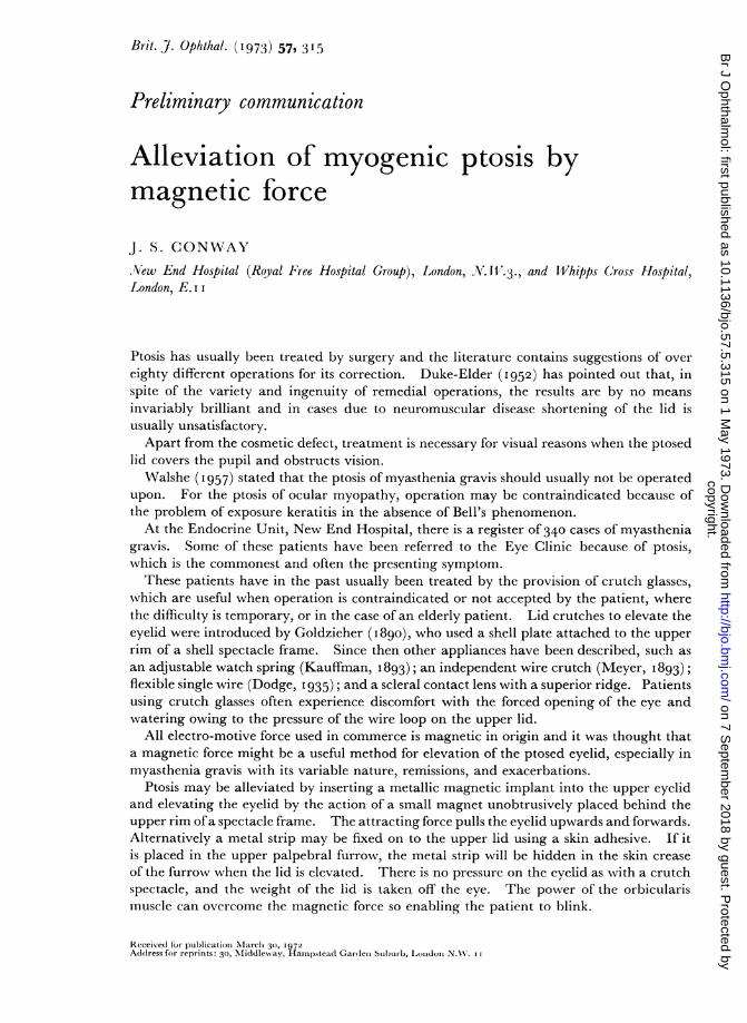
served to fix it to the tarsal plate. A magnet held in the hand shows the magnetic elevation of theright upper lid which is then clear of the pupil (Fig. 3). This implant has been in the eyelid for3 years and shows no sign of extrusion. Temporary spectacles with a small bar magnet on the upperrim of the frame alleviate the right ptosis and improve vision (Fig. 4). Permanent spectaclesincorporating a hidden magnet are under construction.
~~~~~~~~~~~~~~~~~~~V.-N
FIG. 3 Case i elevation of upper lid by magnet FIG. 4 Case i, alleviation of right ptosis by magnetattached to upper rim of spectacle
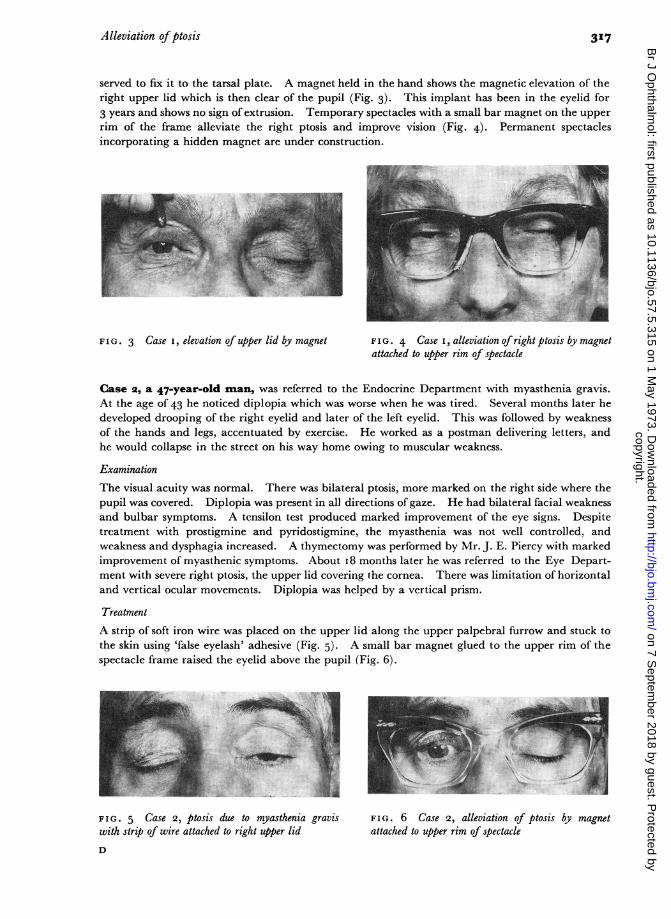
Case 2, a 47-year-old man, was referred to the Endocrine Department with myasthenia gravis.At the age of 43 he noticed diplopia which was worse when he was tired. Several months later hedeveloped drooping of the right eyelid and later of the left eyelid. This was followed by weaknessof the hands and legs, accentuated by exercise. He worked as a postman delivering letters, andhe would collapse in the street on his way home owing to muscular weakness.
Examination
The visual acuity was normal. There was bilateral ptosis, more marked on the right side where thepupil was covered. Diplopia was present in all directions of gaze. He had bilateral facial weaknessand bulbar symptoms. A tensilon test produced marked improvement of the eye signs. Despitetreatment with prostigmine and pyridostigmine, the myasthenia was not well controlled, andweakness and dysphagia increased. A thymectomy was performed by Mr. J. E. Piercy with markedimprovement of myasthenic symptoms. About i8 months later he was referred to the Eye Depart-ment with severe right ptosis, the upper lid covering the cornea. There was limitation of horizontaland vertical ocular movements. Diplopia was helped by a vertical prism.
TreatmentA strip of soft iron wire was placed on the upper lid along the upper palpebral furrow and stuck tothe skin using 'false eyelash' adhesive (Fig. 5). A small bar magnet glued to the upper rim of thespectacle frame raised the eyelid above the pupil (Fig. 6).
FIG. 5 Case 2, ptosis due to myasthenia graviswith strip of wire attached to right upper lidD
F IG. 6 Case 2, alleviation of ptosis by magnetattached to upper rim of spectacle
3I17copyright.
on 7 Septem
ber 2018 by guest. Protected by
http://bjo.bmj.com
/B
r J Ophthalm
ol: first published as 10.1136/bjo.57.5.315 on 1 May 1973. D
ownloaded from
J. S. Conway
Later a similar implant to that used in Case i was placed in the upper lid but this was extrudedafter a few months. Extrusion may have been due to the presence of unreacted monomer or catalystin the methyl-methacrylate coating of the implant or to faulty technique in placing the incisionover the ends of the implant and not as far from the site of implantation as possible.
Satisfactory results have been obtained with a metallic strip adherent to the upper lid for the past34 years. The patient is adept at fixing the strip to the upper lid each morning and removing it inthe evening. He has found 'Out-door Girl' eyelash adhesive to be a satisfactory preparation.
Case 3, a 3x-year-old married woman, had complained for 2 years of double vision and droopingof the left upper eyelid. This was followed by the development of dysarthria, dysphagia, andperipheral muscle weakness, and the ptosis became bilateral. A tensilon test gave prompt relieffrom most of her symptoms.
She was treated with increasing doses of prostigmine and mestinon.Owing to increasing myasthenia and side effects of the drugs, thymectomy was performed by Mr.
M. J. Lange.There was an improvement in her general condition in that she was able to look after her two
children, do more housework, and manage reasonable walks.3 years later she was referred to the Eye Department because of bilateral ptosis, which obscured
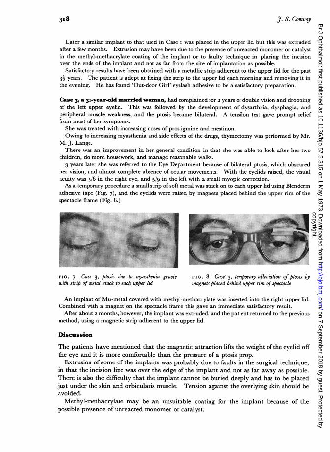
her vision, and almost complete absence of ocular movements. With the eyelids raised, the visualacuity was 5/6 in the right eye, and 5/9 in the left with a small myopic correction.As a temporary procedure a small strip of soft metal was stuck on to each upper lid using Blenderm
adhesive tape (Fig. 7), and the eyelids were raised by magnets placed behind the upper rim of thespectacle frame (Fig. 8.)
'VFIG. 7 Case 3, ptosis due to myasthenia gravis FIG. 8 Case 3, temporary alleviation of ptosis bywith strip of metal stuck to each upper lid magnets placed behind upper rim of spectacle
An implant of Mu-metal covered with methyl-methacrylate was inserted into the right upper lid.Combined with a magnet on the spectacle frame this gave an immediate satisfactory result.
After about 2 months, however, the implant was extruded, and the patient returned to the previousmethod, using a magnetic strip adherent to the upper lid.
Discussion
The patients have mentioned that the magnetic attraction lifts the weight ofthe eyelid offthe eye and it is more comfortable than the pressure of a ptosis prop.
Extrusion of some of the implants was probably due to faults in the surgical technique,in that the incision line was over the edge of the implant and not as far away as possible.There is also the difficulty that the implant cannot be buried deeply and has to be placedjust under the skin and orbicularis muscle. Tension against the overlying skin should beavoided.
Methyl-methacrylate may be an unsuitable coating for the implant because of thepossible presence of unreacted monomer or catalyst.
3I8copyright.
on 7 Septem
ber 2018 by guest. Protected by
http://bjo.bmj.com
/B
r J Ophthalm
ol: first published as 10.1136/bjo.57.5.315 on 1 May 1973. D
ownloaded from
Alleviation ofptosis 319
An implant of a cobalt-platinum magnet alloy coated with medical grade silicone is nowin use. This alloy has outstanding magnetic properties combined with good workability.It can be machined, rolled, or drawn, and lends itself to the manufacture of very smallmagnets with a high energy product and coercive force.The combination of two very small cobalt-platinum magnets, one in or on the eyelid
and another in the upper rim of the spectacle frame, gives improved elevation of the eyelidwhen compared with the conventional magnetic material previously used.
I wish to express my gratitude to the Endowment Fund of the Royal Free Hospital for a Department ofHealthGrant in support of this work.
I wish to thank Mr. Nevies for arousing my interest in this subject and Dr. J. S. Portnoy for advice and muchhelpful discussion.The photographs were kindly provided by the Photographic Department of the Royal Free Hospital.
References
ABRAMS, J. D. (I970) Exp. Eye Res., 9, I 14DODGE, w. M. (I935) Arch. Ophthal., 14, 989DUKE-ELDER, S. (1952) "Textbook of Ophthalmology", vol. 5, p. 5150. Kimpton, LondonGOLDZIEHER, W. (1890) Zbl. prakt. Augenheilk., 14, 34KAUFFMAN, N. F. (I893) Ibid., I8, 75
MEYER (1893) Arch. Augenheilk., 26, I 53WALSH, F. B. (1957) "Clinical Neuro-Ophthalmology", 2nd ed., p. 782. Williams and Wilkins,Baltimore
copyright. on 7 S
eptember 2018 by guest. P
rotected byhttp://bjo.bm
j.com/
Br J O
phthalmol: first published as 10.1136/bjo.57.5.315 on 1 M
ay 1973. Dow
nloaded from