all previously published papers were reproduced with the
TRANSCRIPT


All previously published papers were reproduced with the permission of the publisher. Published by Karolinska Institutet. Printed by Larserics Digital Print AB, Sundbyberg. © Charlotta Olivecrona, 2013 ISBN 978-91-7549-071-7

Change will not come if we wait for some other person or some other time. We are the ones we've been waiting for. We are the change that we seek.
Barack Obama, 2008


ABSTRACT Use of the tourniquet in extremity surgery is considered to be an important tool because it prevents intraoperative bleeding and thereby improves visualization of the surgical field. However, its use is not without risks, and complications may occur. The overall aim of this thesis was to increase our knowledge of tourniquet use in order to improve patient safety during knee arthroplasty surgery. In Study I, a Randomized Controlled Trial (RCT) including 94 patients undergoing Total Knee Arthroplasty (TKA) surgery, the aim was to determine whether there were any differences between different protecting materials and no protective material regarding skin injuries after TKA surgery with tourniquet use. The elastic stockinette was significantly better than having no protective material and there was a trend towards better results in the elastic stockinette group compared to the cast padding group. In Study II, a RCT in 164 patients undergoing TKA surgery, the aim was to investigate whether the limb occlusion pressure (LOP) method reduces the cuff pressure used during surgery and if this would affect postoperative pain, knee range of motion (ROM) and wound complications. Patients in the LOP group had a tourniquet cuff pressure of ≤ 225 mmHg more often than those in the control group. The mean tourniquet cuff pressure was also generally lower in patients in the LOP group, but this difference was not significant. Ratings of postoperative pain on the WOMAC questionnaire did not differ between the randomization groups. An important secondary finding was that patients with a cuff pressure of ≤ 225 mmHg had no postoperative infections and a lower rate of wound complications. In Study III, a part of Study II with 20 consecutively enrolled patients, the aim was to determine the incidence of nerve injuries related to the use of a tourniquet after TKA surgery and to analyze the results of neurophysiological examinations in this patient group. Electromyographic signs of denervation were found in one patient who also had the highest cuff pressure in the study population (294 mm Hg). The sensory nerve response amplitudes were lower in the operated leg. Otherwise, the neurophysiological examinations showed no differences between the legs. In Study IV, a prospective register study of 641 patients undergoing knee arthro-plasty surgery, the aim was to investigate whether tourniquet time influenced the risk of postoperative complications after a knee arthroplasty and whether factors such as age, sex, the American Society of Anesthesiologists (ASA) classification, diabetes, smoking, or tourniquet cuff pressure affected the risk of postoperative complications. Tourniquet time > 100 minutes was associated with a significantly increased risk of complications after knee arthroplasty surgery. When tourniquet time was analyzed as a continuous variable the odds for having a complication increased by 20% for every 10 minutes of longer tourniquet time. Conclusions Tourniquet use can be a safe and reliable tool. Key factors to avoid postoperative complications after tourniquet use are, according to this thesis, use of an elastic stockinette underneath the tourniquet cuff, a cuff pressure of 225 mm Hg or lower, and an as short as possible tourniquet time, preferably 100 minutes or less.

LIST OF PUBLICATIONS
I. Skin protection underneath the pneumatic tourniquet during total knee arthroplasty: A randomized controlled trial of 92 patients. C Olivecrona, J Tidermark, P Hamberg, S Ponzer, C Cederfjäll. Acta Orthopaedica 2006; 77 (3): 519-523
II. Lower tourniquet cuff pressure reduces postoperative wound complications after a total knee arthroplasty: A randomized controlled study of 164 patients. C Olivecrona, S Ponzer, P Hamberg, R Blomfeldt. The Journal of Bone and Joint Surgery (am) 2012; 94:2216-21
III. Tourniquet cuff pressure and nerve injury in knee arthroplasty in a bloodless field: A neurophysiological study. C Olivecrona, R Blomfeldt, S Ponzer, B Ribalta Stanford, B Y Nilsson. In press Acta Orthopaedica
IV. Tourniquet time affects postoperative complications after knee arthroplasty. C Olivecrona, L J Lapidus, L Benson, R Blomfeldt. International Orthopaedics E-pub 2013/02/16

CONTENTS 1 Introduction ................................................................................................... 1 2 Background ................................................................................................... 2
History........................................................................................................... 2 Tourniquet Equipment and Supplies ............................................................ 2 Complications in Tourniquet Use ................................................................ 3 Factors Related to the Outcome after Tourniquet Use ................................ 7 Knee Arthroplasty ........................................................................................ 9
3 Aims of the Studies .................................................................................... 11 4 Ethics ........................................................................................................... 12 5 Material and Methods ................................................................................. 13
Participants.................................................................................................. 13 Study Designs ............................................................................................. 17 Outcome Measurements/Study Instruments .............................................. 19 Neurophysiological Examinations ............................................................. 22
6 Statistical Methods ..................................................................................... 24 7 Results ......................................................................................................... 25
Study I ......................................................................................................... 25 Study II........................................................................................................ 25 Study III ...................................................................................................... 26 Study IV ...................................................................................................... 27
8 Discussion ................................................................................................... 29 General Discussion ..................................................................................... 29 Limitations .................................................................................................. 34 Implications for Clinical Practice ............................................................... 35 Future Research .......................................................................................... 35
9 Conclusions ................................................................................................. 37 10 Sammanfattning (Summary in Swedish) ................................................... 38 11 Acknowledgments ...................................................................................... 40 12 References ................................................................................................... 42 13 Original Papers (I – IV) .............................................................................. 47

LIST OF ABBREVIATIONS AORN The Association of periOperative Registered Nurses (formerly
the Association of Operating Room Nurses)
ASA American Society of Anesthesiologists
BMI
CI
EMG
LOP
mm Hg
NCS
OR
OR nurse
RCT
Body mass index
Confidence interval
Electromyography
Limb occlusion pressure
Millimeters of mercury
Nerve conduction study (Electroneurography, ENeG)
Odds ratio
Operating room nurse
Randomized controlled trial
TKA
ROM
RTP
SBP
Total knee arthroplasty
Range of motion
Recommended tourniquet pressure
Systolic blood pressure
UCA Unicompartmental knee arthroplasty
VAS Visual analog scale
WOMAC The Western Ontario and McMaster Universities Osteoarthritis
Index

1
1 INTRODUCTION The use of tourniquets in extremity surgery is common practice. The tourniquet is considered to be an important tool because it prevents intraoperative bleeding and thereby improves visualization of the surgical field. However, its use is not without risks and complications may occur. The use of a tourniquet in orthopedic extremity surgery is sometimes a precondition, while in other cases, the surgery might be managed without it. Whether it is beneficial to use a tourniquet or not in knee arthroplasty surgery is debatable and the impact of its use on individual patients is uncertain. However, in Sweden, 90% of the knee arthoplasties were reported as being performed with the use of a tourniquet in 2011 (The Swedish Knee Arthroplasty Register 2012). The operating room (OR) nurse shares the responsibility with the perioperative team of preparing and caring for patients undergoing surgery requiring the use of a tourniquet in a safe and professional manner. Clinical situations involving tourniquet use require knowledge concerning several decisions, e.g. cuff type, skin protection material, cuff pressure, and duration of the tourniquet pressure. There are contradictory data on these options in both the research and the recommendations. There is a need for more clinical and scientific evidence concerning tourniquet use since no consensus has yet been reached that provides patients with safe and reliable care during surgery with tourniquet use.

2
2 BACKGROUND Tourniquets are routinely used in knee arthroplasty surgery worldwide. Tourniquet implies the state that occurs when a special tourniquet cuff is used to occlude the venous and arterial circulation to an extremity which, distal to the tourniquet cuff, is regarded as a bloodless field. The use of a tourniquet to obtain a bloodless field during total knee arthroplasty (TKA) improves visualization by preventing intraoperative bleeding and allows the surgeon to work with greater technical precision. Tourniquet use during TKA surgery decreases total blood loss and decreases operating time (Tai et al., 2012). HISTORY
Use of the tourniquet goes back to early Roman times when constricting bandages were used to control bleeding during limb amputations. The goal was to save a life without regard to the limb. Jean Louis Petit was the first to use the term “tourniquet”, which is a derivation of the French verb “tourner” meaning to turn. Petit described his screw tourniquet at the Académie Royal des Sciences in Paris in 1718. The tourniquet consisted of a strap which passed around the limb and to which a screw portion was attached. This was a definite advance because it did not require an assistant to hold the instrument in place. Joseph Lister was probably the first surgeon to use the bloodless field for operations other than amputation in 1864. Lister emphasized the importance of elevation of the limb before the tourniquet was applied. In 1873 Friedrich von Esmarch published a flat rubber bandage for exsanguinations and to stop blood flow. Harvey Cushing introduced the first inflatable (pneumatic) tourniquet for limb surgery in 1904. He designed a rubber cuff which could be quickly filled by connecting it to a large bicycle pump and, as a refinement, he inserted a manometer in the tube (Klenerman, 1962). TOURNIQUET EQUIPMENT AND SUPPLIES
Tourniquet Apparatus Modern pneumatic tourniquets have three basic components: an inflatable cuff, a compressed gas source, and an instrument which automatically monitors and controls cuff pressure. The cuff is secured around the limb proximal to the surgical site. Pressure is exerted on the circumference of the limb by means of compressed gas, which is introduced into the tourniquet cuff by a microprocessor-controlled source, via connection tubing. While the cuff is inflated, the tourniquet system automatically monitors and maintains the pressure chosen by the user. Cuff pressure and inflation time are displayed, and an audiovisual alarm alerts the user to alarm conditions, such as a cuff leak.

3
Automatic tourniquet systems can monitor the cuff inflation time as well as regulate the cuff pressure to a known pressure throughout the surgical procedure. Some microprocessor controlled tourniquets are capable of calculating the proper pressure to ensure complete blood occlusion in about 30 seconds (limb occlusion pressure, LOP). This assists the operating room staff in deciding the pressure at which the tourniquet should be set on a per-patient basis (Younger et al., 2004).
Tourniquet Cuffs Many different types of tourniquet cuffs are available, and the appropriate choice is determined primarily by proper fit and surgical procedures (Finsen & Kasseth, 1997; Rudkin et al., 2004; Delgado-Martinez et al., 2004; Karalezli et al., 2007). The choice of a tourniquet cuff should be individualized, taking into consideration the size and shape of the patient's limb and the specific demands of the operative procedure. Wide tourniquet cuffs require a lower tourniquet pressure to stop blood flow (Pedowitz et al., 1993; Graham et al., 1993; Younger et al., 2004). Additionally, a contoured cuff is desirable for limbs that are excessively tapered (i.e., very muscular, obese) since it occludes blood flow at a lower inflation pressure than a straight one of equivalent width does. This may be attributed to a better fit of the cuff to the limb and thus more efficient transmission of pressure to the underlying tissue (Pedowitz et al., 1993). Both reusable and single-use tourniquet cuffs are available. COMPLICATIONS IN TOURNIQUET USE
Complications associated with tourniquet use are skin burns, soft tissue and muscle damage, injury of calcified vessels, increased swelling and stiffness of the joints, nerve injury and, rarely, acute pulmonary edema and cardiac arrest (Alcelik et al., 2012). Systemic effects are usually related to inflation and deflation of the tourniquet, whereas local effects and complications may result from either direct pressure to the underlying tissues underneath the tourniquet cuff or ischemia in tissues distal to the tourniquet cuff (Kam et al., 2001). Skin Injury The skin underneath the tourniquet cuff may be injured from the compression, or by pinching and shearing forces that occur. These injuries may lead to skin abrasions, blisters, breaks, and even pressure necrosis (Din & Geddes, 2004; Choudhary et al., 1998). A pressure injury develops as a result of unrelieved pressure, either in isolation or in combination with shearing forces (European Pressure Ulcer Advisory Panel, EPUAP 2009). Reported rates of pressure injuries in surgical patients vary (range 12%–49%), depending on the study population, type of surgery, the pressure-relieving support surfaces used, and whether Category I pressure injuries are included (Bulfone et al.,

4
2012; Feuchtinger et al., 2007; Lindgren et al., 2005; Schoonhoven et al., 2002). Examples of demonstrated risk factors have been female gender, patients classified as ASA 2 or higher, and low food intake (Lindgren et al., 2005). When the skin is moist it is in generally more sensitive to pressure, shearing, and friction (McDonagh, 2008). Fluid running underneath the tourniquet cuff and absorbed by the protective material may cause skin injury (Dickinson & Bailey, 1988; Committee, 2007; Chiang et al., 2011). Alcohol-based skin disinfectants running underneath the cuff and absorbed by the padding have been shown to result in chemical burns. The basic mechanism for these chemical burns involves irritation by antiseptics coupled with maceration, compression pressure, duration of compression, and wetness underneath the tourniquet (Yang et al., 2012). Since shearing forces may damage underlying skin and tissues, the wrapping or roll-on process when the limb is exsanguinated, should stop a few centimeters short of the tourniquet cuff. For the same reason, the cuff should not be rotated to a new position after it has been inflated (Estebe et al., 2011). Friction burns can arise if the cuff is unpadded, or telescopes away from its padding during surgery (Choudhary et al., 1998). Nerve Injury The use of the tourniquet is considered to be a risk for nerve injury in the affected extremity. There may be different reasons for this, but the injury is not considered to be mainly due to ischemia distal to the tourniquet cuff, but depends primarily on the combination of ischemia, pressure forces, and shear forces underneath the inflated cuff (Nitz & Dobner, 1989; Nitz & Matulionis, 1982; Ochoa et al., 1972). Compression is the most common cause of localized injury to human peripheral nerves. Two types of nerve injury may be involved. Firstly, damage to the myelin sheath can lead to a local demyelination. The result can be a conduction block, which can take at least two months to disappear. Secondly, the nerve fiber, the axon, may be damaged. The nerve fiber distal to the lesion will degenerate, and all function is stopped. Regeneration of the nerve is slow and symptoms may persist for more than 6 months. A compressive injury can lead to different degrees of disordered nerve function and symptoms, ranging from paresthesias to complete sensory loss and from slight muscle weakness to paralysis (Lundborg & Dahlin, 1996). Large myelinated fibers are more sensitive to compression than small non-myelinated fibers. However, smaller non-myelinated fibers may be more sensitive to ischemia (Dahlin et al., 1989). Peripheral nerves are also susceptible to injury when the pressure distributions are uneven, an event to be expected at the borders of a tourniquet cuff (Ochoa et al., 1972). The difference between soft tissue pressures at the cuff midpoint and those at the cuff edges increases at higher levels of cuff inflation (Noordin et al., 2009). In a randomized controlled trial (RCT) conducted in the 1980s, electromyographic (EMG) abnormalities were reported in 71% of patients after lower-extremity tourniquet

5
use. The evidence of denervation lasted from two to six months. EMG abnormalities correlated with impaired postoperative function and delayed recovery (Dobner & Nitz, 1982). However, in that study the mean tourniquet cuff pressure was 393 mm Hg (300–450 mm Hg) and with today’s lower cuff pressures, this finding may no longer be relevant. Muscle Injury Skeletal muscle may be more sensitive to an ischemic condition than peripheral nerves (Sapega et al., 1985). The tourniquet causes tissue ischemia underneath and distal to the cuff, resulting in a variety of metabolic, cellular, and microvascular changes that become more severe with prolonged tourniquet inflation. Experimental and clinical evidence suggest that the injury is greater in the compressed and ischemic tissues underneath the cuff than in the strictly ischemic distal tissues (Ochoa et al., 1972). The degree of muscle injury induced by tourniquet compression and ischemia is related to a complex interaction of both the magnitude and duration of tissue compression (Pedowitz et al., 1991). The combined effect of muscle ischemia, edema and micro vascular congestion leads to the “post-tourniquet syndrome” (Kam et al., 2001). This syndrome was first described by Bruner (1951) and is characterized by weakness, stiffness, edema, dysesthesia (a condition in which an unpleasant sensation is produced by ordinary stimuli), and pain in the extremity (Pedowitz et al., 1991). The recovery of knee flexion in the early postoperative period has been shown to be significantly better when a tourniquet has not been used (Abdel-Salam & Eyres, 1995; Li et al., 2009; Wakankar et al., 1999). In a study conducted by Abdel-Salam and Eyres (1995), patients without tourniquet use were also able to do straight-leg raising significantly earlier, at a mean 2.4 days, compared to 4.6 days in the tourniquet group. Also when a shorter tourniquet time was achieved in a study comparing tourniquet use with an intraoperative release after arthrotomy closure vs. tourniquet use throughout the procedure, the range of knee motion was significantly better: 108.8° vs. 100.5° at six weeks after TKA surgery (Chang et al., 2012). However, the differences between groups decreased with time. Systemic Changes Tourniquet application and deflation causes several hemodynamic and metabolic changes. The degree is largely determined by the duration of ischemia (Girardis et al., 2000; Song et al., 2012). Interruption of the blood supply results in cellular hypoxia, tissue acidosis, and hyperkalemi, which are corrected on reperfusion in the systemic circulation (Noordin et al., 2009). After deflation a decrease in blood pressure occurs as blood is shunted to the extremity (Townsend et al., 1996). Reactive hyperemia has been noted, which may cause substantial bleeding (Smith & Hing, 2010). Furthermore, a decrease in the core

6
body temperature occurs as a result of the return of hypothermic venous blood from the tourniquet limb into the systemic circulation (Akata et al., 1998; Estebe et al., 2011; Kam et al., 2001). The return of toxic metabolites to the circulation results in systemic metabolic dysfunction and is characterized by decreased pH, decreased pO2, increased pCO2, increased K+, and increased lactate. The period of recovery from these metabolic changes after two hours of tourniquet ischemia is almost 20 minutes (Wilgis, 1971). Despite the significant risk of postoperative deep venous thrombosis (DVT) in orthopedic extremity surgery, the tourniquet does not appear to be implicated as an independent risk factor (Alcelik et al., 2012). However, on studying 78 consecutive patients undergoing TKA surgery in a bloodless field, Hernandez et al. (2012) concluded that a total operating time of > 120 minutes significantly increases the risk of proximal DVT (p = 0.05) and, although not statistically significant, a tourniquet time of > 90 minutes should be regarded as a risk factor for proximal DVT (p = 0.08). Furthermore, in the context of intramedullary instrumentation, when cementing or insertion of a prosthesis in the lower limb, tourniquet use adds the risk of a sudden release of large venous emboli (Tai et al., 2011; Murphy et al., 2005). Use of an Esmarch bandage in an animal study has been shown to activate platelets (Yoshida et al., 1989) and fatal intra-operative pulmonary embolism following application of an Esmarch bandage has also been described (Darmanis et al., 2002). Postoperative Pain Some studies have shown less postoperative pain in patients undergoing lower limb surgery without tourniquet use (Abdel-Salam & Eyres, 1995; Konrad et al., 2005; Ledin et al., 2012; Tai et al., 2012), while other have not demonstrated any difference at all (Tsarouhas et al., 2012, Wakankar et al., 1999). Lower tourniquet cuff pressures have also been suggested to decrease postoperative pain (Worland et al., 1997; Younger et al., 2004). When a standard cuff pressure (350 mm Hg) was compared with 100 mm Hg plus systolic blood pressure (mean cuff pressure 230 mm Hg) the results showed significantly less postoperative thigh pain during the first three days of the lower pressure (Worland et al., 1997). Since the studies have used different cuff pressures and different regimes for tourniquet time, some using the tourniquet for the whole surgery and some only until cementation, it is difficult to compare the results and draw conclusions from these studies. Surgical Wound Complications Wound hypoxia during surgery and reduced postoperative tissue perfusion is greater with tourniquet use than without and may be relevant for an increased incidence of wound healing disorders and early infections (Abdel-Salam & Eyres, 1995; Clarke et al., 2001; Smith & Hing, 2010; Maffulli et al., 1993). Tourniquet cuffs and exsanguinators can also be a source of infection (Brennan et al., 2009; Ahmed et al., 2009). When bacteria samples were taken from the tourniquet cuffs

7
the result showed that all cuffs were contaminated with colony counts ranging from nine to > 385. Coagulase-negative Staphylococcus was the most common organism and was present on every tourniquet cuff. However, there was a 99% reduction in contamination after they were cleansed with alcohol wipes (Ahmed et al., 2009). FACTORS RELATED TO THE OUTCOME AFTER TOURNIQUET USE
Tourniquet Cuff Protection Different cuff protection materials have been used worldwide, but there has been no clear consensus about what type of skin protection underneath the cuff could best prevent or minimize the risk of skin injuries. The Association of periOperative Registered Nurses, AORN (Committee, 2007), recommends that a soft wrinkle-free protection should be carefully placed around the limb so it does not pinch the skin. Din and Geddes (2004) compared two different skin protections vs. no protection material in patients undergoing arthroscopy and TKA. The results showed that skin protection lowered the skin complication rate, but no statistically significant difference between the skin protection groups could be detected. In another study testing different protective materials, cast padding did not protect as well against wrinkles and pinches as an elastic stockinette (McEwen, 2002). However, the examinations were performed on healthy volunteers with a cuff pressure of 200 mm Hg and for just a couple of minutes. Use of excessive tourniquet cuff padding has been shown to reduce the efficiency of the obtained tourniquet pressure (Rajpura et al., 2007). Tourniquet Exsanguination In order to reduce the amount of blood in the limb, it is exsanguinated before the tourniquet cuff is inflated. The cuff should be inflated quickly to prevent the superficial veins from filling before the arterial blood flow is stopped (Committee, 2007). Careful and complete exsanguinations results in a longer period of painless tourniquet duration. Usually, exsanguination is done by either wrapping an Esmarch bandage or by applying an inflated roll-cuff around the extremity from a distal to a proximal direction. Elevation is also an acceptable method for exsanguination of the limb. However, external methods for exsanguinations (Esmarch bandage, gauze bandage, the Pomidor roll-cuff) are more effective than elevation alone (Blond & Madsen, 2003). Tourniquet Cuff Pressure Use of the lowest possible cuff pressure is regarded as probably the most important factor to avoid complications connected with tourniquet use. However, low tourniquet cuff pressures can still be associated with severe muscle injury underneath the cuff, particularly when longer tourniquet times are used (Pedowitz et al., 1991).

8
Too low cuff pressures may also cause venous congestion and edema by allowing the arterial flow to enter the limb but occluding the venous return. This results in blood entering the surgical site, and subsequently interrupting and lengthening the procedure (Younger et al., 2004). The pressure to which the tourniquet cuff should be inflated depends on a number of variables, including the patient’s blood pressure, vessel characteristics, the shape and circumference of the extremity, as well as the dimensions of the cuff. A number of methods to determine the optimal inflation pressure have been described. One method is to add a predetermined margin suitable for the chosen tourniquet cuff, i.e. 50–75 mm Hg and 70–100 mm Hg above the systolic pressure on the upper limb and the lower limb, respectively. Alternatives include a standard pressure based on proven experience or to add 50–75 mm Hg to the pressure required to obliterate the peripheral pulse on a Doppler probe (Murphy et al., 2005). The limb occlusion pressure (LOP) is the minimum tourniquet cuff pressure required to stop the bloodflow to a specific patient's limb at a specific time, distal to the tourniquet cuff. Measuring the LOP takes into account such variables as the type and width of the cuff, the tightness of cuff application, and the fit of the cuff to the limb, as well as the properties of the patient’s soft tissues and vessels, and is therefore suggested to result in a more optimal cuff pressure (Noordin et al., 2009). LOP can be determined by gradually increasing tourniquet pressure until distal arterial pulses cease, as indicated by a device sensing blood flow, such as a Doppler stethoscope. Some automatic tourniquet systems are capable of calculating the proper pressure to ensure complete blood occlusion (LOP) in about 30 seconds by means of an automated photo-plethysmographic sensor connected to a tourniquet apparatus. This assists the operating room staff in deciding the level at which the tourniquet pressure should be set on a per-patient basis. Studies examining the LOP measuring technique have shown that the thigh tourniquet cuff pressure can be reduced from typically 300–350 mm Hg to 202 mm Hg when using a wide conical cuff and to 242 mm Hg with a standard cuff and to 151 mm Hg in pediatric patients (Reilly et al., 2009; Younger et al., 2004). Tourniquet time Tourniquet time limits from one to three hours have been suggested (Estebe et al., 2011; Fitzgibbons et al., 2012). Despite the lack of RCT studies designed to define a safe time limit, a two-hour time limit should probably be considered the most common recommendation (Noordin et al., 2009; Wakai et al., 2001; Flatt, 1972). However, evidence regarding the pressure and time thresholds of muscle injury caused by tourniquet compression is still considered somewhat weak (Fitzgibbons et al., 2012). Two-hour tourniquet application times resulted in muscle fatigue (Wilgis, 1971). Horlocker et al. (2006) found a strong correlation between prolonging tourniquet time and nerve injury in a study investigating 1001 patients undergoing primary or secondary TKA with tourniquet times ≥ 120 min (Horlocker et al., 2006). Pedowitz et al. (1992a) demonstrated significant skeletal muscle necrosis after two-hour tourniquet applications at 350 mm Hg. In their study limiting the tourniquet time to 90 minutes prevented the majority of ischemic injuries to muscle.

9
Deflating the tourniquet to allow a period of reperfusion when a longer tourniquet time is necessary is recommended, but remains largely unstudied. An experimental study suggested a 10-minute reperfusion interval after each hour to minimize muscle injury (Pedowitz et al., 1992). However, this method may involve some practical limitations when a longer tourniquet time is demanded. Horlocker et al. (2006) found evidence indicating that a longer duration (≥ 30 min) of deflation was associated with a decreased frequency of neurologic complications among patients with longer tourniquet times. KNEE ARTHROPLASTY
In 2011, 12,753 primary arthroplasties were reported in the Swedish Knee Arthroplasy Register (SKAR). Knee arthroplasty is more common in females than in males; at present, females account for 58%. The mean age in 2011 was barely 69 years (SKAR, 2012). Several different knee arthroplasty methods are mentioned in the literature. In this thesis, the following are included:
1. Unicompartmental knee arthroplasty (UKA) is an arthroplasty that resurfaces the medial or lateral femorotibial compartment separately (Figure 1).
2. Total knee arthroplasty (TKA) is a knee arthroplasty in which the femoral
component has a flange and thus all three compartments of the knee are affected. Even in cases where a patellar button is absent, the flange resurfaces half of the femoropatellar compartment and the arthroplasty is still considered to be a TKA (Figure 2).
3. Patellar supplementing, a patellar button is added to an earlier TKA when the
patella was not replaced (Figure 3).
4. Revision knee arthroplasty is defined as a new operation in a previously resurfaced knee during which one or more of the components are exchanged, removed, or added (Figure 4).


11
3 AIMS OF THE STUDIES The overall aim of this thesis was to increase our knowledge of tourniquet use in order to improve patient-safety during knee arthroplasty surgery in a bloodless field. Specific aims were: Study I The primary aim was to determine whether there are any differences between an elastic stockinette, cast padding, or no protective material at all regarding skin injuries after a primary TKA in a bloodless field using a pneumatic tourniquet. The secondary aim was to investigate the overall incidence of skin injuries directly after the use of a pneumatic tourniquet during TKA and their potential to progress to clinically significant injuries requiring treatment. Study II The primary aim of this study was to investigate whether the LOP method reduces the tourniquet cuff pressure used during TKA surgery and if this leads to less postoperative pain. The secondary aim was to investigate whether there were any differences between the groups regarding the quality of the bloodless field, range of motion, and postoperative wound complications. Study III The primary aim of this study was to determine the incidence of nerve injuries related to the use of bloodless field after TKA. The secondary aim was to analyze the results of neurophysiological examinations in this patient group. Study IV The primary aim of this study was to investigate whether tourniquet time influences the risk of postoperative complications after a knee arthroplasty. The secondary aim was to investigate whether factors such as age, sex, the American Society of Anesthesiologists (ASA) classification, diabetes, smoking, or tourniquet cuff pressure affect the risk of postoperative complications.
12
4 ETHICS All studies were conducted according to the principles of the WMA Declaration of Helsinki – Ethical Principles for Medical Research Involving Human Subjects (WMA, 2008). Ethical approval has been obtained for all studies before initiation from the local Ethical Review Board at Karolinska Institutet, 244/00, or the Regional Ethical Review Board Stockholm, 2007/757-31/1-4, 2009/1152-31/2. Studies II and III were also registered at ClinicalTrial.gov (NCT01442298).
13
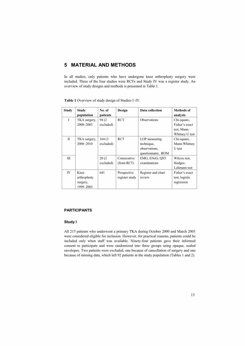
5 MATERIAL AND METHODS In all studies, only patients who have undergone knee arthroplasty surgery were included. Three of the four studies were RCTs and Study IV was a register study. An overview of study designs and methods is presented in Table 1. Table 1 Overview of study design of Studies I–IV. Study Study
population No. of patients
Design Data collection Methods of analysis
I TKA surgery, 2000–2003
94 (2 excluded)
RCT
Observations Chi-square, Fisher’s exact test, Mann-Whitney U test
II TKA surgery, 2008–2010
164 (3 excluded)
RCT
LOP measuring technique, observations, questionnaire, ROM
Chi-square, Mann-Whitney U test
III 20 (2 excluded)
Consecutive (from RCT)
EMG, ENeG, QST examinations
Wilcox test, Hodges-Lehmann test
IV Knee arthroplasty surgery, 1999–2003
641 Prospective register study
Register and chart review
Fisher’s exact test, logistic regression
PARTICIPANTS
Study I All 215 patients who underwent a primary TKA during October 2000 and March 2003 were considered eligible for inclusion. However, for practical reasons, patients could be included only when staff was available. Ninety-four patients gave their informed consent to participate and were randomized into three groups using opaque, sealed envelopes. Two patients were excluded, one because of cancellation of surgery and one because of missing data, which left 92 patients in the study population (Tables 1 and 2).

14
Study II Patients scheduled to undergo primary total knee arthroplasty (TKA) who were 75 years of age or younger and who were classified as American Society of Anesthesiologists (ASA) 1, 2, or 3 were considered eligible for inclusion. Patients who were unable to read and understand Swedish, had a systolic blood pressure of > 200 mm Hg, or had a thigh girth of > 78 cm were excluded. 164 patients gave their informed consent to participate and were randomized preoperatively to a control or intervention (LOP) group with the use of opaque sealed envelopes. Three patients were excluded after inclusion: one because of changing to a unicompartmental knee arthroplasty, one because of changing to a high flex prosthesis, and one who was scheduled for revision surgery and thus had been incorrectly included in the study. In total, 161 patients, 83 in the control group and 78 in the LOP group, were included in the analysis (Tables 1 and 2). Two patients were lost to follow-up at two months (Figure 5). Study III This study was part of Study II. All patients in Study II were given written and verbal information stating that ten patients from each randomization group would be asked to participate in neurophysiological examinations postoperatively on day three and then again two months after their surgery. Twenty consecutive patients were enrolled in this neurophysiological study between November 2009 and June 2010. Patients with diabetes mellitus or spinal disorders, as well as those who had received chemotherapy or had a body mass index (BMI) > 30 were excluded. Two patients were excluded after inclusion: one who was incorrectly included due to a high BMI (40) and one who declined to participate after the first examination (Tables 1 and 2, Figure 5).

15
Figure 5 Flowchart of all patients in Studies II and III.
RCT Oct 2008-June 2010
n = 456
Randomized n =164
Exclusion Age > 75 years
n = 153 No staff available for
inclusion n = 139
Control group n = 84
LOP group n = 80
Altered surgery method n = 1
Altered surgery method, revision
n = 2
Follow-up postop n = 78
Follow-up postop n = 83 Lost
n = 2
Surgery on a Tuesday LOP n = 11
Surgery on a Tuesday Control n = 9
ENeG day 3 n = 9
ENeG day 3 n = 10
ENeG and EMG 2 months
n = 9
ENeG and EMG 2 months
n = 9
Lost n = 1
Lost n = 1
RCT
Neurophysiological Study, Nov 2009–June 2010 Inclusion: Tuesday surgery Exclusion: diabetes, back diseases, earlier chemotherapy, BMI > 30

16
Study IV All patients who had undergone knee arthroplasty surgery and were registered in the local clinical audit database during the period 1999–2003 were considered to be eligible for inclusion. We did not include patients after 2003 because the register had been redesigned and it was not possible to record a tourniquet complication. A consecutive series of 577 patients with primary knee arthroplasties (465 TKA and 112 UKA), 46 revision knee arthroplasties, and 18 patellar supplementing knee arthroplasties were identified and included in the study, in total, 641 patients (Tables 1 and 2). Table 2 Overview of participants and baseline characteristics of all patients included. Study I Study II Study III Study IV Sex # Female Male
60 (65%) 32 (35%)
85 (53%) 76 (47%)
10 (56%) 8 (44%)
420 (65%) 221 (34%)
Age, years* 71 (11) (42–94) 65 (6) (44–75) 65 (8) (46–74) 70 (10) (28–94) ASA # 1 2 3 4 Missing
No co- morbidities 46 (50%)
28 (17%) 89 (55%) 40 (25%) 4 (3%)
7 (39%) 11 (61%)
95 (15%) 297 (46%) 150 (23%) 99 (15%)
Diabetes # 5 (5%) 23 (14%) 89 (14%) BMI #, * Overweight
12 (13%) 29 (4) 26 (3) 29 (5)
Op method # UKA TKA Patella suppl Revision
92
161
18
112 (17%) 465 (73%) 18 (3%) 46 (7%)
Tourniquet time* , minutes
96 (22) (56–154) 87 (21) (3–145) 82 (16) (56–122) 97 (22) (39–156)
Tourniquet cuff pressure*, mm Hg
257 (18) (200–280)
249 (33) (150–300)
237 (33) (172-294)
259 (20) (200-300)
*The values are given as the mean, standard deviation, and minimum to maximum. #The values are given as the number of patients with the percentage in parenthesis.

17
STUDY DESIGNS
Study I was a RCT. Patients were randomized into three groups and for each group different methods for protection underneath the tourniquet cuff were used: the elastic stockinette, group (E) (DeltaNet, Smith and Nephew, Mölndal, Sweden); the cast padding group (C) (Soffban Synthetic, Smith and Nephew, Mölndal, Sweden); and the group (N) in which no protective material was used. The OR nurse chose between a cylindrical 100-mm wide or a conical 140-mm thigh cuff depending on the circumference and shape of the patient’s thigh. The conical cuff was used in 46%. Directly after the surgery, when the tourniquet was removed, the OR nurse inspected the patient’s skin under the cuff and recorded the presence or absence of blisters. Superficial injuries such as indentation and redness were not regarded as skin injuries. In order to evaluate the clinical importance of blisters registered directly after surgery, the incidence of these blisters was compared with the incidence of clinically significant skin injuries after the use of a tourniquet cuff in primary TKA in a clinical audit. Clinically significant skin injuries registered in the audit were defined as an injury requiring any form of treatment, e.g. repeated dressing, wound care, or surgical intervention. Study II was a RCT. The sample size was calculated to detect a difference of 5 points (standard deviation (SD), 10 points) in the WOMAC pain score between the control group and the LOP group on day four. A total of 64 patients in each group (128 patients in the series) were required to detect this difference with an 80% power at the 5% significance level, two-tailed. Since we anticipated a drop-out rate of about 25%, the recruitment goal was determined to be somewhat higher. When we were planning for this study, there were no earlier published randomized studies with cuff pressure as the outcome measure, so the sample size was calculated to detect differences in postoperative pain (Kirkley et al., 2000). For the patients randomized to the control group, the standard method at our department was used, i.e. the tourniquet cuff pressure was based on the patient’s systolic blood pressure and a margin that was decided by the surgeon. For patients randomized in the LOP group, the tourniquet cuff pressure was determined by measuring the LOP using an automated photoplethysmographic sensor connected to an ATS 3000 tourniquet apparatus (Zimmer Sweden Inc®). The OR nurse chose the tourniquet cuff and measured the LOP pressure when the patient was prepared for the surgery. The conical cuff was used in 90%. The skin underneath the tourniquet cuff was protected with a double elastic stockinette (DeltaNet, Smith and Nephew, Mölndal, Sweden). Before starting the surgery, the surgeon determined the tourniquet cuff pressure according to the standard method. The OR personnel applied this pressure if the patient was randomized to the control group, and if the patient was randomized to the LOP group, they applied the pressure suggested by the LOP method. The surgeon was blinded to the randomization and was not informed of the tourniquet pressure that was actually applied. The surgery was

18
performed according to the routine at our department. All patients received perioperative antibiotics and low-molecular-weight heparin. All patients also received a local infiltration analgesic (LIA) at the end of the surgery by infiltration into the fascia, muscles, and subcutaneous tissue, regardless of whether they had had spinal or general anesthesia. In 147 patients a catheter was also inserted into the knee joint for pain treatment with a local anesthetic. It was used during the day after the surgery and was then withdrawn (Kerr & Kohan, 2008; Essving et al., 2011). Study III was a neurophysiological investigation and part of Study II. The number of patients included was chosen on the basis of earlier studies with EMG examinations in which 20 to 25 patients had been included (Weingarden et al., 1979; Saunders et al., 1979; Dobner & Nitz, 1982; Arciero et al., 1996). Electroneurography (ENeG) (Nerve conduction studies, NCS) was performed bi-laterally on day three and two months later with surface electrodes according to the standard routine at the Karolinska Department of Clinical Neurophysiology, using a Nicolet VikingSelect EMG system (Care Fusion, Middleton WI, USA). When necessary, the limbs were warmed with heating bags to keep skin temperature at 32˚C. Motor nerve conduction was studied in the peroneal and tibial nerves and included conduction velocity, distal latency, response amplitude, and F-wave latencies. Sensory nerve conduction studies were done on the superficial peroneal nerve and the sural nerve (conduction velocity and amplitude). Concentric needle electrode electromyography (EMG) was performed bilaterally at the second visit using the same equipment. EMG activity was studied in the vastus lateralis and anterior tibial muscles and in the medial head of the gastrocnemius muscle. Quantitative sensory tests (QSTs) were performed on day three and after two months using the Medoc TSA-II Neurosensory Analyzer (Medoc Ltd, Ramat Yishai, Israel) and included determinations of temperature thresholds anteriorly at the middle of the lower legs and on the dorsum of the feet. Differences between the operated and the unoperated leg were calculated at both visits. At the second visit, the test results were also compared with those from the first visit. The neurophysiologist who performed the examinations was blinded to the allocation group and to any information about the surgery, i.e. tourniquet cuff pressure, duration of tourniquet application, complications, or any clinical nerve symptoms from the operated leg. Study IV was a prospective register study. All patients that had undergone surgery at the department since 1996 and had been prospectively registered in a clinical audit database in which all complications within six weeks after surgery had been recorded and validated. This audit was part of a routine quality control and a follow-up rate of 98% was achieved.

19
Since we wanted dispersion in tourniquet times, a consecutive series of 577 primary knee arthroplasties (465 total knee arthroplasties and 112 unicompartmental knee arthroplasties), 46 revision knee arthroplasties, and 18 patellar supplementing knee arthroplasties was identified in the registry during the period 1999–2003 and included in the study. The postoperative complications recorded for this study were: superficial wound infections (treated with antibiotics), deep wound infections (requiring surgical intervention), deep vein thrombosis (verified by ultrasonography or phlebography), pulmonary embolism (verified by computed tomography), nerve injuries (verified by clinical examination or EMG), compartment syndrome (verified by clinical examination and fasciotomy), cuff pressure injury (documented on the medical chart), and bandage injury. These complications were chosen because they have been described earlier as possibly being associated with the use of a bloodless field (Alcelik et al., 2012; Butt et al., 2011; Noordin et al., 2009; Wakai et al., 2001). An additional review of complications was conducted using medical charts.
OUTCOME MEASUREMENTS/STUDY INSTRUMENTS
An overview of outcome measures used in Study II is presented in Table 3 and described in detail below. Limb Occlusion Pressure (LOP) In the intervention group (LOP group) the tourniquet cuff pressure was determined by measuring the LOP just before surgery using an automated photo plethysmographic sensor connected to an ATS 3000 tourniquet apparatus (Zimmer Sweden Inc® Sävedalen, Sweden). The recommended tourniquet pressure (RTP) was defined as the LOP plus a safety margin of 50 mm Hg for LOP < 130 mm Hg, 75 mm Hg for those with LOP between 131 and 190 mm Hg, and 100 mm Hg for those with LOP > 190 mm Hg. These recommended margins were higher than reported earlier (Noordin et al., 2009; Younger et al., 2004), but were the preadjustable margins indicated by the manufacturer (Zimmer Sweden Inc®). WOMAC There is a consensus that health-related quality-of-life (HRQoL) outcome measures should be used in research and in clinical settings. Assessments of HRQoL can detect small, but clinically relevant, differences and can be used to measure the impact the disease has on an individual (Jones & Pohar, 2012). The Western Ontario and McMaster Universities Osteoarthritis Index (WOMAC) questionnaire was used in Study II to evaluate the patient’s opinion concerning their knee pain and associated knee function. WOMAC is a disease-specific, widely used
20
self-administered health status measure, assessing three separate dimensions, i.e., pain (5 questions), stiffness (2 questions), and function (17 questions), in patients with osteoarthritis (OA) of the hip or knee (Bellamy et al., 1988). WOMAC has been used in several studies to assess pain, stiffness, and function after knee arthroplasty surgery. The Swedish format with five Likert boxes was used and a summary score was calculated for each dimension. All questions are graded from 0 to 4 points (Roos et al., 1999). VAS After the surgery was completed in Study II, the surgeon was asked to rate the quality of the bloodless field on a visual analog scale (VAS) where 1 indicated the worst possible and 10 the most optimal rating. They were also asked to rate whether they had had any technical difficulties due to the quality of the bloodless field (1 indicating no difficulties and 10 extreme difficulties). Pain assessments were made daily in Study II by using the VAS. A nurse asked the patients to rate their pain on the scale, where 0 represented “no pain” and 10 “worst possible pain.” Additional Patient Evaluations In Study II, on postoperative days two and four, the patient’s experience of the quadriceps muscle underneath the tourniquet cuff was analyzed with the question: Do you experience any discomfort from the thigh underneath the tourniquet cuff? The answer was graded at three levels: (1) none, (2) muscle soreness, and (3) pain. Wound Inspection An inspection of the skin underneath the tourniquet was done directly after the surgery and on day four with four possible answers: (1) without remark, (2) clear indentation and redness, (3) blisters, and (4) other skin injuries underneath the tourniquet cuff. A surgical wound check was done when the patient was discharged from the ward, most often on day four. Possible answers were: (1) without remark, (2) hematoma, (3) blisters, (4) ozzing wound, and (5) signs of wound infection. An inspection of the surgical wound was also done at the follow-up visit two months postoperatively. Possible answers were: (1) without remark, (2) hematoma, (3) blisters, (4) delayed wound healing, and (5) postoperative wound infection.

21
Range of Knee Motion The functional assessment in Study II included measuring the range of motion (ROM) of the knee joint (lying down and sitting up) with a goniometer on the third postoperative day by an independent physiotherapist. A straight-leg lifting test (1-kg weight) lying down was also done and measured by five different high levels: from (1) ≤ 10 centimeters to (5) > 40 centimeters. All documented measurements were taken on the patient’s fourth attempt. At the two-month follow-up, the ROM of the knee joint was measured again, by a blinded orthopedic surgeon. Table 3 Overview of outcome measures in Study II. Outcome measures
Measures Time Responders Analysis
Quality of tourniquet
VAS, worst possible to most optimal
Directly after surgery
Surgeon Mann-Whitney U test
Technical difficulties due to the quality
VAS, no difficulties to extreme difficulties
Directly after surgery
Surgeon Mann-Whitney U test
Skin check underneath the cuff
Without remark/blisters /other skin injuries
Directly after surgery
OR nurse Chi-square test
Pain WOMAC Days 1,2,3, Patient Mann-Whitney U test
Surgical wound check
Without remark/ blisters /oozing from the wound /signs of infection
Day 4 Nurse Chi-square test
Skin check underneath the cuff
Without remark/ blisters/other skin injuries
Day 4 Nurse Chi-square test
Pain in thigh muscle
None/pain Days 2,4 Nurse Chi-square test
Knee ROM Extension/flexion Day 3 Physio- therapist
Mann-Whitney U test
Pain, stiffness, and physical function
WOMAC Day 4 Patient Mann-Whitney U test
Surgical wound check
Without remark/delayed wound healing / postoperative wound infection
2 months Surgeon Chi-square test
Knee ROM Extension /flexion 2 months Surgeon Mann-Whitney U test
Pain, stiffness, and physical function
WOMAC 2 months Patient Mann-Whitney U test

22
NEUROPHYSIOLOGICAL EXAMINATIONS
Neurophysiological testing is important in defining the neurogenic basis of weakness and localizing the site of the nerve lesion. It is also helpful in determining the severity of injury. However, it does not indicate the etiology, which must be inferred on clinical grounds. Electromyography The striated muscles are functionally built up by motor units. Each motor unit consists of an anterior horn cell, its motor nerve fiber, and the muscle fibers innervated by intramuscular branches from this axon. A single nerve impulse activates almost simultaneously all muscle fibers within the motor unit. The contraction of the muscle fibers is, in turn, dependent on a depolarizing activity spreading along the muscle fiber membrane. This electrical activity can be picked up by a needle electrode within the muscle and constitutes the basis for electromyography (EMG). The electrical activity from muscle fibers of one motor unit is summated to form a motor unit potential, a MUP. The EMG represents all MUP activity in a muscle. A nerve injury will be reflected in the EMG activity in several ways. Loss of activity in motor nerve fibers implies a reduction in the number of MUPs that can be recruited in a contraction. The extent of the loss of MUPs depends on how many nerve fibers have been severed. The axonal degeneration of a motor nerve fiber results in a group of denervated muscle fibers. After a certain delay (usually 1–4 weeks), these fibers will exhibit individual spontaneous contractions due to an increased sensitivity to small amounts of circulating acetylcholine. The electrical activity from these feeble contractions in single muscle fibers is seen in the EMG as low amplitude fibrillation potentials and positive sharp waves, so-called denervation activity (Aminoff, 2004). Nerve Conduction Studies Nerve conduction studies (NCSs), or electroneurography (ENeG), allow assessments of function in motor and sensory nerves (Aminoff, 2004). Motor NCSs are made by electrical stimulation of a peripheral nerve and recording from a muscle supplied by this nerve. The time it takes for the nerve impulse to travel from the stimulation to the recording site is determined. This latency (in ms), as well as the size of the response, the amplitude (in mV), is measured. By stimulating in two or more different locations along the same nerve, the nerve conduction velocity (NCV, m/s) across different segments can be determined. Calculations are made using the distance (mm) between the different stimulation points and the difference in latencies (ms). In this study, the motor NCS comprised the peroneal and tibial nerves on both sides.
Sensory NCSs are made by electrical stimulation of a purely sensory portion of a peripheral nerve and recording the sensory nerve impulse at a distance along the nerve. Sensory amplitudes are much smaller than motor amplitudes – only a few

23
microV (µV), for which reason, ten responses are averaged to diminish background noise. Calculation of the sensory NCV (m/s) is based on the latency (ms) and the distance (mm) between the stimulating and recording electrodes (Aminoff, 2004). Sensory NCSs were made bilaterally in the sural nerve and in the superficial peroneal nerve.
A volley of nerve impulses initiated by electrical nerve stimulation at the ankle will travel antidromically (“backwards”) in the motor nerve fibers up to the spinal cord so as to depolarize the spinal motor neurons, leading to many impulses bouncing back down the leg to give a twitch in the foot muscles. This response is called an F wave, and the latency (ms) is determined from the stimulation to the muscle twitch (Aminoff, 2004). Thus, these nerve impulses pass the knee and the thigh twice, and a small change in nerve conduction might be doubled and thereby easier to reveal. An injury to the motor nerve fibers might appear as an increase in latency and/or a reduction in persistence, i.e. fewer impulses are returning to the foot muscles. F waves were studied in the peroneal and tibial nerves in both legs.

24
6 STATISTICAL METHODS Different versions of the statistical software SPSS (IBM Corp, Armonk, New York, USA) were used in all studies. In Study IV the statistical software R version 2.15.1 (R Foundation for Statistical Computing, Vienna, Austria) was also used for analyses of collinearity, consistency, and the receiver operator curve (ROC). For all analyses, the level of significance was set to 0.05, and all p values were two-sided. In Study 1 we analyzed the result using the Chi–square test or Fisher’s exact test with nominal variables and the Mann-Whitney U test for scale and ordinal variables. In Study II categorical variables were tested for differences between randomization groups using the Chi-square test. Continuous variables were tested with the Mann-Whitney U test. The non-parametric test was used because it was deemed more appropriate for the subjective scale variables and for the remaining continuous variables, for which the assumption of normality was not satisfied. All analyses were performed according to the intention-to-treat (ITT) principle. In Study III the scale variables were tested with the non-parametric Wilcox test because of the small number of included patients and the risk regarding the assumption of normality. The medians of the differences are estimated using the Hodges-Lehmann estimator and are presented with a 95% confidence interval (CI). In Study IV categorical variables were tested with Fisher’s exact test and, for continuous variables, Student’s t test was used. Associations were investigated using univariable and multivariable logistic regression analyses. The Hosmer-Lemeshow goodness-of-fit test was used to examine the multivariable model with p = 0.525 for the model presented in the article with cuff pressure analyzed as a continuous variable and p = 0.407 in the model with all variables analyzed as categorical variables, indicating an acceptable fit. Outliers were investigated using Cook’s distance and no extreme outliers were detected. The variance inflation factor (VIF) was relatively low, < 3 for all variables, indicating that no multicollinearity was present. Linearity for the continuous variables was investigated using smoothed partial residual plots and by modeling the variables with restricted cubic splines and plotting the functional form.

25
7 RESULTS STUDY I
Primary Outcome The elastic stockinette (E) was significantly better than no protective material (N) (p = 0.004) and there was a trend towards better results in the elastic stockinette group than in the cast padding group (p = 0.09). Ten patients developed skin blisters underneath the tourniquet cuff, seven from the N group and three from the cast padding group (C), giving an overall incidence of 11%. Patients who developed blisters had a longer duration of the bloodless field than those without blisters: 112 (SD 29) min and 94 (SD 21) min, respectively (p = 0.04). There were no significant differences in cuff pressure, 255 (SD 11) mm Hg and 257 (SD 18) mm Hg, thigh circumference, 55 (SD 9) cm and 54 (SD 7) cm, or age, 72 (SD 12) years and 71 (SD 11) years, between patients who developed blisters and those who did not. An additional result not presented in the article was that 13% of women compared with 6% of men developed blisters. Secondary Outcome The overall incidence of skin injuries in our local quality audit requiring any form of treatment recorded during the first six weeks after TKA surgery with tourniquet use was 1.5% (11/728) during the years 1997–2003 (personal communication). STUDY II
Primary Outcome Patients in the LOP group had a tourniquet cuff pressure of ≤ 225 mm Hg more often than those in the control group (p < 0.001). The mean tourniquet cuff pressure was also generally lower in patients in the LOP group, but this difference was not statistically significant (p = 0.362). Ratings of postoperative pain on the self-administered WOMAC questionnaire during hospitalization did not differ between the randomization groups. Secondary Outcome No significant difference between the groups could be detected regarding the quality of the bloodless field or the technical difficulties as judged by the surgeon.

26
Ratings of function on the self-administered WOMAC questionnaire during hospitalization did not differ between the randomization groups, but the patients in the LOP group reported being significantly less stiff in their knee on day four (p = 0.02) The range of motion of the knee on postoperative day three and the ability to do a straight-leg lift did not differ between the groups. Neither did the knee ROM, assessed by a blinded orthopedic surgeon, at the two-month follow-up visit. At the time of discharge, 47 patients (30% of the 158 available ones) had developed a surgical wound complication such as blisters, oozing from the wound, or signs of infection. Forty of these 47 patients had had a cuff pressure of > 225 mm Hg during surgery. None of the patients with a cuff pressure of ≤ 225 mm Hg had any signs of wound secretion or infection, but seven patients had blisters around the knee. There were no significant differences between the LOP and the control groups (p = 0.149). At the two-month follow-up time, seven patients (4%) had a postoperative wound infection and nine (6%) were recorded as having delayed wound healing. Four of these patients had developed a deep wound infection after they were discharged from the ward and had been rehospitalized and reoperated on. Fourteen of the 16 patients with a surgical wound complication at the two-month follow-up visit had had a cuff pressure of > 225 mm Hg. Two patients who had had a cuff pressure of ≤ 225 mm Hg had delayed wound healing. There were no differences between the LOP group and the control groups (p = 0.869). Six of the seven patients who had a postoperative wound infection and all four patients who had a deep wound infection were men. We found no difference in cuff pressures between men and women, but men had significantly longer bloodless field times (mean, 89 min compared with 73 min for women; p = 0.027). STUDY III
Primary Outcome Electromyographic signs of recent denervation were found in one patient. This patient had had the highest cuff pressure in the study, 294 mm Hg for 100 minutes. At two months this patient had fibrillations in the vastus lateralis and the gastrocnemius muscles which were associated with reduced voluntary activity. Nerve conduction studies in the peroneal nerve on day three and at two months showed a reduction in response amplitude, a prolongation of F-wave latency, and a reduction in F-wave persistence. An increase in latency in the tibial nerve was seen only on day three. Fifteen out of 18 included patients were reached in a telephone follow-up. Only the patient with an EMG-evident denervation reported radiating pain and a tingling sensation in the operated leg and foot for about six months after the operation, after which the symptoms disappeared. None of the other patients had had any similar symptoms.

27
Secondary Outcome In three other patients, EMG revealed minor signs of chronic changes with motor unit potentials showing increased duration and amplitude. These patients had no signs of ongoing denervation. Five patients had one or more minor deviations in electro-neurography or QST values that might indicate a change in nerve function related to the surgery. However, the deviations were small and were not considered to be definite signs of recent nerve injury. None of these patients showed any clinical symptoms of nerve damage. The analysis of motor or sensory conduction velocity showed no statistically significant difference (median of differences) when operated and unoperated legs were compared. Sensory nerve amplitudes were lower in the operated leg on day three for both the sural nerve (2 microV, CI 0.5–4; p = 0.01) and the superficial peroneal nerve (1.5 microV, CI 0–3; p = 0.06) and at two months (sural nerve 2.5 microV, CI 0–5; p = 0.04) and the superficial peroneal nerve (1 microV, CI -0.5–2.5; p = 0.08). There was no statistically significant difference in F-wave latencies and persistences between day three and at two months or between the operated and the unoperated leg. STUDY IV
Primary Outcome We found an association between tourniquet time and an increased risk of a complication after knee arthroplasty surgery in a bloodless field. When the tourniquet time exceeded 100 minutes, a univariable analysis showed an increased risk for a complication compared with a tourniquet time of ≤ 100 minutes (OR 2.2, CI 1.5–3.1). This result remained after adjusting for cuff pressure (continuous), sex, age (≤ 70 vs. > 70 years), ASA classification (1–3), smoking (yes/no), diabetes (yes/no), and surgery indication (primary, revision, patella supplementing) (OR 2.4, CI 1.6–3.6). For every 10-minute-longer duration of tourniquet time, the odds for having a complication increased by 20% (OR 1.2, CI 1.1–1.4). Since the recommended time limit at our department is 120 minutes, we also analyzed the tourniquet time variable as ≤ 120 vs. > 120 minutes. These results also showed increased odds for suffering a complication (OR 1.9, CI 1.1–3.2). Secondary Outcome Age, sex, surgery indication, diabetes, smoking, and cuff pressure showed no statistically significant association with suffering a complication in neither the univariable model nor the different multivariable models. However, patients classified as ASA 2 and 3 did show an association for suffering a complication compared to ASA 1 (ASA 2, OR 2.5, CI 1.3–4.7, p = 0.006 and ASA 3, OR 2.9, CI 1.4–5.9, p = 0.003) (Table 4).

28
Table 4 Factors associated with risk of suffering a complication.
In total, 168 patients (26%) had a complication recorded. Ninety-four of these patients (35%) had had a bloodless field time longer than 100 minutes. The mean bloodless field time for the patients who had a recorded complication was 104 minutes compared to 95 minutes for those who had no complication. Three of the 16 patients with a reinflated tourniquet cuff had a complication (2 wound complications and 1 DVT). Among women, the impact of tourniquet time on suffering a complication was greater (OR 3.0, CI 1.8–5.0) than for men (OR 1.6, CI 0.8–3.0), although this difference was not statistically significant: 39% of the women who had a tourniquet time of > 100 minutes had a complication compared to 29% of the men.
Predictor variable
Level Crude measures
Univariable
p value
Multivariable
p value
Adjusted for the final model*
n OR (95%CI) OR (95%CI) Age, years
≤ 70 > 70
291 350
Reference 1 (0.7–1.4)
0.9
Sex
Male Female
420 Reference 1 (0.7–1.4)
0.8
ASA#
1 2 3
95 297 150
Reference 2.4 (1.3–4.4) 2.6 (1.3–5.0)
0.014
2.5 (1.3–4.7) 2.9 (1.4–5.9)
0.009
Diabetes
No Yes
549 89
Reference 1 (0.6–1.6)
0.9
Smokers
No Yes
561 80
Reference 1.1 (0.6–1.8)
0.8
Indication
Primary Revision Patella suppl
577 46 18
Reference 1.7 (0.9–3.2) 0.8 (0.3–2.6)
0.2
Tourniquet time, min
≤ 100 > 100
373 268
Reference 2.2 (1.5–3.1)
<0.001
2.4 (1.6–3.6)
<0.001
Cuff pressure, mm Hg
≤ 260 > 260
345 293
Reference 1.1 (0.7–1.5)
0.8
# 99 patients with missing ASA recording, *Hosmer and Lemeshow, p = 0.40

29
8 DISCUSSION GENERAL DISCUSSION
This thesis shows that tourniquet use can affect the outcome after knee arthroplasty surgery. The results from the four studies demonstrate key factors that appear to be important to avoid complications associated with the use of tourniquets. Longer tourniquet times were associated with an increased risk of complications and the use of higher tourniquet cuff pressures resulted in more frequent wound complications. The LOP measuring technique led to lower cuff pressures. However, the results did not show any differences between the LOP group and the control group regarding postoperative pain. In the neurophysiological study, only one patient, who had the highest cuff pressure, developed a nerve injury while patients with lower cuff pressures had no nerve injuries. The total incidence of nerve injuries was 2% in Studies II and III. Protective material underneath the tourniquet cuff reduced the risk of skin injuries, i.e. blisters. An elastic stockinette appeared to be best even if it did not entirely eliminate the risk. Is the choice of protective material used underneath the tourniquet cuff of any importance? The application of protective material underneath the tourniquet cuff appears to influence the risk of a skin injury after TKA surgery using a tourniquet. None of the patients with the elastic stockinette developed any skin injuries underneath the cuff in Study I. These findings are consistent with those in other published studies (Din & Geddes, 2004; McEwen, 2002). The highest incidence of skin blisters was found in patients with no protective material underneath the tourniquet cuff. In Study II the skin underneath the cuff was protected with a double-elastic stockinette in all patients, but still, 2% of them had blisters underneath the cuff immediately after surgery. These patients with blisters in Study II belonged to the group with higher cuff pressures of 260 mm Hg or higher. However, the reasons for the development of these blisters are not well known. One reason could be the fluid running underneath the cuff and another could be the presence of a pressure ulcer (EPUAP, 2009). On postoperative day four, 7% of the patients in Study II had developed blisters or other pressure-related injuries underneath the tourniquet cuff. This is consistent with the literature describing how pressure-related ulcers develop during a rather long time after the triggering event (Schoonhoven et al., 2002; Feuchtinger et al., 2007). Unfortunately, in Study I, we were not able to follow up the patients and therefore have no information on the further development of skin blisters among these patients.
30
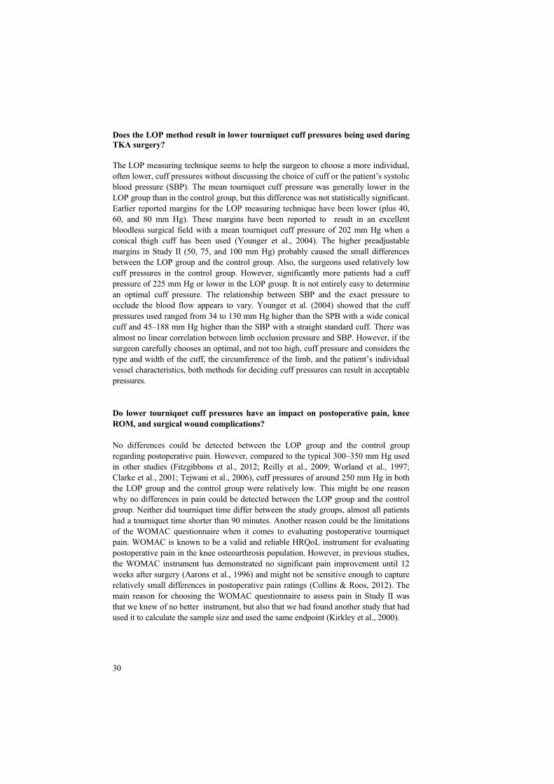
Does the LOP method result in lower tourniquet cuff pressures being used during TKA surgery? The LOP measuring technique seems to help the surgeon to choose a more individual, often lower, cuff pressures without discussing the choice of cuff or the patient’s systolic blood pressure (SBP). The mean tourniquet cuff pressure was generally lower in the LOP group than in the control group, but this difference was not statistically significant. Earlier reported margins for the LOP measuring technique have been lower (plus 40, 60, and 80 mm Hg). These margins have been reported to result in an excellent bloodless surgical field with a mean tourniquet cuff pressure of 202 mm Hg when a conical thigh cuff has been used (Younger et al., 2004). The higher preadjustable margins in Study II (50, 75, and 100 mm Hg) probably caused the small differences between the LOP group and the control group. Also, the surgeons used relatively low cuff pressures in the control group. However, significantly more patients had a cuff pressure of 225 mm Hg or lower in the LOP group. It is not entirely easy to determine an optimal cuff pressure. The relationship between SBP and the exact pressure to occlude the blood flow appears to vary. Younger et al. (2004) showed that the cuff pressures used ranged from 34 to 130 mm Hg higher than the SPB with a wide conical cuff and 45–188 mm Hg higher than the SBP with a straight standard cuff. There was almost no linear correlation between limb occlusion pressure and SBP. However, if the surgeon carefully chooses an optimal, and not too high, cuff pressure and considers the type and width of the cuff, the circumference of the limb, and the patient’s individual vessel characteristics, both methods for deciding cuff pressures can result in acceptable pressures. Do lower tourniquet cuff pressures have an impact on postoperative pain, knee ROM, and surgical wound complications? No differences could be detected between the LOP group and the control group regarding postoperative pain. However, compared to the typical 300–350 mm Hg used in other studies (Fitzgibbons et al., 2012; Reilly et al., 2009; Worland et al., 1997; Clarke et al., 2001; Tejwani et al., 2006), cuff pressures of around 250 mm Hg in both the LOP group and the control group were relatively low. This might be one reason why no differences in pain could be detected between the LOP group and the control group. Neither did tourniquet time differ between the study groups, almost all patients had a tourniquet time shorter than 90 minutes. Another reason could be the limitations of the WOMAC questionnaire when it comes to evaluating postoperative tourniquet pain. WOMAC is known to be a valid and reliable HRQoL instrument for evaluating postoperative pain in the knee osteoarthrosis population. However, in previous studies, the WOMAC instrument has demonstrated no significant pain improvement until 12 weeks after surgery (Aarons et al., 1996) and might not be sensitive enough to capture relatively small differences in postoperative pain ratings (Collins & Roos, 2012). The main reason for choosing the WOMAC questionnaire to assess pain in Study II was that we knew of no better instrument, but also that we had found another study that had used it to calculate the sample size and used the same endpoint (Kirkley et al., 2000).

31
On the fourth postoperative day, 40% of the patients reported that they had pain in the quadriceps muscle underneath the area where the tourniquet cuff had been placed. This was probably not captured either by the WOMAC questionnaire. Thigh pain after tourniquet use is not discussed very often. However, Tai et al. (2012) showed differences in the experience of thigh pain and knee pain between a tourniquet group and a non-tourniquet group after TKA surgery up to the fourth postoperative day. Yet, they reported a rather short duration of tourniquet inflation of only 52 minutes. Previous studies comparing knee or ankle surgery with and without the use of a tourniquet have shown significantly better knee flexion after surgery without a tourniquet (Konrad et al., 2005; Li et al., 2009; Wakankar et al., 1999). However, in Study II, no differences in knee ROM between the LOP and the control group could be demonstrated, neither on postoperative day three nor at the two-month follow-up. All patients achieved good knee flexion on the third day and at the two-month follow-up visit. In contrast to this finding, Ledin et al. (2012) demonstrated differences between groups when a tourniquet had been used or not used after two years. These differences in postoperative knee motion in earlier studies have decreased with time (Abdel-Salam & Eyres, 1995; Chang et al., 2012; Li et al., 2009). Chang et al. (2010) found a difference in knee ROM six weeks after TKA surgery in the group with a shorter tourniquet time (early release) compared to the group with tourniquet use throughout the procedure, but, after three months, no differences were found. Again, the relatively small differences in cuff pressures and tourniquet time in Study II may have had an impact on our results. There was a rather high incidence of surgical wound complications in Studies II and IV. One reason might be that we did not exclude patients with diabetes, as has been done in other published studies (Abdel-Salam & Eyres, 1995; Konrad et al., 2005; Li et al., 2009; Wakankar et al., 1999; Kvederas et al., 2012). In our studies, even patients classified as ASA 3, and some later classified as ASA 4, were included and, in Study IV, patients classified as ASA 2 and ASA 3 showed a statistically significantly higher risk of suffering a complication. Cuff pressures seem to be of importance when assessing the risk of surgical wound complications. No patient in Study II who had a cuff pressure of 225 mm Hg or lower developed a postoperative wound infection and they had also fewer surgical wound complications. An analysis of the data from Study IV with the same cuff pressure levels showed the same results. This finding is in accord with the report by Clarke et al. (2001), who studied the pattern of postoperative wound hypoxia seven days after knee surgery and found that a tourniquet cuff pressure of about 225 mm Hg yielded a significantly better return of the oxygen levels compared to cuff pressures of 350 mm Hg. Does lower tourniquet cuff pressure have an impact on the incidence of nerve injuries? The findings in Study III showed that lower tourniquet cuff pressures of 240 mm Hg resulted in few nerve function disturbances. Only one patient, who also had the highest

32
cuff pressure, had a post-operative nerve injury. The neurophysiological findings in this study are in contrast to those in a rather old study (1982) by Dobner and Nitz who reported that two thirds of patients who had undergone knee surgery in a bloodless field showed EMG evidence of denervation. However, their study was conducted when high cuff pressures were used and today’s lower tourniquet cuff pressures indicate that these older studies with cuff pressures of up to 450 mm Hg are probably no longer relevant. A wide variation in the incidence of nerve injuries has been demonstrated and discussed earlier. The reported rates range from 0.03% to 7.7% (Horlocker et al., 2006; Odinsson & Finsen, 2006). The reported severity of nerve injuries varies from a mild transient loss of function to permanent, irreversible damage. The important differences in study designs, ranging from prospective neurophysiological examinations of a whole study population to a retrospective report of clinically evident nerve injuries may provide an explanation. The findings from Studies II and III showed a total incidence of 2% of nerve injuries, which should be regarded as a rather reliable result since the patients were carefully followed up. Correlations between nerve injuries and prolonged tourniquet time have been reported (Horlocker et al., 2006; Jacob et al., 2011). The results in our Study III may not be comparable with theirs since we had a mean bloodless field time of 81 minutes, with a longest duration of 122 minutes. Furthermore, the cited authors did not report cuff pressures, which are perhaps even more important when discussing post-surgery nerve injuries. Does tourniquet time have an association with the incidence of suffering a complication after knee arthroplasty? The findings in Study IV are in accord with earlier published studies, i.e. a longer tourniquet time increases the risk of complications (Butt et al., 2011; Hernandez et al., 2012; Horlocker et al., 2006; Jacob et al., 2011). Every additional 10 minutes of tourniquet time was associated with an increased risk for complications. When the tourniquet time exceeded 100 minutes 35% of the patients developed a complication compared with 20% of those with a shorter tourniquet time in Study IV. These complications may increase the risk of other complications, such as secondary infection of the surgical wound or, even worse, at the site of the endoprosthesis, and can lead to suffering by the patient and a prolonged hospital stay. It is generally recommended that the use of a tourniquet should be limited to two hours due to the risk of complications. Fitzgibbons et al. (2012) concluded that tourniquet complications are mostly of a minor and short-term nature and that there are no contraindications against longer tourniquet times when necessary. However, every complication may interfere with the postoperative functional recovery and could lead to unnecessary discomfort for the patient. It is therefore important that the tourniquet time should be minimized for an optimal outcome.

33
Do men and women tolerate tourniquet use differently? The results of the studies in this thesis have pointed out some differences between the sexes in terms of tolerating a tourniquet. In Study IV this was part of the research question, but in the other studies, this question was not raised when designing the studies. Nor are gender differences something that has received attention in the literature. Both in Study I and Study IV, the women had a higher rate of skin injuries underneath the cuff than men. This finding is in accordance with a study by Lindgren et al. (2005) indicating that women are at a higher risk of developing pressure ulcers after surgery. However, both in Study I and Study IV, the patients in whom skin injuries underneath the cuff were recorded also had longer tourniquet times. In Study II, when men had statistically significantly longer tourniquet times there were no differences between them in developing skin injuries underneath the cuff. Six of the seven patients who had a postoperative wound infection and all four patients who had a deep wound infection in Study II were men. We found no difference in cuff pressures between men and women, but men had significantly longer bloodless field times. Also in Study IV, more men were recorded as having a surgical wound complication, 20%, compared to 13% of the women. These results are consistent with reports from the Swedish Knee Arthroplasty Register showing that men have a higher risk of knee infection after knee arthroplasty surgery (SKAR, 2012). A register study including almost 70,000 patients undergoing TKA surgery also confirms these results (Kurtz et al., 2010). However, there are no clear reasons for this given in the literature and the phenomenon needs to be further explored. Women appeared to tolerate longer tourniquet times worse than men in Study IV. Women had almost double the odds for a complication (OR 3.0) compared to men (OR 1.6) after a tourniquet time of over 100 minutes. However, this difference was not statistically significant. Still, a difference between men and women in terms of tolerating tourniquet time or perhaps tolerating tourniquet use differently in general has never been discussed earlier and this is something that needs further research. What impact does the OR nurse’s nursing actions have on the care of the patient when a tourniquet is used? Professional care of the patients throughout surgery with a tourniquet requires knowledge about tourniquet use from the whole OR team. The OR nurse has a responsibility to select a suitable tourniquet cuff and protective material underneath the cuff, to apply the cuff correctly and know how to avoid fluid running in underneath the cuff when the patient’s skin is being disinfected. These are routine procedures on the OR ward. However, these procedures require attention since we know that adverse events can occur, e.g. skin injuries underneath the cuff, and unnecessarily high cuff pressures when using a narrow straight cuff. The OR nurse has an important role in the care of patients who undergo surgery with the use of a tourniquet.
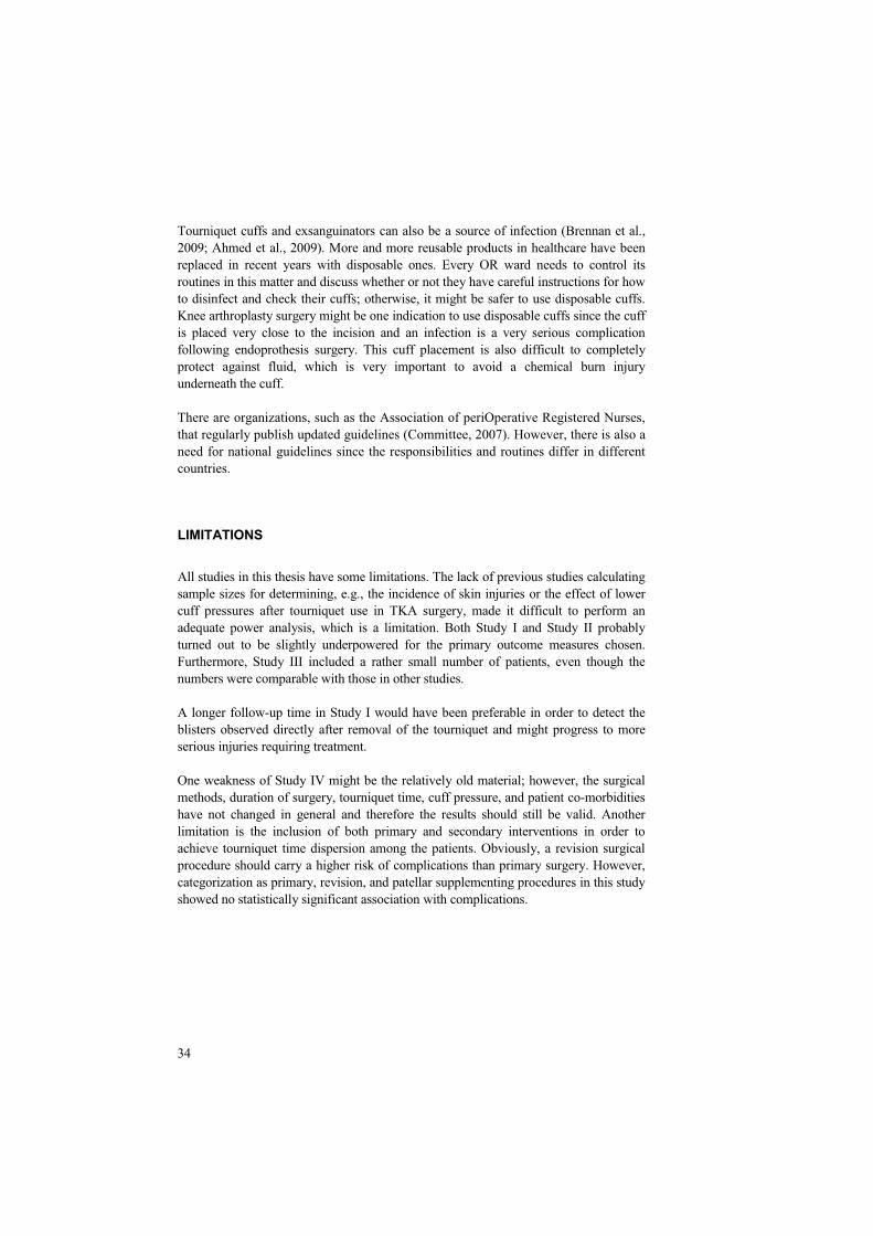
34
Tourniquet cuffs and exsanguinators can also be a source of infection (Brennan et al., 2009; Ahmed et al., 2009). More and more reusable products in healthcare have been replaced in recent years with disposable ones. Every OR ward needs to control its routines in this matter and discuss whether or not they have careful instructions for how to disinfect and check their cuffs; otherwise, it might be safer to use disposable cuffs. Knee arthroplasty surgery might be one indication to use disposable cuffs since the cuff is placed very close to the incision and an infection is a very serious complication following endoprothesis surgery. This cuff placement is also difficult to completely protect against fluid, which is very important to avoid a chemical burn injury underneath the cuff. There are organizations, such as the Association of periOperative Registered Nurses, that regularly publish updated guidelines (Committee, 2007). However, there is also a need for national guidelines since the responsibilities and routines differ in different countries. LIMITATIONS
All studies in this thesis have some limitations. The lack of previous studies calculating sample sizes for determining, e.g., the incidence of skin injuries or the effect of lower cuff pressures after tourniquet use in TKA surgery, made it difficult to perform an adequate power analysis, which is a limitation. Both Study I and Study II probably turned out to be slightly underpowered for the primary outcome measures chosen. Furthermore, Study III included a rather small number of patients, even though the numbers were comparable with those in other studies. A longer follow-up time in Study I would have been preferable in order to detect the blisters observed directly after removal of the tourniquet and might progress to more serious injuries requiring treatment. One weakness of Study IV might be the relatively old material; however, the surgical methods, duration of surgery, tourniquet time, cuff pressure, and patient co-morbidities have not changed in general and therefore the results should still be valid. Another limitation is the inclusion of both primary and secondary interventions in order to achieve tourniquet time dispersion among the patients. Obviously, a revision surgical procedure should carry a higher risk of complications than primary surgery. However, categorization as primary, revision, and patellar supplementing procedures in this study showed no statistically significant association with complications.

35
IMPLICATIONS FOR CLINICAL PRACTICE
The results of this thesis pinpoint some important issues that should be taken into consideration when a tourniquet is used. Patient co-morbidities: when the patient is classified as ASA 2 or higher in the ASA classification, evaluate whether tourniquet use is necessary and if it is helpful to release the tourniquet cuff when it is not needed any more. Cuff type: use the widest cuff as possible and when the shape of the extremity is suitable, choose a conical cuff. Protection material: use a double elastic stockinette underneath the tourniquet cuff to protect against skin injuries. Cuff pressure: use the LOP measuring technique, if possible, to choose the lowest possible cuff pressure. Otherwise, carefully choose an optimal and not too high cuff pressure and consider the type and width of the cuff, the circumference of the limb, and the patient’s individual vessel characteristics. Tourniquet time: use as short a tourniquet time as possible, e.g., release the tourniquet cuff after cementation in TKA surgery. Postoperative check: perform an immediate check of the skin underneath the cuff after surgery and a follow-up on the ward before discharge. Documentation should be designed so that it is useful for checking and following up relevant factors for tourniquet use, and also to continue the control on the ward. FUTURE RESEARCH
There are still many questions that need to be addressed regarding tourniquet use. How does the pressure affect the skin underneath the tourniquet cuff? Besides the chemical burns, no research is available on the way in which the cuff pressure affects the skin. For example, what is the cause of the blisters that develop underneath the cuff? Does the extremely high pressure from the tourniquet cuff result in a different event, or is the influence of tourniquet pressure of the same kind as for other pressure injuries according to the EPUAP classification? Are there differences between men and women in tolerating tourniquet use? Several findings in the Results section in this thesis indicate that this might be the case. However, since the studies were not designed primarily with this research question or hypothesis in mind, the results may not be suitable for drawing conclusions about these indications. Therefore, new studies to explore this matter would be recommendable.

36
To enrich and back up statistically reliable information obtained from numerical measurements with information including the participants’ explanations and experiences is a combination of both quantitative and qualitative methods that is recommended in future research (Sale et al. 2002). Throughout my work with this thesis I have not been able to find any studies describing patients’ experience of tourniquet use. Not even when surgery has been performed with local anesthesia have patients been asked to describe their experience. A study with a qualitative approach would probably provide new valuable knowledge and an opportunity to explore the experiences of patients from undergoing surgery with tourniquet use.

37
9 CONCLUSIONS
� Tourniquet use can have an impact on the outcome in knee arthroplasty surgery.
� The use of an elastic stockinette lowers the risk of skin complications
underneath the tourniquet cuff even if it does not entirely eliminate the risk.
� The LOP measuring technique helps the surgeon to choose a more individual, often lower, tourniquet cuff pressure.
� To use low cuff pressures appears to be an important factor for avoiding
surgical wound complications and wound infections.
� Also, low cuff pressures results in few minor nerve disturbances and rarely in nerve injuries.
� There is an association between tourniquet time and complications after TKA
surgery. The use of tourniquet times shorter than 100 minutes decreases the risk of a complication.

38
10 SAMMANFATTNING (SUMMARY IN SWEDISH) Vid en knäplastikoperation anses det vara en fördel att kunna utföra ingreppet i blodtomt fält (BTF). Operationen kan då genomföras med exakt kontroll över anatomin genom att inga störande blödningar uppstår. Användandet av BTF har i studier visats minska den totala blödningen och leda till kortare operationstid vid knäplastikkirurgi. Metoden är dock inte helt riskfri och komplikationer kan uppstå till följd av att BTF används. Syfte Det övergripande syftet med studierna i denna avhandling var att förbättra kunskaperna om BTF för att öka patientsäkerheten när knäplastikoperationer genomförs i blodtomt fält. I Studie I, en randomiserad kontrollerad studie (RCT) där 94 patienter inkluderades som skulle genomgå en total knäplastik (TKA) operation, var syftet att undersöka huruvida det var någon skillnad mellan en elastisk strumpa, vadd eller inget material alls under blodtomhetsmanschetten i uppkomst av hudskador efter en TKA operation i BTF. En elastisk strumpa var statistiskt signifikant bättre jämfört med att inte använda något skyddande material alls under manschetten och det fanns en trend för att elastisk strumpa var bättre än att använda vadd. I Studie II, en RCT med 164 inkluderade patienter som genomgick en TKA operation, var syftet att undersöka om användandet av limb occlusion pressure (LOP) metoden resulterade i att lägre manschettryck användes och om det ledde till mindre postoperativ smärta, skillnad i knärörlighet och skillnad i sårläkning för patienterna. Fler patienter i LOP gruppen hade ett manschettryck på 225 mm Hg eller lägre. Medelmanschettrycket var också generellt lägre i LOP gruppen jämfört med kontrollgruppen men denna skillnad var ej statistiskt signifikant. Svaren från patientenkäterna visade inte någon skillnad i postoperativ smärta och ingen skillnad i knärörlighet mellan LOP och kontrollgruppen upptäcktes. Ett viktigt bifynd var dock att oavsett randomiseringsgrupp drabbades ingen av de patienter som haft ett manschettryck på 225 mm Hg eller lägre av någon postoperativ sårinfektion och de hade också betydligt färre sårkomplikationer. I Studie III, som var en del av Studie II med 20 konsekutivt inkluderade patienter, var syftet att kontrollera hur ofta en nervskada till följd av BTF vid en TKA operation uppstår och att analysera resultaten efter de neurofysiologiska undersökningarna bland dessa patienter. En patient hade en med EMG bekräftad nervskada. Denna patient hade haft högst manschettryck bland studiepatienterna (294 mm Hg). Förutom att de sensoriska svaren var sämre i det opererade benet efter operationen noterades inga statistiska signifikanta skillnader i ENeG analyserna mellan patienternas opererade ben och icke opererade ben 2 dagar eller 2 månader efter operationen. I Studie IV, en prospektiv registerstudie med 641 inkluderade patienter som genomgått någon form av knäplastikoperation, var syftet att undersöka om blodtomhetstiden påverkade uppkomsten av postoperativa komplikationer och huruvida faktorer som

39
ålder, kön, ASA klassifikation, diabetes, rökning eller manschettryck påverkade risken för att drabbas av en postoperativ komplikation. Resultatet visade på ett samband mellan patienter som hade haft en blodtomhetstid längre än 100 minuter och en högre risk för att drabbas av en postoperativ komplikation. Även när blodtomhetstiden analyserades som en fortlöpande variabel visade resultatet att för varje 10 minuter med längre blodtomhetstid ökade oddset med 20 % att drabbas av en postoperativ komplikation. Slutsatser Användandet av blodtomt fält är oftast ett säkert och tillförlitligt hjälpmedel vid knäplastikoperationer. Viktiga faktorer att beakta som kan påverka resultatet av knäplastikoperationen i blodtomt fält är bl.a. typ av polster under manschetten, manschettrycket och blodtomhetstiden. Genom att skydda huden under manschetten med en dubbel elastisk strumpa, använda så lågt manschettryck som möjligt, helst 225 mm Hg eller lägre, och att undvika blodtomhetstider längre än 100 minuter minskar risken för att patienten drabbas av en komplikation som kan relateras till BTF användning vid knäplastikoperationer.

40
11 ACKNOWLEDGMENTS I wish to express my sincere gratitude to all of you who helped and supported me to make this thesis possible. I could not have done this without the support from all of you. I want to thank all patients who generously participated in the studies and also all colleagues, both doctors and nurses, who have encouraged me enormously. Especially, I want to express my gratitude to: My main supervisor, Richard Blomfeldt: Thank you for giving me this opportunity by accepting to be my supervisor; your friendly attitude and encouragement throughout the work on the thesis – and you have always believed in my capacity. Sari Ponzer, co-supervisor and also head of the Department of Orthopaedics, Södersjukhuset, with your great knowledge and experience of research, together with your encouragement and enthusiasm, you have given me invaluable guidance and support (also financial) throughout this work: Thank you! Per Hamberg, co-supervisor: Thank you for your support and encouragement, for initially supporting me in my first ethical application and for operating on so many knee arthroplasty patients who were suitable for inclusion in the studies. Ludmilla von Zweigbergk, my external mentor, for listening and giving feedback and support many evenings in nice restaurants. Claes Cederfjäll, in memorial, co-author in Study I, for support in my first writing of a manuscript. Jan Tidermark, co-author, for your immediate and without hesitation Yes to becoming a co-author of Study I, which gave me the confidence to continue my research project. Bengt Y Nilsson, co-author, who performed most of the EMG examinations and, with your long experience and enormous knowledge in this neurophysiological field, was invaluable for Study III and for helping me with this thesis. Benjamin Ribalta Stanford, co-author, who performed some of the EMG examinations and contributed to the manuscript for Study III. Lasse J Lapidus, co-author, your experience and constructive criticism resulted in a good manuscript for Study IV. Lina Benson, co-author, for your support with the statistics throughout my studies, especially in Study IV, and for your patience when you had to explain it all several times. Isaac Austin, for quick and excellent linguistic corrections.
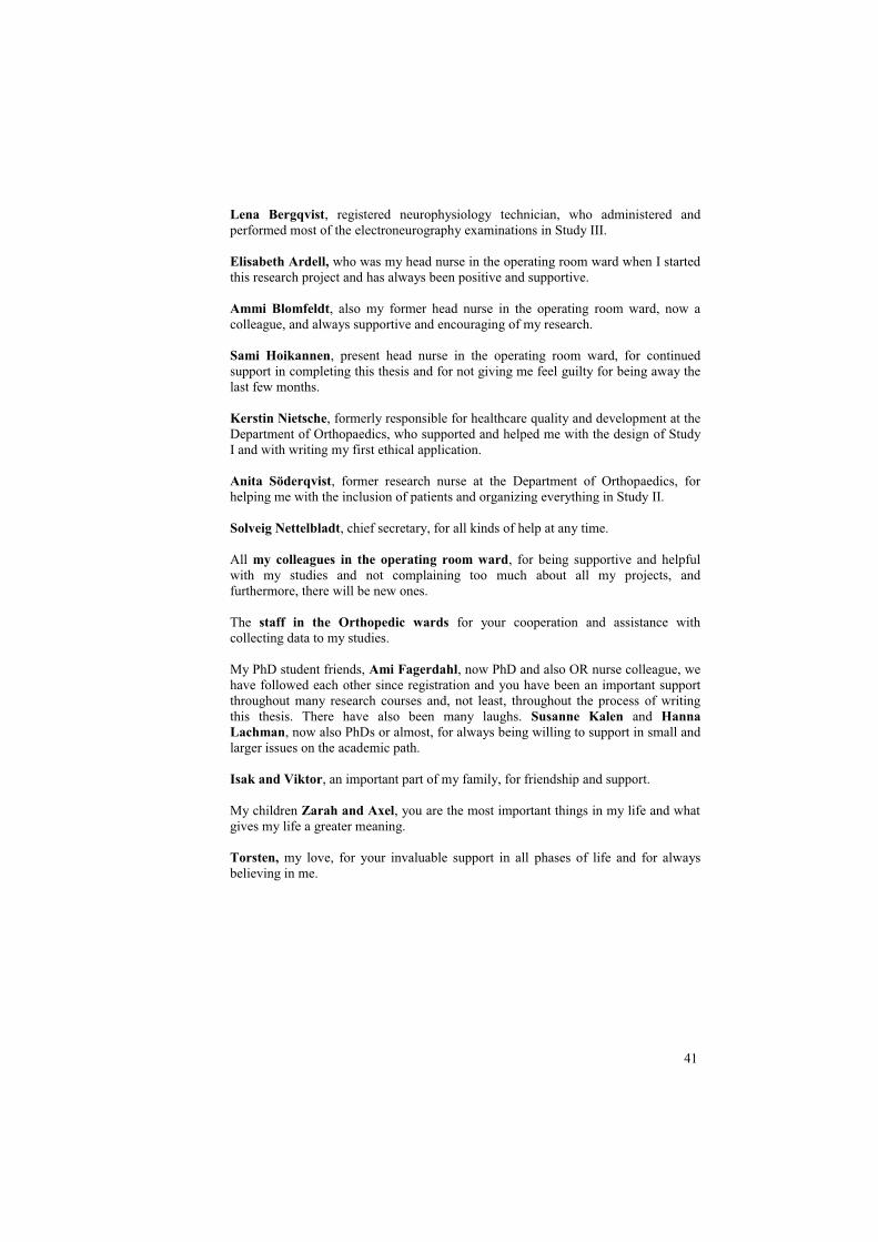
41
Lena Bergqvist, registered neurophysiology technician, who administered and performed most of the electroneurography examinations in Study III. Elisabeth Ardell, who was my head nurse in the operating room ward when I started this research project and has always been positive and supportive. Ammi Blomfeldt, also my former head nurse in the operating room ward, now a colleague, and always supportive and encouraging of my research. Sami Hoikannen, present head nurse in the operating room ward, for continued support in completing this thesis and for not giving me feel guilty for being away the last few months. Kerstin Nietsche, formerly responsible for healthcare quality and development at the Department of Orthopaedics, who supported and helped me with the design of Study I and with writing my first ethical application. Anita Söderqvist, former research nurse at the Department of Orthopaedics, for helping me with the inclusion of patients and organizing everything in Study II. Solveig Nettelbladt, chief secretary, for all kinds of help at any time. All my colleagues in the operating room ward, for being supportive and helpful with my studies and not complaining too much about all my projects, and furthermore, there will be new ones. The staff in the Orthopedic wards for your cooperation and assistance with collecting data to my studies. My PhD student friends, Ami Fagerdahl, now PhD and also OR nurse colleague, we have followed each other since registration and you have been an important support throughout many research courses and, not least, throughout the process of writing this thesis. There have also been many laughs. Susanne Kalen and Hanna Lachman, now also PhDs or almost, for always being willing to support in small and larger issues on the academic path. Isak and Viktor, an important part of my family, for friendship and support. My children Zarah and Axel, you are the most important things in my life and what gives my life a greater meaning. Torsten, my love, for your invaluable support in all phases of life and for always believing in me.

42
12 REFERENCES Aarons H, Hall G, Hughes S, Salmon P. Short-term recovery from hip and knee
arthroplasty. J Bone Joint Surg Br 1996; 78 (4): 555-8. Abdel-Salam A, Eyres K S. Effects of tourniquet during total knee arthroplasty. A
prospective randomised study. J Bone Joint Surg Br 1995; 77 (2): 250-3. Ahmed S M, Ahmad R, Case R, Spencer R F. A study of microbial colonisation of
orthopaedic tourniquets. Ann R Coll Surg Engl 2009; 91 (2): 131-4. Akata T, Kanna T, Izumi K, Kodama K, Takahashi S. Changes in body temperature
following deflation of limb pneumatic tourniquet. J Clin Anesth 1998; 10 (1): 17-22.
Alcelik I, Pollock R D, Sukeik M, Bettany-Saltikov J, Armstrong P M, Fismer P. A comparison of outcomes with and without a tourniquet in total knee arthroplasty: a systematic review and meta-analysis of randomized controlled trials. J Arthroplasty 2012; 27 (3): 331-40.
Aminoff M J. Electrophysiologic testing for the diagnosis of peripheral nerve injuries. Anesthesiology 2004; 100 (5): 1298-303.
Arciero R A, Scoville C R, Hayda R A, Snyder R J. The effect of tourniquet use in anterior cruciate ligament reconstruction. A prospective, randomized study. Am J Sports Med 1996; 24 (6): 758-64.
Bellamy N, Buchanan W W, Goldsmith C H, Campbell J, Stitt L W. Validation study of WOMAC: a health status instrument for measuring clinically important patient relevant outcomes to antirheumatic drug therapy in patients with osteoarthritis of the hip or knee. J Rheumatol 1988; 15 (12): 1833-40.
Blond L, Madsen J L. Exsanguination of the limbs in elderly volunteers. Int Orthop 2003; 27 (2): 114-6.
Brennan S A, Walls R J, Smyth E, Al Mulla T, O'byrne J M. Tourniquets and exsanguinators: a potential source of infection in the orthopedic operating theater? Acta Orthop 2009; 80 (2): 251-5.
Bulfone G, Marzoli I, Quattrin R, Fabbro C, Palese A. A longitudinal study of the incidence of pressure sores and the associated risks and strategies adopted in Italian operating theatres. J Perioper Pract 2012; 22 (2): 50-6.
Butt U, Ahmad R, Aspros D, Bannister G C. Factors affecting wound ooze in total knee replacement. Ann R Coll Surg Engl 2011; 93 (1): 54-6.
Chang C W, Lan S M, Tai T W, Lai K A, Yang C Y. An effective method to reduce ischemia time during total knee arthroplasty. J Formos Med Assoc 2012; 111 (1): 19-23.
Chiang Y C, Lin T S, Yeh M C. Povidone-iodine-related burn under the tourniquet of a child--a case report and literature review. J Plast Reconstr Aesthet Surg 2011; 64 (3): 412-5.
Choudhary S, Koshy C, Ahmed J, Evans J. Friction burns to thigh caused by tourniquet. Br J Plast Surg 1998; 51 (2): 142-3.
Clarke M T, Longstaff L, Edwards D, Rushton N. Tourniquet-induced wound hypoxia after total knee replacement. J Bone Joint Surg Br 2001; 83 (1): 40-4.
Collins N J, Roos E M. Patient-reported outcomes for total hip and knee arthroplasty: commonly used instruments and attributes of a "good" measure. Clin Geriatr Med 2012; 28 (3): 367-94.
Committee A R P. Recommended practices for the use of the pneumatic tourniquet in the perioperative practice setting. AORN J 2007; 86 (4): 640-655.

43
Dahlin L B, Shyu B C, Danielsen N, Andersson S A. Effects of nerve compression or ischaemia on conduction properties of myelinated and non-myelinated nerve fibres. An experimental study in the rabbit common peroneal nerve. Acta Physiol Scand 1989; 136 (1): 97-105.
Darmanis S, Papanikolaou A, Pavlakis D. Fatal intra-operative pulmonary embolism following application of an Esmarch bandage. Injury 2002; 33 (9): 761-4.
Delgado-Martinez A D, Marchal J M, Blanco F, Molina M, Palma A. Distal forearm tourniquet for hand surgery. Int Orthop 2004; 28 (5): 267-9.
Dickinson J C, Bailey B N. Chemical burns beneath tourniquets. BMJ 1988; 297 (6662): 1513.
Din R, Geddes T. Skin protection beneath the tourniquet. A prospective randomized trial. ANZ J Surg 2004; 74 (9): 721-2.
Dobner J J, Nitz A J. Postmeniscectomy tourniquet palsy and functional sequelae. Am J Sports Med 1982; 10 (4): 211-4.
EPUAP, European Pressure Ulcer Advisory Panel and National Pressure Ulcer Advisory Panel. Treatment of pressure ulcers: Quick reference guide. 2009. www.epuap.org/guidelines/Final_Quick_Treatment.pdf
Essving P, Axelsson K, Aberg E, Spannar H, Gupta A, Lundin A. Local infiltration analgesia versus intrathecal morphine for postoperative pain management after total knee arthroplasty: a randomized controlled trial. Anesth Analg 2011; 113 (4): 926-33.
Estebe J P, Davies J M, Richebe P. The pneumatic tourniquet: mechanical, ischaemia-reperfusion and systemic effects. Eur J Anaesthesiol 2011; 28 (6): 404-11.
Feuchtinger J, Halfens R, Dassen T. Pressure ulcer risk assessment immediately after cardiac surgery--does it make a difference? A comparison of three pressure ulcer risk assessment instruments within a cardiac surgery population. Nurs Crit Care 2007; 12 (1): 42-9.
Finsen V, Kasseth A M. Tourniquets in forefoot surgery: less pain when placed at the ankle. J Bone Joint Surg Br 1997; 79 (1): 99-101.
Fitzgibbons P G, Digiovanni C, Hares S, Akelman E. Safe tourniquet use: a review of the evidence. J Am Acad Orthop Surg 2012; 20 (5): 310-9.
Flatt A E. Tourniquet time in hand surgery. Arch Surg 1972; 104 (2): 190-2. Girardis M, Milesi S, Donato S, Raffaelli M, Spasiano A, Antonutto G, Pasqualucci A,
Pasetto A. The hemodynamic and metabolic effects of tourniquet application during knee surgery. Anesth Analg 2000; 91 (3): 727-31.
Graham B, Breault M J, Mcewen J A, Mcgraw R W. Occlusion of arterial flow in the extremities at subsystolic pressures through the use of wide tourniquet cuffs. Clin Orthop Relat Res 1993; (286): 257-61.
Hernandez A J, Almeida A M, Favaro E, Sguizzato G T. The influence of tourniquet use and operative time on the incidence of deep vein thrombosis in total knee arthroplasty. Clinics (Sao Paulo) 2012; 67 (9): 1053-7.
Horlocker T T, Hebl J R, Gali B, Jankowski C J, Burkle C M, Berry D J, Zepeda F A, Stevens S R, Schroeder D R. Anesthetic, patient, and surgical risk factors for neurologic complications after prolonged total tourniquet time during total knee arthroplasty. Anesth Analg 2006; 102 (3): 950-5.
Jacob A K, Mantilla C B, Sviggum H P, Schroeder D R, Pagnano M W, Hebl J R. Perioperative nerve injury after total knee arthroplasty: regional anesthesia risk during a 20-year cohort study. Anesthesiology 2011; 114 (2): 311-7.
Jones C A, Pohar S. Health-related quality of life after total joint arthroplasty: a scoping review. Clin Geriatr Med 2012; 28 (3): 395-429.
Kam P C, Kavanagh R, Yoong F F. The arterial tourniquet: pathophysiological consequences and anaesthetic implications. Anaesthesia 2001; 56 (6): 534-45.

44
Karalezli N, Ogun C O, Ogun T C, Yildirim S, Tuncay I. Wrist tourniquet: the most patient-friendly way of bloodless hand surgery. J Trauma 2007; 62 (3): 750-4.
Kerr D R, Kohan L. Local infiltration analgesia: a technique for the control of acute postoperative pain following knee and hip surgery: a case study of 325 patients. Acta Orthop 2008; 79 (2): 174-83.
Kirkley A, Rampersaud R, Griffin S, Amendola A, Litchfield R, Fowler P. Tourniquet versus no tourniquet use in routine knee arthroscopy: a prospective, double-blind, randomized clinical trial. Arthroscopy 2000; 16 (2): 121-6.
Klenerman L. The tourniquet in surgery. J Bone Joint Surg Br 1962; 44-B 937-43. Konrad G, Markmiller M, Lenich A, Mayr E, Ruter A. Tourniquets may increase
postoperative swelling and pain after internal fixation of ankle fractures. Clin Orthop Relat Res 2005; (433): 189-94.
Kurtz S M, Ong K L, Lau E, Bozic K J, Berry D, Parvizi J. Prosthetic joint infection risk after TKA in the Medicare population. Clin Orthop Relat Res 2010; 468 (1): 52-6.
Kvederas G, Porvaneckas N, Andrijauskas A, Svensen C H, Ivaskevicius J, Mazunaitis J, Marmaite U, Andrijauskas P. A randomized double-blind clinical trial of tourniquet application strategies for total knee arthroplasty. Knee Surg Sports Traumatol Arthrosc 2012.
Ledin H, Aspenberg Per, Good L. Tourniquet use in total knee replacement does not improve fixation, but appears to reduce final range of motion. Acta Ortopaedica 2012; 83 (5): 499-503.
Li B, Wen Y, Wu H, Qian Q, Lin X, Zhao H. The effect of tourniquet use on hidden blood loss in total knee arthroplasty. Int Orthop 2009; 33 (5): 1263-8.
Lindgren M, Unosson M, Krantz A M, Ek A C. Pressure ulcer risk factors in patients undergoing surgery. J Adv Nurs 2005; 50 (6): 605-12.
Lundborg G, Dahlin L B. Anatomy, function, and pathophysiology of peripheral nerves and nerve compression. Hand Clin 1996; 12 (2): 185-93.
Maffulli N, Testa V, Capasso G. Use of a tourniquet in the internal fixation of fractures of the distal part of the fibula. A prospective, randomized trial. J Bone Joint Surg Am 1993; 75 (5): 700-3.
Mcdonagh D. Moisture lesion or pressure ulcer? A review of the literature. J Wound Care 2008; 17 (11): 461-4, 466.
Mcewen J A, Inkpen, K. Tourniquet safety preventing skin injuries. Surgical Technol. 2002; 34 7-15.
Murphy C G, Winter D C, Bouchier-Hayes D J. Tourniquet injuries: pathogenesis and modalities for attenuation. Acta Orthop Belg 2005; 71 (6): 635-45.
Nitz A J, Dobner J J. Upper extremity tourniquet effects in carpal tunnel release. J Hand Surg Am 1989; 14 (3): 499-504.
Nitz A J, Matulionis D H. Ultrastructural changes in rat peripheral nerve following pneumatic tourniquet compression. J Neurosurg 1982; 57 (5): 660-6.
Noordin S, Mcewen J A, Kragh J F, Jr., Eisen A, Masri B A. Surgical tourniquets in orthopaedics. J Bone Joint Surg Am 2009; 91 (12): 2958-67.
Ochoa J, Fowler T J, Gilliatt R W. Anatomical changes in peripheral nerves compressed by a pneumatic tourniquet. J Anat 1972; 113 (Pt 3): 433-55.
Odinsson A, Finsen V. Tourniquet use and its complications in Norway. J Bone Joint Surg Br 2006; 88 (8): 1090-2.
Pedowitz R A, Gershuni D H, Botte M J, Kuiper S, Rydevik B L, Hargens A R. The use of lower tourniquet inflation pressures in extremity surgery facilitated by curved and wide tourniquets and an integrated cuff inflation system. Clin Orthop Relat Res 1993; (287): 237-44.

45
Pedowitz R A, Gershuni D H, Friden J, Garfin S R, Rydevik B L, Hargens A R. Effects of reperfusion intervals on skeletal muscle injury beneath and distal to a pneumatic tourniquet. J Hand Surg Am 1992; 17 (2): 245-55.
Pedowitz R A, Gershuni D H, Schmidt A H, Friden J, Rydevik B L, Hargens A R. Muscle injury induced beneath and distal to a pneumatic tourniquet: a quantitative animal study of effects of tourniquet pressure and duration. J Hand Surg Am 1991; 16 (4): 610-21.
Rajpura A, Somanchi B V, Muir L T. The effect of tourniquet padding on the efficiency of tourniquets of the upper limb. J Bone Joint Surg Br 2007; 89 (4): 532-4.
Reilly C W, Mcewen J A, Leveille L, Perdios A, Mulpuri K. Minimizing tourniquet pressure in pediatric anterior cruciate ligament reconstructive surgery: a blinded, prospective randomized controlled trial. J Pediatr Orthop 2009; 29 (3): 275-80.
Roos E M, Klassbo M, Lohmander L S. WOMAC osteoarthritis index. Reliability, validity, and responsiveness in patients with arthroscopically assessed osteoarthritis. Western Ontario and MacMaster Universities. Scand J Rheumatol 1999; 28 (4): 210-5.
Rudkin A K, Rudkin G E, Dracopoulos G C. Acceptability of ankle tourniquet use in midfoot and forefoot surgery: audit of 1000 cases. Foot Ankle Int 2004; 25 (11): 788-94.
Sale J E M, Lohfeld L H, Brazil K. Revisiting the Quantitative-Qualitative Debate: Implications for Mixed-Methods Research. Quality and Quantity 2002; 36 (1): 43-53.
Sapega A A, Heppenstall R B, Chance B, Park Y S, Sokolow D. Optimizing tourniquet application and release times in extremity surgery. A biochemical and ultrastructural study. J Bone Joint Surg Am 1985; 67 (2): 303-14.
Saunders K C, Louis D L, Weingarden S I, Waylonis G W. Effect of tourniquet time on postoperative quadriceps function. Clin Orthop Relat Res 1979; (143): 194-9.
Schoonhoven L, Defloor T, Grypdonck M H. Incidence of pressure ulcers due to surgery. J Clin Nurs 2002; 11 (4): 479-87.
Smith T O, Hing C B. Is a tourniquet beneficial in total knee replacement surgery? A meta-analysis and systematic review. Knee 2010; 17 (2): 141-7.
Song I, Kim D Y, Kim Y J. The effect of tourniquet deflation on hemodynamics and regional cerebral oxygen saturation in aged patients undergoing total knee replacement surgery. Korean J Anesthesiol 2012; 63 (5): 425-30.
Tai T W, Chang C W, Lai K A, Lin C J, Yang C Y. Effects of tourniquet use on blood loss and soft-tissue damage in total knee arthroplasty: a randomized controlled trial. J Bone Joint Surg Am 2012; 94 (24): 2209-15.
Tai T W, Lin C J, Jou I M, Chang C W, Lai K A, Yang C Y. Tourniquet use in total knee arthroplasty: a meta analysis. Knee Surg Sports Traumatol Arthrosc 2011; 19:1121-30.
Tejwani N C, Immerman I, Achan P, Egol K A, Mclaurin T. Tourniquet cuff pressure: The gulf between science and practice. J Trauma 2006; 61 (6): 1415-8.
Townsend H S, Goodman S B, Schurman D J, Hackel A, Brock-Utne J G. Tourniquet release: systemic and metabolic effects. Acta Anaesthesiol Scand 1996; 40 (10): 1234-7.
Tsarouhas A, Hantes M E, Tsougias G, Dailiana Z, Malizos K N. Tourniquet Use Does Not Affect Rehabilitation, Return to Activities, and Muscle Damage After Arthroscopic Meniscectomy: A Prospective Randomized Clinical Study. Arthroscopy 2012.

46
Wakai A, Winter D C, Street J T, Redmond P H. Pneumatic tourniquets in extremity surgery. J Am Acad Orthop Surg 2001; 9 (5): 345-51.
Wakankar H M, Nicholl J E, Koka R, D'arcy J C. The tourniquet in total knee arthroplasty. A prospective, randomised study. J Bone Joint Surg Br 1999; 81 (1): 30-3.
Weingarden S I, Louis D L, Waylonis G W. Electromyographic changes in postmeniscectomy patients. Role of the pneumatic tourniquet. JAMA 1979; 241 (12): 1248-50.
Wilgis E F. Observations on the effects of tourniquet ischemia. J Bone Joint Surg Am 1971; 53 (7): 1343-6.
Wma T W M a D O H. Ethical principles for medical research involving human subjects. 2008.
Worland R L, Arredondo J, Angles F, Lopez-Jimenez F, Jessup D E. Thigh pain following tourniquet application in simultaneous bilateral total knee replacement arthroplasty. J Arthroplasty 1997; 12 (8): 848-52.
Yang J H, Lim H, Yoon J R, Jeong H I. Tourniquet associated chemical burn. Indian J Orthop 2012; 46 (3): 356-9.
Yoshida W B, Thomazini I A, Carvalho I, Lastoria S, Curi P R, Maffei F H. Platelet activation following application of an Esmarch bandage and tourniquet in rabbits. Braz J Med Biol Res 1989; 22 (9): 1091-3.
Younger A S, Mcewen J A, Inkpen K. Wide contoured thigh cuffs and automated limb occlusion measurement allow lower tourniquet pressures. Clin Orthop Relat Res 2004; (428): 286-93.


Lower Tourniquet Cuff Pressure ReducesPostoperative Wound Complications After
Total Knee ArthroplastyA Randomized Controlled Study of 164 Patients
Charlotta Olivecrona, RN, Sari Ponzer, MD, PhD, Per Hamberg, MD, PhD, and Richard Blomfeldt, MD, PhD
Investigation performed at the Department of Orthopaedics, Sodersjukhuset Karolinska Institutet, Stockholm, Sweden
Background: Measurement of limb occlusion pressure before surgery might lead to the use of a lower tourniquet cuffpressure during surgery and thereby reduce the risk of postoperative pain and complications. The primary aim of this studywas to investigate whether the limb-occlusion-pressure method reduces the tourniquet cuff pressure used during totalknee arthroplasty and if this leads to less postoperative pain compared with that experienced by patients on whom thismethod is not used. The secondary aim was to investigate whether there were any differences regarding the quality of thebloodless field, range of motion, and postoperative wound complications.
Methods: One hundred and sixty-four patients scheduled to be treated with a total knee arthroplasty were randomized toa control group or to undergo the intervention under study (the limb-occlusion-pressure [LOP] group). In the control group,the tourniquet cuff pressure was based on the patient’s systolic blood pressure and a margin decided by the surgeon (theroutine method). In the LOP group, the tourniquet cuff pressure was based on the measurement of the limb occlusionpressure. The primary outcome measure was postoperative pain, and the secondary outcome measures were the qualityof the bloodless field, knee motion, and wound-related complications at discharge and two months after surgery.
Results: The tourniquet cuff pressure was significantly lower in the LOP group than in the control group (p < 0.001). Wecould not demonstrate any differences between the groups regarding postoperative pain or complications, although thenumber of postoperative complications was relatively high in both groups. However, at discharge forty of the forty-sevenpatients with a wound complication had had a cuff pressure above 225 mm Hg and at the two-month follow-up evaluationfourteen of the sixteen patients with a wound complication had had a cuff pressure above 225 mm Hg.
Conclusions: The limb-occlusion-pressure method reduces the cuff pressure without reducing the quality of the blood-less field, but there were no differences in outcomes between the groups. An important secondary finding was thatpatients with a cuff pressure of £225 mm Hg had no postoperative infections and a lower rate of wound complications.
Level of Evidence: Therapeutic Level I. See Instructions for Authors for a complete description of levels of evidence.
The use of a pneumatic tourniquet during a total kneearthroplasty improves visualization by preventing intra-operative bleeding. However, some studies have dem-
onstrated lower rates of postoperative complications and betterinitial recovery of knee movement when the knee arthroplastywas performed without use of a pneumatic tourniquet1-3. Whena tourniquet is used, the cuff pressure should be minimized to
reduce the risk of tourniquet-related postoperative complica-tions2,4. Setting the thigh tourniquet cuff pressure on the basis ofthe systolic blood pressure plus a margin of 100 mm Hg has beenreported to reduce the cuff pressure and early postoperative pain5.According to another study, limb occlusion pressure and systolicblood pressure were not correlated well enough for the systolicblood pressure alone to be used to determine the optimal cuff
Disclosure: None of the authors received payments or services, either directly or indirectly (i.e., via his or her institution), from a third party in support of anyaspect of this work. None of the authors, or their institution(s), have had any financial relationship, in the thirty-sixmonths prior to submission of thiswork, withany entity in the biomedical arena that could be perceived to influence or have the potential to influencewhat iswritten in thiswork. Also, no author has had anyother relationships, or hasengaged in any other activities, that could be perceived to influence or have the potential to influencewhat iswritten in thiswork. Thecomplete Disclosures of Potential Conflicts of Interest submitted by authors are always provided with the online version of the article.
2216
COPYRIGHT � 2012 BY THE JOURNAL OF BONE AND JOINT SURGERY, INCORPORATED
J Bone Joint Surg Am. 2012;94:2216-21 d http://dx.doi.org/10.2106/JBJS.K.01492
pressure6.Wide conical cuffs require a lower cuff pressure7,8 and areless painful7, although the authors of most published studies didnot report whether a straight or awider conical cuff had been used.
Measuring the limb occlusion pressure just before sur-gery by means of an automated photoplethysmographic sensorconnected to a tourniquet apparatus takes into account suchvariables as the type and width of the cuff, the tightness of cuffapplication, the fit of the cuff to the limb, and the properties ofthe patient’s soft tissues and vessels6 and therefore has beensuggested to result in a more optimal cuff pressure. However,automated measurement of limb occlusion pressure has beeninvestigated in only a few clinical studies6,9,10.
The primary aim of this study was to investigate whetherthe limb-occlusion-pressure method reduces the tourniquet cuffpressure used during surgery and if this leads to less postoperativepain. The secondary aim was to investigate whether there wereany differences between the group treated with this method anda control group regarding the quality of the bloodless field, rangeof knee motion, and postoperative wound complications. Wehypothesized that there would be no differences between the limb-occlusion-pressuremeasurementmethod and the controlmethod.
Materials and Methods
This prospective randomized controlled clinical trial was performed fromOctober2008 to July 2010. Patients scheduled to be treated with a primary total knee
arthroplasty, whowere seventy-five years of age or younger, andwhowere classified asAmerican Society of Anesthesiologists (ASA) 1, 2, or 3 were considered eligible forinclusion
11,12. Patients who were unable to read and understand Swedish, had a
systolic blood pressure of >200mmHg, or had a thigh girth of >78 cmwere excluded.All patients gave their informed consent to participate and were ran-
domized preoperatively to a control or intervention (limb-occlusion-pressure[LOP]) group with use of opaque sealed envelopes.
The study was conducted according to the principles of the Helsinki Dec-laration andwas approved by the Ethics Committee of Karolinska Institutet (Ref. No.2007/757-31/1-4), Stockholm. It was registered at ClinicalTrials.gov (NCT01442298).
The standard method at our department was used for the patients in thecontrol group. The tourniquet cuff pressure was based on the patient’s systolicblood pressure and a margin that was decided by the surgeon. In the LOP group,the tourniquet cuff pressure was decided by measuring the limb occlusionpressure with use of an automated photoplethysmographic sensor connected toan ATS 3000 tourniquet apparatus (Zimmer Sweden, Savedalen, Sweden). Therecommended tourniquet pressure was defined as the limb occlusion pressureplus a safety margin of 50 mm Hg for those with a limb occlusion pressure of£130 mmHg, 75mmHg for those with a pressure between 131 and 190mmHg,and 100 mmHg for those with a pressure of >190 mmHg. These recommendedmargins were higher than those reported earlier
4,6, but were the preadjustable
margins indicated by the manufacturer (Zimmer Sweden).The limb underneath the tourniquet cuff was protected by a two-layer
elastic stockinette13. The operating room nurse applied a standard 140-mm-
wide contour thigh cuff for all but seven patients (three in the control groupand four in the LOP group), for whom the operating room nurse selected a 100-mm-wide cylindrical cuff because of difficulties in positioning the contour cuffdue to a short lower limb or a very straight thigh.
The operating room nurse chose the tourniquet cuff and measured thelimb occlusion pressure. Before starting the surgery, the surgeon determined thetourniquet cuff pressure according to the standard method. The operating roompersonnel applied this pressure if the patient had been randomized to the controlgroup. If the patient had been randomized to the LOP group, they applied thepressure suggested by the limb-occlusion-pressure method. The surgeon wasblinded to the randomization and was not told which tourniquet pressure wasapplied.The surgery was performed according to the routine at our department.
The surgeon used a midline skin incision with a medial parapatellarcapsular incision of the joint, and all patients received a cemented NexGenCruciate Retaining total knee arthroplasty (Zimmer Sweden).
All patients received perioperative antibiotics (cloxacillin, 2 g · 3) andlow-molecular-weight heparin (dalteparin). All patients also received a localinfiltration analgesic (300 mg of ropivacaine/0.5 mg of epinephrine/30 mg ofketorolac) at the end of the surgery by infiltration into the fascia, muscles, andsubcutaneous tissue, regardless of whether they had had spinal or general an-esthesia. In 147 patients, a catheter was also inserted into the knee joint for paintreatment with a local anesthetic (150 mg of ropivacain/30 mg of ketorolac). Itwas used during the day after the surgery and was then withdrawn
14.
After the surgery was completed, the surgeon was asked to rate thequality of the bloodless field on a visual analog scale (VAS), with 1 indicating theworst possible rating and 10, the most optimal. The surgeon was also asked torate whether any technical difficulties had been encountered due to the qualityof the bloodless field (1 indicating no difficulties and 10, extreme difficulties).
The skin underneath the tourniquet was inspected immediately after thesurgery and on day four, when a ward nurse also performed a wound check. Thefunctional assessment was done on the third postoperative day by an independentphysiotherapist and included measurement of knee motion (with the patientlying down and sitting up) and a straight-leg lifting test (with a 1-kg weight).Furthermore, the patients filled out themodifiedWestern Ontario andMcMasterUniversities Osteoarthritis Index (WOMAC) questionnaire daily during theirhospital stay. The WOMAC is a self-administered, well-validated health statusinstrument with three domains: pain, physical function, and stiffness
15,16.
All patients were followed at two months postoperatively (with in-spection of the wound, measurement of knee motion, and completion of theWOMAC questionnaire). The medical records were scanned for complicationssuch as nerve injury and deep venous thrombosis. The orthopaedic surgeon incharge and all staff at the department, except the operating room personnelwho measured the limb occlusion pressure, were blinded to the allocationgroup during the hospital stay and at the follow-up evaluations.
Power AnalysisThe sample size was calculated to detect a difference of 5 points (standard deviation[SD], 10 points) in the WOMAC pain score between the control and LOP groupson day four. A total of sixty-four patients in each group (128 patients in the series)were required to detect this difference with an 80% power at the 5% significancelevel, two-tailed. As we anticipated a drop-out rate of about 25%, the recruitmentgoal was determined to be 164 patients. When we were planning for this study, weknew of no earlier randomized study with cuff pressure as the outcome, so oursample size was calculated for detecting differences in postoperative pain.
Statistical MethodsCategorical variables were presented as absolute and relative frequencies and testedfor differences between randomization groups with the chi-square test. Continuousvariables were presented throughout as the mean and SD and tested with theMann-Whitney U test. The nonparametric test was used because it was deemedmore appropriate for the subjective scale variables, and for the remaining contin-uous variables the assumption of normality was not satisfied. Cuff pressure waspartly analyzed as a continuous variable and partly as a categorical variable, wherebythe cutoffs of 225 and 260 mm Hg were chosen on the basis of their clinicalrelevance. The results were regarded as significant if p was < 0.05 (two-tailed).
All analyses were performed according to the intention-to-treat principle.
Source of FundingThe authors did not receive any external funding or grants in support of theirresearch for this work.
Results
Three patients were excluded after inclusion: one because of achange to a unicompartmental knee arthroplasty, one because
of a change to a high-flex prosthesis, and one who was scheduled
2217
THE JOURNAL OF BONE & JOINT SURGERY d J B J S .ORG
VOLUME 94-A d NUMBER 24 d DECEMBER 19, 2012LOWER TOURNIQUET PRESSURE REDUCES POSTOPERAT IVE WOUND
COMPL ICAT IONS AFTER TOTAL KNEE ARTHROPLASTY

for revision surgery and thus had been incorrectly included in thestudy. In total, 161 patients—eighty-three in the control group andseventy-eight in the LOP group—were included in the analysis.Two patients were lost to follow-up at two months (Fig. 1).
There were no differences between the LOP group and thecontrol group regarding sex, age, ASA class, body mass index(BMI), preoperative systolic blood pressure, or the bloodless-field time during surgery (see Appendix). Systolic blood pres-sure, measured on the arm routinely at the start of surgery, was amean (and SD) of 122 ± 20 mmHg in the LOP group comparedwith 119 ± 19 mm Hg in the control group (difference notsignificant). The mean limb occlusion pressure (measured onthe thigh in the LOP group) was 169 ± 34 mm Hg.
As shown in Table I, a tourniquet cuff pressure of £225 mmHg was found more often in the LOP group than in the controlgroup (p < 0.001). The mean tourniquet cuff pressure was alsogenerally lower in the patients in the LOP group, but this differencewas not significant (p = 0.362). No significant difference betweenthe groups could be detected regarding the quality of the bloodlessfield or the technical difficulties as judged by the surgeon (Table I).There were incidents of break-through bleeding in three cases. Inone of these cases, the tourniquet was deflated after only threeminutes and the surgery was performed without a bloodless field.
Ratings of postoperative pain on the self-administeredWOMAC questionnaire during hospitalization did not differbetween the randomization groups, but the patients in the LOPgroup reported significantly less stiffness in the knee on dayfour (p = 0.020) (see Appendix). The range of motion of theknee on postoperative day three and the ability to do a straight-leg lift did not differ between the groups (see Appendix).
Three patients (out of 131 available) had blisters under-neath the tourniquet cuff immediately after surgery, but there wasno difference between the LOP group and the control group (p =0.227). All three patients had had a cuff pressure of ‡260mmHg.
On day four, eight patients (out of the 111 available) haddeveloped blisters or other pressure-related injuries under-neath the tourniquet cuff and sixty-five patients reported thatthey had pain in the quadriceps muscle underneath the tour-niquet cuff (no differences between the groups, p = 0.400).
At the time of discharge, forty-seven patients (30% of the158 available) had developed a surgical wound complicationsuch as blisters, oozing from the wound, or signs of infection.As shown in Table II, forty of these forty-seven patients had hada cuff pressure of >225 mm Hg during surgery. None of thepatients with a cuff pressure of £225 mm Hg had any signs ofwound secretion or infection, but seven patients had blisters
Fig. 1
CONSORT (Consolidated Standards of Reporting Trials) diagram. TKA = total knee arthroplasty.
2218
THE JOURNAL OF BONE & JOINT SURGERY d J B J S .ORG
VOLUME 94-A d NUMBER 24 d DECEMBER 19, 2012LOWER TOURNIQUET PRESSURE REDUCES POSTOPERAT IVE WOUND
COMPL ICAT IONS AFTER TOTAL KNEE ARTHROPLASTY

around the knee. There were no significant differences betweenthe LOP and the control groups (p = 0.149).
At the time of follow-up two months after surgery,seven patients (4%) had a postoperative wound infection andnine patients (6%) were recorded as having delayed wound-healing. Four of these patients had developed a deep woundinfection after they were discharged from the ward and had
been rehospitalized and reoperated on. Of the sixteen pa-tients with a surgical wound complication at the two-monthfollow-up visit, fourteen had had a cuff pressure of >225 mmHg. Two patients who had had a cuff pressure of £225 mmHg had delayed wound-healing (Table III). There were nodifferences between the LOP group and the control group(p = 0.869).
TABLE I Cuff Pressures and Ratings by the Surgeon Regarding the Quality of the Bloodless Field and Technical Difficulties
Control Group (N = 83) LOP Group (N = 78) P Value
Cuff pressure (no. [%] of patients) <0.001£225 mm Hg 7 (8) 26 (33)226-259 mm Hg 45 (54) 22 (28)‡260 mm Hg 31 (37) 30 (38)
Mean cuff pressure (±SD) (mm Hg) 252 ± 17* 246 ± 45† 0.362
Mean preanesthetic systolic blood pressure (±SD) (mm Hg) 154 ± 18 155 ± 21 0.426
Mean preoperative systolic blood pressure/limb occlusionpressure (±SD) (mm Hg)
119 ± 19‡ 169 ± 34§
Mean VAS score (±SD) for quality of bloodless field 9.4 ± 1.3 8.7 ± 2.4 0.364
Mean VAS score (±SD) for technical difficulties 1.4 ± 1.3 2.04 ± 2.3 0.307
*Chosen by the surgeon.†Recommended by the apparatus.‡Preoperative systolic blood pressure measured on the arm, immediately before thetourniquet was applied and the skin incision was made, for determination of the cuff pressure by the surgeon. §Limb occlusion pressure measuredon the thigh by the limb-occlusion-pressure apparatus.
TABLE II Results of Wound Check on Postoperative Day Four, According to Group and Cuff Pressure, for the 158 Patients Available*
Control Group LOP Group
£225 mmHg (N = 7)
226-259 mmHg (N = 45)
‡260 mmHg (N = 31)
£225 mmHg (N = 24)
226-259 mmHg (N = 22)
‡260 mmHg (N = 29)
No wound complication 5 31 24 19 15 17
Blisters 2 8 4 5 5 10
Oozing from the wound 0 5 3 0 1 1
Signs of infection 0 1 0 0 1 1
*The values are given as the number of patients. P = 0.149 for the difference between the LOP and the control groups.
TABLE III Results of Wound Check at Two-Month Follow-up Visit for the 156 Patients Available*
Control Group LOP Group
£225 mmHg (N = 7)
226-259 mmHg (N = 44)
‡260 mmHg (N = 30)
£225 mmHg (N = 25)
226-259 mmHg (N = 21)
‡260 mmHg (N = 29)
No wound complications 7 39 27 23 19 25
Blisters 0 0 0 0 0 0
Delayed wound-healing 0 3 1 2 1 2
Wound infection 0 2 2 0 1 2
*The values are given as the number of patients. P = 0.869 for the difference between the LOP and the control groups.
2219
THE JOURNAL OF BONE & JOINT SURGERY d J B J S .ORG
VOLUME 94-A d NUMBER 24 d DECEMBER 19, 2012LOWER TOURNIQUET PRESSURE REDUCES POSTOPERAT IVE WOUND
COMPL ICAT IONS AFTER TOTAL KNEE ARTHROPLASTY

Six of the seven patients who had a postoperative woundinfection and all four patients who had a deep wound infectionwere men. We found no difference in cuff pressures betweenmen and women, but men had significantly longer bloodless-field times (mean, eighty-nine minutes compared with seventy-three minutes for women; p = 0.027).
Two patients had a deep venous thrombosis, and onepatient had a nerve injury. There were no differences be-tween the LOP group and the control group (p = 0.752),but the patient with a nerve injury had had a cuff pressure>260 mm Hg.
At the two-month follow-up visit, knee motion, as-sessed by a blinded orthopaedic surgeon, did not differ be-tween the groups. No difference in any of the three domainsof the self-administered WOMAC questionnaire was de-tected between the LOP group and the control group (datanot shown).
Discussion
This study shows that the limb-occlusion-pressuremeasuringtechnique reduces the tourniquet cuff pressures in patients
undergoing a total knee arthroplasty, but we could not dem-onstrate any differences in postoperative pain between our LOPand control groups.
The generally lower cuff pressure in the LOP groupprovided a good-quality bloodless field but did not have anyimpact on the risk of developing a postoperative wound com-plication or on the range of motion, although the patients in theLOP group reported less stiffness of the knee on postoperativeday four.
An important secondary finding was that, regardless of therandomization group, patients with a cuff pressure of £225mmHg had no postoperative infections and a lower rate of woundcomplications at discharge and at the two-month follow-upevaluation.
In contrast to the reports by Reilly et al.9 and Youngeret al.6,10, we could not demonstrate any significant differences inthe mean cuff pressures between the LOP group and the controlgroup. The chosen cuff pressure depends on such factors asthe experience of the surgeon as well as on local traditions.The surgeons in our study generally used a rather low cuffpressure, with a mean of 252 mm Hg in the control group,compared with surgeons in other published studies, in whichcuff pressures of 300 to 350 mmHg have been reported2,4,6,17,18.However, the limb-occlusion-pressure method led to moreindividual cuff pressures among our patients, since thepressures ranged from 150 to 300 mmHg (SD, ±45) in the LOPgroup compared with 200 to 300 mm Hg (SD, ±17) in thecontrol group. The mean measured limb occlusion pressure inour study was 169 mm Hg, which is higher than the mean of142 mm Hg in the study by Younger et al.6. Our tourniquetapparatus also had a higher preadjustable margin from themanufacturer than earlier described6,9,10, which resulted inhigher recommended tourniquet pressures and therefore alsohigher mean values—246 mm Hg in our study compared with202 mm Hg in the study by Younger et al. and 198 mm Hg in
the study by Reilly et al. Nevertheless, the limb-occlusion-pressure measuring technique led to lower margins: 77 mmHgin the LOP group compared with 133 mm Hg in the controlgroup.
All of our patients received local infiltration analgesia atthe end of surgery, and most of them received it the next day aswell. This was a rather new routine at our department and waseffective for treatment of postoperative pain. The fact thatpostoperative pain treatment was good in all patients could beone of the reasons why we could not demonstrate any differ-ences in postoperative pain between groups. Another reasoncould be that the WOMAC questionnaire might not be sensi-tive enough to capture relatively small differences in postop-erative pain ratings.
We could not demonstrate any differences in knee mo-tion between the groups. All patients achieved good kneeflexion as early as on day three: the mean was 78� in the controlgroup and 77� in the LOP group, compared with 47� on daythree after total knee arthroplasty done with a tourniquet in thestudy by Li et al.3. Earlier studies comparing knee or anklesurgery with and without a tourniquet have shown significantlybetter knee flexion after surgery without a tourniquet3,17,19. Theauthors suggested that the swelling of the limb after use of atourniquet might be an explanation. Tourniquet release isknown to be associated with an immediate 10% increase inlimb girth20, which was reported to increase up to 50% over thefirst postoperative day17.
There was a rather large number of wound compli-cations in our study. One reason could be that we did notexclude patients with diabetes, as has been done in otherstudies1,3,17,19. In our study, even patients classified as ASA 3,and some later classified as ASA 4, were included. However,no patient in our study who had a cuff pressure of £225 mmHg developed a postoperative wound infection. This find-ing is in accordance with the report by Clarke et al.2, whostudied the pattern of postoperative wound hypoxia sevendays after knee surgery and found that a tourniquet cuffpressure of about 225 mm Hg yielded a significantly betterreturn of the oxygen levels compared with cuff pressures of350 mm Hg.
Butt et al.21 demonstrated a significant association be-tween increased tourniquet time and wound oozing after totalknee arthroplasty. They had a mean tourniquet time of eighty-three minutes with a range of thirty-eight to 125 minutes. Inour study, the mean tourniquet time was eighty-seven minutes.Six of the seven patients who had a postoperative wound in-fection in our study were men. We found no other differencesbetween men and women other than a significantly longerbloodless-field time for men. In this study, we focused on cuffpressures and complications; however, the duration of thebloodless field is probably another important factor that re-quires further investigation.
The strength of this study is that it was a randomizedcontrolled trial of a ‘‘nonselected’’ study population, with fewexclusion criteria. A limitation might be that we used theWOMAC questionnaire as the primary outcome measure
2220
THE JOURNAL OF BONE & JOINT SURGERY d J B J S .ORG
VOLUME 94-A d NUMBER 24 d DECEMBER 19, 2012LOWER TOURNIQUET PRESSURE REDUCES POSTOPERAT IVE WOUND
COMPL ICAT IONS AFTER TOTAL KNEE ARTHROPLASTY

since it may not be reliable enough to capture the small dif-ferences regarding postoperative pain. Another limitationof our study is that very few of our patients had a BMI of >35kg/m2 and therefore our results may not be valid for that pa-tient group.
In conclusion, the generally lower tourniquet cuffpressure in the LOP group did not decrease the postoper-ative pain or other outcomes in our patients. However,patients who had undergone total knee arthroplasty in abloodless field with a cuff pressure of £225 mmHg had a lowerrate of wound complications such as delayed healing andinfections.
The limb-occlusion-pressure measuring techniquecan help the surgeon to choose more individual, often lower,cuff pressures. However, if the surgeon carefully chooses anoptimal and not too high cuff pressure and considers thetype and width of the cuff, the circumference of the limb,and the patient’s individual vessel characteristics, bothmethods appear to yield a good outcome in terms of post-operative complications.
AppendixTables showing demographic baseline data and the du-ration of the bloodless field as well as the results of the
WOMAC, range of motion, and straight-leg lifting are availablewith the online version of this article as a data supplement atjbjs.org. n
Charlotta Olivecrona, RNSari Ponzer, MD, PhDPer Hamberg, MD, PhDRichard Blomfeldt, MD, PhDSection of Orthopaedics,Department of Clinical Science and Education,Sodersjukhuset Karolinska Institutet,SE-118 83 Stockholm, Sweden.E-mail address for C. Olivecrona: [email protected] address for S. Ponzer: [email protected] address for P. Hamberg: [email protected] address for R. Blomfeldt: [email protected]
References
1. Abdel-Salam A, Eyres KS. Effects of tourniquet during total knee arthroplasty.A prospective randomised study. J Bone Joint Surg Br. 1995 Mar;77(2):250-3.2. Clarke MT, Longstaff L, Edwards D, Rushton N. Tourniquet-induced woundhypoxia after total knee replacement. J Bone Joint Surg Br. 2001 Jan;83(1):40-4.3. Li B,Wen Y,WuH,QianQ, Lin X, ZhaoH. The effect of tourniquet use on hidden bloodloss in total knee arthroplasty. Int Orthop. 2009 Oct;33(5):1263-8. Epub 2008 Aug 27.4. Noordin S, McEwen JA, Kragh JF Jr, Eisen A, Masri BA. Surgical tourniquets inorthopaedics. J Bone Joint Surg Am. 2009 Dec;91(12):2958-67.5. Worland RL, Arredondo J, Angles F, Lopez-Jimenez F, Jessup DE. Thigh painfollowing tourniquet application in simultaneous bilateral total knee replacementarthroplasty. J Arthroplasty. 1997 Dec;12(8):848-52.6. Younger AS, McEwen JA, Inkpen K. Wide contoured thigh cuffs and automatedlimb occlusion measurement allow lower tourniquet pressures. Clin Orthop RelatRes. 2004 Nov;(428):286-93.7. Estebe JP, LeNaoures A, Chemaly L, Ecoffey C. Tourniquet pain in a volunteer study:effect of changes in cuff width and pressure. Anaesthesia. 2000 Jan;55(1):21-6.8. Pedowitz RA, Gershuni DH, Botte MJ, Kuiper S, Rydevik BL, Hargens AR. The useof lower tourniquet inflation pressures in extremity surgery facilitated by curved andwide tourniquets and an integrated cuff inflation system. Clin Orthop Relat Res.1993 Feb;(287):237-44.9. Reilly CW, McEwen JA, Leveille L, Perdios A, Mulpuri K. Minimizing tourniquetpressure in pediatric anterior cruciate ligament reconstructive surgery: a blinded, pro-spective randomized controlled trial. J Pediatr Orthop. 2009 Apr-May;29(3):275-80.10. Younger AS, Manzary M, Wing KJ, Stothers K. Automated cuff occlusion pres-sure effect on quality of operative fields in foot and ankle surgery: a randomizedprospective study. Foot Ankle Int. 2011 Mar;32(3):239-43.11. American Society of Anesthesiologists. ASA Physical Status ClassificationSystem. http://www.asahq.org/home/for-members/clinical-information/asa-physical-status-classification-system. Accessed 2012 Sep 4.
12. Dripps RD, Lamont A, Eckenhoff JE. The role of anesthesia in surgical mortality.JAMA. 1961 Oct 21;178:261-6.13. Olivecrona C, Tidermark J, Hamberg P, Ponzer S, Cederfjall C. Skin protectionunderneath the pneumatic tourniquet during total knee arthroplasty: a randomizedcontrolled trial of 92 patients. Acta Orthop. 2006 Jun;77(3):519-23.14. Kerr DR, Kohan L. Local infiltration analgesia: a technique for the controlof acute postoperative pain following knee and hip surgery: a case study of 325patients. Acta Orthop. 2008 Apr;79(2):174-83.15. Bellamy N, Buchanan WW, Goldsmith CH, Campbell J, Stitt LW. Validation studyof WOMAC: a health status instrument for measuring clinically important patientrelevant outcomes to antirheumatic drug therapy in patients with osteoarthritis of thehip or knee. J Rheumatol. 1988 Dec;15(12):1833-40.16. Roos EM, Klassbo M, Lohmander LS. WOMAC osteoarthritis index. Reliability,validity, and responsiveness in patients with arthroscopically assessed osteoar-thritis. Western Ontario and MacMaster Universities. Scand J Rheumatol.1999;28(4):210-5.17. Konrad G, Markmiller M, Lenich A, Mayr E, Ruter A. Tourniquets may increasepostoperative swelling and pain after internal fixation of ankle fractures. Clin OrthopRelat Res. 2005 Apr;(433):189-94.18. Tejwani NC, Immerman I, Achan P, Egol KA, McLaurin T. Tourniquet cuff pres-sure: the gulf between science and practice. J Trauma. 2006 Dec;61(6):1415-8.19. Wakankar HM, Nicholl JE, Koka R, D’Arcy JC. The tourniquet in total knee ar-throplasty. A prospective, randomised study. J Bone Joint Surg Br. 1999Jan;81(1):30-3.20. Estebe JP, Davies JM, Richebe P. The pneumatic tourniquet: mechanical,ischaemia-reperfusion and systemic effects. Eur J Anaesthesiol. 2011Jun;28(6):404-11.21. Butt U, Ahmad R, Aspros D, Bannister GC. Factors affecting wound ooze in totalknee replacement. Ann R Coll Surg Engl. 2011 Jan;93(1):54-6. Epub 2010 Sep 10.
2221
THE JOURNAL OF BONE & JOINT SURGERY d J B J S .ORG
VOLUME 94-A d NUMBER 24 d DECEMBER 19, 2012LOWER TOURNIQUET PRESSURE REDUCES POSTOPERAT IVE WOUND
COMPL ICAT IONS AFTER TOTAL KNEE ARTHROPLASTY


5615
Tourniquet cuff pressure and nerve injury in knee arthroplasty in a bloodless field A neurophysiological study
Charlotta Olivecrona1, Richard Blomfeldt1, Sari Ponzer1, Benjamin Ribalta Stanford2, and Bengt Y Nilsson2
1Orthopedics Section, Department of Clinical Science and Education, Södersjukhuset; 2Clinical Neurophysiology Section, Department of Clinical Neuroscience, Karolinska Institutet, Stockholm, Sweden.Correspondence: [email protected] Submitted 12-04-25. Accepted 12-12-28
Open Access - This article is distributed under the terms of the Creative Commons Attribution Noncommercial License which permits any noncommercial use, distribution, and reproduction in any medium, provided the source is credited.DOI
Background and purpose Tourniquet-related nerve injuries remain a concern in orthopedic surgery. The cuff pressures used today are generally lower, and therefore a decreasing incidence of peripheral nerve injuries might also be expected. However, there have been few neurophysiological studies describing the outcome after bloodless field surgery. We describe the results of neurophys-iological examinations and report the incidence of nerve injuries after total knee arthroplasty (TKA) in a bloodless field.
Patients and methods This study was part of a prospective, randomized controlled clinical trial in patients scheduled for TKA in a bloodless field. 20 consecutive patients were enrolled. Elec-troneurography (ENeG) and quantitative sensory testing (QST) of thermal thresholds were performed on day 3. These tests were repeated 2 months after surgery when electromyography (EMG) with a concentric-needle electrode was also performed.
Results The mean tourniquet cuff pressure was 237 (SD 33) mmHg. Electromyographic signs of denervation were found in 1 patient, who also had the highest cuff pressure in the study popu-lation (294 mmHg). The sensory nerve response amplitudes were lower in the operated leg on day 3; otherwise, the neurophysi-ological examinations showed no differences between the legs.
Interpretation When low tourniquet cuff pressures are used the risk of nerve injury is minor.
■
Nerve injuries, such as peroneal nerve palsy, after a total knee arthroplasty (TKA) can be a devastating complication (Ner-cessian et al. 2005). However, the reported incidence and the severity of nerve injuries vary—ranging from a mild transient loss of function to permanent, irreversible damage (Noordin et al. 2009). The symptoms reported may not reflect the true inci-dence of complications; minor nerve injuries are diagnosed
by electrodiagnostic tests such as EMG or nerve conduction studies. Suggested risk factors are valgus deformity, flexion contracture, pre-existing neuropathy, rheumatoid arthritis, hematoma, postoperative epidural analgesia, and pneumatic tourniquet (Idusuyi and Morrey 1996, Schinsky et al. 2001, Nercessian et al. 2005). The use of a pneumatic tourniquet may be helpful during TKA surgery, but it involves a risk , of nerve injury (Smith and Hing 2010, Tai et al. 2011, Alcelik et al. 2012).
The 2 main mechanisms of pneumatic tourniquet-induced nerve injury are ischemia and direct mechanical effects (Hodg-son 1994). Ochoa et al. (1972) found that compressive neura-praxia rather than ischemic neuropathy or muscle damage was the underlying cause of tourniquet paralysis. They described paranodal myelin invagination as the pathophysiological mechanism of nerve injury after tourniquet use. When a lower tourniquet cuff pressure and a shorter duration have been used, this mechanism has been difficult to confirm (Pedowitz et al. 1991). Nitz et al. (1989) suggested that tourniquet compres-sion is associated with increased microvascular permeability and intraneural edema, with persistent tissue ischemia and subsequent nerve degeneration. Hodgson (1994) suggested that increased tourniquet time increases the probability of an injury, the severity of which will be primarily determined by the tourniquet cuff pressure. Today, tourniquet cuff pressures are usually lower, and fewer nerve injuries might be expected. However, there is a need for further neurophysiological stud-ies to determine whether this is the case (Younger et al. 2011).
The main aim of this study was to determine the incidence of nerve injuries related to the use of bloodless field after TKA. A secondary aim was to analyze the results of neurophysiologi-cal examinations in this patient group
6

5615
Patients and methodsPatientsThis study was part of a randomized controlled clinical trial (RCT) in patients scheduled for a primary TKA in a bloodless field (Olivecrona et al. 2012). In that particular study we inves-tigated whether measuring the limb occlusion pressure would lead to lower tourniquet cuff pressure and if this would lead to less postoperative pain and wound complications. In that study, patients aged 75 years or younger and classified accord-ing to the American Society of Anesthesiologists (ASA) as ASA 1–3 were considered to be eligible for inclusion. Patients with a systolic blood pressure of over 200 mmHg and those with a thigh girth of over 78 cm were excluded. 164 patients gave their informed consent to participate, and were random-ized preoperatively to a control group (routine method) or to an intervention group (limb occlusion pressure, LOP). All the patients were given written and verbal information and they were also informed that 10 patients from each randomiza-tion group would be asked to participate in neurophysiologi-cal examinations postoperatively on day 3 and then again 2 months after their surgery. Patients with diabetes mellitus or spinal disorders and those who had received chemotherapy or had a body mass index (BMI) of > 30 were also excluded from this neurophysiological study. The neurophysiologist who performed the examinations was blinded regarding the alloca-tion group and to any information about tourniquet use or any clinical nerve symptoms from the operated leg.
20 patients were enrolled in this neurophysiological study between November 2009 and June 2010. 2 patients were excluded after inclusion: one with a BMI of 40 who was incor-rectly included, and one who decided to drop out after the first examination.
Neurophysiological examinationsElectroneurography (ENeG) was performed bilaterally with surface electrodes according to the standard routine at the Karolinska Department of Clinical Neurophysiology, using a Nicolet VikingSelect EMG system (Care Fusion, Middleton, WI) Where necessary, the limbs were warmed with heating bags to keep the skin temperature at 32˚C. Motor nerve con-duction studies in the peroneal and the tibial nerves included conduction velocity from knee level to ankle (in m/s), distal latency from ankle to the extensor digitorum brevis muscle and the abductor hallucis muscle, respectively (in ms), muscle response amplitude to distal stimulation (in mV), and also F-wave latencies in both nerves (ms). The peroneal nerve motor conduction velocity was also calculated for the segment across the fibular head. Sensory nerve conduction studies were done on the superficial peroneal nerve and the sural nerve (conduction velocity in m/s and amplitude in μV).
Concentric-needle electrode electromyography (EMG) was performed bilaterally using the same equipment. EMG activ-ity was studied in the vastus lateralis muscle in the thigh, and
in the anterior tibial and medial head of the gastrocnemius muscles in the lower limbs. We looked for spontaneous fibril-lations and positive sharp waves signaling ongoing denerva-tion, for changes in motor unit configuration, and for dimin-ished activity at maximal voluntary contraction.
Quantitative sensory tests (QSTs) were performed using the Medoc TSA-II Neurosensory Analyzer (Medoc Ltd., Ramat Yishai, Israel) and included determinations of temperature thresholds anteriorly at the middle of the lower legs and on the dorsum of the feet. The method of limits was used. The temperature was changed by 1°C/s.The results were also expressed as the value for the neutral zone (°C) that is the dif-ference between thresholds for warmth and cold. Differences between the operated leg and the unoperated leg were calcu-lated at both visits. At the second visit, the test results were also compared with results from the first study.
The neurophysiological tests were planned as a wide approach to discover any intraoperative nerve injury (Aminoff 2004). However, several of the measurements were made below the knee and thus on nerve sections not directly involved in the tourniquet area or the surgical field. F-waves were obtained by nerve stimulation at the ankle. The volley of nerve impulses initiated by electrical nerve stimulation will travel antidromi-cally (“backwards”) in the motor nerve fibers up to the spinal cord, so as to depolarize the spinal motor neurons, leading to many impulses bouncing back down the leg to give a twitch in the foot muscles. Thus, these nerve impulses pass the knee and the thigh twice, and a small change in nerve conduction might be doubled and thereby easier to reveal. An injury to the motor nerve fibers might appear as an increase in latency and/or a reduction in persistence, i.e. fewer impulses return to the foot muscles. We therefore analyzed these responses in more detail.
ENeG and QST were performed on day 3 and after 2 months postoperatively, while EMG was performed only after 2 months.
All patients were followed up by phone 18 months after the examinations in order to find out if any of them had any clini-cal symptoms of nerve injury.
StatisticsThe number of patients included was chosen based on ear-lier studies with EMG examinations where 20–25 patients had been included (Saunders et al. 1979, Weingarden et al. 1979, Dobner and Nitz 1982, Arciero et al. 1996).
Continuous variables are presented as mean (SD) or median (minimum and maximum). The scale variables were tested with the non-parametric Wilcox test because of the small number of patients included and the risk regarding the assumption of normality. The medians of the differences are estimated with the Hodges-Lehmann estimator and presented with a 95% confidence interval (CI). All tests were two-sided and the results were considered significant at p-values of < 0.05. For the statistical analysis, PASW (SPSS) version 18

5615
was used.
Ethics and registration of the studyThe study was conducted according to the tenets of the Hel-sinki Declaration and was approved by the Ethics Committee of Karolinska Institutet, Stockholm, on July 3, 2009 (ref. no. 2007/757-31/1-4, 2007/1164-32) and registered at Clinical-Trial.gov (NCT01442298).
Results
Mean tourniquet cuff pressure (TCP) was 237 mmHg and mean tourniquet time was 81 min. The 9 patients from the LOP group had a mean TCP of 223 (41) mmHg and the 9 con-trols had a mean TCP of 251 (12) mmHg (Table 1).
Electromyographic signs of recent denervation were found in 1 patient (no. 16). At 2 months, this patient had fibrillations in the vastus lateralis and the gastrocnemius muscles, asso-ciated with reduced voluntary activity. Studies in the pero-neal nerve at 3 days and at 2 months showed a reduction in response amplitude, a prolongation of F-wave latency, and a reduction in F-wave persistence. No abnormalities were found in the unoperated leg. This patient had the highest cuff pres-sure in the study, 294 mmHg for 100 min. In 3 other patients (nos. 17, 18, and 20) EMG revealed minor signs of chronic changes with motor unit potentials showing increased dura-tion and amplitude. These patients had no signs of ongoing denervation. 5 patients had one or more minor deviations in electroneurography or QST values (nos. 1, 7, 8, 9, and 17)
that might indicate a change in nerve function related to the operation. However, the deviations were small and were not considered to be definite signs of recent nerve injury. The cuff pressures in these 5 patients ranged from 173 to 280 mmHg. None of these patients showed any clinical symptoms of nerve damage.
The analysis of the patients as a group showed no statis-tically significant difference in motor or sensory conduction velocity when operated and unoperated legs were compared. Motor response amplitudes in the peroneal nerve on day 3 were slightly lower in the operated leg. The median of dif-ferences between operated leg and control leg was 0.5 mV (CI: –0.1 to 1.9) (p = 0.1) on day 3 and 0.3 mV (–0.5 to 1.2) (p = 0.5) at 2 months. This slightly lower—although not sta-tistically significant—response amplitude was not seen in the tibial nerve. Sensory nerve amplitudes were lower in the oper-ated leg on day 3 for both the sural (2 (0.5–4) μV; p = 0.01) and the superficial peroneal nerve (1.5 (0 – 3) μV; p = 0.06) and at 2 months (sural nerve 2.5 (0–5) μV; p = 0.04; and super-ficial peroneal nerve 1 (–0.5 to 2.5) μV; p = 0.08).
There was no statistically significant difference in F-wave latencies and persistencies between day 3 and 2 months (data not shown), or between the operated and unoperated leg (Table 2, Figures 2 and 3). The patient with electromyographic signs of denervation (no. 16) showed prolonged F-wave latency and a reduction in F-wave persistence in the peroneal nerve both on day 3 and at 2 months postoperatively. An increase in latency in the tibial nerve was seen only on day 3.
The thresholds for heat and cold perception (QST) showed no statistically significant differences between the operated leg and the unoperated leg (data not shown).
Table 1. Demographic baseline data for all patients included (n = 18)

5615
In a telephone follow-up of 15 of the patients included, only the patient with an EMG-evident denervation reported radiating pain and a tingling sensation in the operated leg and foot for about 6 months after the operation, after which the symptoms disappeared. None of the other patients had had any similar symptoms.
Discussion
In this study, the mean TCP was 237 (33) mmHg, which appears to be a safe cuff pressure regarding the risk of nerve injuries. Only one patient had clinical and EMG evidence of nerve injury, and this patient had a cuff pressure of 294 mmHg—which is a rather high pressure, but still within rec-ommended limits. Denervation in both the thigh and the lower leg supports the view that the injury was caused by the tourni-quet and not by the surgical procedures at knee level.
Chronic neurogenic EMG changes were seen in 3 patients. The cause was unknown, and we regarded it as being unre-lated to the surgery since there were no signs of ongoing denervation.
A study conducted in the 1980s reported that two-thirds of patients who had undergone knee surgery in a bloodless field showed electromyographic (EMG) evidence of denervation and a functional capacity of one third of the unoperated leg. The control group was operated on without a bloodless field, and showed no evidence of denervation and had a functional capacity of four-fifths (Dobner and Nitz 1982). In that study, the mean TCP was 393 (300–450) mmHg and the mean blood-less field time was 42 (8–90) min. Our neurophysiological results contrast with that study, which may be due to the fact that we had a mean cuff pressure of 237 (33) mmHg. Today’s lower TCPs mean that these older studies with cuff pressures of up to 450 mmHg are no longer relevant.
In a retrospective questionnaire survey, 265 orthopedic sur-
Table 2. Results of F-wave studies in the operated leg and median of the differences between the unoperated and the operated leg on day 3 and 2 months postoperatively
*
*
100
80
60
40
20
0
100
80
60
40
20
0
F-response latency (ms) F-wave persistence (%)
16
16 16
16
10
11
11
13
Day 3 2 months Day 3 2 months
100
80
60
40
20
0
100
80
60
40
20
0
F-response latency (ms) F-wave persistence (%)
16
9
1310 9
1
Day 3 2 months Day 3 2 months
**
*
*4
12
5615
geons in Norway were asked to report any complications due to tourniquet use during the previous 2 years. 12 clinically evident nerve complications in the lower limb were reported, with an overall incidence of 0.03% (Odinsson and Finsen 2006). In our RCT study population of 164 patients, 2 patients were scheduled for EMG/ENeG examinations because they had clinical symptoms at the follow-up 2 months after surgery. 1 patient had minor signs of an injury of the tibial nerve and 1 additional patient had EMG-confirmed denervation in muscles innervated by the peroneal nerve. Together with 1 patient with a nerve injury from this study, this gives an incidence of 2%. This variation in incidence of nerve injuries has been shown and discussed earlier. We want to draw attention to the impor-tant differences in study design, ranging from prospective neurophysiological examination of a whole study population to a retrospective report of clinically evident nerve injuries.
Horlocker et al. (2006) reported a strong correlation between nerve injuries and prolonged tourniquet time. They studied 1,001 patients who underwent a primary or revision knee replacement, with a mean tourniquet time of 145 (25) min. Clinical neurological complications were noted in 7.7%. Odinsson and Finsen (2006) reported that 2 of the major com-plications occurred with tourniquet times of 130 and 180 min. Since we had a mean bloodless field time of 81 min with a longest duration of 122 min, our material is not comparable to theirs. A correlation between nerve injury and tourniquet time was also found in another retrospective 20-year cohort study of 11,645 TKA patients (OR = 1.3; p = 0.003). Unfor-tunately, no data on the duration of tourniquet time were pre-sented (Jacob et al. 2011). In their study, Saunders et al. (1979) showed a distinct difference between patients with short tour-niquet time, i.e. less than 15 min—with 22% EMG abnormali-ties—and patients with more than 60 min of bloodless field time, 85% of whom had EMG abnormalities. We did not find such a difference, but on the other hand none of the patients in our study had a tourniquet time shorter than 63 min. The high incidence of EMG abnormalities in the study by Saunders et al. could have been due to the high TCPs of 350–450 mmHg, regardless of tourniquet time.
Sensory nerve examinations showed differences between the operated leg and the unoperated leg. We were surprised because this might be interpreted as an axonal injury, which is a more severe injury than a local demyelination. None of the patients appeared to be troubled by or even noticed any sensory loss. Nor was this something that was noticed at the clinical follow- up. However, technical considerations such an edema in the operated leg may have affected the results.
Our quantitative sensory test results support the results of earlier studies indicating that small myelinated and non-myelinated fibers are not as sensitive to compression as large myelinated fibers (Dahlin et al. 1989, Nitz and Matulionis, 1982).
One limitation of our study was that the number of patients was rather small, even though it was comparable with other
studies. On the other hand, the neurophysiological series was large enough for the statistical analysis. The strengths of the present study were that it was a consecutive series of well-described patients who were examined with both EMG and ENeG after TKA surgery in a bloodless field. As far as we know, there have been no other studies published in which this type of broad protocol has been used.
We conclude that TCPs of around 240 mmHg for up to 80 min appear to be safe regarding the risk of nerve injury in patients undergoing TKA in a bloodless field.
CO: study design, inclusion of patients, data analysis, follow-up, and writing of the manuscript. RB and SP: study design and provision of feedback on the manuscript. BRS: study design, neurophysiological examinations, and provi-son of feedback on the manuscript. BYN: study design, neurophysiological examinations, recording of data, data analysis, and provision of feedback on the manuscript.
We thank Lena Bergqvist, registered neurophysiology technician, who orga-nized and performed most of the electroneurography examinations. The first author was given time for research work by the Department of Orthopedics, Södersjukhuset, Stockholm. The examinations carried out were financed by funds from the Karolinska Institute.
No competing interests declared.
Alcelik I, Pollock R D, Sukeik M, Bettany-Saltikov J, Armstrong P M, Fismer P. A comparison of outcomes with and without a tourniquet in total knee arthroplasty: a systematic review and meta-analysis of randomized con-trolled trials. J Arthroplasty 2012; 27 (3): 331-40.
Aminoff M J. Electrophysiologic testing for the diagnosis of peripheral nerve injuries. Anesthesiology 2004; 100 (5): 1298-303.
Arciero R A, Scoville C R, Hayda R A, Snyder R J. The effect of tourniquet use in anterior cruciate ligament reconstruction. A prospective, randomized study. Am J Sports Med 1996; 24 (6): 758-64.
Dahlin L B, Shyu B C, Danielsen N, Andersson S A. Effects of nerve com-pression or ischaemia on conduction properties of myelinated and non-myelinated nerve fibres. An experimental study in the rabbit common pero-neal nerve. Acta Physiol Scand 1989; 136 (1): 97-105.
Dobner J J, Nitz A J. Postmeniscectomy tourniquet palsy and functional sequelae. Am J Sports Med 1982; 10 (4): 211-4.
Hodgson A J. A proposed etiology for tourniquet-induced neuropathies. J Biomech Eng 1994; 116 (2): 224-7.
Idusuyi O B, Morrey B F. Peroneal nerve palsy after total knee arthroplasty. Assessment of predisposing and prognostic factors. J Bone Joint Surg Am 1996; 78 (2): 177-84.
Jacob A K, Mantilla C B, Sviggum H P, Schroeder D R, Pagnano M W, Hebl J R. Perioperative nerve injury after total knee arthroplasty: regional anes-thesia risk during a 20-year cohort study. Anesthesiology 2011; 114 (2): 311-7.
Nercessian O A, Ugwonali O F, Park S. Peroneal nerve palsy after total knee arthroplasty. J Arthroplasty 2005; 20 (8): 1068-73.
Nitz A J, Matulionis D H. Ultrastructural changes in rat peripheral nerve following pneumatic tourniquet compression. J Neurosurg 1982; 57 (5): 660-6.
Nitz A J, Dobner J J, Matulionis D H. Structural assessment of rat sciatic nerve following tourniquet compression and vascular manipulation. Ana-tomical Rec 1989; 225: 67-76.

5615
Noordin S, Mcewen J A, Kragh J F, Jr., Eisen A, Masri B A. Surgical tourni-quets in orthopaedics. J Bone Joint Surg Am 2009; 91 (12): 2958-67.
Odinsson A, Finsen V. Tourniquet use and its complications in Norway. J Bone Joint Surg (Br) 2006; 88 (8): 1090-2.
Olivecrona C, Ponzer S, Hamberg P, Blomfeldt R. Lower tourniquet cuff pressure reduces postoperative wound complications after total knee arthroplasty. A randomized controlled study of 164 patients. J Bone Joint Surg (Am) 2012; 94 (24): 2216-21.
Pedowitz R A, Nordborg C, Rosenqvist A L, Rydevik B L. Nerve function and structure beneath and distal to a pneumatic tourniquet applied to rabbit hindlimbs. Scand J Plast Reconstr Surg Hand Surg 1991; 25 (2): 109-20.
Saunders K C, Louis D L, Weingarden S I, Waylonis G W. Effect of tourni-quet time on postoperative quadriceps function. Clin Orthop 1979; (143): 194-9.
Schinsky M F, Macaulay W, Parks M L, Kiernan H, Nercessian O A. Nerve injury after primary total knee arthroplasty. J Arthroplasty 2001; 16 (8): 1048-54.
Smith T O, Hing C B. Is tourniquet beneficial in total knee replacement sur-gery? A meta-analysis and systematic review. Knee 2010; 17 (2): 141-7.
Tai T W, Lin C J, Jou I M, Chang C W, Lai K A, Yang C Y. Tourniquet use in total knee arthroplasty: a meta-analysis. Knee Surg Sports Traumatol Arthrosc 2011; 19 (7): 1121-30.
Weingarden S I, Louis D L, Waylonis G W. Electromyographic changes in postmeniscectomy patients. Role of the pneumatic tourniquet. JAMA 1979; 241 (12): 1248-50.
Younger A S, Manzary M, Wing K J, Stothers K. Automated cuff occlusion pressure effect on quality of operative fields in foot and ankle surgery: a randomized prospective study. Foot Ankle Int 2011; 32 (3): 239-43.


ORIGINAL PAPER
Tourniquet time affects postoperative complicationsafter knee arthroplasty
Charlotta Olivecrona & Lasse J. Lapidus & Lina Benson &
Richard Blomfeldt
Received: 22 January 2013 /Accepted: 3 February 2013# Springer-Verlag Berlin Heidelberg 2013
AbstractPurpose Pneumatic tourniquets are frequently used in kneearthroplasty surgery. However, there is a lack of evidence todefine safe tourniquet time in lower limb surgery. Theprimary aim of this study was to investigate whether tour-niquet time influences the risk of postoperative complica-tions after primary and secondary knee arthroplasty.Methods This study was a prospective register study. Sincewe wanted dispersion in tourniquet time, we included aconsecutive series of 577 primary knee arthroplasties, 46revision knee arthroplasties, and 18 patellar supplementingknee arthroplasties from a clinical audit database over aperiod of five years. The following postoperative complica-tions were recorded: superficial wound infections, deepwound infections, deep vein thrombosis, pulmonary embo-lism, nerve injuries, compartment syndrome, cuff pressureinjuries, and bandage injuries.Results Tourniquet time over 100 minutes was associatedwith an increased risk of complications after knee arthro-plasty surgery (OR 2.2, CI 1.5–3.1). This increase in riskremained after adjusting for cuff pressure, sex, age, Amer-ican Society of Anesthesiologists (ASA) classification,smoking, diabetes, and surgery indication (OR 2.4, CI1.6–3.6).
Conclusions Tourniquet time over 100 minutes increases therisk of complications after knee arthroplasty surgery andspecial attention is advocated to reduce the tourniquet time.
Introduction
The use of a pneumatic tourniquet may be helpful duringknee arthroplasty surgery [1]. However, a number of dis-advantages have been reported and their use is not withoutrisk as complications may still occur [2–4]. The reportedinjuries are often pressure-related, but they can also be causedby excessive tourniquet time [5].
There is a lack of evidence to define a safe tourniquettime in lower limb surgery [6]. Recommendations suggest atime limit of two hours for healthy patients [5, 7, 8], butelderly, trauma patients and those with peripheral vasculardisease are probably more susceptible [7]. Most studies oftourniquet time are of the experimental animal type [9–11],and few clinical human studies use tourniquet times of morethan two hours. In a retrospective study, Horlocker et al.(2006) found that tourniquet times over 120 minutes wereassociated with an increased risk of nerve injury in totalknee arthroplasties (TKA). Other studies have shown anincreased rate of re-operations, a higher incidence of nerveinjuries, inferior knee mobility, and more surgical woundcomplications when longer tourniquet times have been used[12–15]. In a review evaluating tourniquet use, Fitzgibbonset al. (2012) concluded that the existing assumption of atwo-hour safe time limit is mainly based on animal studies,and because the reported complications are mostly minorand of a short-term nature are therefore questionable.
Since earlier investigations have different endpoints andfew are human prospective studies, there is still a lack ofevidence for clear clinical guidance regarding a safe tourni-quet time.
C. Olivecrona : L. J. Lapidus : R. BlomfeldtSection of Orthopedics, Department of Clinical Scienceand Education, Södersjukhuset Karolinska Institutet,Stockholm, Sweden
L. BensonDepartment of Clinical Science and Education,Södersjukhuset Karolinska Institutet, Stockholm, Sweden
C. Olivecrona (*)Section of Orthopedics, Södersjukhuset,118 83, Stockholm, Swedene-mail: [email protected]
International Orthopaedics (SICOT)DOI 10.1007/s00264-013-1826-4

The primary aim of this study was to investigate whethertourniquet time influences the risk of postoperative compli-cations after a knee arthroplasty. The secondary aim was toinvestigate whether factors such as age, sex, the AmericanSociety of Anesthesiologists (ASA) classification, diabetes,smoking, or tourniquet cuff pressure affect the risk of post-operative complications.
Patients and methods
This study was a prospective register study conducted at theDepartment of Orthopedics at Södersjukhuset in Stockholm,Sweden. All patients who have undergone surgery at thedepartment since 1996 have been registered prospectively ina clinical audit database where all complications withinsix weeks after surgery have been recorded and validated.This audit was part of a routine quality control and a follow-up rate of 99.3 % was achieved.
Since we wanted dispersion in tourniquet time, a consec-utive series of 577 primary knee arthroplasties (465 totalknee arthroplasties and 112 unicompartmental knee arthro-plasties), 46 revision knee arthroplasties, and 18 patellarsupplementing knee arthroplasties was identified in the reg-istry during the period 1999–2003 and included in the study.
In 16 patients undergoing revision knee arthroplasty, thetourniquet cuff was deflated and then re-inflated because of aprolonged surgery time. The mean reperfusion interval was29 minutes, (standard deviation [SD] 12) and the second tour-niquet time ranged from 15 to 76 minutes. The longest totalbloodless field period was 193 minutes. These minutes fromthe re-inflated period are not included in the statistical analysis.
The postoperative complications recorded for this studywere: superficial wound infections (treated with antibiotics),deep wound infections (requiring surgical intervention), deepvein thrombosis (verified by ultrasonography or phlebogra-phy), pulmonary embolism (verified by computed tomogra-phy), nerve injuries (verified by clinical examination orelectromyography (EMG), compartment syndrome (verifiedby clinical examination and faciotomy), cuff pressure injury(documented on the medical chart), and bandage injury (blis-ters or other surgical skin related complications) (Table 3).These complications were chosen because they have beendescribed earlier as possibly being associated with the use ofa bloodless field [2, 5, 8, 12]. All surgical procedures wereperformed according to local protocols. Patients received pro-phylactic intravenous antibiotics at induction and two furtherdoses postoperatively. Low molecular weight heparin(LMWH) was used for thromboprophylaxis. The limb under-neath the tourniquet cuff was protected by cast padding, a two-layer elastic stockinette or, in some patients, no protection atall, since this was our routine over some years. A standard140-mm-wide contour thigh tourniquet cuff or a 100-mm-
wide cylindrical tourniquet cuff has been used. The tourniquetcuff pressure was decided by the surgeon, often based on thepatient’s systolic blood pressure plus a margin.
Statistical analysis
Statistical analyses were performed using SPSS statistics 20(IBM Corp, Armonk, New York, USA) and R version 2.15.1(R Foundation for Statistical Computing, Vienna, Austria). Forall analyses, the level of significance was set to 0.05, and all pvalues are two-sided. Baseline data are presented as 100minutes or less vs. over 100 minutes and tested with a Fisher’sexact test for categorical variables and student’s t test forcontinuous variables (Table 1). Associations between tourni-quet time and complication were investigated with univariableand multivariable logistic regression analyses and the resultsare presented as odds ratios (ORs) and with 95 % confidenceintervals (CIs). The receiver Operator Curve (ROC) analysesshowed that a cut-off of 106minutes would be appropriated fortourniquet time, but since it is clinically more suitable, we haveused the categories 100 minutes or less vs. more than100 minutes. We have also analysed tourniquet time catego-rised as 120 minutes or less vs. over 120 minutes since that isthe general recommendation in the literature [5, 7, 8], and alsoas a continuous variable (Table 2). Variables included in themultivariable model besides tourniquet time were sex, age,ASA classification, surgery indication, diabetes, smoking,and cuff pressure. These factors have been described earlierin complications related to the use of pneumatic tourniquets orwound complications in general [2, 5, 7, 12, 16–18].
The Hosmer-Lemeshow goodness-of-fit test was used toexamine the multivariable model, with P=0.525 indicating anacceptable fit. Outliers were investigated using Cook’s dis-tance and no extreme outliers were detected. The VarianceInflation Factors (VIF) was relatively low, less than 3 for allvariables, indicating that no multicollinearity was present.Linearity for the continuous variables was investigated usingsmoothed partial residual plots and by modelling the variableswith restricted cubic splines and plotting the functional form.Tourniquet time and cuff pressure were deemed to have alinear association with complications, but age was not, andwas therefore categorized as 70 years or less vs. over 70 years,based on visual inspection of the plots.
Interactions between tourniquet time and all variablesfrom the multivariable model were tested, and they arepresented in a Forest plot (Fig. 1). Cuff pressure was dicho-tomised for presentational purposes in the Forest plot. Mod-elling interactions make it possible to interpret the results forsubgroups of the population, but with the additional benefitof formally testing for differences in effect of tourniquettime on complications between the different levels of therespective variables.
International Orthopaedics (SICOT)

Two sensitivity analyses were performed. One withoutthe ASA classification, since ASA recording was missing on99 patients, and one for tourniquet time, excluding the 16patients with a re-inflated tourniquet cuff.
The study was conducted according to the Helsinki Dec-laration and was approved by the Ethics Committee of theKarolinska Institute (2009/1152-31/2).
Results
In our eligibility assessment, we identified 641 patients whohad all undergone knee arthroplasty surgery. Tourniquet
time ranged from 39 minutes to 156 minutes. Baseline datafor all patients and by ≤100 min and >100 min tourniquettimes are presented in Table 1. As shown in the Table, theindication for surgery, mean cuff pressure, and of course themean tourniquet time are not equally distributed.
We found an association between tourniquet time and anincreased risk of a complication after knee arthroplasty surgeryin a bloodless field. When tourniquet time exceeded100 minutes, a crude model analysis showed an increased riskfor a complication comparedwith a tourniquet time 100minutesor less (OR 2.2, CI 1.5–3.1). This result remained after adjustingfor cuff pressure (continuous), sex, age (70 years and under vs.over 70 years), ASA classification (1–3), smoking (yes/no),diabetes (yes/no), surgery indication (primary, revision, patellasupplementing) (OR 2.4, CI 1.6–3.6) (Table 2). When tourni-quet time was analysed as a continuous variable, the odds forhaving a complication increased by 20 % for every ten minutesof longer tourniquet time (OR 1.2, CI 1.1–1.4). Since therecommended time limit at our department is 120 minutes, wealso analysed the tourniquet time variable as 120minutes or lessvs. over 120 minutes. These results also showed increased oddsfor suffering a complication (OR 1.9, CI 1.1–3.2) (Table 2).
In sensitivity analyses without the ASA classification orwithout patients with a re-inflated tourniquet cuff, the asso-ciation between tourniquet time of over 100 min and com-plications remained (without ASA OR 2.3, CI 1.6-3.3 andwithout re-inflated OR 2.4, CI 1.6–3.7).
Interactions between tourniquet time and the other vari-ables included in the multivariable model are shown inFig. 1, where the number of complications in each subgroup
Table 2 Odds ratio for tourniquet time in relation to a complicationafter total knee arthroplasty (TKA)
Model Tourniquettime, min
OR 95 % CI p value
Crude model > 100b 2.2 1.5–3.1 < 0.001
Adjusted modela > 100b 2.4 1.6–3.6 < 0.001
Adjusted modela > 120c 1.9 1.1–3.2 0.014
Adjusted modela Continuousd 1.2 1.1–1.4 < 0.001
a Adjusted for sex, age, ASA classification, smokers, diabetes, surgeryindication, and cuff pressureb Reference≤100 minc reference≤120 mind the variable was analyzed as continuous but is presented as per10 min
Table 1 Demographic baselinedata for all patients included(n=641)
ASA American Society ofAnesthesiologists
p values have been tested withFisher’s exact test andstudent’s t testaThe values are given as thenumber of patients with thepercentage in parenthesesbThe values are given as themean and standard deviationc99 patients with missing ASArecording
All patients ≤ 100 min (n=373) > 100 min (n=268) p value
Sexa 0.110
Female 420 (65) 254 (68) 166 (62)
Male 221 (34) 119 (32) 102 (38)
Age, yearsb 70 (10) 70 (11) 70 (10) 0.767
ASA c 0.729
1 95 (15) 58 (16) 37 (14)
2 297 (46) 170 (46) 127 (47)
3 150 (23) 84 (23) 66 (25)
Diabetes 0.418
Yes 89 (14) 48 (13) 41(15)
Smokers 0.904
Yes 80 (12) 46 (12) 34 (13)
Surgery indication < 0.001
Primary 577 (90) 346 (93) 231(86)
Revision 46 (7) 9 (2) 37 (14)
Patellar- supplementing 18 (3) 18 (5) 0
Cuff pressure, mmHgb 259 (20) 261 (20) 256 (20) 0.001
Tourniquet time, minb 97 (22) 82 (12) 119 (12) < 0.001
International Orthopaedics (SICOT)

is also presented (Fig. 1). We found no statistically significantinteraction between a tourniquet time of over 100 minutes andthe other variables in our adjusted model. Even patients classi-fied as ASA 2 and ASA 3 or as diabetics or smokers tourniquettime showed no association with complications (Fig. 1). How-ever, amongwomen, the impact of tourniquet time for sufferinga complication was higher (OR 3.0, CI 1.8–5.0) compared tomen (OR 1.6, CI 0.8–3.0), although this difference was notstatistically significant. Thirty -nine percent of the women who
had a tourniquet time of over 100 minutes had a complicationcompared to 29 % of the men.
Age, sex, surgery indication, diabetes, smoking, and cuffpressure showed no statistically significant association forsuffering a complication in either the crude model or thedifferent adjusted models. However, patients classified asASA 2 and ASA 3 did show an association for suffering acomplication compared to ASA 1 (ASA 2, OR 2.5, CI 1.3–4.7, p=0.006 and ASA 3, OR 2.9, CI 1.4–5.9, p=0.003).
0.25 0.5 1 2.5 5 10 20 40
≥260
<260
Cuff pressure
Yes
No
Smokers
Yes
No
Diabetes
Revision
Primary
Surgery indication
3
2
1
ASA
>70
≤70
Age, years
Female
Male
Sex
Subgroup
0.374
0.103
0.139
0.703
0.352
0.834
0.116
P value
78 / 293
88 / 345
22 / 80
146 / 561
23 / 89
145 / 549
17 / 46
147 / 577
46 / 150
87 / 297
14 / 95
91 / 350
77 / 291
109 / 420
59 / 221
Complications / Total Odds Ratio (95% CI)Fig. 1 Multivariable oddsratio (OR) for suffering acomplication by subgroups andp values are for interactionsbetween tourniquet time≤100min vs. > 100 min and sex,age, ASA, surgery indication,diabetes, smokers, andcuff pressure. Numbers ofcomplications by subgroups
Table 3 Incidence of complica-tions for all patients, and alsowhen the bloodless field timewas shorter or longer than100 min (n=641)
aSome patients have more thanone complication and are there-fore presented in multiple cellsbThe values are given as thenumber of patients with the per-centage in parentheses
DVT Deep vein thrombosis,PE Pulmonary embolism
Complicationsb ≤ 100b (n=373) > 100b (n=268)
Complicationa
Yes 168 (26) 74 (20) 94 (35)
Wound complication 99 (15) 41(11) 58 (22)
Superficial wound infection 92 (14) 37 (10) 55 (21)
Deep wound infection 6 (1) 4 (1) 2 (1)
Bleeding 1 (0.2) 0 1(0.4)
Cuff pressure injury 49 (8) 17 (5) 32 (12)
Bandage injury 21 (3) 10 (2) 11 (4)
Peripheral nerve injury 4 (0.6) 2 (0.5) 2 (1)
Compartment syndrome 2 (0.3) 1 (0.3) 1 (0.4)
DVT 22 (3) 11 (3) 11 (4)
PE 1 (0.2) 1 (0.3) 0
International Orthopaedics (SICOT)

In total, 168 (26 %) patients had a complication recorded.Ninety-four (35 %) of these patients had had a bloodlessfield time longer than 100 minutes (Table 3). The meanbloodless field time for the patients who had a recordedcomplication was 104 minutes compared to 95 minutes forthose who had no complication. Three of the 16 patientswith a re-inflated tourniquet cuff had a complication (twowound complications and one DVT) (data not shown).
Discussion
This study shows a statistically significant association be-tween tourniquet time and complications after knee arthro-plasty surgery.
Our findings are in accordance with earlier publishedstudies, i.e. a longer tourniquet time increases the risk ofcomplications [12–14]. In a meta-analysis, comparing earlytourniquet release with releasing the tourniquet after woundclosure, Rama et al. (2007) reported a significantly in-creased rate of re-operations, due to postoperative compli-cations when the tourniquet was left inflated until woundclosure was completed. Tourniquet time had a mean of80 minutes compared to 69 minutes in the early releasegroup (p=0.002). Jacob et al. (2011) demonstrated a corre-lation between tourniquet time and nerve injury after a TKA(OR 1.28, 95 % CI 1.09–1.50, p=0.003). Chang et al.(2012) found that at six-week follow-up after a TKA,patients with a mean tourniquet time of 53 (SD 8.7) minutes,compared to those with 75.5 (SD 9.9) minutes, had betteractive knee flexion and subjective knee performance. Butt etal. (2011) found an association between cessation of oozingwounds and the duration of the bloodless field (p=0.03).
It is generally recommended that the use of tourniquetshould be limited to two hours due to the risk complications.Fitzgibbons et al. (2012) concluded, however, that these com-plications are mostly minor and of a short-term nature and thatthere are no contraindications to longer tourniquet time whennecessary. In our study, we found that every additional tenminutes of tourniquet time was associated with an increasedrisk for complications. In our experience, every complicationmay interfere with the postoperative functional recovery andcould lead to unnecessary discomfort for the patient. Wetherefore believe that it is important that the tourniquet timeis minimised for an optimal outcome. Individual patientcomorbidities might also be worthy of consideration, sincepatients classified as ASA 2 or ASA 3 also had statisticallysignificant higher odds for suffering a complication comparedto patients with ASA 1 (OR 2.5, 2.9). This finding that apatient with comorbidities will have an increased risk forcomplications connected with knee surgery also seems logi-cal. In this study, it appears that women might be less tolerantto tourniquet time than men. Women had almost doubled the
odds for a complication (OR 3) compared to men (OR 1.6)after a tourniquet time of over 100 minutes. A differencebetween the sexes in terms of tolerating tourniquet time hasnever been demonstrated before. However, this difference wasnot statistically significant. Still, this is maybe something thatneeds further research.
When discussing disadvantages of pneumatic tourniquets,not only the tourniquet time, but also the cuff pressure, has tobe considered. However, cuff pressure showed no associationwith complications in this study. The mean tourniquet cuffpressure was 259 mmHg. This is a rather low mean cuffpressure compared to other published studies, where pressuresof 300–350 mmHg have been reported [5, 19–21]. The rela-tively low cuff pressures used by our surgeons might have hadan influence on the results in this study with no impact of cuffpressure in the multivariable model. However, we have seenearlier that even lower cuff pressures of 225 mmHg or lessresult in fewer wound complications [22]. In this study, only afew patients have had a cuff pressure of less than 225 mmHg.
The strength of this study is the consecutive cohort of quite asubstantial number of knee arthroplasty surgery patients. Thepatients were prospectively followed and all complicationswere verified with a follow-up rate of 99.3 %. One weaknessof the study might be the relatively old material; however, thesurgical methods, length of surgery, tourniquet time, cuff pres-sure, and patient comorbidities have not generally changed, andtherefore the results should still be valid. Another limitation isthe inclusion of both primary and secondary interventions inorder to achieve tourniquet time dispersion among the patients.Obviously, a revision surgical procedure should carry a higherrisk of complications than primary surgery. However, catego-risation as primary, revision, and patellar supplementing pro-cedures in this study showed no statistically significantassociation with complications.
Conclusions
The results of this study indicate that tourniquet time has animpact on the complication rate after knee arthroplasty.
Conflict of interest The authors declare that they have no conflict ofinterest.
References
1. Tai TW, Chang CW, Lai KA, Lin CJ, Yang CY (2012) Effects oftourniquet use on blood loss and soft-tissue damage in total kneearthroplasty: a randomized controlled trial. J Bone Joint Surg Am94:2209–2215
2. Alcelik I, Pollock RD, Sukeik M, Bettany-Saltikov J, ArmstrongPM, Fismer P (2012) A comparison of outcomes with and without
International Orthopaedics (SICOT)

a tourniquet in total knee arthroplasty: a systematic review andmeta-analysis of randomized controlled trials. J Arthroplasty27:331–340
3. Smith TO, Hing CB (2010) Is a tourniquet beneficial in total kneereplacement surgery? A meta-analysis and systematic review.Knee 17:141–147
4. Ledin H, Aspenberg P, Good L (2012) Tourniquet use in total kneereplacement does not improve fixation, but appears to reduce finalrange of motion. Acta Ortopaedica 83:499–503
5. Noordin S, McEwen JA, Kragh JF, Eisen A Jr, Masri BA (2009)Surgical tourniquets in orthopaedics. J Bone Joint Surg Am91:2958–2967
6. Fitzgibbons PG, Digiovanni C, Hares S, Akelman E (2012) Safetourniquet use: a review of the evidence. J Am Acad Orthop Surg20:310–319
7. Kam PC, Kavanagh R, Yoong FF (2001) The arterial tourniquet:pathophysiological consequences and anaesthetic implications.Anaesthesia 56:534–545
8. Wakai A, Winter DC, Street JT, Redmond PH (2001) Pneumatictourniquets in extremity surgery. J Am Acad Orthop Surg 9:345–351
9. Nitz AJ, Matulionis DH (1982) Ultrastructural changes in ratperipheral nerve following pneumatic tourniquet compression. JNeurosurg 57:660–666
10. Ochoa J, Fowler TJ, Gilliatt RW (1972) Anatomical changes inperipheral nerves compressed by a pneumatic tourniquet. J Anat113:433–455
11. Pedowitz RA, Gershuni DH, Schmidt AH, Friden J, Rydevik BL,Hargens AR (1991) Muscle injury induced beneath and distal to apneumatic tourniquet: a quantitative animal study of effects oftourniquet pressure and duration. J Hand Surg Am 16:610–621
12. Butt U, Ahmad R, Aspros D, Bannister GC (2011) Factors affect-ing wound ooze in total knee replacement. Ann R Coll Surg Engl93:54–56
13. Jacob AK, Mantilla CB, Sviggum HP, Schroeder DR, PagnanoMW, Hebl JR (2011) Perioperative nerve injury after total kneearthroplasty: regional anesthesia risk during a 20-year cohort study.Anesthesiology 114:311–317
14. Rama KR, Apsingi S, Poovali S, Jetti A (2007) Timing of tourni-quet release in knee arthroplasty. Meta-analysis of randomized,controlled trials. J Bone Joint Surg Am 89:699–705
15. Tai TW, Lin CJ, Jou IM, Chang CW, Lai KA, Yang CY (2011)Tourniquet use in total knee arthroplasty: a meta-analysis. KneeSurg Sports Traumatol Arthrosc 19:1121–1130
16. Horlocker TT, Hebl JR, Gali B, Jankowski CJ, Burkle CM, BerryD (2006) Anesthetic, patient, and surgical risk factors for neuro-logic complications after prolonged total tourniquet time duringtotal knee arthroplasty. Anesth Analg 102:950–955
17. Nasell H, Ottosson C, Tornqvist H, Linde J, Ponzer S (2011) Theimpact of smoking on complications after operatively treated anklefractures–a follow-up study of 906 patients. J Orthop Trauma25:748–755
18. Owens WD, Felts JA, Spitznagel EL Jr (1978) ASA physical statusclassifications: a study of consistency of ratings. Anesthesiology49:239–243
19. Clarke MT, Longstaff L, Edwards D, Rushton N (2001) Tourniquet-induced wound hypoxia after total knee replacement. J Bone JointSurg Br 83:40–44
20. Konrad G, Markmiller M, Lenich A, Mayr E, Ruter A (2005)Tourniquets may increase postoperative swelling and pain afterinternal fixation of ankle fractures. Clin Orthop Relat Res 189–94
21. Tejwani NC, Immerman I, Achan P, Egol KA, McLaurin T (2006)Tourniquet cuff pressure: the gulf between science and practice. JTrauma 61:1415–1418
22. Olivecrona C, Ponzer S, Hamberg P, Blomfeldt R (2012) Lowertourniquet cuff pressure reduces postoperative wound complica-tions after total knee arthroplasty: a randomized controlled study of164 patients. J Bone Joint Surg Am 94:2216–2221
International Orthopaedics (SICOT)