all-ceramic systems: laboratory and clinical performance · all-ceramic systems: laboratory and...
TRANSCRIPT

1
234
567
8910
11121314
151617
181920
212223
24252627
282930
313233
34353637
383940
414243
444546
4748
All-Ceramic Systems:Laboratory andClinical Performance
Q2 Q3
Petra C. Guess, DDS, Dr Med Denta,*, Stefan Schultheis, DDSa,Estevam A. Bonfante, DDSb, Paulo G. Coelho, DDS, PhDc,Jonathan L. Ferenczd, Nelson R.F.A. Silva, DDS, PhDd
Q4 Q5
Q7
KEYWORDS
� All-ceramic material � CAM/CAD � Zirconia � Alumina
Over the last 3 decades, a trend to shift toward metal-free restorations has beenobserved in the dental field. To meet these increased demands of patients anddentists for highly aesthetic, biocompatible, and long-lasting restorations, severaltypes of all-ceramic systems have been developed.1 Silicate and glass ceramics areused as a veneer for metal or all-ceramic cores to optimize form and aesthetics. Ina monolithic application, small-sized restorations such as inlays, onlays, laminateveneers, and crowns can also be fabricated. High-strength ceramics such asaluminum and zirconium oxide ceramics were developed as a core material for crownsand fixed dental prostheses (FDPs) to extend the indication ranges to the high-loadbearing areas.2 Most recently, monolithic zirconia restorations are increasinglypromoted for single-crown and full-mouth rehabilitation, in particular for patientswith parafunctional habits.Because of advances in computer-aided design and computer-aided
manufacturing (CAD/CAM) technologies, the high-strength ceramic systems havebecome increasingly popular. Zirconia, specifically yttria-containing tetragonalzirconia polycrystal (Y-TZP), with unsurpassed mechanical properties (Table 1), hashad its clinical application expanded from single crowns and short-span FDPs tomultiunit and full-arch zirconia frameworks as well as to implant abutments andcomplex implant superstructures to support fixed and removable prostheses.3,4
Zirconia in its pristine form can be considered as a reliable framework material withreported failure rates lower than those observed for silicate or alumina frameworks
a Department of Prosthodontics, Dental School, Albert-Ludwigs University, Hugstetter Street 55,79106 Freiburg, Germanyb Department of Prosthodontics, Bauru School of Dentistry, University of Sao Paulo, Brazilc Department of Biomaterials and Biomimetics, New York University College of Dentistry, NewYork, NY, USAd Department of Prosthodontics, New York University College of Dentistry, New York, NY, USA Q6
* Corresponding author.E-mail address: [email protected]
DCL513_proof ■ 9 February 2011 ■ 4:23 pm
Dent Clin N Am - (2011) -–-doi:10.1016/j.cden.2011.01.005 dental.theclinics.com0011-8532/11/$ – see front matter � 2011 Elsevier Inc. All rights reserved.
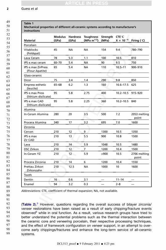
Table 1Mechanical properties of different all-ceramic systems according to manufacturer’sinstructions
MaterialModulus(GPa)
Hardness(GPa)
Toughness(MPa m1/2)
Strength(MPa)
CTE�CX 3 10L6 Firing (�C)
Porcelain
Vitablocks(Feldspat)
45 NA NA 154 9.4 780–790
Lava Ceram 78 5.3 1.1 100 10.5 810
IPS e.max ceram 60–70 5.4 NA 90 9.5 750
IPS e.max ZirPress(Flour-Apatite)
65 5.4 NA 110 10.5–11 900–910
Glass-ceramic
Dicor 75 3.4 1.4 290 9.8 850
Empress esthetic(leucite)
65–68 6.2 1.3 160 16.6–17.5 625
IPS e.max Press(lithium disilicate)
95 5.8 2.75 400 10.2–10.5 915–920
IPS e.max CAD(lithium disilicate)
95 5.8 2.25 360 10.2–10.5 840
Alumina
In-Ceram Alumina 280 20 3.5 500 7.2 2053 meltingpoint
Procera Alumina 340 17 3.2 695 7.0 1600
Zirconia
Cercon 210 12 9 1300 10.5 1350
IPS e.maxZir CAD
210 13 5.5 900 10.8 1500
Lava 210 14 5.9 1048 10.5 1480
DSC Zirkon 210 12 7 1200 10.4 1500
In-Ceram YZ 210 12 5.9 >900 10.5 2706 meltingpoint
Procera Zirconia 210 14 6 1200 10.4 1550
Prettau ZirkonZirkonzahn
210 12.5 NA 1000 10 1600
Tooth
Dentin 16 0.6 3.1 — 11–14 —
Enamel 94 3.2 0.3 — 2–8 —
Abbreviations: CTE, coefficient of thermal expansion; NA, not available.
Guess et al2
49
505152
535455
565758
59606162
636465
666768
697071
72737475
767778
798081
82838485
868788
899091
929394
95969798
99
(Table 2).5 However, questions regarding the overall success of bilayer zirconia/veneer restorations have been raised as a result of early chipping/fracture eventsobserved6 while in oral function. As a result, various research groups have tried tobetter understand the potential problems such as the thermal interaction betweenthe ceramic core and veneering porcelain, their respective processing techniques,and the effect of framework configuration on veneer support, in an attempt to over-come early chippings/fractures and enhance the long-term service of all-ceramicsystems.
DCL513_proof ■ 9 February 2011 ■ 4:23 pm

Q8
Q1All-Ceramic Systems 3
100
101102103
104105106
107108109
110111112113
114115116
117118119
120121122
123124125126
127128129
130131132
133134135136
137138139
140141142
143144145
146147148149
150
This overview presents the current knowledge of monolithic and bilayer all-ceramicsystems and addresses composition and processing mechanisms, laboratory andclinical performance, and possible future trends.
REINFORCED GLASS CERAMICS
The interest in nonmetallic and biocompatible restorative materials increased after theintroduction of the feldspathic porcelain crown in 1903 by Land.7 In 1965, McLean8
pioneered the concept of adding aluminum oxide to feldspathic porcelain in anattempt to enhance mechanical properties. However, the clinical shortcomings ofthese materials, such as brittleness, crack propagation, low tensile strength, wearresistance, and marginal accuracy, discontinued its use.9
Increased strength in glassy ceramics can be achieved with added appropriatefillers that are uniformly dispersed throughout the glass, a technique termed disper-sion strengthening. Leucite is used as a reinforcing crystalline phase at a concentrationof 35 to 45 vol%.10 In the early 1990s, the lost wax press technique was introduced todentistry as an innovative processing method for all-ceramic restorations. Examplesof leucite-reinforced glass ceramics are VITA VMK 68 (VITA Zahnfabrik, Bad Sackin-gen, Germany), Finesse All-Ceramic, (Dentsply, York, PA, USA), Optec OPC (Jeneric,Wallingford, CT, USA), and IPS Empress (Ivoclar Vivadent, Schaan, Principality ofLiechtenstein). The molding procedure of IPS Empress is conducted at 1080�C ina special, automatically controlled furnace. Leucite crystals are formed througha controlled surface crystallization process in the SiO2-Al2O3-K2O glass system.Tangential compressive stresses develop around the crystals on cooling because ofthe difference in the coefficient of thermal expansion (CTE) between leucite crystalsand the glassy matrix. These stresses contribute to crack deflection and improvedmechanical performance.11 IPS Empress, for instance, exhibits a flexural strength of120 to 180 MPa and a CTE of 15 to 18.5 � 10�6 K�1 m/m.12 The material is suitablefor fabrication of inlays, onlays, veneers, and crowns. Favorable clinical long-termdata with high survival rates are described for IPS Empress inlays, onlays (90% after8 years13), veneers (94.4% after 12 years14), and crowns (95.2% after 11 years15) in thedental literature.Leucite glass ceramics can also be machined with various CAD/CAM systems.
Multicolored blocks were recently developed to reproduce color transitions andshading as well as different levels of translucency to reproduce natural teeth(Fig. 1).16 The use of leucite-reinforced glass ceramic has significantly declinedbecause of the introduction of lithium glass ceramics with significantly improvedmechanical and aesthetic properties and is addressed in detail in the next section.
LITHIUM DISILICATE GLASS CERAMICS
A significantly higher strength of 350 MPa was achieved with a glass ceramic of theSiO2-Li2O-K2O-ZnO-P2O5-Al2O3-La2O3 system by precipitating lithium disilicate(Li2Si2O5) crystals. The crystal content of up to 70 vol% is considerably higher thanthat of leucite materials.11 High-temperature x-ray diffraction studies revealed thatboth lithium metasilicate (Li2SiO3) and crystobalite form during the crystallizationprocess before the growth of lithium disilicate (Li2Si2O5) crystals.
17 The final micro-structure consists of highly interlocked lithium disilicate crystals, 5 mm in length and0.8 mm in diameter. Thermal expansion mismatch between lithium disilicate crystalsand glassy matrix results in tangential compressive stresses around the crystals,potentially responsible for crack deflection and strength increase. Crystal alignmentafter heat pressing of the lithium disilicate glass ceramic leads to multiple crack
DCL513_proof ■ 9 February 2011 ■ 4:23 pm
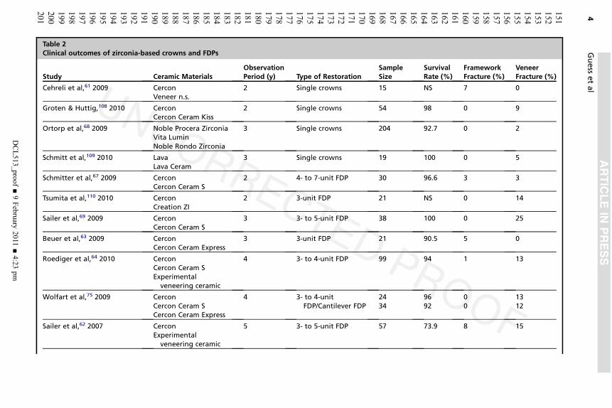
Table 2Clinical outcomes of zirconia-based crowns and FDPs
Study Ceramic MaterialsObservationPeriod (y) Type of Restoration
SampleSize
SurvivalRate (%)
FrameworkFracture (%)
VeneerFracture (%)
Cehreli et al,61 2009 CerconVeneer n.s.
2 Single crowns 15 NS 7 0
Groten & Huttig,108 2010 CerconCercon Ceram Kiss
2 Single crowns 54 98 0 9
Ortorp et al,68 2009 Noble Procera ZirconiaVita LuminNoble Rondo Zirconia
3 Single crowns 204 92.7 0 2
Schmitt et al,109 2010 LavaLava Ceram
3 Single crowns 19 100 0 5
Schmitter et al,67 2009 CerconCercon Ceram S
2 4- to 7-unit FDP 30 96.6 3 3
Tsumita et al,110 2010 CerconCreation ZI
2 3-unit FDP 21 NS 0 14
Sailer et al,69 2009 CerconCercon Ceram S
3 3- to 5-unit FDP 38 100 0 25
Beuer et al,63 2009 CerconCercon Ceram Express
3 3-unit FDP 21 90.5 5 0
Roediger et al,64 2010 CerconCercon Ceram SExperimental
veneering ceramic
4 3- to 4-unit FDP 99 94 1 13
Wolfart et al,75 2009 CerconCercon Ceram SCercon Ceram Express
4 3- to 4-unitFDP/Cantilever FDP
2434
9692
00
1312
Sailer et al,62 2007 CerconExperimental
veneering ceramic
5 3- to 5-unit FDP 57 73.9 8 15
Guess
etal
DCL513_proof
■9February
2011■4:23
pm
4
151
152
153
154
155
156
157
158
159
160
161
162
163
164
165
166
167
168
169
170
171
172
173
174
175
176
177
178
179
180
181
182
183
184
185
186
187
188
189
190
191
192
193
194
195
196
197
198
199
200
201

Crisp et al,111 2008 LavaLava Ceram
1 3- to 4-unit FDP 38 100 0 3
Raigrodski et al,112 2006 LavaVeneer n.s.
2.5 3-unit FDP 20 NS 0 25
Schmitt et al,113 2009 LavaLava Ceram
3 3- to 4-unit FDP 27 100 0 11
Vult von Steyern et al,114 2005 DC ZirkonVita D
2 3- to 5-unit FDP 20 NS 0 15
Tinschert et al,83 2008 DC ZirkonVita D
3 3- to 10-unitFDP/Cantilever FDP
65 NS 0 6
Edelhoff et al,115 2008 DigidentInitial ZirKeramik GC
3 3- to 6-unit FDP 21 90.5 0 10
Molin and Karlsson,116 2008 DenzirVita DIPS Empress
5 3-unit FDP 19 100 0 36
Nothdurft & Pospiech,84 2009 CerconCercon Ceram Kiss
0.5 Implant-supportedsingle crowns
40 NS 0 8
Larsson et al,49 2006 DenzirEsprident Triceram
1 Implant-supportedFDP
13 NS 0 53
Larsson et al,3 2010 CerconCercon Ceram S
3 Implant-supportedfull-arch FDP
10 100 0 34
Abbreviati Q14on: NS, not significant.
All-C
eramic
Systems
DCL513_proof
■9February
2011■4:23
pm
5
202
203
204
205
206
207
208
209
210
211
212
213
214
215
216
217
218
219
220
221
222
223
224
225
226
227
228
229
230
231
232
233
234
235
236
237
238
239
240
241
242
243
244
245
246
247
248
249
250
251
252

print&
web4C=FPO
Fig. 1. Before and after treatment with all-ceramic restorations (lithium disilicate IPS e.maxPress Tooth 24 inlay; onlays 26, 27; and crowns14, 15, 25) and CAD/CAM-fabricated leuciteProCAD onlay 16, 26.
print&
web4C=FPO
Guess et al6
253
254255256
257258259
260261262
263264265266
267268269
270271272
273274275
276277278279
280281282
283284285
286287288289
290291292
293294295
296297298
299300301302
303
deflections. The lithium disilicate ceramic was introduced as IPS Empress 2 (IvoclarVivadent) in 1998 and is moldable as leucite glass ceramics, but at a lower tempera-ture of 920�C. The CTE is 10.5 � 10�6 K�1 m/m.18
High survival rates were observed for anterior and posterior IPS Empress 2 crowns(95.5% after 10 years19) in long-term clinical studies (Fig. 2). The clinical success of 3-unit IPS Empress 2 FDPs is predominately limited by bulk fracture failure within theconnector area, in particular when the connector size is less than the recommendeddimensions (70% survival rate after 5 years20)(Fig. 3).A newly developed pressable lithium disilicate glass ceramic (IPS e.max Press, Ivo-
clar Vivadent) with improved physical properties (flexural strength, 440 MPa) andtranslucency through different firing process has been developed in the SiO2-Li2O-K2O-ZnO-P2O5-Al2O3-ZrO2 system. The pressable lithium disilicate ceramic can beused in monolithic application for inlays, onlays, and posterior crowns or as a corematerial for crowns and 3-unit FDPs in the anterior region. Apatite glass ceramicsare recommended for veneering.
Fig. 2. Lithium disilicate crowns (IPS Empress 2) at baseline and functionally and aestheti-cally successful performance after 5 years.
DCL513_proof ■ 9 February 2011 ■ 4:23 pm

print&
web4C=FPO
Fig. 3. Three-unit lithium disilicate (IPS Empress 2) FDPs at baseline and a connector bulkfracture after 11 months.
All-Ceramic Systems 7
304
305306307
308309310
311312313
314315316317
318319320
321322323
324325326
327328329330
331332333
334335336
337338339340
341342343
344345346
347348349
350351352353
354
Clinical data exhibited high survival rates on IPS e.max Press onlays (100% after3 years),21 crowns (96,6% after 3 years),22 monolithic inlay retained FDPs (100% after4 years),23 and full crown retained FDPs (93% after 8 years).24
Recently, a lithium disilicate glass ceramic (IPS e.max CAD, Ivoclar Vivadent) hasbeen designed for CAD/CAM processing technology. The milled lithium disilicateblock is exposed to a 2-stage crystallization process. Lithium metasilicate crystalsare precipitated during the first stage. The resulting glass ceramic has a crystal sizerange of 0.2 to 1.0 mm with approximately 40 vol% lithium metasilicate crystals. Atthis precrystallized state, the CAD/CAM block exhibits a flexural strength of 130 to150 MPa, which allows simplified machining and intraoral occlusal adjustment. Thefinal crystallization process occurs after milling of the restoration at 850�C in vacuum.The metasilicate crystal phase is dissolved completely, and the lithium disilicate crys-tallizes. This process also converts the blue shade of the precrystallized block to theselected tooth shade and results in a glass ceramic with a fine grain size of approxi-mately 1.5 mm and a 70% crystal volume incorporated in a glass matrix.16 CAD/CAM-processed lithium disilicate glass ceramic demonstrates a flexural strength of360 MPa (see Table 1). Because of the favorable translucency and shade variety,the material can be used for fully anatomic (monolithic) restorations with subsequentstaining characterization or as a core material with subsequent coating with veneeringceramics. The manufacturer recommends its use for anterior or posterior crowns,implant crowns, inlays, onlays, and veneers. Preliminary clinical results on singlecrowns revealed high survival rates (100% after 2 years25) and are hence promising.
GLASS-INFILTRATED CERAMICS
Another group of all-ceramic systems involves the glass-infiltrated ceramics. In-Ceram Alumina (VITA Zahnfabrik) was the first all-ceramic system available forsingle-unit restorations and 3-unit anterior bridges26 with a high-strength ceramiccore fabricated through the slip-casting technique.27 A slurry of densely packed (70–80 wt%) Al2O3 is applied and sintered to a refractory die at 1120�C for 10 hours.28,29
This process produces a porous skeleton of alumina particles, which is infiltrated
DCL513_proof ■ 9 February 2011 ■ 4:23 pm
Guess et al8
355
356357358
359360361
362363364
365366367368
369370371
372373374
375376377
378379380381
382383384
385386387
388389390391
392393394
395396397
398399400
401402403404
405
with lanthanum glass in a second firing at 1100�C for 4 hours to eliminate porosity andincrease strength.29 The core is veneered with feldspathic porcelain.26 In-CeramZirconia (VITA Zahnfabrik) is a modification of the original In-Ceram Alumina system,with an addition of 35% partially stabilized zirconia oxide to the slip composition tostrengthen the ceramic.27 Traditional slip-casting techniques can be used, or themate-rial can be copy-milled from prefabricated, partially sintered blanks and then veneeredwith feldspathic porcelain.30 Because the core is opaque and lacks translucency, theuse of this material for anterior regions becomes problematic.27 Clinical performanceof glass-infiltrated ceramics for crowns and FDPs indication has been evaluated ina systematic review.31 However, with the development of high-strength ceramics,recent literature on glass-infiltrated ceramics became sparse.
Q9
HIGH-STRENGTH OXIDE CERAMICS
Although high-strength ceramic in dentistry includes alumina and zirconia, this sectionfocuses mainly on zirconia because of its superior mechanical properties comparedwith alumina and the current controversial aspects of its clinical performance.Zirconia ceramics have gained a remarkable interest in biomedical sciences.4 The
first reference concerning their application in medicine appeared in the late sixties;20 years later, Christel and colleagues32 studied their use in orthopedic surgery.33
In the early 1990s, zirconia was introduced to dentistry,34 and in recent years, a largenumber of publications appeared in the literature.Zirconia, characterized by a dense, monocrystalline homogeneity, possesses low
thermal conductivity, low corrosion potential, and good radiopacity.4 High biocompat-ibility, low bacterial surface adhesion, and favorable optical properties of zirconiaceramics are reported.35–37 Zirconia in its pure form is a polymorph that has 3temperature-dependant phases that are monoclinic (room temperature to 1170�C),tetragonal (1170�C–2370�C), and cubic (2370�C to melting point).34 With the additionof stabilizing oxides such asmagnesia, ceria, yttria, and calcium to zirconia, the tetrag-onal phase is retained in a metastable condition at room temperature, enablinga phenomenon called transformation toughening to occur. In response to mechanicalstimuli, the partially stabilized crystalline tetragonal zirconia transforms to the more-stable monoclinic phase, with a local volume increase of approximately 4%. Thisincrease in volume counteracts further crack propagation by compression at the tipof the crack.38 Compared with high-strength alumina ceramic, zirconia has twicethe flexural strength (900–1200 MPa).39,40 In addition, high fracture toughness (9–10 MPa∙m1/2) has been described.39 The most commonly used dental zirconia formu-lations are glass-infiltrated zirconia toughened alumina ceramics (In-Ceram, VITAZahnfabrik) and3mol%Y-TZP.Y-TZPhasbeenused in root canal posts,41 frameworksfor posterior teeth, implant-supported crowns,multiunit FDPs,42 resin-bonded FDPs,43
custom-made bars to support fixed and removable dental prostheses,44 implantabutments,45,46 and dental implants.47,48
A magnesium-doped partially stabilized zirconia (Mg-PSZ, Denzir M, Dentronic AB,Sweden) is available, but porosity and large grain size has limited its success.49
Zirconia and CAD/CAM Technology
An array of CAD/CAM systems has evolved since F. Duret introduced the concept in1971.50,51 Some CAD/CAM systems (Denzir, Cadesthetics, Sweden, and DC-Zircon,DCS Dental AG, Switzerland) machine fully sintered Y-TZP blocks, which have beenprocessed by hot isostatic pressing. Because of the hardness and poor machinabilityof fully sintered Y-TZP, a robust milling system and extended milling periods are
DCL513_proof ■ 9 February 2011 ■ 4:23 pm

Q10
All-Ceramic Systems 9
406
407408409
410411412
413414415
416417418419
420421422
423424425
426427428
429430431432
433434435
436437438
439440441442
443444445
446447448
449450451
452453454455
456
required. Most of the available CAD/CAM systems shape blocks of partially sinteredzirconia.34 Milling from partially sintered blocks involves machining enlarged frame-works in a so-called green state. These blocks are then sintered to their full strength,which is accompanied by shrinkage of the milled framework by approximately 25% tothe desired dimensions. Examples of these systems are CERCON (Dentsply Friadent,Mannheim, Germany), LAVA (3M ESPE, Germany), Procera (Nobel Biocare; Sweden),Etkon (Straumann, Switzerland), and Cerec (Sirona, Bensheim, Germany).The advantage of industrialized blank fabrication and reproducible and consistent
CAM resulted in increased process reliability and cost-effectiveness of CAD/CAM-fabricated restorations.52 Labor-intensive waxing, casting, and soldering of frame-works accompanied with conventional laboratory procedures can be avoided withthe application of CAD/CAM technology. With extensive frameworks, particularlyused in implant dentistry, and the escalating costs of precious alloys, all-ceramicrestorations are competitive with conventional metal-ceramic restorations (MCRs)from a cost perspective.53
Zirconia and Low-Temperature Degradation
A major drawback of zirconia restorations compared with metal-ceramics is thematerial-inherent accelerated aging that has been observed to occur in zirconia inthe presence of moisture.54,55 This aging phenomenon is known as low-temperaturedegradation (LTD) and was first described by Kobayashi and colleagues56 in 1981.At relatively low temperatures (150�C–400�C), slow tetragonal to monoclinic transfor-mations occur, initiating at the surface of polycrystalline zirconia and subsequentlyprogressing into the bulk of the material.34,57 Transformation of 1 grain is accompa-nied by an increase in volume, which causes stress on the surrounding grains andmicrocracking. Water penetration into these cracks then exacerbates the process ofsurface degradation, and the transformation progresses. The growth of the transfor-mation zone results in severe microcracking, grain pullout, and finally surface rough-ening, which ultimately leads to strength degradation. Any factor that is detrimental tothe stability of tetragonal zirconia is susceptible to promote LTD. Among these factorsare grain size,58 the amount of stabilizer,59 and the presence of residual stress.54,60 Atpresent, there is no clear correlation between LTD and failure predictability whenzirconia is used as a dental bioceramic.33
Clinical Data on Zirconia-Supported Restorations
Although zirconia was introduced to dentistry in the early 1990s, limited informationabout its clinical performance can be found. A literature search from 1990 throughSeptember 2010 using electronic databases (PubMed and MEDLINE) revealed a totalof 21 clinical trials on zirconia-supported restorations (see Table 2). Long-term clinicaldata for zirconia-supported restorations with observation periods exceeding 5-yeartime frames are not yet available. Most studies (n 5 14) investigated predominatelyposterior FDPs, whereas a small number focused on (n 5 4) crowns. Only 3 studiesdescribed the clinical behavior of implant-supported zirconia-supported FDPs andsingle crowns.
Veneered Zirconia Clinical Failure Modes
Core/framework fracturesCatastrophic fractures within the zirconia core ceramic are reported at 7% forsingle crowns after 2 years and at 1% to 8% for FDPs after 2 to 5 years (seeTable 2). Occlusal overloading caused by bruxism (crown fracture after 1 month61)or trauma (connector fracture 5-unit FDP after 38months62) and insufficient framework
DCL513_proof ■ 9 February 2011 ■ 4:23 pm
Guess et al10
457
458459460
461462463
464465466
467468469470
471472473
474475476
477478479
480481482483
484485486
487488489
490491492493
494495496
497498499
500501502
503504505506
507
thickness at 0.3 mm (crown-abutment fractures in 3-unit FDP63,64) were mentioned asthe main reasons for zirconia core bulk fractures. Fractographic analyses of clinicallyfailed zirconia crowns showed that radial fractures propagating upwards from thecementation surface site resulted in bulk fractures and caused catastrophic failuresof the restorations.65 Microscopic examinations of failed zirconia-based FDPsrevealed that core bulk fractures were most commonly located in the connectorarea and initiated from the gingival surface, where tensile stresses were the greatestbecause of occlusal loading.65,66 Core bulk fracture in the connector area was alsoattributed to damage induced during fabrication.67 A higher susceptibility to connectorfracture was noted with an increased span of FDPs.65
Veneering ceramic cohesive fracturesThe major drawbacks noted in all clinical studies on zirconia-supported restorationswere related to a lesser extent to the framework integrity andmainly to wear and failureof the veneering ceramics. Cohesive fractures within the veneering ceramic (chipping)were described as the most frequent reason for failures, irrespective of the appliedzirconia veneer system (see Table 2). Veneer fracture rates are reported at 2% to9% for single crowns after 2 to 3 years and at 3% to 36% for FDPs after 1 to 5 years.Implant-supported zirconia-based restorations revealed even higher rates at 8% forsingle crowns after 6 months and at 53% for FDPs after 1 year. Impaired propriocep-tion and rigidity of osseointegrated implants correlated with higher functional impactforces might further exacerbate cohesive veneer fractures. To date, only one 3-yearprospective clinical study involving implant-supported full-arch Y-TZP reconstructionsis available. Despite the short follow-up period and limited number of patients (n5 10),no framework fractures were observed; however, chip-off fractures were observed in34% of the units cemented in 9 of 10 patients.3
Dependingon thesizeand localization,cracks leading toveneer fracturescanseverelycompromise the aesthetics and function of zirconia-supported restorations.68,69 Fracto-graphic analysis of clinically failed veneered zirconia restorations revealed cohesiveveneer failures, with cracks originating from the occlusal surface and propagating tothe core veneer interface, leaving an intact core.65 In many clinical studies, it seemedthat these veneer failures were associated with roughness in the veneering ceramicbecause of occlusal function or occlusal adjustment.49,69,70 Therefore, special attentionhas to be paid to the static and dynamic aspects of occlusion in zirconia-based restora-tions. Occlusal adjustments should be performed only with fine-grain diamonds underwater irrigation followed by a thorough polishing sequence.
Reasons for veneering ceramic failureAs the veneering ceramic material (flexural strength, approximately 90–120 MPa) isweak compared with the high-strength core material (900–1200 MPa),39,40 theveneering ceramic seems to be prone to failure at low loads during masticatory func-tion. Hence, the use of higher-strength veneering ceramic was proposed to reduce theincidence of veneer chippings/fractures. However, attempts to improve the micro-structure and mechanical properties of veneering ceramics with the development ofglass-ceramic ingots for pressing veneering ceramics onto zirconia frameworks didnot result in an increased reliability of the veneering ceramic.71,72 In addition, identicalchip-failure patterns were observed.71–74 Although a high density of the veneeringlayer has been expected with the press technique, spherical porosities were notedwithin the veneer layer and also at the interface.72 In clinical trials on zirconia FDPs,chipping fractures have also been reported with pressable veneering ceramics, andthe chipping problem does not seem to be solved.75
DCL513_proof ■ 9 February 2011 ■ 4:23 pm
Q11
All-Ceramic Systems 11
508
509510511
512513514
515516517
518519520521
522523524
525526527
528529530
531532533534
535536537
538539540
541542543544
545546547
548549550
551552553
554555556557
558
Moreover, the CTE is frequently discussed as a contributing factor for veneer failure.However, the problem of chipping may not be limited only to the mismatch of CTE; itseems more complex. Residual stresses in bilayer crowns and FDPs are associatedwith the possibility of thermal gradients being developed in these structures duringcooling. For zirconia veneer all-ceramic systems, the low thermal conductivity of thezirconia (approximately 3 Wm/K)76 results in the highest temperature difference and,therefore, very high residual stresses. In addition, thick layers of veneering ceramicson zirconia cores are highly susceptible to generating high tensile subsurface residualstresses, ultimately resulting in unstable cracking or chipping.77 Slow cooling of therestoration above the glass transition temperature of the porcelain could preventthe development of high tensile subsurface residual stresses in the porcelain, whichmay result in unstable cracking or chipping. Most manufacturers now recommendthe approach of a reduced cooling rate after final firing or glazing, and even an addi-tional 6 minutes has shown to be effective.78
Zirconia framework designThe lack of a uniform support of the veneering ceramic because of improper frame-work design has been discussed as a possible reason for chipping fractures. Remark-ably little scientific clinical data on optimal design of zirconia-supported restorationshave been published.53,79,80 With the introduction of CAD/CAM technologies indentistry, excessive veneer layer thickness (>2.5 mm) was created because of theuniform layer thickness of the copings for crowns and bar-shaped connectorsfor FDPs. Improved customized zirconia coping design derived from the conventionalPFM technique has been recommended to provide adequate support for the veneeringceramic.80 A dual-scan procedure of the die and full-contour wax pattern hasbeen merged to fabricate the desired framework. Preliminary in vitro studies showedthat cohesive fractures within the veneering ceramic could not be avoided with theimproved support, but the size of the fractures was significantly decreased81,82 andfailure initiation was shifted toward higher loads.73 In clinical observation, conflictingresults on the framework design modification have been described.83,84 Hence, theeffect of framework design modifications on residual stress states needs to be betterelucidated.77
Minimizing Zirconia Core Failures
Some fundamental aspects affecting the clinical performance of zirconia as a frame-work material need to be addressed. Laboratory technicians and clinicians shouldfollow precise sequence steps in manufacturing zirconia-based restorations, withthe knowledge that zirconia as a framework material is highly susceptible to surfacemodifications and improper laboratory and clinical handling techniques.85
Grinding or sandblasting of surfaces with high (or mild/low) pressure ranges isdiscussed to induce the formation of surface microcracking85–87 that could be detri-mental to the long-term performance of the restorations and lead to unexpectedfailures.88 Several research groups have claimed an increased flexural strength inY-TZP89 because of phase transformation after grinding and alumina abrasion.90–92
However, any temporary beneficial effect related to phase transformation inducedby different surface treatment methods may be negated by fatigue-related crackgrowth phenomena and following heat treatments associated with veneerapplication.90 With respect to the highly deleterious effect on zirconia reliability,93
postsintering surface modifications of zirconia frameworks at the dental laboratoryor under clinical circumstances should be avoided.
DCL513_proof ■ 9 February 2011 ■ 4:23 pm

Q12
Q13
Guess et al12
559
560561562
563564565
566567568
569570571572
573574575
576577578
579580581
582583584585
586587588
589590591
592593594595
596597598
599600601
602603604
605606607608
609
LABORATORY PERFORMANCE OF ALL-CERAMIC MATERIALS
Randomized prospective clinical trials are the first choice for evaluating clinical long-term behavior of dental materials and techniques. However, the results of clinicalinvestigations are restricted by high costs, and meaningful conclusions can be drawnonly if an adequate number of restorations are incorporated and observed for at least 3to 4 years.94 In addition, fractures are occasionally observed after 4 to 5 years of clin-ical service (Ferencz J, personal communication). Therefore, in vitro simulations andlaboratory tests are developed to investigate new dental materials and predict life-times and failures.Various testing methods have been used to investigate the mechanical properties of
dental materials. Many concerns have been raised lately regarding the clinical signif-icance of simple traditional mechanical testing methodologies and single load tofailure tests of dental restorations, loosely termed “crunch the crown test”95 or flexuralstrength tests. These tests report unrealistically high fracture strength values, largelyoverestimating the actual failure load. Furthermore, the obtained failure modes differsignificantly from those reported in clinical observations. Hence, little insight intodamage initiation and propagation can be provided.96 In material science, fatigue isthe progressive and localized structural damage that occurs when a material is sub-jected to cyclic loading. Fatigue is a significant factor limiting the lifespan ofzirconia-based restorations and, therefore, represents a prerequisite for valid in vitrotesting.97 A recently developed mouth-motion fatigue model has demonstrated theability to duplicate clinical fractures98 and predict the material’s reliability to validateits clinical potential.Guess and colleagues99 examined the failure and fatigue behavior of a hand-layer
veneered zirconia core ceramic and compared it with a monolithic CAD/CAM-fabricated lithium disilicate ceramic. A sliding contact mouth-motion fatigue testingin water on anatomically correct crowns was applied to simulate approximating toothsurfaces during mastication. Hand-layer veneered zirconia crowns supported by toothreplicas revealed a high single-cycle load to failure strength of 1195 � 221 N. Butduring mouth-motion fatigue testing, hand-layer veneered zirconia-based crownsresulted in a very limited reliability; approximately 90%of specimens failed from veneerchip-off fracture by 100 K cycles at 200 N. Failure progressed from the contact areathrough the body of the veneering ceramic as noted in clinical cases.65 The observedfatigue behavior of zirconia-supported crowns does not seem to be material or systemdependent, as similar results were reported in previous findings using the identical testmethodology.100 In contrast, CAD/CAM lithium disilicate crowns showed no failures upto load levels exceeding posterior physiologic chewing forces and seemed to be resis-tant to mouth-motion step-stress (900 N/180K cycles) and also to staircase fatigue(1000 N/1000K cycles).101 A threshold for damage and bulk fracture could be detectedin the range of 1100 to 1200 N. CAD/CAM processing of the lithium disilicate ceramicuses industrialized fabricated homogenous ceramic blanks; therefore, the materialreveals a high density with a minimum of inherent flaws. Increased Weibull modulusand reliability have been reported for CAD/CAM-fabricated materials.102 Conversely,the presence of structural inhomogeneities within the veneer layer of zirconia-supported ceramics is a known drawback of bilayer all-ceramic systems.71
The Gold-Standard Metal-Ceramic
Although MCRs are frequently referred to as the gold standard and still representa common procedure to rehabilitate missing dental structures and teeth, limited infor-mation is available regarding their clinical and laboratory performance when
DCL513_proof ■ 9 February 2011 ■ 4:23 pm

print&web4C=FPO
All-Ceramic Systems 13
610
611612613
614615616
617618619
620621622623
624625626
627628629
630631632
633634635636
637638639
640641642
643644645646
647648649
650651652
653654655
656657658659
660
compared with all-ceramic systems. After the introduction of zirconia to dentistry,more elaborate clinical and laboratory investigations were performed, but MCRswere, in most instances, not included as control.103,104 Moreover, the understandingof the failure mechanisms of MCRs is mainly related to biological issues rather thanfracture or chipping of the veneer porcelain.105
In one of the few laboratory studies presented in the dental literature, Silva andcolleagues74 performed a sliding contact fatigue test study on anatomically correctMCR crowns and compared the reliability and failure modes with zirconia-supported crowns. Fig. 4 shows a series of representative images of laboratory-tested MCRs, zirconia-supported crowns, and its clinical-matched fractures. Theresemblance of failure modes for both systems shows the potential of use slidingcontact fatigue concept to preclinical scenarios. The investigators74 observed thatthe MCR system presented a significantly higher reliability compared with Y-TZP,
Fig. 4. Comparison between clinical and laboratory fractures. (A) Metal-ceramic molarcrown fractured after about 13 years of oral function. (B) Representative metal-ceramicmolar crown after step-stress mouth-motion fatigue test. White arrows in (A) and (B)show typical veneer cohesive fractures. (C) Typical clinical veneer cohesive fracture (blackarrow) at the proximal aspect of a Y-TZP-supported all-ceramic crown after 2.5 years inoral function. (D) Typical fracture (black arrow) at the proximal aspect of a Y-TZP-supportedall-ceramic crown after a mouth-motion fatigue test. Note similar fracture patterncompared with clinical failure mode shown in (C). ([A] Courtesy of Dr Carlo Marinello,University of Basel, Switzerland.)
DCL513_proof ■ 9 February 2011 ■ 4:23 pm
Guess et al14
661
662663664
665666667
668669670
671672673674
675676677
678679680
681682683
684685686687
688689690
691692693
694695696697
698699700
701702703
704705706
707708709710
711
which may explain the fact that not many reports of veneer fractures for MCRs can befound. As new ceramic systems, including more robust monolithic materials, are avail-able in the market, it becomes imperative that further laboratory and clinical investiga-tions including MCR as control are needed. To date, only 1 randomized controlledclinical trial involving 3- to 5-unit zirconia-supported fixed partial dentures has beenperformed with metal-ceramic as controls. The findings of this 3-year follow-uphave shown no differences among biological or technical complications betweenthe 2 systems. Chip-off fractures occurred in 25% of the zirconia-supported pros-theses and in 19.4% of the metal-ceramics. Although the chipping occurrence wasnot statistically different between the groups, only the zirconia-supported prosthesespresented unacceptable major fractures of the veneering ceramic relative to the minorchips observed in the metal-ceramic system.69
NEW CONCEPTS FOR THE ALL-CERAMIC SYSTEMS
Many manufacturers have shifted their attention to the development of monolithic all-ceramic materials to simply remove the most common failing layer of the system andto avoid inherent residual thermal stresses in bilayer all-ceramic systems. In combina-tion with CAD/CAM fabrication, monolithic/full-anatomic crown restorations seemedto be reliable and robust. Most recently, monolithic zirconia ceramic restorationsrefined with superficial glazing and staining and functionally graded glass/zirconia/glass structures106 are being explored in high-load bearing areas.In addition, CAD/CAM capabilities creating separate core and veneer layers that
could then be joined with nonthermal methods will continue to evolve and are ofparticular interest for extended restorations.107 Subtractive CAD/CAM approaches,removing material from a block to create a shape, will be complemented by additiveapproaches, depositing materials only in places where it is needed to form arestoration.74 Although issues are still remaining with each of these approaches,they show great promise.
SUMMARY
Fracture of the veneering ceramics, thermal interaction between core and ceramicmaterials, and susceptibility of zirconia to aging are still beingdebated in thedental liter-ature. Refinement of frameworkdesign and innovativeCAD/CAMveneering techniquesare promising tools to improve the clinical performance of zirconia-supported restora-tions. The use of monolithic leucite and lithium disilicate glass ceramic systems haveshown promising laboratory and clinical results for small restorations such as inlays,onlays, crowns, and laminates. Further laboratory and clinical investigations using all-ceramic materials compared with metal-ceramic systems are strongly recommended.
ACKNOWLEDGMENTS
The authors thank the Department of Biomaterials and Biomimetics at the New YorkUniversity College of Dentistry, USA. The authors also thank Lenny Marotta (MarottaDental Studios) for substantial collaboration and support.
REFERENCES
1. Denry I, Holloway JA. Ceramics for dental applications: a review. Materials2010;3:351–68.
2. ConradHJ, SeongWJ, Pesun IJ. Current ceramicmaterials and systemswith clin-ical recommendations: a systematic review. J Prosthet Dent 2007;98(5):389–404.
DCL513_proof ■ 9 February 2011 ■ 4:23 pm
All-Ceramic Systems 15
712
713714715
716717718
719720721
722723724725
726727728
729730731
732733734
735736737738
739740741
742743744
745746747748
749750751
752753754
755756757
758759760761
762
3. Larsson C, Vult von Steyern P, Nilner K. A prospective study of implant-supported full-arch yttria-stabilized tetragonal zirconia polycrystal mandibularfixed dental prostheses: three-year results. Int J Prosthodont 2010;23(4):364–9.
4. Manicone PF, Rossi Iommetti P, Raffaelli L. An overview of zirconia ceramics:basic properties and clinical applications. J Dent 2007;35(11):819–26.
5. Della Bona A, Kelly JR. The clinical success of all-ceramic restorations. J AmDent Assoc 2008;139(Suppl):8S–13S.
6. Al-Amleh B, Lyons K, Swain M. Clinical trials in zirconia: a systematic review.J Oral Rehabil 2010;37(8):641–52.
7. Land CH. Porcelain dental art. Dental Cosmos 1903;45:437–615.8. McLean JW, Hughes TH. The reinforcement of dental porcelain with ceramic
oxides. Br Dent J 1965;119:251–67.9. Sjogren G, Lantto R, Granberg A, et al. Clinical examination of leucite-reinforced
glass-ceramic crowns (Empress) in general practice: a retrospective study. Int JProsthodont 1999;12(2):122–8.
10. Denry I, Rosenstiel SF. Phase tranformation in feldspathic dental porcelains. In:Fischman G, editor. Bioceramics: materials and applications. Westervill (OH):The American Ceramic Society; 1995.
11. Guazzato M, Albakry M, Ringer SP, et al. Strength, fracture toughness andmicrostructure of a selection of all-ceramic materials. Part I. Pressable andalumina glass-infiltrated ceramics. Dent Mater 2004;20(5):441–8.
12. Dong JK, Luthy H, Wohlwend A, et al. Heat-pressed ceramics: technology andstrength. Int J Prosthodont 1992;5(1):9–16.
13. Kramer N, Taschner M, Lohbauer U, et al. Totally bonded ceramic inlays andonlays after eight years. J Adhes Dent 2008;10(4):307–14.
14. Fradeani M, Redemagni M, Corrado M. Porcelain laminate veneers: 6- to 12-year clinical evaluation–a retrospective study. Int J Periodontics RestorativeDent 2005;25(1):9–17.
15. Fradeani M, Redemagni M. An 11-year clinical evaluation of leucite-reinforcedglass-ceramiccrowns:a retrospectivestudy.Quintessence Int2002;33(7):503–10.
16. Holand W, Schweiger M, Watzke R, et al. Ceramics as biomaterials for dentalrestoration. Expert Rev Med Devices 2008;5(6):729–45.
17. Holand W, Apel E, va�nt Hoen C, et al. Studies of crystal phase formation in theearly stage crystallization of lithium disilicate glass-ceramics. J Non Cryst Solids2006;352:4041–50.
18. Holand W, Schweiger M, Frank M, et al. A comparison of the microstructure andproperties of the IPS Empress 2 and the IPS Empress glass-ceramics. J BiomedMater Res 2000;53(4):297–303.
19. Valenti M, Valenti A. Retrospective survival analysis of 261 lithium disilicatecrowns in a private general practice. Quintessence Int 2009;40(7):573–9.
20. Marquardt P, Strub JR. Survival rates of IPS empress 2 all-ceramic crowns andfixed partial dentures: results of a 5-year prospective clinical study. Quintes-sence Int 2006;37(4):253–9.
21. Guess PC, Strub JR, Steinhart N, et al. All-ceramic partial coverage restora-tions–midterm results of a 5-year prospective clinical splitmouth study. J Dent2009;37(8):627–37.
22. Etman MK, Woolford MJ. Three-year clinical evaluation of two ceramic crownsystems: a preliminary study. J Prosthet Dent 2010;103(2):80–90.
23. Wolfart S, Bohlsen F, Wegner SM, et al. A preliminary prospective evaluation ofall-ceramic crown-retained and inlay-retained fixed partial dentures. Int J Pros-thodont 2005;18(6):497–505.
DCL513_proof ■ 9 February 2011 ■ 4:23 pm

Guess et al16
763
764765766
767768769
770771772
773774775776
777778779
780781782
783784785
786787788789
790791792
793794795
796797798799
800801802
803804805
806807808
809810811812
813
24. Wolfart S, Eschbach S, Scherrer S, et al. Clinical outcome of three-unit lithium-disilicate glass-ceramic fixed dental prostheses: up to 8 years results. DentMater 2009;25(9):e63–e71.
25. Fasbinder DJ, Dennison JB, Heys D, et al. A clinical evaluation of chairsidelithium disilicate CAD/CAM crowns: a two-year report. J Am Dent Assoc 2010;141(Suppl 2):10S–4S.
26. Haselton DR, Diaz-Arnold AM, Hillis SL. Clinical assessment of high-strength all-ceramic crowns. J Prosthet Dent 2000;83(4):396–401.
27. Sundh A, Sjogren G. A comparison of fracture strength of yttrium-oxide-partially-stabilized zirconia ceramic crowns with varying core thickness, shapesand veneer ceramics. J Oral Rehabil 2004;31(7):682–8.
28. Chai J, Takahashi Y, Sulaiman F, et al. Probability of fracture of all-ceramiccrowns. Int J Prosthodont 2000;13(5):420–4.
29. Xiao-ping L, Jie-mo T, Yun-long Z, et al. Strength and fracture toughness ofMgO-modified glass infiltrated alumina for CAD/CAM. Dent Mater 2002;18(3):216–20.
30. Raigrodski AJ. Contemporary materials and technologies for all-ceramic fixedpartial dentures: a review of the literature. J Prosthet Dent 2004;92(6):557–62.
31. Wassermann A, Kaiser M, Strub JR. Clinical long-term results of VITA In-CeramClassic crowns and fixed partial dentures: a systematic literature review. Int JProsthodont 2006;19(4):355–63.
32. Christel P, Meunier A, Dorlot JM, et al. Biomechanical compatibility and designof ceramic implants for orthopedic surgery. Ann N YAcad Sci 1988;523:234–56.
33. Chevalier J. What future for zirconia as a biomaterial? Biomaterials 2006;27(4):535–43.
34. Denry I, Kelly JR. State of the art of zirconia for dental applications. Dent Mater2008;24(3):299–307.
35. Aboushelib MN, Dozic A, Liem JK. Influence of framework color and layeringtechnique on the final color of zirconia veneered restorations. QuintessenceInt 2010;41(5):e84–e89.
36. Degidi M, Artese L, Scarano A, et al. Inflammatory infiltrate, microvessel density,nitric oxide synthase expression, vascular endothelial growth factor expression,and proliferative activity in peri-implant soft tissues around titanium and zirco-nium oxide healing caps. J Periodontol 2006;77(1):73–80.
37. Rimondini L, Cerroni L, Carrassi A, et al. Bacterial colonization of zirconiaceramic surfaces: an in vitro and in vivo study. Int J Oral Maxillofac Implants2002;17(6):793–8.
38. Garvie RC, Hannink RH, Pascoe RT. Ceramic steel? Nature 1975;258:703–4.39. Christel P, Meunier A, Heller M, et al. Mechanical properties and short-term in-
vivo evaluation of yttrium-oxide-partially-stabilized zirconia. J Biomed Mater Res1989;23(1):45–61.
40. Piconi C, Maccauro G. Zirconia as a ceramic biomaterial. Biomaterials 1999;20(1):1–25.
41. Meyenberg KH, Luthy H, Scharer P. Zirconia posts: a new all-ceramic conceptfor nonvital abutment teeth. J Esthet Dent 1995;7(2):73–80.
42. Sturzenegger B, Feher A, Luthy H, et al. Clinical evaluation of zirconium oxidebridges in the posterior segments fabricated with the DCM system. Acta MedDent Helv 2000;5:131–9.
43. Komine F, Tomic M. A single-retainer zirconium dioxide ceramic resin-bondedfixed partial denture for single tooth replacement: a clinical report. J Oral Sci2005;47(3):139–42.
DCL513_proof ■ 9 February 2011 ■ 4:23 pm

All-Ceramic Systems 17
814
815816817
818819820
821822823
824825826827
828829830
831832833
834835836
837838839840
841842843
844845846
847848849850
851852853
854855856
857858859
860861862863
864
44. Bergler M, Holst S, Blatz MB, et al. CAD/CAM and telescopic technology:design options for implant-supported overdentures. Eur J Esthet Dent 2008;3(1):66–88.
45. Glauser R, Sailer I, Wohlwend A, et al. Experimental zirconia abutments forimplant-supported single-tooth restorations in esthetically demanding regions:4-year results of a prospective clinical study. Int J Prosthodont 2004;17(3):285–90.
46. Sailer I, Philipp A, Zembic A, et al. A systematic review of the performance ofceramic and metal implant abutments supporting fixed implant reconstructions.Clin Oral Implants Res 2009;20(Suppl 4):4–31.
47. Andreiotelli M, Wenz HJ, Kohal RJ. Are ceramic implants a viable alternative totitanium implants? A systematic literature review. Clin Oral Implants Res 2009;20(Suppl 4):32–47.
48. Wenz HJ, Bartsch J, Wolfart S, et al. Osseointegration and clinical success ofzirconia dental implants: a systematic review. Int J Prosthodont 2008;21(1):27–36.
49. Larsson C, Vult von Steyern P, Sunzel B, et al. All-ceramic two- to five-unitimplant-supported reconstructions. A randomized, prospective clinical trial.Swed Dent J 2006;30(2):45–53.
50. Miyazaki T, Hotta Y, Kunii J, et al. A review of dental CAD/CAM: current statusand future perspectives from 20 years of experience. Dent Mater J 2009;28(1):44–56.
51. Rekow ED. Dental CAD/CAM systems: a 20-year success story. J Am DentAssoc 2006;137(Suppl):5S–6S.
52. Tinschert J, Natt G, Hassenpflug S, et al. Status of current CAD/CAM technologyin dental medicine. Int J Comput Dent 2004;7(1):25–45.
53. Donovan TE. Factors essential for successful all-ceramic restorations. J AmDent Assoc 2008;139(Suppl):14S–8S.
54. Deville S, Chevalier J, Gremillard L. Influence of surface finish and residualstresses on the ageing sensitivity of biomedical grade zirconia. Biomaterials2006;27(10):2186–92.
55. Tholey MJ, Berthold C, Swain MV, et al. XRD(2) micro-diffraction analysis of theinterface between Y-TZP and veneering porcelain: role of application methods.Dent Mater 2010;26(6):545–52.
56. Kobayashi K, Kuwajima H, Masaki T. Phase change and mechanical propertiesof ZrO2-Y2O3 solid electrolyte after aging. Solid State Ionics 1981;3:489–93.
57. Kelly JR, Denry I. Stabilized zirconia as a structural ceramic: an overview. DentMater 2008;24(3):289–98.
58. Lawson S. Environmental degradation of zirconia ceramics. J Eur Ceram Soc1995;15:485–502.
59. Hannink R, Kelly PM, Muddle B. Transformation toughening in zirconia contain-ing ceramics. J Am Ceram Soc 2000;83(3):461–87.
60. Kim JW, Covel NS, Guess PC, et al. Concerns of hydrothermal degradation inCAD/CAM zirconia. J Dent Res 2010;89(1):91–5.
61. Cehreli MC, Kokat AM, Akca K. CAD/CAM Zirconia vs. slip-cast glass-infiltratedAlumina/Zirconia all-ceramic crowns: 2-year results of a randomized controlledclinical trial. J Appl Oral Sci 2009;17(1):49–55.
62. Sailer I, Feher A, Filser F, et al. Five-year clinical results of zirconia frameworksfor posterior fixed partial dentures. Int J Prosthodont 2007;20(4):383–8.
63. Beuer F, Edelhoff D, Gernet W, et al. Three-year clinical prospective evaluationof zirconia-based posterior fixed dental prostheses (FDPs). Clin Oral Investig2009;13(4):445–51.
DCL513_proof ■ 9 February 2011 ■ 4:23 pm

Guess et al18
865
866867868
869870871
872873874
875876877878
879880881
882883884
885886887
888889890891
892893894
895896897
898899900901
902903904
905906907
908909910
911912913914
915
64. Roediger M, Gersdorff N, Huels A, et al. Prospective evaluation of zirconiaposterior fixed partial dentures: four-year clinical results. Int J Prosthodont2010;23(2):141–8.
65. Aboushelib MN, Feilzer AJ, Kleverlaan CJ. Bridging the gap between clinicalfailure and laboratory fracture strength tests using a fractographic approach.Dent Mater 2009;25(3):383–91.
66. Taskonak B, Yan J, Mecholsky JJ Jr, et al. Fractographic analyses of zirconia-based fixed partial dentures. Dent Mater 2008;24(8):1077–82.
67. Schmitter M, Mussotter K, Rammelsberg P, et al. Clinical performance ofextended zirconia frameworks for fixed dental prostheses: two-year results.J Oral Rehabil 2009;36(8):610–5.
68. Ortorp A, Kihl ML, Carlsson GE. A 3-year retrospective and clinical follow-upstudy of zirconia single crowns performed in a private practice. J Dent 2009;37(9):731–6.
69. Sailer I, Gottnerb J, Kanelb S, et al. Randomized controlled clinical trial of zirconia-ceramic andmetal-ceramic posterior fixed dental prostheses: a 3-year Follow-up.Int J Prosthodont 2009;22(6):553–60.
70. Hobkirk JA, Wiskott HW. Ceramics in implant dentistry (Working Group 1). ClinOral Implants Res 2009;20(Suppl 4):55–7.
71. Tsalouchou E, Cattell MJ, Knowles JC, et al. Fatigue and fracture properties ofyttria partially stabilized zirconia crown systems. Dent Mater 2007;24(3):308–18.
72. Guess PC, Zhang Y, Thompson VP. Effect of veneering techniques on damageand reliability of Y-TZP trilayers. Eur J Esthet Dent 2009;4(3):262–76.
73. Bonfante EA, Rafferty B, Zavanelli RA, et al. Thermal/mechanical simulation andlaboratory fatigue testing of an alternative yttria tetragonal zirconia polycrystalcore-veneer all-ceramic layered crown design. Eur J Oral Sci 2010;118(2):202–9.
74. Silva NR, Bonfante EA, Zavanelli RA, et al. Reliability of metalloceramic andzirconia-based ceramic crowns. J Dent Res 2010;89(10):1051–6.
75. Wolfart S, Harder S, Eschbach S, et al. Four-year clinical results of fixed dentalprostheses with zirconia substructures (Cercon): end abutments vs. cantileverdesign. Eur J Oral Sci 2009;117(6):741–9.
76. Birkby I, Stevens R. Applications of zirconia ceramics. Key Eng Mater 1996;122–124:527–52.
77. Swain MV. Unstable cracking (chipping) of veneering porcelain on all-ceramicdental crowns and fixed partial dentures. Acta Biomater 2009;5(5):1668–77.
78. Rues S, Kroger E, Muller D, et al. Effect of firing protocols on cohesive failure ofall-ceramic crowns. J Dent 2010;38:987–94.
79. Pogoncheff CM, Duff RE. Use of zirconia collar to prevent interproximal porce-lain fracture: a clinical report. J Prosthet Dent 2010;104(2):77–9.
80. Marchack B, Futatsuki Y, Marchack C, et al. Customization of milled zirconiacopings for all-ceramic crowns: a clinical report. J Prosthet Dent 2008;99(3):163–73.
81. Lorenzoni FC, Martins LM, Silva NR, et al. Fatigue life and failure modes ofcrowns systems with a modified framework design. J Dent 2010;38(8):626–34.
82. Rosentritt M, Steiger D, Behr M, et al. Influence of substructure design andspacer settings on the in vitro performance of molar zirconia crowns. J Dent2009;37(12):978–83.
83. Tinschert J, Schulze KA, Natt G, et al. Clinical behavior of zirconia-based fixedpartial dentures made of DC-Zirkon: 3-year results. Int J Prosthodont 2008;21(3):217–22.
DCL513_proof ■ 9 February 2011 ■ 4:23 pm

All-Ceramic Systems 19
916
917918919
920921922
923924925
926927928929
930931932
933934935
936937938
939940941942
943944945
946947948
949950951952
953954955
956957958
959960961
962963964965
966
84. Nothdurft FP, Pospiech PR. Zirconium dioxide implant abutments for posteriorsingle-tooth replacement: first results. J Periodontol 2009;80(12):2065–72.
85. Luthardt RG, Holzhuter MS, Rudolph H, et al. CAD/CAM-machining effects onY-TZP zirconia. Dent Mater 2004;20(7):655–62.
86. Zhang Y, Lawn BR, Rekow ED, et al. Effect of sandblasting on the long-termperformance of dental ceramics. J Biomed Mater Res B Appl Biomater 2004;71(2):381–6.
87. Zhang Y, Lawn BR, Malament KA, et al. Damage accumulation and fatigue life ofparticle-abraded ceramics. Int J Prosthodont 2006;19(5):442–8.
88. Wang H, Aboushelib MN, Feilzer AJ. Strength influencing variables on CAD/CAM zirconia frameworks. Dent Mater 2008;24(5):633–8.
89. Sato H, Yamada K, Pezzotti G, et al. Mechanical properties of dental zirconiaceramics changed with sandblasting and heat treatment. Dent Mater J 2008;27(3):408–14.
90. Guazzato M, Quach L, Albakry M, et al. Influence of surface and heat treatmentson the flexural strength of Y-TZP dental ceramic. J Dent 2005;33(1):9–18.
91. Kosmac T, Oblak C, Jevnikar P, et al. The effect of surface grinding and sand-blasting on flexural strength and reliability of Y-TZP zirconia ceramic. Dent Mater1999;15(6):426–33.
92. Kosmac T, Oblak C, Jevnikar P, et al. Strength and reliability of surface treatedY-TZP dental ceramics. J Biomed Mater Res 2000;53(4):304–13.
93. Guess PC, Zhang Y, Kim JW, et al. Damage and reliability of Y-TZP after cemen-tation surface treatment. J Dent Res 2010;89(6):592–6.
94. Hickel R, Roulet JF, Bayne S, et al. Recommendations for conducting controlledclinical studies of dental restorative materials. J Adhes Dent 2007;9(Suppl 1):121–47.
95. Kelly JR. Perspectives on strength. Dent Mater 1995;11(2):103–10.96. Kelly JR. Clinically relevant approach to failure testing of all-ceramic restora-
tions. J Prosthet Dent 1999;81(6):652–61.97. Suttor D, Bunke K, Hoescheler S, et al. LAVA–the system for all-ceramic ZrO2
crown and bridge frameworks. Int J Comput Dent 2001;4(3):195–206.98. Coelho PG, Bonfante EA, Silva NR, et al. Laboratory simulation of Y-TZP
all-ceramic crown clinical failures. J Dent Res 2009;88(4):382–6.99. Guess PC, Zavanelli RA, Silva NR, et al. Monolithic CD/CAM lithium disilicate
versus veneered Y-TZP crowns: comparison of failure modes and reliability afterfatigue. Int J Prosthodont 2010;23:434–42.
100. Coelho PG, Silva NR, Bonfante EA, et al. Fatigue testing of two porcelain-zirconia all-ceramic crown systems. Dent Mater 2009;25(9):1122–7.
101. Drummond J, Eliades G. Ceramic behavior under different environmental andloading conditions. Dental materials in vivo: aging and related phenomena.Chicago: Quintessence; 2003.
102. Wolf D, Bindl A, Schmidlin PR, et al. Strength of CAD/CAM-generated estheticceramic molar implant crowns. Int J Oral Maxillofac Implants 2008;23(4):609–17.
103. Pjetursson BE, Sailer I, Zwahlen M, et al. A systematic review of the survival andcomplication rates of all-ceramic and metal-ceramic reconstructions after anobservation period of at least 3 years. Part I. Single crowns. Clin Oral ImplantsRes 2007;18(Suppl 3):73–85.
104. Sailer I, Pjetursson BE, Zwahlen M, et al. A systematic review of the survival andcomplication rates of all-ceramic and metal-ceramic reconstructions after anobservation period of at least 3 years. Part II. Fixed dental prostheses. ClinOral Implants Res 2007;18(Suppl 3):86–96.
DCL513_proof ■ 9 February 2011 ■ 4:23 pm
Guess et al20
967968969970971972973974975976977978979980981982983984985986987988989990991992993994995996997998
105. Napankangas R, Raustia A. Twenty-year follow-up of metal-ceramic singlecrowns: a retrospective study. Int J Prosthodont 2008;21(4):307–11.
106. Zhang Y, Kim JW. Graded structures for damage resistant and aestheticall-ceramic restorations. Dent Mater 2009;25(6):781–90.
107. Beuer F, Schweiger J, Eichberger M, et al. High-strength CAD/CAM-fabricatedveneering material sintered to zirconia copings–a new fabrication mode forall-ceramic restorations. Dent Mater 2009;25(1):121–8.
108. Groten M, Huttig F. The performance of zirconium dioxide crowns: a clinicalfollow-up. Int J Prosthodont 2010;23(5):429–31.
109. Schmitt J, Wichmann M, Holst S, et al. Restoring severely compromised anteriorteeth with zirconia crowns and feather-edged margin preparations: a 3-yearfollow-up of a prospective clinical trial. Int J Prosthodont 2010;23(2):107–9.
110. Tsumita M, Kokubo Y, Ohkubo C, et al. Clinical evaluation of posterior all-ceramic FPDs (Cercon): a prospective clinical pilot study. J Prosthodont Res2010;54(2):102–5.
111. Crisp RJ, Cowan AJ, Lamb J, et al. A clinical evaluation of all-ceramic bridgesplaced in UK general dental practices: first-year results. Br Dent J 2008;205(9):477–82.
112. Raigrodski AJ, Chiche GJ, Potiket N, et al. The efficacy of posterior three-unitzirconium-oxide-based ceramic fixed partial dental prostheses: a prospectiveclinical pilot study. J Prosthet Dent 2006;96(4):237–44.
113. Schmitt J, Holst S, Wichmann M, et al. Zirconia posterior fixed partial dentures:a prospective clinical 3-year follow-up. Int J Prosthodont 2009;22(6):597–603.
114. Vult von Steyern P, Carlson P, Nilner K. All-ceramic fixed partial denturesdesigned according to the DC-Zirkon technique. A 2-year clinical study.J Oral Rehabil 2005;32(3):180–7.
115. Edelhoff D, Florian B, Florian W, et al. HIP zirconia fixed partial dentures–clinicalresults after 3 years of clinical service. Quintessence Int 2008;39(6):459–71.
116. Molin MK, Karlsson SL. Five-year clinical prospective evaluation of zirconia-based Denzir 3-unit FPDs. Int J Prosthodont 2008;21(3):223–7.
DCL513_proof ■ 9 February 2011 ■ 4:23 pm
Our reference: DCL 513 P-authorquery-v9
AUTHOR QUERY FORM
Journal: DCL
Article Number: 513
Dear Author,
Please check your proof carefully and mark all corrections at the appropriate place in the proof (e.g., by using on-screen
annotation in the PDF file) or compile them in a separate list. To ensure fast publication of your paper please return your
corrections within 48 hours.
For correction or revision of any artwork, please consult http://www.elsevier.com/artworkinstructions.
Any queries or remarks that have arisen during the processing of your manuscript are listed below and highlighted by flags in
the proof.
Location
in articleQuery / Remark: Click on the Q link to find the query’s location in text
Please insert your reply or correction at the corresponding line in the proof
Q1 Please approve the short title to be used in the running head at the top of each right-hand page.
Q2 This is how your name will appear on the contributor’s list. Please add your academic title and any other
necessary titles and professional affiliations, verify the information, and OK
PETRA C GUESS, DDS, Dr Med Dent, Department of Prosthodontics, Dental School, Albert-Ludwigs
University, Freiburg, Germany
STEFAN SCHULTHEIS, DDS, Department of Prosthodontics, Dental School, Albert-Ludwigs
University, Freiburg, Germany
ESTEVAM A. BONFANTE, DDS, Department of Prosthodontics, Bauru School of Dentistry, University
of Sao Paulo, Brazil
PAULO G. COELHO, DDS, PhD, Department of Biomaterials and Biomimetics, New York University
College of Dentistry, New York, New York
JONATHAN L. FERENCZ, Department of Prosthodontics, New York University College of Dentistry,
New York, New York
NELSON R.F.A. SILVA, DDS, PhD, Department of Prosthodontics, New York University College of
Dentistry, New York, New York
Q3 Are author names and order of authors OK as set?
Q4 Please provide professional degrees (eg, PhD, MD) for the author “Jonathan L. Ferencz”.
Q5 The following synopsis is the one that you supplied, but lightly copyedited. Please confirm OK. Please note
that the synopsis will appear in PubMed: Several all-ceramic systems have been developed in dentistry to
meet the increased expectations of patients and dentists for highly aesthetic, biocompatible, and long-
lasting restorations. However, early bulk fractures or chippings have led the research community to
investigate the mechanical performance of the all-ceramic systems. This overview explores the current
knowledge of monolithic and bilayer dental all-ceramic systems, addressing composition and processing
mechanisms, laboratory and clinical performance, and possible future trends for all-ceramic materials.

Q6 Please verify the affiliation addresses and provide the missing information (street name and zip code for
affiliations “b, c, and d” and city for affiliation “b”.
Q7 Please verify expansion for “CAD/CAM.”
Q8 Please verify the edits made to all manufacturer names and locations throughout the article.
Q9 In the sentence “The most commonly.,” should it be “3 vol%”?
Q10 Please provide cities for 3M ESPE, Nobel Biocare, and Straumann.
Q11 Please expand “PFM.”
Q12 Please provide the date for the personal communication cited in the text (Ferencz J).
Q13 Does “100 K cycles” mean “100,000 cycles”? Also, please change “180K” to “180,000” and “1000K” to “1
million.”
Q14 Please verify abbreviation footnote in Table 2.
Thank you for your assistance.