aids and its ocular presentation
TRANSCRIPT

AIDS , PATHOPHYSIOLOGY SIGN SYMPTOMS DIAGNOSIS AND OCULAR MANIFESTATIONS
1
Presenter : Pabita Dhungel
B.optometry

Presentation layout• Introduction• Global prevalence • Mode of transmission • Pathophysiology • Symptoms • Sign • Ocular manifestation • Summary
2

introduction
• AIDS – caused by retrovirus ( HIV- virus)• Eye involvement – 90% Autopsy cases• Ocular complication in 75% of pts with AIDS• Visual morbidity & blindness – leading cause
of suicide in pt. with AIDS• Maybe the first sign of HIV infection – imp.
Role of Eye consultant to make a sight saving & life sustaining Diagnosis
3

Classification of HIV
• Two serological types• HIV – 1 (world wide)• HIV – 2 (West Africa & Portugal)• HIV – 1 • HIV – 2
1.Type M 1. Type A, B,C,D and E
2. Type O
4
Global prevalence
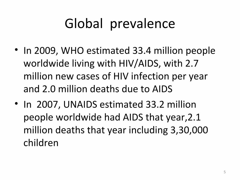
• In 2009, WHO estimated 33.4 million people worldwide living with HIV/AIDS, with 2.7 million new cases of HIV infection per year and 2.0 million deaths due to AIDS
• In 2007, UNAIDS estimated 33.2 million people worldwide had AIDS that year,2.1 million deaths that year including 3,30,000 children
5

Contd…
• According to UNAIDS 2009 report, 60 million people have been infected since the start of pandemic , with 25 million deaths, and 14 million orphaned children in southern Africa alone
6

Global picture
7

AIDS prevalence in Nepal
National centre for Aids &Std Control ,Teku 2009/12/1
• Cumulative no.of reported HIV infections(17th OCT 09)
=14,787( M=9701 +F=5086)
Enrolled in HIV care=13,005
Enrolled in ART = 3,423 Total Deaths =450People living with HIV as of 31 Dec 2006 (Nepal)
Total -8893Adults- 5999Women -2510Children under 15 years- 364Newly infected with HIV in 2006- 2681AIDS deaths in 2006 - 81 8
History
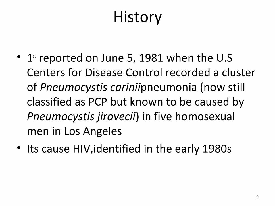
• 1st reported on June 5, 1981 when the U.S Centers for Disease Control recorded a cluster of Pneumocystis cariniipneumonia (now still classified as PCP but known to be caused by Pneumocystis jirovecii) in five homosexual men in Los Angeles
• Its cause HIV,identified in the early 1980s
9

Mode of transmission
• Sexual contact – 70% of cases• IV drug use – 27%• Blood transfusion – 2-3%• Perinatal transmission – 1%• HIV has been isolated from all body fluids
including tears, semen, vaginal fluids, preseminal fluids, and breast milk from infected person
10
Pathophysiology
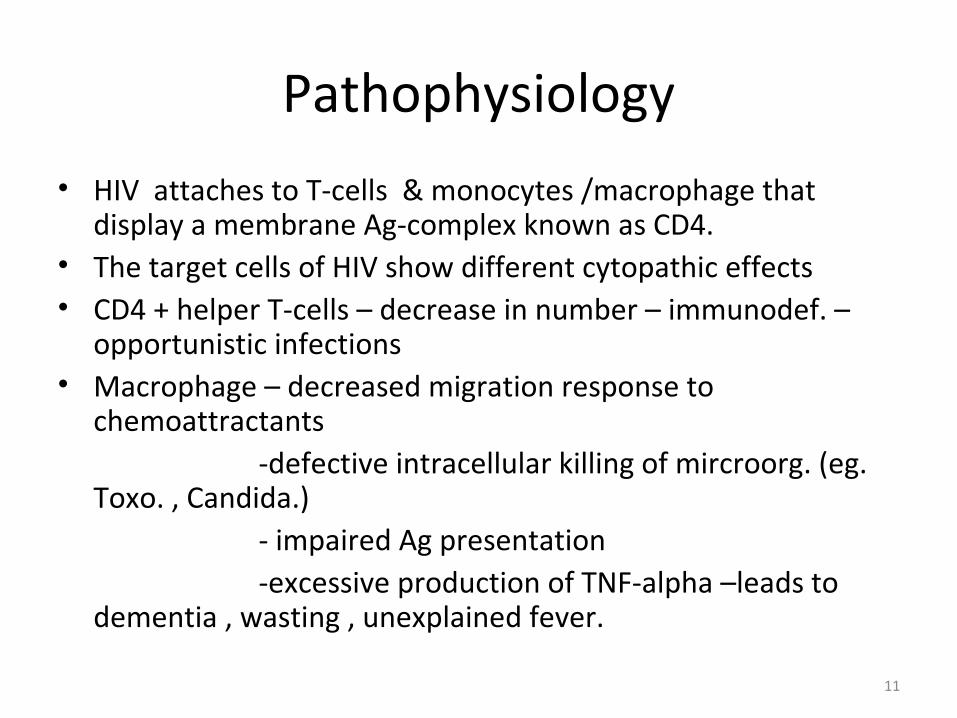
• HIV attaches to T-cells & monocytes /macrophage that display a membrane Ag-complex known as CD4.
• The target cells of HIV show different cytopathic effects• CD4 + helper T-cells – decrease in number – immunodef. –
opportunistic infections• Macrophage – decreased migration response to
chemoattractants -defective intracellular killing of mircroorg. (eg.
Toxo. , Candida.) - impaired Ag presentation -excessive production of TNF-alpha –leads to
dementia , wasting , unexplained fever.
11
Flowchart
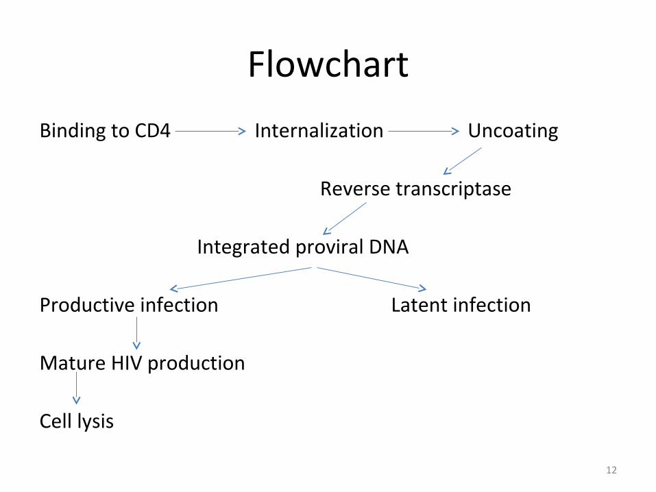
Binding to CD4 Internalization Uncoating Reverse transcriptase
Integrated proviral DNA
Productive infection Latent infection
Mature HIV production
Cell lysis
12
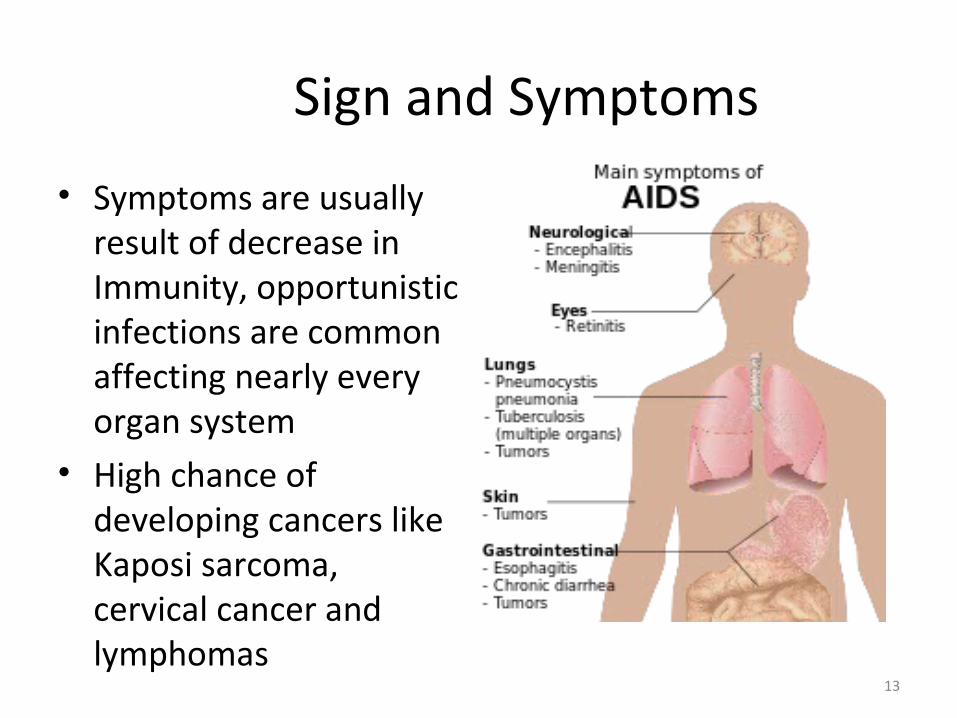
Sign and Symptoms
• Symptoms are usually result of decrease in Immunity, opportunistic infections are common affecting nearly every organ system
• High chance of developing cancers like Kaposi sarcoma, cervical cancer and lymphomas
13
System related Symptoms
• Pulmonary• Pneumocystis pneumonia is common in HIV infected
individuals• 1st indication of AIDS in untested individuals• Doesn’t occur unless the CD4 count is less than 200
cells/µL of blood• TB with HIV co-infection is major world health
problem
14
Pulmonary contd…
• In early HIV(CD4 count >300 cells/µL) TB typically present as pulmonary disease
• In advanced cases of HIV it occurs as extrapulmonary affecting bone marrow, bone, urinary and gastrointestinal tracts , liver, regional lymph nodes and the CNS
15

Gastrointestinal
• Esophagitis is common due to fungal (candidiasis) or viral (herpes simplex-1 or CMV) infections, rarely Mycobacterium
• Unexplained chronic diarrhoea due to bacterial (Salmonella, Shigella, Listeria or Campylobacter) and parasitic and opportunistic infections like cryptosporidiosis, microsporidiosis and viruses viz astrovirus, adenovirus, rotavirus , CMV etc
16

Neurological and psychiatric
• Toxoplasmosis caused by Toxoplasma gonadii infects the brain causing toxoplasma encephalitis affecting eyes and lungs
• Cryptococcal meningi caused by Cryptococcus neoforman causing fevers, headache, fatigue, vomiting and also seizures and confusion
17

Contd…
• Progressive multifocal leukoencephalopathy (PML) is a demylinating disease with gradual destruction of myelin sheath impairing transmission of nerve impulses
• Caused by virus called JC virus which occurs in 70% of population in latent form, causing disease only when immune system has been weakened as in the case of AIDS patients
18

Contd…
• AIDS dementia complex (ADC) is a metabolic encephalopathy induced by HIV infection and fueled by immune activation of HIV infected brain macrophages and microglia
• Prevalence is 10-20% in Western countries
19

Tumors
• High incidence due to co-infection with oncogenic DNA virus, especially Epstein – Barr virus (EBV), Kaposi’s Sarcoma- Associated Herpesvirus (KSHV) and Human Papillomavirus (HPV)
• Kaposi’s sarcoma is the most common tumor in HIV infected patients
20

• Lymphomas often arises in extranodal sites such as gastrointestinal tracts
• AIDS patients are at increased risk of certain tumors like Hodgkin’s disease , rectal carcinomas, hepatocellular carcinomas, head and neck cancers and lungs cancer
21

Other infections
• Includes opportunistic infections causing low grade fevers and weight loss
• Opportunistic inf. with Mycobacterium avium intracellulare and CMV causes colitis, CMV retinitis leading to blindness
• Penicilliosis due to Penicillium marneffei is 3rd most common opportunistic infection (after extrapulmonary TB and cryptococcosis)
22

Diagnosis
• Lab. Inv. – depends on : • Demonstration of virus sp. Ab by ELISA and
Western Blot • Viral Ag by EIA( Enzyme immunoassay)• Detection of HIV Nucleic acid by PCR• Viral P24 Ag detection
23

WHO staging diagnosis
• Given by WHO in September 2005• Stage I: asymptomatic and not catagorized as
AIDS• Stage II: minor mucocutaneous and recurrent
URTI(upper respiratory tract infection)• Stage III: chronic diarrhoea, pulmonary TB• Stage IV: toxoplasmosis of brain, candiasis of
oesophagus, trachea, lungs, Kaposi’s sarcoma
24

HIV Staging with CD4 counts
• CD4 counts Normal- 600- 1500 cells/cumm• 250-500/cumm– oral candidiasis , disseminated TB• 150-200/cumm- Kaposi sarcoma, Lymphoma,
Cryptosporidiosis• 75-125/cumm – pneumocystis carinii, mycobact.
avium, HS, toxo, cryptococcosis, esophageal candida• <50 cell/cumm- CMV retinitis
25

Ophthalmic Manifestations of HIV Infection
26

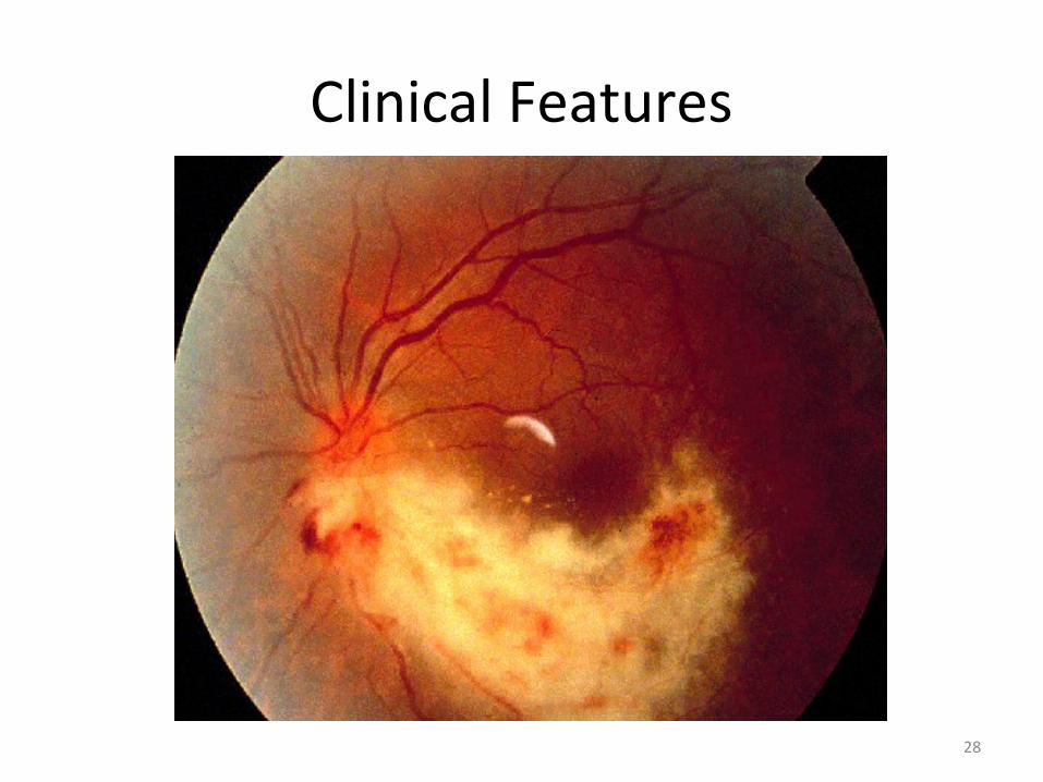
CMV Retinitis
• Most common ocular infection in pts. with AIDS, & maybe the initial manifestation
• Prior to HAART (Highly Active Antiretroviral Therapy)- 15-40% of AIDS pts.
• Untreated CMV – is a progressive and destructive infection- leads to blindness
• CMV – double stranded DNA virus, herpetoviridae
27
Clinical Features
28

• Brush Fire- leading active border due to spread by direct extension
• optic nerve involvement
• RD
29
Diagnosis
• Blood C/S• Urine C/S• PCR( sensitive & specific – 46 days – 6 months)
to dev. CMV retinitis• Fundus exam. Is essential – pt. with CMV +
blood ,urine c/s or extraocular CMV disease
30
MANAGEMENT
• Drugs Used: ( Virostatic)
1.Gancyclovir 2.Foscarnet 3.Cidofovir4.Formivirsen
31

Toxoplasma Retinochoroiditis
32

• Decreased vision • Moderate granulomatous uveitis ( AC+Vit-
cells)• Retina : Focal areas of necrotizing retinitis Abs of pre existing scars (pr 4-6%)• ~ 25% pts. –intracranial involvement( Imp. to
do intracranial imaging study in all AIDS pts. With ocular toxpl.)
33

• D/D- CMV retinitis , PORN
• T/T: Pyrimethamine(50mg/dailly)+ sulfanamide(4-
6gm/daily)+clindamycin(300mg x QID) –folinic acid
34
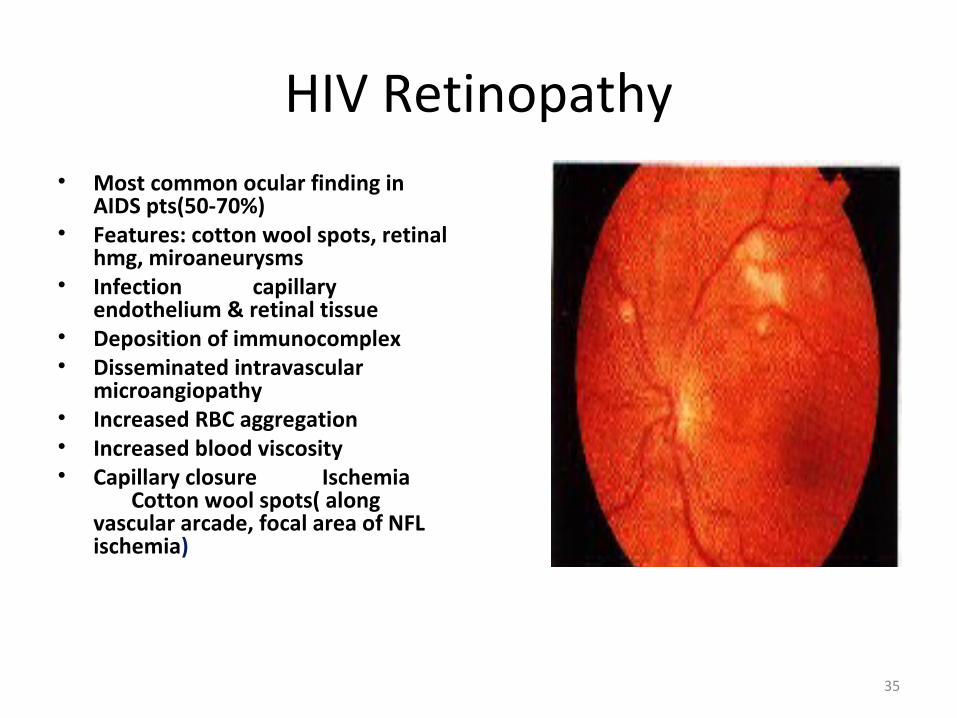
HIV Retinopathy• Most common ocular finding in
AIDS pts(50-70%)• Features: cotton wool spots, retinal
hmg, miroaneurysms• Infection capillary
endothelium & retinal tissue• Deposition of immunocomplex• Disseminated intravascular
microangiopathy• Increased RBC aggregation• Increased blood viscosity• Capillary closure Ischemia
Cotton wool spots( along vascular arcade, focal area of NFL ischemia)
35

Progressive Outer Retinal Necrosis (PORN)
• Caused by Herpes Zoster (HZ)
• Rapid destruction of retina
• Disease starts in one eye – fellow eye usu. Involves subsequently
36

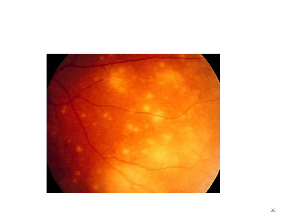
• Deep outer retinal lesions in a circumferential pattern in the peripheral retina
• lesion coalesce ,progress to full thickness retinal necrosis in a matter of days
• Rapid diminution of vision ( sparing of the perivascular retina)
37
38

PORN - Diagnosis
• H/o HZ inf. In the skin or elsewhere • Rapid progressive & sparing of the retinal
vessels and the adj. area• CD4 < 50 cells/mm3D/D• CMV retinitis /toxoplasmosis.
RetinochoroiditisT/T• I.V. gangciclovir /foscarnet with acyclovir• Oral sorivudine
39

Acute Retinal Necrosis (ARN)
• Caused by: HZ & HS• Clinical features:• Decreased vision with pain • Photophobia • Floaters • Granulomatous uveitis • Marked vitritis• Multiple white opaque patches of
thickened retina (periphery) enlarges gradually & coalaesce
• RD• 1/3 B/L involvement
40

Other Forms Of Retinitis
• Protozoa : Pneumocystis carinii choroiditis• Fungal : Cryptococcosis• Candidiasis• Bacterial : Tubercular retinitis• Spirochaete : Treponema Pallidum
Syphilitic retinitis
41

Herpes Zoster ophthalmicus
• If young pts have HZ of the face or eyelids Suspect HIV infection
• Corneal involvement: persistent, chr. Epithelial keratitis• T/T: Systemic acyclovir : I.V 10mg/kg/8hrly oral 600-800mg 5x daily conjunctivitis, keratitis, uveitis –t/t accordingly• Regular FU
42

Herpes Simplex infection
Keratitis:• Prolonged course• Multiple recurrence• Involve the limbus
Bacterial & Fungal Keratitis– More aggressive & likely to cause perforation– Difficult or non responsive to t/t– Microsporidia– Punctate epithelial keratopathy– Mild conjunctivitis
43

Molluscum Contagiosum (MC)• Causative agent: • DNA Virus ( Poxvirus)• Eyelids:• Umbilicated skin papule• Multiple >10 B/L• Size: large >5mm• Resistant to therapy• Follicular conjunctivitis• T/T:• Surgical excision• cautery, cryotherapy to the
base
44
conjunctival Squamous cell carcinoma
• third most common neoplasm associated to HIV infection.• occurs due to Papilloma Virus infection.• appears as a pink, gelatinous growth, usually in the
interpalpebral area. Often an engorgedblood vessel feeding the tumour is seen.
• It may extend onto the cornea, but deep invasion and metastasis are rare.
• •The treatment of choice is local excision and cryotherapy• but the presence of orbital invasion is an indication of
exenteration
45

Conjuctival SCC
46

Trichomegaly
• Trichomegaly or hypertrichosis is an exaggerated growth of the eye lashes found in the later stages of the disease
• The cause is not known• When symptomatic or
for cosmetic reasons the eyelashes can be trimmed
47

Dry Eye
• • Sicca syndrome is frequent among patients with HIV infection
• •Patients complain of burning uncomfortable red
• eyes.• causes of dry eye in HIV
infection from blepharitis, due to destruction of the
• lacrimal glands.• •T/T with tear supplements
48
Anterior uveitis
• HIV related anterioruveitis can be:• – Direct manifestation of the human
immunodeficiency virus infection• – autoimmnune in origin• – drug induced ie: rifabutin, secondary to direct toxic
effect upon the non-pigmented epithelium of the ciliary body
• –Any of the different infections• associated with AIDS, ie: Herpes Zoster Virus, Herpes
Simplex Virus,Cytomegalovirus, Toxoplasma gondii, Syphilis 49

• Candida albicans endophthalmitis• •Infection with candida albicansis rare. • Candida albicans is the commonest cause of fungal
endophthalmitis• •Affected patients usually have a history of drug
abuse • •In the initial stages, floaters are the main symptom.
As the condition progresses, whitish “puff-balls”and vitreous strands develop. Later, similar infiltrates appear in the choroid and retina
50

• •The treatment depends on the severity of the ocular involvement and systemic disease. The original foci should be removed. The drugs of choice are Amphotericine BBand Fluconazol
51

Non Infectious Ocular Manifestation
Kaposi’s Sarcoma• 30% of the pts/ with AIDS• Multifocal malignant
sarcomaOcular• Eyelid• Conjunctiva inf. Fornix• Orbit ProptosisPtosisOcular nerve palsy
52

Other Non infective Ocular Manifestation
• Keratoconjunctivitis sicca: 10-15%• Thrombocytopenia – Subconjunctival
Hemorrhage• Peripheral corneal ulceration• CN palsy• Papilloedema
53

54