ahmed malibary, m.d. primary postpartum hemorrhage
Post on 19-Dec-2015
226 views
TRANSCRIPT

AHMED MALIBARY, M.D.
Primary Postpartum hemorrhage

Hemorrhage is the underlying causative factor
in at least 25% of maternal deaths in industrialized and
underdeveloped countries

Maternal physiology is well prepared for hemorrhage:
increase in blood volume
hypercoagulable state
the “tourniquet” effect of uterine contractions
vital signs may remain near normal until more than 30% of blood volume is lost
tachycardia can be attributed to pregnancy, stress, pain, and delivery

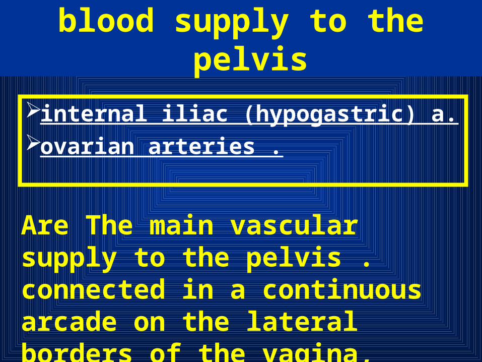
blood supply to the pelvis
blood supply to the pelvis
internal iliac (hypogastric) a.ovarian arteries .
Are The main vascular supply to the pelvis . connected in a continuous arcade on the lateral borders of the vagina, uterus, and adnexa

o/The ovarian arteries :are direct branches of the aorta
beneath the renal arteries. They traverse bilaterally and retroperitoneally to enter the infundibulopelvic ligaments.
blood supply to the pelvis

h/The hypogastric artery:
retroperitoneally posterior to the ureter it divides into an anterior and posterior divisions
blood supply to the pelvis
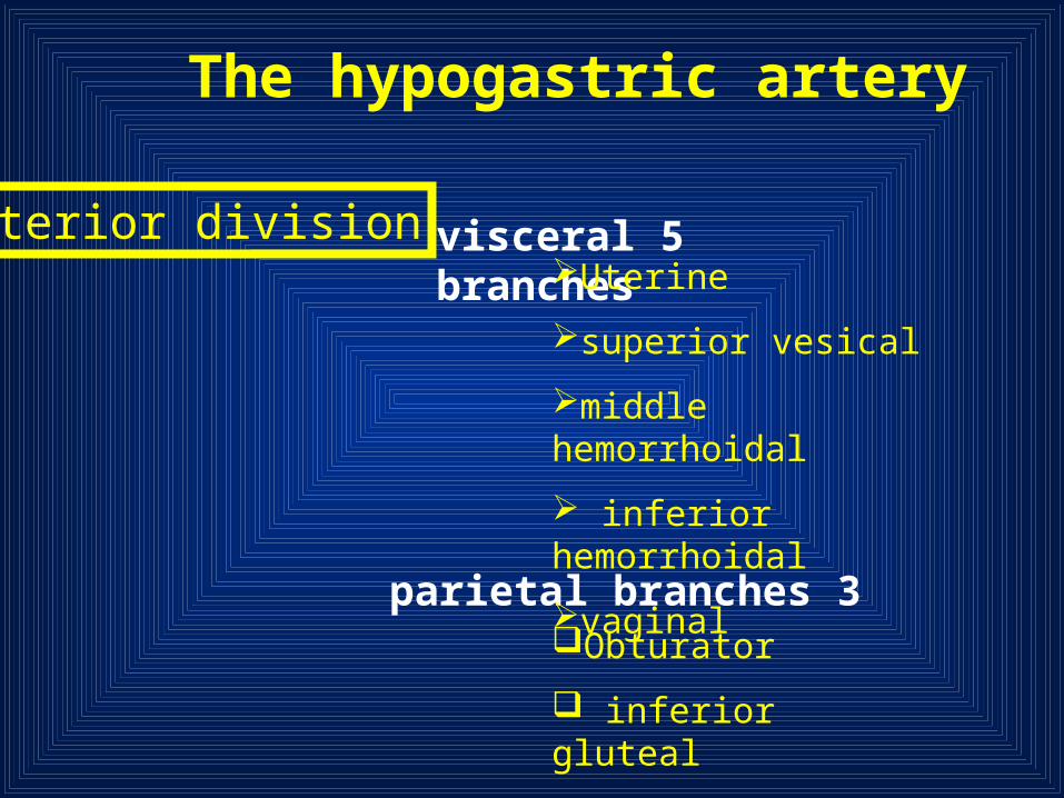
The hypogastric artery
anterior division
3 parietal branches
5 visceral branches
Obturator
inferior gluteal
internal pudendal
Uterine
superior vesical
middle hemorrhoidal
inferior hemorrhoidal
vaginal

The hypogastric artery
posterior division
important collateral to the pelvis.Iliolumbar lateral sacralsuperior gluteal

PHYSIOLOGY OF COAGULATION
PHYSIOLOGY OF COAGULATION
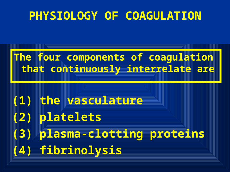
The four components of coagulation that continuously interrelate are
(1) the vasculature
(2) platelets
(3) plasma-clotting proteins
(4) fibrinolysis

the vasculature
A disruption in the vessel wall removes the protective
covering of the endothelial cells and releases tissue thromboplastin, which activates the clotting
mechanism
platelets
Activation of surface receptors causes morphologic changes in the platelets
(changing first to a sphere and then to a spiderlike structure with pseudopods)
and the generation of thromboxane A2 These lead to platelet aggregation
and eventual formation of a platelet plug
plasma-clotting proteins
Activation of the clotting system is initiated in two ways:
the intrinsic or extrinsic pathway
Intrinsic Pathway
requires no extravascular component for initiation and begins with Factor XII, which is activated by contact with injured epithelium

Extrinsic Pathway
is activated by the tissue factor thromboplastin (which subsequently activates Factor VII) when vascular disruption occurs. Prothrombin is converted to thrombin, which catalyzes the conversion of fibrinogen to fibrin. A clot is eventually formed at the site of vascular injury

fibrinolysis
plasma substrate plasminogen is activated This substrate is converted to the active enzyme plasmin, which lyses fibrin clots and destroys fibrinogen and Factors XII and VII

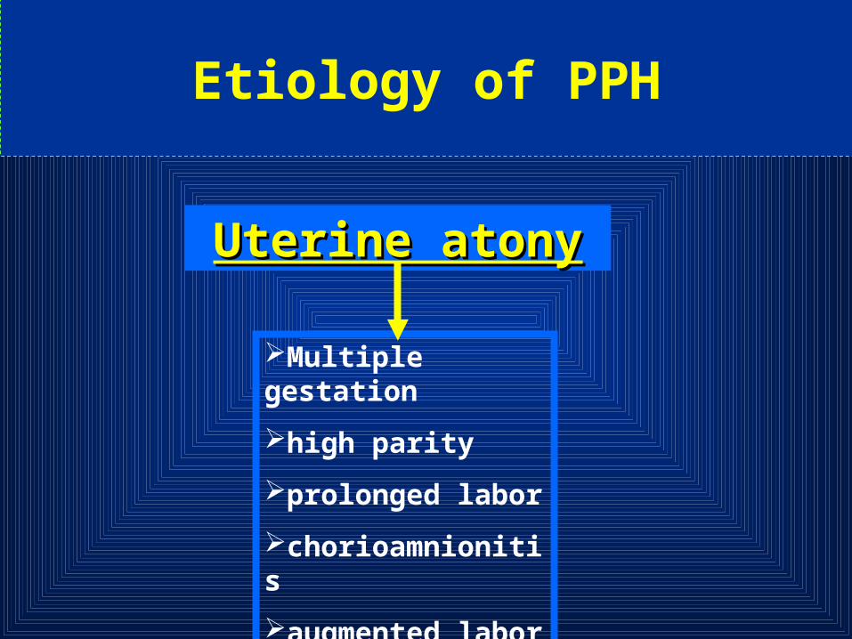
Etiology of PPH
Uterine atonyUterine atony
Multiple gestation
high parity
prolonged labor
chorioamnionitis
augmented labor
tocolytic agents

Etiology of PPH
Retained uterine Retained uterine contentscontents
Products of conception,
blood clots
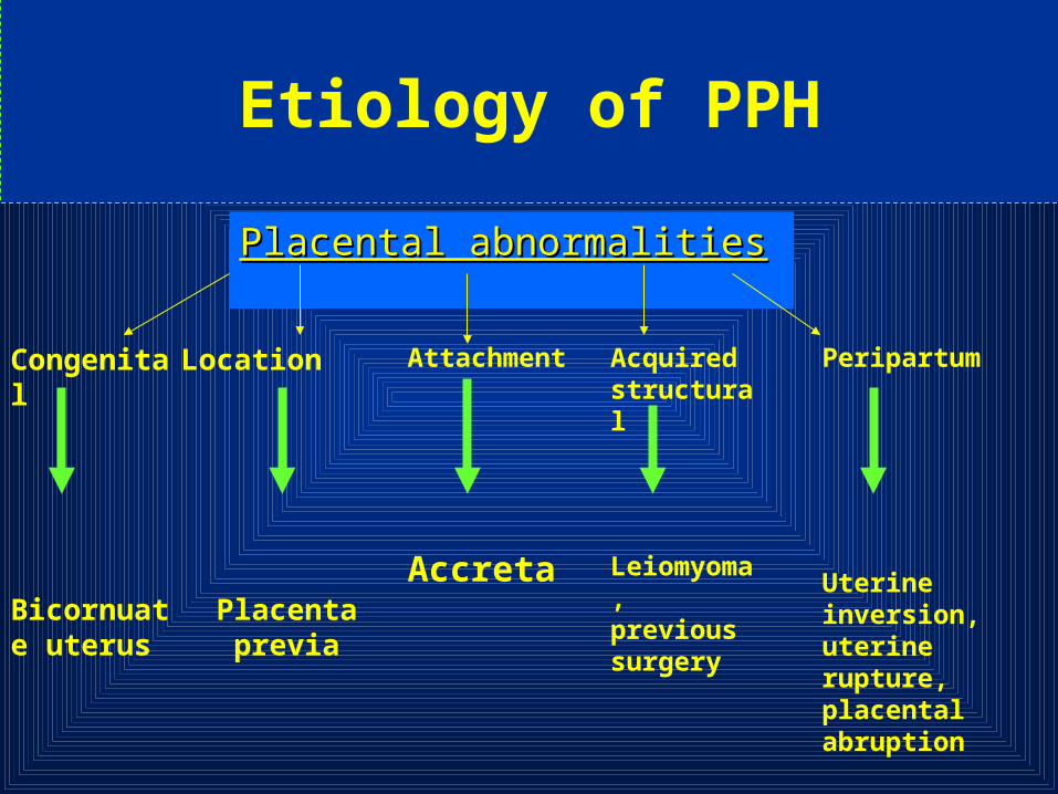
Placental abnormalitiesPlacental abnormalities
Congenital
Bicornuate uterus
Location
Placenta previa
Attachment
Accreta
Acquired structural
Leiomyoma, previous surgery
Peripartum
Uterine inversion, uterine rupture, placental abruption
Etiology of PPH

Lacerations and traumaLacerations and trauma
Planned
•Cesarean section
•episiotomy
Unplanned
•Vaginal/cervical tear
•surgical trauma
Etiology of PPH
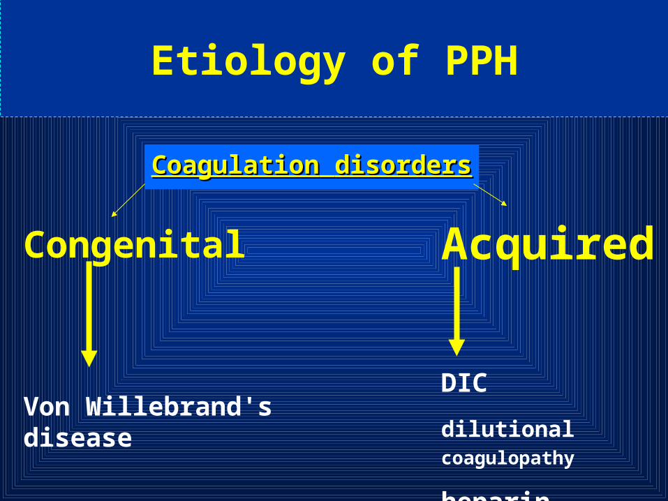
CoagulationCoagulation disordersdisorders
Etiology of PPH
Congenital
Von Willebrand's disease
Acquired
DIC
dilutional coagulopathy
heparin


Women in whom these factors have been identified should be advised to deliver in a specialist obstetric unit( GRADE B )
Risk Factorodds ratio for PPH
•Proven abruptio placentae
•Known placenta praevia
•Multiple pregnancy
•Pre-eclampsia/gestational hypertension
13
12
5
4
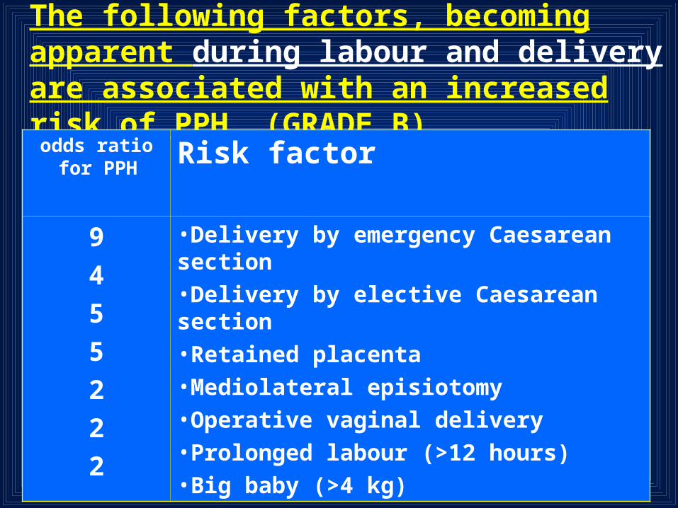
The following factors, becoming apparent during labour and delivery are associated with an increased risk of PPH. (GRADE B)
Risk factorodds ratio for PPH
•Delivery by emergency Caesarean section
•Delivery by elective Caesarean section
•Retained placenta
•Mediolateral episiotomy
•Operative vaginal delivery
•Prolonged labour (>12 hours) •Big baby (>4 kg)
9
4
5
5
2
2
2

Prophylactic oxytocics should be offered routinely in the management of the third stage of labour as they reduce the risk of PPH by about 60%.
(GRADE A)

• Women considered at high risk of thromboembolism may be receiving prophylaxis in the form of Unfractionated Heparin (UH) or Low Molecular Weight Heparin (LMWH) antenatally
• Women with a lower level of increased risk of thromboembolism may be receiving aspirin (75mg daily) antenatally and may begin intrapartum prophylaxis with the above agents
In prophylactic dosage, these agents do not present a haemorrhagic hazard and should be continued intrapartum
(ALL GRADE C)

In the event of a woman coming to delivery while receiving therapeutic heparin
•GRADE C
the infusion should be stopped. Heparin activity will fall to safe levels within an hour. Protamine sulphate will reverse activity more rapidly, if required

UH and LMWH in prophylactic dosage are not felt to present a haemorrhagic hazard
However, in overdosage there can be bleeding problems and protamine sulphate is less effective at reversing the effects of these agents (particularly LMWH) than of therapeutic heparin administered by infusion
Letsky EA. Peripartum prophylaxis of thromboembolism. In: Greer IA, ed. Thromboembolic disease in obstetrics and gynaecology. 1997

If women with inherited bleeding disorders present for preconceptual counselling, they should be tested for immunity against hepatitis B ,and immunised if required (as a safeguard should blood products be required at delivery). Immunisation during pregnancy is also safe
GRADE C

The existence of some of the obstetric risk factors may be known early in pregnancy from history and examination
Antenatal assessment
history

Antenatal assessment
anemia
Detection of anemia more than physiologic anemia of pregnancy is important, because anemia at delivery increases the likelihood of a woman requiring blood transfusion
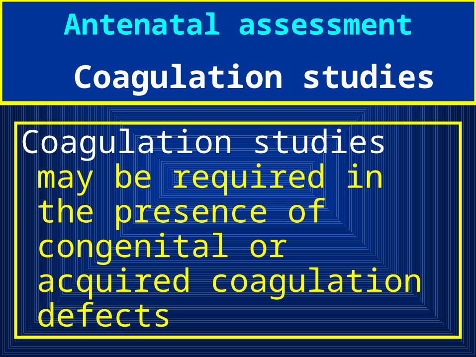
Coagulation studies may be required in the presence of congenital or acquired coagulation defects
Antenatal assessment
Coagulation studies

Imaging investigations are useful in the detection of placental abnormalities, with placenta
previa and placenta accreta the most important identifiable risk
factors for massive hemorrhage
Antenatal assessment
Imaging investigations
Conventional gray-scale assessment has a sensitivity of 93%, a specificity of 79%, and a positive predictive value of 78% in the diagnosis of placenta accreta when previa and previous cesarean scar are present
Antenatal assessment
Imaging investigations
Finberg HJ, Williams JW. Placenta accreta: prospective sonographic diagnosis in patients with placenta previa and prior cesarean section. J Ultrasound Med 1992;11:333-43.

Certain characteristics, such as the ”Swiss cheese appearance” with placenta previa, are associated with a threefold increase in mean blood loss during cesarean section
Guy GP, Peisner DB, Timor-Tritsch IE. Ultrasonographic evaluation of uteroplacental blood flow patterns of abnormally located and adherent placenta. Am J Obstet Gynecol 1990;163:723-7.
Antenatal assessment
Imaging investigations
Color Doppler may increase the specificity to 96%, which gives a positive predictive value in high-risk patients of 87% and a negative predictive value of 95% and allows better assessment of the depth of placental myometrial or serosal invasion
Antenatal assessment
Imaging investigations
Chou MM, Ho ESC, Lee YH. Prenatal diagnosis of placenta previa accreta by transabdominal color Doppler ultrasound. Ultrasound Obstet Gynecol 2000;15:28-35.

Further imaging by MRI is recommended to assess bladder involvement in percreta and assess high-risk cases
Thorp Jr. JM, Councell RB, Sandridge DA, et al. Antepartum diagnosis of placenta previa percreta by magnetic resonance imaging. Obstet Gynecol 1992;80:506-8.
Antenatal assessment
Imaging investigations

Guidelines by the Scottish Executive Committee of the RCOG
COMMUNICATE. RESUSCITATE. MONITOR / INVESTIGATE. STOP THE BLEEDING.

COMMUNICATEcall 6
• Call experienced midwife
• Call obstetric registrar & alert consultant
• Call anaesthetic registrar , alert consultant
• Alert haematologist
• Alert Blood Transfusion Service
• Call porters for delivery of specimens / blood

RESUSCITATE• IV access with 14 G cannula X 2 • Head down tilt • Oxygen by mask, 8 litres / min• Transfuse
•Crystalloid (eg Hartmann’s)
•Colloid (eg Gelofusine)
•once 3.5 litres infused, GIVE ‘O NEG’ If no cross-matched blood available OR give uncross-matched own-group blood, as available
•Give up to 1 liter Fresh Frozen Plasma and 10 units cryoprecipitate if clinically indicated

MONITOR / INVESTIGATE
• Cross-match 6 units • Full blood count • Clotting screen • Continuous pulse / BP / • ECG / Oximeter • Foley catheter: urine output • CVP monitoring • Discuss transfer to ITU

STOP THE BLEEDING
• Exclude causes of bleeding other than uterine atony • Ensure bladder empty • Uterine compression • IV syntocinon 10 units • IV ergometrine 500 g • Syntocinon infusion (30 units in 500 ml) • IM Carboprost (500 g) • Surgery earlier rather than late • Hysterctomy early rather than late (GRADE B)

If conservative measures fail to control haemorrhage, initiate surgical haemostasis SOONER RATHER THAN LATER I. At laparotomy, direct
intramyometrial injection of Carboprost (Haemabate) 0.5mg
II. Bilateral ligation of uterine arteries III. Bilateral ligation of internal iliac
(hypogastric arteries) IV. Hysterectomy (GRADE C)

Resort to hysterectomy SOONER RATHER THAN LATER (especially in cases of placenta accreta or uterine rupture)(GRADE C)
Whole blood frequently is used for rapid correction of volume loss because of its ready availability, but component therapy is ideal. A general practice has been to transfuse 1 unit of fresh-frozen plasma for every 3 to 4 units of red cells given to patients who are bleeding profusely
Genital tract lacerations
Genital trauma always must be eliminated first if the uterus is
firm

• Explore the uterine cavity
• Inspect vagina and cervix for lacerations
• If the cavity is empty, Massage and give methylergonovine 0.2 mg, the dose can be repeated every 2 to 4 hours
• Rectal Misoprostol 800mg is beneficial
Management of uterine atony
During the administration of uterotonic agents, bimanual compression may control hemorrhage. The physician places his or her fist in the vagina and presses on the anterior surface of the uterus while an abdominal hand placed above the fundus presses on the posterior wall. This while the Blood for transfusion made available
Management of uterine atony
Retained placenta
Retained placental fragments are a leading cause of early and delayed postpartum hemorrhage. Treatment is manual removal, General anesthesia with any volatile agent (1.5–2 minimum alveolar concentration (MAC)) may be necessary for uterine relaxation
On rare occasions, a retained placenta is an undiagnosed placenta accreta, and massive bleeding may occur during attempted manual removal
Placenta accreta • Placenta accreta is defined as an abnormal
implantation of the placenta in the uterine wall, of which there are three types:
(1) accreta vera in which the placenta adheres to the myometrium without invasion into the muscle.
(2) increta in which it invades into the myometrium. (3) percreta in which it invades the full thickness of the uterine wall and possibly other pelvic structures, most frequently the bladder
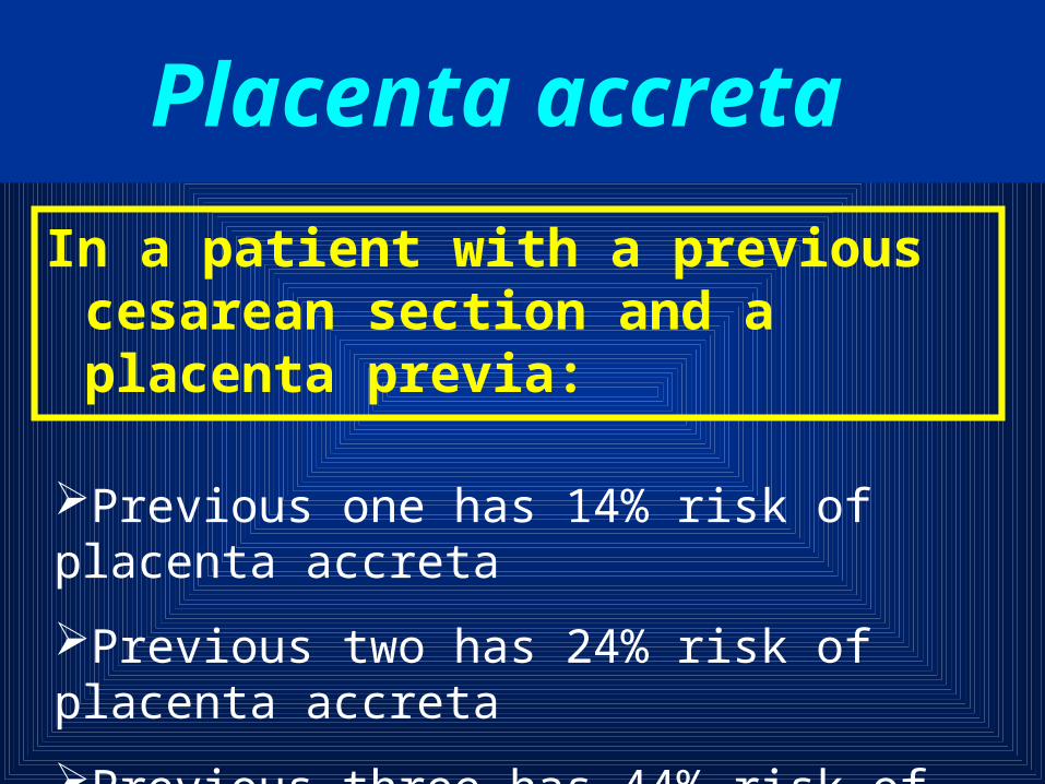
In a patient with a previous cesarean section and a placenta previa:
Placenta accreta
Previous one has 14% risk of placenta accreta
Previous two has 24% risk of placenta accreta
Previous three has 44% risk of placenta accreta

UTERINE RUPTURE
Rupture of the uterus is described as complete or incomplete and should be differentiated from dehiscence of a cesarean section scar
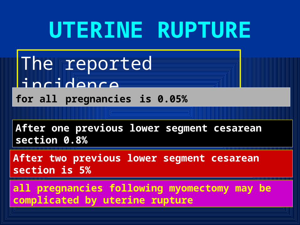
UTERINE RUPTURE
The reported incidence
for all pregnancies is 0.05%
After one previous lower segment cesarean section 0.8%
After two previous lower segment cesarean section is 5%
all pregnancies following myomectomy may be complicated by uterine rupture
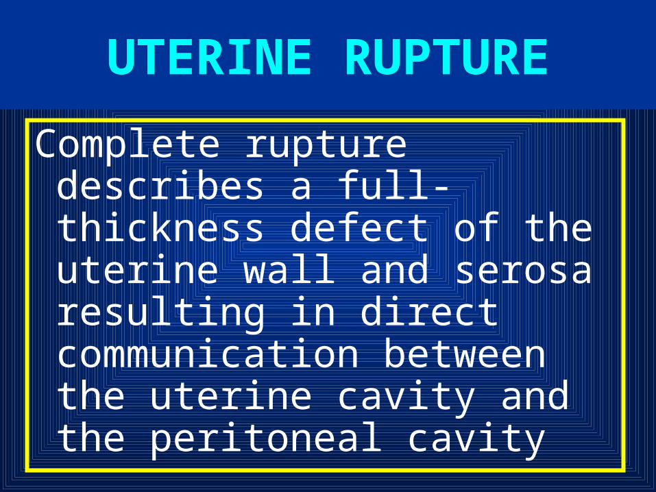
Complete rupture describes a full-thickness defect of the uterine wall and serosa resulting in direct communication between the uterine cavity and the peritoneal cavity
UTERINE RUPTURE

Incomplete rupture describes a defect of the uterine wall that is contained by the visceral peritoneum or broad ligament. In patients with prior cesarean section
UTERINE RUPTURE

dehiscence describes partial separation of the scar with minimal bleeding, with the peritoneum and fetal membranes remaining intact
UTERINE RUPTURE

The identification or suspicion of uterine rupture must be followed by an immediate and simultaneous response from the obstetric team
Surgery should not be delayed owing to hypovolemic shock because it may not be easily reversible until the hemorrhage is controlled
Management of Rupture Uterus

Upon entering the abdomen, aortic compression can be applied to decrease bleeding
Oxytocin should be administered to effect uterine contraction to assist in vessel constriction and to decrease bleeding
Hemostasis can then be achieved by ligation of the hypogastric artery, uterine artery, or ovarian arteries
Management of Rupture Uterus

At this point, a decision must be made to perform hysterectomy or to repair the rupture site. In most cases, hysterectomy should be performed
In selected cases, repair of the rupture can be attempted. When rupture occurs in the body of the uterus
bladder rupture must be ruled out by clearly mobilizing and inspecting the bladder to ensure that it is intact. This avoids injury on repair of the defect as well
Management of Rupture Uterus

A lower segment lateral rupture can cause transection of the uterine vessels. The vessels can retract toward the pelvic side wall, and the site of bleeding must be isolated before placing clamps to avoid injury to the ureter and iliac vessels
Typically, longitudinal tears, especially those in a lateral position, should be treated by hysterectomy, whereas low transverse tears may be repaired
Management of Rupture Uterus

Uterine Artery Ligation
Uterine artery ligation involves taking large purchases through the uterine wall to ligate the artery at the cervical isthmus above the bladder flap
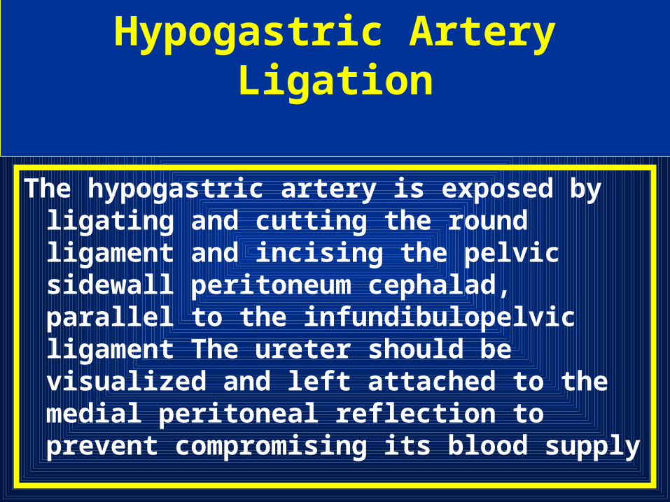
Hypogastric Artery Ligation
The hypogastric artery is exposed by ligating and cutting the round ligament and incising the pelvic sidewall peritoneum cephalad, parallel to the infundibulopelvic ligament The ureter should be visualized and left attached to the medial peritoneal reflection to prevent compromising its blood supply
.The common, internal, and external iliac arteries must be identified clearly. The hypogastric vein, which lies deep and lateral to the artery, may be injured as instruments are passed beneath the artery, resulting in massive, potentially fatal bleeding
Hypogastric Artery Ligation

The hypogastric artery should be completely visualized. A blunt-tipped, right-angle clamp is gently placed around the hypogastric artery, 2.5 to 3.0 cm distal to the bifurcation of the common iliac artery. Passing the tips of the clamp from lateral to medial under the artery is crucial in preventing injuries to the underlying hypogastric vein
Hypogastric Artery Ligation

the artery is double-ligated with a nonabsorbable suture, with 1-0 silk, but not divided .The ligation is then performed on the contralateral side in the same manner
Hypogastric Artery Ligation

A.MALIBARY, M.D.