affect submitted to in partial fulfillment of requirements for
TRANSCRIPT

i
HYPOCRETIN/OREXIN AND THE VENTRAL MIDBRAIN: TOPOGRAPHY
AND FUNCTION ASSOCIATED WITH PSYCHOSTIMULANT-TAKING AND
AFFECT
A Dissertation Submitted to
Temple University Graduate Board
In partial fulfillment of requirements for Doctor of Philosophy (Ph.D.)
In Biomedical Sciences (Neuroscience)
By Steven J. Simmons
Examining Committee Members
John W. Muschamp PhD – Advisory Chair, CSAR (Temple University) Ellen M. Unterwald PhD – Committee Member, CSAR (Temple University) Lynn G. Kirby PhD – Committee Member, CSAR (Temple University) Lee-Yuan Liu-Chen PhD – Committee Member, CSAR (Temple University) David E. Moorman PhD – External Dissertation Examiner, Psychological & Brain Sciences (University of Massachusetts [Amherst])

ii
ABSTRACT
Abuse of psychostimulants including cocaine and new synthetic formulations
remains an international public health problem and economic burden. Addiction develops
consequential to positive and negative drives that underlie “getting” and “staying” high.
Dopamine (DA), arising from ventral tegmental area (VTA), projects to ventral striatal
targets to encode reward signals and reward prediction. Mesolimbic DA is implicated in
both the immediate rewarding effects of psychostimulants, and its hypoactivity underlies
negative affect as drug levels decline. Accordingly, modulating inputs to midbrain DA
possesses capacity to mediate positive/rewarding and negative/aversive effects of drugs.
Hypocretin/orexin (hcrt/ox) is a family of excitatory hypothalamic peptides that projects
widely throughout the central nervous system including to VTA DA cells, and hcrt/ox
mediates brain reward function and motivation for self-administered drugs. Notably, the
first-in-class hcrt/ox receptor antagonist (suvorexant) was approved for management of
insomnia in the summer of 2014. Also within the past decade, the caudal division of VTA
(termed “tail of VTA” and “rostromedial tegmental nucleus [RMTg]”) was detailed for
its ability to negatively regulate VTA DA. Functionally, stimulation of the GABA-
producing RMTg population encodes aversion and responds to aversive cues. Curiously,
anatomy work depicts the hypothalamus as a principal input to the RMTg although the
cellular phenotypes and functions of hypothalamic projections to RMTg have not been
fully resolved.
Work in this thesis was designed to map hcrt/ox projections to VTA and RMTg in
effort to understand functionally-relevant topographical arrangement. In preliminary
assessments, we test for the first time the ability of suvorexant to modulate reward and

iii
reinforcement associated with psychostimulant use in rats. Additionally, we profile how
self-administered cocaine and “bath salt” synthetic cathinone 3,4-
methylenedioxypyrovalerone (MDPV) influence affective states in rats by measuring
ultrasonic vocalizations (USVs) and comparing patterns of responding. Subsequently, we
test the ability of suvorexant to influence MDPV-taking and affective changes that
promote self-administration. Finally, we utilize direct-site pharmacology to assess the
degree to which hcrt/ox transmission within VTA and RMTg contributes to motivated
responding for and affective processing of self-administered cocaine across two doses.
Specifically, we hypothesized that intra-VTA suvorexant would suppress drug-taking by
reducing the rewarding value of self-administered cocaine, whereas intra-RMTg hcrt/ox
peptide injection would suppress drug-taking by increasing aversive value of self-
administered cocaine.
We observed that systemic suvorexant effectively reduces motivated cocaine-
taking, and that this reduction relates in part to reductions in subjective reward of self-
administered cocaine as interpreted by reductions in positively-valenced 50-kHz USVs.
Retrograde tracing supports that hcrt/ox projects to both VTA and RMTg without
discernible topographical arrangement. Target-site pharmacology finds that intra-VTA
suvorexant has no appreciable effects on motivated cocaine-taking but tends to elevate
50-kHz USVs during the pre-drug “anticipation” time epoch in low-dose cocaine self-
administering rats (0.375 mg/kg/inf). While intra-RMTg hcrt/ox pre-treatment sparsely
affected USVs, 0.3 nmol/hemisphere hcrt/ox significantly enhanced cocaine-taking in
low-dose cocaine self-administering rats, and, in high-dose (0.750 mg/kg/inf) cocaine

iv
self-administering rats, intra-RMTg hcrt/ox significantly suppressed responding when
pre-treated with 1.0 and 3.0 nmol/hemisphere.
Collectively, studies within this thesis promote the use of hcrt/ox receptor
antagonists as adjunct pharmacotherapy in managing psychostimulant use disorders,
although the circuitries through which aberrant motivated behaviors are modulated are
not entirely clear. Future work will need to be performed to understand how hcrt/ox
transmits to neurochemically-defined cell populations residing within VTA and RMTg—
these pathways are recruited for processing stimuli as “rewarding” and “aversive” which
are critical contributors in the development of substance use disorders and other
psychiatric disorders characterized by dysregulated reward processing.

v
ACKNOWLEDGEMENTS
It seems most reasonable to start at the beginning; birth—thanks to mother (Clare)
and father (Malcolm “Bud” Simmons) for making me. My parents are unconditionally
supportive and loving, which I am extremely grateful of and for which I dedicate my
successes to. Family members who I cherish dearly for their care (and great food/parties)
include the Christmas crew: Aunt Marianne and Uncle Buddy, Aunt Marlene and Uncle
Rich, cousins Jaime (and husband Josh; their children Avery and Teegan), Buddy (and
wife Becky; their child Lucas), Ryan (and wife Stephanie), Richie and Ron. Tradition has
withstood for as long as I remember when it comes to Thanksgiving dinner—all my love
to Aunt Arlene, Uncle Gene and cousin Gino. I also warmly recognize family members I
visit far too infrequently—cousin Colette (husband Michael), Will and Su. Much love for
my godparents, too—Aunt Stephanie and Uncle Joe. I thank our black cat Prince for
entering our lives and blessing our home with a warm and gentle presence. “Home Is
Where My Rats Are”—this thesis is dedicated in part to the memories of my first pet rats
Rexi and Dyna.
My brother, Mark, was born a year before me—in recent years, he has provided
me with experiences of social frivolity which help me to decompress from long work
weeks. We had a somewhat tumultuous relationship in grade school, but since college I
have really appreciated his company. I look up to my loving and career-driven sister Jiji
and her husband Greg. Much love also to my nephew Josh and niece Sarah. A “big
brother” in life as well as in my undergraduate fraternity, love to Russell (wife Catina),
niece Catena and nephew Dominic. I am very thankful, too, for caring friends who
helped, since high school, shape my social self including Philip Sanville, Ben Robinson,

vi
Jay Hotaling, Dan Carreon, Brian and Jacob Peacock, and Kirsten King. Our trips
together (lake house in NH, cabin in NY), weekend hangouts, and themed parties have
made for incredibly fond and unforgettable memories despite the rowdiness that often
ensues.
I sincerely appreciate guidance and advisory provided by my undergraduate thesis
advisor, Dr. Mark West, who was the first research scientist to both stimulate my interest
and provide an opportunity to begin my career as a research scientist. The West Lab at
this time was populated by, who I consider to be, an unsurpassable team of bright and
committed minds—namely, David Barker, David Root, and Sisi Ma. All three then-
graduate-students continue to impress me and provide continuing mentorship for which I
am forever indebted. I owe my successes to them and to the opportunities they have
provided for me. During my undergraduate education, I additionally recognize Dr. John
Ackroff for his mentorship, availability to discuss course material, and for the
opportunities he provided me as supplementary instructor and tutor for two courses—
these early peer-to-peer instruction sessions made clear in my mind the great joys in
information dissemination and understanding.
Support from graduate student peers and lab members was paramount to my
ability to successfully complete a PhD program. From my former lab, I sincerely
appreciate friendship and exchange of knowledge with David Connor, Rachel Poole,
Erica Holliday and Emre Yildirim. Special shout-out to Rob Cole and Brittney Yegla of
the Parikh lab. I appreciate the kindness, understanding, warmth and intellect provided by
my PhD thesis advisor, John Muschamp. In the trenches, a tremendous “thanks” to my
partner-in-crime, yang-to-my-yin, Taylor Gentile—he and I remained a tag-team through

vii
much of our work and churned out original research reports I am very proud of. We
complimented each other as well as any great duo could, and from working alongside
him I consider myself extremely fortunate. On work in the lab, I owe so much to
dedicated, passionate undergraduate research scientists—Lili Mo, Fionya Tran, Rose
Martorana and Emily Clark as well as the undergraduate-turned-graduate student Stacia
Lewandowski. Literally, the work would not have gotten done without them, and I
greatly appreciate their flexibility and commitment. I additionally appreciate Mia Watson
for her dedicated role in the lab as well as classmates and peers that encouraged my
progress throughout the Biomedical Sciences PhD program—notably, in the recent year,
Allison Andrews, Kimmy Ferrero and a continuing faculty mentor Servio Ramirez. I
additionally appreciate encouragement from friends and publication co-authors Chicora
Oliver and Callum Hicks.
I sincerely thank my thesis committee—Ellen Unterwald, Lynn Kirby and Lee-
Yuan Liu-Chen as well as my external thesis examiner David Moorman. My committee
provided critical yet productive advice through many meetings, and I have all positive
memories in reflecting through the semesters. While not a formal committee member, I
am extremely grateful for the mentorship, advisory and friendship provided by Scott
Rawls who effectively assumed a “secondary mentor” role for me. Through a difficult
programmatic shift, I thank Scott Shore and Diane Soprano for facilitating my admission
to the Biomedical Sciences program. I sincerely appreciate the organization reflected by
the program’s administrators—namely, Tracy Burton and Denise Rykard as well as
administrators of the Center for Substance Abuse Research, Marc Graver and Melva
Smith. I additionally recognize the very important veterinary care staff of our facility who

viii
regularly checked on the health and wellbeing of our experimental critters—Shanna
Mcalarnen, Shannon Morton and Kim Gilmore. On this, I thank the experimental rat
subjects who enabled this research to be performed and for scientific progress to occur
and for whom this thesis is also dedicated to.
The past two years have been my best with company of Arielle Ridolfino, who I
love dearly and who supports and encourages both my career progression and personal
growth. After my defense, Ari and I will head to Disney World for yet another incredibly
magical, fun, and loving experience (thanking Disney for the movies and all-around
incredible experience in each theme park). Thank you from the bottom of my heart, Ari,
for being so flexible, accommodating and encouraging—you mean the world to me, and I
remain incredibly excited and optimistic to begin our new chapter of life together. I
recognize here, too, Ari’s mother Rachel Blumenfeld and father Ralph Ridolfino—two
folks I hope continue to be a major part of my life.
Finally, I thank the entities who have made my day-to-day life extremely
enjoyable, places and people alike. My mornings begin with a hearty breakfast from local
diners—Westmont Diner and the Crystal Lake Diner. No morning commute is without
coffee from Dunkin Donuts (Starbucks on the weekends to facilitate extended writing
sessions). I thank insightful podcast/TV show hosts including Dan Savage, Joe
Scarborough/Mika Brzezinski/Willie Geist/Mark Halperin/Richard Haass/David
Iganatius/Katty Kay/Steve Rattner/Sam Stein/Nicolle Wallace, Rachel Maddow, Steve
Kornacki, Jad Abumrad/Robert Krulwich, and Amy Baldwin/April Lampert. I thank the
incredibly talented and uplifting musicians, most notably Judith “Malukah” de los Santos,
Peter and Evynne Hollens, Christopher Tin and Yanni.

ix
I would like to acknowledge generous grant support from the National Institute on
Drug Abuse (DA007237 [SJS], DA031767 [John W. Muschamp], DA013429 [Ellen M.
Unterwald], DA039139 [Scott M. Rawls]) of the National Institutes of Health.
Additionally, I greatly appreciate success of a crowdfunded campaign for a pilot
study entitled “Antibiotics and Affect: Avenues for Addiction Management” on
Experiment.com. Specifically, the author thanks Arielle Ridolfino, Mark DeFeo, Mark
Martorana, Louis Di Meglio, Jae Kyun Kim, Linnet Ramos, Janice Peracchia, Eileen
Sanville, Arnab Sengupta, Junko Kanero, Brendan Reap, Bud Simmons, Marlene
Simmons Pikunis, Claire Martorana, Elaine Fee, Rachel Blumenfeld, Kirsten King,
Calvin Main, Phil Sanville, Anthony Pawlak, Carly Reyes, Anthony Belardo, Dustin
Lynn-Becket, Sisi Ma, Jaime Martorana, Natasha Utlik, Anne McMonagle, Sam Hauser,
Ryan Gibbons, Haley Ragsdale, Jack Gibbons, Paul Shapiro, Salim Short, Eric Damon
Walters, and Larry Martinez for their generous contributions that led to the success of
this campaign.

x
DEDICATION
See “Acknowledgements”.

xi
TABLE OF CONTENTS
ABSTRACT ........................................................................................................................ ii
ACKNOWLEDGEMENTS ................................................................................................ v
DEDICATION .................................................................................................................... x
TABLE OF CONTENTS ................................................................................................... xi
LIST OF FIGURES .......................................................................................................... xv
LIST OF TABLES .......................................................................................................... xvii
CHAPTER 1 – BEHAVIORAL EFFECTS AND NEUROBIOLOGY OF PSYCHOSTIMULANT DRUGS OF ABUSE ................................................................... 1
1.1) Discovery of cocaine and abuse potential of psychostimulant drugs ................... 1
1.2) Novel psychoactive substances mimic cocaine, are potently rewarding and abused .............................................................................................................................. 3
1.3) Behavioral features of reward and reinforcement: relevance to addiction ........... 5
1.3.1) Delivery of electrical current to brain sites is reinforcing ............................ 5
1.3.2) Place conditioning enables associative reward learning ............................... 7
1.3.3) Modeling volitional drug-taking using intravenous self-administration ...... 8
1.4) Neurophysiology of reward prediction and receipt: role of mesolimbic dopamine transmission in the context of psychostimulant use ...................................................... 10
CHAPTER 2 – HYPOCRETIN/OREXIN ANATOMY, CONNECTIVITY AND PHYSIOLOGY ................................................................................................................. 15
2.1) Discoveries of a novel neuropeptide system: hypocretin/orexin ....................... 15
2.2) Connectivity and physiology of the hypocretin/orexin neuropeptide system .... 18
CHAPTER 3 – HYPOCRETIN/OREXIN FUNCTION AND ROLES IN PATHOLOGY........................................................................................................................................... 25
3.1) Orexigenic and thirst-regulating properties of hypocretin/orexin ...................... 25
3.2) Wakefulness and arousal promotion by hypocretin/orexin ................................ 27
3.2.1) Suvorexant (MK-4305) becomes first clinically-available hypocretin/orexin 29
receptor antagonist ..................................................................................................... 29
3.3) Hypocretin/orexin and motivation for natural and drug rewards ....................... 30
CHAPTER 4 – VENTRAL MIDBRAIN: CELLULAR HETEROGENEITY, CONNECTIVITY AND ROLES IN REWARD-SEEKING ........................................... 36

xii
4.1) Cellular heterogeneity within ventral tegmental area confers divergent functions 36
4.2) Rostromedial tegmental nucleus/tail of ventral tegmental area: anatomy and participation in aversive state processing ...................................................................... 39
CHAPTER 5 – AFFECT AND ADDICTION: ULTRASONIC VOCALIZATIONS CAPTURE HIGHS AND LOWS ASSOCIATED WITH SUBSTANCE USE DISORDERS .................................................................................................................... 46
5.1) Production and ethological relevance of rat ultrasonic vocalizations ................ 46
5.2) Elicitation of 22- and 50-kHz ultrasonic vocalizations in laboratory settings ... 47
5.2.1) Ultrasonic vocalizations in preclinical models of substance use disorders 49
5.2.1.1) 50-kHz ultrasonic vocalizations are elicited after psychostimulant use ... 53
5.2.1.2) 22-kHz ultrasonic vocalizations align with negative affect experienced during drug withdrawal .......................................................................................... 56
5.2.1.3) Ultrasonic vocalizations as sensitive indicator of bivalent mood states in cocaine self-administering rats: revisiting the opponent-process model of addiction................................................................................................................................ 57
addiction ........................................................................................................................ 60
CHAPTER 6 – HYPOCRETIN/OREXIN ROLE IN PSYCHOSTIMULANT-ASSOCIATED REINFORCEMENT AND AFFECTIVE CHANGES ........................... 64
6.1) Suvorexant, a hypocretin/orexin receptor antagonist, attenuates motivational and hedonic properties of cocaine (Gentile et al. 2018) ...................................................... 65
6.2) Comparing rewarding and reinforcing properties between ‘bath salt’ 3,4-methylenedioxypyrovalerone and cocaine using ultrasonic vocalizations in rats (Simmons et al. 2018) ................................................................................................... 69
6.3) Role of hypocretin/orexin receptor blockade on drug-taking and ultrasonic vocalizations associated with self-administration of ‘bath salt’ 3,4-methylenedioxypyrovalerone in rats (Simmons et al. 2017) ......................................... 74
SPECIFIC AIMS .............................................................................................................. 79
CHAPTER 7 – HYPOCRETIN/OREXIN INNERVATION TO VENTRAL MIDBRAIN........................................................................................................................................... 82
7.1) Materials and methods ........................................................................................... 82
7.1.1) Animals ........................................................................................................... 82
7.1.2) Experimental procedures ................................................................................. 83
7.1.3) Stereotaxic cannulation ................................................................................... 83
7.1.4) Retrograde tracing ........................................................................................... 84
7.1.5) Tissue collection and analysis ......................................................................... 85

xiii
7.1.6) Statistical analyses........................................................................................... 88
7.2) Results .................................................................................................................... 89
7.3) Interim discussion .................................................................................................. 90
CHAPTER 8 – ROLE OF HYPOCRETIN/OREXIN TRANSMISSION WITHIN VENTRAL MIDBRAIN ON PSYCHOSTIMULANT-ASSOCIATED REINFORCEMENT AND AFFECTIVE CHANGES ..................................................... 92
8.1) Materials and methods ........................................................................................... 92
8.1.1) Animals ........................................................................................................... 92
8.1.2) Drugs ............................................................................................................... 93
8.1.3) Experimental procedures ................................................................................. 93
8.1.4) Stereotaxic cannulation ................................................................................... 94
8.1.5) Intravenous drug self-administration: jugular vein catheterization, apparatus and behavioral procedures ......................................................................................... 95
8.1.6) Ultrasonic vocalization recording and analysis ............................................... 95
8.1.7) Statistical analyses........................................................................................... 95
8.2) Results .................................................................................................................... 97
8.3) Interim discussion ................................................................................................ 104
DISCUSSION ................................................................................................................. 106
Suvorexant decreases rewarding and reinforcing properties of psychostimulants ..... 108
An evaluation of 50-kHz ultrasonic vocalizations as representating a “positively-valenced” subjective state ........................................................................................... 110
Functional connectivity: hypocretin/orexin afferents to rostrocaudal extent of ventral midbrain ...................................................................................................................... 111
Technical considerations: retrograde tracing and recording of ultrasonic vocalizations ..................................................................................................................................... 115
Distribution of OX1Rs and OX2Rs across neurocehmically-defined cell populations of ventral midbrain .......................................................................................................... 117
Beyond ventral midbrain: alternative targets of hypocretin/orexin in the mediation of psychostimulant-associated reward and reinforcement ............................................... 119
BIBLIOGRAPHY ........................................................................................................... 122
APPENDIX A ................................................................................................................. 150
S1.1) Locomotor activity ....................................................................................... 150
S1.2) Place conditioning ........................................................................................ 150
S1.3) Intravenous drug self-administration: jugular vein catheterization, apparatus and behavioral procedures ........................................................................................... 151

xiv
S1.4) Ultrasonic vocalization recording and analysis ............................................ 152
S1.5) Fast-scan cyclic voltammetry ....................................................................... 153
S1.6) Metrics and statistics .................................................................................... 154
APPENDIX B ................................................................................................................. 156
S2.1) Experimental procedures .............................................................................. 156
S2.2) Ultrasonic vocalization recording and analysis ............................................ 156
S2.3) Metrics and statistics .................................................................................... 156
APPENDIX C ................................................................................................................. 158
S3.1) Experimental procedures .............................................................................. 158
S3.2) Ultrasonic vocalization recording and analysis ............................................ 158
S3.3) Metrics and statistics .................................................................................... 158
APPENDIX D ................................................................................................................. 160

xv
LIST OF FIGURES
(1) Hcrt/ox peptide structures, signaling and connectivity, p. 16.
(2) Ventral midbrain connectivity and functions in stimulus processing, p. 44.
(3) USVs and opponent processes of self-administered psychostimulants, p. 60.
(4) Suvorexant on motivated cocaine-taking, p. 65.
(5) Suvorexant on cocaine place conditioning and hedonic reactivity to cocaine, p. 66.
(6) Suvorexant on cocaine-elicited locomotor activity, p. 67.
(7) Suvorexant on stimulated DA release in nucleus accumbens in vivo, p. 68.
(8) USVs associated with systemic cocaine and MDPV injections, p. 70.
(9) USVs and self-administration of cocaine and MDPV, p. 72.
(10) USVs during “load-up” of self-administered cocaine and MDPV, p. 73.
(11) Suvorexant on MDPV self-administration, p. 75.
(12) Suvorexant on USVs associated with MDPV self-administration, p. 76.
(13) Associating effects of suvorexant on MDPV infusions earned and USVs, p. 77.
(14) Hypothesized effects of specific aims, p. 81.
(15) Retrograde tracing schematic to examine hcrt/ox afferents to VTA/RMTg, p. 87.
(16) Example retrograde tracer deposits in target structures, p. 88.
(17) Experimental design of target-site pharmacology experiment, p. 94.
(18) Target-site pharmacology: cannula placement histology, p. 96.
(19) Target-site pharmacology: intra-VTA suvorexant on cocaine-taking, p. 97.
(20) Target-site pharmacology: intra-RMTg hcrt/ox on cocaine-taking, p. 98.
(21) Target-site pharmacology: ventral midbrain hcrt/ox transmission perturbation on
USVs during cocaine self-administration, p. 102.

xvi
(22) Target-site pharmacology: ventral midbrain hcrt/ox transmission perturbation on
USV profiles across cocaine self-administration, p. 103.

xvii
LIST OF TABLES
(1) Pharmacological antagonists against hcrt/ox receptors and effects on cocaine self-
administration, p. 33.
(2) Effects of drugs of abuse on and neuropharmacology of rat USVs, p. 50.
(3) Retrograde tracing data expanded: individual subject cell count data of hcrt/ox
afferents targeting VTA and RMTg, p. 90.
(4) Retrograde tracing data expanded: relative innervation densities of hcrt/ox to
VTA and RMTg, p. 91.
(5) Target-site pharmacology data expanded: raw behavioral metrics per subject
during cocaine self-administration for intra-VTA cannulated rats, p. 99.
(6) Target-site pharmacology data expanded: raw behavioral metrics per subject
during cocaine self-administration for intra-RMTg cannulated rats, p. 100.

1
CHAPTER 1 – BEHAVIORAL EFFECTS AND NEUROBIOLOGY OF
PSYCHOSTIMULANT DRUGS OF ABUSE
1.1) Discovery of cocaine and abuse potential of psychostimulant drugs
Cocaine was isolated in the mid-1800s by German chemist Albert Niemann from
the coca leaf (Erythroxylon coca) native to South America which is chewed for its ability
to allay fatigue. In 1865, cocaine’s chemical structure was resolved and, in the late 1880s,
academicians Halsted and Hall contributed to the discovery of cocaine as an injectable
nerve block anesthetic compared to relatively unsuccessful efforts using nitrous oxide
and ether for systemic anesthesia. A surge of international interest in cocaine’s anesthetic
properties, including from Halsted and Hall, was brought about following successful
demonstration of its use prior to corneal surgery during a German Ophthalmological
Society meeting in 1884 by Carl Koller (for discussion, see Goerig et al. 2012).
Thereafter, Halsted used injectable cocaine throughout “thousands” of dental and oral
clinical procedures, but formal publication of these findings failed due to manuscript
incomprehension and incompleteness. His colleagues at the time recognized coincident
“erratic social and professional behavior” (López-Valverde et al. 2011) which was found
to stem from self-experimentation and eventual dependence on cocaine, although at the
time Surgeon General William Hammond considered cocaine use as nothing more
serious than coffee consumption (e.g., Olch and Williams 1975). Hall, possessing
competence and prowess in rigorous scientific research, left his New York academic
position in 1889 for private practice in Santa Barbara, California. In 1895, Hall wrote to
Halsted on the matter of his abrupt exit and delayed communication: “It is now quite a
long time since I receive a letter from you and a very kind one. I am not sure that I ever

2
answered it, for at that time I was only pulling myself together after a long period of
misery, the causes of which I do not need to describe… I am like an artist banished from
Paris… my greatest grief is my isolation.” (Hall 1895). Hall died two years later. For
Halsted, treating cocaine dependence meant turning to morphine and alcohol. Halsted
missed academic lectures, and his attendance at New York Surgical Society meetings
dropped precipitously. Olch and Williams (1975) described Halsted’s personality as
shifting from “a relative extrovert” to “a recluse who frequently avoided social
intercourse”. Indeed, it appears Halsted maintained regular use of morphine until his
death in 1932. The contributions of Halstead and Hall in detailing cocaine’s anesthetic
utility remain paramount as do reports of cocaine’s apparent abuse potential.
Today, cocaine use disorder is recognized as a psychiatric disease per the
Diagnostic and Statistical Manual of Mental Disorders. Criteria for diagnosis of
substance use disorders include: (i) inability to reduce consumption, (ii) feelings of
craving/strong desire, (iii) inability to successfully perform work-related tasks, (iv)
reduced engagement in social/occupational/recreational activities, (v) tolerance as
defined by increased use to produce desired effect, and (vi) withdrawal symptom
emergence upon cessation (American Psychiatric Association 2013). In 2016, the
National Survey of Drug Use and Health reported a 16.60% lifetime prevalence risk of
cocaine use among adult men and women, although it is clear that not all instances of use
lead to development of dependence. Cocaine overdoses were responsible for ~6,500
recorded deaths in 2015 alone—a 62% increase from 2010 (CDC Wonder 2017).
Common methods of administration include absorption of cocaine hydrochloride powder
through nasal passages (“snorting”) or smoking the freebase form (“crack”) through a

3
glass pipe both of which elicit sympathomimesis and subjective euphoria which wane and
devolve, in turn, to negative affect and craving together with somatic withdrawal
symptoms. While opiates and synthetic opioids (fentanyl and fentanyl analogs) have
surged in abuse this past decade and have led to alarming death rates, Shiels and
colleagues (2017) showed that more overdoses are, in fact, attributed to cocaine among
black men and women compared to heroin and synthetic opioids across all 3-year time
periods charted from 2000 to 2015. Despite remaining a decades-long public health
problem, the Food and Drug Administration (FDA) has not approved a single medication
for managing cocaine use disorder, and behavioral intervention remains the only
available treatment for those attempting to quit.
1.2) Novel psychoactive substances mimic cocaine, are potently rewarding and abused
To compound the societal problems associated with cocaine abuse, clandestine
laboratories in the past 15 years have synthesized new agents to mimic effects of
popularly abused “parent” drugs phenylisopropylamine (amphetamine), Δ9-
tetrahydrocannabinol (THC; active agent in cannabis), and cathinone—an endogenous
alkaloid that, similarly to cocaine, is the principal psychoactive ingredient in leaves of a
perennial shrub (“khat”) that are chewed for stimulant-like effects. From self-reports,
khat leaves are also chewed for subjective improvements in concentration and libido
(Numan 2004). Unlike cocaine, purified cathinone does not itself appear as prone to
abuse. Capitalizing on comparable biochemistry, home laboratories altered structural
motifs of cathinone to produce synthetic agents which have been classified under the
umbrella of “novel psychoactive substances” (NPS). The most popular “synthetic
cathinones” include 4-methylmethcathinone (mephedrone), 3,4-

4
methylenedioxypyrovaerlone (MDPV) and the empthogenic agent 3,4-
methylenedioxymethcathinone (methylone).
The stark rise in synthetic cathinone production and aggressive marketing arrived
on an international scale beginning around 2008. Around then, synthetic cathinones (and
other synthetic drug formulations) were advertised and sold online in packages marked
“not for human consumption” although users purchased these “plant food” and “bath
salt” products for recreational use—this mode of distribution bypassed regulations
imposed by the FDA. In 2010, 3,200 calls were made to the Poison Control Centers from
emergency departments related to synthetic drug abuse including from “bath salt”
products. By the end of 2011, the number of calls made rose beyond 13,000 (AAPCC
2012). Administering synthetic cathinones produces adverse risks ranging from local
tissue injury to death following multi organ failure. In one of the first case reports related
to synthetic cathinone use, Belhadj-Tahar and Sadeg (2005) describe the misfortune of a
29-year-old woman who was admitted to the emergency department following a
toxicologically-verified coma following use of methcathinone—a methylated analog of
cathinone—presented with mydriasis and hyperpnea. Abuse of MDPV was reported in a
case report documenting a 25-year-old who was “markedly combative and foaming at the
mouth” (Borek and Holstege 2012). The patient was hyperthermic, hypertensive and
mydriatic upon arrival to the emergency department and subsequently developed renal
failure and rhabdomyolysis indicative of multi organ failure although eventually
recovered after a prolonged hospital stay. In addition to violent behaviors, MDPV has
also been reported to lead to paranoid delusions and hallucinatory delirium which
presents management obstacle as methods to control erratic behaviors including physical

5
restraints, tasers, and antipsychotics can worsen somatic toxicity (e.g., Penders et al.
2012).
While cocaine and cocaine-like synthetic psychostimulants including synthetic
cathinones still detrimentally impact many communities, basic and preclinical research
has worked to understand much about the circuits, behavioral effects and mechanisms of
action underlying the progression from recreational psychostimulant use to dependence.
In the following sections, the development of behavioral assays used to measure reward
and reinforcement in laboratory animals is provided—these assays capture principal
features of substance use disorders as can be seen in addicts. Moreover, important
advances in our understanding of how psychostimulants including cocaine alter brain
chemistry and physiology are discussed.
1.3) Behavioral features of reward and reinforcement: relevance to addiction
1.3.1) Delivery of electrical current to brain sites is reinforcing
Early studies designed to directly probe “brain reward function” in laboratory
animals involved delivery of electrical currents via steel wires to target sites in brain
contingent on behavioral responses from the implanted animal; this assay is henceforth
termed intracranial self-stimulation (ICSS) and has been practiced since the mid-1950s
(Olds and Milner 1954). Typically, the intensity of current needed to support self-
stimulation is used as a metric to determine shifts in reward function, but the rate of
responding at a fixed current can also be used. Upon current injection, it can be surmised
that surrounding cells and fibers alike rapidly depolarize to permit cellular cation influx,
action potential propagation and synaptic release. Numerous sites in the rodent central
nervous system (CNS) support self-stimulation including hypothalamus, septal nucleus

6
and preoptic area (Corbett and Wise 1980). While this assay is not natural for laboratory
animals, it has provided a consensus that acute administration of euphorogenic drugs
which are abused by humans invariably “prime” reward function (Goodall and Carey
1975; Koob et al. 1975; Esposito et al. 1978). Remarkably, pairing a novel tastant with
the subsequent opportunity to electrically self-stimulate imbues “reward” to the tastant
and in turn produces conditioned taste place preference (Ettenberg 1979)—this assay is
described in greater detail in Section 1.3.2. Interestingly, the acquisition of positive
reinforcement associated with ICSS is in part regulated by environmental factors
including experimental handling and housing conditions (e.g., Schaefer and Michael
1991). In these experiments, for example, significantly higher self-stimulation rates were
found from socially-housed rats compared to isolated rats.
Generally, deficits in reward function are thought to reflect an “anhedonic”
(dysphoric) state that corresponds in part to negative mood/depressed states in humans
although no such “self-report” from animal subjects exists to confirm this relationship.
Accordingly, drug withdrawal states are characterized by deficient reward function as
evidenced by elevated self-stimulation thresholds and/or reduced rates of self-stimulation
at a fixed current (Schaefer and Michael 1983; Kokkinidis and McCarter 1990).
Moreover, systemic injection of lithium chloride (LiCl), which induces behavioral
suppression (for review, O’Donnell and Gould 2007), produces transient decrements in
self-stimulation (Edelson et al. 1976). Taken together, these studies reveal ICSS as a
flexible behavioral tool that probes neurobiological substrates of reward in a manner
sensitive to environmental and pharmacological experimental treatments including drugs
of abuse.

7
1.3.2) Place conditioning enables associative reward learning
Associative learning can imbue unique stimuli with “rewarding” or “aversive”
value as interpreted from “approach-avoid” behaviors measured in laboratory animals.
Often utilized in place conditioning is a shuttle-box apparatus composed of two
contextually-distinguished compartments (“Contexts”) whereby constellations of visual,
tactile, and olfactory cues enable the formation of two unique environments. Test subjects
may be probed in a pre-conditioning screening trial to shuttle to one compartment
(“Context A”) or the adjacent compartment (“Context B”). Time spent on each Context is
recorded which may be used as a determinant of which Context an experimental
treatment will be given versus the alternate Context in which a control treatment may be
given—this is referred to as a biased design. Test subjects then undergo a series of
conditioning trials during which experimental and control treatments become associated
with each of two Contexts. Experimenters will pair an “experimental treatment” with
confinement in Context A whereas, in different sessions during the conditioning phase, a
“control treatment” will be paired with confinement in Context B. Following repeated
conditioning sessions, test subjects are once again allowed to freely shuttle between two
Contexts, and time on each Context is measured as the primary dependent measure. A
place preference for the Context paired with experimental treatment (i.e. greater time
spent on Context A compared to Context B during the post-conditioning test session) is
interpreted as developing after the formation of a positive associative memory.
Oppositely, a place aversion is formed when a test subject spends less time within the
Context paired with experimental treatment and is thought to reflect the formation of a

8
negative associative memory. In utilizing place conditioning, researchers can assess both
the rewarding and aversive properties of experimental treatments.
In initial assessment of the acute effects following injection of drugs of abuse,
numerous teams captured an initial post-drug rewarding state as interpreted from the
development of place preferences (Kumar 1972; Mucha et al. 1982). In other work,
pairing an environment with precipitation of withdrawal in drug-dependent rats was
shown to produce place aversion (Hand et al. 1988; Suzuki et al. 1996). Readers should
note that, contrary to acute drug effects delineated from place conditioning studies, taste
aversions frequently develop in test subjects receiving drug of abuse immediately
following tastant access (for review, Goudie 1979). For cocaine, taste aversions develop
when rats are injected with relatively high doses (20 and 40 mg/kg, IP) after tastant
access (Heinrichs et al. 1998). Conditioned taste aversions are interpreted as reflecting
“toxicity” and/or “sickness” induced in response to administration of experimental
treatment, although temporal contiguity is inconsistent with place conditioning designs
(i.e. drug is injected prior to place conditioning but after conditioning in tests of
associative taste learning). As is detailed in Section 5.2.1.3, the duration of conditioning
trials—prior to or post-drug—could permit the experience of sequential epochs
characterized by opposing subjective states such that a mixed associative memory
develops. Place conditioning enables researchers to evaluate “passive” reward learning
through Pavlovian-like associative memory formation.
1.3.3) Modeling volitional drug-taking using intravenous self-administration
In 1962, James Weeks announced his “experimental morphine addiction” model
whereby unrestrained rats could perform an operant response, such as lever pressing, for

9
intravenous drug injection. Intravenous drug self-administration remains to-date a model
with face and construct validity that measures abuse potential and captures the volitional
aspect of drug-taking. Nearly all drugs that are abused by humans are reinforcing and
self-administered intravenously by laboratory subjects including non-human primates and
rats (Thompson and Schuster 1964). Important differences in the subjective effects
associated with drug receipt have been determined between volitional as compared to
experimental injection including toxicity and withdrawal symptom severity (Dworkin et
al. 1995). As drugs of abuse are taken invariably by the user, self-administration better
models the psychiatric conditions as they manifest in humans.
Several paradigmatic iterations of intravenous drug self-administration can be
created by altering response-outcome contingencies. For example, a test subject’s
“motivational drive” may be assessed by measuring the number of operant responses (or
frequency of operant responding) exerted to retrieve drug injection. Session length can be
adjusted to model clinically-documented “binge” episodes and additionally appears to be
a critical determinant in modeling the diagnostic feature of “escalation” of drug intake in
self-administering rats when permitting extended-access availability (Ahmed and Koob
1998). Moreover, the rat’s drug-taking environment as well as discrete cues are often
imbued with behaviorally-activating salience following context- and cue-drug associative
memory formation. Taking advantage of this, experimenters can model the extent to
which contexts and cues drive drug-seeking in tests of relapse, which can be presented
alongside “triggers” including stress and drug “priming” thought to behaviorally
disinhibit test subjects. Collectively, drug self-administration presents a customizable
model allowing researchers to probe how pharmacological and behavioral interventions

10
alter drug-taking and -seeking under controlled laboratory conditions. In improving how
models reflect psychiatric states seen in humans, we are afforded improved accuracy in
understanding how the brain and body become affected in such states.
1.4) Neurophysiology of reward prediction and receipt: role of mesolimbic dopamine
transmission in the context of psychostimulant use
Of significance to the content of this thesis, the ventral tegmental area (VTA)—
which houses neurons producing the catecholamine neurotransmitter dopamine (DA)—
was revealed as a site readily supporting electrical self-stimulation as has the medial
forebrain bundle (MFB) which encompasses DA fibers that project to ascending
forebrain structures. A principal target of VTA DA output is the nucleus accumbens
(NAcc) which itself is housed within the ventral striatum of rats. DA signals via two
broad categories of G-protein coupled receptors (GPCRs) stratified based on net
physiological effect—“D1-like receptors” include D1 and D5 GPCRs, are Gαs-coupled
and stimulate adenyl cyclase to elevate intracellular cyclic adenosine monophosphate
(cAMP) producing net excitation whereas “D2-like receptors” include D2, D3 and D4
GPCRs, are Gαi-coupled and produce an opposite, inhibitory effect when bound to (for
review, Beaulieu et al. 2015). Electrical self-stimulation of MFB and VTA were found to
evoke elevations in extracellular NAcc DA content and enhance firing rates of NAcc
single units in vivo (Wolske et al. 1993; Young and Michael 1993). Years earlier, it was
shown that local perfusion of DA and, to a lesser extent, noradrenaline (NA) into NAcc
facilitates ICSS (Redgrave 1978). In agreement with abused drugs’ ability to facilitate
reward function, neurochemical evidence shows that cocaine and amphetamine elevate
NAcc DA content (Baird and Lewis 1964; Di Chiara and Imperato 1988; Pettit and

11
Justice Jr. 1989); these effects are attributed to pre-synaptic inhibition and release
facilitation mechanisms (Ross and Renyi 1967). Decades of work have thereafter helped
shape our understanding on how rewards including drugs of abuse and reward-predictive
cues affect VTA DA physiology and neurochemistry.
For example, Schultz and team performed work in non-human primates to
demonstrate that unexpected intra-oral delivery of a fruit juice reward robustly elevates
phasic (“burst”) activity of VTA DA neurons in vivo (e.g., Mirenowicz and Schultz 1996;
for review, Schultz et al. 2017). These same neurons became “tuned” to fire to a reward-
predictive cue after an associative learning training period. In rats, “reward prediction”
was expanded to find that initial receipt of a palatable, natural reward elevates
extracellular DA content in NAcc, and that a similar effect can be observed following
presentation of a reward-predictive cue in trained rats which is sensitive to VTA ablation
(Yun et al. 2004). Complimentary elevations in NAcc DA were observed when rats were
trained to associate a cue with the opportunity to electrically self-stimulate (Owesson-
White et al. 2008). Notably, self-administration of cocaine evokes burst firing of NAcc
single units which remain sensitive to drug-predictive cues weeks after abstinence
(Peoples and West 1996; Ghitza et al. 2003). Neurochemical work shows that both
cocaine and the synthetic cathinone MDPV significantly elevate extracellular NAcc DA
content in vivo—the latter with approximately ten-fold greater potency (Baumann et al.
2013). Together, this work supported the integration of reward-predictive cues in
behavioral studies including drug self-administration which has allowed researchers to
probe the salience and persistence of maladaptive drug-cue memories.

12
Mixed results have circulated interrogating how cocaine affects cellular
physiology of VTA DA and nearby cells. In the 1980s, intravenous cocaine was shown to
inhibit firing of a majority of recorded VTA DA neurons in vivo (Pitts and Marwah 1987;
Einhorn et al. 1988). In contrast, Cameron and Williams (1994) provided compelling
evidence that cocaine may lead to VTA DA disinhibition by way of suppressed local γ-
aminobutyric acid (GABA) release. Newer reports find that both excitatory and inhibitory
acute actions of intravenous cocaine on VTA DA neurophysiology can be observed and
that a likely explanation is heterogeneous neurochemical composition and input
regulation (Shi et al. 2004; Mejias-Aponte et al. 2015; see Section 4.1 of this thesis). In
the nearby ventral pallidum (VP)—a structure positioned to integrate signals from both
VTA and NAcc that outputs in large part to thalamus—self-administered cocaine evokes
distinctive patterns of either excitation or inhibition from single units recorded in vivo
(Root et al. 2010). Further, VP inputs from NAcc were found to critically regulate
cocaine-seeking in relapse tests (Stefanik et al. 2013). Additional evidence finds that
terminal release of DA within NAcc mediates cocaine reinforcement as local lesioning
with 6-hydroxydopamine (6-OHDA) robustly blunts cocaine self-administration (Pettit et
al. 1984). It seems clear, though, that cocaine withdrawal leads to disrupted VTA DA
physiology, although the dosing regimen and means of injection appear to impact
directionality of withdrawal-associated effects (e.g., Lee et al. 1999).
Mixed results acquired with other drug types (i.e., non-cocaine) further support
“cell type” heterogeneity within VTA. Early in vivo electrophysiology work showed that
acute amphetamine injection inhibits VTA DA activity likely via negative feedback from
striatal inputs whereas neuroleptic agents such as haloperidol augment VTA DA

13
physiology (Bunney et al. 1973; Bunney and Achajanian 1976). Matthews and German
(1984) showed that VTA DA neurons are strongly activated by morphine whereas cells in
nearby substantia nigra (SN) fail to show morphine-elicited excitation. Contrarily, a
subset of “tertiary” VTA DA cells hyperpolarized upon bath application of µ opioid
receptor (MOR) agonists ex vivo (Cameron et al. 1997). Other lines of work show that
systemic caffeine acutely depresses VTA DA activity (Stoner et al. 1988). The
dissociative drug of abuse phencyclidine (PCP) elicits bimodal influence on VTA DA
cell physiology—a rapid though transient depolarizing effect followed by relatively
protracted inhibition (French et al. 1991). Cannabinoid receptor agonists transiently
enhance firing rate in a majority of VTA DA cells in a CB1-dependent manner (Cheer et
al. 2003). Likewise, the commonly-abused inhalant toluene elevates firing rate of VTA
DA neurons in vivo as well as terminal DA content in NAcc (Riegel et al. 2007). Ex vivo,
ethanol depolarized VTA DA neurons but with appreciably reduced potency compared to
toluene (Nimitvilai et al. 2016). Like cocaine, mixed effects measured from VTA DA
neurons upon administration/application of other drug types support that differential
neurophysiological effects are produced which are likely regulated by inputs to and
neurochemical composition of recorded cells.
Collectively, seminal works described above provide foundation for how we
understand rewards to be processed within mammalian brains. Basic and pre-clinical
researchers have rightfully focused on how central catecholamine transmission is
modulated, and a vast number of transmitter and peptide systems have been uncovered
accordingly as important contributors. These systems at times provide pharmacological
targeting opportunity which is performed in effort to artificially modulate catecholamine

14
transmission. Certainly, rewards (natural and drugs) can alter genetic landscapes within
nuclei of certain cells, and this provides additional opportunity for targeting. While new
technology focused on optically and/or chemogenetically targeting receptors (which
largely require viral-mediated expression) has surged in the past decade and the first
“optogenetics” clinical trial has started recruitment (NCT02556736 from
https://ClinicalTrials.gov), pharmacological tools remain an effective way to normalize
aberrant neural circuits known to underlie psychiatric disease states.

15
CHAPTER 2 – HYPOCRETIN/OREXIN ANATOMY, CONNECTIVITY AND
PHYSIOLOGY
G-protein coupled receptors (GPCRs) present attractive targets for drug
development to aide treatment regimens of nearly all known psychiatric disorders. In
1998, two independent research groups described their discoveries of a novel
neuropeptide system produced exclusively within hypothalamic compartments with
functions in appetite regulation and neuroexcitation. The following sections detail the
discoveries of hypocretin/orexin (hcrt/ox) peptides, their receptors and initial functions
ascribed to their transmission. For explicit clarification, the terms hypocretin and orexin
refer to identical mRNA/protein. Advances in our understanding of the physiology,
connectivity and transmission capacity of hcrt/ox cells will also be provided.
2.1) Discoveries of a novel neuropeptide system: hypocretin/orexin
While the primary discovery paper was published in 1998 by first-author Luis de
Lecea, much of the work of this Scripps-based research team was provided by
investigator Gautvik and team 2 years earlier (Gautvik et al. 1996). Using a directional
tag polymerase chain reaction (PCR) subtraction identification strategy (for detail, see
Usui et al. 1994) of RNA extracts from rat hypothalamus, 23 highly enriched novel
mRNAs were detected. Compared to oxytocin (clone 2 from this report) which showed
predominant signal within the hypothalamus, northern blot analysis of clone 35 also
showed unique presence within hypothalamus with scant signal from cortical
homogenates whereas all other novel sequences showed relatively promiscuous labeling
in other brain regions including thalamus, hippocampus and pituitary. The authors
concluded, “Preliminary nucleotide sequence data suggest that the clone 35 mRNA

16
encodes a novel small secretory
protein that contains sites for
proteolytic maturation, possibly the
hallmarks of a new hypothalamic
peptide hormone precursor”
(Gautvik et al. 1996). Indeed, in a
more comprehensive report (de
Lecea et al. 1998), the hypocretin
peptide (previously termed clone
35) was named for its localization
within hypothalamus (hypo-) and
sequential homology to the gut
hormone secretin (-cretin) (Figure
1a). Cleavage sites within the precursor sequence were identified for production of two
mature peptides termed hypocretin-1 and -2. Notably, application of a synthetic
hypocretin-2 peptide (corresponding to residues 69-96 of the precursor sequence; 1 µM)
to synaptically-coupled rat hypothalamic cells in vitro enhanced post-synaptic
Figure 1. (A) Hcrt/ox mRNA and two mature protein products (hcrt-1/OX-A and hcrt-2/OX-B). (B) Mechanisms through which hcrt/ox exerts neuroexcitatory actions. (C) Origin and projections of hypothalamic orexin neurons with relative quantitative distribution of receptors derived from in situ hybridization study and pathway intensity from fiber innervation using studies immunofluorescence. Amy – amygdala, BNST – bed nucleus of stria terminalis, Cb – cerebellum, Ctx – cortex, DRN – dorsal raphe nucleus, Hp – hippocampus, Hy – hypothalamus, LC – locus coeruleus, NAcc – nucleus accumbens, OB – olfactory bulb, POA – preoptic area, VTA – ventral tegmental area. (D) Immunohistochemical characterization of the orexin field within rat hypothalamus. Atlas image adapted from Brain Maps: Structure of the Rat Brain, Third Edition (Swanson 2004). IIIv – third ventricle, fx – fornix, mtt – mammillothalamic tract. Figure originally contained within and adapted from Gentile et al. 2017a.

17
depolarizations suggesting neuroexcitatory action. The authors suggested that hypocretin
may be functionally involved in the promotion of food consumption or possess anoretic
and/or satiety-promoting properties (e.g., as is known with cholecystokinin [CCK]).
A second research team, based in Japan, identified the same hypothalamic
precursor mRNA sequence and mature peptides but advanced our understanding of its
receptors. Using over 50 stable cell lines transfected with unique orphan GPCR cDNA
constructs and measuring intracellular signaling readouts following exposure to tissue
fractions resolved by high-performance liquid chromatography (HPLC), Sakurai and
colleagues (1998) discovered the peptide orexin which was named after the Greek word
orexis meaning “appetite”. Contained in this discovery report is the mRNA sequences for
the orexin precursor peptide as well as its two excitatory cognate receptors presumed as
Gq-linked (termed OX1R and OX2R). The orexin peptide precursor is cleaved at its Gln33
site via N-terminal pyroglutamyl residue transamidation to match the purified orexin-A
(OX-A) mature peptide mRNA sequence and is tailed by C-terminal amidated Gly66. A
second sequence of the precursor mRNA (Arg69-Met96) encodes the mature orexin-B
peptide and is also C-terminal amidated. Northern blot tissue screening yielded a nearly
exclusive compartmentalization of orexin peptide precursor and OX1R/OX2R mRNAs
within the brain of rat subjects. Immunohistochemical and in situ hybridization supported
that the orexin precursor mRNA and mature protein are localized to hypothalamus
(namely within lateral, posterior and perifornical compartments). Orexin cell body
structures were generally categorized as medium-sized yet varied from thin/fusiform to
robust/multipolar. The authors took note that no signals were detected within
paraventricular thalamus or arcuate nucleus—two structures critical for regulating food

18
consumption (e.g., Bernardis and Bellinger 1996). Behaviorally, central injection of OX-
A or -B stimulated feeding above consumption measured from vehicle-injected control
subjects at 1- to 4-h post-injection. Moreover, hypothalamic orexin precursor mRNA was
found to be significantly upregulated (2.4-fold) in rats subjected to a 48-h fasting period
relative to freely-fed control rats.
2.2) Connectivity and physiology of the hypocretin/orexin neuropeptide system
While the innervation of hypothalamic hcrt/ox to target structures is still being
explored, its first characterization was provided within a year following discovery.
Peyron and colleagues (1998) used rabbit-raised antiserum to target the 17 amino acids of
the hcrt/ox precursor protein (sequence: CPTATATALAPRGGSRV) to profile cell
bodies and fibers of the neuropeptide across rat brain (Figure 1c). To compliment, the
team also utilized digoxigenin-amplified in situ hybridization to probe for the hcrt/ox
precursor mRNA to support initial findings that code for the precursor is restricted to
hypothalamus. Double-labeling with a riboprobe targeting melanin-concentrating
hormone (MCH), which is known to be synthesized predominantly within hypothalamus,
revealed no overlap suggesting unique populations of cells produce the hcrt/ox and MCH
peptides. Ultrastructural analyses further revealed that hcrt/ox peptide can be found
proximal to cytoplasmic dense core granules as well as Golgi apparatus. Dense hcrt/ox
fiber immunoreactivity was observed in locus coeruleus (LC) followed by other
structures including raphe nuclei, compacta division of substantia nigra (SN), bed nucleus
of stria terminalis (BNST), central grey and VTA. This anatomical characterization paper
is widely cited and provided a basis for possible functions across domains ranging from

19
energy homeostasis (i.e. feeding, thermoregulation), regulation of sleep/wakefulness via
innervation to LC as well as motivated behaviors via innervation to midbrain.
Marcus and colleagues (2001) provided comprehensive profiling of hcrt/ox
receptor subtype (OX1R and OX2R) mRNA across rat brain using X-ray autoradiography
(Figure 1c). Strong OX1R mRNA signal was found within BNST, hypothalamus, A4/5/7
midbrain cell groups and LC. OX2R mRNA was found in high density within
septal/hippocampal regions, hypothalamus with relatively low labeling detected across
midbrain and brainstem. A second report of comparable ambition using similar
methodologies reported similar densities of OX1R and OX2R across rat brain (Trivedi et
al. 1998).
Years later, Sakurai and colleagues (2005) developed a genetically-encoded
retrograde tracing method to further map inputs to the hcrt/ox cells. Primary input sites
included the ventral striatum, amygdala and preoptic areas. A reciprocal connection
between cholinergic neurons of ventral striatum and hcrt/ox cells was uncovered, and
slice physiology showed that one-third of hcrt/ox are excited by carbachol, an
acetylcholine receptor agonist. Other work using retrograde and anterograde tracers
within hypothalamus and within sites suspected to provide input to hcrt/ox cells,
respectively, revealed dense local input to hcrt/ox as approximately one-third of
retrogradely-traced cells were found in hypothalamus (Yoshida et al. 2006). Dense inputs
were also traced from cortex, ventral striatum and amygdala. To complicate topography
further, España and colleagues (2005) found that single hcrt/ox cells are capable of
targeting multiple far-reaching targets throughout brainstem and forebrain. In addition to
rodent work, comparative anatomical studies reveal similar patterns of hcrt/ox cell

20
populations and projections across non-human primates, domestic pigs, certain avian
species, and even fish (Singletary et al. 2006; Amiya et al. 2007; Chometton et al. 2014).
Physiology study finds that hcrt/ox peptide application enhances GABA- and
glutamate-mediated spontaneous post-synaptic currents, suggesting influence of hcrt/ox
transmission on excitatory and inhibitory cell types (van den Pol et al. 1998). Using in
vitro Ca2+ imaging, hcrt/ox peptides significantly and dose-dependently elevated
intracellular Ca2+ levels when bath-applied to cultured hypothalamic cells. Hcrt/ox was
able to elevate intracellular Ca2+ levels in the presence of tetrodotoxin (TTX) suggesting a
mechanism of neuroexcitation beyond synaptic release. Numerous direct and indirect
intracellular signaling mechanisms have been proposed as contributors to net
physiological excitation in neurons ex vivo following hcrt/ox ligand binding including: (i)
Gs/q-mediated cyclic adenosine monophosphate (cAMP) production, (ii) Gα/q-mediated
inhibition of two-pore-domain and inward rectifier K+ channels (K2P and Kir,
respectively), (iii) activation of non-selective cation channels (NSCC) and/or Na+/Ca2+
exchanger (NCX), (iv) inositol-1,4,5-triphosphate (IP3)-mediated release of Ca2+ from
endoplasmic reticular stores following Gq-mediated activation of phospholipase C (PLC),
(v) Na+ influx via PLC-/PLA2/PLD-mediated activation of transient receptor potential
(TRP) channels, (vi) voltage-gated L-type Ca2+ channel opening following PLC and
protein kinase C (PKC) activation, and (vii) PLC-mediated 2-arachidonoyl glycerol (2-
AG) production following lipase-mediated diacylglycerol (DAG) hydrolysis (for reviews,
see Kukkonen 2014, 2016; Figure 1b).
With regard to regulation of hypothalamic output, it appears that hcrt/ox output is
propagated following hcrt/ox-mediated excitation via local glutamate signaling (Li et al.

21
2002). Other work finds that hcrt/ox cells are sensitive to other peptides—glucagon-like
peptide 1 depolarizes hcrt/ox cells whereas neuropeptide Y (NPY) produces
hyperpolarization (Acuna-Goycolea and van den Pol 2004; Fu et al. 2004).
Neuroexcitatory actions of hcrt/ox are shown ex vivo on putative VTA DA-producing
cells (Korotkova et al. 2003; Muschamp et al. 2007) as well as noradrenaline (NA)
producing neurons of LC (Hagan et al. 1999), histamine-producing cells of the
tuberomammillary nucleus (Eriksson et al. 2001), GABA-producing cells of arcuate
nucleus (Burdakov et al. 2003) and dorsal root ganglion (DRG) cells of spinal cord (Yan
et al. 2008). Interestingly, neuroexcitation of VTA DA neurons following stimulation of
putative glutamate-producing inputs from medial prefrontal cortex (mPFC) was
augmented by local application of hcrt/ox (Moorman and Aston-Jones 2010) suggesting
probable indirect modulatory actions of hcrt/ox in midbrain. Taken together, hcrt/ox
produces excitation by cation transport regulation (enhancing Ca2+ influx, suppressing K+
efflux) and itself is promiscuously regulated by local peptides as well as by canonical
neurotransmitters.
Of particular relevance to the experiments of this thesis, original tract tracing
work using iontophoretically-injected FluroGold revealed ~20% of hcrt/ox cells target
VTA, and that hcrt/ox terminals closely appose DA-producing cells of VTA (Fadel and
Deutch 2002). Additional support for functional transmission of hcrt/ox in VTA was
provided by Borgland and colleagues (2006) showing that hcrt/ox induces OX1R-
dependent NMDA-mediated plasticity of VTA DA synapses. Curiously, electron
microscopy analysis revealed that the majority of hcrt/ox fibers within VTA pass to more
caudal structures or signal within VTA non-synaptically, although a small proportion

22
were seen synaptically-linked to DA- and GABA-producing VTA neurons (Balcita-
Pedicino and Sesack 2007). Even if synapsing onto a minority of VTA cells, OX1R
blockade significantly suppresses firing of VTA DA neurons during the active/wake
phase in tested animals (Moorman and Aston-Jones 2010). Extensive reciprocal
connectivity exists between hypothalamic hcrt/ox cells and with NA-producing LC cells
(Baldo et al. 2003; Carter et al. 2009) prompting functional interrogation of hcrt/ox
within arousal-promoting brainstem nuclei. In support of a functional hcrt/ox�LC
pathway, intra-LC administration of OX-A led to a robust yet transient elevation in
extracellular NA within hippocampal targets (Walling et al. 2004). Separately, neurons
producing serotonin (5-HT) arising from DRN can hyperpolarize hcrt/ox cells via 5-
HT1ARs (Muraki et al. 2004). Hcrt/ox cells are additionally sensitive to purines by way of
P2X2Rs suggesting direct influence of local adenosine triphosphate (ATP) release
(Florenzano et al. 2006). Whereas other hypothalamic cell populations are excited by
cannabinoids, hcrt/ox cells show a CB1R-dependent reduction in spike frequency upon
application of WIN55,212,2 (Huang et al. 2007). Strikingly, CB1Rs and OX1Rs have
been shown to directly interact and form heteromultimers in vitro (Ward et al. 2011).
Collectively, these studies support that hcrt/ox synaptic inputs and outputs are diversely
regulated, and that reciprocal connections are thus far known to exist between hcrt/ox and
monoaminergic nuclei of LC, DRN and VTA.
Like many neuronal cell types, hcrt/ox populations are capable of packaging and
releasing other peptide and canonical neurotransmitters. mRNA for the vesicular
glutamate transporter type-2 (VGluT2) was detected in ~50% of hcrt/ox-immunolabeled
cells of rat brain (Rosin et al. 2003). As a testament to hcrt/ox neuroexcitatory action, no

23
overlap with glutamic acid decarboxylase 67 (GAD67; marker of GABA-producing cell
bodies) was found. Curiously, hcrt/ox cells were found to produce mRNA for the
precursor peptide of dynorphin, the endogenous ligand of kappa opioid receptors (KORs)
(Chou et al. 2001)—this finding was supported using both in situ hybridization to target
mRNAs as well as co-immunolabeling methods against the mature peptides. Li and Van
den pol (2006) then showed that hcrt/ox cells synapse onto other local hypothalamic
hcrt/ox cells but that sensitivity of this pathway is biased towards dynorphin signaling. In
an ex vivo preparation, dynorphin suppressed activity of hcrt/ox cells by altering spike
frequency and calcium currents; opposingly, bath application of a KOR antagonist
augmented hcrt/ox activity supporting the interpretation that hypothalamic-based
dynorphin exerts suppressive tone over the hcrt/ox cell population. Williams and Behn
(2011) modeled the dynamic interplay between hcrt/ox and dynorphin based on
experimental slice physiology data and concluded that desensitization of hcrt/ox cells to
KOR stimulation permits a rapid shift of these cells from KOR-mediated inhibition to
hcrt/ox-driven excitation—in turn, hcrt/ox release is stimulated and excitatory activity is
signaled to targets. Indeed, Muschamp and colleagues (2014) found that unique
populations of putative DA-producing cells within VTA exhibit preferential sensitivity to
excitatory actions of hcrt/ox, inhibitory actions of dynorphin, or mixed sensitivity
whereby endogenous ligands of both transmitter types influence VTA DA cell activity.
Follow-up retrograde tracing work revealed preferential excitatory actions of VTA DA
cells targeting NAcc whereas VTA DA cells targeting amygdala were preferentially
inhibited by dynorphin (Baimel et al. 2017). These papers set the stage for ongoing

24
studies interrogating functional interactions between hcrt/ox and dynorphin in behavioral
states influenced by motivation and mood/affect which are mentioned in Chapter 3.

25
CHAPTER 3 – HYPOCRETIN/OREXIN FUNCTION AND ROLES IN
PATHOLOGY
3.1) Orexigenic and thirst-regulating properties of hypocretin/orexin
Contained in their discovery paper, Sakurai and colleagues (1998) found that
central injection of OX-A stimulates feeding in freely-fed rats—an effect that has since
been replicated (e.g., Sweet et al. 1999) but clarified as diurnally linked to the inactive
phase (Haynes et al. 1999; Yamanaka et al. 1999). Lubkin and Stricker-Krongrad (1998)
used indirect calorimetry to reveal metabolism enhancing effects of central OX-A
injection which would in part drive food-seeking and consumption. Indeed, central
injection of OX-A was soon found to stimulate the cephalic phase of gastric acid
secretion (i.e. prior to food consumption) (Takahashi et al. 1999) which aligns with the
finding that fasting can enhance hypothalamic hcrt/ox content (Mondal et al. 1999).
Relatedly, cues linked to palatable food consumption enhance Fos expression in hcrt/ox
cells (Choi et al. 2010). In fact, hcrt/ox Fos expression following presentation of a food-
paired cue persisted in rats that experienced punishment-induced suppression of food
consumption (Campbell et al. 2017) suggesting resiliency of hcrt/ox to motivate food-
seeking despite suspected adverse outcomes. Food-seeking is likely driven via OX1R
signaling as systemic OX1R blockade suppresses hyperphagia induced by central
injection of OX-A (Rodgers et al. 2001). In a stress-associated binge-eating paradigm,
pharmacological blockade of OX1Rs was effective in reducing palatable food
consumption (Piccoli et al. 2012). Relatedly, central blockade of OX1Rs disrupts sucrose
taste preference (Mediavilla et al. 2011) and consumption (Cason and Aston-Jones 2014).
Whereas hcrt/ox knockout mice are not impaired in responding for food reinforcement,

26
RNAi-mediated knockdown of hcrt/ox in adult mice impairs operant responses for food
under progressive-ratio (PR) and variable-ratio (VR) schedules of reinforcement (Sharf et
al. 2010)—these reinforcement schedules tax motivational drives of test subjects by
employing high-effort reward contingencies. Taken together, amounting evidence
supports a functional role of hcrt/ox in motivated food-seeking which is driven in large
part via OX1R transmission, although contributing target loci are not yet elucidated.
These studies additionally demonstrate that hcrt/ox mediates energy homeostasis by
engaging preparatory systems prior to food consumption, and that disorders characterized
by pathological eating may be normalized in part by pharmacological intervention of
hcrt/ox transmission.
In other lines of work delineating the conditions whereby hcrt/ox possesses
orexigenic activity, Hagar and colleagues (2017) showed that hcrt/ox normalizes age-
related deficits in feeding behaviors. The orexigenic property of hcrt/ox transmission is
phylogenetically mixed—goldfish and bullfrog larvae consume more food upon central
injection of OX-A (Volkoff et al. 1999; Shimizu et al. 2014) whereas neonatal chicks do
not (Furuse et al. 1999). Further, activation of hcrt/ox cell populations in male voles was
associated with feeding behavior (Zhang et al. 2011).
Orexigenic actions of hcrt/ox may in part be due to interactions with nearby
peptide-sensitive hypothalamus cell populations including interactions with
neuropeptide-Y, corticotropin-releasing factor and leptin (Funahashi et al. 2000; Ida et al.
2000; Jain et al. 2000). Moreover, OX-A can normalize hypophagia associated with
injection of the satiety-signaling peptide cholecystokinin (CCK) (Asakawa et al. 2002).
Hyperphagia induced by hypothalamic ghrelin injection was associated with increased

27
Fos activity of hcrt/ox cells suggesting these peptides may interact within hypothalamus
to stimulate feeding (Olszewski et al. 2003). shRNA-mediated knockdown of OX1Rs
within the paraventricular thalamic nucleus (PVT) reduced high-fat food consumption
which the authors attribute in part to PVT OX1R regulation of extracellular NAcc DA
transmission (Choi et al. 2012). To compliment this work, Cole and colleagues (2015)
showed that suppression of cue-induced food consumption following OX1R blockade is
met with concurrent increases in PVT Fos expression, suggesting that PVT may
participate in inhibiting an otherwise motivated response to seek food. Within midbrain,
OX1Rs were shown to regulate palatable food consumption as intra-VTA OX-A injection
stimulated feeding whereas bilateral intra-VTA OX1R blockade reduced feeding (Terrill
et al. 2016). Similarly, OX-A injection within the nucleus of the solitary tract (NTS) of
brainstem was shown to increase high-fat food consumption (Kay et al. 2014). Hcrt/ox-
mediated food consumption seems to also recruit endocannabinoid signaling as
pretreatment with the CB1R antagonist rimonabant can block hyperphagia following
central OX-A injection (Crespo et al. 2008). Collectively, whereas hcrt/ox transmission in
midbrain and brainstem seem to energize motivated food-seeking, select forebrain
structures including PVT may participate in behavioral inhibition in part through
regulating striatal DA content.
3.2) Wakefulness and arousal promotion by hypocretin/orexin
Two seminal Cell papers were published in 1999 that linked hcrt/ox transmission
to regulation of sleep. One study found that genetic mutation in the gene encoding hcrt-r2
linked with narcolepsy—a debilitating condition characterized by unsignaled and
uncontrollable bouts of sleep onset and dysregulated rapid eye movement (REM) sleep—

28
in canine subjects (Lin et al. 1999). A second paper by Chemelli and colleagues (1999)
phenotyped the hcrt/ox knockout mouse as displaying prominent narcolepsy-like
behaviors and disrupted REM sleep which were later confirmed using an ataxin-3 genetic
ablation strategy of hcrt/ox cells. Clinical reports subsequently confirmed the regulatory
role of hcrt/ox transmission in arousal/sleep (Kubota et al. 2002; Kato et al. 2003) as did
studies performed in non-human primates (Zeitzer et al. 2003). In direct support of a
causal role, systemic treatment with OX-A was shown to rescue sleep deficits and
cataplectic episodes in narcoleptic dogs (John et al. 2000). Similarly, expression of a
hcrt/ox transgene in hcrt/ox-ablated mice can rescue sleep deficits and prevent cataplexy
(Mieda et al. 2004).
Taheri and colleagues (1999) showed that hcrt/ox peptide content fluctuates
diurnally in rats with greatest release to arousal-regulating pontine structures (e.g., LC)
captured in the dark phase of light cycle when rats are active, and that sleep deprivation
can significantly elevate central hcrt/ox content (Yoshida et al. 2001). Circadian
regulation of plasma hcrt/ox content has also been captured with greatest content
observed in wake epochs (Kiyashchenko et al. 2002). Within the central nervous system,
OX-A evokes Fos expression within structures known to regulate circadian rhythm
including LC and suprachiasmatic nucleus (Date et al. 1999). Follow-up study confirmed
that central OX-A injection concurrently suppresses REM sleep in part via depolarizing
LC neurons in vivo (Bourgin et al. 2000). Subsequent studies reveal that optical as well as
chemogenetic stimulation of the hcrt/ox cell population can wake mice from sleep states
and concurrently suppress REM sleep (Adamantidis et al. 2007; Sasaki et al. 2011). It
appears hcrt/ox projections to LC and tuberomammillary nucleus (TMN) are critical

29
regulators of wakefulness as wake states are promoted when hcrt/ox innervation within
these structures is restored (Mochizuki et al. 2011; Hasegawa et al. 2014). Similarly,
forebrain cholinergic structures including ventral pallidum and preoptic area (collectively
termed the “basal forebrain”) additionally promote wake states (España et al. 2001;
Thakkar et al. 2001). Sleep deprivation leads to depleted hcrt/ox cellular excitability
which is in part mediated by altered local glutamate content and regulation of perisomatic
hcrt/ox inputs (Briggs et al. 2018). Thus, wake states are supported by hcrt/ox
transmission and deficits therein can lead to development of pathological sleep disorders,
and a unique network of nuclei across the central nervous system participate in hcrt/ox-
mediated wake promotion.
3.2.1) Suvorexant (MK-4305) becomes first clinically-available hypocretin/orexin
receptor antagonist
With the clear role of hcrt/ox regulation of sleep, numerous major pharmaceutical
companies raced to produce the first hcrt/ox-based medication for treating sleep
disorders. While earlier compounds were deemed “research only” agents for undesirable
pharmacokinetics, toxicity profiles, and/or complications associated with Phase I-III
clinical trials, Merck Research Laboratories synthesized and tested a hcrt/ox receptor
antagonist that eventually became the first-in-class medication approved by the Food and
Drug Administration for management of primary insomnia—a disorder characterized by
chronic sleep deficiencies (Whitman et al. 2009; Cox et al. 2010). Suvorexant (MK-4305)
possesses a 7-methyldiazepane core, chlorobenzoxazole for improved lipophilicity for
brain penetrability and boasts favorable potencies against OX1R (Ki = 0.55 nM) and
OX2R (Ki = 0.35 nM) with clean ancillary profile against 170 screened off-target
30
enzymes, receptors and ion channels (Cox et al. 2010). Suvorexant produced significant
enhancement in delta and REM sleep epochs with appreciable reductions in light sleep
(no movement, predominantly consisting of theta waves, moderate electromyogram
activity) from telemetry-implanted rats. In a multi-center trial of 522 patients diagnosed
with primary insomnia, Michelson and colleagues (2014) reported significant
improvement in subjective total sleep time and time to sleep onset in suvorexant-treated
patients compared to the placebo-treated control population (NCT01021813 from
https://ClinicalTrials.gov).
3.3) Hypocretin/orexin and motivation for natural and drug rewards
DiLeone and colleagues (2003) conjectured that hcrt/ox may participate in reward
processing based on available anatomical and physiological data performed on midbrain
cell populations. Published that year, Gulia and colleagues (2003) observed that local
infusion of OX-A within the medial preoptic area (mPOA) can enhance sexual behaviors
in male rats supporting a novel role for hcrt/ox in reward-seeking. Subsequent work
revealed that hcrt/ox cells express significantly elevated Fos after male rats are given
opportunities to copulate with sexually-receptive females (Muschamp et al. 2007).
Curiously, castration led to a gradual deterioration of hcrt/ox cells commensurate with
reductions in copulatory behavior—an effect in part normalized by gonadal hormone
(estradiol) supplementation. Consistent with the hypothesis that hcrt/ox transmission
mediates copulatory behavior, pharmacological OX1R blockade increased intromission
latency and decreased ejaculation frequency. Earlier work demonstrated sexually
dimorphic expression of hcrt/ox receptor mRNAs in peripheral tissues including
appreciably high steroid-sensitive OX2R mRNA within adrenal glands of male rats

31
suggesting a controlling role of hcrt/ox in endocrine functions (Jöhren et al. 2001, 2003).
Further still, OX-A can enhance testosterone secretion in a OX1R-sensitive manner
suggesting a role along the reproductive axis (Barreiro et al. 2004). These experiments set
the stage for further interrogation of if/how hcrt/ox transmission regulates pathological
states characterized by aberrant reward processing.
In the first documentation implicating hcrt/ox transmission in reward and
reinforcement associated with drugs of abuse, Harris and colleagues (2005) showed that
exposure to a morphine-paired context significantly elevates Fos expression within
hcrt/ox cells. Moreover, morphine place preference was re-expressed following
pharmacological activation of hcrt/ox cells. Finally, morphine place preference could
additionally be re-expressed following bilateral injection of OX-A directed into VTA,
suggesting that hcrt/ox transmission in VTA facilitates the retrieval of drug-paired
associative memories. Not long thereafter, hcrt/ox genetically-deficient mice were found
to have significantly attenuated morphine place preference relative to wildtype control
subjects (Narita et al. 2006).
Numerous behavioral pharmacology reports followed to demonstrate that
psychostimulant-associated reward and reinforcement is modulated by hcrt/ox
transmission; these effects are summarized in Table 1. The development of locomotor
sensitization from repeated cocaine injections is prevented in rats pre-treated systemically
with an OX1R antagonist (Borgland et al. 2006). OX1R blockade was also shown to
decrease motivated operant responding for intravenous cocaine (as well as for sucrose
pellets) but failed to appreciably alter responding for normal food chow suggesting a
selective role for hcrt/ox transmission via OX1Rs in the retrieval of palatable reinforcers

32
(Borgland et al. 2009). These data supported prior work finding that central OX-A
injection enhances the amount of work expended by rats for sucrose pellets (Thorpe et al.
2005) as well as intake of liquid saccharin (Furudono et al. 2006), and that OX1R
blockade suppresses responding for high-fat food pellets (Nair et al. 2008). A role for
hcrt/ox in the motivation required to self-administer cocaine was then made clear by
España and colleagues (2010) showing that systemic OX1R blockade has no effect when
employing a low-effort, fixed-ratio 1 (FR-1) schedule of reinforcement but does reduce
breakpoint in PR test sessions. OX1R blockade additionally decreases cocaine infusions

33
when receipt is unpredictable under VR access conditions (Hutcheson et al. 2011).
Muschamp and colleagues (2014) soon revealed VTA OX1Rs as direct contributors to
Table 1. Summary of select findings from studies utilizing pharmacological agents against hcrt/ox receptors on behaviors associated with cocaine self-administration and reinstatement of cocaine-seeking. Adapted from Gentile et al. 2017a.

34
motivated cocaine taking—effects of which are counteracted by KOR activation.
Complimentary work showed that direct infusion of OX-A into VTA elevated ventral
striatal DA and facilitated operant responding for intravenous cocaine (España et al.
2011). Systemic pre-treatment with OX1R as well as OX2R antagonists decreased
cocaine-evoked elevations in ventral striatal DA (Prince et al. 2014; Brodnik et al.
2015)—an effect additionally supported by the preliminary published studies summarized
in Chapter 6 of this thesis.
Hcrt/ox transmission appears to underlie seeking for other drug types as well.
Moorman and Aston-Jones (2009) observed that OX1R blockade suppresses alcohol
consumption in a stratified subset of “highly motivated” rats which was later confirmed
with alternative pharmacological tools (Lopez et al. 2016). Separately, systemic OX2R
blockade was shown to reduce escalation of self-administered heroin—a characteristic
feature in the development of addiction (Ahmed and Koob 1998)—under extended access
conditions (Schmeichel et al. 2015). Divergent from findings using cocaine self-
administration, systemic OX1R blockade suppresses low-effort (FR-1) responding for
heroin (Smith and Aston-Jones 2012).
Hcrt/ox receptor blockade additionally shows partial therapeutic potential in its
ability to modulate drug-seeking in tests of relapse. Boutrel and team (2005) first showed
that OX1R antagonism reduces cocaine-seeking following footshock-evoked
reinstatement. Other converging evidence finds that systemic or intra-VTA injection of
hcrt/ox receptor antagonists can reduce cocaine-seeking in reinstatement tests triggered
by cues or contexts but not by priming injections (Smith et al. 2009; Zhou et al. 2012;
Mahler et al. 2013; Martin-Fardon and Weiss 2014). OX1R blockade is additionally

35
effective in reducing cued reinstatement of alcohol-seeking (Moorman et al. 2017)—prior
topographical analyses suggested that hcrt/ox cells within lateral hypothalamus may
promote cued drug-seeking as compared to hcrt/ox cells in dorsomedial hypothalamus
(e.g., Moorman et al. 2016). Further still, OX1R blockade was effective in reducing cue-
elicited heroin seeking (Smith and Aston-Jones 2012). However, OX1R blockade does
not reduce seeking for high-fat food pellets when provoked by pellet consumption or
central OX-A injection (Nair et al. 2008). Convincing new work reveals that hcrt/ox
knockout mice show resilience in tests of cued reinstatement of cocaine-seeking
compared to wildtype littermate control subjects (Steiner et al. 2018)—reductions in
reward-seeking were additionally seen for saccharine. Taken together, these experiments
support that hcrt/ox transmission underlies seeking behavior for multiple drug types
under some but not all methods of relapse provocation.

36
CHAPTER 4 – VENTRAL MIDBRAIN: CELLULAR HETEROGENEITY,
CONNECTIVITY AND ROLES IN REWARD-SEEKING
The recent two decades have been met with remarkable gain in our understanding
of neural circuit complexities. Notably, circuit perturbation techniques that achieve cell-
type and pathway-specific resolution including photostimulation/inhibition have
introduced new contributors that participate in stimulus processing and reward-seeking.
Contained in this section is a developing history on our understanding of midbrain
anatomy in laboratory animals including new insights on how neurochemical
heterogeneity underlies functions in the context of reward-seeking.
4.1) Cellular heterogeneity within ventral tegmental area confers divergent functions
The anatomical boundaries of VTA and cytoarchitectural features therein were
introduced in the late 1950s as a structure containing one of twelve monoaminergic cell
clusters (A10 of A1-12) (Montagu 1957; Nauta 1958). DA-producing cells can be
immunohistochemically targeted upon application of a probe against tyrosine
hydroxylase—the rate-limiting enzyme used in DA biosynthesis—and most of these cells
were found to reside in the lateral parabrachial pigmented and paranigral compartments
of VTA (Swanson 1982). Electrophysiological characteristics of immunohistochemically-
confirmed VTA DA cells recorded ex vivo were provided in 1989 (Grace and Onn 1989)
confirming slow depolarizing potentials and a relatively sustained spike threshold around
-36 mV but distinguishing a regular “pacemaker” activity not identified in prior in vivo
work. Kiyatkin and Rebec (1998) identified three subsets of VTA neurons in awake,
behaving rats based on sensitivity to DA, GABA and glutamate, discharge stability, and
relationship of firing to movement. Only one type (“Type I”) matched properties of

37
earlier-studied VTA DA neurons, whereas Type II and Type III neurons were speculated
as non-DA. Steffensen and colleagues (1998) used in vivo electrophysiology and electron
microscopy to characterize putative GABA-producing cells within VTA which
demonstrated rapid-firing and non-bursting actions and were also found to become
inhibited following NAcc stimulation.
Numerous transmitters and neuropeptides conveyed from unique circuits
influence VTA cellular physiology and synaptic release in targets. Suaud-Chagny and
colleagues (1992) showed bidirectional effects of intra-VTA pressure-injected GABA
and glutamate—the former produced inhibition of DA cells along with decreased NAcc
DA release whereas the latter produced physiological excitation and robustly elevated
NAcc DA content. Mesolimbic DA pathway activity is additionally influenced by inputs
from the medial prefrontal cortex (mPFC). Using a combination of in vivo
electrophysiology, voltammetry and target-site pharmacology, Murase and colleagues
(1993) showed that activation of mPFC via local glutamate application enhances VTA
DA cellular activity and NAcc DA content concurrently. Using genetically-assisted
retrograde tracing with Rabies viruses, Beier and colleagues (2015) mapped inputs to
putative VTA DA and GABA neurons finding principal influence of neuropeptides
hcrt/ox and neurotensin onto VTA DA cells whereas oxytocin and vasopressin more
abundantly innervated VTA GABA cells. Together, a constellation of neurochemically-
defined efferents target VTA DA cells and influence their net physiological activity.
Notably, electrophysiological properties of VTA DA neurons have undergone
repeated refinement since their initial description and are still a topic of debate (e.g.,
Margolis et al. 2006; Ungless and Grace 2012). A general consensus is that VTA DA

38
neurons possess a low-firing “tonic” state which can be interrupted by occasional rapid-
fire “bursts” that can be precipitated from salient stimuli and are thought to drive
transmitter release for strongest influence post-synaptically. It was known for a couple
preceding decades that some cells in VTA contained code for packaging and releasing
both GABA and glutamate (Nagai et al. 1983; Bellocchio et al. 1998). Aligned with the
notion of neurochemical heterogeneity within VTA, several teams provided evidence that
VTA cells could co-transmit DA along with GABA and/or glutamate (e.g., Kosaka et al.
1987; Hnasko et al. 2010; Root et al. 2014a). It reasons that the physiological
heterogeneity of VTA DA cells is likely influenced by neurochemical composition and,
possibly, divergent synaptic inputs.
Our understanding of the functions associated with neurochemically-defined
transmission from VTA afferents to various targets has widely expanded this recent
decade. Stamatakis and colleagues (2013) observed that putative DA-producing cells
within anterior VTA innervate the lateral habenula (LHb) and, upon photostimulation of
terminals within LHb, encode reward as determined by real-time place preference. Using
single-cell PCR of TH+ afferents projecting to LHb, many of the cells contained code for
the vesicular GABA transporter (VGaT) suggesting possible GABA co-release in
contrast to TH+ VTA afferents targeting NAcc which contained sparse VGaT mRNA.
Taking advantage of the fact that LHb cells predominantly transmit glutamate to synaptic
targets, it was then observed that stimulation of TH+ VTA�LHb terminals suppressed
activity of recorded cells in the caudal division of VTA—a structure termed the
rostromedial tegmentum (RMTg), described in detail in Section 4.2—which the LHb
richly innervates. Thus, it was concluded that TH+ VTA�LHb transmission suppresses

39
glutamatergic output from LHb to targets including within RMTg, and that a likely
mechanism of this circuit is by way of GABA co-release. Additional experiments
revealed that terminal stimulation of the “mesohabenular” (i.e. VTA�LHb) pathway
additionally produced reward and was reinforcing when optical channels were encoded
under the glutamatic acid decarboxylase (GAD) promoter (Lammel et al. 2015).
Conversely, stimulation of glutamatergic mesohabenular terminals (encoded under the
VGluT2 promoter) produces real-time place aversion (Root et al. 2014b). Further still,
single mesoaccumbens projections (VTA�NAcc) were shown to contain mRNA to
package both glutamate and DA and were additionally shown to release both transmitters
albeit at different synaptic output sites. Other lines of work found that photostimulation
of local GABA neurons in VTA promotes place aversion (Creed et al. 2014), whereas
stimulation of glutamatergic terminals arising from the dorsal raphe nucleus (DRN)
participates in reward processing and concomitantly elevates NAcc DA release (Qi et al.
2014). Separately, both VTA DA and GABA neurons have been shown to contain
mRNA for enzymes needed for endocannabinoid biosynthesis (Merrill et al. 2015). These
few studies highlight the significance of neurochemical identification via mRNA and
protein measurement when examining midbrain functional anatomy.
4.2) Rostromedial tegmental nucleus/tail of ventral tegmental area: anatomy and
participation in aversive state processing
Atlases of Larry Swanson (2004) and of George Paxinos and Charles Watson
(2013) superbly depict structural boundaries of the rat brain. Within a given anatomical
structure, however, lie ever-changing sub-regions often populated by cells that produce or
express receptors for a unique peptide, transmitter or signaling molecule. Serendipity and

40
breakthrough technologies permit sub-structure discovery, and the uncovering of a
nucleus within the caudal division of VTA, which would later be termed RMTg, is no
exception. In testing how modafinil (Provigil), a widely prescribed stimulant for treating
narcolepsy and sleep apnea, alters cellular activity within the rat brain, Scammell and
colleagues report, “…modafinil consistently produced large increases in Fos expression
in a region that we refer to as the retro-ventral tegmental area; these Fos-immunoreactive
neurons were clustered dorsolateral to the most caudal portion of the interpeduncular
nucleus, along the lateral margin of the caudal linear and median raphe nuclei”
(Scammell et al. 2000). At a Society for Neuroscience meeting in 2004, Chou presented
data revealing the caudal division of VTA—an anatomically identical cluster of cells
showing modafinil-evoked Fos—regulates fear-induced freezing behavior and targets the
DA-producing cell population within relatively rostral compartments of VTA (Chou et al.
2004). Contained within a special issue entitled “The Anatomy of the Soul” published in
the Journal of Comparative Neurology, Chou elaborates that this “retro-VTA” nucleus is
predominantly comprised of GABA-producing cells and may participate as a final
common pathway in the inhibition of DA-producing cells during receipt or prediction of
aversive stimuli (Jhou 2005). Other work around this time showed that the GABA-
producing nucleus within the “posterior tail of VTA” expresses the nuclear activation
marker ΔFosB after systemic injection of cocaine as well as following cocaine self-
administration (Perrotti et al. 2005).
The RMTg was anatomically detailed in 2009 as being situated dorsolateral to the
caudal division of the interpeduncular nucleus (IPN) (Jhou et al. 2009a). Labeling
strategies revealed a cluster of GABA-producing heterogeneous cells enriched in MOR

41
and somatostatin proteins. Across 3 cases of subjects injected with retrograde tracer
within RMTg, dense inputs from lateral habenula (LHb), lateral hypothalamus, prelimbic
cortex, lateral preoptic area (LPO), zona incerta and supramammillary nucleus were
revealed which supported pioneering systematic neuroanatomy studies (e.g., Herkenham
and Nauta 1979). Subsequent anterograde tracing with Phaseolus vulgaris
leucoagglutinin (PHA-L) supported a robust LHb�RMTg connection with served as
strong basis for functional interrogation. RMTg densely innervated targets including
substantia nigra (SN) and VTA with rostrally-extending targets spanning ventral pallidum
(VP), LPO and lateral hypothalamus. In brainstem, periaqueductal grey (PAG), dorsal
raphe nucleus (DRN), pedunculopontine tegmental nucleus (PPTg), laterodorsal
tegmental nucleus (LDTg) and locus coeruleus (LC) were revealed as RMTg targets.
Additional ultrastructural evidence supported that glutamatergic terminals of LHb
terminate on GABA-producing cells of RMTg which provide inhibition to DA-producing
cells of VTA (e.g., Brinschwitz et al. 2010, Balcita-Pedicino et al. 2011, Gonçalves et al.
2012).
Functionally, psychostimulant-responsive VTA-projecting RMTg cells were
shown to robustly express Fos following footshock as well as following a footshock-
predictive auditory cue (Jhou et al. 2009b). In vivo electrophysiology found that the
majority of footshock-responsive single units resided within RMTg boundaries.
Oppositely of putative DA-producing cells within VTA (e.g., Mirenowicz and Schultz
1996), RMTg cells transiently inhibit following presentation of a sucrose-predicting cue,
and this “reward prediction” modulatory role of RMTg cells was recapitulated in non-
human primates (Hong et al. 2011). Finally, RMTg lesions resulted in impaired

42
conditioned freezing following a footshock-predictive tone as well as impaired defensive
behaviors following trimethylthiazoline (TMT) exposure—an ethologically-relevant fear-
inducing predatory odorant found in fox urine. Stamatakis and Stuber (2012) provided
the first direct evidence that RMTg cell activation (via optical stimulation of
glutamatergic LHb terminals) produces real-time place aversion, complimenting an anti-
reward (negative reward) function of excitatory LHb cells as shown in seminal work by
Matsumoto and Hikosaka (2007). Stimulation of LHb terminals in RMTg additionally
disrupted operant responding for sucrose rewards suggesting it negatively regulates
positive reinforcement. Further, systemic injection of cocaine was shown to transiently
inhibit LHb cell activity in vivo, and a proportion of these cells spiked in activity during
the post-injection time epoch at which point cocaine is known to be aversive (Jhou et al.
2013). Indeed, the majority of activated LHb cells (i.e. during the ‘aversive’ post-cocaine
time epoch) targeted the RMTg.
A new body of work has uncovered functional inputs from VP and LPO into
ventral midbrain circuitry either directly or via LHb. Glutamate-producing ventral
pallidal cells are suspected to produce aversion through an LHb�RMTg relay (Tooley et
al. 2018). While pathway-specific cell targeting was not performed, general stimulation
to the glutamate-producing VP population produced place aversion and concurrently
enhanced a population of LHb and RMTg cell activity in vivo. These results are
corroborated by a second report by Faget and colleagues (2018) who observe that
stimulation of LHb-projecting glutamatergic VP cells produces place aversion while
stimulation of VTA-projecting GABAergic VP cells drives preference thus conferring
“opponent” actions of heterogenous VP populations recruited for encoding reward and

43
aversion. In a functional mapping study, Barker and colleagues (2017) reveal that GABA
transmission from LPO—which itself lies just caudal to VP—to LHb promotes reward
whereas glutamatergic projections along the same pathway promote aversion. Yet, in vivo
Ca2+ imaging further showed that a footshock-predictive tone, as well as footshock itself,
elicited concordant elevations from GABAergic and glutamatergic projection fibers from
LPO to LHb which were suspected to influence DA-producing cell activity in VTA via
the RMTg. To complicate circuitry further, diverse inhibitory inputs to VTA DA cells
were captured in work by Polter and colleagues (2018) which additionally suggests that
inhibitory RMTg inputs may co-transmit glycine in addition to GABA as inhibitory
synaptic currents were normalized with bath-applied strychnine. Collectively, it was
concluded that RMTg actively “brakes” reward function via inhibitory connections to
DA-producing cells of VTA and in part via a LHb relay; in turn, this circuit is
functionally recruited for aversive behaviors including freezing and behavioral avoidance
(for comment, see Barrot et al. 2012).
Beyond the psychostimulants, RMTg cells were shown to be quieted by morphine
as well as WIN55212-2 (cannabinoid receptor agonist) in and ex vivo (Lecca et al. 2011).
In a subsequent report, VTA-projecting RMTg cells were uncovered as uniquely sensitive
to MOR stimulation (evidenced via application of DAMGO) but insensitive to delta and
kappa opioid receptor (DOR and KOR, respectively) stimulation (Matsui and Williams
2011; see also Matsui et al. 2014). Interestingly, enhancing cholinergic influence of
MOR-responsive RMTg cells (via viral-mediated transfection of the M5 AChR) led to a
pronounced decrease in morphine-induced locomotion whereas transfection into VTA
more than doubled the morphine-induced locomotor response (Wasserman et al. 2013).

44
The collection of work implicating RMTg in effects from psychostimulants and other
drugs of abuse was recently summarized by Fakhoury (2018).
RMTg cell activity is also implicated in motor execution via the nigrostriatal DA
pathway. With tract tracing and electron microscopy, Bourdy and colleagues (2014)
initially revealed a RMTg�SNc�dorsal striatum pathway. Interestingly, RMTg-
lesioned rats showed improved performance in tests of motor coordination and motor
skill learning. Further evidence found that direct bilateral injections of the GABA-A
receptor agonist muscimol significantly elevated locomotor activity whereas bicuculline-
mediated antagonism tended to inhibit locomotor responses (Lavezzi et al. 2015).
Together, it can be concluded that the RMTg provides inhibitory control over SNc
neurons that are critical for motor execution and coordination, and that this site should be
considered a prominent contributor within the circuits of basal ganglia.
The ventral midbrain consists of VTA and RMTg structures that appear to exert
opposing actions on
reward
processing—the
former facilitates
whereas the latter
constrains reward
processing by
promoting the
subjective
experience of
Figure 2. Circuit schematic of neurochemically-defined midbrain pathways participating in the encoding of reward and aversion. * indicates a hypothesized/presumed but untested circuit with corresponding function. Hyp – hypothalamus, LHb – lateral habenula, LPO – lateral preoptic area, NAcc – nucleus accumbens, RMTg – rostromedial tegmentum, VP – ventral pallidum, VTA – ventral tegmental area.

45
aversion. Anatomy and select findings are described in Figure 2. Generally underlining
the subjective experience of “rewarding” versus “aversive” are states of positive and
negative affect, respectively. Ventral midbrain circuits are recruited in part to process an
experience as rewarding or aversive, but quantifiably assessing whether positive or
negative affect is concurrently experienced in laboratory subjects has remained relatively
elusive. Ultimately, understanding circuits involved in affective processing can provide
insight into the motivational “seeking” drives evident for natural and drug rewards.

46
CHAPTER 5 – AFFECT AND ADDICTION: ULTRASONIC VOCALIZATIONS
CAPTURE HIGHS AND LOWS ASSOCIATED WITH SUBSTANCE USE
DISORDERS
Affect can exert powerful influence over decisions and behavior in humans.
Fluctuations in mood invariably manifest in psychiatric conditions including substance
use disorders. Throughout its course, addiction is often conceived as a flux of positive
and negative mood states related to the timing and frequency of drug administration. The
opponent-process model of addiction derived from clinical observations captures these
states by positing that, following drug administration, an initial positive mood state (“A-
Phase”) is experienced followed by a consequential negative mood state (“B-Phase”) that
tends to exacerbate in intensity after repeated drug use (Solomon and Corbit 1974). In
studies probing affective states in humans, self-reports are employed whereby a user is
asked to rank feelings of euphoria (or dysphoria) typically on a Likert scale. In rodents,
no such self-report exists. Instead, four decades of research collectively support a role in
measuring ultrasonic vocalizations (USVs) as analogs of mood/affect which can be non-
invasively recorded for incorporation in behavioral studies. This chapter will detail initial
studies that support the idea of USVs as sensitive, bivalent measures of positive and
negative mood states. First, a brief introduction to our understanding of how USVs are
produced as well as how they are detected will be provided.
5.1) Production and ethological relevance of rat ultrasonic vocalizations
Like other mammals, rats vocalize to convey information. Vocalizations within
the human auditory range (0- to approximately 14-kHz) are emitted by rats typically in
situations where predatory or conspecific contact is occurring or about to occur (circa-

47
strike – e.g., Litvin et al 2007). Rat vocalizations also include those within ultrasonic
frequency ranges (>14-kHz) that can be detected using portable ultrasonic frequency
detectors (“bat detectors”) or condenser microphone recording instruments. USVs are
emitted by air flow through a constricted larynx and produce a “whistle-like” sound as
determined ex vivo (Johnson et al. 2010). Acoustically, USVs are emitted within two
broad categories based on frequency. USVs around 22-kHz can be relatively long in
duration (up to 3.5 s) and are often monotonic following brief pitch fluctuation upon
emission onset (Brudzynski et al. 1991, 1993). USVs with a mean frequency around 50-
kHz are typically shorter in length (15-100 ms) and acoustically varied by way of pitch
modulations (jumps/sweeps)—a contiguous emission characterized by rapid sinusoidal-
like fluctuations between 40- and 90-kHz is termed “trill”. One research team categorized
14 USV sub-types within the 50-kHz category (Wright et al. 2010), but functional
relevance is still not clear.
5.2) Elicitation of 22- and 50-kHz ultrasonic vocalizations in laboratory settings
The production of 22-kHz USVs has tightly linked to experimental conditions,
either in isolation or in social settings, thought to evoke affective distress or alarm. Sales
(1972) reported that long exhalations around 25-kHz are emitted by submissive male rats
when pinned by a dominant conspecific. Similarly, when pairs of male rats were placed
in an experimental setting encouraging aggressive behavior, “hissing” was observed with
bouts of audible (12-kHz) and ultrasonic (20-kHz) calls (Berg and Baenninger 1973). It
should be noted that Sales (1972) observed short bouts of frequency-modulated 50-kHz
USVs prior to aggressive encounters which were perhaps emitted in effort to decrease
intensity of imminent encounter. Separate work soon showed that male rats emit 22-kHz

48
USVs after copulating with sexually-responsive females, and that these USVs correspond
to an absolute refractory period during which males do not re-initiate copulation
suggesting an “avoidance” function (Barfield and Geyer 1972). 22-kHz USVs have
additionally been observed following social isolation, electrical footshock, and upon
exposure to the sight of a predator (Tonoue et al. 1986; Blanchard et al. 1991). More
recently, playback of 22-kHz USVs was shown to promote general indices of behavioral
avoidance (Wöhr and Schwarting 2007). Other work has positioned 22-kHz USV
measurement as an indicator of subjective pain state in rats finding that pharmacological
agents that alleviate behavioral measurements of pain intensity can additionally suppress
USVs (e.g., Han et al. 2005).
Whereas 22-kHz USVs are elicited under experimental conditions associated with
fear, alarm and/or distress, 50-kHz USVs have generally populated in appetitively-linked
laboratory tests. Arguably the most consistent elicitation method of 50-kHz USVs in rats
is manual “tickling” by an experimenter which was the subject of a recent sensationalized
Science report (Ishiyama and Brecht 2016); this manipulation mimics rough-and-tumble
play prominently exhibited by juvenile and adolescent rats. Initial work revealed that rats
emit 50-kHz USVs (“chirps”) after this physical manipulation and will emit USVs in
response to a tickling-predictive cue (Panksepp and Burgdorf 2000). Curiously, rats
observed to elicit high rates of tickle-induced USVs were shown to have relatively
blunted novelty-associated coping strategies as evaluated by open-field exploratory
behavior and forced-swim test compared to low-calling comparator rats (Mällo et al.
2007). Perhaps the most comprehensive USV-related behavioral testing battery to-date
and somewhat converse to prior work, Burgdorf and colleagues (2009) revealed that rats

49
bred for high rates of 50-kHz USVs exhibit increased exploratory behavior as well as
preference for a sweetened solution and were relatively less aggressive compared to
control subjects whereas rats bred for low rates of spontaneous 50-kHz USVs tended to
show more anxiogenic/antisocial responses. Initial work demonstrated a robust
communicative function of 50-kHz USVs from rat pups as playback would elicit an
“orient-approach” response most abundantly in lactating adult female rats (but found to
lesser extent in male and virgin female rats) (Allin and Banks 1972). Recorded from adult
rats, 50-kHz USVs also induce approach behavior and in fact are responded for in
operant tests (Wöhr and Schwarting 2007; Burgdorf et al. 2008).
5.2.1) Ultrasonic vocalizations in preclinical models of substance use disorders
The recent two decades have proven fruitful in expanding our understanding of
how drugs of abuse alter USV calls in rats (for reviews, see Barker et al. 2015 and
Simmons et al. in press). The following passages detail initial work utilizing systemic
injection methods followed by a description of findings from intravenous self-
administration experiments. Importantly, researchers have additionally captured how rats
vocalize following chronic drug administration (i.e. during withdrawal) as well as in tests
of drug relapse/reinstatement—these studies will then be described to collectively support
the notion that drug-associated bivalent mood states, as are observed in humans, can be
measured from rats in controlled laboratory settings. Findings from select studying
capturing USVs at different stages of addiction in preclinical models are described in
Table 2.

50

51

52

53
5.2.1.1) 50-kHz ultrasonic vocalizations are elicited after psychostimulant use
Systemic cocaine and amphetamine elicit high rates of 50-kHz USVs which are
sensitive to incubation/sensitization which crudely align with changes in locomotor
activity (Ahrens et al. 2009; Mu et al. 2009). Indeed, individual differences in the rate of
amphetamine-elicited USVs upon initial exposure related to locomotor sensitization (Mu
et al. 2009) which posed an alluring hypothesis that USVs possess predictive utility in
gauging abuse potential. In further support of this hypothesis, rats with high rates of 50-
kHz USVs at baseline tend to show greater anticipatory responses for drug administration
and develop stronger place preferences relative to low-calling comparator rats (Ahrens et
al. 2013; Taracha et al. 2014). Rats selectively bred for high rates of 50-kHz USVs
showed more robust amphetamine-evoked elevations in locomotor activity compared to
low-calling control rats (Brudzynski et al. 2011). Psychostimulant-elicited 50-kHz USVs
Table 2. Summary of select findings from studies measuring USVs from rats across several stages of addiction (anticipation for drug, administration, and withdrawal). Adapted from Barker et al. 2015.

54
are additionally sensitive to environmental factors including sociability and bedding
availability—specifically, socially-housed rats elicit greater rates of 50-kHz USVs
following amphetamine injection as do rats tested in chambers with bedding (Natusch
and Schwarting 2010; Wright et al. 2010). Systemic administration of other drugs of
abuse including morphine, nicotine, caffeine and MDMA do not readily elicit 50-kHz
USVs (Simola et al. 2010, 2012, 2014; Sandananda et al. 2012; Wright et al. 2012;
Manduca et al. 2013); however, returning rats to drug-paired testing environments can
elicit greater rates of 50-kHz USVs compared to saline-injected control rats. These
studies show that widely abused psychostimulant drugs, but not necessarily other drug
types, elicit 50-kHz USVs after injection in laboratory rats.
Several studies utilizing intravenous drug self-administration demonstrate a robust
presence of 50-kHz USVs following volitional drug receipt. In the first study of its kind,
Barker and colleagues (2010) showed that self-administration of relatively high doses of
cocaine (~0.71 mg/kg/inf) under variable-interval extended access conditions is
characterized predominantly by emission of 50-kHz USVs whereas no such behavioral
signature was observed in low-dose (~0.355 mg/kg/inf) cocaine self-administering rats. A
second report soon followed restricting the euphorogenic effects of self-administered
cocaine as interpreted by the presence of 50-kHz USVs to initial infusions only—referred
to as drug “load-up” (Barker et al. 2014a). Thus, it was concluded that the transition from
sobriety to intoxication produces 50-kHz USVs whereas maintenance of the drug “high”
does not (for detail, see Section 5.2.1.3). Other studies determined that 50-kHz USVs
escalate across initial training sessions as rats learn that an operant response yields drug
infusion but decline thereafter suggesting a tolerance-like effect (Maier et al. 2012). Reno

55
and colleagues (2013) captured a strong positive anticipatory response to cocaine self-
administration is observed in rats exhibiting high baseline USV activity. Interestingly,
50-kHz USVs emitted following the first self-administered cocaine infusion positively
correlate with rates at which rats learn the lever-drug pairing (i.e., rate of acquisition;
Browning et al. 2011).
Self-administration of other drugs including psychostimulants as well as heroin
and alcohol can produce transient bouts of 50-kHz USVs similarly as cocaine self-
administration. Intravenous self-administration of methamphetamine is met with robust
emission of 50-kHz USVs most consistently in well-trained rats (Mahler et al. 2012). Our
team recently showed that intravenous self-administration of cathinone-derived
psychostimulant 4-methymethcathinone (mephedrone) elicits strong rates of 50-kHz
USVs in an enantiomer-sensitive manner (Philogene-Khalid et al. 2017) as does self-
administration of 3,4-methylenedioxypyrovalerone (MDPV; Sections 6.2 and 6.3 of
present thesis). Separate lines of work show that, in alcohol-dependent rats, anticipatory
50-kHz USVs are positively correlated with escalation of intake (Buck et al. 2014).
Additionally, intravenous heroin self-administration leads to emission of 50-kHz USVs
which are more pronounced when taking drug in a “resident” (home cage) setting unlike
cocaine which was shown to strongly elicit 50-kHz USVs following self-administration
in a non-resident setting (Avvisati et al. 2016). Taken together, these studies illustrate a
consistent presence of 50-kHz USVs following self-administration across several drug
types that vary based on numerous situational factors including self-administration
context and prior drug exposure.

56
5.2.1.2) 22-kHz ultrasonic vocalizations align with negative affect experienced
during drug withdrawal
The first studies capturing USVs by researchers studying addiction were designed
in effort to capture dysphoria during drug withdrawal. Readers should note that
withdrawal-associated USVs are most abundantly observed following induction from an
otherwise ineffectual air-puff stimulus which is delivered to the nape of the rat by a
trained experimenter (e.g., Browning et al. 2017). Barros and Miczek (1996) observed
puff-elicited 22-kHz USVs following self-administration of an oral cocaine solution that
emerged 72-h into withdrawal and ceased by 7-d. Mutschler and Miczek (1998a) soon
found that puff-elicited 22-kHz USVs are produced following withdrawal from a 12- or
48-h intravenous cocaine binge—these post-cocaine USVs were detected 6-h after
cessation but waned by 72-h. Soon thereafter, the same group reported that 22-kHz USVs
following an intravenous cocaine binge intensify when cocaine is administered non-
contingently (Mutschler and Miczek 1998b) which corroborated earlier evidence that
post-cocaine aversion and toxicity are more pronounced following passive administration
compared to volitional receipt (Dworkin et al. 1995). In the study introduced above by
Barker and colleagues (2014), restricting rats’ drug level to 50% of individually-assigned
satiety points led to significant emission of spontaneous 22-kHz USVs as well as
heightened operant responding despite inability to self-administer bolus infusions as rats
had been trained to do throughout two weeks of acquisition. Beyond psychostimulants,
22-kHz USVs have been observed from rats experiencing withdrawal from opiates and
alcohol (Vivian and Miczek 1991; Knapp et al. 1993, 1998; Moy et al. 2000; Williams et

57
al. 2012) but were not present during precipitated nicotine withdrawal (Simmons,
unpublished observations).
5.2.1.3) Ultrasonic vocalizations as sensitive indicator of bivalent mood states in
cocaine self-administering rats: revisiting the opponent-process model of addiction
In the mid-1970s, Solomon and Corbit (1974) synthesized numerous examples,
empirical and anecdotal, within a cohesive theorem entitled “An Opponent-Process
Theory of Motivation: Temporal Dynamics of Affect”. Here, the authors suggest that
stimulus presentation, pleasurable or aversive, yields an affective state (“State A”;
reaction) which wanes in intensity as the stimulus continues to be applied. Upon stimulus
termination, a state of opposite affective valence is then experienced (“State B”; after-
reaction) after which a normal baseline state resumes. A supporting example providing
behavioral and physiological measurements conducted in laboratory dogs, after receiving
numerous 10-s electrical shocks, is provided—shock onset yields an anxious/afraid
behavioral state aligned with protracted tachycardia whereas shock termination yields a
euphoric/social state aligned with abrupt bradycardia (Church et al. 1966). Appropriate to
the content of this thesis, the authors surmise from earlier studies conducted in opiate
users that an intense euphoric “rush” is evident after administration which wanes
gradually and is dominated thereafter by a period in which “the user suffers aversive,
painful, and frightening somatic withdrawal symptoms, together with a feeling of
craving” (Solomon and Corbit 1974). Comparable subjective and physiological
“opponent/after-reaction” effects are seen in psychostimulant addicts following drug use,
and this provided a basis for Koob and colleagues (1997) to position psychostimulant
addiction as being propagated by similar “opponent-process” motivational impetuses.

58
In efforts to capture the opponent processes of psychostimulants in an
experimental setting, Ettenberg and colleagues (1999) intravenously injected rats with
cocaine and incorporated a wait-period of 0-, 5- or 15-min before placing in one of two
partitioned chambers of a shuttle-box apparatus. On alternate days of the conditioning
period, all rats were injected with saline. In agreement with prior place conditioning
work, a 0-min post-cocaine delay period conditioned a significant place preference in rats
relative to pre-conditioning baseline. As rewarding stimuli including mating opportunity
and electrical self-stimulation all produce place preference (e.g., Miller and Baum 1987;
Duvauchelle et al. 1992), results suggest that acute cocaine produces “reward”.
Incorporation of a 15-min post-cocaine delay period induced a significant place aversion
as indicated by less time spent on the cocaine-paired context relative to baseline. Thus, it
appears cocaine exerts “…dual or biphasic properties—an initial positive or rewarding
action followed temporally by one that is negative or aversive in nature” (Ettenberg et al.
1999). Several years earlier, Ettenberg and Geist (1991) documented cocaine’s rewarding
as well as its anxiogenic effects in an operant runway (“goal-box”) test whereby rats
learned to traverse a runway for intravenous cocaine injection. The authors took note of
episodic “retreat” behavior which were interpreted as internal conflict—that is, rats
would traverse for cocaine but with hesitation. These early studies corroborate in support
that cocaine produces time-locked positive and negative states which could be quantified
by measuring preference and goal-box retreat behaviors.
In 2014, Barker and colleagues determined how drug level of intravenous cocaine
influenced emission of 22- and 50-kHz USVs (Barker et al. 2014). The hypothesis that
rising and sustained cocaine levels would facilitate emission of 50-kHz USVs was tested.

59
Interestingly, 50-kHz USVs were only detected during the initial few self-administered
injections of cocaine (often termed “load-up” phase). A near absence of any USVs
occurred when rats titrated cocaine level to a “satiety” point which is conceived as the
point at which rats prefer to maintain drug level around during cocaine self-
administration despite having access to escalate drug level further (Wise and Bozarth
1987; Tsibulsky and Norman 1999). In a series of experimental sessions whereby
calculated drug level was manipulated at, below or above the aforementioned satiety
point via a series of computer-controlled micro-infusions, the role of drug level on USVs
was explicitly tested. Here, results found that keeping rats around the point of satiety
yields a near absence of any USVs and is met with infrequent lever pressing—these data
matched those from sessions at which point cocaine level was maintained slightly above
each rat’s individually-defined satiety point. Curiously, maintaining cocaine levels below
satiety (50%) led to a significant increase in 22-kHz USVs and lever pressing. The
authors concluded that, whereas positive affect (euphoria) is experienced when rats titrate
from sobriety to a state of intoxication but not thereafter, a negative affective state may
emerge as drug levels decline but without necessity for drug to be completely removed
from the animal’s system. This report, coupled with cocaine-associated place
preference/aversion and “conflict behavior” by Ettenberg and colleagues (1991, 1999),
supports that mixed positive and negative effects (“opponent processes”) can be observed
after systemic and self-administration of cocaine in rats—these behavioral phenomena
are depicted in Figure 3.

60
5.3) Underlying
neurobiology of
ultrasonic
vocalizations in
rats: relevance to
addiction
As is
discussed in the
Introduction, drugs
of abuse elicit
elevations in
catecholamines by
release
augmentation and/or pre-synaptic reuptake inhibition mechanisms. Accordingly,
mesolimbic DA transmission has been implicated in the production of “appetitive” 50-
kHz USVs in rats. Early studies revealed that direct injection of amphetamine into NAcc
elicits a robust bout of 50-kHz USVs whereas no such response is elicited following
injection into dorsal striatum, and that neuronal structures supporting electrical self-
stimulation concomitantly yield 50-kHz USVs upon stimulation (Burgdorf et al. 2001).
Thompson and team (2006) expanded this work to find that that central injection of
amphetamine into the shell compartment of NAcc produces greater rates of 50-kHz USVs
relative to injections into the core compartment of NAcc. Several investigators found that
systemic pretreatment with D1- or D2-like receptor antagonists suppresses amphetamine-
Figure 3. Ultrasonic vocalizations (USVs) capture affective opponent processes during cocaine self-administration in rats. Positive (bluish-cyan) and negative (reddish-orange) affective drives underlie drug-seeking behavior at different stages of cocaine self-administration. While anticipation and load-up of cocaine evoke positive affective 50-kHz USVs, decaying drug levels during the maintenance phase of self-administration as well as withdrawal from cocaine induce negative affective 22-kHz USVs. Representative 50- and 22-kHz USVs from rats are provided in lower panels. Figure originally contained within and adapted from Simmons et al. in press.

61
and cocaine-elicited 50-kHz USVs (Thompson et al. 2006; Williams and Undieh 2010;
Wright et al. 2013). Curiously, though, neither administration of D1-/D2-like receptor
agonists nor DAT/NAT inhibitors appreciably elevate 50-kHz USVs compared to rates
produced by psychostimulant injection (Williams and Undieh 2010; Wright et al. 2010).
Additional evidence by Ciucci and colleagues (2009) found that central DA-depleting
lesions reduce acoustic complexity of 50-kHz USVs but do not altogether eliminate their
basal emission. Scardochio and colleagues (2015) observed that photostimulation of pre-
synaptic DA-releasing terminals within NAcc elicited a transient bout of 50-kHz USVs
following self-stimulation or non-contingent stimulation. Expanding on functional roles
of noradrenergic transmission, α1 antagonist as well as α2 agonist pretreatment were
shown to effectively suppress amphetamine-elicited 50-kHz USVs (Wright et al. 2012)
whereas β1/2 antagonists (“beta blockers”) altered the profile of 50-kHz USV sub-types
but did not reduce net call rates. Taken together, the above-mentioned studies provide a
firm association between catecholamine transmission—notably mesolimbic DA
transmission—on the production of psychostimulant-associated 50-kHz USVs.
A handful of studies position the periaqueductal grey (PAG) as an active locus in
producing 22-kHz USVs during drug withdrawal in rats. Initially, the inducible
transcription factor zif268—used as a proxy for structural activation—was appreciably
elevated in the PAG in rats withdrawn from a 16-h cocaine binge (Mutschler et al. 2000).
Stemming from reports capturing 22-kHz USVs during cocaine withdrawal (Mutschler
and Miczek 1998a, b) and that 22-kHz USVs can be elicited following pharmacological
activation of PAG (Depaulis et al. 1992), the authors reasoned that the PAG may be a
sensitive structure for the production of negative affect during drug withdrawal. The PAG

62
has long been known as a structure mediating anxiety- and pain-related
behaviors/feelings in humans and rats (for review, see Brandäo 1993). A complimentary
study observed that intra-PAG pre-treatment with morphine, a selective µ opioid receptor
(MOR) agonist, suppressed 22-kHz USVs elicited from socially defeated rats (Vivian and
Miczek 1999). Ascending cholinergic transmission originating from the laterodorsal
tegmental nucleus (LDTg)—housed within the ventral division of PAG—has repeatedly
been shown as a critical modulator for the production of 22-kHz USVs (for review, see
Brudzynski 2014). Thus, amounting evidence supports PAG transmission as participating
in the generation of negative affect, perhaps also anxiety and pain, during drug
withdrawal.
More recently, studies have implicated kappa opioid receptor (KOR) signaling in
the elicitation of 22-kHz USVs during drug withdrawal. Using an intermittent alcohol
vapor administration model to induce dependence in rats, Williams and colleagues (2012)
captured a strong presence of 22-kHz USVs in alcohol-withdrawn rats. This group further
observed that pre-treating alcohol-withdrawing rats with the long-acting KOR antagonist
nor-binaltorphimine (nor-BNI) significantly reduces the emission rate of 22-kHz USVs
(Berger et al. 2013). In alcohol-naïve rats, systemic administration of the selective KOR
agonist U50,488 dose-dependently enhanced rates of 22-kHz USVs. Additionally, pre-
treatment with nor-BNI decreased alcohol consumption following presentation of an
alcohol-predictive cue. Coupled with seminal work finding that KOR antagonism
normalizes suppressed brain reward function in drug-withdrawn rats (Chartoff et al.
2012), these studies highlight an important role of KOR transmission in the production of
a negative affective state—measured in part by rates of 22-kHz USVs—during drug

63
withdrawal, and ultimately that suppressing KOR transmission may therapeutically
benefit humans attempting to maintain drug abstinence.
The LHb�RMTg�VTA circuit could bidirectionally modulate positive and
negative affective states and, in doing so, the production of 50- and 22-kHz USVs,
respectively. In a Fos mapping study, Sadananda and colleagues (2008) observed that
playback of 50-kHz USVs significantly reduces Fos activation within LHb. A possible
explanation is that VTA DA cells become disinhibited because of reduced GABAergic
RMTg input which itself is quieted via suppressed glutamatergic LHb input. This notion
is supported by neurochemical evidence showing that playback of 50-kHz USVs
transiently elevates DA efflux within NAcc (Willuhn et al. 2014). Separate evidence
finds that glutamate-producing LHb neurons become transiently suppressed immediately
following a cocaine injection but spike in firing ~15 min post-injection—an epoch at
which cocaine is empirically evidenced as aversive (Jhou et al. 2013). Additional support
for bidirectional control of affect from the LHb�RMTg�VTA circuit includes
photostimulation experiments showing that excitation of glutamate-producing LHb
afferents is aversive whereas LHb inhibition is rewarding as demonstrated by real-time
place preference (Root et al. 2014a; Lammel et al. 2015). Taken together, several studies
support that the LHb�RMTg�VTA circuit may provide bidirectional control of
affective states which can be behaviorally demonstrated via USVs as well as assays
probing reward and aversion. Still, other circuit mediators including from hypothalamus
may be involved in the regulation of VTA/RMTg actions and ultimately on regulation of
the encoding of stimulus valence.
64
CHAPTER 6 – HYPOCRETIN/OREXIN ROLE IN PSYCHOSTIMULANT-
ASSOCIATED REINFORCEMENT AND AFFECTIVE CHANGES
So far, we recognize that psychostimulant abuse remains a pervasive societal
problem, and that no medications are available to manage psychostimulant use disorders.
Hypothalamic hcrt/ox, in large part via connections to ventral midbrain, regulates
motivated, effortful states including those needed for procurement of drugs. In reaching
for high translational value, Section 6.1 describes our work evaluating the first-in-class
clinically-available hcrt/ox receptor antagonist suvorexant in behavioral models of
cocaine addiction. In response to an emerging crisis concerning synthetic
psychostimulant drug manufacture and distribution, work in Section 6.2 is designed to
characterize patterns of responding and “affective responses” to self-administered
cocaine compared to the potent synthetic psychostimulant MDPV which acts at central
sites by comparable mechanisms as cocaine. We profile emission of USVs as a non-
invasive supplemental measure as rats actively self-administer their assigned
psychostimulant drug. In continuing our work with MDPV, Section 6.3 details a study
that merges the prior two reports—that is, we test how systemic suvorexant influences
drug-taking and USVs associated with MDPV self-administration. Collectively, these
three published preliminary reports provide encouraging results as to the potential
efficacy of hcrt/ox receptor antagonists for the management of psychostimulant use
disorders in humans.

65
6.1) Suvorexant, a
hypocretin/orexin receptor
antagonist, attenuates motivational
and hedonic properties of cocaine
(Gentile et al. 2018)
Our team first sought to
assess effects of the clinically-
available hcrt/ox receptor
antagonist suvorexant in models of
psychostimulant addiction. In this
work, we test how suvorexant
influences passive reward learning
using cocaine place conditioning
and how suvorexant impacts
motivated cocaine-taking using intravenous self-administration. Additionally, we
examine how suvorexant influences “hedonic reactivity” to cocaine by analyzing
cocaine-elicited USVs in rats pre-treated either with or without suvorexant. In effort to
control for suvorexant-associated somnolence, we test how suvorexant influences
cocaine-elicited locomotor activity. Finally, we use electrochemical measurement of
ventral striatal DA to test if suvorexant can suppress cocaine-elicited mesolimbic DA
activation. For description of specific metrics and analyses, readers are referred to
supplemental Section S1.6.
Figure 4. (A) Timeline of events to train rats in cocaine self-administration. Effects of suvorexant on the number of (B) infusions and (C) correct responses relative to prior-day baseline performance. * p < 0.05 relative to vehicle-pretreated control data (Bonferroni-corrected contrasts against vehicle group). Data are presented as mean ± S.E.M. n=12. Figure originally contained within and adapted from Gentile et al. 2018.

66
In this study, we show for the first time
that the clinically-available hcrt/ox receptor
antagonist suvorexant is effective in suppressing
some measures of cocaine-associated reward
and reinforcement. Suvorexant did not
significantly alter place preference for a
cocaine-paired chamber but significantly
reduced cocaine infusions earned when self-
administering rats were taxed under high-effort
access conditions (Figures 4 and 5A). One
reason suvorexant may not have significantly
reduced place preference for cocaine is because
of the seemingly selective role for hcrt/ox
transmission in providing motivational drive for
reward receipt. For example, pre-treatment with
hcrt/ox receptor antagonists generally fails to
alter cocaine infusions earned under low-effort
fixed-ratio 1 (FR-1) access conditions (e.g., España et al. 2010).
It does not appear that high-dose suvorexant (30 mg/kg, IP) interferes with motor
initiation to the point where task performance would be obfuscated. In a locomotor
ambulation assay, we observed that rats were comparably active following systemic
cocaine injection irrespective of pre-treatment (i.e. no difference between Suvo-Coc and
Veh-Coc groups; Figure 6). We also observed that suvorexant-pretreated rats still self-
Figure 5. Effects of suvorexant on (A) CPP Score and (B) Δ USV Score. * p < 0.05, ** p < 0.01, *** p < 0.001 compared to Veh-Sal in (A; Bonferroni-corrected contrasts against Veh-Sal control group) or to “0” in (B; independent samples t-test). All data are presented as mean ± S.E.M. n=6-8/group. Figure originally contained within and adapted from Gentile et al. 2018.

67
administered cocaine albeit significantly
fewer infusions as compared to infusions
earned following a vehicle pre-treated
baseline session. We surmise that a
salient environment, such as one paired
with opportunity to perform for rewards,
accommodates a behaviorally active state
that negates somnolence associated with
hcrt/ox receptor blockade. During place
conditioning in which rats are confined to
one chamber without employment of an
operant task, suvorexant-treated rats
tended to exhibit signs of behavioral
suppression (Simmons and Gentile,
unpublished observations). However, in
an earlier study from our team, we observed that suvorexant (30 mg/kg, IP) failed to
significantly alter measures in the 5-choice serial reaction time task (5-CSRTT) used to
probe for possible confounding locomotor effects (e.g., latency to retrieve reward, trials
omitted, % accuracy) (Gentile et al. 2017b). Thus, it appears that suvorexant may induce
somnolence when rats are confined in an environment removed of stimuli to interact
with, but “spikes” in hcrt/ox transmission during operant reward-based tests promote a
state of wakefulness and vigilance such that sleep is not promoted.
Figure 6. Effects of suvorexant on cocaine-induced locomotor activity. Solid vertical line indicates pretreatment with either suvorexant (30 mg/kg) or vehicle at -30 minutes. Solid vertical line indicates acute treatment with either cocaine (10 mg/kg) or vehicle at 0 minutes. * p < 0.05, ** p < 0.01, *** p < 0.001 indicates activity count difference between Veh-Sal and Suvo-Coc groups (ANOVA main effect). † p < 0.05, †† p < 0.01, ††† p < 0.001 indicates activity count difference between Veh-Sal and Veh-Coc groups. Data are presented as mean ± S.E.M. n=8/group. Figure originally contained within and adapted from Gentile et al. 2018.
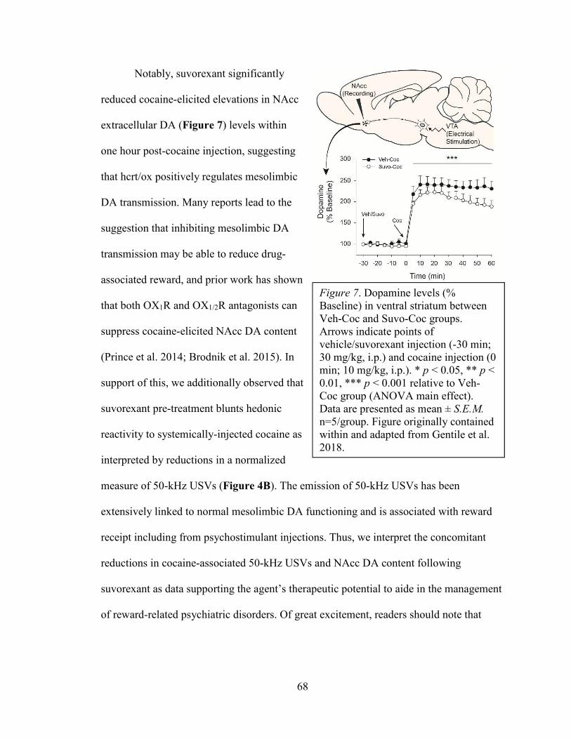
68
Notably, suvorexant significantly
reduced cocaine-elicited elevations in NAcc
extracellular DA (Figure 7) levels within
one hour post-cocaine injection, suggesting
that hcrt/ox positively regulates mesolimbic
DA transmission. Many reports lead to the
suggestion that inhibiting mesolimbic DA
transmission may be able to reduce drug-
associated reward, and prior work has shown
that both OX1R and OX1/2R antagonists can
suppress cocaine-elicited NAcc DA content
(Prince et al. 2014; Brodnik et al. 2015). In
support of this, we additionally observed that
suvorexant pre-treatment blunts hedonic
reactivity to systemically-injected cocaine as
interpreted by reductions in a normalized
measure of 50-kHz USVs (Figure 4B). The emission of 50-kHz USVs has been
extensively linked to normal mesolimbic DA functioning and is associated with reward
receipt including from psychostimulant injections. Thus, we interpret the concomitant
reductions in cocaine-associated 50-kHz USVs and NAcc DA content following
suvorexant as data supporting the agent’s therapeutic potential to aide in the management
of reward-related psychiatric disorders. Of great excitement, readers should note that
Figure 7. Dopamine levels (% Baseline) in ventral striatum between Veh-Coc and Suvo-Coc groups. Arrows indicate points of vehicle/suvorexant injection (-30 min; 30 mg/kg, i.p.) and cocaine injection (0 min; 10 mg/kg, i.p.). * p < 0.05, ** p < 0.01, *** p < 0.001 relative to Veh-Coc group (ANOVA main effect). Data are presented as mean ± S.E.M.
n=5/group. Figure originally contained within and adapted from Gentile et al. 2018.

69
suvorexant is currently in clinical testing for the management of cocaine use disorder
(NCT02785406 from https://ClinicalTrials.gov).
6.2) Comparing rewarding and reinforcing properties between ‘bath salt’ 3,4-
methylenedioxypyrovalerone and cocaine using ultrasonic vocalizations in rats (Simmons
et al. 2018)
Several studies show that rats elicit “positive affective” 50-kHz USVs during
receipt of initial infusions of self-administered cocaine as well as upon systemic
injection. The rise in cocaine-like synthetic stimulants, including the synthetic cathinone
MDPV, warrant characterization of behavioral effects used to measure abuse potential.
The purpose of this study was to compare the reinforcing efficacy and subjective state
associated with anticipation and self-administration of cocaine compared MDPV. We
additionally conducted studies to compare the extent to which non-contingent
administration of MDPV altered 50-kHz USVs. Experimental procedures can be found in
supplemental Section S2.1, and metrics/statistics in supplemental Section S2.3.
In this study, we utilized intravenous self-administration and trained rats across
daily 2-h sessions for 14 d to self-administer cocaine (0.56 mg/kg/inf) or MDPV (0.056
mg/kg/inf) according to an approximate ten-fold greater potency attributed to MDPV
compared to cocaine (Baumann et al. 2013). We replicated earlier work demonstrating
that 50-kHz USVs associated with cocaine self-administration wane following a “load-
up” period (Barker et al. 2014; for methodological detail, readers are referred to
supplemental Section S2.2) which was additionally observed from MDPV self-
administering rats. These results support that euphoria is experienced during the
transition from sobriety to psychostimulant intoxication but not necessarily thereafter

70
despite high drug levels. These data contribute to an ongoing hypothesis that the
underlying drive to self-administer psychostimulants shifts from positive to negative
reinforcement upon titrating to a point of preferred drug level (satiety). Barker and
colleagues (2014) additionally observed that the “maintenance” phase of cocaine self-
administration (i.e. at drug levels after titration to satiety is achieved) is characterized by
periodic bouts of 22-kHz USVs that predominate when calculated cocaine levels are in
the lowest quartile, suggesting these “negative affective” USVs dissipate following bolus
injections. The underlying neural circuits that are recruited as drug levels wane and
negative affect emerges during cocaine self-administration may correspond to stress-
associated circuits that are known to activate during withdrawal. Transmitters including
dynorphin and corticotropin releasing factor (CRF), notably in extended amygdala
structures, dynamically activate and underlie withdrawal-associated reward hypofunction
(for review, see Koob 2015). Real-time measurements of circuit-specific transmitter and
peptide release using voltammetry and fiber photometry can better determine systems
Figure 8. Systemic injection dose-response. (A) 50-kHz USVs across 30-minute recording session on day 7 of cocaine (solid bars) or MDPV (hashed bars) administration. (B-C) Time course of cocaine- or MDPV-elicited 50-kHz USVs in 10-minute time bins. * p < 0.05 against saline control group, Mann Whitney tests. Data for (A) are means ± S.E.M., and data for (B-C) are medians. n=5-8/group. Figure originally contained within and adapted from Simmons et al. 2018.

71
underlying the shift from positive to negative reinforcement during psychostimulant self-
administration.
This is the first study to measure USVs following MDPV injection (experimenter-
and self-administration). As MDPV is readily self-administered and induces place
preference in laboratory animals (e.g., Karlsson et al. 2014), we anticipated that MDPV
injection would elicit 50-kHz USVs and, indeed, observed such behavioral effects.
Moreover, MDPV facilitates brain reward function as determined by electrical self-
stimulation—a measure explicitly measuring “hedonic state” of the rat (Watterson et al.
2014). Experimenter-delivered MDPV injection elicited dose-related elevations in 50-
kHz USVs similarly to cocaine but with high variability (Figure 8). While we did not
stratify subjects into “high” and “low” responders to better accommodate individual
differences, this strategy has been applied in prior work and is suspected to capture
individual vulnerabilities to drug abuse (e.g., Tripi et al. 2017). Self-administered MDPV

72
elicited stronger rates of 50-
kHz USVs with relatively
lower variation and greater
persistence through “load-up”
compared to cocaine (Figure
9 and 10). MDPV
additionally appeared to be
intravenously self-
administered more quickly
than cocaine, although a
within-subjects comparison
would have allowed for better
assessment. We surmise that
MDPV, like cocaine, elicits
high rates of 50-kHz USVs
and can be measured during
intravenous self-
administration which may be useful in evaluating subjective euphorogenisis.
Figure 9. Self-administration of cocaine or MDPV, whole session. (A) Number of infusions earned during second week of self-administration. Mean number of 50-kHz USVs during anticipation (B) or administration (C) of either cocaine (grey bars) or MDPV (black bars). (D, upper) 50-kHz USVs throughout 2-hours of drug self-administration in 10-s time bins. (D, lower) Drug infusion vertical line plots for individual rats. Vertical dashed line represents lever extension. ** p < 0.01 against respective anticipation data (independent samples t-test). Data in A, B and C are mean ± S.E.M. n=7-8/group. Figure originally contained within and adapted from Simmons et al. 2018.

73
Self-administration of MDPV showed a profile of USV emissions like self-
administered cocaine but at one-tenth the dose. MDPV blocks catecholamine transporters
and elevates NAcc DA content with ~10-fold greater potency compared to intravenous
cocaine (Baumann et al. 2013). Indeed, several teams have published that MDPV is
intravenously self-administered at doses as low as 0.03 to 0.05 mg/kg/inf (Watterson et
al. 2014; Schindler e al. 2016). Qualitative analysis on the patterns of responding for
MDPV versus cocaine across
subjects revealed relatively
“regulated” injection
intervals, suggesting perhaps
greater titration efficiency
with intravenous MDPV
which may relate to its ability
to move quickly across the
blood-brain barrier and exert
central effects (Simmler et al.
2013). Collectively, these
results confirm the alarming
potency of MDPV with
respect to the amount of drug
needed to facilitate self-
administration.
Figure 10. Self-administration of cocaine or MDPV, load-up. (A) Latency to receive infusions 1-10 of cocaine (grey bars) or MDPV (black bars) relative to lever extension. (B) 50-kHz USVs per infusion where infusion "0" represents USV rate prior to infusion 1. (C, upper) Cumulative time to earn infusions 1-10 plotted using horizontal lines with corresponding 50-kHz USVs across anticipation and load-up (C, lower). Vertical dashed line represents lever extension. * p < 0.05, ** p < 0.01, *** p < 0.001 main effect of Drug Type. Data in A and B are mean ± S.E.M. n=7-8/group. Figure originally contained within and adapted from Simmons et al. 2018.

74
Finally, this study was the first to capture robust rates of 50-kHz USVs from
exposure to a chamber paired with psychostimulant (MDPV and cocaine) self-
administration. Earlier work showed that rats emit 50-kHz USVs upon exposure to an
environment associated with experimenter-delivered cocaine injections (Ma et al. 2010),
and thus it is not altogether surprising to observe strong USV rates from a salient, drug-
paired context in which rats engage in an operant task to volitionally take drugs. In some
subjects, the rate of 50-kHz USVs from placement into the drug-paired operant chamber
surpassed that which followed intravenous drug self-administration. Notably, in the study
performed by Barker and colleagues (2014), USVs prior to the first self-administered
cocaine injection were measured and found to be approaching near-zero levels. Aligned
with the idea that a salient context drives pre-drug USV responses, rats in the study
described by Barker and colleagues (2014) resided permanently within their operant
chamber after catheterization, whereas rats in our study lived in separate home-cages.
This finding contributes to a well-documented phenomenon that salient contextual cues
can lead to behavioral disinhibition and drive motivational/mood states.
6.3) Role of hypocretin/orexin receptor blockade on drug-taking and ultrasonic
vocalizations associated with self-administration of ‘bath salt’ 3,4-
methylenedioxypyrovalerone in rats (Simmons et al. 2017)
The following study was the first to capture effects of svuorexant pre-treatment on
infusions earned and USVs associated with MDPV self-administration. In prior work
(Simmons et al. 2018), we identified that MDPV, comparably as cocaine but with greater
persistence, elicits 50-kHz USVs following systemic and self-administration injection
methods. We additionally examined the influence of suvorexant of acoustically-defined

75
USV call types as briefly described in Chapter 5.
For additional detail on this study, experimental
procedures are provided in supplemental Section
S3.1 as is detail on USV recording and analysis
(supplemental Section S3.2) and metric/statistics
(supplemental Section S3.3).
Briefly, rats were trained to self-administer
MDPV (0.03 mg/kg/inf) across daily 2-h sessions
for 14 d. A principal finding from this report is
that, while suvorexant failed to significantly
decrease infusions earned under a low-effort FR-1
self-administration task (Figure 11), pre-treatment
with a moderate dose (10 mg/kg, IP) did, indeed,
significantly suppress 50-kHz USVs during
“anticipation” and “post-lever” time epochs
(Figure 12). Non-significant suppression of 50-
kHz USVs during each of the above-mentioned
time epochs was additionally seen upon high-dose
(30 mg/kg, IP) suvorexant pre-treatment. Finally,
the degrees to which suvorexant altered 50-kHz USV emissions and infusions earned
trended to positively correlate (Figure 13), promoting potential utility in measuring
USVs to predict subsequent drug-taking.
Figure 11. Effect of suvorexant on MDPV self-administration. (A) Change in infusions relative to each rat's vehicle pre-treated control data. (B) Latency to self-administer the first 10 infusions of MDPV. Data in (A-B) are means + S.E.M. * p < 0.05 relative to "100"–the point of no-change from vehicle pre-treatment. n=7-14/group. Figure originally contained within and adapted from Simmons et al. 2017.

76
Similar to our
previously published
work (Simmons et al.
2018), we observed
that trained rats elicit
robust rates of 50-kHz
USVs from exposure
to a context paired
with MDPV self-
administration (i.e.
during
“Anticipation”).
Whereas Simmons and
colleagues (2018)
incorporated a 5-min
“Anticipation”
recording, the present
study used a 30-min recording window in effort to better resolve bouts of 50-kHz USVs
occurring prior to and after operant lever extension. Indeed, in our prior study the rate of
50-kHz USVs prior to lever extension often overshadowed 50-kHz USVs emerging
following intravenous receipt of MDPV. Incorporation of an “Anticipation” time window
longer than 5 min, but not necessarily as long as 30 min, is useful in capturing distinct
signatures of USVs associated with “Anticipation” and self-administration of MDPV.
Figure 12. Effect of suvorexant on ultrasonic vocalizations (USVs) associated with MDPV self-administration. (A) Vertical bar charts depicting change in 50-kHz USVs relative to vehicle pre-treatment control data during "anticipation" (upper panel) and "post-drug" (lower panel) time-points. Plotted using right y-axis are individual data points showing change in number of 50-kHz USVs from vehicle during "anticipation" and "post-drug" time-points. (B) Pie charts showing USV type composition of 5-min samples during "anticipation" and "post-drug" time-points during pre-treatment sessions. (C) 50- and 22-kHz USVs plotted across session with black vertical line showing lever extension and access to MDPV. Data in (A) are means + S.E.M. * p < 0.05, ** p < 0.01 relative vehicle pre-treated control data (non-parametric corrected pairwise comparisons). n=7-14/group. Figure originally contained within and adapted from Simmons et al. 2017.
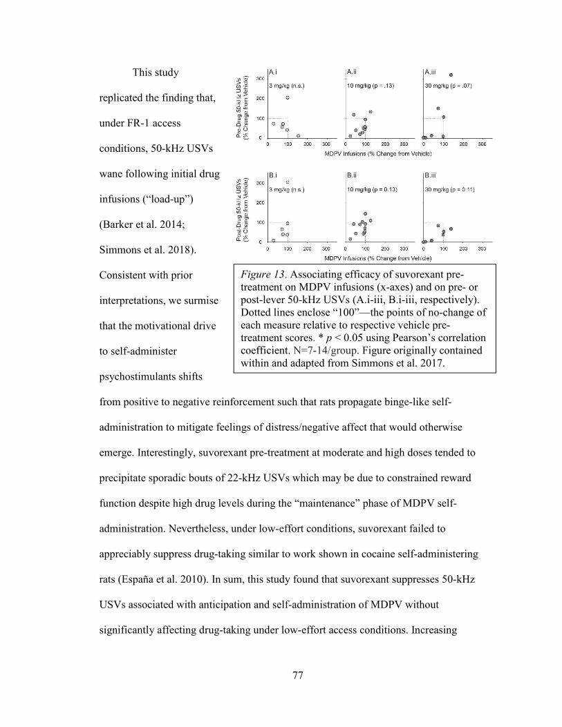
77
This study
replicated the finding that,
under FR-1 access
conditions, 50-kHz USVs
wane following initial drug
infusions (“load-up”)
(Barker et al. 2014;
Simmons et al. 2018).
Consistent with prior
interpretations, we surmise
that the motivational drive
to self-administer
psychostimulants shifts
from positive to negative reinforcement such that rats propagate binge-like self-
administration to mitigate feelings of distress/negative affect that would otherwise
emerge. Interestingly, suvorexant pre-treatment at moderate and high doses tended to
precipitate sporadic bouts of 22-kHz USVs which may be due to constrained reward
function despite high drug levels during the “maintenance” phase of MDPV self-
administration. Nevertheless, under low-effort conditions, suvorexant failed to
appreciably suppress drug-taking similar to work shown in cocaine self-administering
rats (España et al. 2010). In sum, this study found that suvorexant suppresses 50-kHz
USVs associated with anticipation and self-administration of MDPV without
significantly affecting drug-taking under low-effort access conditions. Increasing
Figure 13. Associating efficacy of suvorexant pre-treatment on MDPV infusions (x-axes) and on pre- or post-lever 50-kHz USVs (A.i-iii, B.i-iii, respectively). Dotted lines enclose “100”—the points of no-change of each measure relative to respective vehicle pre-treatment scores. * p < 0.05 using Pearson’s correlation coefficient. N=7-14/group. Figure originally contained within and adapted from Simmons et al. 2017.

78
response requirements, such as through employment of PR reinforcement, may capture
suvorexant’s reward-attenuating properties and may aide in understanding possible
relationships between USVs and drug-taking.

79
SPECIFIC AIMS
Addictions to psychostimulants collectively form a crippling epidemic facing
many communities on an international scale. Drugs of abuse augment extracellular
monoamine content—notably, DA—in structures associated with reward and emotion
processing. Transmitters and peptides that regulate the ability of drugs to perturb central
neurochemical transmission present attractive targets in effort to normalize motivation
and affective changes associated with their abuse. Among contributors, hypothalamic
hcrt/ox innervates midbrain and forebrain structures implicated in reward- and mood-
related pathologies. Pharmacological studies support that hcrt/ox receptor blockade,
systemically and directly within ventral midbrain, attenuates pathological motivation
including for drugs of abuse. Our understanding of ventral midbrain structural
heterogeneity, however, is continually evolving—within the past decade, the caudal
division of VTA (now termed RMTg) was highlighted for its ability to negatively
regulate VTA DA activity. Curiously, anatomy work depicts the hypothalamus as a
principal input to RMTg, though the cellular phenotypes and functions of this connection
have not been fully resolved. Thus, the continuum residing within ventral midbrain is
composed of rostral and caudal divisions that lie adjacently yet function in opposition.
Aim 1. Identify topographic organization of hcrt/ox afferents to ventral tegmental area
(VTA) and rostromedial tegmentum (RMTg).
For Aim 1, rats will be intracranially cannulated for injections of fluorescent
microspheres within VTA and RMTg targets. Immunolabeling will be used to label and
map putative hcrt/ox afferents to each structure. Mapping hcrt/ox afferents within ventral

80
midbrain will provide support for potential functional roles in the domains of reward and
emotion regulation.
Aim 2. Determine functional involvement of hcrt/ox transmission within VTA and RMTg
on affect and motivated drug-taking.
For Aim 2, rats will receive bilateral cannulation and jugular vein catheterization prior to
cocaine self-administration training. Rats will train under fixed-ratio 1 access conditions
followed by progressive-ratio during which one of two self-administered cocaine doses
(0.375 and 0.750 mg/kg/inf) will be available. During experimental sessions, intra-VTA
pre-treatment with suvorexant (0.0, 0.3, 1.0, 3.0 µg/hemi) or intra-RMTg pre-treatment
with hcrt/ox (0.0, 0.3, 1.0, 3.0 nmol/hemi) will precede placement in operant chambers
and initiation of the self-administration session. Ultrasonic vocalizations (USVs) will be
recorded throughout as a proxy for shifts in affective state. Aim 2 will test the hypothesis
that intra-VTA suvorexant will attenuate positively-valenced 50-kHz USVs and decrease
motivated drug-taking whereas augmenting intra-RMTg hcrt/ox transmission will
enhance negatively-valenced 22-kHz USVs and also reduce motivated drug-taking.
Collectively, these experiments are designed to determine topographical
arrangement of hcrt/ox afferents to VTA and RMTg and will test the central
hypothesis that, while hcrt/ox signaling in VTA contributes to reward/positive
affect, hcrt/ox signaling in RMTg contributes to aversion/negative affect (depicted in
Figure 14).

81
Figure 14. Hypothesis of predicted effects of hcrt/ox receptor perturbation within VTA and RMTg on motivated cocaine-taking and USVs. It is predicted that hcrt/ox transmission within RMTg and/or receptor blockade within VTA will diminish mesolimbic DA transmission to reduce the number of cocaine infusions earned under high-effort access conditions. Additionally, blockade of hcrt/ox receptor in VTA is hypothesized to reduce the rate of ‘positive affective’ 50-kHz USVs while hcrt/ox transmission augmentation in RMTg is hypothesized to promote aversion and, in turn, enhance the rate of ‘negative affective’ 22-kHz USVs.

82
CHAPTER 7 – HYPOCRETIN/OREXIN INNERVATION TO VENTRAL
MIDBRAIN
Several studies have supported a critical role of VTA hcrt/ox in motivated
responding for salient rewards including palatable food and drugs of abuse. Early work
revealed that neurons containing hcrt/ox project to midbrain and indeed influence VTA
DA cellular physiology (Fadel and Deutch 2001; Korotokova et al. 2003; Borgland et al.
2009). Since 2009, several research teams worked to uncover a distinct structure within
caudal division of ventral midbrain named RMTg (“tail of VTA”). GABA-producing
cells within RMTg negatively regulated VTA DA neurons and participate in encoding
aversive stimuli (Jhou et al. 2009a, b). The hypothalamus is a principal input to RMTg
(Jhou et al. 2009a), but the cellular phenotypes of these afferents have not been
uncovered. To the best of our knowledge, explicit study of hcrt/ox innervation to VTA
and RMTg of ventral midbrain has not been done. Thus, this study utilized retrograde
tract tracing and immunolabeling methods to map hcrt/ox afferents projecting to VTA
and RMTg.
7.1) Materials and methods
7.1.1) Animals
Adult male Sprague-Dawley rats (Charles River Laboratories; Horsham, PA,
USA) weighing ~300 g at experimental onset were used for the present experiment. Rats
were provided food and water freely upon arrival to vivarium, were pair-housed until
cannulation surgery after which rats were individually-housed and maintained on a
reverse 12-h : 12-h light cycle (lights off at 9:00 AM) in a temperature- and humidity-

83
controlled vivarium. All experimental procedures were approved by the Institutional
Animal Care and Use Committee of Temple University.
7.1.2) Experimental procedures
Rats underwent unilateral stereotaxic cannulation surgery as specified below
followed by retrograde tracer injections. After allowing time for retrograde transport (10-
14 d), tissue was harvested and processed to label hcrt/ox afferents to target structures in
ventral midbrain (Figure 15A.i).
7.1.3) Stereotaxic cannulation
Rats received cannulation with 26-gauge guide cannulae directed 1 mm above
target structure. Unilateral cannulations were performed and were implanted without
angling the cannula (VTA: A/P -5.6 mm, M/L ±0.6 mm, D/V -6.6; RMTg: A/P -6.8 mm,
M/L ±0.4 mm, D/V -6.4 mm from dura rupture). Rats were anesthetized using isoflurane
gas anesthesia (5% induction, 3-4% maintenance) at a flow rate of 1.2 – 1.5 L/min mixed
with O2, and hair on head region was shaved using electric clippers. Rats were
administered ketoprofen (5 mg/kg, SC) as pre-operative analgesia and secured in ear bars
of stereotaxic frame atop water-circulating heating pad. Shaved head region was wiped
intermittently with betadine and alcohol, and a midline incision was thereafter performed
using a stainless steel scalpel blade (#10). Connective tissues were gently cleared to
reveal skull, blood was wiped using cotton-tipped applicators, and clamps were used to
keep skin pulled laterally to facilitate visualization. Epinephrine-soaked cotton was
thereafter applied to minimize bleeding, and a sickle scaler was used to etch skull surface
to improve skull-cap adhesion. If needed, Z-plane was calibrated to allow for even
anterior-posterior positioning. Burr holes were drilled using a 106 engraving cutter (1/8”

84
shank) of drill—two incomplete holes for jeweler screw installations (each per
hemisphere approximately 2 mm caudolateral from bregma) and holes according to atlas
coordinates to affix guide cannulae above target structure as detailed above. Guide
cannulae were lowered, and dura rupture was observed; if not observed, guide tip would
be lowered 0.8 – 1.0 mm beneath outer skull surface prior to lowering per D/V
coordinate. Guide cannulae were lowered at a rate of ~1.5 mm/min. A resin-ionomer
dental paste was applied directly proximal to lower threads of guide and allowed 3-5 min
to dry. Fast-curing acrylic resin powder was sprayed to form outer skull-cap and
hardened following liquid initiator application. Rats were supplemented with 4 mL of
0.9% saline prior to waking from anesthesia, and lidocaine gel analgesic was topically
applied around skull-cap. A 33-gauge dummy cannula (0-mm projection) beyond the end
of the guide cannula was tightened in place to prevent backflow and clogging of cannula
track.
7.1.4) Retrograde tracing
Rats were anesthetized with isoflurane gas for retrograde tracer injections, and
dummy cannulae were thereafter removed. Fluorophore-conjugated latex microspheres
(Lumafluor [Red RetroBeads IX]; Durham, NC, USA) with 530-/590-nm maximum
excitation/emission spectra, diluted 1:1 with aCSF, were deposited into target structure
using a 33-gauge locking injector extending 1-mm beyond pedestal of guide cannulae. A
series of 3 150-nL injections per day across three days was performed on each rat.
Injections were separated by 1-2 h, and each day of injections was separated by ~24 h.
PE-50 tubing connected the locking injector to a 22-gauge microliter syringe which itself
was secured on an automated syringe pump. Injection parameters were established at 50

85
nL/min flow rate, and locking injectors were kept in place for an additional 1 min to
allow for diffusion from injector tip. Patency/flow of injector was checked in between
each injection. Dummy cannulae were then re-tightened onto guides, and rats were placed
in homecage and back into vivarium upon waking from anesthesia.
7.1.5) Tissue collection and analysis
After allowing 10-14 d for retrograde transport of tracers, rats were deeply
anesthetized with isoflurane anesthesia for intracardiac perfusion. Anteriorally-extending
lateral incisions from mid-section of rat were made using large sharp scissors. Diaphragm
was snipped and rib-cage clamped against anterior aspect to reveal beating heart.
Connective tissues were carefully separated, and a 22-gauge needle connected to
peristaltic perfusion pump (LongerPump; Boonton, NJ, USA) was inserted into left
ventricle. Perfusion with ice-cold phosphate buffer (30-34 rpm) was then initiated, and
the right atrium was snipped with small sharp scissors to permit exsanguination. About 7-
8 min later (until drained content was pale in color), the perfusion pump was briefly
halted to switch uptake solution to ice-cold paraformaldehyde (PFA; 4% mixed in
phosphate buffer, pH = 7.40). Rats were perfused with PFA for 10 min, and heads were
then decapitated to allow for whole brain dissection through skull removal using rangeurs
and small sharp scissors. Brains were kept at 4o C in PFA for 24-72 h followed by 30%
sucrose in phosphate buffer for 5-7 d. Dehydrated brains were then sectioned on a
cryostat (Leica) at after blocking cerebellum for even mounting position. For some
brains, a marking was made using a 22-gauge needle on one hemisphere of lateral
midbrain (i.e. outside of hypothalamus) to demarcate ipsilateral versus contralateral
hemisphere relative to tracer deposit. Tissue containing guide cannula track was

86
sectioned at 60 µm, and hypothalamus was sectioned at 30 um. Tissue was stored in
cryoprotectant (30% ethylene glycol and 30% sucrose in 0.1M phosphate-buffered saline
[PBS]) and kept at -20o C until processed for immunolabeling.
Hypothalamus-containing tissue (constituting a 1-in-3 series of entire
hypothalamus) was processed throughout under conditions minimizing exposure to white
light. Tissue was first washed in PBS 6 times (5-min each on rocker) to clear away
residual cryoprotectant. Tissue was then blocked with 5% donkey serum in PBS
containing 0.3% Triton X-100 (hereafter termed "PBS+") for 60 min at room
temperature. Tissue was then transferred to 1.5-mL microcentrifuge tubes containing goat
anti-orexinA primary antibody (1:1,000; SC8070, Santa Cruz) in 1.5% donkey serum in
PBS+ and gently rocked for 24-48 h at 4o C. Thereafter, tissue was washed with PBS 6x
(5-min each on rocker) and then transferred to 1.5-mL microcentrifuge tubes containing
donkey anti-goat 488 (AlexaFluor) secondary antibody (1:750) in 1.5% donkey serum in
PBS+ and gently rocked for 3-4 h at room temperature. Tissue containing cannula track
and tracer deposit site was rinsed in phosphate buffer and gently rocked in fluorescent
Nissl (NeuroTrace 500/525) at 1:500 concentration in PBS+. Finally, all tissue was again
washed in PBS (3x, 5-min each on rocker) and kept at 4o C until mounted on charged
microscope slides using a petri dish containing ddH2O. Tissue was allowed to dry on
slides before coverslipping with medium that protects against fluorophore decay
(ProLong Gold).
Sections from confirmed "hits" to target structure (based on histology of sections
containing cannula track) were imaged on an Eclipse 80i upright fluorescent microscope
(Nikon; Minato, Tokyo, Japan). Hypothalamic sections containing orexinA-
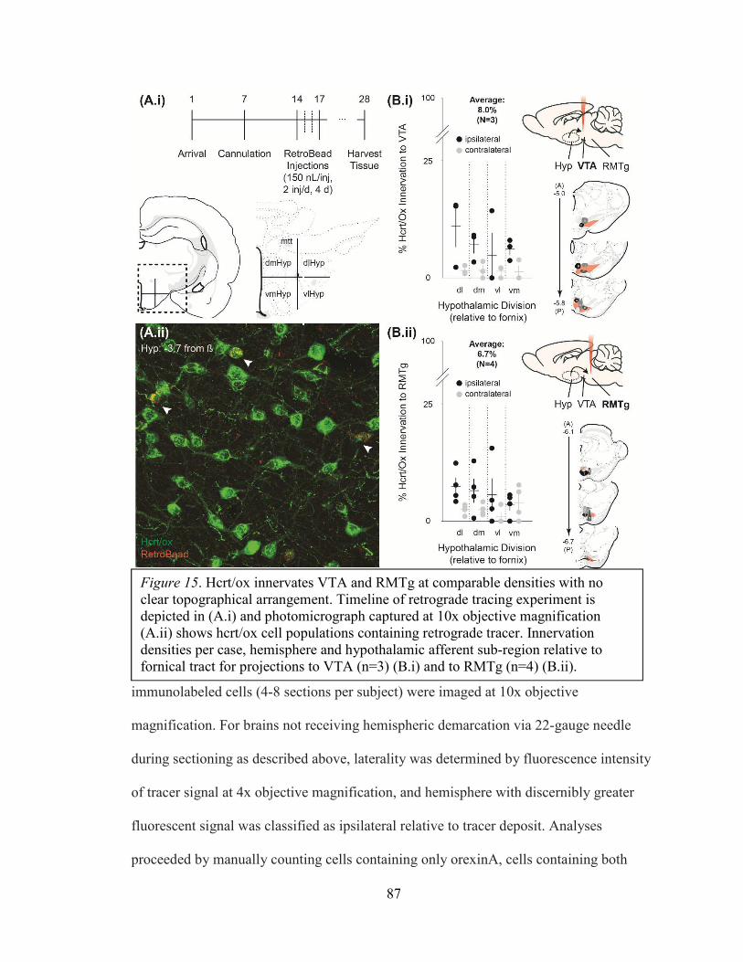
87
immunolabeled cells (4-8 sections per subject) were imaged at 10x objective
magnification. For brains not receiving hemispheric demarcation via 22-gauge needle
during sectioning as described above, laterality was determined by fluorescence intensity
of tracer signal at 4x objective magnification, and hemisphere with discernibly greater
fluorescent signal was classified as ipsilateral relative to tracer deposit. Analyses
proceeded by manually counting cells containing only orexinA, cells containing both
Figure 15. Hcrt/ox innervates VTA and RMTg at comparable densities with no clear topographical arrangement. Timeline of retrograde tracing experiment is depicted in (A.i) and photomicrograph captured at 10x objective magnification (A.ii) shows hcrt/ox cell populations containing retrograde tracer. Innervation densities per case, hemisphere and hypothalamic afferent sub-region relative to fornical tract for projections to VTA (n=3) (B.i) and to RMTg (n=4) (B.ii).

88
orexinA and tracer,
and tracer-
containing cells
without orexinA.
Each hypothalamus
section used for
anterior-posterior
topographical
analysis was
assigned to atlas
levels 26 through
32 (Swanson 2004)
based on gross
morphological
features including extent of hippocampus across medial-lateral and dorsal-ventral axes,
positions of fornical and mammillothalamic tracts, and shape of ventral division of third
ventricle.
7.1.6) Statistical analyses
Independent samples t-tests were conducted to compare total % innervation of
hcrt/ox-producing cells targeting VTA and RMTg as well as comparisons between each
sub-region (i.e., dorsolateral, dorsomedial, ventrolateral, ventromedial). Separate t-tests
were conducted for % innervation comparisons of contralateral versus ipsilateral
afferents.
Figure 16. Representative retrograde tracer deposit histology. Dye plumes are shown for retrograde tracer deposits to VTA (A) and to RMTg (B). aq – central aqueduct, cpd – cerebellar peduncle, EW – Eddinger-Westphal nucleus, IPN – interpeduncular nucleus, MGN – medial geniculate nucleus, ml – medial lemniscus, PAG – periaqueductal grey area, RMTg – rostromedial tegmental nucleus, RN – red nucleus, SC – superior colliculus, SN – substantia nigra, VTA – ventral tegmental area.

89
7.2) Results
Histological analysis confirmed retrograde tracer deposit within VTA (n=3) and
RMTg (n=4) with minimal tracer spillover into adjacent structures (Figure 16). Analysis
of putative hcrt/ox-producing afferent cells indicated comparable targeting density to
each of VTA (8.0%) and RMTg (6.7%) targets from entire hcrt/ox-producing cell
population of ipsilaterally-injected hemisphere (Figure 15B.i-ii). No significant
differences were found when examining % innervation of total hcrt/ox cells of ipisilateral
or contralateral hemisphere relative to retrograde tracer deposit between putative hcrt/ox-
producing VTA- and RMTg-projecting afferents [all |t(5)| ≤ 0.40, n.s.]. Similarly, no
significant differences between VTA- and RMTg-projecting hcrt/ox afferents were found
when examining afferent cells arising from ipsilaterally- or contralaterally-injected
dorsolateral, dorsomedial, ventrolateral or ventrolmedial hypothalamus. Sparse labeling
was found on hemisphere contralateral to tracer deposit (VTA: 1.7%; RMTg: 2.5%). The
majority of hcrt/ox-producing cells were found within dorsolateral and dorsomedial
hypothalamus relative to fornical tract, and these sub-regions contained the greatest
proportion of retrogradely-traced hcrt/ox cells compared to the field of hcrt/ox-producing
cells within sub-region (VTA: 11.6% and 7.8%, respectively; RMTg: 7.4% and 7.9%,
respectively). Data per each analyzed case are reported in Table 3. Topographical
analysis across anterior-posterior extent of hypothalamus revealed that sections
containing the densest quantity of hcrt/ox cells (i.e. at atlas levels 28-30) tended to most
robustly target VTA or RMTg relative to anterior or posterior extent of hypothalamus
(i.e. at atlas levels 26 and 32, respectively) (Table 3). Innervation densities can be found
in Table 4. Overall, these data show that hcrt/ox afferents target both VTA and RMTg of

90
ventral midbrain with comparable densities and suggest possible opponent functions of
hcrt/ox in RMTg to known reward-mediating functions in VTA. Specifically, hcrt/ox
may provide excitatory transmission to GABA-producing cells of the RMTg which in
turn may inhibit the DA-producing cells of VTA to “brake” reward signals and/or
participate in the encoding of aversive stimuli.
7.3) Interim discussion
Outputs from hypothalamic hcrt/ox-producing neurons are of interest in
determining the loci underlying behavioral effects of hcrt/ox transmission modulation
(i.e., with pharmacological agents). Specifically, numerous teams reveal that hcrt/ox
receptor antagonism normalizes pathological motivation in the context of drug-seeking
(e.g., Borgland et al. 2009; Zhou et al. 2012). Prior studies additionally find that hcrt/ox
Table 3. Retrograde tract tracing data expanded; cell count data per case are included as are proportions of afferent innervation from hypothalamic hcrt/ox (by sub-region) to VTA and RMTg.

91
innervates VTA (Peyron et al. 1998; Fadel and Deutch 2001) and direct-site
pharmacology finds that blockade of OX1Rs within VTA reduces motivated cocaine-
taking (Muschamp et al. 2014). In 2009, the caudal division of VTA—a structure termed
“tail of VTA” and RMTg—was anatomically and functionally characterized (Jhou et al.
2009a, b). Interestingly, this GABAergic nuclei inhibits the rostrally-positioned DA-
producing cell population in VTA to inhibit mesolimbic DA transmission, and the RMTg
is activated following presentation of aversive stimuli. Our tract-tracing study was
conducted to reveal if hcrt/ox additionally transmits to RMTg which would support an
omnibus modulatory role in positively and negatively regulating motivated reward-
seeking. Indeed, we observed comparable inneration of putative hcrt/ox afferents to VTA
and RMTg and, thereafter, begged to question potential functions of these connections.
Table 4. Retrograde tract tracing data expanded; relative innervation densities of putative hypothalamic hcrt/ox afferents to VTA and RMTg are included and divided by sub-region of hypothalamus based on position of fornix.

92
CHAPTER 8 – ROLE OF HYPOCRETIN/OREXIN TRANSMISSION WITHIN
VENTRAL MIDBRAIN ON PSYCHOSTIMULANT-ASSOCIATED
REINFORCEMENT AND AFFECTIVE CHANGES
To-date, no studies have examined possible functions of hcrt/ox transmission
within RMTg. Numerous behavioral pharmacological reports demonstrate therapeutic
utility of hcrt/ox receptor blockade within VTA in the context of attenuating pathological
motivation, and work contained within Chapter 7 of this thesis supports that hcrt/ox
additionally innervates RMTg. Critically, the RMTg negatively regulates DA-producing
cells housed within VTA and thus hcrt/ox within RMTg may participate in the
physiological encoding of aversive states. In this study, we tested the functional impact of
hcrt/ox perturbation within ventral midbrain structures using local injections of hcrt/ox
peptide (bilaterally into RMTg) or of the clinically-available hcrt/ox receptor antagonist
suvorexant (bilaterally into VTA). Specifically, we measured the impact of site-directed
hcrt/ox receptor agonist and antagonist administration on USVs and drug-taking
associated with cocaine self-administration in a high-effort task. Moreover, we used two
doses of self-administered cocaine (0.375 and 0.75 mg/kg/inf) to evaluate potential dose-
related differences as has been shown in prior pharmacological studies using hcrt/ox
receptor antagonists (España et al. 2010).
8.1) Materials and methods
8.1.1) Animals
Rats used for the present study were adult male Sprague-Dawley rats (Charles
River Laboratories; Horsham, PA, USA) weighing ~250 g from experimental onset. Rats
were pair-housed until cannulation surgery after which rats were singly-housed and

93
maintained on a 12-h : 12-h reverse light cycle in a humidity- and temperature-controlled
vivarium. Rats were provided food and water ad libitum except during experimental
sessions (drug self-administration). All experimental procedures were approved by the
Institutional Animal Care and Use Committee of Temple University.
8.1.2) Drugs
Cocaine hydrochloride (Sigma-Aldrich; St. Louis, MO, USA) and suvorexant
(AstaTech; Bristol, PA, USA) were commercially purchased. OrexinA peptide was
generously provided by the Drug Supply Program of the National Institute on Drug
Abuse (PolyPeptide Group; San Diego, CA, USA). Cocaine hydrochloride was stored at
4o C and was dissolved in 0.9% saline, vortexed, and filtered through 0.45 µm cellulose
acetate prior to being loaded in syringes for self-administration—the concentration was
adjusted for target infusion dose as well as average bodyweight of cohort every 5-7 d.
OrexinA was prepared for intracerebral injections by dissolving in artificial cerebrospinal
fluid (aCSF, ion concentrations in mM: Na+ 150, K+ 3.0, Ca2+ 1.4, Mg2+ 0.8, P3- 1.0, Cl-
155) and was kept desiccated at -20o C prior to loading in injectors. Suvorexant was
stored at 4o C and was prepared for intracerebral injections immediately prior to each
treatment session by dissolving in dimethyl sulfoxide (DMSO) through vortexing and
ultrasonication.
8.1.3) Experimental procedures
Rats underwent bilateral cannulation and jugular vein catheterization surgeries
prior to cocaine self-administration. Using a within-subjects design, effects of intra-VTA
suvorexant (0.0, 0.3, 1.0, 3.0 µg/hemisphere) were assessed on motivated responding for
and ultrasonic vocalizations (USVs) associated with either low-dose (~0.375 mg/kg/inf)

94
or moderate-dose (~0.75 mg/kg/inf) of self-administered cocaine. In separate cohorts,
effects of intra-RMTg orexinA (0.0, 0.3, 1.0, 3.0 nmol/hemisphere) were assessed on
identical dependent measures. Following pharmacological agent pretreatment, rats were
immediately placed in operant chamber, and USVs were recorded for 15 min prior to
lever extension and onset of 2-h self-administration session. Following all experimental
sessions, tissue was harvested to histologically verify cannula placements. Experimental
timelines and designs are depicted in Figure 17.
8.1.4) Stereotaxic cannulation
For detail of stereotaxic cannulation surgery, readers are referred to Section 7.1.3.
Briefly, bilateral cannulations were performed, and each of two individual guides were
angled at 10o and lowered to reach depth of target structure (VTA: -5.4 mm, M/L ± 2.1,
D/V -6.6; RMTg: A/P -6.8 mm, M/L ±0.4 mm, D/V -6.6 mm from dura rupture). Dummy
cannulae were tightened onto guide cannulae to prevent backflow and clogging.
Figure 17. Experimental designs for target site pharmacology probing hcrt/ox function in VTA and RMTg on motivated cocaine-taking. FR1 – fixed ratio 1, PR – progressive ratio, USV – ultrasonic vocalization.

95
8.1.5) Intravenous drug self-administration: jugular vein catheterization, apparatus and
behavioral procedures
For full description, readers are referred to supplemental Section S1.3. Briefly,
rats were trained to self-administer cocaine (~0.75 mg/kg/inf) under fixed-ratio 1 (FR-1)
access conditions during 2-h sessions for 10-14 d, 5-7 d/wk. Thereafter, rats were shifted
to a progressive-ratio (PR) schedule of reinforcement for at least 5 d before receiving pre-
treatments. At least 1 d of PR cocaine self-administration without pre-treatment
proceeded between each of 4 pre-treatment sessions as depicted in Figure 17.
8.1.6) Ultrasonic vocalization recording and analysis
USVs were recorded and analyzed as described in supplemental Section S1.4.
Briefly, USV recordings began 15-min prior to the onset of self-administration sessions
(i.e. before triggering MED-PC programs). Recordings proceeded throughout the 2-h PR
sessions.
8.1.7) Statistical analyses
For cocaine self-administrating training sessions, mixed model ANOVAs were
used to compare the number of infusions earned examining session (10 sessions of FR-1
or 7 sessions of PR) by cocaine dose (0.375 or 0.750 mg/kg/inf) for each target-site (VTA
or RMTg). For experimental test sessions, analyses proceeded on the % of infusions
earned, active and inactive lever presses performed, and breakpoints relative to prior-day
baseline performance using one-way ANOVAs with pre-treatment dose (vehicle, low,
moderate, high) as the repeated-measures factor. Separate ANOVAs were performed
dependent on cannulated target site (VTA and RMTg) which influenced levels of the
“pre-treatment dose” variable (for VTA, suvorexant doses were: 0.0, 0.3, 1.0, 3.0 µg; for

96
RMTg, hcrt/ox peptide doses
were: 0.0, 0.3, 1.0, 3.0 nmol) as
well as on cocaine dose (0.375
and 0.750 mg/kg/inf); thus, four
one-way repeated-measures
ANOVAs were conducted in total
for each analyzed %-change
metric (VTA-0.375, VTA-0.750,
RMTg-0.375, RMTg-0.750).
Greenhouse Geisser adjustments
were implemented when
sphericity violations were
detected. Contrast analyses
proceeded to compare each “%
change” from pre-treatment
metric (low, moderate, high)
against vehicle pre-treatment. 22-
and 50-kHz USV count data
during “Anticipation” and “Post-
Lever” time epochs were analyzed using mixed ANOVAs as described above for lever
press data1. Correlations between breakpoint and USVs proceeded using Pearson
coefficients1. Statistical significance was defined as Type I error rate (α) below 0.05.
Figure 18. Cannula placement histology for rats used for target-site behavioral pharmacology experiments. Histologically-confirmed “hits” for intra-VTA injected rats are indicated by lower-case (a, b, c) and upper-case (A, B, C) letters for low-dose (0.375 mg/kg/inf; n=5) and high-dose (0.750 mg/kg/inf; n=4) cocaine self-administering rats, respectively. Similarly, Roman numerals (i, ii, iii) and Arabic numerals (1, 2, 3) are used to depict cannula placement for intra-RMTg injected rats self-administering low-dose (n=5) versus high-dose (n=6) cocaine, respectively. Red highlight indicates intended target structure (VTA in left panels, RMTg in right panels). Representative histology sections with cannula track outlines are shown in lower panels. cpd – cerebellar peduncle, IPN – interpeduncular nucleus, ml – medial lemniscus, RMTg – rostromedial tegmental nucleus, VTA – ventral tegmental area.
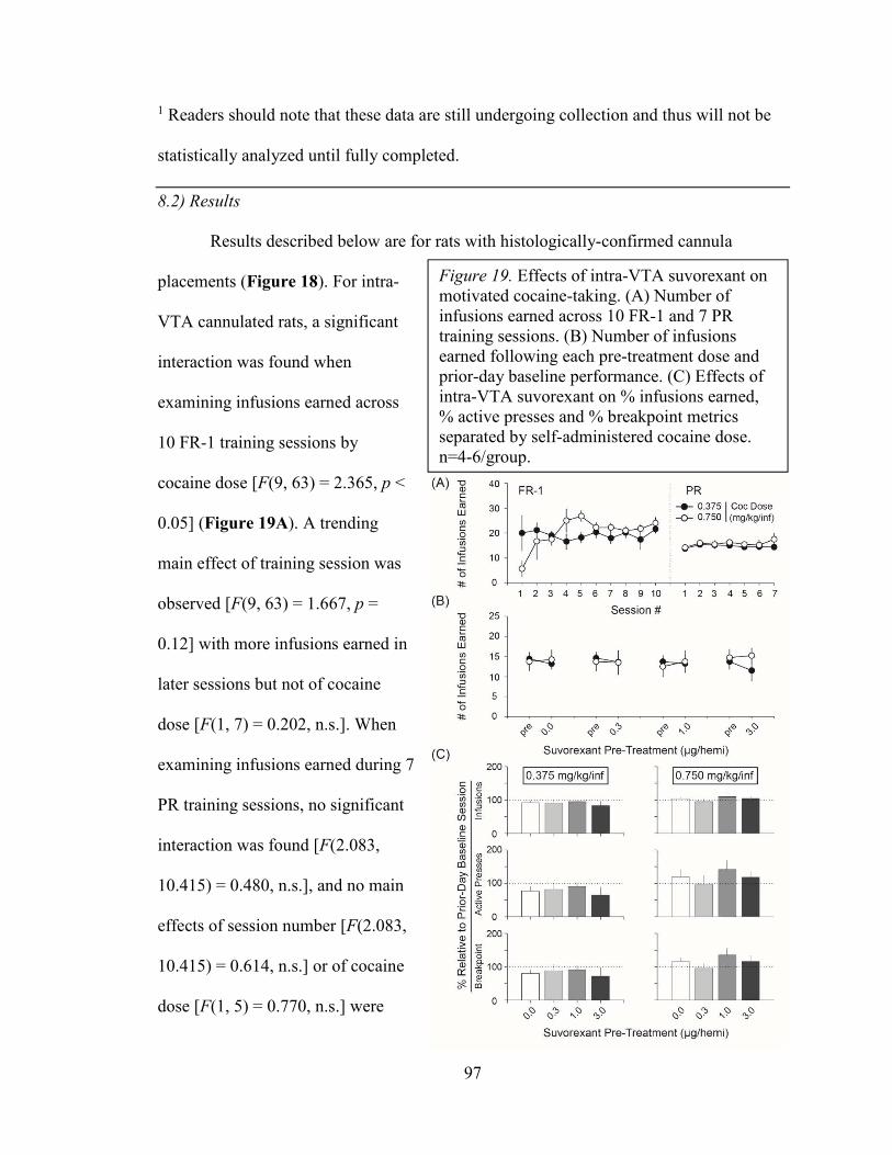
97
1 Readers should note that these data are still undergoing collection and thus will not be
statistically analyzed until fully completed.
8.2) Results
Results described below are for rats with histologically-confirmed cannula
placements (Figure 18). For intra-
VTA cannulated rats, a significant
interaction was found when
examining infusions earned across
10 FR-1 training sessions by
cocaine dose [F(9, 63) = 2.365, p <
0.05] (Figure 19A). A trending
main effect of training session was
observed [F(9, 63) = 1.667, p =
0.12] with more infusions earned in
later sessions but not of cocaine
dose [F(1, 7) = 0.202, n.s.]. When
examining infusions earned during 7
PR training sessions, no significant
interaction was found [F(2.083,
10.415) = 0.480, n.s.], and no main
effects of session number [F(2.083,
10.415) = 0.614, n.s.] or of cocaine
dose [F(1, 5) = 0.770, n.s.] were
Figure 19. Effects of intra-VTA suvorexant on motivated cocaine-taking. (A) Number of infusions earned across 10 FR-1 and 7 PR training sessions. (B) Number of infusions earned following each pre-treatment dose and prior-day baseline performance. (C) Effects of intra-VTA suvorexant on % infusions earned, % active presses and % breakpoint metrics separated by self-administered cocaine dose. n=4-6/group.

98
observed. When examining
infusions earned during 10 FR-1
training sessions of intra-RMTg
cannulated rats, no significant
interaction was found [F(9, 81) =
1.565, n.s.] (Figure 20A), but main
effects of training session [F(9, 81)
= 10.669, p < 0.001] and, of lesser
power, cocaine dose [F(1, 9) =
6.010, p < 0.05] were detected.
When examining infusions earned
during 7 PR sessions of intra-RMTg
cannulated rats, no significant
interaction [F(2.344, 21.098) =
2.088, n.s.] or main effects of
training session [F(2.344, 21.098) =
1.160, n.s.] or cocaine dose [F(1, 9)
= 0.224, n.s.] were observed. These
data show that rats across groups
can train to self-administer cocaine
under FR-1 and PR schedules of
reinforcement.
Figure 20. Effects of intra-RMTg hcrt/ox on motivated cocaine-taking. (A) Number of infusions earned across 10 FR-1 and 7 PR training sessions. (B) Number of infusions earned following each pre-treatment dose and prior-day baseline performance. (C) Effects of intra-RMTg hcrt/ox on % infusions earned, % active presses and % breakpoint metrics separated by self-administered cocaine dose. * p < 0.05 and # p = 0.06 relative to vehicle pre-treatment comparator scores (Bonferroni-corrected contrasts). n=4-6/group.

99
For experimental sessions, intra-VTA suvorexant in rats self-administering low-
dose cocaine (0.375 mg/kg/inf) had no effect on % infusions earned [F(3, 9) = 0.303,
n.s.], % active presses [F(3, 9) = 0.332, n.s.], % inactive presses [F(3, 9) = 0.321, n.s.], or
% breakpoint [F(3, 9) = 0.214, n.s.] (Figure 19B-C). Similarly, no effect of intra-VTA
suvorexant was found in high-dose cocaine (0.750 mg/kg/inf) self-administering rats
when examining % infusions earned [F(3, 9) = 0.895, n.s.], % active presses [F(3, 9) =
0.551, n.s.], % inactive presses [F(3, 9) = 0.264, n.s.], and % breakpoint [F(3, 9) = 0.804,
n.s.]. Raw behavioral metrics for intra-VTA cannulated rats subjected to suvorexant pre-
Table 5. Behavioral metrics per subject for intra-VTA cannulated rats receiving suvorexant pre-treatment prior to cocaine self-administration. Data in parenthesees indicates prior-day baseline performance from which % change metrics were computed throughout analyses.

100
treatment prior to cocaine self-administration are reported in Table 5. These results show
that intra-VTA suvorexant has negligible effects on motivated cocaine-taking.
For rats self-administering low-dose cocaine, the effect of intra-RMTg hcrt/ox on
% infusions earned trended towards significance [F(1.090, 4.359) = 3.256, p = 0.14]
(Figure 20B-C). Contrast analyses revealed that rats self-administered significantly
greater infusions when pre-treated with 0.3 nmol hcrt/ox relative to vehicle pre-treatment
[p < 0.05]. Similarly, a main effect of hcrt/ox pre-treatment was found for % active
presses [F(3, 12) = 4.653, p < 0.05] and % breakpoint [F(3, 12) = 4.373, p < 0.05] but not
for inactive presses [F(1.063, 3.189 = 0.802, n.s.]. Consistent with % infusion contrast
analyses,
0.3 nmol
Table 6. Behavioral metrics per subject for intra-RMTg cannulated rats receiving hcrt/ox pre-treatment prior to cocaine self-administration. Data in parenthesees indicates prior-day baseline performance from which % change metrics were computed throughout analyses.

101
significantly increased % active presses [p < 0.05] and % breakpoint [p < 0.05] relative to
vehicle pre-treatment comparator data. Finally, for intra-RMTg cannulated high-dose
cocaine self-administering rats, a significant main effect of hcrt/ox pre-treatment was
found for % infusions [F(3, 15) = 5.499, p < 0.01], % active presses [F(3, 15) = 3.708, p
< 0.05], and % breakpoint [F(3, 15) = 4.899, p < 0.05] but not for % inactive presses
[F(1.403, 7.015) = 0.719, n.s.]. Contrast analyses revealed that 1.0 and 3.0 nmol hcrt/ox
pre-treatment significantly suppressed % infusions [both p < 0.05], % active presses [1.0
nmol: p < 0.05; 3.0 nmol: p = 0.06], and % breakpoint [both p < 0.05]. A significant
reduction inactive presses was found for rats pre-treated with 3.0 nmol hcrt/ox [p < 0.05]
but not 1.0 nmol. Raw behavioral metrics for intra-RMTg cannulated rats subjected to
hcrt/ox pre-treatment prior to cocaine self-administration are reported in Table 6.
Interestingly, these data show that hcrt/ox peptide injection in RMTg significantly
influence motivated cocaine-taking, but direction of effect is based in part on self-
administered cocaine dose. Insufficient data have been generated for missed cannula
placements which generally tended to target dorsal to intended structure. For low-dose
cocaine self-administering rats receiving intra-VTA suvorexant with confirmed “missed”
cannula placement, mean % infusions ranged from 75.2 to 122.2, but only one to three
data points per pre-treatment condition have been collected so far. Similarly, intra-VTA
cannulated rats self-administering high-dose cocaine, means for % infusions earned
relative to prior-day baseline ranged from 81.3 to 104.0 with only two data points per
pre-treatment condition. One subject was a confirmed “miss” for intra-RMTg cannulated
rats self-administering low-dose cocaine, and, likewise, two subjects were confirmed
“misses” for intra-RMTg cannulated high-dose cocaine self-administering rats.

102
Readers
should note that
studies examining
intra-VTA suvorexant
and intra-RMTg
hcrt/ox on USV
emissions is still
being collected, and
thus sample sizes
(n=2-4/group) are
inadequate for
statistical analyses
and do not reflect all
histologically-
confirmed rats as described for above-mentioned infusion data. However, trending effects
which can be found on Figures 21 and 22 will herein be reported. In terms of
anticipatory USVs, intra-VTA suvorexant tended to enhance 50-kHz USVs at 0.3 and 1.0
µg/hemi doses whereas this effect destabilized with high-dose (3.0 µg/hemi) pre-
treatment in rats trained to self-administer low-dose (0.375 mg/kg/inf) cocaine. No
appreciable effects of either intra-VTA suvorexant or intra-RMTg hcrt/ox on 22-kHz
USVs across groups was observed. In the low-dose cocaine self-administering, intra-
RMTg hcrt/ox suppressed anticipatory 50-kHz USVs across pre-treatment doses,
however no effects were observed in rats trained to self-administer high-dose (0.750
Figure 21. Effects of intra-VTA suvorexant and intra-RMTg hcrt/ox on USVs associated with anticipation and self-administration of low-dose (0.375 mg/kg/inf) and high-dose (0.750 mg/kg/inf) cocaine. Data are mean ± S.E.M. Solid bars indicate 50-kHz USVs whereas hatched bars indicate 22-kHz USVs. n=2-4/group.

103
mg/kg/inf) cocaine.
Secondly, nearly all rats
(excepting intra-VTA
cannulated rats self-
administering low-dose
cocaine) emitted a
paucity of 50-kHz
USVs following time
“0” at which point self-
administered cocaine
was available. Bouts of
post-lever 22-kHz
USVs were present
across groups, notably
in intra-VTA
cannulated low-dose
cocaine self-administering rats. Collectively, these data suggest that hcrt/ox transmission
in RMTg influences motivated cocaine-taking, but effects are dependent on reward
magnitude (i.e., dose of self-adminsitered cocaine). It remains possible that histories of
cocaine self-administration influenced the extent to which RMTg cells are impacted by
hcrt/ox transmission.
Figure 22. Effects of pharmacological pre-treatments (intra-VTA suvorexant, intra-RMTg hcrt/ox) on profile of 22- and 50-kHz USVs across cocaine self-administration sessions.

104
8.3) Interim discussion
In target-site pharamacoloy experiments, we failed to observe appreciable effects
of intra-VTA hcrt/ox receptor blockade (via suvorexant) on motivated cocaine-taking.
Previous work finds that OX1R blockade with VTA-directed SB334867 significantly
reduces the number of cocaine infusions earned under progressive-ratio access conditions
(Muschamp et al. 2014). Systemic injection of almorexant or suvorexant dose-
dependently decreases motivated cocaine-taking (Prince et al. 2015; Gentile et al. 2018).
In situ hybridization data supports that both OX1R and OX2R exist in comparable
densities within VTA (Marcus et al. 2001), but we lack appreciation of which cell
populations hcrt/ox receptors are produced within. As intra-VTA suvorexant had
negligible effects on cocaine self-administration, we suspect that OX2Rs may be on
GABA-producing interneuron populations which may negate motivation-suppressing
effects normally observed following OX1R blockade. Interestingly, hcrt/ox peptide
injection within RMTg produced mixed effects on motivated cocaine-taking dependent
on self-administered cocaine dose (i.e., 0.375 vs 0.750 mg/kg/inf). This preliminary data
supports that hcrt/ox functionally transmits to RMTg, however we lack an understanding
of how hcrt/ox influences physiology of cell populations within RMTg. Interestingly,
prior work shows that putative GABA-producing cells within RMTg are insensitive to
KOR stimulation (Matsui and Williams 2011) whereas a DA-producing VTA cell sub-
population possesses robust sensitivity to KOR stimulation (determined via bath
application of dynorphin—the endogenous ligand of KORs) (Muschamp et al. 2014;
Baimel et al. 2017). Unique sensitivity of neurochemically-defined midbrain cells to
KOR and hcrt/ox receptor stimulation suggests selective influence of hcrt/ox versus
105
dynorphin—peptides that are vastly co-localized (Chou et al. 2001; Muschamp et al.
2014) and are suspected to co-release. The balance between hcrt/ox and dynorphin
transmission in midbrain warrants further attention, and preferential sensitivity of VTA
and RMTg to these peptides may provide a second layer through which hcrt/ox-
producing cells bivalently regulates motivated reward-seeking.

106
DISCUSSION
Psychiatric disorders characterized by aberrantly motivated states including those
seen with substance use disorders can significantly deteriorate lives of the afflicted as
well as whole families and communities. Medications indicated to manage substance use
disorders prove effective by suppressing drug-associated reward and/or by reducing the
intensity of withdrawal-related negative affective symptoms. For psychostimulants
including cocaine, amphetamine and novel synthetic psychostimulants, no FDA-approved
medications are available to manage their abuse. An underlying theme throughout work
in this thesis is to better understand the transmitters and circuits that support subjective
experiences of positive and negative affect—in the context of addiction, these “drives”
propagate binge-like drug consumption and facilitate the escalation to a state of
dependence. Seminal work focused on reward neurobiology promotes studying the
ventral midbrain—notably, the DA-producing afferent population residing in VTA—as a
key locus in reward processing.
As stated in Chapter 4, the ventral midbrain is richly heterogeneous and possesses
unique, adjacently-lying structures that seem to function in opposition. Groups have
demonstrated that the RMTg—resting caudally in midbrain, dorsolateral to IPN,
populated by GABA-producing cells—negatively regulates the DA-producing cells of
rostrally-positioned VTA. These two structures, then, may work to provide net output
that encodes a stimulus as rewarding or aversive. It was tantalizing to consider a “master
controller” that innervates these structures to regulate affective valence and drive
motivated behavioral states. We placed our attention on a hypothalamic neuropeptide—
hcrt/ox—which has a ~12-year history of being implicated pre-clinically in motivated

107
states associated with consumption and seeking of drugs. Interestingly, a single
nucleotide polymorphism in the hcrt-r1 gene is associated with greater risk of developing
either major depressive disorder or bipolar disorder, suggesting a possible of function of
hcrt/ox transmission in the regulation of negative affective states (Rainero et al. 2011).
Hcrt/ox regulates an array of behaviors concerning homeostasis and vigilance, and its
discovery positioned hcrt/ox as orexigenic and physiologically excitatory. Within a year,
teams showed critical involvement of hcrt/ox as a sleep regulator noting circadian-linked
fluctuations in systemic peptide content. Of note, the FDA approved the first-in-class
hcrt/ox receptor antagonist—suvorexant—for the management of primary insomnia in
late 2014.
Studying clinically-available pharmacotherapeutic agents affords rapid
translational power, and thus in preliminary assessment our team explored the efficacy of
suvorexant in normalizing motivated drug-taking. We additionally phenotyped affective
responses in rats trained to self-administer cocaine and the synthetic psychostimulant
MDPV. While not as widely studied, MDPV mechanistically inhibits clearance of DA via
blockade of DATs with approximately ten-fold greater potency compared to cocaine
(Baumann et al. 2013; Kolanos et al. 2013). MDPV is readily self-administered by
rodents and acutely facilitates brain reward function (Watterson et al. 2014). Our work
showed that MDPV, like cocaine, elicits predominantly positive affective 50-kHz USVs.
Further, we provided additional evidence that the “positive affective” 50-kHz USV
response observed early in sessions corresponds with the transition from sobriety to
intoxication, but that 50-kHz USVs diminish to near-zero rates thereafter (Barker et al.
2014; Simmons et al. 2018). In a follow-up study, we determined that systemic

108
suvorexant could attenuate USVs associated with anticipation and self-administration of
MDPV. Although, as mentioned below, no effects of suvorexant were observed on the
number of MDPV infusions earned (Simmons et al. 2017). Together, these lines of work
promoted further interrogation of possible circuits whereby hcrt/ox could regulate
affective states and, in doing so, pathological motivation.
Suvorexant decreases rewarding and reinforcing properties of psychostimulants
Consistent with prior studies demonstrating therapeutic value of hcrt/ox receptor
antagonists to normalize pathological motivation (e.g., Borgland et al. 2009; Smith et al.
2009), we observed that systemic suvorexant decreases infusions earned when rats are
taxed under high-effort access conditions for intravenous cocaine. From our study, it
appeared that a means through which hcrt/ox receptor blockade decreased motivated
cocaine-taking was through attenuating cocaine-associated positive affect commensurate
with suppression of mesolimbic DA transmission. Prior studies observed that hcrt/ox
innervates VTA and modulates cocaine-elicited DA efflux in NAcc targets (Fadel and
Deutch 2002; España et al. 2011; Bernstein et al. 2017). Additionally, the production of
50-kHz USVs is supported by mesolimbic DA transmission as DA receptor antagonists
suppress psychostimulant-elicited 50-kHz USVs, and electrical/optical excitation of DA
release in NAcc robustly, yet transiently, elicits 50-kHz USVs (Ciucci et al. 2009;
Williams and Undieh 2010; Scardochio and Clarke 2013; Scardochio et al. 2015). Thus,
the ability of suvorexant to reduce cocaine-elicited 50-kHz USVs aligns with its ability to
suppress cocaine-stimulated NAcc DA release (Gentile et al. 2018). However, consistent
with prior work, we failed to find significant effects of systemic hcrt/ox receptor
blockade on low-effort (FR-1) psychostimulant self-administration (Simmons et al.

109
2017). These results collectively support that hcrt/ox is selectively recruited for motivated
action directed on seeking/consuming positive reinforcers, and that hcrt/ox influence on
VTA DA transmission supports reward-seeking.
When measuring motivated behaviors, blockade of hcrt/ox receptors has been
shown to decrease reproductive behaviors in male rats as well as high-effort responding
for “natural” rewards typically associated with an obesogenic diet (i.e. high in fats and
sugar) and for drugs of abuse (Muschamp et al. 2007; Borgland et al. 2009). Indeed, we
observed that suvorexant (30 mg/kg, IP) pre-treatment significantly decreased the effort
expended for intravenous cocaine (Gentile et al. 2018). There is clear evidence that
suvorexant can induce somnolence as its indication is for managing primary insomnia
(Winrow et al. 2011). However, in our test, we observed that systemic cocaine injection
elicited comparable elevations in ambulation irrespective of pre-treatment (i.e.,
suvorexant vs. vehicle). Moreover, in a separate assessment, we observed significant
effects of suvorexant on cocaine-elicited elevations in premature responding in the 5-
CSRTT, but no apparent differences were found for “control” measures including trial
accuracy and latency to retrieve sugar reward (Gentile et al. 2017b). Finally, in a series of
unpublished observations, we observed that suvorexant does, indeed, promote sleep when
rats are placed in e.g., a place conditioning or homecage test setting. However, we
consistently observe an absence of somnolence when rats are placed in settings
demanding behavioral performance (i.e., operant responses) such as in 5-CSRTT or
during intravenous drug self-administration. We interpret from these observations that
sleep-promoting effects of suvorexant will manifest when subjects are returned to an
uninteresting environment where interactions (with conspecifics and environment) are

110
limited or altogether absent. Indeed, suvorexant in humans is suggested to be taken
before bed. It does not appear that suvorexant significantly impacted the ability for rats to
perform the self-administration task, and so we interpret suvorexant-mediated reductions
in cocaine-taking as an effect on the animal’s motivated state.
An evaluation of 50-kHz ultrasonic vocalizations as representating a “positively-
valenced” subjective state
The observed results that suvorexant suppresses cocaine-elicited 50-kHz USVs
(Gentile et al. 2018) and 50-kHz USVs occurring prior to and during MDPV self-
administration (Simmons et al. 2018) is captivating based on the interpretation that this
behavioral signature indexes a shift in positive affect. While drugs of abuse including
psychostimulants generally produce 50-kHz USVs above baseline rates (Ahrens et al.
2009; Ma et al. 2010), other work finds that 50-kHz USVs can be produced in general
states associated with behavioral activation/arousal. For example, in addition to
unpublished observations made by our team, Taylor and colleagues (2017) reported that a
strong yet transient bout of 50-kHz USVs are elicited following an electrical footshock
(~0.5 mA) which is generally agreed upon to be an aversive stimulus. After repeated
presentations, the pattern of USVs invariably shifts to production of “negative affective”
22-kHz USVs, and thus novelty associated with initial footshock experience is suspected
as the underlying factor driving 50-kHz USV emission. Here, it should be noted that the
authors further define the acoustic features of shock-elicited 50-kHz USVs as belonging
to a “constant frequency” category in contrast to “frequency-modulated”/“trill-like” 50-
kHz USVs more commonly observed following acute or repeated psychostimulant
injections (Ahrens et al. 2009; Wright et al. 2010). As rats throughout our studies were

111
familiar with their experimental test chambers, we do not believe that observed USV
emissions were influenced by stimulus novelty. We additionally found that systemic
suvorexant produced sporadic bouts of 22-kHz USVs which are generally tightly linked
to experimental conditions associated with affective distress, fear and/or alarm.
Moreover, we conducted a cursory analysis on the proportion of 50-kHz USV call types
(divided based on number of frequency modulations [i.e., ±3-kHz contiguous “sweep”,
“jump” or “step”]) during MDPV self-administration and observed, consistent with
earlier work in cocaine-injected rats, many calls possessed 1 or more frequency
modulation—these call type profiles were largely unaffected by suvorexant (Figure
12B).
Functional connectivity: hypocretin/orexin afferents to rostrocaudal extent of ventral
midbrain
To help further elucidate hcrt/ox-mediated circuits that participate in positive and
negative affective states, we interrogated anatomically distinct ventral midbrain structures
that are suspected to act in functional opposition. We sought to define for the first time
the topographical organization of hcrt/ox afferents to VTA and RMTg to evaluate
potential loci of hcrt/ox-mediated affective control. We observed comparable innervation
densities across the two adjacent structures and explored functional differences of hcrt/ox
transmission to each of VTA and RMTg. Specifically, we tested the hypothesis that
hcrt/ox transmission in VTA positively mediates motivated cocaine-taking and
participates in the encoding of rewarding experiences whereas hcrt/ox in RMTg brakes
this transmission sequence to bias stimulus processing as aversively-valenced.

112
Geisler and Zahm (2005) describe the VTA as “a neuronal continuum with
overlapping dendritic fields” as is evident from inputs arising from many anatomically
diffuse sources (see also description of “isodendritic core” as articulated by Ramón-
Moliner and Nauta 1966). Since 2009, several teams have recognized the caudal division
of VTA as a unique structure based on cellular architecture and neurochemical
composition and thus termed it based on anatomical location—the RMTg residing
caudally/dorsolaterally to DA-producing midline VTA cells and caudally/dorsomedially
to DA-producing SN cells. In fact, Lahti and colleagues (2016) ascribed the nuclear
transcription factor FoxP1 as a relatively selective marker for RMTg which was later
found to overlap selectively to psychostimulant-evoked ΔFosB. Inputs to VTA and
RMTg of the ventral midbrain are of significance for their roles in encoding stimuli as
rewarding or aversive. A comprehensive tracing study from hcrt/ox afferents has not been
conducted beforehand and thus was the aim of current investigation.
In 2009, Kaufling and colleagues (2009) published a report detailing inputs to
RMTg using two retrograde tracer types (FluroGold [FG] and cholera toxin) deposited
iontophoretically. Semiquantitative analyses revealed relatively strong input to RMTg
from medial prefrontal cortex (mPFC), zona incerta, LHb, LPO, hypothalamus, inferior
division of superior colliculus, dorsal raphe nucleus (DRN), PAG, parabrachial nucleus,
and laterodorsal tegmental nucleus. As described in Section 4.2, cocaine injection was
used to elicit ΔFosB within RMTg, and immunohistochemical confirmation of
ΔFosB/GAD co-labeling was made whereas no cells were ΔFosB+ were co-labeled TH.
Anterograde tracing using biotinylated dextran amines supported that fibers extending
from mPFC, LHb, LPO and hypothalamus appose cocaine-elicited ΔFosB+ nuclei within

113
RMTg whereas no such appositions were found along the amygdala�RMTg pathway.
Figures 15 and 16 of this report clarify that virtually none of the FG+ cells traced from
DRN contained the immunolabel against tryptophan hydroxylase—the rate-limiting
enzyme used in serotonin biosynthesis—which otherwise predominates within DRN.
Likewise, a small population (1.5%) of hcrt/ox-immunoreactive cells within
hypothalamus co-labeled with FG. This afferent proportion is lower than estimates
derived from our retrograde tracing (6.7%) which may be due to differences in tracer type
versus extent and rigor of tissue studied. The “retrobeads” used in the present study are
latex microspheres conjugated to a fluorophore which are passively endocytosed by
nearby terminals (e.g., Katz et al. 1984) similarly to FG whereas other work finds that the
beta-subunit of cholera toxin may be actively transported and capable of trans-synaptic
movement following lysosome-like vacuole packaging (e.g., van der Want et al. 1997).
Additionally, our study was directed around comparing innervation between hcrt/ox
afferents to VTA and to RMTg whereas no such comparison work was performed by
Kaufling and colleagues (2009). It should also be mentioned that continuous refinement
on ventral midbrain structural heterogeneity continues to take place. For example, Petzel
and colleauges (2017) provide elegant neuroanatomical tracing work revealing unique
input to anterior and posterior divisions of VTA as well as to RMTg (furtherest posterior)
from LHb. Specifically, only the lateral division of LHb tends to innervate RMTg
whereas mixed inputs across LHb target the posterior division of VTA. Our study
assumed a relatively homogenous stretch of nuclei residing in the caudal division of
ventral midbrain—future work utilizing anterograde tracing from the hcrt/ox population
across ventral midbrain will further elucidate relative innervation densities.

114
While no raw numbers are reported, Richardson and Aston-Jones (2012) show
that VTA-projecting hcrt/ox-immunoreactive cells are activated (interpreted by Fos
immunoreactivity) upon exposure to a morphine-paired context during a place
conditioning test session. Moreover, hcrt/ox cells arising from the lateral hypothalamic
division were most strongly activated following place conditioning testing, and a % Fos+
metric significantly correlated with the behavioral measure of preference score for the
morphine-paired context. However, no report to-date has utilized a similar projection-
specific Fos labeling approach in the context of psychostimulant addiction. Nonetheless,
we suspected that putative hcrt/ox afferents to ventral midbrain (including to VTA) are
functionally recruited for reward processing psychostimulant drugs of abuse. From our
tract-tracing data, we additionally suspected that innervation to the caudal division of
ventral midbrain (RMTg) may promote a subjective state of opposing affective valence.
Functionally, we tested how hcrt/ox transmission perturbation within VTA and
RMTg influences the rate of 22- and 50-kHz USV emission as an analog measure of
positive and negative affect. We additionally tested the extent to which ventral midbrain
hcrt/ox transmission perturbation impacts motivated cocaine-taking in rats trained to self-
administer a low-dose (0.375 mg/kg/inf) or a relatively higher-dose (0.750 mg/kg/inf) of
cocaine as prior work showed that OX1R blockade selectively impacts self-
administration behavior at relatively higher doses only (España et al. 2010). USV data
from intra-VTA suvorexant and intra-RMTg hcrt/ox produced mixed, largely
inconclusive results from data collected thus far. It should be noted that, to our
knowledge, this study is the first to measure USVs during high-effort drug self-
administration (i.e. progressive-ratio) test. Results from our earlier work (Simmons et al.

115
2017, 2018) show a robust presence of anticipatory 50-kHz USVs which diminishes to
near-zero levels over the course of ~30 mins. These studies additionally captured a strong
bout of 50-kHz USVs upon and following lever extension during which intravenous drug
injections became available. The majority of rats evaluated in the current study, except
intra-VTA cannulated rats self-administering low-dose (0.375 mg/kg/inf) cocaine, failed
to emit appreciable bouts of 50-kHz USVs after lever extension. Interestingly, too, a
comparable “low-dose” of self-administered cocaine (0.355 mg/kg/inf) was shown to
lead to predominantly 22-kHz USVs under low-effort, FR-1 access conditions (Barker et
al. 2010). At present, we attribute the lack of a “positive affective” response observed in
present work to the employment of a high-effort, less predictable reward contingency
task.
Technical considerations: retrograde tracing and recording of ultrasonic vocalizations
Newer technologies allowing monosynaptic retrograde tracing from
neurochemically-defined cell populations (e.g., those that produce distinct
neurotransmitters) enable improved understanding of how the VTA receives and conveys
information. To date, we have relied in large part on conventional tracing methods that
possess relatively promiscuous retrograde transport activity. Direct inputs to cells that
produce DA, GABA and glutamate across ventral midbrain will address whether unique
or overlapping cell populations (e.g., from hcrt/ox afferents) target the ventral midbrain.
Ongoing efforts from our laboratory are attempting to deposit cholera toxin retrograde
tracers conjugated to unique fluorophores to each of VTA and RMTg from single
subjects.

116
It should be noted, here, that a majority of detected 22-kHz USVs from the
experiment described in Chapter 8 were spectrographically “quiet” and suffered from
poor signal:noise quality (1.05-1.10), and thus their presence should be interpreted
cautiously. One possibility is that “quiet” 22-kHz USVs are organic signals from nearby
animals and not the ones within the chamber being recorded from—all of our recordings
were contained within wooden, sound-attenuating chambers but microphone sensitivities
remain an untested contributing factor. Indeed, all detections carried a mean, monotonic
frequency ~22-kHz and did not appear influenced from clearly-discernible artifact noises
(e.g., spring leash tether contacting Plexiglas) which promoted their inclusion. Coupling
the facts that 50-kHz USVs recorded from the same microphones possessed excellent
signal:noise (1.20-1.30) and that the same microphones were able to capture shock-
elicited USVs in a separate study without detection difficulty, we do not suspect the
microphones suffer from an inability to detect 22-kHz USVs. Instead, it remains possible
that 22-kHz USVs elicited under experimental conditions described in Chapter 8 are, in
fact, quiet, monotonic and of varied lengths unlike acoustic characteristics of 22-kHz
USVs described in earlier studies (e.g., Barker et al. 2010). Microphones with improved
amplifier units, such as the CM16/CMPA model manufactured by Avisoft Bioacoustics,
can likely detect these possible 22-kHz USVs with greater accuracy but are prohibitive
for purchase by young laboratories carrying a cost ~13.7x greater than USB plug-and-
play microphones (Dodotronic) used by our group. Recordings using improved
microphones should be conducted to confirm the existence of 22-kHz USVs described in
Chapter 8 as the functional implications from their emission are of great value.

117
Distribution of OX1Rs and OX2Rs across neurocehmically-defined cell populations of
ventral midbrain
An additional finding from intra-VTA suvorexant deposit was its tendency to
enhance rates of anticipatory 50-kHz USVs—an effect that destabilizes with high-dose
(3.0 µg/hemisphere) suvorexant pre-treatment—in low-dose cocaine self-administering
rats suggesting hcrt/ox receptor blockade may augment positive anticipation for self-
administered cocaine. Intra-VTA hcrt/ox receptor blockade studies thus far have utilized
selective OX1R antagonists and have invariably demonstrated that OX1Rs positively
mediate drug-taking and -seeking. Systemic blockade of OX1Rs and OX2Rs suppresses
motivated responding in cocaine self-administering rats, and it appears too that dual
hcrt/ox receptor blockade decreases positive affective to systemically-injected cocaine
(Gentile et al. 2018). It remains possible that, while OX1Rs may predominantly localize
on VTA DA cells, OX2Rs may more densely populate on VTA GABA interneurons that
function to inhibit the VTA DA population. Pharmacological agents, including
suvorexant, that antagonize both hcrt/ox receptor subtypes may elicit opposing effects on
VTA DA cellular physiology and transmission to targets including NAcc. Under the
simplistic view that net DA transmission in this manner effectively encodes reward
signals and its suppression participates in encoding aversive stimuli, a mixed affective
response from intra-VTA suvorexant could be generated. Studies utilizing selective
blockade of hcrt/ox receptor subtypes on neurochemically-defined VTA populations
could test the hypothesis blockade of VTA OX2Rs disinhibits VTA DA to facilitate
reward encoding.

118
Curiously, we observed that low-dose hcrt/ox (0.3 nmol/hemi) enhanced
responding for low-dose self-administered cocaine whereas higher doses of intra-RMTg
injected hcrt/ox (1.0 and 3.0 nmol/hemi) significantly decreased motivated cocaine-
taking in high-dose cocaine self-administering rats. Reward-attenuating effects of intra-
RMTg hcrt/ox (i.e. reducing motivated cocaine-taking) align with the idea that hcrt/ox
functionally brakes cocaine-associated reward and reinforcement likely through
augmenting activity of GABA-producing RMTg neurons that project and inhibit DA-
producing neurons of VTA. In preliminary assessment, we failed to see any clear effect
of intra-RMTg hcrt/ox on USVs during cocaine self-administration, so it is unclear as to
whether RMTg hcrt/ox participates in the affective/emotional node underlying
pathological motivation. It would be of great value to delineate the ventral midbrain
cellular populations through which hcrt/ox transmits to—this knowledge is lacking at the
moment.
In situ hybridization studies confirm that both OX1Rs and OX2Rs can be found
within ventral midbrain, but to-date we do not know which neurochemically-defined cell
populations hcrt/ox receptors are produced within. To help address this, we piloted a
combined in situ hybridization (RNAscope) with immunohistochemistry study to
visualize TH+, VGaT+, and VGluT2+ cells within ventral midbrain labeled along with
probes targeting either hcrt-r1 or hcrt-r2 mRNA. However, poor signal:noise and non-
specific labeling on negative-control tissue sections ended these experiments pre-
maturely. It remains possible that OX2Rs are densely populated on GABA-producing
interneurons within VTA whereas OX1Rs cluster on DA-producing cellular populations.
If this is the case, blockade of both hcrt/ox receptor subtypes would produce a mixed

119
physiological effect within the structure which may explain a lack of intra-VTA
suvorexant. While unsubstantiated at the moment, mixed effects of intra-RMTg hcrt/ox
pre-treatment on cocaine-taking between low- and high-doses of self-administered
cocaine suggest that cocaine may influence the RMTg cellular population in a dose-
dependent manner—specifically, our results support that low doses of self-administered
cocaine bias the RMTg population to favor reward-seeking upon transmission of hcrt/ox,
whereas a bias towards promoting aversion would be produced following high doses of
self-administered cocaine.
Beyond ventral midbrain: alternative targets of hypocretin/orexin in the mediation of
psychostimulant-associated reward and reinforcement
While we have placed our attention solely on hcrt/ox innervation within midbrain,
effects of transmission to forebrain structures including to NAcc should not be
discounted. Recent study finds that hcrt/ox signals to D2-containing NAcc cells to
functionally contribute to innate risk avoidance (Blomeley et al. 2018) despite anatomical
work showing relatively scant hcrt/ox fiber density within striatum (e.g., Peyron et al.
1998). In this report, chemogenetic silencing of D2-containing NAcc cells as well as
systemic OX1R blockade increased time spent in the center of an open field arena and
decreased reward-seeking latency across a chamber paired with predatory odor. Notably,
co-dependency of the hcrt/ox�D2(NAcc) circuit was interpreted from hcrt/ox-driven risk
avoidance requiring an intact D2-containing NAcc cell population. Monosynaptic tracing
using truncated G-segment Rabies virus confirmed ~25% of hcrt/ox cells directly target
D2-containing NAcc cells whereas a relatively scarce proportion (~5%) of hypothalalmic
outputs contained MCH. Physiologically, photostimulation of hcrt/ox terminals within

120
NAcc activated D2-containing NAcc cells ex vivo which was abolished with bath
application of hcrt/ox receptor blocking agents. This study reveals a pivotal role of
hcrt/ox in risk avoidance by signaling directly to ventral striatal targets. Furthermore,
neuropeptide proteomic analysis indicates that a functional role of hcrt/ox transmission to
LHb is likely in large part via OX-B which has relatively greater affinity for OX2Rs
(Yang et al. 2018). While we speculate that the function of hcrt/ox in reward processing,
including for abused drugs, relays in large part through midbrain structures, possible
effects of direct hcrt/ox transmission to ventral striatum on reward-related behaviors
should be considered. However, a technical limitation of the above-mentioned research is
the use of a “selective” hcrt/ox cre mouse line—hcrt/ox mRNA is transiently expressed
across many cells during development which leads in turn to translation of cre in cells
that ultimately fail to produce hcrt/ox. Genetic tools for selective targeting of hcrt/ox cells
at present carry considerable limitations—namely, poor and/or non-selective transfection.
Many nuclei within limbic and forebrain structures receive functionally-relevant
transmission from hcrt/ox. In this thesis, we explored the idea that hcrt/ox may innervate
VTA and RMTg to functionally participate in the production of opposing affective states
upon stimulus presentation/receipt in the context of psychostimulant addiction. We
provided evidence that, indeed, comparable innervation densities of hcrt/ox to each of
VTA and RMTg can be observed, although functions remain uncertain. It appears that
blockade of both OX1Rs and OX2Rs in the VTA produces no discernible effect on
cocaine-taking, although some interesting dose-related effects on USVs are emerging.
Intra-RMTg hcrt/ox transmission appears to be recruited in a relatively more significant
manner to influence motivated cocaine-taking, although alterations in affective state at

121
the moment do not appear as underlying contributors. Explicit examination of hcrt/ox
receptor subtype expression within neurochemically-defined cellular populations of
ventral midbrain will add important clarity to the above-mentioned results. Further, the
effects of drug-taking history on hcrt/ox receptor distribution on midbrain cellular
populations should be assessed. Armed with this knowledge, more effective and
individualized targeting strategies can be developed which could collectively promote
pharmacotherapeutic intervention to better manage psychostimulant abuse.

122
BIBLIOGRAPHY
Acuna-Goycolea, C., & van den Pol, A. (2004). Glucagon-like peptide 1 excites hypocretin/orexin neurons by direct and indirect mechanisms: implications for viscera-mediated arousal. Journal of Neuroscience, 24(37), 8141-8152.
Adamantidis, A. R., Zhang, F., Aravanis, A. M., Deisseroth, K., & De Lecea, L. (2007). Neural substrates of awakening probed with optogenetic control of hypocretin neurons. Nature, 450(7168), 420.
Ahmed, S. H., & Koob, G. F. (1998). Transition from moderate to excessive drug intake: change in hedonic set point. Science, 282(5387), 298-300.
Ahrens, A. M., Ma, S. T., Maier, E. Y., Duvauchelle, C. L., & Schallert, T. (2009). Repeated intravenous amphetamine exposure: rapid and persistent sensitization of 50-kHz ultrasonic trill calls in rats. Behavioural brain research, 197(1), 205-209.
Ahrens, A. M., Nobile, C. W., Page, L. E., Maier, E. Y., Duvauchelle, C. L., & Schallert, T. (2013). Individual differences in the conditioned and unconditioned rat 50-kHz ultrasonic vocalizations elicited by repeated amphetamine exposure. Psychopharmacology, 229(4), 687-700.
Allin, J. T., & Banks, E. M. (1972). Functional aspects of ultrasound production by infant albino rats (Rattus norvegicus). Animal Behaviour, 20(1), 175-185.
American Association of Poison Control Centers (2012). AAPCC Issues Statement on Synthetic Drug Abuse Prevention Act. Available at: http://www.aapcc.org/press/2.
American Psychiatric Assoication (2013). Diagnostic and statistical manual of mental
disorders (5th ed.). Arlington, VA: American Psychiatric Publishing.
Amiya, N., Amano, M., Oka, Y., Iigo, M., Takahashi, A., & Yamamori, K. (2007). Immunohistochemical localization of orexin/hypocretin-like immunoreactive peptides and melanin-concentrating hormone in the brain and pituitary of medaka. Neuroscience
letters, 427(1), 16-21.
Asakawa, A., Inui, A., Inui, T., Katsuura, G., Fujino, M. A., & Kasuga, M. (2002). Orexin reverses cholecystokinin‐induced reduction in feeding. Diabetes, Obesity and
Metabolism, 4(6), 399-401.
Avvisati, R., Contu, L., Stendardo, E., Michetti, C., Montanari, C., Scattoni, M. L., & Badiani, A. (2016). Ultrasonic vocalization in rats self-administering heroin and cocaine in different settings: evidence of substance-specific interactions between drug and setting. Psychopharmacology, 233(8), 1501-1511.
Baimel, C., Lau, B. K., Qiao, M., & Borgland, S. L. (2017). Projection-target-defined effects of orexin and dynorphin on VTA dopamine neurons. Cell reports, 18(6), 1346-1355.

123
Baird, J. R. C., & Lewis, J. J. (1964). The effects of cocaine, amphetamine and some amphetamine-like compounds on the in vivo levels of noradrenaline and dopamine in the rat brain. Biochemical pharmacology, 13(11), 1475-1482.
Balcita‐Pedicino, J. J., & Sesack, S. R. (2007). Orexin axons in the rat ventral tegmental area synapse infrequently onto dopamine and γ‐aminobutyric acid neurons. Journal of
Comparative Neurology, 503(5), 668-684.
Balcita‐Pedicino, J. J., Omelchenko, N., Bell, R., & Sesack, S. R. (2011). The inhibitory influence of the lateral habenula on midbrain dopamine cells: ultrastructural evidence for indirect mediation via the rostromedial mesopontine tegmental nucleus. Journal of
Comparative Neurology, 519(6), 1143-1164.
Baldo, B. A., Daniel, R. A., Berridge, C. W., & Kelley, A. E. (2003). Overlapping distributions of orexin/hypocretin‐and dopamine‐β‐hydroxylase immunoreactive fibers in rat brain regions mediating arousal, motivation, and stress. Journal of
Comparative Neurology, 464(2), 220-237.
Barfield, R. J., & Geyer, L. A. (1972). Sexual behavior: Ultrasonic postejaculatory song of the male rat. Science, 176(4041), 1349-1350.
Barker, D. J., Miranda-Barrientos, J., Zhang, S., Root, D. H., Wang, H. L., Liu, B., ... & Morales, M. (2017). Lateral preoptic control of the lateral habenula through convergent glutamate and GABA transmission. Cell reports, 21(7), 1757-1769.
Barker, D. J., Root, D. H., Ma, S., Jha, S., Megehee, L., Pawlak, A. P., & West, M. O. (2010). Dose-dependent differences in short ultrasonic vocalizations emitted by rats during cocaine self-administration. Psychopharmacology, 211(4), 435-442.
Barker, D. J., Simmons, S. J., & West, M. O. (2015). Ultrasonic vocalizations as a measure of affect in preclinical models of drug abuse: a review of current findings. Current neuropharmacology, 13(2), 193-210.
Barker, D. J., Simmons, S. J., Servilio, L. C., Bercovicz, D., Ma, S., Root, D. H., ... & West, M. O. (2014). Ultrasonic vocalizations: evidence for an affective opponent process during cocaine self-administration. Psychopharmacology, 231(5), 909-918.
Barreiro, M. L., Pineda, R., Navarro, V. M., Lopez, M., Suominen, J. S., Pinilla, L., ... & Tena-Sempere, M. (2004). Orexin 1 receptor messenger ribonucleic acid expression and stimulation of testosterone secretion by orexin-A in rat testis. Endocrinology, 145(5), 2297-2306.
Barros, H. M. T., Miczek, K. A. (1996). Withdrawal from oral cocaine in rats: ultrasonic vocalizations and tactile startle. Psychopharmacology, 125(4), 379-384.
Barrot, M., Sesack, S. R., Georges, F., Pistis, M., Hong, S., & Jhou, T. C. (2012). Braking dopamine systems: a new GABA master structure for mesolimbic and nigrostriatal functions. Journal of Neuroscience, 32(41), 14094-14101.

124
Baumann, M. H., Partilla, J. S., Lehner, K. R., Thorndike, E. B., Hoffman, A. F., Holy, M., ... & Brandt, S. D. (2013). Powerful cocaine-like actions of 3, 4-methylenedioxypyrovalerone (MDPV), a principal constituent of psychoactive ‘bath salts’ products. Neuropsychopharmacology, 38(4), 552.
Beaulieu, J. M., Espinoza, S., & Gainetdinov, R. R. (2015). Dopamine receptors–IUPHAR review 13. British journal of pharmacology, 172(1), 1-23.
Beier, K. T., Steinberg, E. E., DeLoach, K. E., Xie, S., Miyamichi, K., Schwarz, L., ... & Luo, L. (2015). Circuit architecture of VTA dopamine neurons revealed by systematic input-output mapping. Cell, 162(3), 622-634.
Belhadj-Tahar, H., & Sadeg, N. (2005). Methcathinone: a new postindustrial drug. Forensic science international, 153(1), 99-101.
Bellocchio, E. E., Hu, H., Pohorille, A., Chan, J., Pickel, V. M., & Edwards, R. H. (1998). The localization of the brain-specific inorganic phosphate transporter suggests a specific presynaptic role in glutamatergic transmission. Journal of Neuroscience, 18(21), 8648-8659.
Berg, D. S., & Baenninger, R. (1973). Hissing by laboratory rats during fighting encounters. Behavioral biology, 8(6), 733-741.
Berger, A. L., Williams, A. M., McGinnis, M. M., & Walker, B. M. (2013). Affective cue-induced escalation of alcohol self-administration and increased 22-kHz ultrasonic vocalizations during alcohol withdrawal: role of kappa-opioid receptors. Neuropsychopharmacology, 38(4), 647.
Bernardis, L. L., & Bellinger, L. L. (1996). The lateral hypothalamic area revisited: ingestive behavior. Neuroscience & Biobehavioral Reviews, 20(2), 189-287.
Blanchard, R. J., Blanchard, D. C., Agullana, R., & Weiss, S. M. (1991). Twenty-two kHz alarm cries to presentation of a predator, by laboratory rats living in visible burrow systems. Physiology & behavior, 50(5), 967-972.
Blomeley, C., Garau, C., & Burdakov, D. (2018). Accumbal D2 cells orchestrate innate risk-avoidance according to orexin signals. Nature neuroscience, 21(1), 29.
Borek, H. A., & Holstege, C. P. (2012). Hyperthermia and multiorgan failure after abuse of “bath salts” containing 3, 4-methylenedioxypyrovalerone. Annals of emergency
medicine, 60(1), 103-105.
Borgland, S. L., Chang, S. J., Bowers, M. S., Thompson, J. L., Vittoz, N., Floresco, S. B., ... & Bonci, A. (2009). Orexin A/hypocretin-1 selectively promotes motivation for positive reinforcers. Journal of Neuroscience, 29(36), 11215-11225.

125
Borgland, S. L., Taha, S. A., Sarti, F., Fields, H. L., & Bonci, A. (2006). Orexin A in the VTA is critical for the induction of synaptic plasticity and behavioral sensitization to cocaine. Neuron, 49(4), 589-601.
Bourdy, R., Sánchez-Catalán, M. J., Kaufling, J., Balcita-Pedicino, J. J., Freund-Mercier, M. J., Veinante, P., ... & Barrot, M. (2014). Control of the nigrostriatal dopamine neuron activity and motor function by the tail of the ventral tegmental area. Neuropsychopharmacology, 39(12), 2788.
Bourgin, P., Huitrón-Reséndiz, S., Spier, A. D., Fabre, V., Morte, B., Criado, J. R., ... & De Lecea, L. (2000). Hypocretin-1 modulates rapid eye movement sleep through activation of locus coeruleus neurons. Journal of neuroscience, 20(20), 7760-7765.
Boutrel, B., Kenny, P. J., Specio, S. E., Martin-Fardon, R., Markou, A., Koob, G. F., & de Lecea, L. (2005). Role for hypocretin in mediating stress-induced reinstatement of cocaine-seeking behavior. Proceedings of the National Academy of Sciences of the
United States of America, 102(52), 19168-19173.
Brandäo, M. L. (1993). Involvement of opioid mechanisms in the dorsal periaqueductal gray in drug abuse. Reviews in the Neurosciences, 4(4), 397-406.
Briggs, C., Hirasawa, M., & Semba, K. (2018). Sleep Deprivation Distinctly Alters Glutamate Transporter 1 Apposition and Excitatory Transmission to Orexin and MCH Neurons. Journal of Neuroscience, 38(10), 2505-2518.
Brinschwitz, K., Dittgen, A., Madai, V. I., Lommel, R., Geisler, S., & Veh, R. W. (2010). Glutamatergic axons from the lateral habenula mainly terminate on GABAergic neurons of the ventral midbrain. Neuroscience, 168(2), 463-476.
Brodnik, Z. D., Bernstein, D. L., Prince, C. D., & España, R. A. (2015). Hypocretin receptor 1 blockade preferentially reduces high effort responding for cocaine without promoting sleep. Behavioural brain research, 291, 377-384.
Browning, J. R., Browning, D. A., Maxwell, A. O., Dong, Y., Jansen, H. T., Panksepp, J., & Sorg, B. A. (2011). Positive affective vocalizations during cocaine and sucrose self-administration: a model for spontaneous drug desire in rats. Neuropharmacology, 61(1-2), 268-275.
Browning, J. R., Whiteman, A. C., Leung, L. Y., Lu, X. C. M., & Shear, D. A. (2017). Air-puff induced vocalizations: A novel approach to detecting negative affective state following concussion in rats. Journal of neuroscience methods, 275, 45-49.
Brudzynski, S. M. (2014). The Ascending Mesolimbic Cholinergic System—A Specific Division of the Reticular Activating System Involved in the Initiation of Negative Emotional States. Journal of Molecular Neuroscience, 53(3), 436-445.

126
Brudzynski, S. M., Bihari, F., Ociepa, D., & Fu, X. W. (1993). Analysis of 22 kHz ultrasonic vocalization in laboratory rats: long and short calls. Physiology &
behavior, 54(2), 215-221.
Brudzynski, S. M., Gibson, B., Silkstone, M., Burgdorf, J., Kroes, R. A., Moskal, J. R., & Panksepp, J. (2011). Motor and locomotor responses to systemic amphetamine in three lines of selectively bred Long-Evans rats. Pharmacology Biochemistry and
Behavior, 100(1), 119-124.
Brudzynski, S. M., Ociepa, D., & Bihari, F. (1991). Comparison between cholinergically and naturally induced ultrasonic vocalization in the rat. Journal of Psychiatry and
Neuroscience, 16(4), 221.
Buck, C. L., Malavar, J. C., George, O., Koob, G. F., & Vendruscolo, L. F. (2014). Anticipatory 50 kHz ultrasonic vocalizations are associated with escalated alcohol intake in dependent rats. Behavioural brain research, 271, 171-176.
Bunney, B. S., & Achajanian, G. K. (1976). d-Amphetamine-induced inhibition of central dopaminergic neurons: mediation by a striato-nigral feedback pathway. Science, 192(4237), 391-393.
Bunney, B. S., Walters S, J. R., Roth, R. H., & Aghajanian, G. K. (1973). Dopaminergic neurons: effect of antipsychotic drugs and amphetamine on single cell activity. Journal of
Pharmacology and Experimental Therapeutics, 185(3), 560-571.
Burdakov, D., Liss, B., & Ashcroft, F. M. (2003). Orexin excites GABAergic neurons of the arcuate nucleus by activating the sodium—calcium exchanger. Journal of
Neuroscience, 23(12), 4951-4957.
Burgdorf, J., Knutson, B., Panksepp, J., & Ikemoto, S. (2001). Nucleus accumbens amphetamine microinjections unconditionally elicit 50-kHz ultrasonic vocalizations in rats. Behavioral neuroscience, 115(4), 940.
Burgdorf, J., Kroes, R. A., Moskal, J. R., Pfaus, J. G., Brudzynski, S. M., & Panksepp, J. (2008). Ultrasonic vocalizations of rats (Rattus norvegicus) during mating, play, and aggression: Behavioral concomitants, relationship to reward, and self-administration of playback.. Journal of comparative psychology, 122(4), 357.
Burgdorf, J., Panksepp, J., Brudzynski, S. M., Beinfeld, M. C., Cromwell, H. C., Kroes, R. A., & Moskal, J. R. (2009). The effects of selective breeding for differential rates of 50‐kHz ultrasonic vocalizations on emotional behavior in rats. Developmental
psychobiology, 51(1), 34-46.
Cameron, D. L., & Williams, J. T. (1994). Cocaine inhibits GABA release in the VTA through endogenous 5-HT. Journal of Neuroscience, 14(11), 6763-6767.

127
Cameron, D. L., Wessendorf, M. W., & Williams, J. T. (1997). A subset of ventral tegmental area neurons is inhibited by dopamine, 5-hydroxytryptamine and opioids. Neuroscience, 77(1), 155-166.
Campbell, E. J., Barker, D. J., Nasser, H. M., Kaganovsky, K., Dayas, C. V., & Marchant, N. J. (2017). Cue-induced food seeking after punishment is associated with increased Fos expression in the lateral hypothalamus and basolateral and medial amygdala. Behavioral neuroscience, 131(2), 155.
Carter, M. E., Adamantidis, A., Ohtsu, H., Deisseroth, K., & de Lecea, L. (2009). Sleep homeostasis modulates hypocretin-mediated sleep-to-wake transitions. Journal of
Neuroscience, 29(35), 10939-10949.
Cason, A. M., & Aston-Jones, G. (2014). Role of orexin/hypocretin in conditioned sucrose-seeking in female rats. Neuropharmacology, 86, 97-102.
Center for Disease Control and Prevention (2017). CDC Wonder. U.S. Department of Health and Human Services. Accessed 10 February 2018.
Chartoff, E., Sawyer, A., Rachlin, A., Potter, D., Pliakas, A., & Carlezon, W. A. (2012). Blockade of kappa opioid receptors attenuates the development of depressive-like behaviors induced by cocaine withdrawal in rats. Neuropharmacology, 62(1), 167-176.
Cheer, J. F., Kendall, D. A., Mason, R., & Marsden, C. A. (2003). Differential cannabinoid-induced electrophysiological effects in rat ventral tegmentum. Neuropharmacology, 44(5), 633-641.
Chemelli, R. M., Willie, J. T., Sinton, C. M., Elmquist, J. K., Scammell, T., Lee, C., ... & Fitch, T. E. (1999). Narcolepsy in orexin knockout mice: molecular genetics of sleep regulation. Cell, 98(4), 437-451.
Choi, D. L., Davis, J. F., Fitzgerald, M. E., & Benoit, S. C. (2010). The role of orexin-A in food motivation, reward-based feeding behavior and food-induced neuronal activation in rats. Neuroscience, 167(1), 11-20.
Choi, D. L., Davis, J. F., Magrisso, I. J., Fitzgerald, M. E., Lipton, J. W., & Benoit, S. C. (2012). Orexin signaling in the paraventricular thalamic nucleus modulates mesolimbic dopamine and hedonic feeding in the rat. Neuroscience, 210, 243-248.
Chometton, S., Franchi, G., Houdayer, C., Mariot, A., Poncet, F., Fellmann, D., ... & Risold, P. Y. (2014). Different distributions of preproMCH and hypocretin/orexin in the forebrain of the pig (Sus scrofa domesticus). Journal of chemical neuroanatomy, 61, 72-82.
Chou, T. C., Lee, C. E., Lu, J., Elmquist, J. K., Hara, J., Willie, J. T., ... & Saper, C. B. (2001). Orexin (hypocretin) neurons contain dynorphin. Journal of Neuroscience, 21(19), RC168-RC168.

128
Church, R. M., LoLordo, V., Overmier, J. B., Solomon, R. L., & Turner, L. H. (1966). Cardiac responses to shock in curarized dogs: Effects of shock intensity and duration, warning signal, and prior experience with shock. Journal of Comparative and
Physiological Psychology, 62(1), 1.
Ciucci, M. R., Ahrens, A. M., Ma, S. T., Kane, J. R., Windham, E. B., Woodlee, M. T., & Schallert, T. (2009). Reduction of dopamine synaptic activity: degradation of 50-kHz ultrasonic vocalization in rats. Behavioral neuroscience, 123(2), 328.
Cole, S., Mayer, H. S., & Petrovich, G. D. (2015). Orexin/hypocretin-1 receptor antagonism selectively reduces cue-induced feeding in sated rats and recruits medial prefrontal cortex and thalamus. Scientific reports, 5, 16143.
Corbett, D., & Wise, R. A. (1980). Intracranial self-stimulation in relation to the ascending dopaminergic systems of the midbrain: a moveable electrode mapping study. Brain research, 185(1), 1-15.
Cox, C. D., Breslin, M. J., Whitman, D. B., Schreier, J. D., McGaughey, G. B., Bogusky, M. J., ... & Bruno, J. G. (2010). Discovery of the dual orexin receptor antagonist [(7 R)-4-(5-chloro-1, 3-benzoxazol-2-yl)-7-methyl-1, 4-diazepan-1-yl][5-methyl-2-(2 H-1, 2, 3-triazol-2-yl) phenyl] methanone (MK-4305) for the treatment of insomnia. Journal of
medicinal chemistry, 53(14), 5320-5332.
Creed, M. C., Ntamati, N. R., & Tan, K. R. (2014). VTA GABA neurons modulate specific learning behaviors through the control of dopamine and cholinergic systems. Frontiers in behavioral neuroscience, 8, 8.
Crespo, I., de Heras, R. G., de Fonseca, F. R., & Navarro, M. (2008). Pretreatment with subeffective doses of Rimonabant attenuates orexigenic actions of orexin A-hypocretin 1. Neuropharmacology, 54(1), 219-225.
Date, Y., Ueta, Y., Yamashita, H., Yamaguchi, H., Matsukura, S., Kangawa, K., ... & Nakazato, M. (1999). Orexins, orexigenic hypothalamic peptides, interact with autonomic, neuroendocrine and neuroregulatory systems. Proceedings of the National
Academy of Sciences, 96(2), 748-753.
De Lecea, L., Kilduff, T. S., Peyron, C., Gao, X. B., Foye, P. E., Danielson, P. E., ... & Frankel, W. N. (1998). The hypocretins: hypothalamus-specific peptides with neuroexcitatory activity. Proceedings of the National Academy of Sciences, 95(1), 322-327.
Depaulis, A., Keay, K. A., & Bandler, R. (1992). Longitudinal neuronal organization of defensive reactions in the midbrain periaqueductal gray region of the rat. Experimental
Brain Research, 90(2), 307-318.
Di Chiara, G., & Imperato, A. (1988). Drugs abused by humans preferentially increase synaptic dopamine concentrations in the mesolimbic system of freely moving rats. Proceedings of the National Academy of Sciences, 85(14), 5274-5278.

129
DiLeone, R. J., Georgescu, D., & Nestler, E. J. (2003). Lateral hypothalamic neuropeptides in reward and drug addiction. Life sciences, 73(6), 759-768.
Duvauchelle, C. L., MacConell, L. A., Eremia, A. D., & Ettenberg, A. (1992). Pimozide prevents the development of conditioned place preferences induced by rewarding locus coeruleus stimulation. Behavioural brain research, 50(1-2), 85-92.
Dworkin, S. I., Mirkis, S., & Smith, J. E. (1995). Response-dependent versus response-independent presentation of cocaine: differences in the lethal effects of the drug. Psychopharmacology, 117(3), 262-266.
Dworkin, S. I., Mirkis, S., & Smith, J. E. (1995). Response-dependent versus response-independent presentation of cocaine: differences in the lethal effects of the drug. Psychopharmacology, 117(3), 262-266.
Edelson, A., Gottesfeld, Z., Samuel, D., & Yuwiler, A. (1976). Effect of lithium and other alkali metals on brain chemistry and behavior. Psychopharmacologia, 45(3), 233-237.
Einhorn, L. C., Johansen, P. A., & White, F. J. (1988). Electrophysiological effects of cocaine in the mesoaccumbens dopamine system: studies in the ventral tegmental area. Journal of Neuroscience, 8(1), 100-112.
Eriksson, K. S., Sergeeva, O., Brown, R. E., & Haas, H. L. (2001). Orexin/hypocretin excites the histaminergic neurons of the tuberomammillary nucleus. Journal of
Neuroscience, 21(23), 9273-9279.
España, R. A., Baldo, B. A., Kelley, A. E., & Berridge, C. W. (2001). Wake-promoting and sleep-suppressing actions of hypocretin (orexin): basal forebrain sites of action. Neuroscience, 106(4), 699-715.
España, R. A., Melchior, J. R., Roberts, D. C., & Jones, S. R. (2011). Hypocretin 1/orexin A in the ventral tegmental area enhances dopamine responses to cocaine and promotes cocaine self-administration. Psychopharmacology, 214(2), 415-426.
España, R. A., Oleson, E. B., Locke, J. L., Brookshire, B. R., Roberts, D., & Jones, S. R. (2010). The hypocretin–orexin system regulates cocaine self‐administration via actions on the mesolimbic dopamine system. European Journal of Neuroscience, 31(2), 336-348.
España, R. A., Reis, K. M., Valentino, R. J., & Berridge, C. W. (2005). Organization of hypocretin/orexin efferents to locus coeruleus and basal forebrain arousal‐related structures. Journal of comparative neurology, 481(2), 160-178.
Esposito, R. U., Motola, A. H., & Kornetsky, C. (1978). Cocaine: Acute effects of reinforcement thresholds for self-stimulation behavior to the medial forebrain bundle. Pharmacology Biochemistry and Behavior, 8(4), 437-439.
130
Ettenberg, A. (1979). Conditioned taste preferences as a measure of brain-stimulation reward in rats. Physiology & behavior, 23(1), 167-172.
Ettenberg, A., & Geist, T. D. (1991). Animal model for investigating the anxiogenic effects of self-administered cocaine. Psychopharmacology, 103(4), 455-461.
Ettenberg, A., Raven, M. A., Danluck, D. A., & Necessary, B. D. (1999). Evidence for opponent-process actions of intravenous cocaine. Pharmacology Biochemistry and
Behavior, 64(3), 507-512.
Fadel, J., & Deutch, A. Y. (2002). Anatomical substrates of orexin–dopamine interactions: lateral hypothalamic projections to the ventral tegmental area. Neuroscience, 111(2), 379-387.
Faget, L., Zell, V., Souter, E., McPherson, A., Ressler, R., Gutierrez-Reed, N., ... & Hnasko, T. S. (2018). Opponent control of behavioral reinforcement by inhibitory and excitatory projections from the ventral pallidum. Nature communications, 9(1), 849.
Fakhoury, M. (2018). The tail of the ventral tegmental area in behavioral processes and in the effect of psychostimulants and drugs of abuse. Progress in Neuro-
Psychopharmacology and Biological Psychiatry.
Florenzano, F., Viscomi, M. T., Mercaldo, V., Longone, P., Bernardi, G., Bagni, C., ... & Carrive, P. (2006). P2X2R purinergic receptor subunit mRNA and protein are expressed by all hypothalamic hypocretin/orexin neurons. Journal of Comparative
Neurology, 498(1), 58-67.
French, E. D., Ferkany, J., Abreu, M., & Levenson, S. (1991). Effects of competitive N-methyl--aspartate antagonists on midbrain dopamine neurons: An electrophysiological and behavioral comparison to phencyclidine. Neuropharmacology, 30(10), 1039-1046.
Fu, L. Y., Acuna-Goycolea, C., & Van Den Pol, A. N. (2004). Neuropeptide Y inhibits hypocretin/orexin neurons by multiple presynaptic and postsynaptic mechanisms: tonic depression of the hypothalamic arousal system. Journal of Neuroscience, 24(40), 8741-8751.
Funahashi, H., Hori, T., Shimoda, Y., Mizushima, H., Ryushi, T., Katoh, S., & Shioda, S. (2000). Morphological evidence for neural interactions between leptin and orexin in the hypothalamus. Regulatory peptides, 92(1-3), 31-35.
Furudono, Y., Ando, C., Yamamoto, C., Kobashi, M., & Yamamoto, T. (2006). Involvement of specific orexigenic neuropeptides in sweetener-induced overconsumption in rats. Behavioural brain research, 175(2), 241-248.
Furuse, M., Ando, R., Bungo, T., Shimojo, M., & Masuda, Y. (1999). Intracerebroventricular injection of orexins does not stimulate food intake in neonatal chicks. British poultry science, 40(5), 698-700.
131
Gautvik, K. M., De Lecea, L., Gautvik, V. T., Danielson, P. E., Tranque, P., Dopazo, A., ... & Sutcliffe, J. G. (1996). Overview of the most prevalent hypothalamus-specific mRNAs, as identified by directional tag PCR subtraction. Proceedings of the National
Academy of Sciences, 93(16), 8733-8738.
Geisler, S., & Zahm, D. S. (2005). Afferents of the ventral tegmental area in the rat‐anatomical substratum for integrative functions. Journal of Comparative
Neurology, 490(3), 270-294.
Gentile, T. A., Simmons, S. J., Barker, D. J., Shaw, J. K., España, R. A., & Muschamp, J. W. (2018). Suvorexant, an orexin/hypocretin receptor antagonist, attenuates motivational and hedonic properties of cocaine. Addiction biology, 23(1), 247-255.
Gentile, T. A., Simmons, S. J., & Muschamp, J. W. (2017a). Hypocretin (Orexin) in Models of Cocaine Addiction. In The Neuroscience of Cocaine (pp. 235-245).
Gentile, T. A., Simmons, S. J., Watson, M. N., Connelly, K. L., Brailoiu, E., Zhang, Y., & Muschamp, J. W. (2017b). Effects of Suvorexant, a Dual Orexin/Hypocretin Receptor Antagonist, on Impulsive Behavior Associated with Cocaine. Neuropsychopharmacology.
Ghitza, U. E., Fabbricatore, A. T., Prokopenko, V., Pawlak, A. P., & West, M. O. (2003). Persistent cue-evoked activity of accumbens neurons after prolonged abstinence from self-administered cocaine. Journal of Neuroscience, 23(19), 7239-7245.
Goerig, M., Bacon, D., & van Zundert, A. (2012). Carl Koller, cocaine, and local anesthesia: some less known and forgotten facts. Regional anesthesia and pain
medicine, 37(3), 318-324.
Gonçalves, L., Sego, C., & Metzger, M. (2012). Differential projections from the lateral habenula to the rostromedial tegmental nucleus and ventral tegmental area in the rat. Journal of Comparative Neurology, 520(6), 1278-1300.
Goodall, E. B., & Carey, R. J. (1975). Effects of d-versus l-amphetamine, food deprivation, and current intensity on self-stimulation of the lateral hypothalamus, substantia nigra, & medial frontal cortex of the rat. Journal of comparative and
physiological psychology, 89(9), 1029.
Goudie, A. J. (1979). Aversive stimulus properties of drugs. Neuropharmacology, 18(12), 971-979.
Grace, A. A., & Onn, S. P. (1989). Morphology and electrophysiological properties of immunocytochemically identified rat dopamine neurons recorded in vitro. Journal of
Neuroscience, 9(10), 3463-3481.
Gulia, K. K., Mallick, H. N., & Kumar, V. M. (2003). Orexin A (hypocretin-1) application at the medial preoptic area potentiates male sexual behavior in rats. Neuroscience, 116(4), 921-923.

132
Hagan, J. J., Leslie, R. A., Patel, S., Evans, M. L., Wattam, T. A., Holmes, S., ... & Munton, R. P. (1999). Orexin A activates locus coeruleus cell firing and increases arousal in the rat. Proceedings of the National Academy of Sciences, 96(19), 10911-10916.
Hagar, J. M., Macht, V. A., Wilson, S. P., & Fadel, J. R. (2017). Upregulation of orexin/hypocretin expression in aged rats: Effects on feeding latency and neurotransmission in the insular cortex. Neuroscience, 350, 124-132.
Han, J. S., Li, W., & Neugebauer, V. (2005). Critical role of calcitonin gene-related peptide 1 receptors in the amygdala in synaptic plasticity and pain behavior. Journal of
Neuroscience, 25(46), 10717-10728.
Hand, T. H., Koob, G. F., Stinus, L., & Le Moal, M. (1988). Aversive properties of opiate receptor blockade: evidence for exclusively central mediation in naive and morphine-dependent rats. Brain research, 474(2), 364-368.
Harris, G. C., Wimmer, M., & Aston-Jones, G. (2005). A role for lateral hypothalamic orexin neurons in reward seeking. Nature, 437(7058), 556.
Hasegawa, E., Yanagisawa, M., Sakurai, T., & Mieda, M. (2014). Orexin neurons suppress narcolepsy via 2 distinct efferent pathways. The Journal of clinical
investigation, 124(2), 604-616.
Haynes, A. C., Jackson, B., Overend, P., Buckingham, R. E., Wilson, S., Tadayyon, M., & Arch, J. R. (1999). Effects of single and chronic intracerebroventricular administration of the orexins on feeding in the rat. Peptides, 20(9), 1099-1105.
Heinrichs, S. C., Klaassen, A., Koob, G. F., Schulteis, G., Ahmed, S., & De Souza, E. B. (1998). Corticotropin-releasing factor receptor blockade enhances conditioned aversive properties of cocaine in rats. Psychopharmacology, 136(3), 247-255.
Herkenham, M., & Nauta, W. J. (1979). Efferent connections of the habenular nuclei in the rat. Journal of Comparative Neurology, 187(1), 19-47.
Hnasko, T. S., Chuhma, N., Zhang, H., Goh, G. Y., Sulzer, D., Palmiter, R. D., ... & Edwards, R. H. (2010). Vesicular glutamate transport promotes dopamine storage and glutamate corelease in vivo. Neuron, 65(5), 643-656.
Hong, S., Jhou, T. C., Smith, M., Saleem, K. S., & Hikosaka, O. (2011). Negative reward signals from the lateral habenula to dopamine neurons are mediated by rostromedial tegmental nucleus in primates. Journal of Neuroscience, 31(32), 11457-11471.
Huang, H., Acuna-Goycolea, C., Li, Y., Cheng, H. M., Obrietan, K., & van den Pol, A. N. (2007). Cannabinoids excite hypothalamic melanin-concentrating hormone but inhibit hypocretin/orexin neurons: implications for cannabinoid actions on food intake and cognitive arousal. Journal of Neuroscience, 27(18), 4870-4881.

133
Hutcheson, D. M., Quarta, D., Halbout, B., Rigal, A., Valerio, E., & Heidbreder, C. (2011). Orexin-1 receptor antagonist SB-334867 reduces the acquisition and expression of cocaine-conditioned reinforcement and the expression of amphetamine-conditioned reward. Behavioural pharmacology, 22(2), 173-181.
Ida, T., Nakahara, K., Kuroiwa, T., Fukui, K., Nakazato, M., Murakami, T., & Murakami, N. (2000). Both corticotropin releasing factor and neuropeptide Y are involved in the effect of orexin (hypocretin) on the food intake in rats. Neuroscience letters, 293(2), 119-122.
Ishiyama, S., & Brecht, M. (2016). Neural correlates of ticklishness in the rat somatosensory cortex. Science, 354(6313), 757-760.
Jain, M. R., Horvath, T. L., Kalra, P. S., & Kalra, S. P. (2000). Evidence that NPY Y1 receptors are involved in stimulation of feeding by orexins (hypocretins) in sated rats. Regulatory peptides, 87(1-3), 19-24.
Jhou, T. C. (2005). Neural mechanisms of freezing and passive aversive behaviors. Journal of Comparative Neurology, 493(1), 111-114.
Jhou, T. C., Fields, H. L., Baxter, M. G., Saper, C. B., & Holland, P. C. (2009b). The rostromedial tegmental nucleus (RMTg), a GABAergic afferent to midbrain dopamine neurons, encodes aversive stimuli and inhibits motor responses. Neuron, 61(5), 786-800.
Jhou, T. C., Geisler, S., Marinelli, M., Degarmo, B. A., & Zahm, D. S. (2009a). The mesopontine rostromedial tegmental nucleus: a structure targeted by the lateral habenula that projects to the ventral tegmental area of Tsai and substantia nigra compacta. Journal
of Comparative Neurology, 513(6), 566-596.
Jhou, T. C., Good, C. H., Rowley, C. S., Xu, S. P., Wang, H., Burnham, N. W., ... & Ikemoto, S. (2013). Cocaine drives aversive conditioning via delayed activation of dopamine-responsive habenular and midbrain pathways. Journal of
Neuroscience, 33(17), 7501-7512.
John, J., Wu, M. F., & Siegel, J. M. (2000). Systemic administration of hypocretin-1 reduces cataplexy and normalizes sleep and waking durations in narcoleptic dogs. Sleep
Res Online, 3(1), 23-28.
Johnson, A. M., Ciucci, M. R., Russell, J. A., Hammer, M. J., & Connor, N. P. (2010). Ultrasonic output from the excised rat larynx. The Journal of the Acoustical Society of
America, 128(2), EL75-EL79.
Jöhren, O., Brüggemann, N., Dendorfer, A., & Dominiak, P. (2003). Gonadal steroids differentially regulate the messenger ribonucleic acid expression of pituitary orexin type 1 receptors and adrenal orexin type 2 receptors. Endocrinology, 144(4), 1219-1225.

134
Jöhren, O., Neidert, S. J., Kummer, M., Dendorfer, A., & Dominiak, P. (2001). Prepro-orexin and orexin receptor mRNAs are differentially expressed in peripheral tissues of male and female rats. Endocrinology, 142(8), 3324-3331.
Kato, T., Kanbayashi, T., Yamamoto, K., Nakano, T., SfflMIZU, T., Hashimoto, T., & Ikeda, S. I. (2003). Hypersomnia and low CSF hypocretin-1 (orexin-A) concentration in a patient with multiple sclerosis showing bilateral hypothalamic lesions. Internal
medicine, 42(8), 743-745.
Katz, L. C., Burkhalter, A., & Dreyer, W. J. (1984). Fluorescent latex microspheres as a retrograde neuronal marker for in vivo and in vitro studies of visual cortex. Nature, 310(5977), 498.
Kaufling, J., Veinante, P., Pawlowski, S. A., Freund‐Mercier, M. J., & Barrot, M. (2009). Afferents to the GABAergic tail of the ventral tegmental area in the rat. Journal
of Comparative Neurology, 513(6), 597-621.
Kay, K., Parise, E. M., Lilly, N., & Williams, D. L. (2014). Hindbrain orexin 1 receptors influence palatable food intake, operant responding for food, and food-conditioned place preference in rats. Psychopharmacology, 231(2), 419-427.
Kiyashchenko, L. I., Mileykovskiy, B. Y., Maidment, N., Lam, H. A., Wu, M. F., John, J., ... & Siegel, J. M. (2002). Release of hypocretin (orexin) during waking and sleep states. Journal of Neuroscience, 22(13), 5282-5286.
Kiyatkin, E. A., & Rebec, G. V. (1998). Heterogeneity of ventral tegmental area neurons: single-unit recording and iontophoresis in awake, unrestrained rats. Neuroscience, 85(4), 1285-1309.
Knapp, D. J., Duncan, G. E., Crews, F. T., & Breese, G. R. (1998). Induction of Fos‐Like Proteins and Ultrasonic Vocalizations during Ethanol Withdrawal: Further Evidence for Withdrawal‐Induced Anxiety. Alcoholism: Clinical and Experimental
Research, 22(2), 481-493.
Knapp, D. J., Saiers, J. A., & Pohorecky, L. A. (1993). Observations of novel behaviors as indices of ethanol withdrawal-induced anxiety. Alcohol and alcoholism (Oxford,
Oxfordshire). Supplement, 2, 489-493.
Kokkinidis, L., & McCarter, B. D. (1990). Postcocaine depression and sensitization of brain-stimulation reward: analysis of reinforcement and performance effects. Pharmacology Biochemistry and Behavior, 36(3), 463-471.
Koob, G. F. (2015). The dark side of emotion: the addiction perspective. European
journal of pharmacology, 753, 73-87.
Koob, G. F., Caine, S. B., Parsons, L., Markou, A., & Weiss, F. (1997). Opponent process model and psychostimulant addiction. Pharmacology Biochemistry and
Behavior, 57(3), 513-521.

135
Koob, G. F., Spector, N. H., & Meyerhoff, J. L. (1975). Effects of heroin on lever pressing for intracranial self-stimulation, food and water in the rat. Psychopharmacologia, 42(3), 231-234.
Korotkova, T. M., Sergeeva, O. A., Eriksson, K. S., Haas, H. L., & Brown, R. E. (2003). Excitation of ventral tegmental area dopaminergic and nondopaminergic neurons by orexins/hypocretins. Journal of Neuroscience, 23(1), 7-11.
Kosaka, T., Kosaka, K., Hataguchi, Y., Nagatsu, I., Wu, J. Y., Ottersen, O. P., ... & Hama, K. (1987). Catecholaminergic neurons containing GABA-like and/or glutamic acid decarboxylase-like immunoreactivities in various brain regions of the rat. Experimental brain research, 66(1), 191-210.
Kubota, H., Kanbayashi, T., Tanabe, Y., & Kohno, Y. (2002). A case of acute disseminated encephalomyelitis presenting hypersomnia with decreased hypocretin level in cerebrospinal fluid. Journal of child neurology, 17(7), 537-539.
Kukkonen, J. P. (2014). Lipid signaling cascades of orexin/hypocretin receptors. Biochimie, 96, 158-165.
Kukkonen, J. P. (2016). Orexin/hypocretin signaling. In Behavioral Neuroscience of
Orexin/Hypocretin (pp. 17-50). Springer, Cham.
Kumar, R. (1972). Morphine dependence in rats: Secondary reinforcement from environmental stimuli. Psychopharmacologia, 25(4), 332-338.
Lahti, L., Haugas, M., Tikker, L., Airavaara, M., Voutilainen, M. H., Anttila, J., ... & Partanen, J. (2016). Differentiation and molecular heterogeneity of inhibitory and excitatory neurons associated with midbrain dopaminergic nuclei. Development, 143(3), 516-529.
Lammel, S., Steinberg, E. E., Földy, C., Wall, N. R., Beier, K., Luo, L., & Malenka, R. C. (2015). Diversity of transgenic mouse models for selective targeting of midbrain dopamine neurons. Neuron, 85(2), 429-438.
Lavezzi, H. N., Parsley, K. P., & Zahm, D. S. (2015). Modulation of locomotor activation by the rostromedial tegmental nucleus. Neuropsychopharmacology, 40(3), 676.
Lecca, S., Melis, M., Luchicchi, A., Ennas, M. G., Castelli, M. P., Muntoni, A. L., & Pistis, M. (2011). Effects of drugs of abuse on putative rostromedial tegmental neurons, inhibitory afferents to midbrain dopamine cells. Neuropsychopharmacology, 36(3), 589.
Lee, T. H., Gao, W. Y., Davidson, C., & Ellinwood, E. H. (1999). Altered activity of midbrain dopamine neurons following 7-day withdrawal from chronic cocaine abuse is normalized by D2 receptor stimulation during the early withdrawal phase. Neuropsychopharmacology, 21(1), 127-136.

136
Li, Y., & van den Pol, A. N. (2006). Differential target-dependent actions of coexpressed inhibitory dynorphin and excitatory hypocretin/orexin neuropeptides. Journal of
Neuroscience, 26(50), 13037-13047.
Li, Y., Gao, X. B., Sakurai, T., & van den Pol, A. N. (2002). Hypocretin/orexin excites hypocretin neurons via a local glutamate neuron—a potential mechanism for orchestrating the hypothalamic arousal system. Neuron, 36(6), 1169-1181.
Lin, L., Faraco, J., Li, R., Kadotani, H., Rogers, W., Lin, X., ... & Mignot, E. (1999). The sleep disorder canine narcolepsy is caused by a mutation in the hypocretin (orexin) receptor 2 gene. Cell, 98(3), 365-376.
Litvin, Y., Blanchard, D. C., & Blanchard, R. J. (2007). Rat 22 kHz ultrasonic vocalizations as alarm cries. Behavioural brain research, 182(2), 166-172.
Lopez, M. F., Moorman, D. E., Aston-Jones, G., & Becker, H. C. (2016). The highly selective orexin/hypocretin 1 receptor antagonist GSK1059865 potently reduces ethanol drinking in ethanol dependent mice. Brain research, 1636, 74-80.
López-Valverde, A., De Vicente, J., & Cutando, A. (2011). The surgeons Halsted and Hall, cocaine and the discovery of dental anaesthesia by nerve blocking. British dental
journal, 211(10), 485.
Lubkin, M., & Stricker-Krongrad, A. (1998). Independent feeding and metabolic actions of orexins in mice. Biochemical and biophysical research communications, 253(2), 241-245.
Mahler, S. V., Moorman, D. E., Feltenstein, M. W., Cox, B. M., Ogburn, K. B., Bachar, M., ... & See, R. E. (2013). A rodent “self-report” measure of methamphetamine craving? Rat ultrasonic vocalizations during methamphetamine self-administration, extinction, and reinstatement. Behavioural brain research, 236, 78-89.
Mahler, S. V., Smith, R. J., & Aston-Jones, G. (2013). Interactions between VTA orexin and glutamate in cue-induced reinstatement of cocaine seeking in rats. Psychopharmacology, 226(4), 687-698.
Maier, E. Y., Abdalla, M., Ahrens, A. M., Schallert, T., & Duvauchelle, C. L. (2012). The missing variable: ultrasonic vocalizations reveal hidden sensitization and tolerance-like effects during long-term cocaine administration. Psychopharmacology, 219(4), 1141-1152.
Mällo, T., Matrov, D., Herm, L., Koiv, K., Eller, M., Rinken, A., & Harro, J. (2007). Tickling-induced 50-kHz ultrasonic vocalization is individually stable and predicts behaviour in tests of anxiety and depression in rats. Behavioural Brain Research, 184(1), 57-71.
Manduca, A., Campolongo, P., Palmery, M., Vanderschuren, L. J., Cuomo, V., & Trezza, V. (2014). Social play behavior, ultrasonic vocalizations and their modulation by

137
morphine and amphetamine in Wistar and Sprague-Dawley rats. Psychopharmacology, 231(8), 1661-1673.
Marcus, J. N., Aschkenasi, C. J., Lee, C. E., Chemelli, R. M., Saper, C. B., Yanagisawa, M., & Elmquist, J. K. (2001). Differential expression of orexin receptors 1 and 2 in the rat brain. Journal of Comparative Neurology, 435(1), 6-25.
Margolis, E. B., Lock, H., Hjelmstad, G. O., & Fields, H. L. (2006). The ventral tegmental area revisited: is there an electrophysiological marker for dopaminergic neurons?. The Journal of physiology, 577(3), 907-924.
Martin-Fardon, R., & Weiss, F. (2014). Blockade of hypocretin receptor-1 preferentially prevents cocaine seeking: comparison with natural reward seeking. Neuroreport, 25(7), 485.
Matsui, A., & Williams, J. T. (2011). Opioid-sensitive GABA inputs from rostromedial tegmental nucleus synapse onto midbrain dopamine neurons. Journal of
Neuroscience, 31(48), 17729-17735.
Matsui, A., Jarvie, B. C., Robinson, B. G., Hentges, S. T., & Williams, J. T. (2014). Separate GABA afferents to dopamine neurons mediate acute action of opioids, development of tolerance, and expression of withdrawal. Neuron, 82(6), 1346-1356.
Matsumoto, M., & Hikosaka, O. (2007). Lateral habenula as a source of negative reward signals in dopamine neurons. Nature, 447(7148), 1111.
Matthews, R. T., & German, D. C. (1984). Electrophysiological evidence for excitation of rat ventral tegmental area dopamine neurons by morphine. Neuroscience, 11(3), 617-625.
Mediavilla, C., Cabello, V., & Risco, S. (2011). SB-334867-A, a selective orexin-1 receptor antagonist, enhances taste aversion learning and blocks taste preference learning in rats. Pharmacology Biochemistry and Behavior, 98(3), 385-391.
Mejias-Aponte, C. A., Ye, C., Bonci, A., Kiyatkin, E. A., & Morales, M. (2015). A subpopulation of neurochemically-identified ventral tegmental area dopamine neurons is excited by intravenous cocaine. Journal of Neuroscience, 35(5), 1965-1978.
Merrill, C. B., Friend, L. N., Newton, S. T., Hopkins, Z. H., & Edwards, J. G. (2015). Ventral tegmental area dopamine and GABA neurons: Physiological properties and expression of mRNA for endocannabinoid biosynthetic elements. Scientific reports, 5, 16176.
Michelson, D., Snyder, E., Paradis, E., Chengan-Liu, M., Snavely, D. B., Hutzelmann, J., ... & Lines, C. (2014). Safety and efficacy of suvorexant during 1-year treatment of insomnia with subsequent abrupt treatment discontinuation: a phase 3 randomised, double-blind, placebo-controlled trial. The Lancet Neurology, 13(5), 461-471.

138
Mieda, M., Willie, J. T., Hara, J., Sinton, C. M., Sakurai, T., & Yanagisawa, M. (2004). Orexin peptides prevent cataplexy and improve wakefulness in an orexin neuron-ablated model of narcolepsy in mice. Proceedings of the national academy of sciences of the
United States of America, 101(13), 4649-4654.
Miller, R. L., & Baum, M. J. (1987). Naloxone inhibits mating and conditioned place preference for an estrous female in male rats soon after castration. Pharmacology
Biochemistry and Behavior, 26(4), 781-789.
Mirenowicz, J., & Schultz, W. (1996). Preferential activation of midbrain dopamine neurons by appetitive rather than aversive stimuli. Nature, 379(6564), 449.
Mirenowicz, J., & Schultz, W. (1996). Preferential activation of midbrain dopamine neurons by appetitive rather than aversive stimuli. Nature, 379(6564), 449.
Mochizuki, T., Arrigoni, E., Marcus, J. N., Clark, E. L., Yamamoto, M., Honer, M., ... & Scammell, T. E. (2011). Orexin receptor 2 expression in the posterior hypothalamus rescues sleepiness in narcoleptic mice. Proceedings of the National Academy of
Sciences, 108(11), 4471-4476.
Mondal, M. S., Nakazato, M., Date, Y., Murakami, N., Yanagisawa, M., & Matsukura, S. (1999). Widespread distribution of orexin in rat brain and its regulation upon fasting. Biochemical and biophysical research communications, 256(3), 495-499.
Montagu, K. A. (1957). Catechol compounds in rat tissues and in brains of different animals. Nature, 180(4579), 244.
Moorman, D. E., & Aston-Jones, G. (2009). Orexin-1 receptor antagonism decreases ethanol consumption and preference selectively in high-ethanol–preferring Sprague–Dawley rats. Alcohol, 43(5), 379-386.
Moorman, D. E., & Aston-Jones, G. (2010). Orexin/hypocretin modulates response of ventral tegmental dopamine neurons to prefrontal activation: diurnal influences. Journal
of Neuroscience, 30(46), 15585-15599.
Moorman, D. E., James, M. H., Kilroy, E. A., & Aston-Jones, G. (2017). Orexin/hypocretin-1 receptor antagonism reduces ethanol self-administration and reinstatement selectively in highly-motivated rats. Brain research, 1654, 34-42.
Moorman, D. E., James, M. H., Kilroy, E. A., & Aston‐Jones, G. (2016). Orexin/hypocretin neuron activation is correlated with alcohol seeking and preference in a topographically specific manner. European Journal of Neuroscience, 43(5), 710-720.
Moy, S. S., Knapp, D. J., Duncan, G. E., & Breese, G. R. (2000). Enhanced ultrasonic vocalization and Fos protein expression following ethanol withdrawal: effects of flumazenil. Psychopharmacology, 152(2), 208-215.

139
Mu, P., Fuchs, T., Saal, D. B., Sorg, B. A., Dong, Y., & Panksepp, J. (2009). Repeated cocaine exposure induces sensitization of ultrasonic vocalization in rats. Neuroscience
letters, 453(1), 31-35.
Mucha, R. F., Van Der Kooy, D., O'Shaughnessy, M., & Bucenieks, P. (1982). Drug reinforcement studied by the use of place conditioning in rat. Brain research, 243(1), 91-105.
Muraki, Y., Yamanaka, A., Tsujino, N., Kilduff, T. S., Goto, K., & Sakurai, T. (2004). Serotonergic regulation of the orexin/hypocretin neurons through the 5-HT1A receptor. Journal of Neuroscience, 24(32), 7159-7166.
Murase, S., Grenhoff, J., Chouvet, G., Gonon, F. G., & Svensson, T. H. (1993). Prefrontal cortex regulates burst firing and transmitter release in rat mesolimbic dopamine neurons studied in vivo. Neuroscience letters, 157(1), 53-56.
Muschamp, J. W., Dominguez, J. M., Sato, S. M., Shen, R. Y., & Hull, E. M. (2007). A role for hypocretin (orexin) in male sexual behavior. Journal of Neuroscience, 27(11), 2837-2845.
Muschamp, J. W., Hollander, J. A., Thompson, J. L., Voren, G., Hassinger, L. C., Onvani, S., ... & Carlezon, W. A. (2014). Hypocretin (orexin) facilitates reward by attenuating the antireward effects of its cotransmitter dynorphin in ventral tegmental area. Proceedings of the National Academy of Sciences, 111(16), E1648-E1655.
Mutschler, N. H., & Miczek, K. A. (1998a). Withdrawal from IV cocaine “binges” in rats: ultrasonic distress calls and startle. Psychopharmacology, 135(2), 161-168.
Mutschler, N. H., & Miczek, K. A. (1998b). Withdrawal from a self-administered or non-contingent cocaine binge: differences in ultrasonic distress vocalizations in rats. Psychopharmacology, 136(4), 402-408.
Mutschler, N. H., Miczek, K. A., & Hammer Jr, R. P. (2000). Reduction of zif268 messenger RNA expression during prolonged withdrawal following “binge” cocaine self-administration in rats. Neuroscience, 100(3), 531-538.
Nagai, T., McGeer, P. L., & McGeer, E. G. (1983). Distribution of GABA‐T‐Intensive neurons in the hat forebrain and midbrain. Journal of Comparative Neurology, 218(2), 220-238.
Nair, S. G., Golden, S. A., & Shaham, Y. (2008). Differential effects of the hypocretin 1 receptor antagonist SB 334867 on high‐fat food self‐administration and reinstatement of food seeking in rats. British journal of pharmacology, 154(2), 406-416.
Narita, M., Nagumo, Y., Hashimoto, S., Narita, M., Khotib, J., Miyatake, M., ... & Suzuki, T. (2006). Direct involvement of orexinergic systems in the activation of the mesolimbic dopamine pathway and related behaviors induced by morphine. Journal of
Neuroscience, 26(2), 398-405.

140
Natusch, C., & Schwarting, R. K. W. (2010). Using bedding in a test environment critically affects 50-kHz ultrasonic vocalizations in laboratory rats. Pharmacology
Biochemistry and Behavior, 96(3), 251-259.
Nauta, W. J. (1958). Hippocampal projections and related neural pathways to the mid-brain in the cat. Brain, 81(3), 319-340.
Nimitvilai, S., You, C., Arora, D. S., McElvain, M. A., Vandegrift, B. J., Brodie, M. S., & Woodward, J. J. (2016). Differential effects of toluene and ethanol on dopaminergic neurons of the ventral tegmental area. Frontiers in neuroscience, 10, 434.
Numan, N. (2004). Exploration of adverse psychological symptoms in Yemeni khat users by the Symptoms Checklist‐90 (SCL‐90). Addiction, 99(1), 61-65.
O’Donnell, K. C., & Gould, T. D. (2007). The behavioral actions of lithium in rodent models: leads to develop novel therapeutics. Neuroscience & Biobehavioral
Reviews, 31(6), 932-962.
Olch, P.D., William, S. (1975). Halsted and local anesthesia: contributions and complications. Anesthesiology, 42, 479-86.
Olds, J., & Milner, P. (1954). Positive reinforcement produced by electrical stimulation of septal area and other regions of rat brain. Journal of comparative and physiological
psychology, 47(6), 419.
Olszewski, P. K., Li, D., Grace, M. K., Billington, C. J., Kotz, C. M., & Levine, A. S. (2003). Neural basis of orexigenic effects of ghrelin acting within lateral hypothalamus. Peptides, 24(4), 597-602.
Owesson-White, C. A., Cheer, J. F., Beyene, M., Carelli, R. M., & Wightman, R. M. (2008). Dynamic changes in accumbens dopamine correlate with learning during intracranial self-stimulation. Proceedings of the National Academy of Sciences, 105(33), 11957-11962.
Panksepp, J., & Burgdorf, J. (2000). 50-kHz chirping (laughter?) in response to conditioned and unconditioned tickle-induced reward in rats: effects of social housing and genetic variables. Behavioural brain research, 115(1), 25-38.
Penders, T. M., Gestring, R. E., & Vilensky, D. A. (2012). Intoxication delirium following use of synthetic cathinone derivatives. The American journal of drug and
alcohol abuse, 38(6), 616-617.
Peoples, L. L., & West, M. O. (1996). Phasic firing of single neurons in the rat nucleus accumbens correlated with the timing of intravenous cocaine self-administration. Journal
of Neuroscience, 16(10), 3459-3473.
Perrotti, L. I., Bolanos, C. A., Choi, K. H., Russo, S. J., Edwards, S., Ulery, P. G., ... & Barrot, M. (2005). ΔFosB accumulates in a GABAergic cell population in the posterior

141
tail of the ventral tegmental area after psychostimulant treatment. European Journal of
Neuroscience, 21(10), 2817-2824.
Pettit, H. O., & Justice Jr, J. B. (1989). Dopamine in the nucleus accumbens during cocaine self-administration as studied by in vivo microdialysis. Pharmacology
Biochemistry and Behavior, 34(4), 899-904.
Pettit, H. O., Ettenberg, A., Bloom, F. E., & Koob, G. F. (1984). Destruction of dopamine in the nucleus accumbens selectively attenuates cocaine but not heroin self-administration in rats. Psychopharmacology, 84(2), 167-173.
Petzel, A., Bernard, R., Poller, W. C., & Veh, R. W. (2017). Anterior and posterior parts of the rat ventral tegmental area and the rostromedial tegmental nucleus receive topographically distinct afferents from the lateral habenular complex. Journal of
Comparative Neurology, 525(10), 2310-2327.
Peyron, C., Tighe, D. K., Van Den Pol, A. N., De Lecea, L., Heller, H. C., Sutcliffe, J. G., & Kilduff, T. S. (1998). Neurons containing hypocretin (orexin) project to multiple neuronal systems. Journal of Neuroscience, 18(23), 9996-10015.
Philogene-Khalid, H. L., Simmons, S. J., Nayak, S., Martorana, R. M., Su, S. H., Caro, Y., ... & Murad, A. (2017). Stereoselective differences between the reinforcing and motivational effects of cathinone-derived 4-methylmethcathinone (mephedrone) in self-administering rats. ACS chemical neuroscience.
Piccoli, L., Di Bonaventura, M. V. M., Cifani, C., Costantini, V. J., Massagrande, M., Montanari, D., ... & Merlo-Pich, E. (2012). Role of orexin-1 receptor mechanisms on compulsive food consumption in a model of binge eating in female rats. Neuropsychopharmacology, 37(9), 1999.
Pitts, D. K., & Marwah, J. (1987). Cocaine modulation of central monoaminergic neurotransmission. Pharmacology Biochemistry and Behavior, 26(2), 453-461.
Polter, A. M., Barcomb, K., Tsuda, A. C., & Kauer, J. A. (2018). Synaptic function and plasticity in identified inhibitory inputs onto VTA dopamine neurons. European Journal
of Neuroscience.
Prince, C. D., Rau, A. R., Yorgason, J. T., & España, R. A. (2014). Hypocretin/orexin regulation of dopamine signaling and cocaine self-administration is mediated predominantly by hypocretin receptor 1. ACS chemical neuroscience, 6(1), 138-146.
Qi, J., Zhang, S., Wang, H. L., Wang, H., Buendia, J. D. J. A., Hoffman, A. F., ... & Morales, M. (2014). A glutamatergic reward input from the dorsal raphe to ventral tegmental area dopamine neurons. Nature communications, 5, 5390.
Rainero, I., Ostacoli, L., Rubino, E., Gallone, S., Picci, L. R., Fenoglio, P., ... & Furlan, P. M. (2011). Association between major mood disorders and the hypocretin receptor 1 gene. Journal of affective disorders, 130(3), 487-491.

142
Ramón‐Moliner, E., & Nauta, W. J. H. (1966). The isodendritic core of the brain stem. Journal of Comparative Neurology, 126(3), 311-335.
Redgrave, P. (1978). Modulation of intracranial self-stimulation behaviour by local perfusions of dopamine, noradrenaline and serotonin within the caudate nucleus and nucleus accumbens. Brain research, 155(2), 277-295.
Richardson, K. A., & Aston-Jones, G. (2012). Lateral hypothalamic orexin/hypocretin neurons that project to ventral tegmental area are differentially activated with morphine preference. Journal of Neuroscience, 32(11), 3809-3817.
Riegel, A. C., Zapata, A., Shippenberg, T. S., & French, E. D. (2007). The abused inhalant toluene increases dopamine release in the nucleus accumbens by directly stimulating ventral tegmental area neurons. Neuropsychopharmacology, 32(7), 1558.
Rodgers, R. J., Halford, J. C. G., Nunes de Souza, R. L., Canto de Souza, A. L., Piper, D. C., Arch, J. R. S., ... & Blundell, J. E. (2001). SB‐334867, a selective orexin‐1 receptor antagonist, enhances behavioural satiety and blocks the hyperphagic effect of orexin‐A in rats. European Journal of Neuroscience, 13(7), 1444-1452.
Root, D. H., Fabbricatore, A. T., Ma, S., Barker, D. J., & West, M. O. (2010). Rapid phasic activity of ventral pallidal neurons during cocaine self‐administration. Synapse, 64(9), 704-713.
Root, D. H., Mejias-Aponte, C. A., Qi, J., & Morales, M. (2014b). Role of glutamatergic projections from ventral tegmental area to lateral habenula in aversive conditioning. Journal of Neuroscience, 34(42), 13906-13910.
Root, D. H., Mejias-Aponte, C. A., Zhang, S., Wang, H. L., Hoffman, A. F., Lupica, C. R., & Morales, M. (2014a). Single rodent mesohabenular axons release glutamate and GABA. Nature neuroscience, 17(11), 1543.
Rosin, D. L., Weston, M. C., Sevigny, C. P., Stornetta, R. L., & Guyenet, P. G. (2003). Hypothalamic orexin (hypocretin) neurons express vesicular glutamate transporters VGLUT1 or VGLUT2. Journal of Comparative Neurology, 465(4), 593-603.
Ross, S. B., & Renyi, A. L. (1967). Inhibition of the uptake of tritiated catecholamines by antidepressant and related agents. European journal of pharmacology, 2(3), 181-186.
Sadananda, M., Natusch, C., Karrenbauer, B., & Schwarting, R. K. (2012). 50-kHz calls in rats: Effects of MDMA and the 5-HT1A receptor agonist 8-OH-DPAT. Pharmacology
Biochemistry and Behavior, 101(2), 258-264.
Sakurai, T., Amemiya, A., Ishii, M., Matsuzaki, I., Chemelli, R. M., Tanaka, H., ... & Arch, J. R. (1998). Orexins and orexin receptors: a family of hypothalamic neuropeptides and G protein-coupled receptors that regulate feeding behavior. Cell, 92(4), 573-585.

143
Sakurai, T., Nagata, R., Yamanaka, A., Kawamura, H., Tsujino, N., Muraki, Y., ... & Koyama, Y. (2005). Input of orexin/hypocretin neurons revealed by a genetically encoded tracer in mice. Neuron, 46(2), 297-308.
Sales, G. D. (1972). Ultrasound and mating behaviour in rodents with some observations on other behavioural situations. J Zool, 168, 149-164.
Sasaki, K., Suzuki, M., Mieda, M., Tsujino, N., Roth, B., & Sakurai, T. (2011). Pharmacogenetic modulation of orexin neurons alters sleep/wakefulness states in mice. PloS one, 6(5), e20360.
Scammell, T. E., Estabrooke, I. V., McCarthy, M. T., Chemelli, R. M., Yanagisawa, M., Miller, M. S., & Saper, C. B. (2000). Hypothalamic arousal regions are activated during modafinil-induced wakefulness. Journal of Neuroscience, 20(22), 8620-8628.
Scardochio, T., & Clarke, P. B. (2013). Inhibition of 50-kHz ultrasonic vocalizations by dopamine receptor subtype-selective agonists and antagonists in adult rats. Psychopharmacology, 226(3), 589-600.
Scardochio, T., Trujillo-Pisanty, I., Conover, K., Shizgal, P., & Clarke, P. (2015). The effects of electrical and optical stimulation of midbrain dopaminergic neurons on rat 50-kHz ultrasonic vocalizations. Frontiers in behavioral neuroscience, 9, 331.
Schaefer, G. J., & Michael, R. P. (1983). Morphine withdrawal produces differential effects on the rate of lever-pressing for brain self-stimulation in the hypothalamus and midbrain in rats. Pharmacology Biochemistry and Behavior, 18(4), 571-577.
Schaefer, G. J., & Michael, R. P. (1991). Housing conditions alter the acquisition of brain self-stimulation and locomotor activity in adult rats. Physiology & behavior, 49(3), 635-638.
Schmeichel, B. E., Barbier, E., Misra, K. K., Contet, C., Schlosburg, J. E., Grigoriadis, D., ... & Koob, G. F. (2015). Hypocretin receptor 2 antagonism dose-dependently reduces escalated heroin self-administration in rats. Neuropsychopharmacology, 40(5), 1123.
Schultz, W., Stauffer, W. R., & Lak, A. (2017). The phasic dopamine signal maturing: from reward via behavioural activation to formal economic utility. Current opinion in
neurobiology, 43, 139-148.
Sharf, R., Sarhan, M., Brayton, C. E., Guarnieri, D. J., Taylor, J. R., & DiLeone, R. J. (2010). Orexin signaling via the orexin 1 receptor mediates operant responding for food reinforcement. Biological psychiatry, 67(8), 753-760.
Shi, W. X., Pun, C. L., & Zhou, Y. (2004). Psychostimulants induce low-frequency oscillations in the firing activity of dopamine neurons. Neuropsychopharmacology, 29(12), 2160.

144
Shiels, M. S., Freedman, N. D., Thomas, D., & de Gonzalez, A. B. (2017). Trends in US Drug Overdose Deaths in Non-Hispanic Black, Hispanic, and Non-Hispanic White Persons, 2000–2015. Annals of internal medicine.
Shimizu, S., Nakamachi, T., Konno, N., & Matsuda, K. (2014). Orexin A enhances food intake in bullfrog larvae. Peptides, 59, 79-82.
Simmons, S. J., Barker, D. J., West, M. O. (in press). Ultrasonic Vocalizations Capture Opposing Affective States During Drug Self-Administration: Revisiting the Opponent-Process Model of Addiction. In Handbook of Ultrasonic Vocalization (Volume 25, 1st edition).
Simmons, S. J., Martorana, R., Philogene-Khalid, H., Tran, F. H., Gentile, T. A., Xu, X., ... & Muschamp, J. W. (2017). Role of hypocretin/orexin receptor blockade on drug-taking and ultrasonic vocalizations (USVs) associated with low-effort self-administration of cathinone-derived 3, 4-methylenedioxypyrovalerone (MDPV) in rats. Psychopharmacology, 234(21), 3207-3215.
Simola, N., Fenu, S., Costa, G., Pinna, A., Plumitallo, A., & Morelli, M. (2012). Pharmacological characterization of 50-kHz ultrasonic vocalizations in rats: comparison of the effects of different psychoactive drugs and relevance in drug-induced reward. Neuropharmacology, 63(2), 224-234.
Simola, N., Frau, L., Plumitallo, A., & Morelli, M. (2014). Direct and long-lasting effects elicited by repeated drug administration on 50-kHz ultrasonic vocalizations are regulated differently: implications for the study of the affective properties of drugs of abuse. International Journal of Neuropsychopharmacology, 17(3), 429-441.
Simola, N., Ma, S. T., & Schallert, T. (2010). Influence of acute caffeine on 50-kHz ultrasonic vocalizations in male adult rats and relevance to caffeine-mediated psychopharmacological effects. International Journal of
Neuropsychopharmacology, 13(1), 123-132.
Singletary, K. G., Deviche, P., Strand, C., & Delville, Y. (2007). Distribution of orexin/hypocretin immunoreactivity in the brain of a male songbird, the house finch, Carpodacus mexicanus. Journal of chemical neuroanatomy, 33(2), 101-109.
Smith, R. J., & Aston‐Jones, G. (2012). Orexin/hypocretin 1 receptor antagonist reduces heroin self‐administration and cue‐induced heroin seeking. European Journal of
Neuroscience, 35(5), 798-804.
Smith, R. J., See, R. E., & Aston‐Jones, G. (2009). Orexin/hypocretin signaling at the orexin 1 receptor regulates cue‐elicited cocaine‐seeking. European Journal of
Neuroscience, 30(3), 493-503.
Solomon, R. L., & Corbit, J. D. (1974). An opponent-process theory of motivation: I. Temporal dynamics of affect. Psychological review, 81(2), 119.

145
Stamatakis, A. M., & Stuber, G. D. (2012). Activation of lateral habenula inputs to the ventral midbrain promotes behavioral avoidance. Nature neuroscience, 15(8), 1105.
Stamatakis, A. M., Jennings, J. H., Ung, R. L., Blair, G. A., Weinberg, R. J., Neve, R. L., ... & Stuber, G. D. (2013). A unique population of ventral tegmental area neurons inhibits the lateral habenula to promote reward. Neuron, 80(4), 1039-1053.
Stefanik, M. T., Kupchik, Y. M., Brown, R. M., & Kalivas, P. W. (2013). Optogenetic evidence that pallidal projections, not nigral projections, from the nucleus accumbens core are necessary for reinstating cocaine seeking. Journal of Neuroscience, 33(34), 13654-13662.
Steiner, N., Rossetti, C., Sakurai, T., Yanagisawa, M., de Lecea, L., Magistretti, P. J., ... & Boutrel, B. (2018). Hypocretin/orexin deficiency decreases cocaine abuse liability. Neuropharmacology, 133, 395-403.
Steffensen, S. C., Svingos, A. L., Pickel, V. M., & Henriksen, S. J. (1998). Electrophysiological characterization of GABAergic neurons in the ventral tegmental area. Journal of neuroscience, 18(19), 8003-8015.
Stoner, G. R., Skirboll, L. R., Werkman, S., & Hommer, D. W. (1988). Preferential effects of caffeine on limbic and cortical dopamine systems. Biological psychiatry, 23(8), 761-768.
Suaud-Chagny, M. F., Chergui, K., Chouvet, G., & Gonon, F. (1992). Relationship between dopamine release in the rat nucleus accumbens and the discharge activity of dopaminergic neurons during local in vivo application of amino acids in the ventral tegmental area. Neuroscience, 49(1), 63-72.
Suzuki, T., Ise, Y., Tsuda, M., Maeda, J., & Misawa, M. (1996). Mecamylamine-precipitated nicotine-withdrawal aversion in rats. European journal of
pharmacology, 314(3), 281-284.
Swanson, L. W. (1982). The projections of the ventral tegmental area and adjacent regions: a combined fluorescent retrograde tracer and immunofluorescence study in the rat. Brain research bulletin, 9(1-6), 321-353.
Sweet, D. C., Levine, A. S., Billington, C. J., & Kotz, C. M. (1999). Feeding response to central orexins. Brain research, 821(2), 535-538.
Taheri, S., Mahmoodi, M., Opacka-Juffry, J., Ghatei, M. A., & Bloom, S. R. (1999). Distribution and quantification of immunoreactive orexin A in rat tissues. FEBS
letters, 457(1), 157-161.
Takahashi, N., Okumura, T., Yamada, H., & Kohgo, Y. (1999). Stimulation of gastric acid secretion by centrally administered orexin-A in conscious rats. Biochemical and
biophysical research communications, 254(3), 623-627.

146
Taracha, E., Kaniuga, E., Chrapusta, S. J., Maciejak, P., Śliwa, L., Hamed, A., & Krząścik, P. (2014). Diverging frequency-modulated 50-kHz vocalization, locomotor activity and conditioned place preference effects in rats given repeated amphetamine treatment. Neuropharmacology, 83, 128-136.
Taylor, J. O., Urbano, C. M., & Cooper, B. G. (2017). Differential patterns of constant frequency 50 and 22 khz usv production are related to intensity of negative affective state. Behavioral neuroscience, 131(1), 115.
Terrill, S. J., Hyde, K. M., Kay, K. E., Greene, H. E., Maske, C. B., Knierim, A. E., ... & Williams, D. L. (2016). Ventral tegmental area orexin 1 receptors promote palatable food intake and oppose postingestive negative feedback. American Journal of Physiology-
Regulatory, Integrative and Comparative Physiology, 311(3), R592-R599.
Thakkar, M. M., Ramesh, V., Strecker, R. E., & McCarley, R. W. (2001). Microdialysis perfusion of orexin-A in the basal forebrain increases wakefulness in freely behaving rats. Archives italiennes de biologie, 139(3), 313-328.
Thompson, B., Leonard, K. C., & Brudzynski, S. M. (2006). Amphetamine-induced 50 kHz calls from rat nucleus accumbens: a quantitative mapping study and acoustic analysis. Behavioural brain research, 168(1), 64-73.
Thompson, T., & Schuster, C. R. (1964). Morphine self-administration, food-reinforced, and avoidance behaviors in rhesus monkeys. Psychopharmacology, 5(2), 87-94.
Thorpe, A. J., Cleary, J. P., Levine, A. S., & Kotz, C. M. (2005). Centrally administered orexin A increases motivation for sweet pellets in rats. Psychopharmacology, 182(1), 75-83.
Tonoue, T., Ashida, Y., Makino, H., & Hata, H. (1986). Inhibition of shock-elicited ultrasonic vocalization by opioid peptides in the rat: a psychotropic effect. Psychoneuroendocrinology, 11(2), 177-184.
Tooley, J., Marconi, L., Alipio, J., Matikainen-Ankney, B., Georgiou, P., Kravitz, A. V., & Creed, M. C. (2018). Glutamatergic ventral pallidal neurons modulate activity of the habenula-tegmental circuitry and constrain reward seeking. Biological Psychiatry.
Tripi, J. A., Dent, M. L., & Meyer, P. J. (2017). Individual differences in food cue responsivity are associated with acute and repeated cocaine-induced vocalizations, but not cue-induced vocalizations. Psychopharmacology, 234(3), 437-446.
Tsibulsky, V. L., & Norman, A. B. (1999). Satiety threshold: a quantitative model of maintained cocaine self-administration. Brain research, 839(1), 85-93.
Ungless, M. A., & Grace, A. A. (2012). Are you or aren’t you? Challenges associated with physiologically identifying dopamine neurons. Trends in neurosciences, 35(7), 422-430.

147
Usui, H., Falk, J. D., Dopazo, A., De Lecea, L., Erlander, M. G., & Sutcliffe, J. G. (1994). Isolation of clones of rat striatum-specific mRNAs by directional tag PCR subtraction. Journal of Neuroscience, 14(8), 4915-4926.
Van Den Pol, A. N., Gao, X. B., Obrietan, K., Kilduff, T. S., & Belousov, A. B. (1998). Presynaptic and postsynaptic actions and modulation of neuroendocrine neurons by a new hypothalamic peptide, hypocretin/orexin. Journal of Neuroscience, 18(19), 7962-7971.
Van der Want, J. J. L., Klooster, J., Cardozo, B. N., De Weerd, H., & Liem, R. S. B. (1997). Tract-tracing in the nervous system of vertebrates using horseradish peroxidase and its conjugates: tracers, chromogens and stabilization for light and electron microscopy. Brain Research Protocols, 1(3), 269-279.
Vivian, J. A., & Miczek, K. A. (1991). Ultrasounds during morphine withdrawal in rats. Psychopharmacology, 104(2), 187-193.
Volkoff, H., Bjorklund, J. M., & Peter, R. E. (1999). Stimulation of feeding behavior and food consumption in the goldfish, Carassius auratus, by orexin-A and orexin-B. Brain
research, 846(2), 204-209.
Walling, S. G., Nutt, D. J., Lalies, M. D., & Harley, C. W. (2004). Orexin-A infusion in the locus ceruleus triggers norepinephrine (NE) release and NE-induced long-term potentiation in the dentate gyrus. Journal of Neuroscience, 24(34), 7421-7426.
Ward, R. J., Pediani, J. D., & Milligan, G. (2011). Heteromultimerization of cannabinoid CB1 receptor and orexin OX1 receptor generates a unique complex in which both protomers are regulated by orexin A. Journal of Biological Chemistry, 286(43), 37414-37428.
Wasserman, D. I., Wang, H. G., Rashid, A. J., Josselyn, S. A., & Yeomans, J. S. (2013). Cholinergic control of morphine‐induced locomotion in rostromedial tegmental nucleus versus ventral tegmental area sites. European Journal of Neuroscience, 38(5), 2774-2785.
Weeks, J. R. (1962). Experimental morphine addiction: method for automatic intravenous injections in unrestrained rats. Science, 138(3537), 143-144.
Whitman, D. B., Cox, C. D., Breslin, M. J., Brashear, K. M., Schreier, J. D., Bogusky, M. J., ... & Reiss, D. R. (2009). Discovery of a Potent, CNS‐Penetrant Orexin Receptor Antagonist Based on an N, N‐Disubstituted‐1, 4‐diazepane Scaffold that Promotes Sleep in Rats. ChemMedChem, 4(7), 1069-1074.
Williams, A. M., Reis, D. J., Powell, A. S., Neira, L. J., Nealey, K. A., Ziegler, C. E., ... & Walker, B. M. (2012). The effect of intermittent alcohol vapor or pulsatile heroin on somatic and negative affective indices during spontaneous withdrawal in Wistar rats. Psychopharmacology, 223(1), 75-88.

148
Williams, K. S., & Diniz Behn, C. G. (2011). Dynamic interactions between orexin and dynorphin may delay onset of functional orexin effects: a modeling study. Journal of
biological rhythms, 26(2), 171-181.
Williams, S. N., & Undieh, A. S. (2010). Brain-derived neurotrophic factor signaling modulates cocaine induction of reward-associated ultrasonic vocalization in rats. Journal
of Pharmacology and Experimental Therapeutics, 332(2), 463-468.
Willuhn, I., Tose, A., Wanat, M. J., Hart, A. S., Hollon, N. G., Phillips, P. E., ... & Wöhr, M. (2014). Phasic dopamine release in the nucleus accumbens in response to pro-social 50 kHz ultrasonic vocalizations in rats. Journal of Neuroscience, 34(32), 10616-10623.
Winrow, C. J., Gotter, A. L., Cox, C. D., Doran, S. M., Tannenbaum, P. L., Breslin, M. J., ... & Reiss, D. R. (2011). Promotion of sleep by suvorexant—a novel dual orexin receptor antagonist. Journal of neurogenetics, 25(1-2), 52-61.
Wise, R. A., & Bozarth, M. A. (1987). A psychomotor stimulant theory of addiction. Psychological review, 94(4), 469.
Wöhr, M., & Schwarting, R. K. (2007). Ultrasonic communication in rats: can playback of 50-kHz calls induce approach behavior?. PloS one, 2(12), e1365.
Wolske, M., Rompre, P. P., Wise, R. A., & West, M. O. (1993). Activation of single neurons in the rat nucleus accumbens during self-stimulation of the ventral tegmental area. Journal of Neuroscience, 13(1), 1-12.
Wright, J. M., Dobosiewicz, M. R., & Clarke, P. B. (2012). α-and β-Adrenergic receptors differentially modulate the emission of spontaneous and amphetamine-induced 50-kHz ultrasonic vocalizations in adult rats. Neuropsychopharmacology, 37(3), 808.
Wright, J. M., Dobosiewicz, M. R., & Clarke, P. B. (2013). The role of dopaminergic transmission through D1-like and D2-like receptors in amphetamine-induced rat ultrasonic vocalizations. Psychopharmacology, 225(4), 853-868.
Wright, J. M., Gourdon, J. C., & Clarke, P. B. (2010). Identification of multiple call categories within the rich repertoire of adult rat 50-kHz ultrasonic vocalizations: effects of amphetamine and social context. Psychopharmacology, 211(1), 1-13.
Yamanaka, A., Sakurai, T., Katsumoto, T., Yanagisawa, M., & Goto, K. (1999). Chronic intracerebroventricular administration of orexin-A to rats increases food intake in daytime, but has no effect on body weight. Brain research, 849(1-2), 248-252.
Yang, N., Anapindi, K. D., Rubakhin, S. S., Wei, P., Yu, Q., Li, L., ... & Sweedler, J. V. (2018). Neuropeptidomics of the Rat Habenular Nuclei. Journal of proteome research.
Yoshida, K., McCormack, S., España, R. A., Crocker, A., & Scammell, T. E. (2006). Afferents to the orexin neurons of the rat brain. Journal of Comparative
Neurology, 494(5), 845-861.

149
Yoshida, Y., Fujiki, N., Nakajima, T., Ripley, B., Matsumura, H., Yoneda, H., ... & Nishino, S. (2001). Fluctuation of extracellular hypocretin‐1 (orexin A) levels in the rat in relation to the light–dark cycle and sleep–wake activities. European Journal of
Neuroscience, 14(7), 1075-1081.
Young, S. D., & Michael, A. C. (1993). Voltammetry of extracellular dopamine in rat striatum during ICSS-like electrical stimulation of the medial forebrain bundle. Brain
research, 600(2), 305-307.
Yun, I. A., Wakabayashi, K. T., Fields, H. L., & Nicola, S. M. (2004). The ventral tegmental area is required for the behavioral and nucleus accumbens neuronal firing responses to incentive cues. Journal of Neuroscience, 24(12), 2923-2933.
Zeitzer, J. M., Buckmaster, C. L., Parker, K. J., Hauck, C. M., Lyons, D. M., & Mignot, E. (2003). Circadian and homeostatic regulation of hypocretin in a primate model: implications for the consolidation of wakefulness. Journal of Neuroscience, 23(8), 3555-3560.
Zhang, X. Y., Yang, H. D., Zhang, Q., Wang, Z., & Wang, D. H. (2011). Increased feeding and food hoarding following food deprivation are associated with activation of dopamine and orexin neurons in male Brandt's voles. PloS one, 6(10), e26408.
Zhou, L., Ghee, S. M., Chan, C., Lin, L., Cameron, M. D., Kenny, P. J., & See, R. E. (2012). Orexin-1 receptor mediation of cocaine seeking in male and female rats. Journal
of Pharmacology and Experimental Therapeutics, 340(3), 801-809.

150
APPENDIX A
METHODOLOGICAL DETAILS ADAPTED FROM GENTILE ET AL. 2018.
S1.1) Locomotor activity
Rats were placed in ambulation chambers for a 30 min acclimation period prior to
pre-treatment with suvorexant (30 mg/kg, IP) or vehicle at which point ambulation was
recorded for an additional 30 mins. Thereafter, systemic cocaine (10 mg/kg, IP) or
vehicle was injected, and ambulation was measured for 120 min. Thus, ambulation was
measured across 180 min. Photodetectors, spanning width of behavioral chambers, and
corresponding breaks were used to measure ambulation via Digiscan DMicro automated
software program (Accuscan, Inc.; Columbus, OH, USA) from a nearby PC.
S1.2) Place conditioning
An apparatus consisting of two distinct contexts (via visual and tactile cues) with
partition ability was used. Rats used for place conditioning were initially subjected to a
30-min pre-test whereby times spent on each of two contexts, freely accessible by
removal of the partitioning door, were measured. The context that rats spent greater time
on during the pre-test was designated as the “preferred” context and will hereafter be
referred to as Context B. Following the pre-test, a series of counterbalanced 30-min daily
conditioning trials ensued across 8 days (4 days/context) at which point rats were
confined to one of two Contexts. For all rats and for all conditioning trials, a 30-min
pretreatment preceded an acute injection after which rats were immediately placed in the
assigned context. For all rats, Context B trials consisted of a vehicle (DMSO)
pretreatment (-30 min) followed by an acute saline injection and placement into Context
B. A forced-choice, biased design was used in which Context A was always the less

151
preferred context relative to times measured during the pre-test. Context A treatment
assignments proceeded as follows: Veh-Sal, Veh-Coc, Suvo-Sal, Suvo-Coc. Following 8
conditioning trials, a 30-min post-test was conducted where rats could freely shuttle
between Contexts A and B. Time spent on each side was measured once again.
S1.3) Intravenous drug self-administration: jugular vein catheterization, apparatus and
behavioral procedures
Rats were maintained under isoflurane anesthesia, and ketoprofen (5 mg/kg, SC)
was administered for pre-operative analgesia. The mid-scapular region of dorsal surface
as well as neck region of ventral surface of rats were shaved and cleaned with betadine
and alcohol. A midline incision was performed on the dorsal surface, and connective
tissues were cleared to allow for placement of mesh back-mount containing 22-gauge
stainless steel tubing with 90o elbow joint. A small incision was performed on ventral
surface of rat, and the right jugular vein was thereafter isolated and secured. A tapered
polyurethane catheter line which connected to back-mount tubing extending parallel to
rat, secured with super glue, was fed using a hemostat to ventral incision site after
pathway clearance with spatula. A sterile 22-gauge needle was used to make entry point
within the lifted jugular vein. The catheter extended ~1-in into jugular vein, and
biocompatible suture thread secured the catheter permanently within jugular vein. 0.2 mL
of heparin- and antibiotic-containing saline was pushed through, and a gentle pull-back
was performed to verify blood uptake. Dorsal and ventral incision sites were secured with
9-mm stainless steel wound clips, and wounds were first wiped with alcohol and then
with topical antibiotic containing bacitracin zinc, neomycin sulfate and polymixin b
sulfate. Each rat was supplemented with 2 mL of saline and was returned to vivarium

152
upon normal ambulation. After surgery, catheter lines were flushed with 100 µL of
filtered 0.9% saline containing heparin (100 USP/mL) and enrofloxacin (0.23 mg/mL) 5-
7 d/wk prior to acquisition of drug self-administration.
During training and test sessions, rats were flushed prior to and following 2-h
self-administration sessions. For the first 10 d, rats were trained to respond for
intravenous cocaine (~0.36 mg/kg/inf) under fixed-ratio 1 (FR-1) access conditions. After
an infusion, a 20-s timeout period commenced at which point responses were not
reinforced. Rats could respond for a maximum of 60 infusions during 2-h sessions.
Following at least 3 consecutive sessions with ≥20 infusions, rats were shifted to 4-h
progressive-ratio (PR) sessions at which point response requirements for infusions
progressively increased (e.g., 1, 1, 2, 4, 6, 9, 12 and 15 operant responses required) to
assess motivational drive exerted by rats to maintain a cocaine “high” within each
session. After rats exhibited consistent response behavior (±1 infusion across ≥3
consecutive sessions), a 3-d repeating suvorexant pre-treatment regimen (in order: FR-1,
PR without pre-treatment, PR with pre-treatment) was utilized to evaluate effects of
suvorexant (0, 3, 10, 30 mg/kg, IP) on motivated cocaine-taking using a within-subjects
design (Figure 6a).
S1.4) Ultrasonic vocalization recording and analysis
To record USVs, condenser microphones (UltraMic200K [Dodotronic; Italy])
were affixed on top of Plexiglas behavioral chambers. In all recording instances, acoustic
transmission was tested to ensure detection of USVs with minimal background
interference. Microphones were connected to nearby recording PCs via mini USB cable.
Audio was sampled at 192-kHz (to enable detection up to 96-kHz in frequency) and was

153
saved in 10-min “.wav” files for offline scoring and analysis. Spectrographic analysis
proceeded offline by trained experimenters using RavenPro software (Cornell Laboratory
of Ornithology; Ithaca, NY, USA) who manually inspected for putative USV detection.
USVs were confirmed on the basis of appearance, and when necessary, playback
proceeded at 1/20th recording speed for characteristic “whistle like” sound. USVs were
categorized grossly by frequency of emission—USVs between 18- and 30-kHz in mean
frequency (average of lower and upper frequency bounds of each detection) and greater
than 15 ms in duration were classified as “22-kHz USVs” whereas USVs greater than 38-
kHz in mean frequency and greater than 15 ms in duration were classified as “50-kHz
USVs”. For some experimental subjects, USVs matching 50-kHz USV classification
criteria were collected using a MATLAB-based automated detection program as
described elsewhere (Barker et al. 2014b). Minimum correlation to template library was
established at 0.30. Briefly, all calls detected using automated software were visually
inspected by a trained experimenter, and artifact/non-organic detections were discarded
prior to analysis.
S1.5) Fast-scan cyclic voltammetry
Mice were fully anesthetized with isofluorane gas (5% induction, 2-3%
maintenance) mixed with oxygen and implanted with a carbon fiber microelectrode
within ventral striatum (A/P +1.3; M/L +1.3; D/V -4.5). A Ag/AgCl reference electrode
was lowered into cortical tissue of the contralateral hemisphere relative to the carbon
fiber recording electrode. Finally, a bipolar stimulating electrode was gradually lowered
within the DA-producing population of VTA (A/P -3.0; M/L +1.1; D/V -4.0) through
tissue of the hemisphere ipsilateral to the carbon fiber recording electrode. The

154
stimulating electrode was lowered until a 60-Hz monophasic pulse (4 ms, 250 uA)
elicited an appreciable DA response from the recording electrode embedded within the
ventral striatum. A stable DA response (within 10% across 3 consecutive recordings,
spaced 5-min apart) was required before proceeding to experimental treatments.
Thereafter, mice were injected with either suvorexant (30 mg/kg, IP) or vehicle as
pretreatment (-30 min) followed a second injection of cocaine (10 mg/kg, IP) after which
ventral striatal DA efflux was recorded for an additional 60 min. Recording electrode
potential was compared against reference via 0.8 V sweeps (-0.4 to 1.2 V) and cyclic
voltammograms were generated every 100 ms via a 400 V/s scanning
voltammeter/amperometer (Chem-Clamp, Dagan Corporation; Minneapolis, MN, USA).
Magnitude and overflow of DA release were measured in part using Labview-based
recording software (Demon Voltammetry and Analysis, National Instruments; Austin,
TX, USA; see Yorgason et al. 2011).
S1.6) Metrics and statistics
For self-administration, the number of cocaine infusions earned and correct
responses performed are expressed as a percentage relative to prior-day baseline data.
One-way repeated measures ANOVA tested the effect of suvorexant dose (0, 3, 10, 30
mg/kg, IP) on aforementioned metrics. Bonferroni-corrected contrast analyses proceeded
by comparing each dose of suvorexant pre-treatment (3, 10, 30 mg/kg, IP) against the
effect of vehicle pre-treatment on cocaine infusions earned and correct responses
performed. CPP data are expressed as “% Baseline” relative to pre-test time measured in
Context B. One-way between-subjects ANOVA proceeded to test the effect of treatment
group (Veh-Sal, Suvo-Sal, Veh-Coc, Suvo-Coc) on place preference, and Bonferroni-
155
corrected contrasts compared each of three groups to the Veh-Sal control group. USV
data are expressed as normalized change-scores (ΔUSV Score) which compares the
number of 50-kHz USVs detected following the first injection of cocaine against USVs
detected following the first injection of saline (i.e. [B – A] / [A + B]). To examine
changes across the 30-min recording session, a two-way mixed ANOVA examining
treatment group by time proceeded. For between-groups analysis, independent samples t-
tests were performed testing each mean against “0”—the point of no-change when
comparing cocaine- to saline-elicited USVs. Activity counts for the locomotor activity
test were analyzed using one-way ANOVAs for pre-cocaine (-30 to 0 min) and post-
cocaine (0 to 120 min) phases. Tukey’s HSD post-hoc tests were used to perform
pairwise comparisons within each 5-min time interval during the post-cocaine phase, and
Bonferroni-corrected t-tests were performed for pairwise comparisons during the pre-
cocaine phase. Finally, ventral striatal DA efflux was expressed as a percentage of
baseline comparing to extracellular DA content recorded following VTA stimulation.
Two-way ANOVAs proceeded to compare DA efflux by treatment group and across
time. For all ANOVAs and pairwise comparisons, significance was established if Type I
error rate (α) was at or lower than 0.05.

156
APPENDIX B
METHODOLOGICAL DETAILS ADAPTED FROM SIMMONS ET AL. 2018.
S2.1) Experimental procedures
We initially assessed USVs following systemic injection of either cocaine (5, 10,
30 mg/kg, IP), MDPV (0.5, 1.0, 3.0 mg/kg, IP) or following a saline control injection.
Rats were injected with one drug type/dose across 7 daily sessions. Following injection,
rats were placed into Plexiglas chambers for 30 min. USVs were recorded following the
last injection (day 7) for 30 min. For rats self-administering either cocaine or MDPV,
USV recordings were triggered immediately as rats were placed into behavioral
chambers, but initiation of the self-administration procedure commenced 5 min after
placement into behavioral chambers. Thus, a 5-min recording session captured context-
elicited “anticipatory” USVs (-5 to 0 min) as well as following lever extension and access
to self-administered drugs (0 to 120 min).
S2.2) Ultrasonic vocalization recording and analysis
USV recording as described in supplemental Section S1.4. For USV recording,
each rat was recorded once on day 7 of systemic injections. For drug self-administering
rats, USVs were recorded from individual subjects once during the second week of
acquisition (i.e. between days 8 and 14).
S2.3) Metrics and statistics
To examine USVs elicited following repeated systemic injections of cocaine or
MDPV, total 50-kHz USVs detected in 30-min recording sessions were analyzed using
one-tail Mann-Whitney U-Tests against the saline-injected control group. To examine
behaviors during drug self-administration, mixed-ANOVAs with infusion number as

157
within-subjects factor and drug type as between-subjects factor were used to examine
changes in infusion latency and 50-kHz USVs across “load-up” (i.e. initial 10 infusions
of self-administered drug). An independent samples t-test was applied to examine onset
to first infusion of drug between cocaine and MDPV self-administration groups.
Additional t-tests were used to examine differences in 50-kHz USVs between drug types
within “anticipation” and “post-lever” time epochs. Statistical significance was
determined based on Type I error rate (α) less than 0.05.

158
APPENDIX C
METHODOLOGICAL DETAILS ADAPTED FROM SIMMONS ET AL. 2017.
S3.1) Experimental procedures
Following 14 days of MDPV self-administration acquisition, rats were pre-treated
with each of three doses of suvorexant (3, 10, 30 mg/kg, IP) or vehicle spaced 1-2 d apart
according to a balanced Latin square design. Rats were pre-treated with suvorexant 15
min prior to being placed in the behavioral chamber (equating to 45 min prior to lever
extension/onset of MDPV self-administration).
S3.2) Ultrasonic vocalization recording and analysis
USV recording proceeded as described in supplemental Section S1.4. Briefly,
each rat was recorded for 30 min prior to lever extension and onset of MDPV self-
administration following pre-treatment with each of four drug doses. USVs were
additionally recorded during the 120 min of MDPV self-administration. Unique to this
study, 5-min samples during “anticipation” and “post-lever” time epochs (described
below) were reviewed to classify 50-kHz USVs as belonging to one of three categories
based on acoustic characteristics: fixed-frequency (FF) if no ≥3-kHz contiguous
fluctuation in frequency was observed or frequency-modulated (FM) if up to 2 (FM1-2) or
3+ (FM3+) fluctuations were observed within a detected call. Typically, USVs with 3 or
more fluctuations in frequency fit within the description of “trill-like” calls marked by
rapid and contiguous frequency jumps from 40- to 80-kHz.
S3.3) Metrics and statistics
Infusions earned were analyzed via a one-way ANOVA with suorexant pre-
treatment dose as between-subjects factor. Non-parametric Wilcoxon signed-rank tests

159
were applied to examine 50-kHz USVs during “anticipation” and “post-lever” time
epochs across suvorexant pre-treatment doses. The ratio of 50-kHz USVs elicited
following suvorexant pre-treatment relative to within-subject elicitation of USVs
following vehicle pre-treatment was computed and compared via Bonferroni-corrected
one-sample t-tests to “0”—the point of no-change relative to vehicle pre-treatment.
Correlations between the % change in infusions and % change in “anticipatory” or “post-
lever” 50-kHz USVs were tested using the method of Spearman after Bonferroni
adjustment for multiple comparisons. Pie charts were created for qualitative analysis of
call type (FF, FM1-2, FM3+) across suvorexant pre-treatment doses as well as by
“anticipation” and “post-lever” time epochs.

160
APPENDIX D
ABBREVIATIONS
IIIv – third ventricle;
2-arachidonoyl glycerol – 2-AG;
3,4-methylenedioxymethamphetamine – MDMA (ecstasy);
3,4-methylenedioxypyrovalerone – MDPV;
4-methylmethcathinone – 4-MMC (mephedrone);
5-choice serial reaction time task – 5CSRTT;
6-hydroxydopamine – 6-OHDA;
Acetylcholine – ACh;
Adenosine triphosphate – ATP;
Amy – amygdala;
Analysis of variance – ANOVA;
Bed nucleus of stria terminalis – BNST;
Central nervous system – CNS;
Cerebellum – Cb;
Cholecystokinin – CCK;
Cortex – Ctx;
Cyclic adenosine monophosphate – cAMP;
Δ9-tetrahydrocannabinol – THC;
Diacylglycerol – DAG;
Dopamine – DA;
Dorsal raphe nucleus – DRN;
Dorsal root ganglion – DRG;

161
Fixed frequency – FF;
Fixed-ratio – FR;
Food and Drug Administration – FDA;
Fornix – fx;
Frequency modulated – FM;
G-protein coupled receptor – GPCR;
γ-aminobutyric acid – GABA;
Glutamic acid decarboxylase – GAD;
High-performance liquid chromatography – HPLC;
Hypocretin/orexin – hcrt/ox;
Hypocretin/orexin peptide, type 1 – hcrt-1;OX-A;
Hypocretin/orexin peptide, type 2 – hcrt-2;OX-B;
Hypocretin/orexin receptor, type 1 – hcrt-r1;OX1R;
Hypocretin/orexin receptor, type 2 – hcrt-r2;OX2R;
Inositol-1,4,5-triphosphate – IP3;
Interpeduncular nucleus – IPN;
Intracranial self-stimulation – ICSS;
IP – intraperitoneal;
Kappa opioid receptor – KOR;
Lateral habenula – LHb;
Lateral preoptic area – LPO;
Laterodorsal tegmental nucleus – LDTg;
Lithium chloride – LiCl;

162
Locus coeruleus – LC;
Mammillothalamic tract – mtt;
Medial forebrain bundle – MFB;
Melanin-concentrating hormone – MCH;
Mu (µ) opioid receptor – MOR;
Neuropeptide Y – NPY;
Non-selective cation channel – NSCC;
Nor-binaltorphimine – norBNI;
Noradrenaline – NA;
Olfactory bulb – OB;
Paraventricular nucleus of the thalamus – PVT;
Pedunculopontine tegmental nucleus – PPTg;
Periaqueductal grey – PAG;
Phaseolus vulgaris leucoagglutinin – PHA-L;
Phencyclidine – PCP;
Phenylisopropylamine – amphetamine;
Phospholipase C – PLC;
Polymerase chain reaction – PCR;
Preoptic area – POA;
Progressive-ratio – PR;
Protein kinase C – PKC;
Rapid eye movement – REM;
Rostromedial tegmentum/rostromedial tegmental nucleus – RMTg;

163
Serotonin (5-hydroxytryptamine) – 5-HT;
Sodium-calcium exchanger – NCX;
Substantia nigra – SN;
Tetrodotoxin – TTX;
Transient receptor potential (channel) – TRP;
Trimethylthiazoline – TMT;
Ultrasonic vocalization – USV;
Variable-ratio – VR;
Ventral pallidum – VP;
Ventral tegmental area – VTA;
Vesicular GABA transporter – VGaT;
Vesicular glutamate transporter type-2 – VGluT2;