afb smear microscopy - aphl home · pdf file · 2016-05-23•afb smear...
TRANSCRIPT

AFB Smear Microscopy
1

Terminology
• AFB Smear Microscopy: Microscopic examination of specially stained smears to detect acid-fast organisms such as Mycobacterium tuberculosis and non-tuberculous mycobacteria (NTM)
• Acid Fast Bacilli (AFB): organisms (including mycobacteria) that resist decolorization with acid alcohol due to the lipid-rich mycolic acids in the cell wall thereby retaining the primary stain
2

Terminology
• Processing: digestion, decontamination, and/or concentration of a primary patient specimen prior to setting up culture and smear
• Smear: A small amount of primary patient specimen (direct or processed) is placed on a slide for the purpose of microscopic examination
3

AFB Microscopy
• Examination of smears is a rapid, convenient and inexpensive test
• All types of specimens can be evaluated – sputum, tissue, body fluids, etc.
• Positive AFB smear results provide a first indication of mycobacterial infection and potential TB disease
• Must be accompanied by additional testing including culture for confirmatory diagnosis
4

AFB Microscopy Results
Guide Decisions • Clinical management
– Patient therapy may be initiated for TB based on smear result and clinical presentation
– Changes in smear status important for monitoring response to therapy
• Laboratory testing – Algorithms for use of nucleic acid amplification tests are often
based on smear positivity
• Public health interventions – Smear status and grade useful for identifying the most
infectious cases
– Contact investigations prioritized based on smear result
– Decisions regarding respiratory isolation based on smear result
5

Smear-positive TB Cases
• Smear-positivity and grade indicates relative bacterial burden and correlates with disease presentation
• Patients that are sputum smear-positive are 5–10 times more infectious than smear negative patients
• Untreated or treated with an inappropriate regimen, a sputum smear-positive patient may infect 10-15 persons/year
6
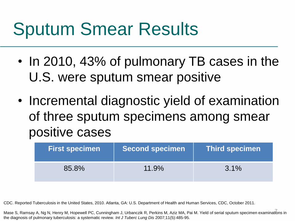
Sputum Smear Results
• In 2010, 43% of pulmonary TB cases in the
U.S. were sputum smear positive
• Incremental diagnostic yield of examination
of three sputum specimens among smear
positive cases First specimen Second specimen Third specimen
85.8% 11.9% 3.1%
CDC. Reported Tuberculosis in the United States, 2010. Atlanta, GA: U.S. Department of Health and Human Services, CDC, October 2011.
Mase S, Ramsay A, Ng N, Henry M, Hopewell PC, Cunningham J, Urbanczik R, Perkins M, Aziz MA, Pai M. Yield of serial sputum specimen examinations in
the diagnosis of pulmonary tuberculosis: a systematic review. Int J Tuberc Lung Dis 2007;11(5):485-95.
7

Limitations of AFB Microscopy
• Does not distinguish between viable and dead organisms
– Follow-up specimens from patients on treatment may be smear positive yet culture negative
• Limited sensitivity
– High bacterial load 5,000-10,000 AFB /mL is required for detection
– Misses >45% of U.S. TB cases
• Limited specificity
– All mycobacteria are acid fast
– Does not provide species identification
– Local prevalence of MTB and NTM determine the predictive values of a positive smear for MTB
8

Smear Types
• Direct smear
– Smear prepared directly from a patient
specimen prior to processing
• Concentrated smear
– Smear prepared from a processed specimen
after centrifugation is used to concentrate the
material
9

Direct Smears
• Rapid and simple
– May be performed for quick results
– Positive results help confirm clinical
suspicions
• Not as sensitive as concentrated smear
– A direct smear should always be followed by a
concentrated smear
10

Concentrated Smear
• 3-10 ml of sputum is processed and
concentrated by centrifugation
• Smears can be made
– Directly from processed pellet
• May increase smear sensitivity
• However, may result in less material available for
NAA testing & culture
– From re-suspended pellet after the addition of
buffer
• Re-suspended in ~2 ml buffer; one drop for smear 11

AFB Microscopy Staining Techniques
• Two basic techniques: same
principle:
• Fluorescence: Auramine staining
– Also known as Fluorochrome staining
– Contrast light & dark
• Brightfield: carbol fuchsin staining
– Contrast red AFB on blue or green
background
12

Carbol-Fuchsin AFB Staining
• Primary stain is Carbol Fuchsin
– Ziehl-Neelson (ZN)
• Requires heat during staining
• Requires higher magnification
• More fields examined (e.g. 300 fields at 1000X)
• Requires more time to read
• Requires use of oil immersion
• Stains all NTM well
– Kinyoun
• Cold carbol fuchsin method
• Less toxic fumes
• Neither method is reccomended for staining primary
specimens
13

Fluorescent AFB Staining
• Primary stain is Fluorescent
– CDC recommends fluorochrome staining
for detecting AFB in primary patient
specimens
– Auramine-O, Auramine Rhodamine • Read at lower magnification, less fields examined
(e.g, 30 fields at 200X)
– Faster screening of smears than with ZN
– ~10% more sensitive than ZN
– Does not require use of oil immersion
14

1. Preparing and Fixing Smears
2. Staining Smears
3. Examining Smears
4. Recording and Reporting Results
Steps in Performing AFB
Microscopy
15
• New, clean, greaseless, and unscratched slides
should be used
• Match identifiers on slide with clinical specimens
• Labeling should be performed with material that stays
permanently affixed to the slide during the staining
procedure (e.g., graphite or diamond tip pencil)
Getting Started…
16

17

• After processing and concentrating the specimen, 1 to 2 drops of material should be smeared on the slide
• Smear material in an area of approximately 2-cm2
Preparing Smears
18

Just
Right
Too
Thick
Too
Thin
19

Fixing Smears
• Prior to heat fixing, smears should be allowed to air dry completely within the biological safety cabinet
• Acceptable methods for heat fixing • Flame fixing by passing over flame 2–3 times for a few seconds smear
side up – Avoid scorching
• 2 hrs minimum at 65-75oC on an electric slide warmer
• 15 min at 80oC1
• 5% phenol in 70% EtOH for 5 min2
– also kills AFB
• Considerations • Flame fixing may aerosolize organisms from smear
• Insufficient heat or time can lead to smear washing off
• Slide warmers may not heat evenly
• Viable AFB remain with some fixing methods
20
1 Bailey & Scott’s Diagnostic Microbiology 2007 12th ed. 2 Chedore et al. 2002. J. Clin. Microbiol. 40:4077
CLSI M48-A: Laboratory Detection & Identification of Mycobacteria

21
Keep slides on
warmer for 2 hours

Heat Fixing Smear
22
Safety Concerns: • Wear gloves at all times • Work inside the BSC • Use open flame for shor periods of time

AFB Staining Principles
• Primary stain penetrates cell wall
• Intense decolorization does not release primary stain from the cell wall of AFB
• Color of AFB-based on primary stain
• Counterstain provides contrasting background
23

Stains Used in Fluorescence
Microscopy
• Primary Stains
– Auramine O
– Auramine O-Rhodamine-B
• Counter Stains
– Potassium Permanganate
– Acridine Orange
24
Fluorescence AFB Microscopy
• Primary fluorochrome
– Auramine O
– Auramine O-Rhodamine B
• Counter Stain – Potassium permanganate
25

Fluorescence AFB Microscopy
• Primary
Fluorochrome
– Auramine O
– Auramine O/
Rhodamine B
– Acridine Orange
• AFB Fluoresces
– Green
– Yellow/Orange
– Yellow/Orange
26

450x
27

Water Quality is Key
• AFB microscopy is not specific – Acid fast environmental contaminants as well as NTM and MTB presence in
the specimen will be detected
• Introduction of an environmental contaminant during wash steps and in reagent preparation must be avoided
• Use of a negative control slide essential for detecting potential environmental contaminants
• Avoid using large containers of reagents and carboys of water
• Use filtered distilled or deoinized water
• Water filtration and distribution systems should be monitored
28

Staining Reagents
• Commercial products are available or
reagents can be prepared in-house
– If prepared in-house, proper precautions must
be taken when handling dyes including
appropriate PPE and the use of a fume hood
• Reagents containing fluorescence stains
should be stored to protect from light
exposure
29

Steps in the Fluorescent Staining
Procedure
1. Place slides on staining rack; slides should not touch
2. Flood slides with fluorochrome stain; no heating. Follow protocol or package insert for timing.
3. Rinse with water; aim flow at edge of slide
4. Decolorize with 0.5% acid-alcohol solution, Follow protocol or package insert for timing.
5. Rinse with water; drain excess
6. Flood slide with counterstain; Follow protocol or package insert for timing.
7. Rinse with water; drain excess
8. Air-dry stained slide; do not blot 30

31

32

33

34

35

36

37

Fluorescence Microscopy
• A fluorescence microscope is required for examining fluorochrome-stained smears:
– Mercury vapor or halogen bulb light source (about 150 hours of use)
– Newer mercury bulbs (about 2,000 hours of use)
– LED Bulbs (about 15,000 hours of use)
– Excitation and emission (barrier) filters are necessary for visualization of the fluorescently-stained smear (specific to the staining method used. Check package insert)
• LED-based Fluorescent Microscopy
– LED modules used to adapt light microscopes for reading fluorescently-stained smears
• May be useful in low income settings
• More research is needed to evaluate performance
38

Systematic Examination of Smears
39
Whichever method you use, BE CONSISTEN!

Number of Fields to Examine
Magnification a Number of Fields b
200x
250x
400x
450x a This final magnification represents the objective lens
magnification multiplied by the eyepiece magnification
b The minimum number of fields to examine before
reporting a smear as negative for acid-fast organisms.
30
30
55
70
40

41

Examining Smears for AFB
• AFB will be rod-shaped, 1–10 µm in length and 0.2–
0.6 µm wide
• Appearance is generally long and slender but may
also appear bent
• Bacilli may contain heavily stained areas called
beads
• Count a clump of bacilli that are touching as one
• Debris, some species of the genera Nocardia and
Corynebacterium, and some fungal spores may
appear acid fast
42

Reporting Smear Results Fluorescence Microscopy
(CDC Scale)
Ziehl-Neelsen Stain
(CDC scale)
250X 450X Report As:
1000X (oil immersion)
0 AFB/ smear 0 AFB/ smear
No AFB seen 0 AFB/ smear
1–2/ 30 fields 1–2/ 70 fields
Report exact count;
order repeat
specimen
1–2/300 fields
1–9/ 10 fields 2–18/ 50 fields 1+ 1–9/ 100 fields
1–9/ field 4–36/ 10 fields 2+ 1–9/ 10 fields
10–90/ field
4–36/ field
3+ 1–9/ field
>90/ field
>36/ field
4+
>9/ field
43

CLSI M48
Fluorescence Microscopy Ziehl-Neelsen Stain
Number of AFB
found at 250x
Number of AFB found
at 450x
Number of AFB
found at 1000x Report as: Or report as:
Negative for AFB Negative for AFB 0 Negative for AFB Negative for AFB
Number seen.
(Order repeat
specimen) **
Number seen.
(Order repeat
specimen)
1-2 per 300 fields
Number seen.
(Order repeat
specimen)
Number seen.
(Order repeat
specimen)
(Number seen/10)
per 100 fields
(Number seen/4)
per 100 fields
1-9 per 100 fields 1+ Number seen per
100 fields
(Number seen/10)
per 10 fields
(Number seen/4)
per 10 fields
1-9 per 10 fields 2+ Number seen per
10 fields
(Number seen/10)
per field
(Number seen/4)
per field
1-9 per field 3+ Number seen per
field
>(Number seen/10)
per field
>(Number seen/4)
per field
>9 per field 4+ >Number seen per
field 44

Manual of Clinical Microbiology.
Acid-fast smear evaluation and reportinga
Report
No. of AFB seen by staining method and magnification
Fluorochrome stain Ziehl-Neelsen Stain
X 250 X 450 X 1000
No AFB seen 0 0 0
Doubtful;
repeat
1 – 2/30 F
(1 sweep)
1 -2/70 F
(1.5 sweeps)
1 – 2/300 Fb
(3 sweeps)c
1+ 1 – 9/10 F 2 – 18/ 50 F 1 – 9/100 F
2+ 1 – 9/F 4 – 36/10 F 1 – 9/10 F
3+ 10 – 90/F 4 – 36/F 1 – 9/F
4+ > 90/F > 36/F >9/F a Adapted from K/K, 1985 b F, microscope fields. c one full sweep refers to scanning the full length (2 cm) of a smear 1 cm wide by 2 cm long.
45

Reporting smear results
• Use one of the previously recommended or CLSI reporting scales to report and approximation of the number of AFB viewed on the slide using a semi-quantitative scale
• Report smear results within 24 hours of specimen receipt
• Smear positive results are considered critical values and should be reported to the health care provider and public health department as soon as results are known.
46

Importance of Control Slides
• Assess the quality of the reagents
• Determine if the staining is
performed properly
• Determine if the microscope is
working properly
• Detect environmental contaminants
• Help find the plane of focus
47

Quality Assurance of AFB
Microscopy • A known positive and known negative smear should
be read with each run and when new reagent used
• QC smears may be prepared in advance, heat-killed,
and stored unstained
• Preferable to prepare QC slides using sediment from a
clinical specimen
• Records should include stain lot numbers, expiration
dates, results of the control slides, and technician
name
• Patient smears should only be examined and reported
when control slides are acceptable
48

Monitoring Performance for Smear Status
and Smear Sensitivity* Numerator Denominator Action
Thresholds
Investigative actions
Potential causes-
increases
Potential causes-
decreases
Positive smears Number of AFB
smears reported as
positive in one month
Total number of
AFB smears
performed in one
month
Patient
population
dependent;
determine
baseline and
monitor trends
False positive smears
(contaminated reagents,
tap water rinse, artifacts,
technologist issues)
False negative
smears (microscope,
centrifuge, or
technologist
problems,
insufficient read
time)
Smear positive/culture
positive rate
(sensitivity)
Number of smear-
positive specimens**
that are culture
positive( 1.) for MTBC
or for (2) any
mycobacteria
1)Total number of
cultures positive for
MTBC*
2)Total number of
cultures positive for
all mycobacterial
sp.**
National
averages: More
smear positive
specimens are
MTBC than
NTMs. MTBC
smear positivity
ranges 30-70%
nationally
May be due to false
positive smears. Patients
on treatment may have
positive smears and
negative cultures.
-Cross contamination
-Use of poor quality
slides
-Use of contaminated or
poor quality reagents
-Technical errors
False negative
smears
-microscope,
centrifuge or
technologist
problems
-Suboptimal
specimens
submitted to the
laboratory
-Inadequate staining
and evaluation of
slides
Correlation culture
positive/ smear
positive (specificity)
Number of smear-
positive specimens
inoculated that were
culture positive for
MTBC or NTM **
Specimens
inoculated for
culture that were
smear positive in
one month**
Should be high
percent, 90-98%
May be due to false
positive smears (see
above, left), or false
negative cultures,
patients on
treatment. *Suggested frequency of monitoring: High volume: monthly. Low volume or low incidence: bi-monthly or quarterly.
** Ideally measured for initial diagnostic specimens only; smears from treated patients may be positive, but yield no growth on culture.
49

Suggestions for Avoiding False-Positive AFB
Smear Results
Cause Corrective Action
Old, used microscope slides retain material
for previous smear
Use only new slides.
AFB transferred from a positive smear to a
negative smear
Use a staining rack and keep slides from
touching each other; do not use staining
jars.
Food particles Request another specimen.
Precipitated stains Use only fresh stains, without precipitates,
or contaminating organisms. If any
precipitate is observed, filter the stain.
AFB transferred in oil on the objective lens Always wipe oil from the oil immersion lens
after each AFB-positive smear is read.
CLSI M48AE: Laboratory Detection and Identification of Mycobacteria; Approved
Guideline 50

Suggestions for Avoiding False-
Negative AFB Smear Results Cause Corrective Action
Smears that are too thick, causing material
to be washed off during staining
Proper digestion of specimen. Avoid making thick
smears.
Smear area is too large, making the smear
too thin
Apply smear to a 2-cm2 area.
Non-staining or poorly staining AFB Protect smear from UV light, direct sunlight,
overheating during smear fixation; store stains in dark
bottles; high chlorine content in rinse water affects
fluorescence stain.
Incorrect slide warmer temperature Set temperature at 65-75°C and monitor weekly
Incomplete slide reading Search smear in uniform manner and read suggested
number of fields
Insufficient centrifugation Ensure centrifugation occurs at 3000 x g for at least
15 minutes
CLSI M48AE: Laboratory Detection and Identification of Mycobacteria; Approved
Guideline
51

Maintaining Proficiency in
Microscopic Smear Examination • Smears should be examined by an experienced
microscopist – Microscopists should meet a level of competency before
being allowed to report smear results.
• Mycobacteriology laboratories should participate in an approved proficiency testing program that includes smear microscopy
• To maintain proficiency, laboratories should process at least 15 AFB smears per week
• Other procifiency testing activities – Participate in multiple proficiency testing programs
– Develop an internal proficiency testing program
– Establish a QA program
52

Achieving Reliable Results
• Obtain good quality specimens is essential
• Prevent contamination of testing reagents and adjacent slides when staining
• Follow established procedures & recommendations
• Ensure accurate reporting and record keeping
53

AFB Fluorescent Smear Microscopy
Example slides


References
• Use of Fluorochrome Staining for Detecting Acid-fast Mycobacteria, CDC
• Manual of Clinical Microbiology, 10th edition
• CLSI M48AE: Laboratory Detection and Identification of Mycobacteria; Approved Guideline
• Mycobacterium tuberculosis: Assessing your laboratory (APHL 2009)
• Monitoring the performance of mycobacteriology laboratories: a proposal for standardized indicators; McCarthy, K.D et al.; The International Journal of Tuberculosis and Lung Disease, Volume 12, Number 9, September 2008 , pp. 1015-1020
67