aesthetic surgery journal onlay fascial grafts to silicone
TRANSCRIPT
RHINOPLASTY
Aesthetic Surgery Journal2019, Vol 39(11) 1182–1190
DOI: 10.1093/asj/sjz121www.aestheticsurgeryjournal.com
Rhinoplasty
© 2019 The American Society for Aesthetic Plastic Surgery, Inc. Reprints and permission: [email protected]
Onlay Fascial Grafts to Silicone-Polytetrafluorethylene Composite Implants in Augmentation Rhinoplasty: A Retrospective Study of 241 Cases
Lee Seng Khoo, MD; Cheng-I Yen, MD; Chun-Shin Chang, MD; Hung-Chang Chen, MD; Chih-Jung Huang, MD; and Yen-Chang Hsiao, MD
AbstractBackground: Silicone-polytetrafluoroethylene composite implants are fast gaining popularity in Asian rhinoplasty. Nonetheless, implant displace-ment, erythematous reactions, and infections still occur in the authors’ patient group during long-term follow-up.Objectives: The authors reported successful experience of combining the utilization of silicone-polytetrafluoroethylene composite implants with onlay temporal fascial grafts to circumvent these complications.Methods: Sixty-four patients of Asian ethnicity underwent augmentation rhinoplasty utilizing an I-shaped composite implant with an onlay fascial graft from January 2015 to June 2018, with a mean follow-up period of 13.5 months. This patient group was compared with a control group of 177 Asian patients who underwent augmentation rhinoplasty utilizing the same composite implant but without the addition of a fascial graft; the control group was treated from February 2012 to June 2015, with a mean follow-up of 42.0 months. Complications were compared between these 2 patient groups, specif-ically focusing on malposition/deviations, erythema, and infections.Results: There was a marked decrease in complication rates with the addition of an onlay temporal fascial graft to cover the composite implant in augmentation rhinoplasty (7.8% vs 14.7%) as well as the rate of erythematous reactions (0% vs 6.2%, P = 0.04), infection (1.6% vs 1.1%), and implant malposition/deviation (0% vs 4.5%). Harvesting the temporal fascia and fashioning the onlay graft added an additional 33 minutes on average per pro-cedure. No donor site morbidity was encountered.Conclusions: Although the operative time increased, the benefits of adding onlay fascial grafts to silicone-polytetrafluoroethylene implants in alloplastic augmentation rhinoplasty outweigh the drawbacks, as evidenced by the decrease in erythematous reactions.
Level of Evidence: 4
Editorial Decision date: April 15, 2019; online publish-ahead-of-print April 26, 2019.
Alloplastic implants, such as silicone, expanded pol-ytetrafluoroethylene (PTFE or Gore-Tex, W.L. Gore Associates, Inc., Phoenix, AZ), and porous high-density polyethylene (Medpor, PPE, Stryker, Kalamazoo, MI), are widely employed in Asian rhinoplasty.1 Initial re-ports in the 1960s and 1970s pointed to a high extrusion rate with silicone alloplastic implants, which cemented a negative perception with Western-trained surgeons.2,3
applyparastyle "fig//caption/p[1]" parastyle "FigCapt"applyparastyle "fig" parastyle "Figure"
From the Department of Plastic and Reconstructive Surgery, Chang Gung Memorial Hospital, College of Medicine, Chang Gung University, Taipei, Taiwan
Corresponding Author: Dr Yen-Chang Hsiao, Department of Plastic and Reconstructive Surgery, Chang Gung Memorial Hospital, 5, Fu-Hsing Street, Kweishan, Taoyuan 333, Taiwan. E-mail: [email protected]
Dow
nloaded from https://academ
ic.oup.com/asj/article-abstract/39/11/1182/5480182 by guest on 28 O
ctober 2019
Khoo et al 1183
Nevertheless, the utilization of alloplastic implants is still widespread in Asian rhinoplasty, with many surgeons re-porting a low extrusion rate (less than 2%) and infection rates of 0% in their practices.1,4 Complication rates with the utilization of silicone implants range from 2% to 7%. The removal rate for both Gore-Tex and Medpor is 3.1%, whereas the removal rate for silicone implants is higher, at 6.5%.1,4
Currently, silicone-polytetrafluoroethylene com-posite implants are quickly gaining popularity in Asian rhinoplasty.5 The Implantech Composite nasal implant (Implantech, Ventura, CA) is marketed as Chimera in Taiwan and as Silitex in Korea .5 These composite implants utilize the pliable nature of silicone implants while miti-gating the capsular formation by coating the surface with polytetrafluoroethylene (Gore-Tex), which theoretically elicits a minimal foreign body reaction and allows the in-growth of tissue for a natural appearance.5 The outer polytetrafluoroethylene surface of the implant imparts a soft texture and reduces implant visibility through the skin. The inner surface is composed of silicone and has a relative firmness, which is suitable for fashioning the nasal shape in Asians with moderately thick to thick skin.
Nonetheless, implant displacement/malposition (4.5%), erythematous reactions (6.2%), and infections (1.1%) still occur in our patient group, as observed during long-term follow-up.5 In this series, we report our suc-cessful experience of combining the utilization of silicone-polytetrafluoroethylene composite implants with onlay temporal fascial grafts to circumvent these complications.
METHODS
This retrospective study was performed at Chang Gung Memorial Hospital after obtaining approval from the in-stitutional review board. This was a single-center, retro-spective study involving 241 consecutive patients who underwent augmentation rhinoplasty with silicone-PTFE composite implants, otherwise known as Chimera im-plants, by a single senior surgeon (H.Y.C.). Sixty-four pa-tients of Asian ethnicity underwent primary (n = 28) or secondary (n = 36) rhinoplasty utilizing an I-shaped com-posite implant with an onlay fascial graft from January 2015 to June 2018. This group of 64 patients was compared with a control group of 177 consecutive Asian patients who underwent primary (n = 63) or secondary (n = 114) rhinoplasty utilizing an I-shaped composite implant without the addition of a fascial graft from February 2012 to June 2015. Chart data and photographs were reviewed for all 241 patients included in the 2 groups. These 2 pa-tient groups were compared for malposition/deviations, erythema, and infections.
Surgical Technique
All surgeries were conducted under general anesthesia. The proposed site of augmentation was delineated and marked with methylene blue prior to the infiltration of local anesthesia. An inverted V incision over columella was performed, and a marginal incision was extended to expose the lower and upper lateral cartilage in the supraperichondrial plane. At the nasal bone, the dissection was made in the subperichondrial plane. Rasping was per-formed for minor dorsal humps. Septal cartilage harvesting was performed when necessary.
Tip work involved the placement of a small septal ex-tension graft supported laterally by 2 extended spreader grafts. Tip grafts, shield grafts, and counter rotation grafts were employed subject to the aesthetic goals. The grafts were fashioned from either conchal or septal cartilage, or a combination of both.
At this stage, deep temporal fascia (DTF) was har-vested for cases where an onlay fascial graft was em-ployed to cover the Chimera composite implant. A 2.5-cm straight incision hidden within the hairline in the temporal region was made after the instillation of local anesthesia. Dissection proceeded through the sub-cutaneous tissue and superficial temporal fascia until the glistening DTF was located. The size of the harvested DTF was determined by the amount required (typically 4 cm × 2.5 cm), and it was imperative to avoid dam-aging vessels in the temporal region (Figure 1; Video, available online as Supplementary Material at www.aestheticsurgeryjournal.com).
Conservative subperiosteal pocket dissection over-lying the nasal dorsum was performed to allow a snug fit with an appropriately sized I-shaped composite implant. The thickness of the chosen implant was determined by the skin and soft tissue characteristics. Implants were handled with strict aseptic precautions, including a change of gloves and “impregnation” with a first-gen-eration antibiotic. This was done by placing the implant in a 50-cc syringe and filling the syringe with the anti-biotic solution. The stopper and cap were replaced after the evacuation of air to create a vacuum, which impreg-nates the pores of the implant with the antibiotic solu-tion (Video).
The harvested fascia was draped over the composite implant and secured with 27G hypodermic needles. It was then sutured in place with 5-0 polydioxanone and the excess fascia was trimmed, making sure that the fascia adequately covered the lateral edges of the im-plant (Figure 2; Video). We wrapped around the dorsal side of the implant with DTF in onlay fashion instead of wrapping it all around. The fascia is more of an onlay graft than a wrap-around. The implant (with or without
Dow
nloaded from https://academ
ic.oup.com/asj/article-abstract/39/11/1182/5480182 by guest on 28 O
ctober 2019

1184 Aesthetic Surgery Journal 39(11)
the addition of fascia coverage) was placed into the dis-sected pocket after irrigating the implant pocket with the antibiotic solution.
The lower pole of the implant extended just to the cephalic margin of the lateral crura, lateral to the tip-defining points. The upper border of the implant was posi-tioned at the level of the intercanthal line or supratarsal crease, depending on the patients’ preference. Two 5-0 polydioxanone sutures were used to loosely anchor the PTFE layer of the implant to the lateral crura of the lower lateral cartilage to maintain the implant position. Adequate hemostasis was achieved and the skin/mucosa was closed as a single layer. Skin closure was performed with 5-0 Dexon and 7-0 nylon in layers. Taping and the placement of thermoplastic splints was routine in all cases. Patients were typically discharged on the day of surgery and seen 1 week after surgery for tape/splint removal. All patients were prescribed a 1-week course of oral anti-biotics postsurgery.
Statistical Analysis
All data were evaluated using SPSS software (SPSS, Inc., Chicago, IL, Version 17.0). Analysis of variance was util-ized to compare the difference in complication rates, taking into account the disparity of the sample size. Statistical significance was defined as P < 0.05.
RESULTS
I-shaped silicone-PTFE composite implants (Chimera implants, Taiwan) were used in all augmentation rhino-plasty cases with either fascial or nonfascial coverage. On average, the implant thickness was 3.7 mm (range, 1.5-5.0 mm) in the group without the addition of an onlay fascial graft and 3.6 mm (range, 1.5-5.0 mm) in the fascia group. Glabellar augmentation was performed in 11 pa-tients (17.2%) in the group that underwent augmentation rhinoplasty with the addition of an onlay fascial graft and in 19 patients (10.7%) in the group without the addition of a graft. Table 1 shows the demographics and comparison of the complication rates between the 2 groups.
For the control group (composite implant augmenta-tion rhinoplasty without the addition of an onlay fascial graft), the patients consisted of 159 women and 18 men, with an age range of 19 to 72 years (mean, 34 years) and the average follow-up period was 42.0 months (range, 37-72 months). There were 26 patients (14.7%) with complications, which necessitated revision in 12 patients (6.8%). Eight patients (4.5%) had persistent or new devi-ation or malposition of the implant, which was the main reason for revision surgery (7 out of 8 surgeries). Eleven patients (6.2%) had idiopathic persistent postoperative er-ythema that was noninfectious in nature that did not re-spond to antimicrobial therapy (Figure 3). Two patients (1.1%) had a postoperative infection, which necessitated implant removal in 1 of the patients. Four patients (2.3%)
Figure 1. Harvest of the deep temporal fascia (typically 4 cm × 2.5 cm).
Figure 2. The harvested deep temporal fascia was trimmed and draped over the composite implant and sutured in place with 5-0 polydioxanone.
Video. Watch now at https://academic.oup.com/asj/article-lookup/doi/10.1093/asj/sjz121
Dow
nloaded from https://academ
ic.oup.com/asj/article-abstract/39/11/1182/5480182 by guest on 28 O
ctober 2019

Khoo et al 1185
required fat grafting for a multitude of reasons, such as recontouring an uneven glabella, prominent implant de-marcation and migration, and to obtain a supratip break. One patient (0.6%) had mild ear keloid formation. There were no reported implant exposures and no capsular con-tractures during the follow-up period.
For the group that underwent composite implant aug-mentation rhinoplasty with the addition of an onlay fascial graft, the patients comprised 55 women and 9 men, with an age range of 21 to 59 years (mean, 33.9 years) and a mean follow-up period of 13.5 months (range, 8-46 months). Five patients (7.8%) had complications, and revision surgery was performed in 4 patients (6.3%). One patient (1.6%) had an infection that necessitated the removal of the im-plant and fascia, 1 patient (1.6%) underwent fat grafting post implant surgery for visible implant demarcation, and 2 other patients underwent revision for the tip (1.6%) and alar (1.6%) region. One patient had septal deviation (1.6%), but no patients (0%) presented with malposition of the implant. No patients (0%) suffered postoperative prolonged erythema. No implant exposures or contractures were noted during the follow-up period. There were no re-ported complications associated with the additional proce-dure of the DTF harvest. Figures 4 and 5 demonstrate the
postoperative outcome of augmentation rhinoplasty with an onlay fascial graft on the composite implant.
Comparing the 2 groups, there was a significant de-crease in postoperative erythema (P = 0.04) after adding the onlay fascial graft to the composite implant. Regarding the overall complication, infection, and implant displace-ment rates, no significant differences between the 2 groups were noted.
DISCUSSION
The major goals of Asian rhinoplasty involve dorsal aug-mentation with tip contouring. Alloplastic augmentation in nasal implants remains the most popular method world-wide in Asian rhinoplasty.2 A paradigm shift in the under-standing of the dynamics of augmentation rhinoplasty and technical advances in both materials and techniques render obsolete the consensus that the use of alloplastic implants is a cardinal sin.
The composite nasal implant places a 0.3-mm layer of PTFE (Gore-Tex) on a silicone surface.5 PTFE implants allow tissue ingrowth into their surface, making them more biocompatible, and they reduce the incidence of capsular contracture.4,5 This hybrid implant exhibits characteristics that make it stable and able to maintain its position better than silicone implants.5 The prominent step-off noticed with the use of silicone implants is also less evident in composite implant rhinoplasties.5 Low rates of infection and capsular contracture make the composite implant a superior option for Asian rhinoplasty.5
The DTF acts as a scaffold that reduces implant dis-placement and malposition by the rearrangement of torque over the implant. This is due to the inherent pro-perty of the DTF that it is rougher and less slippery than the superficial temporal fascia.6 No patient had devia-tion or malposition of the implant with the addition of an onlay fascial graft; in contrast, the rate was 4.5% in the control group. These data compare favorably with data reported by Zeng et al, who documented a 9.4% implant “maldirection” out of 406 patients who under-went silicone rhinoplasty,7 and with data reported by Hong et al, who reported implant displacement in 1.2% of 257 Asian patients who underwent “hard-type” Gore-Tex rhinoplasty.4
Nonetheless, persistent erythema over the implant re-mains an unwelcomed complication that can be a major source of patient dissatisfaction.5 Persistent color changes were reported by Zeng et al in 0.6% of Asians with sili-cone implants.7 Conrad et al noted soft tissue reactions in 4 (0.6%) of 685 Gore-Tex implants.8 The same authors reported the incidence of hyperemia did not respond to antibiotics in a similar review 1 decade earlier,9 suggesting a similar complication.
Table 1. Demographics and Complications of Onlay Fascia Group and Control (Without Fascia) Groups
Onlay fascia n = 64
Control (without fascia) n = 177
P value
Age, y (range) 33.9 (21-59) 34 (19-72)
Male/female, n 9/55 18/159
Primary/secondary, n 28/36 63/114
Implant height, mm (range) 3.6 (1.5-5.0) 3.7 (1.5-5.0)
Complication, n (% of group) 5 (7.8) 26 (14.7) 0.16
Erythema 0 (0) 11 (6.2) 0.04*
Infection 1 (1.6) 2 (1.1) 0.79
Implant malposition 0 (0) 8 (4.5) 0.08
Others 1 (1.6) 5 (2.8) 0.58
Revision, n (% of group) 4 (6.3) 12 (6.8) 0.89
Infection 1 (1.6) 1 (0.6)
Malposition 0 (0) 7 (4.0)
Inadequate correction 0 (0) 4 (2.3)
Fat graft for demarcation 1 (1.6) 0 (0)
Tip refinement 1 (1.6) 0 (0)
Alar refinement 1 (1.6) 0 (0)
*Statistically significant at P < 0.05.
Dow
nloaded from https://academ
ic.oup.com/asj/article-abstract/39/11/1182/5480182 by guest on 28 O
ctober 2019

1186 Aesthetic Surgery Journal 39(11)
Eleven patients (6.2%) in the control group who un-derwent augmentation rhinoplasty with composite im-plants without the addition of onlay fascia grafts had idiopathic persistent postoperative erythema that was noninfectious in nature and did not respond to antimicro-bial therapy. In contrast, virtually no patients (0%) suf-fered from prolonged postoperative erythema in the group
of augmentation rhinoplasty attenuated with onlay fascial grafting. Two patients from the control group (1.1%) and 1 patient (1.6%) from the onlay fascial graft group devel-oped postoperative infections, but no statistically signifi-cant differences were noted.
Because we supposed that the erythematous reaction is a relatively superficial problem and may result from the
A B C D
E F G H
I J
Figure 3. This 45-year-old woman underwent augmentation rhinoplasty without the addition of an onlay fascial graft. Preoperative and 16-month postoperative (A, B) frontal views, (C, D) three-quarter view, (E, F) profile view, (G, H) three-quarter view, and (I, J) profile view photographs are shown. Erythematous change over the dorsal skin was noted.
Dow
nloaded from https://academ
ic.oup.com/asj/article-abstract/39/11/1182/5480182 by guest on 28 O
ctober 2019

Khoo et al 1187
foreign body reaction between the dorsal surface of the implant and skin, we proposed the idea of adding an autol-ogous tissue overlying the implant to decrease this kind of complication. The utilization of autologous temporal fas-cial grafts in rhinoplasty is well established.10-13 The onlay temporal fascia adds a subtle layer of lining over the com-posite implant, protecting it from sebaceous glands that
may trigger foreign body reactions and possible infections. The fascia also possesses histological cell-supportive prop-erties that act as a cast and promote integration with other surrounding tissues.14,15 One study demonstrated favor-able outcomes in immediate nasal dorsum reconstruction without secondary infection by covering silicone implants with DTF; this followed foreign body removal due to
A B C D
E F G H
I J
Figure 4. This 29-year-old woman underwent augmentation rhinoplasty with the addition of an onlay fascial graft. Preoperative and 22-month postoperative (A, B) frontal views, (C, D) three-quarter view, (E, F) profile view, (G, H) three-quarter view, and (I, J) profile view photographs are shown. There was no erythema, infection, or implant malposition.
Dow
nloaded from https://academ
ic.oup.com/asj/article-abstract/39/11/1182/5480182 by guest on 28 O
ctober 2019
1188 Aesthetic Surgery Journal 39(11)
infection resulting from augmentation rhinoplasty with in-dustrial silicone.16
Carving an implant may result in irregularities over its surface, which can be camouflaged with the use of an onlay temporal fascial graft over the implant. The onlay temporal fascial graft adds a layer of approximately 2 mm over the composite implant, conferring a more stable and smoother surface to blend in the dorsum with the lat-eral nasal walls. Two studies confirmed the integrity and
survival of DTF on histological examination when utilized to wrap diced cartilage grafts.17,18 Hence, a Chimera com-posite implant with the addition of an onlay fascial graft results in a more natural looking nose and avoids a prom-inent demarcation, which has been seen with silicone or PTFE alone. An onlay fascial graft over the implant is even more important in secondary rhinoplasties, where the skin overlying the dorsum is particularly thin due to previous implant pressure and capsular tissue removal.
A B C D
E F G H
I J
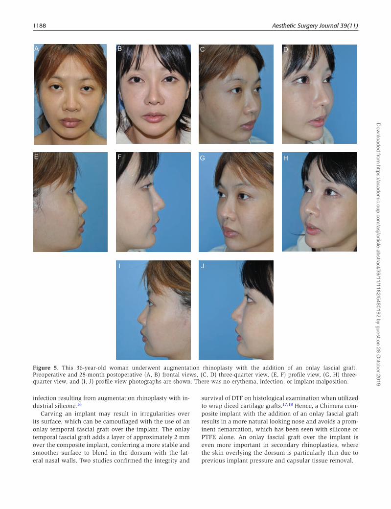
Figure 5. This 36-year-old woman underwent augmentation rhinoplasty with the addition of an onlay fascial graft. Preoperative and 28-month postoperative (A, B) frontal views, (C, D) three-quarter view, (E, F) profile view, (G, H) three-quarter view, and (I, J) profile view photographs are shown. There was no erythema, infection, or implant malposition.
Dow
nloaded from https://academ
ic.oup.com/asj/article-abstract/39/11/1182/5480182 by guest on 28 O
ctober 2019

Khoo et al 1189
Harvesting the temporal fascia and fashioning the onlay graft, however, did add an additional 30 to 40 minutes (mean, 33 minutes) per procedure in our case series. Donor site morbidity includes hematoma, scarring, dehiscence, and possible temporal hollowing. However, this was not seen in our case series. The downtime is typically prolonged when patients must tolerate an early wider and higher dorsal bridge, which resolves with partial absorption of the fascia. Removal of the composite implant in secondary rhinoplasty may be difficult due to the dense adhesion of the fascia to the implant. That said, the donor site scar for harvesting the DTF is hidden within the hairline, rendering it inconspic-uous (Figure 6), and the donor site morbidity incidence is minimal and was not evident in our case series.
The limitation of this study is the relatively short fol-low-up duration and the relatively noncomparable number between fascia group and nonfascial group, and also the different periods during which the surgeries were per-formed between the 2 groups.
To our knowledge, this is the first documented study involving the utilization of temporal fascia onlay grafts with silicone-polytetrafluoroethylene nasal implants, and the results showed the benefit of decreased erythematous reactions after surgery. Further studies need to be con-ducted to confirm the findings of this preliminary study, and the use of other appropriate materials, such as artifi-cial dermis or cadaveric fascia, can be explored in regard to avoiding donor site morbidity.
CONCLUSIONS
The addition of onlay DTF grafts to composite silicone-polytetrafluoroethylene implants decreases the com-plication rate of erythematous reactions in alloplastic augmentation rhinoplasty. Although the operative time is increased, the benefits of this procedure still outweigh the drawbacks.
Supplementary MaterialThis article contains supplementary material located online at www.aestheticsurgeryjournal.com.
DisclosuresThe authors declared no potential conflicts of interest with respect to the research, authorship, and publication of this article.
FundingThe authors received no financial support for the research, authorship, and publication of this article.
REFERENCES
1. Peled ZM, Warren AG, Johnston P, Yaremchuk MJ. The use of alloplastic materials in rhinoplasty surgery: a meta-analysis. Plast Reconstr Surg. 2008;121(3):85e-92e.
2. McCurdy JA Jr. The Asian nose: augmentation rhino-plasty with L-shaped silicone implants. Facial Plast Surg. 2002;18(4):245-252.
3. McCurdy JA, Lam SM. Cosmetic surgery of the Asian face. 2nd ed. New York: Thieme; 2005.
4. Hong JP, Yoon JY, Choi JW. Are polytetrafluoroethylene (Gore-Tex) implants an alternative material for nasal dorsal augmentation in Asians? J Craniofac Surg. 2010;21(6):1750-1754.
5. Zelken JA, Hong JP, Chang CS, Hsiao YC. Silicone-polytetrafluoroethylene composite implants for Asian rhinoplasty. Ann Plast Surg. 2017;78(2):131-137.
6. Calvert J, Brenner K. Autogenous dorsal reconstruction: maximizing the utility of diced cartilage and fascia. Semin Plast Surg. 2008;22(2):110-119.
7. Zeng Y, Wu W, Yu H, Yang J, Chen G. Silicone im-plant in augmentation rhinoplasty. Ann Plast Surg. 2002;49(5):495-499.
Figure 6. This 36-year-old woman underwent augmentation rhinoplasty with the addition of an onlay fascial graft. The donor site scar from harvesting the deep temporal fascia is hidden within the hairline.
Dow
nloaded from https://academ
ic.oup.com/asj/article-abstract/39/11/1182/5480182 by guest on 28 O
ctober 2019

1190 Aesthetic Surgery Journal 39(11)
8. Conrad K, Torgerson CS, Gillman GS. Applications of Gore-Tex implants in rhinoplasty reexamined after 17 years. Arch Facial Plast Surg. 2008;10(4):224-231.
9. Conrad K, Gillman G. A 6-year experience with the use of expanded polytetrafluoroethylene in rhinoplasty. Plast Reconstr Surg. 1998;101(6):1675-1683; discussion 1684.
10. Besharatizadeh R, Ozkan BT, Tabrizi R. Complete or a partial sheet of deep temporal fascial graft as a radix graft for radix augmentation. Eur Arch Otorhinolaryngol. 2011;268(10):1449-1453.
11. Harel M, Margulis A. Dorsal augmentation with diced cartilage enclosed with temporal fascia in secondary endonasal rhinoplasty. Aesthet Surg J. 2013;33(6):809-816.
12. Guerrerosantos J. Temporoparietal free fascia grafts in rhinoplasty. Plast Reconstr Surg. 1984;74(4):465-475.
13. Park SW, Kim JH, Choi CY, Jung KH, Song JW. Various applications of deep temporal fascia in rhinoplasty. Yonsei Med J. 2015;56(1):167-174.
14. Dubay DA, Wang X, Kirk S, Adamson B, Robson MC, Franz MG. Fascial fibroblast kinetic activity is increased during abdominal wall repair compared to dermal fibro-blasts. Wound Repair Regen. 2004;12(5):539-545.
15. Walter AJ, Morse AN, Leslie KO, Hentz JG, Cornella JL. Histologic evaluation of human cadaveric fascia lata in a rabbit vagina model. Int Urogynecol J Pelvic Floor Dysfunct. 2006;17(2):136-142.
16. Lee KC, Ha SU, Park JM, Kim SK, Park SH, Kim JH. Foreign body removal and immediate nasal reconstruc-tion with superficial temporal fascia. Aesthetic Plast Surg. 2006;30(3):351-355.
17. Kelly MH, Bulstrode NW, Waterhouse N. Versatility of diced cartilage-fascia grafts in dorsal nasal augmentation. Plast Reconstr Surg. 2007;120(6):1654-1659; discussion 1654.
18. Calvert JW, Brenner K, DaCosta-Iyer M, Evans GR, Daniel RK. Histological analysis of human diced cartilage grafts. Plast Reconstr Surg. 2006;118(1):230-236.
Dow
nloaded from https://academ
ic.oup.com/asj/article-abstract/39/11/1182/5480182 by guest on 28 O
ctober 2019