aerodynamic forces experienced during e · pdf fileaerodynamic forces experienced during e ......
TRANSCRIPT

AASG91 AIR FORCE AEROSPACE MEDICAL RESEARCH LAB WRIBHT-PATT-ETC F/6 6/19AERODYNAMIC FORCES EXPERIENCED DURING E.,ECTION.CUMAR 81 A .J NESTLE
UNCLASSIFIED AFAMRLTR-80-16 N
E N,11h~I m

11111 * 128 W
3 2
11111L" 10 1.0
L81.2.5 1- 11.6
MICROCOPY RESOLUTION TEST CHARTNATIONAr HUN Al OF T I AN[R , 1A, A

Se
iC3


SECURITY CLASSIFICATION OF THIS PAGE (When Dt.Entered)__
READ INSTRUCTIONSREPORT DOCUMENTATION PAGE BEFORE COMPLETING FORM
. REPORT NUMBER 2. GOVT ACCESSION NO. 3. RECIPIENT'S CATALOG NUMBER
-4. TITLE (and -ubtile) 16OF REPORT A PERIOD COVERED
"'fAERODYNAMIC FORCES EXPERIENCED DURINGjJECTION, Tehia S~gt.6. ERFORMINGN,. REPORT I MBER
7. AUTHOR(*) S CONTRACT OR GRANT NUMSER(s)
Arthur J/Nestle
S9. PERFORMING ORGANIZATION NAME AND ADDRESS 10. PROGRAM ELEMENT, PROJECT. TASKSAREA & WORK UNIT NUMBER
Air Force Aerospace Medical Research Laborator 2 F"/ *Aerospace Medical Divsion, Air Force Systems 62 2Q:}1101 : ;
Command. Wrigqht-Patterson Air Force Base. OH 44 i ' .q.1/
II. CONTROLLING OFFICE NAME AND ADDRESS "1. REPORT DATE
I'll NU*6e*frVAGES(
14, MONITORING AGENCY NAME A ADDRqESS(iI different from Controlling Office) IS. SECURITY CLASS.,ot leo - '
UNCLASSIFIED
ISa. DECLASSIFICATION/DOWNGRADINGSCHEDULE
16. DISTRIBUTION STATEMENT (of this Report)
Approved for public release; distribution unlimited._. ....
ro
17. DISTRIBUTION STATEMENT (of the abstract entered in Block 20, It different from Report.) ""
IS. SUPPLEMENTARY NOTES
* ,,!-.'A~t' Cc-S!
19, KEY WORDS (Continue on revere side if neceseesary and Identify by block number)
Ejection F -4Aircraft Acceleration (abruptWindblast InjuryBiomechanical data Long bones
20. ABSTRACT (Continue on reverse side If necessary and identify by block number)Emergency egress exposes aircrewmembers to abrupt accelerative and windblastforces. For the time period January 1967 to December 1977, 399 ejections weremade from the F-4 aircraft. Forty-three'aircrewmen sustained 95 long bone andjoint injuries. Of this number, 39 were identified as uppper and 21 as lowerextremity injuries. The purpose of this paper is twofold. First, is toidentify the region, nature, and severity of long bone and joint injuriesresulting from aircraft ejection. The second is to review known biomedicaldata on bone and ioint stin h
DO ,R 1473 EDITION OF I NOV65 3 S4LEN
SECURITY CLASSIFICATION OF THIS PAGE (Item VAI Eniered0
/9L

INTRODUCTION
Emergency egress exposes aircrewmembers to abrupt accelerative and windblast forces. For the time periodJanuary 1967 to December 1977, 399 ejections were made from the F-4 aircraft. Forty-three aircrowmen sus-tained 95 long bone and joint Injuries. Of this number, 39 were Identified as upper and 21 as lower extremityInjuries. The purpose of this paper Is twofold. First, Is to Identify the region, nature, and severity of long bone andjoint Injuries resulting trom aircraft ejection. The second Is to review known biomedical data on bone and jointstrength.
OPERATIONAL DATA
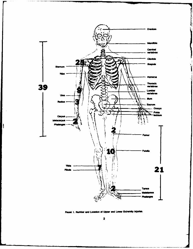
The frequency and distribution of the 39 upper extremity and 21 lower extremity injuries are presented InFigure 1. Discussion of those specific Injury locations follows:
KNEE
The knee joint Is a synovial joint formed by the articulation of the distal femur, proximal tibia, and the posterioraspect of the patella. This joint Is the most massive articulation in the body; and Its exposed position makes Itvulnerable to many types of Injuries, Involving both bony and soft tissue components. The stability of the kneejoint Is dependent upon the ligamentous apparatus and the muscles that motorize the joint.
e Type: Ligaments-medial collateral tear-dislocation
Menisci-medlal meniscus tear
e Frequency: 44%
e Mechanism: The function of the ligament Is to prevent abnormal motion of the joint In a particular direc-tion. If the stress applied to a joint Is of sufficient Intensity to produce abnormal motion, the protecting ligamentwill be injured. For Injury the foot Is forced In external rotation and an abducting force occurs at the outer aspectof the knee, forcing the thigh to rotate Inward and the calf to rotate outward.
e Etiology: Windblast causes the foot to be forced back and out, after the extremity becomes maipoelonedbeyond the geometry of the sat. An abducting force at the outer aspect of the knee forces rotation beyond therange of motion of the joint. Forced external rotation, accompanied by vaigus movement of the foot and calfcause Injury.
The common fractures about the knee are the supracondylar fractures and Intercondylar on the femur (dis-cussed under long bones).
ANKLE
This synovial joint Is of the hinge varlety; however Its axis of rotation Is not fixed but changes between extremesof plantar flexion and dorshfiexion.
" Type: Malsol-Medlal malleolus fractureDislocation-lateral subtalar dislocation
" Frequency. 9%
" Mechanism: The bony configuration of the ankle joint provides inherent stability. The joint Is comrisedof the distal end of the tibia and the medial malleolus on one side, and the distal end of the fllbua and the medialmallelous on the other. The malleoli are of unequal iength and shape. When the foot Is plenta-flexed the narrowposterior portion of the talus advances forward Into the mortise. This position produces lateral Instablity. In
1 m
-l uII "-Ii -- I i II i ~ i n
Humerus
Nlu
RafteH-cum
m's ' <: 21
Pcain 1. Nwnber an LoostW of Upper and Low Extt hMutSs

general, Injuries result from sideward stresses forcing the ankle beyond normal arcs of motion. As the foot InvertIn relation to the leg, the lateral collateral ligamnt Is stretched and tears If the force continues. Further con-tnuation of the force Jams the talus against the medial malleolus. The tip of the malleolus engages the body ofthe talus, providing a fulcrum which cause the talus to rotate over the mafleclua. The resulting rupture of thelateral ligament and fracture of the medial mallolus predisposes dislocation, laterally.
* Etiology: Rotation and Inversion of the foot with Intense lateral foroL This condition is created when thefoot is caught In the windstreem beyond the seat protection and the foot/calf Is thrust backward and outward.
SIHOULDER
The shoulder joint is a synovial joint of the ball and socket variety. There Is marked disproportion between thesmall sallow glenold fooa and the large round head of the humerus. The shoulder is the most movable andpossbly the least stable of all the Joints of the extremities.
" Type: 8cepula-fractureQienold--rm fracture
laloction-subglenoldNote: the humerus is addressed as a long bone In another section.
" Frequency. 13%. Acute dislocations are frequently associated with fractures of the bones of the shouldergirdle. This complication Is found in approximately 25% of all dislocations; the greater tuberoeity is mostfrequently Involved (18-20%) (Depalma, 1970).
* Mechanism: Any alteration from the range of motion of the glenohumeral joint results In spain, dislocationor fracture of the head of the humerus. The complete mobility of the upper arm is possible through action of theglenohumeral Joint as well as articulation with the clavical and scapula. Impairment of any one reflects in impairedfunction of the shoulder, even though independent motion remains possible.
Fractures of the scapula are usually the result of a violent direct force.
Avulion of portions of the glenold rim can occur In acute dislocations of the joint. The fragment may detachanteriorly, posteriorly or Inferiorly, determined by the type of dislocation reflected in the final resting place of thehumeral head.
inferior dislocation occurs when the arm is hyperabducted beyond the limits of the pivotal position. Inferiordislocation occurs If hyperabduction is continued after the arm has reached the pivotal position. The sequenceof the mechanism Is
ForceThe arm Is locked In the pivotal position and the scapula Is completely rotated.
Force continuesThe acromlon, acting as a fulcrum, displaces the greater tuberolty and the Inferior capsule is satched.
Force continue.The head leaves the glenold cavity and Is displaced medially and Inferiorly. The Inferior capsule iS torn. The
rotator cuff Is stretched and the arm drops. The head Is In the subgisnold position.
* Etilogy: Hyperabductlon of the arm. Windblast forces exceed the physical limit of restraint. The forearmand hand (supine) ae forced up and back, the elbow Is fully extended as the scapula completely rotates and thehypeabduction mechanism is established. Associated glenoid rim fracture may be observed.
3

ULfi W
The elbow Is a synovial joint formed by the articulation of the distal humerus and the proximal ulna and radius,forming a humero-ulnar and humero-radlal articulation. The only appreciable movement possible at the elbowjoint is the simple hinge movement of flexion and extension. This movement takes place in a lne oblique to thehumerus.
" Type: Fracture-interarticularDiloretion-posterlor
" Frequency:. 25%
" Mechanism: The general mechanism of Injury Is posterior displacement of both bones of the forearm.Severe concomitant soft tissue injury results. Because the radius and ulna are firmly bound by the annular liga-ment and Interosseous membrane, a dislocation of one Is usually primary to fracture or dislocation of the adja-cent bone. Hyperextenslon of the Joint disrupts the articulation, with subsequent posterior boney displacement.
Comminuted Interartlicular fracture Is caused by trauma that drives the olecranon against the articular end ofthe humerus producing comminution of the distal end of the humerus. FlexIon forces the ulna between thefragments of the distal end of the humerus.
* Etiology:. Hyperextenslon of the joint results from severe arm ioads forcing the forearm back and drivingthe elbow joint beyond the limit of Its range of motion.
FEMUR
The thigh bone extends from the hip to the knee and Is the iongest and strongest bone In the body. Fracturesof the femur are usually the result of severe violence. They may be transverse, oblique, greenstick, spiral orseverely comminuted.
" Type: Fracture, mid-shaft
e Frequency:. 9%
" Mechanism: Fracture of the femur is the result of traumatic Impact, torsion or bending. Torque applied tothe distal femoral shaft will Induce stress at the mid-shaft. Intense local force application results in soft tissueInjury at the Injury eight.
* Etioogy: Torque applied to the distal femoral shaft by rotation/abduction of the knee joint/calf/foot dueto wndblest.
& Severit: Usually, there is cardiovascular shock from the trauma to bone and soft tissue. Fracture of theshaft of the femur may be accompanied by marked concealed blood loss.
The tObia-fbula articulations are bound together by ligamentous fibers and Interoseous membrane. Thepresence of a synovial Joint at the upper and of the two bona Indicates movement, but this movement Is entirelypasive. The tibia articulates with the femur above and the talus below. The fibula articulates above with the tibiaand blow with the tibia and talus.
e Type. Fracture-spiralcomminutedplateau (knee joint surface)
4

" Frequency: 33%
* Mechanism: The leslons are produced by an angulatory or rotational force. Generally, angulatoy forcesproduce transverse or short oblique fractures of both bones at the same level. Rotational forces producetransverse or short oblique fractures of both bones at the same level. Rotational forces produce spiral fracturesat different levels (fibula fracture usually at a higher level than the tiblal fracture). Fracture/dislocation of the anklemay be primary to fibula fracture.
o Etiology: Twisting of the foot/ankle secondary to external rotation-valgus movement of the foot/call dueto windblest. Blow to the fibular head due to dual leg garter configuration.
RADIUS AND ULNA
The radius carries the hand and is stabilized against the ulna for pronation-supination, and against the humerusfor flexion extension of the forearm. The forearm Injuries are marked In that Impact with the aircraft structuregenerally provides the trauma Inducing force.
" Type: Fracture-proximalmid-shaftcompoundstylold processcoronold process (ulna)
" Frequency: 10%
" Mechanism: A direct localized blow precipitates mid-shaft fracture. The type of Injury sustained by the heador neck of the bone depends upon the Intensity of the force applied and the position of rotation of the radius atthe time of Impact. The capitallum drives the radial bead outward, the direction of tilt depends on the rotationalposition of the radius.
e Etiology Fuselage Impact or pronating beyond anatomical limits resulting In posterior dislocation of theelbow by windbast Induced hyperextenslon. After the break in continuity of the bones has occurred, themuscles controlling the different segments come Into action and play a major role In the final position thefragments assume.
HUMERUS
Upper bone of the arm from the elbow to the shoulder Joints, articulating with the ulna and radius and scapula.As such forces predisposing Injury at these joints may involve the humerus.
" Type: Fracture-supracondylarheadgreater tuberoltymid-shafttransverse
" Frequency- 5%
" Mechanism: Most fractures of the shaft of the humerus are the result of direct violence. Involvement withshoulder and elbow injury Is responsible for proximal fractures.
e Etiology. Direct violent blow to the long bone from fuselage Impact. Force from wlndblat abductionl
dislocation of the Shoulder or hyperextndlon of the elbow.
5

RANGE OF MOTION
Three sources of range of motion determination are presented for the major joints In Table 1. The magnitude ofrange Is of Interest because angular momentum (rotational velocity and Its product with the appropriate anatom-ical mass) Is a potentially high energy phenomenon occurring at the shoulder (and knee less dramaticlly) Whenthe lfoor/humerus ae taken Into rotiry/potsrlor motion by windblst. The physiological dynamics of disrlb-uting and dissipating the energy of this motion awe engaged when the limit of the hyperabducted joint Is reached.Fracture Is a major energy dissipation actt.
TABLE 1
NORMAL RANW OF MOTION OF JOINTS IN MALE SUBJECTS
Co~l of PAnges of MoW (D res)
,nfrn A2Weny ofJot OrHvfftw " OWsu. 1) Oepw (O. 2) ftirt @at . INoV. a
Sh1Norkont fIfalow 136 140.7 t 5.9 134 ± 7H ehm actenlon - 45.4± 6.2 46 ± 9Forwrd fladon 16 166.7 4.7 186± 12Bk wctenelon 53 62.3 ±. 01 ±14
Rado 146 142.0 5.6 142 ±10ExtensOln 0 0.0 ± 3.1
Forearm~
Proneton 71 75.8 ±5.1 77± *24Supe 64 82.1 ± 3.8 113 ± 22
Wris
Fladon 73 70.4 ± 6.3 90± 12Extesd on 71 74.9 ± 6.4 99 13
Knee
Radon 134 142.5 ± 6.4 144±0
Ankle
A ldonWt) 48 56.26.1 * V 7Eideneon dorsfluidon) 10 12.6 ± 4.4 3 612
Forepef of tge footIInvmeon 33 3.8 ± 4.5 24±9EveON 1 s 20.7 ±5 .0 23± 7
8TRENGTN
Static capability of caefos bone In Omprev load ISng is psted In Tae 2as determined by sevenInvestigatore, and summarized In Combs, 1978, and Evans, 1967.
The dynamic environn t of operational InJurie was approached In Engin's (1070) measurement of retvemuscle foroe and momnt (Table 3). Selected Joints appear from tht work.
6

TABLE 2
8OMd PROPERTIES OF HUMAN. VERTEBRAL BODY. CANCELLOUS BONE (Ref. 3,7)(- UWae through lmbar)
Ea MXodus LUmnate co oeiwrnAuthor in compressn atrM RAW
MN/rn' MN/rn'
Galmnte, 1970 sup-inf. 2.0a-p 0.8lot. 0.7
McEhslney. 1970 152 4.14 0.14
Rackoff. t W., 1920 2.1-15.8 Intact bodyapprox. 0.8-6.3traacular bone only
Yamada, 1970 70-0 comprsion 1.4-1.9 compression(Sonoda)" (330 tension 1 specimen) (3.7-4.0 tension)
LIn. 1976 sup-nf 1117 0.36
a-p & lateral 558
Llndahl, 1976 1.1-139 1.0-7.0
Kazarlan, 1977 22.0-290.0 2.2-9.5
Evans, 1973(summary of7 authors)
IncludingMcEhalney &G0lant. 1.4-8.0
og" Iu
To allow operational comparison on the laboratory measured moment magnitudes, wind-tunnel pressure coeffi-cient data were used to develop the values In Table 4. Following is the sequence used in deriving the force (N)value of column four In Table 4:
(1) A 1/32 ale F4E aircraft model was Investigated In a 5 foot diameter, low-speed, wind-tunnal. The Scalemodel pilot was affixed at the top of the catapult sequence, Its feet level with the front canopy windshield, bothtotally enveloped in the flow. Static ports were drilled and miaometer Instrumented at selected anatomicallocations. The pressure coefficients were measured.
(2) The recognized formula: F-lpv'CpS was used to calculate N/m 2 at the elbow, knee (side) and foot (toe)locations, for comparison.
(3) The anatomical area was estimated using mean value to determine elbow area, side of the knee area andfrontal area of the foot. This allowed calculation of force (N) at that point.
(4) This value (Table 4, column 4) was then used with the moment arm, column 1, to calculate the magnitudeof moment (N-m) of column 5.
(5) These data can be compared with Table 3 containing resistive moments to develop an appreciation for theWeverIty of the windblast condition.
7

At 500 KTS, all the experienced moment$ are far beyond the resistive values given In Table 3. At a Slower 300KTS there Is relative likeness. Remnember, Tabl 3 data were collected with the participant anticipating theenvironment and focused on a single externiall force. The rapid establishment (1-2 seconds) of wlndblaSt loadingcould further reduce possible Successful engagement of that environment.
TA3LE 3MAGNITUDES OF ACTIVE RESISTIVE MUSCLE FORCE AND MOMENTS (ENGiN: 1979)
Joint, POtlo MeVtude Subject No. I Subject No. 2 Subject No. 3
Shouilder, lateral exteralon Force (N) 140.01 144.91 136.61163.83 147.02 161.92106.14 160.32 177.83
Moment (N-rn) 49.38 55.15 66.2957.76 56.64 62.2666.93 62.29 69.48
ElbowLower arm. 900 aupnation Moment (N-rn) 16.78 14.25 13.53
22.05 14.97 14.7722.91 16.93 15.03
Lower arm, 90* pronatlon Moment (N-rn) 21.95 14.17 16.2329.16 17.18 22.6129.35 20.78 25.10
KnotLowe Mo., rotated lateral limit Moment (N-rn) 36.92 23.07 43.63
- 25.17 46.71- 32.24 49.59
Lower leg, rotated medial limit Moment (N-rn) 55.47 31.07 63.94- 35.65 93.14
-41.61 101.76
AnkleTiblal rotation, medial Moment (N-rn) 41.92 17.63 56.1
47.11 23.40 66.0450.38 25.30 74.14
Tiblal rotation, lateral Moment (N-rn) 30.11 20.99 52.6130.66 22.49 56.6332.04 24.52 61.78

TABLE 4
OPERATIONAL ENVIRONMENT MAGNITUIE OF MOMENTS
Joint Lower (m) Akupwd (KTS) Fboo (N) Movnt (N-m)
Shoukler 0.504' 300 36 18.1500 103 52.0
Knee 0.4532 300 73 33.0500 220 110.0
Ankle 0.263 300 130 34.3500 361 96.0
malt. ultM Ito mguIbOm c. 1076;)
* nMiu, bottm ot to ltS (tkl)* faul, toS to ujid. Oad)
* 94inm 1070)ona dWJW y of ak Und.
BIOMECHANICAL DATA
The blomechanical properties of long bones vary significantly with geometry, material properties, loadingmethod, pathology, etc. Although there are numerous studies on the mechanical properties of bone in theliterature, there are few that investigate intact long bone and joint failure precipitated by specific modes ofdisruption, resulting from experiences with force environments.
The majority of literature deals with the nature and physical properties of bone and, to some extent, static loadingof intact bone. A broad summary of these efforts Is given by Evans (1957) in his book entitled Stress and StrainsIn Bone. More recent surveys have been reported by Swanson (1971) and Kummer (1972).
So-called flail Injuries, occurring as a result of large magnitude aerodynamic forces on the higher and lowerextremities of the body, are represented by failure of the major articulating joints being forced beyond theanatomical limit of range and load of the structure as well as long bone trauma. Frequency of such Injury had beendocumented by Combs (1978). Determination of voluntary range of motions, resistive force and movements andresistive torques for the rotational motion of the body segments about their long bone axis was accomplished byEngin (1979).
There is much that is still to be learned about long bone and joint response to mechanical loading. These Injurieshave significant operational Impact and can be predisposing of long-term degenerative changes. Clinical in-vestigations have been inadequate because there has been no way to assess the forces, their rate and directionof application In an actual Injury. Present research programs are directed to simulate observable and explainableInjury modes produced by windblast flailing, with bones and joints experiencing similar effects and constraintsas those defined by the operational environment.
9
REEMNCES
1. Ametcan Acadwmyof Ohpeedic wesoflJoint lln: ANA Mtd@ eaeu*,g ad A~orl'dV iM&2. Sorter. A.T. atel. (1067) A SteIMbaIWEW Mllof Jointfang Dat WADGTechica Note 57-311, AeroMedca Laborltoy. WVVght
Patterson, AFS. OH.3. Combs. &P. (1078) Corrialff of Mfolnedin of ftjwy OWd Awcmwkmi Factors In Ejection horn F-4 Aftriaft AFAALX-7-4*
Aerospace Medical eserolt Laboratory. WrIght-Pattrson AFB. OH.4.o PaiMs A.F. (1070) "Me Mfimaent of Arcwein &Wd Oblosone-an ads vokame OniL W.B. Sounders Compeny. Ph~ededpMLa5.0Do Palms A.F. (1070) The Maonagmnrt of Fracwy and DMIaloaone-Aw M~eg Vowe Twos. W.B. Saunders Compeny, Pluladeopts.6. Engln, A.E. (1970) Afaseirnnt of Reallelw Torques I Majr hman Joia. AFAMRL-TR-79-4, Aerooeo Medical RAeerch Labors-
tory, WrIght-Patterson AFS, 0OH.7. Evens FAG (1957) Sbwam and S&Oai In fiorme Charles C. Thoas, Springfiel.8. HAnoleNJ and Kroh, G. (1075) A MevfewofAnthrcornt~k Data ofaweman AlrForce andL*~te St~eAfrFveF"VJgPoWlet
108-1N. AQARDogrp 205.9. Last. R.J. (1078) ANATOMY-Regonil and Apphhd. Churchill Livngstone, New York.
10. Schultz. R.J. (1972) The Language of Fractures. The WNNims and ilkins Company. Baltiyme.
10
*U.S.Government Printing OffIce: 1961 - 757-002/475

*1