advances in stem cell therapy for spinal cord...
TRANSCRIPT
Science in medicine
3824 The Journal of Clinical Investigation http://www.jci.org Volume 122 Number 11 November 2012
IntroductionSpinal cord injury (SCI) is a devastating condition, with sudden loss of sensory, motor, and autonomic function distal to the level of trauma. Despite major advances in the medical and surgical care of SCI patients, no effective treatment exists for the neu-rological deficits of major SCI (1). Current treatment includes surgery to decompress and stabilize the injury, prevention of secondary complications, management of any that do occur, and rehabilitation. Unfortunately, neurological recovery is lim-ited, and most SCI patients still face substantial neurological dysfunction and lifelong disability. Stem cell therapy offers sev-eral highly attractive strategies for spinal cord repair, including replacement of damaged neuronal and glial cells, remyelination of spared axons, restoration of neuronal circuitry, bridging of lesion cavities, production of neurotrophic factors, antiinflam-matory cytokines, and other molecules to promote tissue spar-ing and neovascularization, and a permissive environment for plasticity and axonal regeneration. This review builds on several excellent previous reviews (2–8) and discusses the incidence and pathophysiology of SCI as well as the key experimental and clini-cal stem cell strategies for SCI.
Epidemiology, etiology, incidence, and prevalence of SCIWorldwide, the annual incidence of SCI is 15–40 cases per mil-lion people (9). In Canada, the Rick Hansen Institute estimates there are currently 85,000 people living with SCI, with more than 4,000 new cases per year (10), and in the United States, the Christopher and Dana Reeve Foundation estimates a prevalence of over 1 million patients with SCI and more than 12,000 new cases each year (11).
The primary causes of traumatic SCI are motor vehicle crash-es, sports and recreation injuries, falls at home, and trauma at work (12). In young adults, males are four times more likely than females to sustain an SCI (13). Injury incidence shows a bimodal distribution, with the highest incidence in adolescents and young adults, with more than half aged 16–30 years old (10). The second incidence peak is in older adults, primarily as a result of falls, and the aging population has increased the average age of injury.
Clinical trial design and management of SCIAssessment of therapy in patients has improved markedly due to the development of the American Spinal Injury Association (ASIA) grading scale and quantitative scores of sensory and motor function now used worldwide to assess the severity of SCI and response to treatment (1). The ASIA Impairment Scale (AIS) ranges from A to E, where A is a complete SCI and E denotes normal sensory and motor function. Acute treatment often involves surgical management, such as decompression, spi-nal stabilization, or realignment of displaced vertebrae (14) to prevent further injury from impingement on the spinal cord. Acutely injured patients often require intensive care monitoring to treat cardiovascular instability and respiratory insufficiency. Currently, there is limited pharmacotherapy for SCI patients. Methylprednisolone demonstrated some neuroprotective effects in early experimental and clinical studies (15, 16), but its use is controversial because of limited efficacy and harmful side effects. Many SCI centers have stopped using steroids (17). Other neu-roprotective agents with promising results in experimental ani-mals are now being investigated in clinical SCI trials, including riluzole, a sodium channel blocker, and minocycline, an antiin-flammatory agent (1, 18). However, neuroprotective agents alone may be insufficient to promote repair in major SCI where there is extensive tissue loss.
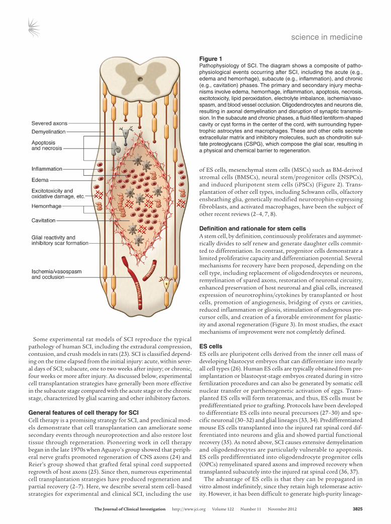
Pathophysiology of SCIThe most frequent type of traumatic SCI is acute compression of the spinal cord (12). Usually, some neurological tissue is preserved, particularly in the subpial region (19, 20). The primary mechani-cal trauma causes necrosis, edema, hemorrhage, and vasospasm. A cascade of secondary pathophysiological mechanisms is induced, including ischemia, apoptosis, fluid and electrolyte disturbances, excitotoxicity, lipid peroxidation, production of free radicals, and an inflammatory response, resulting in further damage due to swelling and blood flow reduction (21). Ultimately, a large fluid-filled cavity or cyst forms in the center of the cord, surrounded by a subpial rim containing some preserved axons, many of which are demyelinated (Figure 1). Hypertrophic astrocytes, macrophages, and other cells secrete extracellular matrix and inhibitory mol-ecules that constitute the glial scar, resulting in a physical and chemical barrier to regeneration (22).
Advances in stem cell therapy for spinal cord injuryAndrea J. Mothe and Charles H. Tator
Toronto Western Research Institute and Krembil Neuroscience Centre, Toronto Western Hospital, Toronto, Ontario, Canada.
Spinal cord injury (SCI) is a devastating condition producing great personal and societal costs and for which there is no effective treatment. Stem cell transplantation is a promising thera-peutic strategy, though much preclinical and clinical research work remains. Here, we briefly describe SCI epidemiology, pathophysiology, and experimental and clinical stem cell strategies. Research in stem cell biology and cell reprogramming is rapidly advancing, with the hope of mov-ing stem cell therapy closer to helping people with SCI. We examine issues important for clinical translation and provide a commentary on recent developments, including termination of the first human embryonic stem cell transplantation trial in human SCI.
Conflict of interest: The authors have declared that no conflict of interest exists.
Citation for this article: J Clin Invest. 2012;122(11):3824–3834. doi:10.1172/JCI64124.
science in medicine
The Journal of Clinical Investigation http://www.jci.org Volume 122 Number 11 November 2012 3825
Some experimental rat models of SCI reproduce the typical pathology of human SCI, including the extradural compression, contusion, and crush models in rats (23). SCI is classified depend-ing on the time elapsed from the initial injury: acute, within sever-al days of SCI; subacute, one to two weeks after injury; or chronic, four weeks or more after injury. As discussed below, experimental cell transplantation strategies have generally been more effective in the subacute stage compared with the acute stage or the chronic stage, characterized by glial scarring and other inhibitory factors.
General features of cell therapy for SCICell therapy is a promising strategy for SCI, and preclinical mod-els demonstrate that cell transplantation can ameliorate some secondary events through neuroprotection and also restore lost tissue through regeneration. Pioneering work in cell therapy began in the late 1970s when Aguayo’s group showed that periph-eral nerve grafts promoted regeneration of CNS axons (24) and Reier’s group showed that grafted fetal spinal cord supported regrowth of host axons (25). Since then, numerous experimental cell transplantation strategies have produced regeneration and partial recovery (2–7). Here, we describe several stem cell–based strategies for experimental and clinical SCI, including the use
of ES cells, mesenchymal stem cells (MSCs) such as BM-derived stromal cells (BMSCs), neural stem/progenitor cells (NSPCs), and induced pluripotent stem cells (iPSCs) (Figure 2). Trans-plantation of other cell types, including Schwann cells, olfactory ensheathing glia, genetically modified neurotrophin-expressing fibroblasts, and activated macrophages, have been the subject of other recent reviews (2–4, 7, 8).
Definition and rationale for stem cellsA stem cell, by definition, continuously proliferates and asymmet-rically divides to self renew and generate daughter cells commit-ted to differentiation. In contrast, progenitor cells demonstrate a limited proliferative capacity and differentiation potential. Several mechanisms for recovery have been proposed, depending on the cell type, including replacement of oligodendrocytes or neurons, remyelination of spared axons, restoration of neuronal circuitry, enhanced preservation of host neuronal and glial cells, increased expression of neurotrophins/cytokines by transplanted or host cells, promotion of angiogenesis, bridging of cysts or cavities, reduced inflammation or gliosis, stimulation of endogenous pre-cursor cells, and creation of a favorable environment for plastic-ity and axonal regeneration (Figure 3). In most studies, the exact mechanisms of improvement were not completely defined.
ES cellsES cells are pluripotent cells derived from the inner cell mass of developing blastocyst embryos that can differentiate into nearly all cell types (26). Human ES cells are typically obtained from pre-implantation or blastocyst-stage embryos created during in vitro fertilization procedures and can also be generated by somatic cell nuclear transfer or parthenogenetic activation of eggs. Trans-planted ES cells will form teratomas, and thus, ES cells must be predifferentiated prior to grafting. Protocols have been developed to differentiate ES cells into neural precursors (27–30) and spe-cific neuronal (30–32) and glial lineages (33, 34). Predifferentiated mouse ES cells transplanted into the injured rat spinal cord dif-ferentiated into neurons and glia and showed partial functional recovery (35). As noted above, SCI causes extensive demyelination and oligodendrocytes are particularly vulnerable to apoptosis. ES cells predifferentiated into oligodendrocyte progenitor cells (OPCs) remyelinated spared axons and improved recovery when transplanted subacutely into the injured rat spinal cord (36, 37).
The advantage of ES cells is that they can be propagated in vitro almost indefinitely, since they retain high telomerase activ-ity. However, it has been difficult to generate high-purity lineage-
Figure 1Pathophysiology of SCI. The diagram shows a composite of patho-physiological events occurring after SCI, including the acute (e.g., edema and hemorrhage), subacute (e.g., inflammation), and chronic (e.g., cavitation) phases. The primary and secondary injury mecha-nisms involve edema, hemorrhage, inflammation, apoptosis, necrosis, excitotoxicity, lipid peroxidation, electrolyte imbalance, ischemia/vaso-spasm, and blood vessel occlusion. Oligodendrocytes and neurons die, resulting in axonal demyelination and disruption of synaptic transmis-sion. In the subacute and chronic phases, a fluid-filled lentiform-shaped cavity or cyst forms in the center of the cord, with surrounding hyper-trophic astrocytes and macrophages. These and other cells secrete extracellular matrix and inhibitory molecules, such as chondroitin sul-fate proteoglycans (CSPG), which compose the glial scar, resulting in a physical and chemical barrier to regeneration.

science in medicine
3826 The Journal of Clinical Investigation http://www.jci.org Volume 122 Number 11 November 2012
specific cell lines without karyotypic abnormalities. The concerns with transplantation of ES cell–derived neural cells for SCI are the ethical issues of cell derivation and the possibility of tumorigen-esis due to incomplete or aberrant differentiation resulting in the formation of nonneural cells (Table 1).
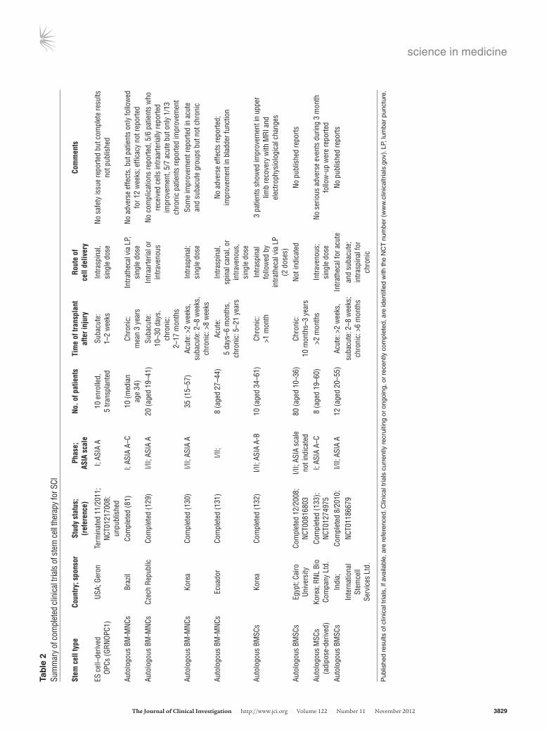
Based on promising preclinical data of human ES cell–derived OPC transplants in rodent SCI models (36, 37), the US Food and Drug Administration (FDA) approved the first human ES cell trial in 2009. This phase I safety trial in SCI sponsored by Geron Corp. began in 2010 after further preclinical safety data were obtained concerning abnormal cyst formation in transplanted animals. The GRNOPC1 cell line (human ES cell–derived OPC) was transplant-ed subacutely (one to two weeks after injury) directly into the spi-nal cord of ASIA A patients with complete thoracic SCI. Patients received 2 million cells and were immunosuppressed for the first two months following transplantation. In 2011, Geron discon-tinued this trial due to funding challenges. No safety issues were reported in the five patients who received GRNOPC1 transplants, but complete results have not been published (Table 2).
NSPCsNSPCs are multipotent cells committed to the neural lineage that can self renew and be readily expanded in vitro. NSPCs are typically grown as free-floating neurospheres in serum-free medium supplemented with EGF and FGF-2. Neurospheres are 3D aggregates comprising a mixture primarily of progeni-tor cells, a small percentage of stem cells, and small numbers of more differentiated cell types over multiple passages in culture. The neural stem cells respond to the growth factors in the cul-ture medium and selectively proliferate in suspension to form neurospheres. When these cells are plated in growth factor–free medium containing serum, they differentiate into neurons, oli-godendrocytes, and astrocytes.
NSPCs are found in both fetal and adult CNS (38). The isolation of adult neural stem cells in mammals was first reported in 1992 by Reynolds and Weiss (39). NSPCs reside within specific niches in the adult CNS, including the subventricular zone lining the lateral ventricles of the forebrain (40, 41), the dentate gyrus of the hip-pocampus (42), and the region of the central canal of the spinal cord (43). Multipotential, self-renewing NSPCs can be isolated and cultured from the adult rodent spinal cord when the cultured tis-sue includes regions of the central canal (44, 45). We have shown that these NSPCs primarily differentiate into oligodendrocytes in vitro (46) and in vivo (47, 48). Transplantation of these NPSCs into SCI rats promoted functional recovery with neuroprotective and neuroregenerative effects (49–51). Most studies with trans-planted NSPCs have shown modest recovery of the injured spi-nal cord (3, 7), although allodynia was associated with astrocytic differentiation of grafted NSPCs (49). Adult mouse brain–derived NSPCs transplanted into the injured rat spinal cord with con-comitant infusion of growth factors promoted oligodendrocyte differentiation of the grafted NSPCs, remyelination, and improved locomotor function (52, 53). NSPCs derived from fetal rat spinal cord differentiated into neurons that integrated into the injured cord and improved recovery (54), and transplanted NSPCs com-bined with valproic acid administration promoted neuronal differ-entiation, resulting in restoration of disrupted neuronal circuitry and enhanced recovery (55). NSPCs have also demonstrated some immunomodulatory and pathotropic ability by homing toward damaged tissue (56, 57) as well as secreting various neurotrophic factors and cytokines (58–60).
Most experimental SCI studies with NSPC transplants have involved rodent cells because human stem cells were either not available or difficult to grow. Human NSPCs have been isolated from fetal brain and spinal cord from aborted fetuses (61–65) and from adult brain from surgical biopsy specimens and post-
Figure 2Sources of stem cells for transplantation into the injured spinal cord. This illustration shows various tissue sources of stem cells, including NSPCs, iPSCs, SKPs, MSCs, ES cells (ESC), and direct conversion methods to yield neural cells for transplantation. NSPCs can be iso-lated from the fetal and adult brain and spinal cord and differentiated into progenitor cells, such as OPCs and mature oligodendrocytes, or astrocytes or neurons depending on culture conditions and exposure to growth factors. ES cells follow a default pathway to neural cells, and specific conditions can promote OPC gen-eration. MSCs can be derived from a variety of tissues, including BM, umbilical cord, adipose tissue, muscle, and dental pulp from decidu-ous baby teeth. In culture, MSCs have shown properties of neural cells. Fibroblasts from the skin can be reprogrammed using various methods into iPSCs,which are then directed along a neural lineage. Recent studies have directly converted fibroblasts to neurons and NSPCs, bypassing the pluripotent stage.

science in medicine
The Journal of Clinical Investigation http://www.jci.org Volume 122 Number 11 November 2012 3827
mortem tissue (66–69). Recently, we demonstrated that self-renewing multipotent NSPCs can be passaged from the adult human spinal cord of organ transplant donors and that these cells differentiate into both neurons and glia following trans-plantation into rats with SCI (70). Stem cells isolated from the human fetal brain were transplanted into NOD/SCID mice with SCI, and the grafted cells expressed neural differentiation markers and improved recovery (71, 72). Extensive neuronal differentiation of human fetal NSPC grafts was reported after transplantation into the adult rat spinal cord (73). In addi-tion, human fetal brain NSPCs (modified to express galectin-1) transplanted subacutely into the contused cervical spinal cord of adult common marmosets produced significantly greater grip strength than controls (74).
A registry of government and privately supported clinical tri-als from all countries is available (http://www.clinicaltrials.gov). Table 2 summarizes completed trials of stem cell therapy for SCI; Table 3 indicates ongoing trials. Recently, Stem Cells Inc. started a phase I/II (safety/efficacy) trial in Switzerland involv-ing transplantation of human fetal brain stem cells into ASIA A–C patients with thoracic SCI. Currently, this is the only human trial involving NSPCs for SCI, and these patients require immu-
nosuppression. Thus, human NSPCs have certain drawbacks, including ethical concerns about fetal-derived cells, difficulties in expanding adult-derived cells to clinically sufficient numbers, and unavailability of autologous sources.
Skin-derived precursorsAn accessible, potentially autologous source of precursor cells for transplantation is skin-derived precursors (SKPs) residing within the dermis of rodents and humans, as described by Miller and other groups (75–77). SKPs are generated during embryo-genesis, persist into adulthood, and share characteristics with embryonic neural crest stem cells, producing both mesodermal progeny and peripheral neurons and Schwann cells (78). SKP-derived Schwann cells transplanted into SCI rats showed lesion sparing, remyelination of spared axons with peripheral myelin, and, unlike other sources of Schwann cells, provided a conducive environment for axonal growth into the lesion, but with limited functional recovery (79).
HSCs and MSCsAdult BM contains several different stem cell populations of non-neural origin, including HSCs and MSCs. HSCs are self-renewing adult stem cells found mainly in the BM that differentiate into blood and immune cells. In the early 1960s, James Till and Ernest McCulloch observed that BM cells injected into irradiated mice grew as colonies of cells in their spleens, and each colony grew from a single cell (80), now known as a stem cell. These Canadian scientists are recognized as the founders of HSC research. HSC transplantation has now been used for decades to treat blood can-cers and other blood disorders (81).
HSCs are identified based on their expression of distinct cell-surface markers and nonadherence to plastic in culture. Typical-ly, erythrocytes and platelets are separated from BM yielding the mononuclear cell fractions (MNCs) comprising HSCs and nonhe-matopoietic cells, including monocytes, macrophages, endothelial progenitor cells, and small numbers of MSCs. The advan-tages of HSCs or BM-derived mononuclear cells (BM-MNCs) are that they can be autologously derived and that they have a record of safety in humans. The drawbacks are that HSCs are rare (1 in 100,000 cells from BM) and pose major risks with graft rejec-tion and graft-versus-host disease (GvHD). Subacute intraspinal transplantation of HSCs was shown to promote functional recov-ery after compression SCI in mice (82, 83). For clinical transla-tion, HSCs are commonly harvested from peripheral blood fol-lowing cytokine administration, which mobilizes the HSCs, and the MNC fraction is administered.
Figure 3Potential mechanisms of spinal cord repair following stem cell trans-plantation. The diagram shows some of the potential mechanisms of repair after transplantation of stem cells into the injured cord. Potential mechanisms include replacement of oligodendrocytes or neurons by transplanted cells (shown in green), remyelination of spared axons, restoration of neuronal circuitry by a new synapse with a transplanted neuron that gives rise to a newly regenerated axon, enhanced preser-vation of host neuronal and glial cells, for example, by secreted neuro-trophins from transplanted cells, promotion of angiogenesis, bridging of the cyst/cavity by transplanted cells, reduction of inflammation or gliosis, stimulation of endogenous precursor cells, and creation of a favorable environment for plasticity and axonal regeneration.

science in medicine
3828 The Journal of Clinical Investigation http://www.jci.org Volume 122 Number 11 November 2012
The most common nonneural cell type for transplantation in experimental SCI is the BMSC. BMSCs are isolated from the MNC fraction of BM and expanded in culture based on their adherence to tissue culture plastic and expression of distinct cell-surface antigens that do not include HSC markers. The major advantage of MSCs is that they can be autologously transplanted and that they express a variety of neurotrophic factors that are beneficial for repair. Other important features are their low immunogenic-ity and their reported immunomodulatory properties (ref. 84 and Table 1). MSCs are widespread throughout a variety of tissues (85), including Wharton jelly of the umbilical cord, adipose tis-sue, adult muscle, and the dental pulp of deciduous baby teeth. Recently, predifferentiated adipose-derived MSCs were transplant-ed into SCI rats, resulting in some functional recovery, perhaps due to paracrine effects of grafted cells wrapping demyelinated host axons and promoting their protection (86). Umbilical cord–derived HSCs or MSCs are attractive because this tissue is readily accessible and frequently discarded, and MSCs are less prone to rejection, as evidenced by a lower risk of developing GvHD (ref. 87 and Table 1). Compared with adult sources, the number of MSCs or HSCs obtained from cord blood or placental tissues is small, although they can be readily expanded and tissue can be frozen and used later for isolation (88).
Transplantation of BMSCs for SCI has been previously reviewed (2, 3, 6, 89, 90). Many studies have examined BMSCs in SCI rodents, with some showing improved locomotor recov-ery (91–93), while others did not (51, 94, 95), likely due to dif-ferences in culture conditions. Several studies have also shown MSC differentiation into neural lineages in vitro, although in vivo, this is controversial (88–90). Cumulative evidence suggests the therapeutic effects of MSCs are likely due to secretion of neurotrophins, angiogenesis, and antiinflammatory actions (60, 93, 96, 97). HSCs and MSCs have also shown variable efficacy when transplanted intravenously or intrathecally demonstrat-ing pathotropism (98–100). Despite these potential benefits,
there are reported adverse effects of MSCs, such as increased recurrence of hematological malignancies and enhanced tumor growth and metastases (90, 101, 102).
There are a number of completed and currently ongoing SCI clinical trials involving autologous BM-MNC or BMSC transplan-tation as summarized in Tables 2 and 3. There are other reports of small numbers of patients treated with MSC transplants show-ing no adverse effects (103, 104). Collectively, evidence suggests that even transient MSC engraftment may exert favorable effects through secretion of cytokines and other paracrine factors that engage and recruit recipient cells in tissue repair (105).
iPSCsiPSCs are generated by reprogramming mature, fully differ-entiated cells into a pluripotent state. The advantage of iPSCs is that easily accessible cells such as skin from an SCI patient could be reprogrammed, differentiated, and transplanted. iPSCs were developed in 2006 by Takahashi and Yamanaka, who showed that mouse somatic cells, such as fibroblasts, could be reprogrammed to pluripotency with retroviral expres-sion of the transcription factors OCT4, SOX2, KLF4, and c-MYC (106). iPSCs can also be generated from human somatic cells (107, 108). The ability to generate pluripotent cells from adult somatic cells without the need for an embryo was a major development in stem cell biology.
Puri and Nagy recently compared ES cells and iPSCs (109). iPSCs share many key properties with ES cells, including morphology, pluripotency, self renewal, and gene expression. During expansion and prolonged passage, human ES cell lines frequently acquire abnormal karyotypes and genetic amplification associated with oncogenic transformation, which is also the case with iPSC lines (109). One of the main problems with generation of iPSCs is the expression of reprogramming factors associated with teratoma formation (110). For this reason, several alternative delivery meth-ods have been developed for reprogramming that do not require
Table 1Summary of advantages and disadvantages of potential stem cell types for SCI
ES cells NSPCs SKPs HSCs/BM-MNCs MSCs iPSCs iNPCs Fetal Adult BM- Other derived sources
Availability Yes Yes No Yes Yes Yes Yes Yes YesEase of isolation No No No Yes Yes Yes Yes No YesAutologous donors No No No Yes Yes Yes Yes Yes YesEthical considerations Yes Yes No No No No No No NoDifferentiation Pluripotent Neural Neural Nonneural Nonneural Nonneural Pluripotent Neural potential and and and peripheral potentially potentially myelin only neural neuralTumorigenicity Yes Some No Unknown No No Unknown Yes UnknownPathotropism Unknown Yes Yes Unknown Yes Yes Yes Unknown UnknownEfficacy in preclinical Yes, in some Inconsistent Yes, Yes, Yes, Inconsistent Yes, Yes, for Unknown studies studies in many but few in some in many preclonesSafety in human Yes, for ES Stem Cells Untested Untested Yes Yes Yes Untested Untested trials cell–derived Inc. OPC Geron trial
science in medicine
The Journal of Clinical Investigation http://www.jci.org Volume 122 Number 11 November 2012 3829
Tab
le 2
Sum
mar
y of
com
plet
ed c
linic
al tr
ials
of s
tem
cel
l the
rapy
for S
CI
Stem
cel
l typ
e Co
untr
y; s
pons
or
Stud
y st
atus
; Ph
ase;
No
. of p
atie
nts
Tim
e of
tran
spla
nt
Rout
e of
Co
mm
ents
(r
efer
ence
) AS
IA s
cale
afte
r inj
ury
cell
deliv
ery
ES c
ell–
deriv
ed
USA;
Ger
on
Term
inat
ed 1
1/20
11;
I; AS
IA A
10
enr
olle
d,
Suba
cute
: In
trasp
inal
, No
saf
ety
issu
e re
porte
d bu
t com
plet
e re
sults
OPCs
(GRN
OPC1
)
NCT0
1217
008;
5 tra
nspl
ante
d 1–
2 w
eeks
si
ngle
dos
e no
t pub
lishe
d
un
publ
ishe
dAu
tolo
gous
BM
-MNC
s Br
azil
Com
plet
ed (8
1)
I; AS
IA A
–C
10 (m
edia
n
Chro
nic:
In
trath
ecal
via
LP,
No
adv
erse
effe
cts,
but
pat
ient
s on
ly fo
llow
ed
age
34)
mea
n 3
year
s si
ngle
dos
e fo
r 12
wee
ks; e
ffica
cy n
ot re
porte
dAu
tolo
gous
BM
-MNC
s Cz
ech
Repu
blic
Co
mpl
eted
(129
) I/I
I; AS
IA A
20
(age
d 19
–41)
Su
bacu
te:
Intra
arte
rial o
r No
com
plic
atio
ns re
porte
d, 5
/6 p
atie
nts
who
10–3
0 da
ys,
intra
veno
us
rece
ived
cel
ls in
traar
teria
lly re
porte
d
ch
roni
c:
im
prov
emen
t, 5/
7 ac
ute
but o
nly
1/13
2–17
mon
ths
ch
roni
c pa
tient
s re
porte
d im
prov
emen
tAu
tolo
gous
BM
-MNC
s Ko
rea
Com
plet
ed (1
30)
I/II;
ASIA
A
35 (1
5–57
) Ac
ute:
>2
wee
ks,
Intra
spin
al;
Som
e im
prov
emen
t rep
orte
d in
acu
te
su
bacu
te: 2
–8 w
eeks
, si
ngle
dos
e an
d su
bacu
te g
roup
s bu
t not
chr
onic
chro
nic:
>8
wee
ksAu
tolo
gous
BM
-MNC
s Ec
uado
r Co
mpl
eted
(131
) I/I
I; 8
(age
d 27
–44)
Ac
ute:
In
trasp
inal
, No
adv
erse
effe
cts
repo
rted;
5 da
ys–6
mon
ths,
sp
inal
can
al, o
r im
prov
emen
t in
blad
der f
unct
ion
ch
roni
c: 5
–21
year
s in
trave
nous
,
si
ngle
dos
eAu
tolo
gous
BM
SCs
Kore
a Co
mpl
eted
(132
) I/I
I; AS
IA A
-B
10 (a
ged
34–6
1)
Chro
nic:
In
trasp
inal
3
patie
nts
show
ed im
prov
emen
t in
uppe
r
>1 m
onth
fo
llow
ed b
y
limb
reco
very
with
MRI
and
in
trath
ecal
via
LP
el
ectro
phys
iolo
gica
l cha
nges
(2
dos
es)
Auto
logo
us B
MSC
s Eg
ypt;
Cairo
Co
mpl
eted
12/
2008
; I/I
I; AS
IA s
cale
80
(age
d 10
–36)
Ch
roni
c:
Not i
ndic
ated
No
pub
lishe
d re
ports
Univ
ersi
ty
NCT0
0816
803
not i
ndic
ated
10 m
onth
s–3
year
sAu
tolo
gous
MSC
s
Kore
a; R
NL B
io
Com
plet
ed (1
33);
I;
ASIA
A–C
8
(age
d 19
–60)
>2
mon
ths
Intra
veno
us;
No s
erio
us a
dver
se e
vent
s du
ring
3 m
onth
(adi
pose
-der
ived
) Co
mpa
ny L
td.
NCT0
1274
975
si
ngle
dos
e fo
llow
-up
wer
e re
porte
dAu
tolo
gous
BM
SCs
Indi
a;
Com
plet
ed 8
/201
0;
I/II;
ASIA
A
12 (a
ged
20–5
5)
Acut
e: >
2 w
eeks
, In
trath
ecal
for a
cute
No
pub
lishe
d re
ports
Inte
rnat
iona
l NC
T011
8667
9
su
bacu
te: 2
–8 w
eeks
; an
d su
bacu
te;
St
emce
ll
ch
roni
c: >
6 m
onth
s in
trasp
inal
for
Se
rvic
es L
td.
chro
nic
Pub
lishe
d re
sults
of c
linic
al tr
ials
, if a
vaila
ble,
are
refe
renc
ed. C
linic
al tr
ials
cur
rent
ly re
crui
ting
or o
ngoi
ng, o
r re
cent
ly c
ompl
eted
, are
iden
tified
with
the
NC
T n
umbe
r (w
ww
.clin
ical
tria
ls.g
ov).
LP, l
umba
r pu
nctu
re.

science in medicine
3830 The Journal of Clinical Investigation http://www.jci.org Volume 122 Number 11 November 2012
Tab
le 3
Sum
mar
y of
ong
oing
clin
ical
tria
ls o
f ste
m c
ell t
hera
py fo
r SCI
Stem
cel
l typ
e Co
untr
y; s
pons
or
Stud
y st
atus
; Ph
ase;
ASI
A sc
ale
No. o
f pat
ient
s Ti
me
of tr
ansp
lant
Ro
ute
of
Com
men
ts
(ref
eren
ce)
afte
r inj
ury
cell
deliv
ery
Hum
an fe
tal b
rain
Sw
itzer
land
; Re
crui
ting;
I/I
I; AS
IA A
–C
12 e
stim
ated
Ch
roni
c:
Intra
spin
al,
Cells
use
d in
Bat
ten
dise
ase
phas
e I t
rial,
NSPC
s (H
u-CN
S-SC
) St
emCe
lls In
c.
initi
ated
3/2
011;
enro
llmen
t (el
igib
le
3–12
mon
ths
sing
le d
ose
no s
afet
y is
sues
repo
rted;
est
imat
ed
NCT0
1321
333
ag
es 1
8–60
)
co
mpl
etio
n da
te: 3
/201
6Au
tolo
gous
BM
SCs
USA;
TCA
On
goin
g, n
ot
I; AS
IA A
10
(age
d 18
–65)
>2
wee
ks
Intra
thec
al v
ia
Estim
ated
com
plet
ion
date
: 6/2
012
Ce
llula
r The
rapy
, re
crui
ting;
LP, s
ingl
e do
se
LL
C in
itiat
ed 7
/201
0;
NCT0
1162
915
Auto
logo
us B
MSC
s Br
azil;
Hos
pita
l On
goin
g, n
ot
I; AS
IA A
20
No
t ind
icat
ed
Intra
spin
al
Estim
ated
com
plet
ion
date
: 1/2
013
Sa
o Ra
fael
re
crui
ting;
(age
d 18
–50)
NC
T013
2510
3Um
bilic
al c
ord
Ch
ina;
Spi
nal C
ord
On
goin
g, n
ot
I/II;
ASIA
A
20 e
nrol
led
Ch
roni
c: >
1 ye
ar
Intra
spin
al
3 gr
oups
: cor
d bl
ood
cell
dose
com
paris
on;
bl
ood
MNC
s In
jury
Net
wor
k re
crui
ting;
(age
d 18
–60)
2
grou
ps: c
ord
bloo
d ce
lls c
ombi
ned
with
in
itiat
ed
met
hylp
redn
isol
one,
cor
d bl
ood
cells
with
9/
2010
;
m
ethy
lpre
dnis
olon
e an
d or
al li
thiu
m;
NCT0
1354
483
estim
ated
com
plet
ion
date
: 8/2
013
MSC
s (u
mbi
lical
Ch
ina;
Gen
eral
Re
crui
ting;
I/I
I; AS
IA s
cale
60
est
imat
ed
Inte
rval
tim
e
Not i
ndic
ated
Es
timat
ed e
nrol
lmen
t: 20
acu
te p
atie
nts,
cord
-der
ived
) Ho
spita
l of C
hine
se
initi
ated
1/2
011;
no
t ind
icat
ed
enro
llmen
t no
t ind
icat
ed; a
cute
20 c
hron
ic, 1
0 pa
tient
s re
habi
litat
ion
only,
Arm
ed P
olic
e Fo
rces
NC
T013
9397
7
(elig
ible
age
s
and
chro
nic
10 p
atie
nts,
no
treat
men
t; es
timat
ed
20–5
0)
trans
plan
ts
co
mpl
etio
n da
te: 5
/201
2Au
tolo
gous
BM
SCs
USA;
Mem
oria
l Re
crui
ting;
I/I
I; AS
IA A
–D
10 e
stim
ated
Ch
roni
c:
Intra
veno
us
Estim
ated
com
plet
ion
date
: 10/
2014
Herm
ann
Heal
thca
re
initi
ated
4/2
011;
ch
ildre
n en
rollm
ent
6 m
onth
s–4
year
s
Syst
em
NCT0
1328
860
(e
ligib
le a
ges
1–15
)Um
bilic
al c
ord
Ch
ina;
Spi
nal C
ord
On
goin
g, n
ot
I/II;
ASIA
A
60 e
nrol
led
Ac
ute/
suba
cute
In
trasp
inal
, 4
grou
ps: c
ord
bloo
d ce
ll tra
nspl
ant
bl
ood
MNC
s In
jury
Net
wor
k re
crui
ting;
(age
d 18
–65)
<
4 w
eeks
si
ngle
dos
e co
mbi
ned
with
ora
l lith
ium
, cor
d bl
ood
cells
,
in
itiat
ed 9
/201
1;
lithi
um, a
nd p
lace
bo c
ontro
l; es
timat
ed
NCT0
1471
613
com
plet
ion
date
: 5/2
013
Auto
logo
us B
MSC
s Ch
ina;
Gua
ngzh
ou
Recr
uitin
g;
I/II;
ASIA
A-B
20
est
imat
ed
2 w
eeks
–1 y
ear
Com
bine
d
Estim
ated
com
plet
ion
date
: 6/2
014
Ge
nera
l Hos
pita
l of
initi
ated
10/
2011
;
enro
llmen
t
intra
veno
us a
nd
Gu
angz
hou
Mili
tary
NC
T014
4664
0
(elig
ible
age
s
in
trath
ecal
via
LP
Co
mm
and
16–6
0)Au
tolo
gous
BM
SCs
Indi
a; T
otip
oten
tRX
Re
crui
ting;
I/I
I; AS
IA A
-C
15 e
stim
ated
Ch
roni
c:
Not i
ndic
ated
Es
timat
ed s
tudy
com
plet
ion
date
: 10/
2013
Cell
Ther
apy
Pvt.
Ltd.
in
itiat
ed 1
0/20
11;
en
rollm
ent (
elig
ible
6
mon
ths–
8 ye
ars
NCT0
1490
242
ag
es 1
8–60
)
Pub
lishe
d re
sults
of c
linic
al tr
ials
, if a
vaila
ble,
are
refe
renc
ed. C
linic
al tr
ials
cur
rent
ly re
crui
ting
or o
ngoi
ng, o
r re
cent
ly c
ompl
eted
, are
iden
tified
with
the
NC
T n
umbe
r.
science in medicine
The Journal of Clinical Investigation http://www.jci.org Volume 122 Number 11 November 2012 3831
permanent transgene integrations, such as adenovirus, the piggy-Bac transposon, and direct protein transduction (109, 111). These reprogramming factors are needed to initiate but not sustain somatic cell transformation into iPSCs, which is very important from a therapeutic standpoint. However, for clinical translation, the development of reproducible protocols for iPSC differentia-tion to specific neural lineages with complete elimination of resid-ual pluripotent stem cells is necessary.
Recently, it was demonstrated that NSPCs can be derived from human iPSCs, but human iPSC differentiation to neural lineag-es occurs at a much lower frequency than with ES cells (112). Also, some types of iPSC-derived neural cells have an increased likelihood of tumor formation after transplantation into the CNS. Thus, safe iPSC-derived clones will need to be screened and selected (113, 114). Experimental studies using preselected “safe” iPSC-derived neurospheres transplanted subacutely after contusion SCI showed remyelination, axonal outgrowth of sero-tonergic fibers, and promotion of locomotor recovery (114). In contrast, transplantation of “unsafe” iPS-derived neurospheres resulted in robust teratoma formation and sudden loss of loco-motor function (114). Transplantation of murine iPSC-derived astrocytes into SCI rats resulted in allodynia (115). Recently, Okano and colleagues grafted human iPSC-derived neuro-spheres into the injured mouse spinal cord and demonstrated improved locomotor recovery with synapse formation between host and grafted cells, expression of neurotrophic factors, angio-genesis, axonal regrowth, and increased myelination (116). No tumor formation occurred in the grafted mice with the prese-lected clones. Also, a recent study showed that transplanted human iPSC-derived self-renewing neuroepithelial-like stem cells differentiated into neuronal progeny in the injured mouse spinal cord and restored synaptic connections, contributing to improved motor function (117).
Direct conversion to neural cellsRecently, direct conversion of a cell into a different cell type bypass-ing the pluripotent stage was shown. For example, hematopoi-etic cells were generated directly from human dermal fibroblasts without establishing pluripotency via the ectopic expression of hematopoietic transcription factors (118), and mouse embryonic fibroblasts were directly reprogrammed to cardiomyocytes (119). Several studies demonstrated direct conversion of mouse and human skin or liver cells into neurons (termed induced neuronal cells) with the combinatorial expression of neural lineage–spe-cific transcription factors (120–122). The reprogramming factors appear to lead to a switch in lineage fate rather than an induction of hybrid phenotypes, although the induced neuronal cells retain a small but detectable epigenetic memory of their donor cells (120). The interconversion between adult cells from ontogenically dif-ferent lineages by an induced transdifferentiation process without the need for establishing pluripotency provides a novel tool for adult cell fate modification (123).
Wernig and colleagues showed recently that mouse embry-onic fibroblasts can be directly converted to self-renewing neural precursor cells that generate both neurons and glia and can be expanded in large numbers (124). The next step will be to deter-mine whether similar induced neural precursor cells (iNPCs) can be generated from adult human fibroblasts and whether these are safe. These new developments in stem cell biology suggest that pluripotency is no longer a prerequisite for somatic cell repro-
gramming. This alternative approach to cellular reprogramming for autologous cell replacement therapies avoids complications associated with the use of human pluripotent stem cells (118), such as tumorigenicity. However, more work needs to be done to elucidate the process of direct conversion.
Summary of challenges for clinical translation of stem cell therapy for SCIClinical translation of stem cell therapy for SCI still faces enor-mous challenges, although much has been learned from previ-ous SCI and other trials. However, most have been phase I trials conducted with small numbers of patients without controls, and thus, assessment of efficacy is not possible (1). Enrolling sufficient numbers of SCI patients for clinical trials has been difficult because of differing severity and level of injury, age of patient, and associated injuries. Generally, for cell transplanta-tion trials, the target SCI population has been ASIA A patients to avoid causing further damage, but these patients may have minimal ability to recover, and demonstration of effectiveness is impaired due to insensitive outcome measures. For example, short-length axonal regeneration would be undetected in tho-racic injuries. Some of the obstacles the Geron trial encountered were the need to screen large numbers of patients, the need to inject a large number of cells (2 million per patient), and a rela-tively long wait time to ascertain clinical efficacy (more than six months). The process of creating clinically acceptable ES cell–derived cells is costly, and the same challenges apply to iPSC-derived cells. Recent developments with direct conversion meth-ods indicate great potential for clinical stem cell therapy, but more work is needed. In contrast, there are 50 years of research in the adult stem cell field with HSCs, which are routinely used to treat patients with leukemia and related bone/blood cancers. Many clinical case reports describing MSC therapy for stroke, multiple sclerosis, and in orthopedic conditions have been pub-lished (125–128). Given the large amount of preclinical data and the safety record, it is understandable that so many clinical tri-als have used MSC-based therapies. However, answers will not come from small, uncoordinated phase I trials. Also, for rea-sons outlined in consensus panels, it is important for patients to avoid experimental therapy outside of a formal, clinical trial. The complexities of attenuating the tissue damage and second-ary complications due to trauma and reconstructing the cyto-architecture of the injured spinal cord are very challenging, but hopefully, the rapid advances being made in stem cell biology will result in effective experimental and clinical trials of stem cell therapy for SCI.
AcknowledgmentsWe thank the Canadian Paraplegic Association (Ontario branch), the Christopher and Dana Reeve Foundation, Physicians’ Services Incorporated, the Ontario Neurotrauma Foundation, and Krembil Foundation SCI-NET (new emerging team) for a grant for finan-cial support. We also thank the reviewers for their helpful sugges-tions and the editors of JCI.
Address correspondence to: Charles H. Tator, Toronto Western Research Institute and Krembil Neuroscience Centre, Toronto Western Hospital, 399 Bathurst Street, Toronto, ON, Canada M5T 2S8. Phone: 416.603.5889; Fax: 416.603.5745; E-mail: [email protected].

science in medicine
3832 The Journal of Clinical Investigation http://www.jci.org Volume 122 Number 11 November 2012
1. Tator CH. Review of treatment trials in human spinal cord injury: issues, difficulties, and recom-mendations. Neurosurgery. 2006;59(5):957–982.
2. Fehlings MG, Vawda R. Cellular treatments for spi-nal cord injury: the time is right for clinical trials. Neurotherapeutics. 2011;8(4):704–720.
3. Tetzlaff W, et al. A systematic review of cellular transplantation therapies for spinal cord injury. J Neurotrauma. 2011;28(8):1611–1682.
4. Sahni V, Kessler JA. Stem cell therapies for spinal cord injury. Nat Rev Neurol. 2010;6(7):363–372.
5. Thomas KE, Moon LD. Will stem cell therapies be safe and effective for treating spinal cord injuries? Br Med Bull. 2011;98:127–142.
6. Wright KT, El Masri W, Osman A, Chowdhury J, Johnson WE. Bone marrow for the treatment of spinal cord injury: mechanisms and clinical appli-cation. Stem Cells. 2011;29(2):169–178.
7. Enzmann GU, Benton RL, Talbott JF, Cao Q, Whit-temore SR. Functional considerations of stem cell transplantation therapy for spinal cord repair. J Neurotrauma. 2006;23(3–4):479–495.
8. Thuret S, Moon LD, Gage FH. Therapeutic interven-tions after spinal cord injury. Nat Rev Neurosci. 2006; 7(8):628–643.
9. Ackery A, Tator C, Krassioukov A. A global per-spective on spinal cord injury epidemiology. J Neu-rotrauma. 2004;21(10):1355–1370.
10. Farry A, Baxter D. The Incidence and Prevalence of Spinal Cord Injury in Canada: Overview and estimates based on current evidence. Vancouver, British Colum-bia, Canada: Rick Hansen Institute; 2010.
11. [No authors listed]. One Degree of Separation: Paralysis and SCI in the United States. Short Hills, New Jersey, USA: Christopher and Dana Reeve Foundation; 2009.
12. Tator CH. Epidemiology and general characteristics of the spinal cord injury patient. In: Benzel EC, ed. Contemporary Management Of Spinal Cord Injury. Park Ridge, Illinois, USA: American Association of Neurological Surgeons; 1995:9–13.
13. Wyndaele M, Wyndaele JJ. Incidence, prevalence and epidemiology of spinal cord injury: what learns a worldwide literature survey? Spinal Cord. 2006; 44(9):523–529.
14. Fehlings MG, Sekhon LH. Acute interventions in spinal cord injury: what do we know, what should we do? Clin Neurosurg. 2001;48:226–242.
15. Bracken MB, et al. Administration of methylpred-nisolone for 24 or 48 hours or tirilazad mesylate for 48 hours in the treatment of acute spinal cord injury. Results of the Third National Acute Spinal Cord Injury Randomized Controlled Trial. Nation-al Acute Spinal Cord Injury Study. JAMA. 1997; 277(20):1597–1604.
16. Braughler JM, Hall ED, Means ED, Waters TR, Anderson DK. Evaluation of an intensive methyl-prednisolone sodium succinate dosing regimen in experimental spinal cord injury. J Neurosurg. 1987; 67(1):102–105.
17. Hurlbert RJ, Hamilton MG. Methylprednisolone for acute spinal cord injury: 5-year practice reversal. Can J Neurol Sci. 2008;35(1):41–45.
18. Kwon BK, et al. A systematic review of non-inva-sive pharmacologic neuroprotective treatments for acute spinal cord injury. J Neurotrauma. 2011; 28(8):1545–1588.
19. Hulsebosch CE. Recent advances in pathophysiol-ogy and treatment of spinal cord injury. Adv Physiol Educ. 2002;26(1–4):238–255.
20. Tator CH. Update on the pathophysiology and pathology of acute spinal cord injury. Brain Pathol. 1995;5(4):407–413.
21. Tator CH, Fehlings MG. Review of the secondary injury theory of acute spinal cord trauma with emphasis on vascular mechanisms. J Neurosurg. 1991;75(1):15–26.
22. Fawcett JW, Asher RA. The glial scar and cen-tral nervous system repair. Brain Res Bull. 1999;
49(6):377–391. 23. Tator C, Poon P. Acute clip impact-compression
model. In: Chen J, Xu ZC, Xiao-Ming X, Zhang JH, eds. Animal Models of Acute Neurological Injuries. New York, New York, USA: Humana Press; 2009:449–460.
24. Richardson PM, McGuinness UM, Aguayo AJ. Axons from CNS neurons regenerate into PNS grafts. Nature. 1980;284(5753):264–265.
25. Bregman BS, Reier PJ. Neural tissue transplants rescue axotomized rubrospinal cells from retro-grade death. J Comp Neurol. 1986;244(1):86–95.
26. Evans MJ, Kaufman MH. Establishment in cul-ture of pluripotential cells from mouse embryos. Nature. 1981;292(5819):154–156.
27. Zhang SC, Wernig M, Duncan ID, Brustle O, Thomson JA. In vitro differentiation of transplant-able neural precursors from human embryonic stem cells. Nat Biotechnol. 2001;19(12):1129–1133.
28. Tropepe V, Hitoshi S, Sirard C, Mak TW, Rossant J, van der Kooy D. Direct neural fate specification from embryonic stem cells: a primitive mammalian neural stem cell stage acquired through a default mechanism. Neuron. 2001;30(1):65–78.
29. Reubinoff BE, et al. Neural progenitors from human embryonic stem cells. Nat Biotechnol. 2001; 19(12):1134–1140.
30. Carpenter MK, Inokuma MS, Denham J, Mujtaba T, Chiu CP, Rao MS. Enrichment of neurons and neural precursors from human embryonic stem cells. Exp Neurol. 2001;172(2):383–397.
31. Wichterle H, Lieberam I, Porter JA, Jessell TM. Directed differentiation of embryonic stem cells into motor neurons. Cell. 2002;110(3):385–397.
32. Wada T, et al. Highly efficient differentiation and enrichment of spinal motor neurons derived from human and monkey embryonic stem cells. PLoS One. 2009;4(8):e6722.
33. Brustle O, et al. Embryonic stem cell-derived glial precursors: a source of myelinating transplants. Science. 1999;285(5428):754–756.
34. Nistor GI, Totoiu MO, Haque N, Carpenter MK, Keirstead HS. Human embryonic stem cells differ-entiate into oligodendrocytes in high purity and myelinate after spinal cord transplantation. Glia. 2005;49(3):385–396.
35. McDonald JW, et al. Transplanted embryonic stem cells survive, differentiate and promote recovery in injured rat spinal cord. Nat Med. 1999; 5(12):1410–1412.
36. Keirstead HS, et al. Human embryonic stem cell-derived oligodendrocyte progenitor cell transplants remyelinate and restore locomotion after spinal cord injury. J Neurosci. 2005;25(19):4694–4705.
37. Sharp J, Frame J, Siegenthaler M, Nistor G, Keirst-ead HS. Human embryonic stem cell-derived oli-godendrocyte progenitor cell transplants improve recovery after cervical spinal cord injury. Stem Cells. 2010;28(1):152–163.
38. Gage FH. Mammalian neural stem cells. Science. 2000;287(5457):1433–1438.
39. Reynolds BA, Weiss S. Generation of neurons and astrocytes from isolated cells of the adult mam-malian central nervous system. Science. 1992; 255(5052):1707–1710.
40. Morshead CM, et al. Neural stem cells in the adult mammalian forebrain: a relatively quiescent sub-population of subependymal cells. Neuron. 1994; 13(5):1071–1082.
41. Gritti A, et al. Multipotential stem cells from the adult mouse brain proliferate and self-renew in response to basic fibroblast growth factor. J Neurosci. 1996;16(3):1091–1100.
42. Palmer TD, Takahashi J, Gage FH. The adult rat hippocampus contains primordial neural stem cells. Mol Cell Neurosci. 1997;8(6):389–404.
43. Weiss S, et al. Multipotent CNS stem cells are pres-ent in the adult mammalian spinal cord and ventric-ular neuroaxis. J Neurosci. 1996;16(23):7599–7609.
44. Kulbatski I, Mothe AJ, Keating A, Hakamata Y, Kobayashi E, Tator CH. Oligodendrocytes and radial glia derived from adult rat spinal cord pro-genitors: morphological and immunocytochemical characterization. J Histochem Cytochem. 2007; 55(3):209–222.
45. Martens DJ, Seaberg RM, van der Kooy D. In vivo infusions of exogenous growth factors into the fourth ventricle of the adult mouse brain increase the proliferation of neural progenitors around the fourth ventricle and the central canal of the spinal cord. Eur J Neurosci. 2002;16(6):1045–1057.
46. Kulbatski I, Mothe AJ, Keating A, Hakamata Y, Kobayashi E, Tator CH. Oligodendrocytes and radial glia derived from adult rat spinal cord pro-genitors: morphological and immunocytochemical characterization. J Histochem Cytochem. 2007; 55(3):209–222.
47. Mothe AJ, Tator CH. Transplanted neural stem/progenitor cells generate myelinating oligoden-drocytes and Schwann cells in spinal cord demy-elination and dysmyelination. Exp Neurol. 2008; 213(1):176–190.
48. Mothe AJ, Kulbatski I, Parr A, Mohareb M, Tator CH. Adult spinal cord stem/progenitor cells trans-planted as neurospheres preferentially differenti-ate into oligodendrocytes in the adult rat spinal cord. Cell Transplant. 2008;17(7):735–751.
49. Hofstetter CP, et al. Allodynia limits the usefulness of intraspinal neural stem cell grafts; directed dif-ferentiation improves outcome. Nat Neurosci. 2005; 8(3):346–353.
50. Moreno-Manzano V, et al. Activated spinal cord ependymal stem cells rescue neurological function. Stem Cells. 2009;27(3):733–743.
51. Parr AM, et al. Transplanted adult spinal cord-derived neural stem/progenitor cells promote early functional recovery after rat spinal cord injury. Neuroscience. 2008;155(3):760–770.
52. Karimi-Abdolrezaee S, Eftekharpour E, Wang J, Morshead CM, Fehlings MG. Delayed trans-plantation of adult neural precursor cells pro-motes remyelination and functional neurological recovery after spinal cord injury. J Neurosci. 2006; 26(13):3377–3389.
53. Karimi-Abdolrezaee S, Eftekharpour E, Wang J, Schut D, Fehlings MG. Synergistic effects of trans-planted adult neural stem/progenitor cells, chon-droitinase, and growth factors promote functional repair and plasticity of the chronically injured spi-nal cord. J Neurosci. 2010;30(5):1657–1676.
54. Ogawa Y, et al. Transplantation of in vitro-expand-ed fetal neural progenitor cells results in neuro-genesis and functional recovery after spinal cord contusion injury in adult rats. J Neurosci Res. 2002; 69(6):925–933.
55. Abematsu M, et al. Neurons derived from trans-planted neural stem cells restore disrupted neuronal circuitry in a mouse model of spinal cord injury. J Clin Invest. 2010;120(9):3255–3266.
56. Ziv Y, Avidan H, Pluchino S, Martino G, Schwartz M. Synergy between immune cells and adult neural stem/progenitor cells promotes functional recov-ery from spinal cord injury. Proc Natl Acad Sci U S A. 2006;103(35):13174–13179.
57. Ferrari D, et al. Differential pathotropism of non-immortalized and immortalized human neural stem cell lines in a focal demyelination model. Cell Mol Life Sci. 2012;69(7):1193–1210.
58. Yan J, Welsh AM, Bora SH, Snyder EY, Koliatsos VE. Differentiation and tropic/trophic effects of exogenous neural precursors in the adult spinal cord. J Comp Neurol. 2004;480(1):101–114.
59. Lu P, Jones LL, Snyder EY, Tuszynski MH. Neu-ral stem cells constitutively secrete neurotroph-ic factors and promote extensive host axonal growth after spinal cord injury. Exp Neurol. 2003; 181(2):115–129.
science in medicine
The Journal of Clinical Investigation http://www.jci.org Volume 122 Number 11 November 2012 3833
60. Hawryluk GW, Mothe AJ, Chamankhah M, Wang J, Tator C, Fehlings MG. In vitro characterization of trophic factor expression in neural precursor cells. Stem Cells Dev. 2012;21(3):432–447.
61. Carpenter MK, et al. In vitro expansion of a mul-tipotent population of human neural progenitor cells. Exp Neurol. 1999;158(2):265–278.
62. Ostenfeld T, et al. Regional specification of rodent and human neurospheres. Brain Res Dev Brain Res. 2002;134(1-2):43–55.
63. Piao JH, et al. Cellular composition of long-term human spinal cord- and forebrain-derived neuro-sphere cultures. J Neurosci Res. 2006;84(3):471–482.
64. Svendsen CN, et al. A new method for the rapid and long term growth of human neural precursor cells. J Neurosci Methods. 1998;85(2):141–152.
65. Vescovi AL, et al. Isolation and cloning of multipo-tential stem cells from the embryonic human CNS and establishment of transplantable human neural stem cell lines by epigenetic stimulation. Exp Neurol. 1999;156(1):71–83.
66. Kukekov VG, et al. Multipotent stem/progenitor cells with similar properties arise from two neu-rogenic regions of adult human brain. Exp Neurol. 1999;156(2):333–344.
67. Palmer TD, Schwartz PH, Taupin P, Kaspar B, Stein SA, Gage FH. Cell culture. Progenitor cells from human brain after death. Nature. 2001;411:42–43.
68. Nunes MC, Roy NS, Keyoung HM, Goodman RR, McKhann G. Identification and isolation of multi-potential neural progenitor cells from the subcor-tical white matter of the adult human brain. Nat Med. 2003;9(4):439–447.
69. Schwartz PH, Bryant PJ, Fuja TJ, Su H, O’Dowd DK, Klassen H. Isolation and characterization of neural progenitor cells from post-mortem human cortex. J Neurosci Res. 2003;74(6):838–851.
70. Mothe AJ, Zahir T, Santaguida C, Cook D, Tator CH. Neural stem/progenitor cells from the adult human spinal cord are multipotent and self-renew-ing and differentiate after transplantation. PLoS One. 2011;6(11):e27079.
71. Cummings BJ, et al. Human neural stem cells dif-ferentiate and promote locomotor recovery in spi-nal cord-injured mice. Proc Natl Acad Sci U S A. 2005; 102(39):14069–14074.
72. Salazar DL, Uchida N, Hamers FP, Cummings BJ, Anderson AJ. Human neural stem cells differenti-ate and promote locomotor recovery in an early chronic spinal cord injury NOD-scid mouse model. PLoS One. 2010;5(8):e12272.
73. Yan J, et al. Extensive neuronal differentiation of human neural stem cell grafts in adult rat spinal cord. PLoS Med. 2007;4(2):e39.
74. Yamane J, et al. Transplantation of galectin-1-ex-pressing human neural stem cells into the injured spinal cord of adult common marmosets. J Neurosci Res. 2010;88(7):1394–1405.
75. Hu YF, Gourab K, Wells C, Clewes O, Schmit BD, Sieber-Blum M. Epidermal neural crest stem cell (EPI-NCSC)—mediated recovery of sensory func-tion in a mouse model of spinal cord injury. Stem Cell Rev. 2010;6(2):186–198.
76. Joannides A, et al. Efficient generation of neural precursors from adult human skin: astrocytes pro-mote neurogenesis from skin-derived stem cells. Lancet. 2004;364(9429):172–178.
77. Toma JG, et al. Isolation of multipotent adult stem cells from the dermis of mammalian skin. Nat Cell Biol. 2001;3(9):778–784.
78. Fernandes KJ, et al. Analysis of the neurogenic potential of multipotent skin-derived precursors. Exp Neurol. 2006;201(1):32–48.
79. Biernaskie J, et al. Skin-derived precursors generate myelinating Schwann cells that promote remyelin-ation and functional recovery after contusion spi-nal cord injury. J Neurosci. 2007;27(36):9545–9559.
80. Becker AJ, McCulloch CE, Till JE. Cytological dem-
onstration of the clonal nature of spleen colonies derived from transplanted mouse marrow cells. Nature. 1963;197:452–454.
81. Callera F, do Nascimento RX. Delivery of autolo-gous bone marrow precursor cells into the spinal cord via lumbar puncture technique in patients with spinal cord injury: a preliminary safety study. Exp Hematol. 2006;34(2):130–131.
82. Koshizuka S, et al. Transplanted hematopoietic stem cells from bone marrow differentiate into neural lineage cells and promote functional recov-ery after spinal cord injury in mice. J Neuropathol Exp Neurol. 2004;63(1):64–72.
83. Koda M, et al. Hematopoietic stem cell and marrow stromal cell for spinal cord injury in mice. Neurore-port. 2005;16(16):1763–1767.
84. Keating A. Mesenchymal stromal cells. Curr Opin Hematol. 2006;13(6):419–425.
85. Young HE, et al. Mesenchymal stem cells reside within the connective tissues of many organs. Dev Dyn. 1995;202(2):137–144.
86. Arboleda D, et al. Transplantation of predifferen-tiated adipose-derived stromal cells for the treat-ment of spinal cord injury. Cell Mol Neurobiol. 2011; 31(7):1113–1122.
87. Laughlin MJ, et al. Hematopoietic engraftment and survival in adult recipients of umbilical-cord blood from unrelated donors. N Engl J Med. 2001; 344(24):1815–1822.
88. Park DH, Lee JH, Borlongan CV, Sanberg PR, Chung YG, Cho TH. Transplantation of umbili-cal cord blood stem cells for treating spinal cord injury. Stem Cell Rev. 2011;7(1):181–194.
89. Parr AM, Tator CH, Keating A. Bone marrow-derived mesenchymal stromal cells for the repair of central nervous system injury. Bone Marrow Trans-plant. 2007;40(7):609–619.
90. Wong RSY. Mesenchymal stem cells: angels or demons? J Biomed Biotechnol. 2011(2011):459510.
91. Chopp M, et al. Spinal cord injury in rat: treatment with bone marrow stromal cell transplantation. Neuroreport. 2000;11(13):3001–3005.
92. Hofstetter CP, et al. Marrow stromal cells form guiding strands in the injured spinal cord and promote recovery. Proc Natl Acad Sci U S A. 2002; 99(4):2199–2204.
93. Himes BT, et al. Recovery of function following grafting of human bone marrow-derived stromal cells into the injured spinal cord. Neurorehabil Neu-ral Repair. 2006;20(2):278–296.
94. Ankeny DP, McTigue DM, Jakeman LB. Bone mar-row transplants provide tissue protection and direc-tional guidance for axons after contusive spinal cord injury in rats. Exp Neurol. 2004;190(1):17–31.
95. Lu P, Jones LL, Tuszynski MH. BDNF-expressing marrow stromal cells support extensive axonal growth at sites of spinal cord injury. Exp Neurol. 2005;191(2):344–360.
96. Hawryluk GW, Mothe A, Wang J, Wang S, Tator C, Fehlings MG. An in vivo characterization of trophic factor production following neural precur-sor cell or bone marrow stromal cell transplanta-tion for spinal cord injury. Stem Cells Dev. 2012; 21(12):2222–2238.
97. Caplan AI, Dennis JE. Mesenchymal stem cells as tro-phic mediators. J Cell Biochem. 2006;98(5):1076–1084.
98. Bakshi A, Hunter C, Swanger S, Lepore A, Fischer I. Minimally invasive delivery of stem cells for spinal cord injury: advantages of the lumbar puncture technique. J Neurosurg Spine. 2004;1(3):330–337.
99. Osaka M, et al. Intravenous administration of mesenchymal stem cells derived from bone mar-row after contusive spinal cord injury improves functional outcome. Brain Res. 2010;1343:226–235.
100. Paul C, Samdani AF, Betz RR, Fischer I, Neuhuber B. Grafting of human bone marrow stromal cells into spinal cord injury: a comparison of delivery methods. Spine. 2009;34(4):328–334.
101. Ramasamy R, Lam EW, Soeiro I, Tisato V, Bonnet D, Dazzi F. Mesenchymal stem cells inhibit prolif-eration and apoptosis of tumor cells: impact on in vivo tumor growth. Leukemia. 2007;21(2):304–310.
102. Wang X, Zhang Z, Yao C. Survivin is upregulated in myeloma cell lines cocultured with mesenchymal stem cells. Leuk Res. 2010;34(10):1325–1329.
103. Attar A, et al. An attempt to treat patients who have injured spinal cords with intralesional implanta-tion of concentrated autologous bone marrow cells. Cytotherapy. 2011;13(1):54–60.
104. Kumar AA, Kumar SR, Narayanan R, Arul K, Bas-karan M. Autologous bone marrow derived mono-nuclear cell therapy for spinal cord injury: A phase I/II clinical safety and primary efficacy data. Exp Clin Transplant. 2009;7(4):241–248.
105. Tolar J, Le Blanc K, Keating A, Blazar BR. Concise review: hitting the right spot with mesenchymal stromal cells. Stem Cells. 2010;28(8):1446–1455.
106. Takahashi K, Yamanaka S. Induction of pluripo-tent stem cells from mouse embryonic and adult fibroblast cultures by defined factors. Cell. 2006; 126(4):663–676.
107. Park IH, et al. Reprogramming of human somatic cells to pluripotency with defined factors. Nature. 2008;451(7175):141–146.
108. Takahashi K, et al. Induction of pluripotent stem cells from adult human fibroblasts by defined fac-tors. Cell. 2007;131(5):861–872.
109. Puri MC, Nagy A. Concise review: Embryonic stem cells versus induced pluripotent stem cells: the game is on. Stem Cells. 2012;30(1):10–14.
110. Ben-David U, Benvenisty N. The tumorigenicity of human embryonic and induced pluripotent stem cells. Nat Rev Cancer. 2011;11(4):268–277.
111. Gonzalez F, Boue S, Izpisua Belmonte JC. Meth-ods for making induced pluripotent stem cells: reprogramming à la carte. Nat Rev Genet. 2011; 12(4):231–242.
112. Hu BY, et al. Neural differentiation of human induced pluripotent stem cells follows develop-mental principles but with variable potency. Proc Natl Acad Sci U S A. 2010;107(9):4335–4340.
113. Miura K, et al. Variation in the safety of induced pluripotent stem cell lines. Nat Biotechnol. 2009; 27(8):743–745.
114. Tsuji O, et al. Therapeutic potential of appropri-ately evaluated safe-induced pluripotent stem cells for spinal cord injury. Proc Natl Acad Sci U S A. 2010;107(28):12704–12709.
115. Hayashi K, et al. Increase of sensitivity to mechani-cal stimulus after transplantation of murine induced pluripotent stem cell-derived astrocytes in a rat spinal cord injury model. J Neurosurg Spine. 2011;15(6):582–593.
116. Nori S, et al. Grafted human-induced pluripotent stem-cell-derived neurospheres promote motor functional recovery after spinal cord injury in mice. Proc Natl Acad Sci U S A. 2011;108(40):16825–16830.
117. Fujimoto Y, et al. Treatment of a mouse model of spinal cord injury by transplantation of human induced pluripotent stem cell-derived long-term self-renewing neuroepithelial-like stem cells. Stem Cells. 2012;30(6):1163–1173.
118. Szabo E, et al. Direct conversion of human fibro-blasts to multilineage blood progenitors. Nature. 2010;468(7323):521–526.
119. Efe JA, et al. Conversion of mouse fibroblasts into cardiomyocytes using a direct reprogramming strategy. Nat Cell Biol. 2011;13(3):215–222.
120. Marro S, et al. Direct lineage conversion of terminal-ly differentiated hepatocytes to functional neurons. Cell Stem Cell. 2011;9(4):374–382.
121. Son EY, et al. Conversion of mouse and human fibroblasts into functional spinal motor neurons. Cell Stem Cell. 2011;9(3):205–218.
122. Vierbuchen T, Ostermeier A, Pang ZP, Kokubu Y, Südhof TC, Wernig M. Direct conversion of fibro-

science in medicine
3834 The Journal of Clinical Investigation http://www.jci.org Volume 122 Number 11 November 2012
blasts to functional neurons by defined factors. Nature. 2010;463(7284):1035–1041.
123. Masip M, Veiga A, Izpisúa Belmonte JC, Simón C. Reprogramming with defined factors: from induced pluripotency to induced transdifferentia-tion. Mol Hum Reprod. 2010;16(11):856–868.
124. Lujan E, Chanda S, Ahlenius H, Südhof TC, Wernig M. Direct conversion of mouse fibroblasts to self-renewing, tripotent neural precursor cells. Proc Natl Acad Sci U S A. 2012;109(7):2527–2532.
125. Borlongan CV, Glover LE, Tajiri N, Kaneko Y, Free-man TB. The great migration of bone marrow-derived stem cells toward the ischemic brain: thera-peutic implications for stroke and other neurological disorders. Prog Neurobiol. 2011;95(2):213–228.
126. Connick P, et al. Autologous mesenchymal stem
cells for the treatment of secondary progressive multiple sclerosis: an open-label phase 2a proof-of-concept study. Lancet Neurol. 2012;11(2):150–156.
127. Gomez-Barrena E, et al. Bone regeneration: stem cell therapies and clinical studies in orthopaedics and traumatology. J Cell Mol Med. 2011;15(6):1266–1286.
128. Uccelli A, Laroni A, Freedman MS. Mesenchymal stem cells for the treatment of multiple sclerosis and other neurological diseases. Lancet Neurol. 2011; 10(7):649–656.
129. Sykova E, et al. Autologous bone marrow transplan-tation in patients with subacute and chronic spinal cord injury. Cell Transplant. 2006;15(8–9):675–687.
130. Yoon SH, et al. Complete spinal cord injury treat-ment using autologous bone marrow cell trans-plantation and bone marrow stimulation with
granulocyte macrophage-colony stimulating factor: Phase I/II clinical trial. Stem Cells. 2007; 25(8):2066–2073.
131. Geffner LF, et al. Administration of autologous bone marrow stem cells into spinal cord injury patients via multiple routes is safe and improves their quality of life: comprehensive case studies. Cell Transplant. 2008;17(12):1277–1293.
132. Park JH, et al. Long-term results of spinal cord inju-ry therapy using mesenchymal stem cells derived from bone marrow in humans. Neurosurgery. 2012;70(5):1238–1247
133. Ra JC, et al. Safety of intravenous infusion of human adipose tissue-derived mesenchymal stem cells in animals and humans. Stem Cells Dev. 2011; 20(8):1297–1308.