advanced drug delivery reviews - hebrew university of...
TRANSCRIPT

1
2
345
6
789101112131415161718192021222324
39
40
4142
43
44
45
46
47
48
49
50
51
52
53
54
55
56
57
58
59
60
61
62
63
64
65
66
67
Advanced Drug Delivery Reviews xxx (2011) xxx–xxx
ADR-12161; No of Pages 21
Contents lists available at ScienceDirect
Advanced Drug Delivery Reviews
j ourna l homepage: www.e lsev ie r.com/ locate /addr
F
Interactions of formulation excipientswith proteins in solution and in the dried state☆
Satoshi Ohtake a, Yoshiko Kita b, Tsutomu Arakawa c,⁎a Aridis Pharmaceuticals, 5941 Optical Court, San Jose, CA 95138, USAb Department of Pharmacology, KEIO University School of Medicine, Tokyo 160-8582, Japanc Alliance Protein Laboratories, Thousand Oaks, CA 91360, USA
O☆ This review is part of the Advanced Drug Delivery Re⁎ Corresponding author at: Alliance Protein Laborator
E-mail address: [email protected] (T. Arakawa).
0169-409X/$ – see front matter © 2011 Published by Edoi:10.1016/j.addr.2011.06.011
Please cite this article as: S. Ohtake, et al., InRev. (2011), doi:10.1016/j.addr.2011.06.011
Oa b s t r a c t
a r t i c l e i n f oE25
26
27
28
29
30
31
32
33
34
35
36
Article history:Received 16 May 2011Accepted 23 June 2011Available online xxxx
Keywords:Protein formulationExcipientsStabilityAttraction pressurePreferential interactionExcluded volumeAmorphous glassSolutionDry
D P
RAvariety of excipients are used to stabilize proteins, suppress protein aggregation, reduce surface adsorption, or tosimply provide physiological osmolality. The stabilizers encompass a wide variety of molecules including sugars,salts, polymers, surfactants, and amino acids, in particular arginine. The effects of these excipients on proteinstability in solution are mainly caused by their interaction with the protein and the container surface, and mostimportantly with water. Some excipients stabilize proteins in solution by direct binding, while others use anumber of fundamentally differentmechanisms that involve indirect interactions. In the dry state, any effects thattheexcipients confer to proteins through their interactionswithwater are irrelevant, aswater isno longerpresent.Rather, the excipients stabilize proteins through direct binding and their effects on the physical properties of thedriedpowder. This reviewwill describe a number ofmechanismsbywhich the excipients interactwith proteins insolution andwith various interfaces, and their effects on the physical properties of the dried protein structure, andexplain how the various interaction forces are related to their observed effects on protein stability.
views theme issue on ”Formulating Biomolecules: Mechies, 3957 Corte Cancion, Thousand Oaks, CA 91360, USA
lsevier B.V.
teractions of formulation excipientswith prote
© 2011 Published by Elsevier B.V.
3738
T
Contents
UNCO
RREC
1. Introduction . . . . . . . . . . . . . . . . . . . . . . . . . . . . . . . . . . . . . . . . . . . . . . . . . . . . . . . . . . . . . . . 02. Protein stabilizers . . . . . . . . . . . . . . . . . . . . . . . . . . . . . . . . . . . . . . . . . . . . . . . . . . . . . . . . . . . . 0
2.1. Solution . . . . . . . . . . . . . . . . . . . . . . . . . . . . . . . . . . . . . . . . . . . . . . . . . . . . . . . . . . . . . 02.2. Dry state . . . . . . . . . . . . . . . . . . . . . . . . . . . . . . . . . . . . . . . . . . . . . . . . . . . . . . . . . . . . . 02.3. Mechanism . . . . . . . . . . . . . . . . . . . . . . . . . . . . . . . . . . . . . . . . . . . . . . . . . . . . . . . . . . . . 0
2.3.1. Cohesive force . . . . . . . . . . . . . . . . . . . . . . . . . . . . . . . . . . . . . . . . . . . . . . . . . . . . . . 02.3.2. Excluded volume effect . . . . . . . . . . . . . . . . . . . . . . . . . . . . . . . . . . . . . . . . . . . . . . . . . . 02.3.4. Unfavorable interaction with peptide bond . . . . . . . . . . . . . . . . . . . . . . . . . . . . . . . . . . . . . . . . . 02.3.5. Preferential interaction . . . . . . . . . . . . . . . . . . . . . . . . . . . . . . . . . . . . . . . . . . . . . . . . . . 0
4. Polymers . . . . . . . . . . . . . . . . . . . . . . . . . . . . . . . . . . . . . . . . . . . . . . . . . . . . . . . . . . . . . . . . 04.1. Solution . . . . . . . . . . . . . . . . . . . . . . . . . . . . . . . . . . . . . . . . . . . . . . . . . . . . . . . . . . . . . 04.2. Dry state . . . . . . . . . . . . . . . . . . . . . . . . . . . . . . . . . . . . . . . . . . . . . . . . . . . . . . . . . . . . . 04.3. Mechanism . . . . . . . . . . . . . . . . . . . . . . . . . . . . . . . . . . . . . . . . . . . . . . . . . . . . . . . . . . . . 0
5. Surfactants . . . . . . . . . . . . . . . . . . . . . . . . . . . . . . . . . . . . . . . . . . . . . . . . . . . . . . . . . . . . . . . 05.1. Solution. . . . . . . . . . . . . . . . . . . . . . . . . . . . . . . . . . . . . . . . . . . . . . . . . . . . . . . . . . . . . . 05.2. Dry state . . . . . . . . . . . . . . . . . . . . . . . . . . . . . . . . . . . . . . . . . . . . . . . . . . . . . . . . . . . . . 05.3. Mechanism . . . . . . . . . . . . . . . . . . . . . . . . . . . . . . . . . . . . . . . . . . . . . . . . . . . . . . . . . . . . 0
6. Arginine . . . . . . . . . . . . . . . . . . . . . . . . . . . . . . . . . . . . . . . . . . . . . . . . . . . . . . . . . . . . . . . . . 06.1. Solution . . . . . . . . . . . . . . . . . . . . . . . . . . . . . . . . . . . . . . . . . . . . . . . . . . . . . . . . . . . . . 06.2. Dry state . . . . . . . . . . . . . . . . . . . . . . . . . . . . . . . . . . . . . . . . . . . . . . . . . . . . . . . . . . . . . 06.3. Mechanism . . . . . . . . . . . . . . . . . . . . . . . . . . . . . . . . . . . . . . . . . . . . . . . . . . . . . . . . . . . . 0
7. Overall discussion on mechanism . . . . . . . . . . . . . . . . . . . . . . . . . . . . . . . . . . . . . . . . . . . . . . . . . . . . . 08. Conclusion. . . . . . . . . . . . . . . . . . . . . . . . . . . . . . . . . . . . . . . . . . . . . . . . . . . . . . . . . . . . . . . . 0References . . . . . . . . . . . . . . . . . . . . . . . . . . . . . . . . . . . . . . . . . . . . . . . . . . . . . . . . . . . . . . . . . . 0
anistics Insights in Molecular Interactions”.. Tel.: +1 805 388 1074; fax: +1 805 388 7252.
ins in solution and in the dried state, Adv. Drug Deliv.

T
68
69
70
71
72
73
74
75
76
77
78
79
80
81
82
83
84
85
86
87
88
89
90
91
92
93
94
95
96
97
98
99
100
101
102
103
104
105
106
107
108
109
110
111
112
113
114
115
116
117
118
119
120
121
122
123
124
125
126
127
128
129
130
131
132
133
134
135
136
137
138
139
140
141
142
143
144
145
146
147
148
149
150
151
152
153
154
155
156
157
158
159
160
161
162
163
164
165
166
167
168
169
170
171
172
173
174
175
176
177
178
1
Sorbitol
0.1
Agg
rega
tion
Rat
e C
onst
ant
Sarcosine
0.010 10 20 30
Osmolye, %
Fig. 1. The effect of osmolyte concentration on the aggregation rate constant ofhemoglobin. Sorbitol and sarcosine were examined in a concentration range between0 and 30%.Data adapted from Domenico and Lavecchia [55].
2 S. Ohtake et al. / Advanced Drug Delivery Reviews xxx (2011) xxx–xxx
UNCO
RREC
1. Introduction
Many proteins are structurally unstable in solution, and aresusceptible to conformational changes due to various stressesencountered during purification, processing, and storage [1–8].These stresses include elevated temperature, exposure to extremepH, shear strain, and surface adsorption, to name a few [5,6]. Thus,protein-based pharmaceuticals have the potential to undergo physicaldegradation (e.g., unfolding, aggregation, and insoluble particulateformation) by a number of mechanisms, which can negatively impactboth the efficacy and safety of the therapeutic product [7,8]. Thesolvent environment of the protein plays a major role in determiningits stability [9]. Numerous solvent additives, the so-called “osmo-lytes”, have been shown to enhance the stability of proteins and, as aconsequence, reduce the aggregation of marginally stable proteins[10–28]. In this case, protein unfolding precedes aggregation, and thestructure-stabilizing co-solvents reduce aggregation by stabilizing thenative structure. The lack of affinity for, or repulsive interaction with,the protein surface is the reason why these co-solvents stabilize theprotein structure. Conversely, excipients such as arginine, surfactants,proteins, and polymers are often used to suppress protein aggregationwithout enhancing its stability [28–38]. These additives exert theireffects by weakly binding to the protein surface, or by competitivelybinding to the surface/interface that have the potential to destabilizethe protein structure. Some of these excipients are also used tostabilize proteins in the dry state. However, in the absence of water,fundamentally different mechanisms are in effect, as any mechanismthat involves excipient-water interactions will not play its part. Thischapter summarizes the effects of additives that are used to mitigateprotein aggregation and will discuss the mechanistic basis of theireffects both in solution and in the dried state. In addition, the effects ofadditives on protein stability during freezing will also be discussed, asfreezing is an intermediate processing step involved in lyophilization.It should be noted that as water is still present, yet is graduallyremoved during ice crystallization, the freezing process involves aninteresting physical state that is described mainly by interactionforces that are present in solution.
2. Protein stabilizers
2.1. Solution
Awide variety of protein stabilizing excipients is used for enhancingthe stability of both pharmaceutical and reagent proteins and they arereferred to as stabilizing co-solvents [9,22–24]. These excipients havebeen reported to stabilize the structure of native proteins at moderate(0.1 M) to high concentrations (1 M). In fact, these compounds played acritical role at the dawn of classical enzymology and biochemistry ofcellular proteins. Many proteins are inactivated when they are isolatedfrom their natural environment. For example, the cytoskeletal proteins,actin and tubulin, have been reported to lose the ability to polymerize assoon as they are purified, i.e., as soon as the protective componentswereremoved. It was discovered that sugars, present at high concentrationsduring purification, were effective in preserving their activity [23,39–41], and thus replacing the role of the protective components that wereinitially present in the cellular environment. Sugars were also reportedto increase themelting temperatures of variousmodel proteins [10–12].Thus, a correlation was drawn between the additives that stabilizeproteins against thermal stress and thosewhich stabilize the function ofunstable proteins during isolation and storage. These co-solvents arealso referred to as osmolytes, or compatible solutes, since they areutilized in nature to raise the osmotic pressure of the cellularenvironment and are compatible with the macromolecular functionand cell viability [42,43]. Several examples of protein stabilization andsuppression of aggregation by osmolytes are presented here, followedby a discussion of their stabilization mechanisms.
Please cite this article as: S. Ohtake, et al., Interactions of formulation excRev. (2011), doi:10.1016/j.addr.2011.06.011
ED P
RO
OF
Protein-stabilizing co-solvents encompass polyols, sugars, aminoacids, amines, and salting out salts. Each class of compounds has a longhistory of use and has been employed interchangeably. It would bedifficult to find a strong reason to choose one over the others, as theyall enhance the stability of proteins. The effects of salts on proteinstability have been studied extensively ever since the discovery of thesalting out effect of proteins by Hofmeister [44]. The main conclusionfrom his study was that the salting out salts increase the stability ofproteins while the salting in salts either decrease or demonstrateinsignificant effect on the stability of proteins. Examples of stabilizersare described below in detail.
Sugars and polyols are often used to stabilize many proteins andprotect them from aggregation [45–48]. Among sugars, sucrose andtrehalose have been themost frequently used. In one application, severalpolyols, including the two mentioned above, have been shown to behighly effective in increasing the melting temperature (Tm) of the two-domain protein, yeast hexokinase A, which resulted in significantpreservation of the enzyme activity upon storage at both 4 and 25 °C[46]. Among the other effective saccharides, sorbitol has been shown toincrease the Tm of human IgG and reduce its aggregation during theheating process, which is employed for viral inactivation [49]. Theefficacyof glycerol in stabilizingproteins varies dependingon theproteinitself [50]; while glycerol conferred protection against thermal inactiva-tion for several enzymes, it has been found to have either no effect or, attimes, destabilizing effect. Sek [51] studied the effect of polyols inincreasing the unfolding temperature of several antibodymolecules andreported that the extent of stabilization increasedwith increasing polyolconcentration, with larger polyols conferring greater stability. Morespecifically, when the data were normalized with respect to the molarconcentration of alcohol groups, smaller polyols, such as glycerol anderythritol, were found to be less effective in stabilizing the antibody.
Pasteurization, normally conducted at 60 °C for 10 h or more, is akey process for virus inactivation of plasma-derived products. Thisprocess, however, can cause denaturation of proteins, often leading toaggregation. Aggregated proteins are one of themajor side products ofpharmaceutical protein therapeutics. Thus, it is essential to stabilizeproteins against heat-induced denaturation. Caprylate and trypto-phanate are the most commonly employed solvent additives for thispurpose. Sorbitol and other polyols have also been demonstrated toincrease the Tm of IgG solutions, thus reducing its propensity foraggregation [49,52,53].
Tetrameric hemoglobin structures readily dissociate and, as aconsequence, aggregate due to thermal stress [54]. The effects of twoosmolytes, sarcosine and sorbitol, were studied for their ability tostabilize hemoglobin against heat-induced dissociation followed byaggregation [55]. Hemoglobin at 1 mg/ml in 50 mM phosphate buffer,pH 7.0, was incubated at 65 °C and the amount of soluble protein wasdetermined as a function of time. The apparent rate constant ofaggregation was determined in the absence and presence of sarcosine
ipientswith proteins in solution and in the dried state, Adv. Drug Deliv.

T
179
180
181
182
183
184
185
186
187
188
189
190
191
192
193
194
195
196
197
198
199
200
201
202
203
204
205
206
207
208
209
210
211
212
213
214
215
216
217
218
219
220
221
222
223
224
225
226
227
228
229
230
231
232
233
234
235
236
237
238
239
240
241
242
243
244
245
246
247
248
249
250
251
252
253
254
255
256
257
258
259
260
261
262
263
264
265
266
267
268
269
270
271
272
3S. Ohtake et al. / Advanced Drug Delivery Reviews xxx (2011) xxx–xxx
ORREC
and sorbitol. Fig. 1 plots the log rate constant against concentration ofthe osmolytes. The rate constant was greatly reduced in the presence ofosmolytes. Sarcosine was more effective than sorbitol, leading to over50-fold reduction in the rate constant, when present at 30%. Thestabilization effects of sorbitol are in line with its effects on many otherglobular proteins [10,19,56–58], as is the case with sarcosine [59,60].
Keratinocyte growth factor (KGF or FGF7) is an approved productto treat oral mucositis [61]. It is a growth factor for epithelial cells andserves as a protector from various cell toxins [62], although it maypromote the growth of solid tumors [63]. KGF has a strong tendency toaggregate in solution due to its inherent instability [26,28]. KGF beginsto melt at ~40 °C in 10 mM phosphate, pH 7.0, which is immediatelyfollowed by an increase in solution turbidity due to aggregation. Themelting of KGF is irreversible, thus the interpretation of its stabilitysolely from the melting data is difficult, as it depends on the rate ofaggregation as well as on the thermodynamic stability of the protein.In this case, the onset temperature of thermal unfolding, To, may bemore meaningful, as it is affected less by the aggregation process. Theshelf life of KGF during storage is short, perhaps reflecting itsinstability and propensity for aggregation; 50% of the monomericform of KGF disappears within 0.35 days when stored in 10 mMphosphate, pH 7.0 at 37 °C. Various protein stabilizers were tested toenhance the thermal stability of the protein in the same buffer [27].Fig. 2 shows the effects of various stabilizing osmolytes and salts,present at 0.2 and 0.5 M, on To (left side panel) and the shelf life of theprotein (right side panel). NaCl (gray bars) and the other osmolytes(black bar) examined in the study were only slightly effective inincreasing To at these concentrations. However, salting out salts,including ammonium sulfate (white bar), sodium phosphate (shadedbar), and sodium citrate (dotted bar) were extremely effective, raisingthe temperature by over ~10 °C and ~12 °C at 0.2 and 0.5 Mconcentrations, respectively. Similarly to their effects on To, onlythese salting out salts were effective in increasing the shelf life of theprotein. Citrate (dotted bars) in particular was extremely effective,leading to an increase in shelf life by more than 300-fold. Largedifferences observed between osmolytes and salts suggest theexistence of specific effects of salt ions. KGF is also characterized byits ability to bind heparin and poly-anions, which results in itsstabilization [64–69]. The observed stabilization of KGF by the threesalts is most likely due to the binding of polyvalent anions to KGF,rather than due to their general stabilization effects of proteins[26,70,71]. Such binding is expected to be most significant fortrivalent ions, such as citrate. Care must be exercised when dealingwith organic acids, such as citrate, or organic bases. Organic acids andbases differ from strong electrolytes, e.g., inorganic salts, in that theirionic states depend on pH. The consequence of which is that theireffects on protein stability vary and are highly dependent on theircharged state.
UNC
10
12
14A
6
8ΔTo
2
4
00.2 M 0.5 M
Excipient Conce
Fig. 2. The effect of various co-solvents on the (A) melting temperature (To) and (B) shelf-lifmannitol, sorbitol, betaine, and sarcosine (black bars), NaCl (gray bars), ammonium sulfate (stonset melting temperature in the presence and absence of the excipient and the value of shelf lData adapted from Chen and Arakawa [27].
Please cite this article as: S. Ohtake, et al., Interactions of formulation excRev. (2011), doi:10.1016/j.addr.2011.06.011
ED P
RO
OF
The melting temperature appears to correlate with the increasedshelf life of proteins in the examples given above. However, suchcorrelation is not universal. This can be explained from the possibleeffects of co-solvents on protein stability and self-association. Asdepicted in Fig. 3, aggregation can occur from the association of eitherthe unfolded or the native state of proteins. Increased meltingtemperature typically translates to a shift in the equilibrium constantof unfolding towards the native state, i.e., decrease in K1. Thus, therewillbe a reduction in the population of unfolded protein leading toaggregation. However, the protein stabilizing excipients can enhanceself-association, i.e., greater k1, indicating that they may enhanceaggregation even when there is a paucity of unfolded proteins. Thestabilizing excipients can also increase the equilibrium constant, K2, ofself-association of the native state. As long as such self-association isreversible, they cause no damage to proteins, although aggregationoften becomes irreversible (reflected on k2) as the extent of self-association increases. Thus, it is clear that the effects of excipients onmelting temperaturemay not always correlatewith the storage stabilityof proteins.
2.2. Dry state
Lyophilization is commonly used in the manufacture of proteinproducts that are insufficiently stable in aqueous solution [72]. In fact,pH-induced and/or temperature-induced hydrolysis and deamidationreactions have been reported to be reducedwhen the protein is storedin the dried state. In addition, lyophilized products are less prone toshear-induced denaturation and precipitation during transport.Freeze drying process parameters and the formulation componentslargely dictate the process-associated loss and consequent stability ofthe lyophilized product during storage [73]. Lyophilization involvestwo orthogonal stress vectors, freezing and drying. Both processescause damage to the protein structure by a variety ofmechanisms, andthus the selected excipients must stabilize the protein effectivelyagainst both stress vectors. In addition, the excipients must protectproteins from various stresses encountered during storage. Manystructure-stabilizing co-solvents were found to be effective againstfreezing, but not against drying. During drying, the removal of waterfrom the vicinity of the protein often perturbs its structure, leading toirreversible aggregation following reconstitution. The structurallyaltered proteins are also prone to chemical degradation [74]. Therehave been several reports suggesting the benefit of leaving a smallamount of water in the dried structure, attesting to the detrimentalconsequence of over-drying. In fact, following his studies on thedehydration of calcein, Pauling [75] suggested that the protein shouldnot be dried exhaustively, and that certain highly polar residues foundon the protein surface should be maintained in the hydrated state, inorder to avoid denaturation during drying. The theory that highly
300
250
200
B
150
Shel
f L
ife
50
100
00.2 M 0.5 M
ntration, M
e of KGF. The effects of osmolytes, including sucrose, trehalose, glycine, proline, glycerol,riped bars), and sodium citrate (dotted bars) are shown. ΔTo represents the difference inife in (B) represent the ratio of KGF shelf life in the presence and absence of the excipient.
ipientswith proteins in solution and in the dried state, Adv. Drug Deliv.

T
F
273
274
275
276
277
278
279
280
281
282
283
284
285
286
287
288
289
290
291
292
293
294
295
296
297
298
299
300
301
302
303
304
305
306
307
308
309
310
311
312
313
314
315
316
317
318
319
320
321
322
323
324
325
326
327
328
329
330
331
332
333
334
335
336
337
338
339
340
341
342
343
344
Native UnfoldedK1 k1
K2
k2
Fig. 3. The effect of excipient on the equilibrium constant of unfolding and association. K1:Equilibrium constant for folding/unfolding, k1: Rate constant for association of unfoldedprotein, K2: Equilibrium constant for association of native structure, k2: Rate constant forassociation of native protein oligomers. Aggregation can occur from the association ofeither the unfolded or the native state of proteins. Increasedmelting temperature typicallytranslates to a shift in the equilibrium constant of unfolding towards the native state, i.e.,decrease inK1. Thus, therewill be a reduction in thepopulation of unfoldedprotein leadingto aggregation. However, the protein stabilizing excipients can enhance self-association,i.e., greater k1, indicating that theymay enhance aggregation evenwhen there is a paucityof unfolded proteins. The stabilizing excipients can also increase the equilibrium constant,K2, of self-association of the native state. As long as such self-association is reversible, theycause no damage to proteins, although aggregation often becomes irreversible as theextent of self-association increases.
120
100
60
80 TrehaloseSucroseD-fructoseD-glucose
Rel
ativ
e ac
tivity
(%
)
20
40
0 2 4 6 8
0
Storage time (days)
Fig. 4. The relative activity of β-galactosidase freeze dried with mono- and di-saccharides following storage at 70 °C for the indicated amount of time.Data adapted from Izutsu et al. [80].
100
80
60
Res
idua
l act
ivity
%
20
40
No additive
0.0 0.5 1.0 1.5 2.0 2.5 3.00
10% sucrose10% dextran
Storage time, weeks
Fig. 5. Residual activity of lyophilized elastase at 40 °C and 75% RH in the presence andabsence of sucrose or dextran.Data adapted from Chang et al. [83].
4 S. Ohtake et al. / Advanced Drug Delivery Reviews xxx (2011) xxx–xxx
UNCO
RREC
polar residues should be maintained in the hydrated state has alsobeen suggested by Hsu et al. [76].
Although sugars are widely used for the preservation of proteinactivity following lyophilization, their amount needs to be optimized.The highest recovered activity of phosphofructokinase (PFK) at50 μg/mlwas 65% in 150 mg/ml trehalose concentration (concentrationprior to lyophilization), however, the recovered activity decreasedwithfurther increases in trehalose concentration [77]. At 400 mg/mltrehalose, no PFK activity remained following freeze drying. At thatlevel of trehalose, approximately 90% of the protein activity wasrecovered following freezing, thus the damage is thought to haveoccurred during desiccation. A similar trend was observed in thestabilization of several other lyophilized proteins in the presence ofincreasing concentrations of excipients, including mannitol for L-asparaginase [78], LDH [78], and β-galactosidase [79], and myo-inositolfor PFK [77]. Typically, however, disaccharides have been reported to bea more effective lyoprotectant than are monosaccharides. This may bedue to the higher glass transition temperature, Tg, of the former as wellas their configurational flexibility. In fact, β-galactosidase freeze driedwith trehalose and sucrose demonstrated no loss in activity duringfreeze drying and storage, whereas monosaccharides, such as glucoseand fructose, were ineffective as stabilizers (Fig. 4) [80]. The simplisticview is that the higher the Tg of the amorphous sample, the greater thestability (another related parameter is the difference between Tg andstorage temperature). This is because a significant change in theviscosity of the system occurs at the glass transition (lower viscosity atTbTg, i.e., in the glassy state), and it is the reduction inmotion that offersstability to the labile biological molecule.
In addition to preserving the activity of proteins and enzymesfollowing lyophilization, saccharides are effective stabilizers of proteinstructure. Several saccharides, including sucrose, lactose, and maltose,have been shown to inhibit the random coil to β-sheet transition ofpoly-L-lysine [81]. Not all saccharides are effective, however, asevidenced by the ineffectiveness of mannitol and myo-inositol inpreventing the conformational transition of poly-L-lysine. The authorsproposed that themechanismof protein stabilization by these additivesduring lyophilization is through the maintenance of its nativeconformation during dehydration, and the ability of each additive ininteracting with the protein determines its efficacy as a stabilizer.
The preservation of protein structure does not always correlate toimproved recovery of protein activity, as demonstrated by the followingexample. Recombinant human Factor XIII (rFXIII) freeze dried and
Please cite this article as: S. Ohtake, et al., Interactions of formulation excRev. (2011), doi:10.1016/j.addr.2011.06.011
ED P
RO
Orehydrated without additives has been reported to exhibit substantialloss of its native structure and catalytic activity [82]. Loss of the nativeprotein appeared to be due mainly to the generation of soluble andinsoluble aggregates, as was evidenced by the change in the infraredspectrum (amide I region) of the dried protein relative to that in itsnative state. When rFXIII was co-lyophilized with 3.5% (w/v) dextranand rehydrated, improved protection with respect to the formulationwithout additives was noted. However, the infrared spectrum of rFXIIIdried with dextran demonstrated greater band broadening. Thus,although dextran caused increased protein unfolding, recovery of theactive, native proteinwas improved as a result of its propensity to favorrefolding over aggregation. Sucrose and trehalose, on the contrary,demonstrated greater recovery of the native structure of the protein,although their addition resulted in the formation of aggregates ofdecreased solubility. Thus the simplistic view of structure stabilityresulting in improved stability should be taken with caution.
Excipients are required not only to confer protection duringprocessing, but also during subsequent storage as demonstrated byChang et al. [83]. Elastase lyophilized without any excipients retainedfull activity immediately following freeze drying, however, itdenatured upon storage at 40 °C and 75% RH, losing ~70% of theinitial activity in 2 weeks (Fig. 5). The addition of sucrose or dextran40 was effective in preventing denaturation; at up to 3 weeks ofstorage, residual activity was at least 80%. In another example, theeffects of various saccharides, including sucrose and dextran, on thestability of a monoclonal antibody (MN12) was investigated [73].Irrespective of the lyoprotectant used, precipitation and concomitantreduction (~10%) in the antigen-binding capacity of MN12 wereobserved upon reconstitution. In contrast, the additives did have adramatic influence on antibody stability during storage. A moderate
ipientswith proteins in solution and in the dried state, Adv. Drug Deliv.
T
345
346
347
348
349
350
351
352
353
354
355
356
357
358
359
360
361
362
363
364
365
366
367
368
369
370
371
372
373
374
375
376
377
378
379
380
381
382
383
384
385
386
387
388
389
390
391
392
393
394
395
396
397
398
399
400
401
402
403
404
405
406
407
408
409
410
411
412
413
414
415
416
417
418
419
420
421
422
423
424
425
426
427
428
429
430
431
432
433
434
435
5S. Ohtake et al. / Advanced Drug Delivery Reviews xxx (2011) xxx–xxx
ORREC
recovery of approximately 30% was obtained upon the addition ofdextran. HPβCD was the most effective stabilizer examined for MN12,for which the antigen-binding recoverywas approximately 70%. In theabsence of lyoprotectants, insignificant amount of antibody wasdetected by ELISA following storage at 56 °C for 18 days.
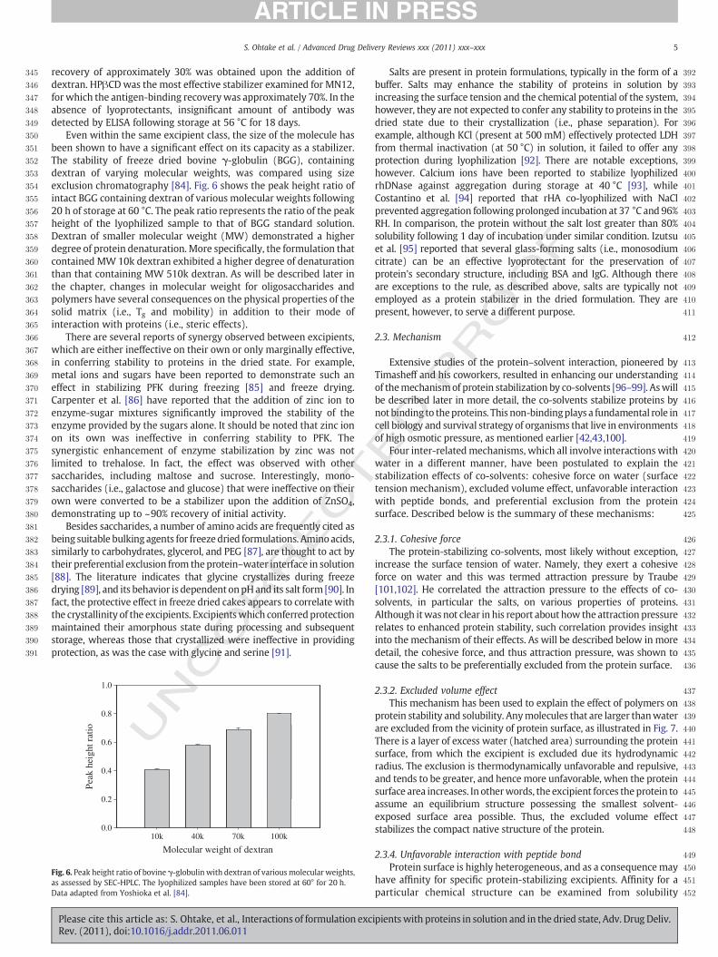
Even within the same excipient class, the size of the molecule hasbeen shown to have a significant effect on its capacity as a stabilizer.The stability of freeze dried bovine γ-globulin (BGG), containingdextran of varying molecular weights, was compared using sizeexclusion chromatography [84]. Fig. 6 shows the peak height ratio ofintact BGG containing dextran of various molecular weights following20 h of storage at 60 °C. The peak ratio represents the ratio of the peakheight of the lyophilized sample to that of BGG standard solution.Dextran of smaller molecular weight (MW) demonstrated a higherdegree of protein denaturation. More specifically, the formulation thatcontained MW 10k dextran exhibited a higher degree of denaturationthan that containing MW 510k dextran. As will be described later inthe chapter, changes in molecular weight for oligosaccharides andpolymers have several consequences on the physical properties of thesolid matrix (i.e., Tg and mobility) in addition to their mode ofinteraction with proteins (i.e., steric effects).
There are several reports of synergy observed between excipients,which are either ineffective on their own or only marginally effective,in conferring stability to proteins in the dried state. For example,metal ions and sugars have been reported to demonstrate such aneffect in stabilizing PFK during freezing [85] and freeze drying.Carpenter et al. [86] have reported that the addition of zinc ion toenzyme-sugar mixtures significantly improved the stability of theenzyme provided by the sugars alone. It should be noted that zinc ionon its own was ineffective in conferring stability to PFK. Thesynergistic enhancement of enzyme stabilization by zinc was notlimited to trehalose. In fact, the effect was observed with othersaccharides, including maltose and sucrose. Interestingly, mono-saccharides (i.e., galactose and glucose) that were ineffective on theirown were converted to be a stabilizer upon the addition of ZnSO4,demonstrating up to ~90% recovery of initial activity.
Besides saccharides, a number of amino acids are frequently cited asbeing suitable bulking agents for freezedried formulations. Aminoacids,similarly to carbohydrates, glycerol, and PEG [87], are thought to act bytheir preferential exclusion from theprotein–water interface in solution[88]. The literature indicates that glycine crystallizes during freezedrying [89], and its behavior is dependent onpHand its salt form [90]. Infact, the protective effect in freeze dried cakes appears to correlate withthe crystallinity of the excipients. Excipientswhich conferredprotectionmaintained their amorphous state during processing and subsequentstorage, whereas those that crystallized were ineffective in providingprotection, as was the case with glycine and serine [91].
UNC 436
437
438
439
440
441
442
443
444
445
446
447
448
449
450
451
452
1.0
0.8
0.6
Peak
hei
ght r
atio
0.2
0.4
10k 40k 70k 100k0.0
Molecular weight of dextran
Fig. 6. Peak height ratio of bovine γ-globulin with dextran of variousmolecular weights,as assessed by SEC-HPLC. The lyophilized samples have been stored at 60° for 20 h.Data adapted from Yoshioka et al. [84].
Please cite this article as: S. Ohtake, et al., Interactions of formulation excRev. (2011), doi:10.1016/j.addr.2011.06.011
ED P
RO
OF
Salts are present in protein formulations, typically in the form of abuffer. Salts may enhance the stability of proteins in solution byincreasing the surface tension and the chemical potential of the system,however, they are not expected to confer any stability to proteins in thedried state due to their crystallization (i.e., phase separation). Forexample, although KCl (present at 500 mM) effectively protected LDHfrom thermal inactivation (at 50 °C) in solution, it failed to offer anyprotection during lyophilization [92]. There are notable exceptions,however. Calcium ions have been reported to stabilize lyophilizedrhDNase against aggregation during storage at 40 °C [93], whileCostantino et al. [94] reported that rHA co-lyophilized with NaClprevented aggregation following prolonged incubation at 37 °C and 96%RH. In comparison, the protein without the salt lost greater than 80%solubility following 1 day of incubation under similar condition. Izutsuet al. [95] reported that several glass-forming salts (i.e., monosodiumcitrate) can be an effective lyoprotectant for the preservation ofprotein's secondary structure, including BSA and IgG. Although thereare exceptions to the rule, as described above, salts are typically notemployed as a protein stabilizer in the dried formulation. They arepresent, however, to serve a different purpose.
2.3. Mechanism
Extensive studies of the protein–solvent interaction, pioneered byTimasheff and his coworkers, resulted in enhancing our understandingof themechanismof protein stabilization by co-solvents [96–99]. Aswillbe described later in more detail, the co-solvents stabilize proteins bynot binding to theproteins. This non-bindingplays a fundamental role incell biology and survival strategy of organisms that live in environmentsof high osmotic pressure, as mentioned earlier [42,43,100].
Four inter-relatedmechanisms, which all involve interactions withwater in a different manner, have been postulated to explain thestabilization effects of co-solvents: cohesive force on water (surfacetension mechanism), excluded volume effect, unfavorable interactionwith peptide bonds, and preferential exclusion from the proteinsurface. Described below is the summary of these mechanisms:
2.3.1. Cohesive forceThe protein-stabilizing co-solvents, most likely without exception,
increase the surface tension of water. Namely, they exert a cohesiveforce on water and this was termed attraction pressure by Traube[101,102]. He correlated the attraction pressure to the effects of co-solvents, in particular the salts, on various properties of proteins.Although itwas not clear in his report about how the attraction pressurerelates to enhanced protein stability, such correlation provides insightinto the mechanism of their effects. As will be described below in moredetail, the cohesive force, and thus attraction pressure, was shown tocause the salts to be preferentially excluded from the protein surface.
2.3.2. Excluded volume effectThis mechanism has been used to explain the effect of polymers on
protein stability and solubility. Anymolecules that are larger thanwaterare excluded from the vicinity of protein surface, as illustrated in Fig. 7.There is a layer of excess water (hatched area) surrounding the proteinsurface, from which the excipient is excluded due its hydrodynamicradius. The exclusion is thermodynamically unfavorable and repulsive,and tends to be greater, and hence more unfavorable, when the proteinsurface area increases. In otherwords, the excipient forces theprotein toassume an equilibrium structure possessing the smallest solvent-exposed surface area possible. Thus, the excluded volume effectstabilizes the compact native structure of the protein.
2.3.4. Unfavorable interaction with peptide bondProtein surface is highly heterogeneous, and as a consequencemay
have affinity for specific protein-stabilizing excipients. Affinity for aparticular chemical structure can be examined from solubility
ipientswith proteins in solution and in the dried state, Adv. Drug Deliv.

453
454
455
456
457
458
459
460
461
462
463
464
465
466
467
468
469
470
471
472
473
474
475
476
477
478
479
480
481
482
483
484
485
486
487
488
489
490
491
492
493
494
495
496
497
498
499
500
501
502
503
504
505
506
507
508
509
510
511
512
513
514
515
516
517
518
519
520
521
522
523
524
525
526
527
528
529
530
531
532
Protein
Hydrodynamic Radius
Excipient
Layer of water free from excluded excipient
Fig. 7. Schematic illustration of the excluded volume effect. The protein is representedby the black circle and the excipient by the white circle. The striped area represents thelayer of water free from the excluded excipient.
6 S. Ohtake et al. / Advanced Drug Delivery Reviews xxx (2011) xxx–xxx
measurements. Nozaki and Tanford [103–106] pioneered suchsolubility experiments and reported a number of important conclu-sions for the mechanism of protein denaturation by urea, GdnHCl, andorganic solvents. Conversely, Gekko [107–109] and Bolen [110–112]examined the interactions between protein stabilizers and aminoacids. While both demonstrated the critical role of unfavorableinteractions between amino acid side chains and peptide bonds, thelatter concluded that the unfavorable interaction present betweenstabilizing excipients and peptide bonds is the primary determinantfor protein stabilization. Such an unfavorable interaction may beclosely related to the cohesive force, excluded volume effect, or both.In reality, both mechanisms should favor the stabilizing excipients toremain in bulk water, creating an entropically unstable condition. Asall of themechanisms cause repulsive interactions between excipientsand proteins, it is generally impossible to pinpoint which mechanismplays a dominant role in stabilizing proteins.
T 533
534
535
536
537
538
539
540
541
542
543
544
545
546
547
548
549
550
551
552
553
554
555
556
557
558
559
560
561
562
563
564
565
566
UNCO
RREC2.3.5. Preferential interaction
Various interactions (both weak and strong) contribute to theoverall interaction of the co-solvents with proteins. These interactionscan be determined from equilibrium dialysis experiments and may beformally grouped into two different modes. In the first case, co-solvents (depicted by black circles) are present in excess in thevicinity of the protein surface compared to its concentration in thebulk phase, as illustrated in Fig. 8 (left panel). This case is termed“preferential interaction”, indicating that the co-solvent concentra-tion is higher at the protein surface than that in the bulk phase(arbitrarily separated by the dashed line). The opposite case is alsoillustrated in Fig. 8 (right panel), in which there is excess water (whitecircles) at the protein surface. This is called “preferential hydration” or“preferential exclusion” of the co-solvent, indicating a deficiency ofco-solvent molecules in the vicinity of the protein. Osmolytesdemonstrate preferential hydration of proteins; in other words,osmolytes are preferentially excluded from the protein surface[13,14]. Many sugars, polyols, and certain salts, which are known tostabilize proteins and decrease their solubility, are all preferentiallyexcluded from the vicinity of the protein [13–15,22,23]. Furthermore,preferential exclusion is in accord with the repulsive interactions ofthese co-solvents with proteins, as described above.
Having introduced these concepts, the question now is, by whatmechanism do these co-solvents/osmolytes increase the stability ofproteins and decrease their solubility? The structure-stabilizingosmolytes are preferentially excluded from the protein surface,indicating that the interaction between osmolytes and protein isthermodynamically unfavorable. This increases the free energy of thenative state of the protein, as schematically depicted in Fig. 9.Although not determined experimentally, a greater exclusion of co-solvent/osmolyte is expected from the unfolded structure, because itpossesses a greater surface area compared to that of the folded, native
Please cite this article as: S. Ohtake, et al., Interactions of formulation excRev. (2011), doi:10.1016/j.addr.2011.06.011
ED P
RO
OF
state. The unfavorable interaction, and thus free energy, wouldincrease even more so for the unfolded state in the presence of theco-solvent. This leads to a greater energy difference between thenative and unfolded structures in the presence of stabilizing co-solvents/osmolytes, i.e., more energy is required to unfold proteins inthe presence of preferentially excluded co-solvents. As preferentialexclusion, and thus unfavorable interaction, increases with co-solvent/osmolyte concentration, the native structure is stabilized toa greater extent at higher co-solvent/osmolyte concentrations (Fig. 9).This concept can be extended to the situation in which there is self-association. During the process of protein self-association, the surfacearea per protein molecule decreases, which in turn reduces theunfavorable interaction present between the co-solvent and theprotein complex or aggregates, also depicted in Fig. 9 (right sidepanel). Thus, the associated state is stable in the presence ofstabilizing co-solvents/osmolytes; i.e., they enhance aggregation. Inthe examples described above, protein unfolding is the key determi-nant in causing aggregation, thus preferentially excluded co-solventsreduce aggregation by stabilizing the native structure.
As mentioned above, during lyophilization, both freezing anddrying stresses need to be taken into account. The stressesencountered, which include cold denaturation, increased concentra-tions of solutes and proteins, pH shift, and dehydration, can causeprotein denaturation and aggregation [113,114]. Protection duringfreezing is provided by a wide variety of co-solvents and is attributedto Timasheff's preferential exclusion mechanism (similar to that insolution), as free water is still present [115]. Nevertheless, as watermolecules gradually crystallize, the amount of free water decreases, sostabilization by preferential exclusion mechanism may be impactedby the rate and extent of crystallization. Carpenter and Crowe [116]reported that high concentrations (N1 M) of sodium acetate,potassium phosphate, and various sulfate salts (all kosmotropes, orwater structure-makers) provide significant cryoprotection of lactatedehydrogenase (LDH). In contrast, the more chaotropic salt, NaCl,yielded a much lower level of activity following freeze-thaw. Duringdrying, the preferential interactionmechanism is no longer applicablebecause the bulk water, as well as the hydration shell of the protein, isremoved [117–119]. It is the water molecules (not the protein) thatthe structure-stabilizing co-solvents influence. Furthermore, dehy-dration stress is different from those associated with freezing, thusmany effective cryoprotectants or stabilizers in solution do notnecessarily stabilize proteins during drying [83]. For many of theproteins examined by Prestrelski et al. [114], including γ-IFN, G-CSF,LDH, α-lactalbumin, bFGF, and α-casein, general disordering of theprotein backbone was observed upon dehydration, as evidenced bythe broadening of the individual amide I components. However, therewere notable differences observed, which mainly depended on theprotein itself. In general, three types of behaviors were observedduring dehydration followed by rehydration. First, the protein can beresistant to conformational change during drying, thus retain itsnative conformation during processing. G-CSF is one example. In thesecond case, the protein may unfold during dehydration but refoldupon rehydration, as was observed for α-lactalbumin and lysozyme.Finally, the protein may unfold during dehydration and remainunfolded during rehydration, resulting in irreversible conformationalchanges. For poly-L-lysine, the dehydration-induced conformationaltransitions appear to arise from its attempt in compensating for thelost hydrogen bonds with water. In solution, the random coilconfiguration has its peptide hydrogen bonding satisfied through itsinteraction with water molecules. Upon dehydration, these hydrogenbonding interactions are lost, and to compensate for the loss, thepolypeptide forms intermolecular hydrogen bonds, resulting inβ-sheet conformation. Furthermore, in the absence of water, thepartial charges of the intermolecular interactions are screened to alesser extent due to the lowered dielectric environment, thus increasingthe electrostatic attraction of opposing charges between the peptides,
ipientswith proteins in solution and in the dried state, Adv. Drug Deliv.

T
RO
OF
567
568
569
570
571
572
573
574
575
576
577
578
579
580
581
582
583
584
585
586
587
588
589
590
591
592
593
594
595
596
597
598
599
600
601
602
603
604
605
606
Dialysis Equilibrium
Preferential Binding of Additive Preferential Hydration
Protein Protein
= WaterDialysis membrane
= Additives
Fig. 8. Schematic presentation of preferential binding (left) and preferential exclusion (right) in a typical dialysis equilibrium experiment. The protein is represented by the whitecircle, water molecule by the blue circle, and the additive by the black circle. (For interpretation of the references to color in this figure legend, the reader is referred to the webversion of this article.)
7S. Ohtake et al. / Advanced Drug Delivery Reviews xxx (2011) xxx–xxx
RREC
leading to aggregation. Similarly to peptides, proteins rearrange theirconformation to maximize both intra- and inter-chain hydrogenbonding to replace the lost hydrogen bonds during dehydration [114].The above data clearly demonstrate the importance of satisfying thehydrogen bonding requirements of the polypeptide side chains andpeptide bonds both in solution and upon desiccation.
Saccharides have been postulated to protect proteins through avariety of mechanisms, but two have been put forth to describe manyobservations, the water replacement hypothesis and vitrification. Themain difference between the two proposed methods is that directinteraction is a pre-requisite for the former [77,114,120], while it is notfor vitrification. For the latter mechanism, the formation of anamorphous glass (vitrification) is the only requirement for providingstability, mainly through retarding molecular motion and providingphysical separation between the proteins (i.e., inhibiting aggregation)[121,122]. Although the underlyingmechanisms differ, both hypothesesrequire theprotein andthestabilizer tobe in thesameamorphousphase.
To support the water replacement hypothesis, many studies haveconfirmed the presence of hydrogen bonding in lyophilized samplesbetween carbohydrates and proteins. Examples include lysozyme,
UNCO
Stabilizing co-solvent
Free energy oUnfolded
Fre
e E
nerg
y
Free energy of unfolding
unfolding
NativeStabilizing co-solvent
Fig. 9. Free energy diagram of protein unfolding and the effect of co-solvent interaction. The l
Please cite this article as: S. Ohtake, et al., Interactions of formulation excRev. (2011), doi:10.1016/j.addr.2011.06.011
ED PBSA, PFK, bFGF, γ-IFN, recombinant G-CSF, bovine α-lactalbumin, and
bovine α-casein, to name a few [77,81,123–125]. Spectroscopicstudies of L-asparaginase freeze dried with trehalose indicated thatthe amide II band of the enzyme was quite similar to that observed insolution, thus suggesting that the level of hydrogen bonding forL-asparaginase was similar in the two states [116,126]. In anotherexample, Prestrelski et al. [81] have demonstrated that the titration ofsucrose with increasing amounts of protein resulted in decreasedamount of residual water following lyophilization. The authorsproposed that water is displaced from the dried protein through itsdirect interaction with the sugars. In fact, examination of thecarboxylate bands in the spectrum of α-lactalbumin indicated thatthe addition of carbohydrate maintained these bands in thehydrogen-bonded or hydrated form after dehydration, as reportedby Carpenter and Crowe [77]. Furthermore, the degree of structuralprotection conferred by saccharides, such as sucrose and trehalose,which are apparent in second derivative amide I infrared spectra, hasbeen shown to correlate with the extent of hydrogen bondingbetween sugar and protein [125]. Direct binding (hydrogen bonding),thoughnecessary, is insufficient to confer stability during lyophilization.
Aggregate
Stabilizing co-solvent
f
Free energy of association
Free energy of association
Monomer
Stabilizing co-solvent
eft panel illustrates protein unfolding and the right panel illustrates protein aggregation.
ipientswith proteins in solution and in the dried state, Adv. Drug Deliv.

T
607
608
609
610
611
612
613
614
615
616
617
618
619
620
621
622
623
624
625
626
627
628
629
630
631
632
633
634
635
636
637
638
639
640
641
642
643
644
645
646
647
648
649
650
651
652
653
654
655
656
657
658
659
660
661
662
663
664
665
666
667
668
669
670
671
672
673
674
675
676
677
678
679
680
681
682
683
684
685
686
687
688
689
690
691
692
693
694
695
696
697
698
699
700
701
702
703
704
705
706
707
708
709
710
711
712
713
714
715
716
717
718
719
720
721
722
723
724
725
726
727
728
729
730
731
732
733
734
735
736
737
738
8 S. Ohtake et al. / Advanced Drug Delivery Reviews xxx (2011) xxx–xxx
UNCO
RREC
For example, glucose has been shown to hydrogen bond effectively todried proteins [127,128], however, this was insufficient to retain thenative structure during freeze drying.
Vitrification hypothesis is based on the premise that the inhibition ofmolecular mobility, whether long-order (i.e., aggregation) or short-order (i.e., deamidation, cyclization, etc.), leads to an improvement instorage stability [129–131]. The parameter that has typically beenexamined for the purpose of comparing the expected stability of variousformulations is the glass transition temperature (Tg). The publication ofseveral reports illustrating the lack of direct correlation between Tg andstability has somewhat discredited the hypothesis as a stand-aloneexplanation for the observed stability of amorphous pharmaceuticals[132,133]. It should benoted that theoccurrence of vitrificationdoes notpreclude the existence of direct interaction between the glassy matrixand the protein (i.e., water-replacement). Furthermore, the importanceof reducing themolecularmobility is a common themebetween the twohypotheses. Besides Tg, other parameters have been reported tocorrelate to stability. In one example, Yoshioka et al. [84] examinedthe effect of the molecular weight of dextran on the stability of freezedried bovine γ-globulin (BGG) using 1H-NMR. Changes in molecularmobility of freeze dried formulations occurring below Tg was detectedand this temperature was called the molecular mobility-changingtemperature (Tmc). Tmc increased as the molecular weight of dextranincreased, which indicated that the molecular mobility of formulationsin the microscopically liquidized state decreased as the molecularweight of dextran increased. In comparison to Tmc, the Tg of the freezedried BGG formulations was determined to be higher. Thus, Tmc
represents the temperature at which molecular mobility begins toincrease in a temperature range below Tg [134], and may be a morerelevant marker for stability indication.
While the amorphous or crystalline nature of excipients is clearlyimportant in achieving optimal protein stability, the effects of thephysical characteristics of the stabilizer cannot be generalized for allproteins. For example, mannitol is often used as a bulking agent inpreparing lyophilized proteins due to its propensity for crystallization,thus it is phase separated from the protein, which is typicallyamorphous. However, the spectra of γ-IFN indicate that mannitol,and other crystallizing components such as myo-inositol, aredestabilizing and induce further unfolding during dehydration [81].This finding suggests that focusing solely on the physical properties ofthe excipients, while necessary, provides a limited view of the effectsof the lyophilization process on protein stability. On the contrary,amorphous excipients form a part of the protein-rich glassyconcentrate and behave differently from the segregated crystallineexcipients. These behaviors can have important implications inregards to the stability of proteins during freezing, freeze drying,and subsequent storage. It should be emphasized that even when thephysical criteria mentioned above are met (i.e., amorphous nature, Tgabove storage temperature, etc.), there are cases in which asubstantial loss of protein structure and activity are observed [82].Chemical degradation, including oxidation and deamidation, couldperturb the protein structure, and thus activity. These types ofreactions may not be slowed sufficiently even upon the formation ofthe highly viscous glass, as would be for aggregation.
For optimal stability of the protein in the dried state, not only do theexcipients have to replace the hydrogen bonding network lost duringdehydration and remain in the amorphous phase, but they must alsooffer structural stabilization through direct binding. That is, theexcipients must be in a specific geometrical orientation to interactfavorably with the protein. This is illustrated by the example of HPβCD,which is a sugar polymer arranged in a cylindrical conformation with ahydrophilic outer surface and a hydrophobic internal cavity [135].Reasons behind the efficacy of HPβCD include its relatively high collapsetemperature (~−9 °C) and its intrinsic amorphous nature [136,137]. Inaddition, the hydrophilic exterior of the lyoprotectant was reported toprovide the protein-HPβCD complex a higher degree of hydration and,
Please cite this article as: S. Ohtake, et al., Interactions of formulation excRev. (2011), doi:10.1016/j.addr.2011.06.011
ED P
RO
OF
thus, promote water structure formation. The most important require-ment for the formation of a stable protein-HPβCD inclusion complex isthe tight fitting, wholly or at least partially, of the protein within thecyclodextrin cavity. The hydrophobic cavity of HPβCD may enclose theamino acid side chains of mAb MN12, thus protecting them from avariety of degradation reactions. In fact, HPβCD has been reported toprotect other drugs against oxidation and gastric acid degradation [135].
The effects of salts on the stability of dried proteins have also beenexamined, although not to the same extent as for sugars. It should benoted that the effects of salts in the dry systems aremore case-specificthan those in solution; in solution, stabilizing salts are universallystabilizing and destabilizing salts consistently demonstrate adverseeffects. Buffer components may favorably or adversely affect thestability of proteins through direct interactions and/or throughmodification of its local environment (pH shift) [138–142]. Changand Randall [127] have classified salts into 3 types based on theirglass-forming tendency at a given cooling rate and subsequentthermal history: (1) crystallizing salts, (2) partially crystallizingsalts, and (3) glass-forming salts. As glass-forming excipients caninhibit salt crystallization, salts can be included in the formulationwhen other amorphous excipients are present [143]. Interestingly,non-glass forming salts, on their own or in combination with glass-forming excipients (i.e., sugars), have been reported to demonstratestabilizing effects on proteins following lyophilization. For example,Costantino et al. [94] reported that recombinant human albumin(rHA) co-lyophilized with NaCl did not exhibit any aggregationfollowing prolonged incubation at 37 °C and 96% RH, while greaterthan 80% loss in solubility following 1 day of incubation under similarcondition was noted for the protein lyophilized in the absence of thesalt. As the inclusion of NaCl did not induce any significant changes tothe secondary structure of lyophilized rHA, the stabilization effect ofthe salt was attributed to its water uptake in the vicinity of theprotein, which may have facilitated protein refolding into its nativeand more stable conformation. Thus following this logic, the greaterthe affinity of salt (or excipient) for water, the greater the stabilizingeffect. However, if a protein is sensitive to residual water, thisstabilization mechanism is not applicable. Another stabilizationmechanism proposed for salts is the prevention of protein–proteininteraction and aggregation by physical dilution and separation ofprotein molecules. Liu et al. [144] attributed the reduced aggregationof lyophilized BSA in the presence of NaCl, along with otherexcipients, to the dilution effect.
In the case of glass-forming salts, the stabilization mechanism isthought to occur through a similar mechanism as that for sugars; i.e.,their direct interactionwith proteins to substitute for water moleculesthat are removed during drying. Carboxylic acid salts have beenshown to provide both hydrogen bonds and electrostatic interactionswith the protein, resulting in high Tg of the amorphous solid. In fact,the observed structural stabilization at specific salt-to-protein ratioindicates the presence of direct interaction. It is highly plausible thatthese buffer salts hydrogen bond to the protein, thereby substitutingfor the lost water molecules. In fact, FTIR analysis of bovine IgG inmonosodium citrate buffer demonstrated the retention of intramo-lecular β-sheet band (1637 cm−1) following lyophilization [95].Furthermore, the lower concentration of monosodium citrate incomparison to sucrose, which was required to stabilize IgG, suggestedan additional mode of interaction present between the salt and theprotein (besides hydrogen bonding), which is most likely electrostaticinteraction [145–147].
Salts have also been incorporated into a lyoprotectant formulationas a structure former. Chang and Randall [127] reported that one ofthemajor stress factors that contribute to protein denaturation duringa lyophilization cycle is the loss of cake structure. It was concludedthat the addition of salts with eutectic melting temperatures (Te)above−20 °C would promote rapid crystallization upon freezing andprevent the collapse of the frozen fraction during dehydration.
ipientswith proteins in solution and in the dried state, Adv. Drug Deliv.

T
739
740
741
742
743
744
745
746
747
748
749
750
751
752
753
754
755
756
757
758
759
760
761
762
763
764
765
766
767
768
769
770
771
772
773
774
775
776
777
778
779
780
781
782
783
784
785
786
787
788
789
790
791
792
793
794
795
796
797
798
799
800
801
802
803
804
805
806
807
808
809
810
811
812
813
814
815
816
817
818
819
820
821
822
823
824
825
826
827
828
829
830
831
9S. Ohtake et al. / Advanced Drug Delivery Reviews xxx (2011) xxx–xxx
CO
RREC
However, the presence of uncrystallized salt in a freeze-concentrateusually depresses Tg', so the salt content in protein formulationsshould be kept to a minimum [121]. It should be noted that theoccurrence of cake collapse (or lack of typical structure) does notnecessarily correlate to instability, as demonstrated by several authors[148–151].
4. Polymers
4.1. Solution
Hydrophilic polymers have often been used to stabilize proteinsand enhance protein assembly [152–154]. Sasahara et al. [155]demonstrated that the stability of a protein against heat treatmentwas increased through the incorporation of dextran. Manning et al.[156] have studied the effects of polymeric excipients on thethermally-induced aggregation of low molecular weight urokinase,and found hydroxyl ethyl (HETA) starch, PEG4000, and gelatin to allbe effective in stabilizing the enzyme, which consequently suppressedaggregation [157]. In contrast, polyvinylpyrrolidone (PVP) and lowmolecular weight PEGs (e.g., PEG 300) were found to be ineffective, astheir hydrophobic nature offset the stabilizing effects of the polymers.Unlike small molecular weight protein-stabilizers, polymers posses-sing a hydrophobic moiety do not always stabilize proteins. Anexample is shown in Fig. 10A, which plots the change in the meltingtemperature of β-lactoglobulin as a function of PEG concentration[158]. Both PEG200 and 1000 greatly decreased the meltingtemperature. Due to the smaller molecular weight of these PEGs, itappears that the excluded volume effects (i.e., stabilizing effects) areoverwhelmed by their hydrophobic nature. In addition, as polymersare strong protein precipitants, they are known to enhance self-association as well as protein–protein and protein–macromoleculeinteractions, leading to protein aggregation [159–161]. Examples ofthese effects include the acceleration of α-synuclein fibril formationupon the addition of PEG, dextran, and Ficoll [162,163].
Chargedpolymers can stabilizeproteins via electrostatic interactionsthrough their multiple charged binding sites [164,165]. This effect israther protein specific, as has been demonstrated for acidic fibroblastgrowth factor (aFGF), which has a constellation of positively chargedgroups on the surface [166–168].Won and co-workers [169] found thata variety of sulfated and phosphorylated anionic polymers (heparin,dextran sulfate, pentosan sulfate, enoxaparin, phosvitin, and phyticacid) were effective at stabilizing aFGF. The only requirement for aFGFstabilization appeared to be the presence of one ormore regions of highnegative charge density [68]. Similarly, other negatively chargedbiopolymers, e.g., nucleic acids,were found to be effective. Furthermore,negatively charged dextran sulfate was found to be effective inpreventing aggregation of basic ribonuclease A [170]. Andersson andHatti-Kaul [171] examined the effect of polyethyleneimine (PEI), a
UN
0
A
-10
-5 PEG1000
-25
-20
-15
Mel
ting
tem
pera
ture
PEG200
Concentration, g/g0 20 40
Fig. 10. Effect of PEG on (A) the melting temperatureData adapted from Arakawa and Timasheff [158].
Please cite this article as: S. Ohtake, et al., Interactions of formulation excRev. (2011), doi:10.1016/j.addr.2011.06.011
ED P
RO
OF
cationic polymer, on the stability of lactate dehydrogenase (LDH) andfound the storage stability of LDH to be improved (and prevented theaggregation) upon the addition of 0.01–1% (w/v) polymer. Unlike theprotein-stabilizing excipients (e.g., sugars and salts), the addition of PEIdid not increase the denaturation temperature of LDH (62 °C), althoughit did suppress the oxidation of free sulfhydryl groups (which arecatalyzedbymetal ions), thus improving the stability of the enzyme. Theprotective effect is attributed to the metal chelating property of PEI[172]. Furthermore, the addition of 0.1% PEIwas effective inmaintainingthe secondary structure of the enzyme, while in its absence significantloss was observed following 2 weeks of storage. Charged DEAE-dextranpolymer conferred no stabilization effect on the green fluorescentprotein, suggesting the protein-specific nature of charged polymers[173].
PEG is different from the more hydrophilic polysaccharides (e.g.,dextran) in that it possesses a small, non-polar moiety. PEG has beenshown to decrease the surface tension of water and act as a surfactant[174]. PEG and other polymers have been used to suppress protein–protein interactions and surface adsorption through hydrophobiccompetitive interaction. Poloxamers, which are non-ionic co-polymersof polyoxypropylene and polyoxyethylene, have also been found to beeffective in preventing aggregation induced by various stresses [175].Poloxamers are amphiphilic, and are thus surface active. The proportionof hydrophobic and hydrophilic moieties can be modulated by therelative sizes of the polypropylene hydrophobic core and the hydro-philic polyoxyethylenemoieties, and thus, a wide variety of poloxamersare commercially available. In this regard, many proteins are alsoamphiphilic. For example, human serum albumin (HSA) has been usedas a stabilizer in pharmaceutical products [176], typically at concentra-tions ranging from 0.1 to 1%, as found in many patent applications andpublications, e.g. [177]. Although HSA has been used as a stabilizingexcipient in a number of protein therapeutics to prevent surfaceadsorption, recent concerns about potential infectious agents in animal-derived products have prompted regulatory agencies to restrict itsusage, and non-ionic surfactants are increasingly finding use as areplacement for serum albumin [178]. As polymers and proteins arecompetitive inhibitors for protein adsorption, their use at lowconcentrations may be sufficient to cover the protein binding sites onthe surface.
4.2. Dry state
Polymers have been demonstrated to be a successful additive insuppressing protein aggregation during lyophilization and to preventthe solubility decrease observed during reconstitution [179–182].Dextran, CMC, DEAE-dextran, and PEG have all been shown to reducethe aggregation of lyophilized BSA significantly during storage at37 °C [144]. The derivatized starch, hydroxypropyl β-cyclodextrin(HPβCD), has been used to improve the solubility and prevent the
1.6B
1
1.2
1.4
0.2
0.4
0.6
0.8
Hyd
rati
on, g
/g
00 200 400 600 800 1000
PEG Molecular Weight
and (B) preferential hydration of β-lactoglobulin.
ipientswith proteins in solution and in the dried state, Adv. Drug Deliv.

T
832
833
834
835
836
837
838
839
840
841
842
843
844
845
846
847
848
849
850
851
852
853
854
855
856
857
858
859
860
861
862
863
864
865
866
867
868
869
870
871
872
873
874
875
876
877
878
879
880
881
882
883
884
885
886
887
888
889
890
891
892
893
894
895
896
897
898
899
900
901
902
903
904
905
906
907
908
909
910
Table 1 t1:1
Effect of polyethyleneimine (PEI) on the stability of LDH during freeze-thaw, freezedrying, and drying. LDH concentration was 50 μg/ml in 50 mM Tris–HCl at pH 7.2.
t1:2Data adapted from Andersson and Hatti-Kaul [171].
t1:3PEI Concentration Residual activity (%)
t1:4Freeze-thaw Freeze dried Drying
t1:5None 84±2.6 27±0.4 43±0.3t1:6Low MW 0.01 82±1.0 43±3.7 NDt1:70.1 79±2.8 47±0.4 NDt1:81 76±4.3 69±3.9 NDt1:9High MW 0.01 77±1.0 52±5.1 77±1.8t1:100.1 83±2.6 64±0.8 79±0.8t1:111 83±1.4 69±0.7 78±2.4
10 S. Ohtake et al. / Advanced Drug Delivery Reviews xxx (2011) xxx–xxx
ORREC
lyophilization-induced insoluble aggregate formation for growthhormone, interleukin-2 (IL-2), and insulin [183]. In addition, HPβCDwas found to stabilize lyophilized mouse monoclonal antibody duringstorage at 56 °C [73] and inhibit the dimerization of lyophilized TNFduring storage at 37 °C [184]. Dextran 40 at 10% concentrationincreased the activity of lyophilized elastase (20 mg/ml) from 33 to82% following storage for 2 weeks at 40 °C and 79% RH [83], whiledextran (162 kD) at 3.5 and 5% (w/v) improved the storage stability oflyophilized rFXIII andHumicola lanuginose lipase, respectively, at 40 or60 °C [82,185]. Several PVP's and maltodextrin were reported tostabilize lyophilized invertase during incubation at 90 °C [133,186],and 1–10% PVP or BSA have been reported to improve the recovery ofLDH activity following freeze-thaw or lyophilization [187] (Fig. 11). Incontrast, PEI addition failed to confer stability to LDH following freeze-thaw (Table 1). The charged polymer was, however, effective inmaintaining the enzyme activity following freeze drying and thedegree of protection was found to depend on the concentration of PEIused.
HSA at concentrations between 0.05 and 0.1% (w/v) has been usedas a lyoprotectant in formulating hydrophobic cytokines, includinginterleukin-1a (IL-1a), IL-1b, IL-3, andmacrophage colony stimulatingfactor (MCSF) [188]. Inclusion of BSA at 0.05% concentration increasedthe recovered activity of LDH (25 μg/ml) from approximately 30 to80% following lyophilization [187]. LDH activity was also maintainedduring lyophilization in the presence of different concentrations of PEI[189] as well as with PVP (40 kD) [187]. Hydroxyethylcellulose (HEC)at 1% completely inhibited the lyophilization-induced aggregation ofaFGF at 100 μg/ml in PBS containing 33 μg/ml heparin [190].
Polymers may not always stabilize proteins in the solid state, and insome cases, have adverse effects. For example, dextran of certainmolecular weight may be unable to provide sufficient stability toproteins during lyophilization due to steric hindrance, which preventsefficient hydrogen bonding with proteins. Dextran (40 kD) at concen-trations of up to 100 mg/ml was ineffective in inhibiting dehydration-induced unfolding of lysozyme [128], and its addition was not effectivein preventing the formation of β-sheets in poly-L-lysine duringdehydration [191]. Certain polymers may also cause phase separationduring freezing, which can adversely affect protein stability. Thepresence of dextran 40 in IL-6-sucrose formulation increased proteinaggregation during storage for 9 months at 30 °C [192]. Also, rehydra-tion of PEG-containing lyophilized sample resulted in protein precip-itation [114]. It is possible that the high concentration of PEG, a strongprotein precipitant [158], induced precipitation of the protein duringrehydration, resulting in its lower activity. PEG was also reported to beineffective in stabilizing lysozyme, even up to concentrations of100 mg/ml [128]. However, in combination with a smaller excipient,such as glucose, PEGwas shown to be an effective lyoprotectant. In fact,as the glucose concentration was increased in lysozyme preparations
UNC
120
100
Freeze-thawFreeze-drying
80
Rec
over
ed a
ctiv
ity, %
40
60
0
20
PVP concentrationNone 0.1% 1% 10%
Fig. 11. Effects of PVP concentration on the recovery of LDH activity following freeze-thaw or lyophilization.Data adapted from Anchordoquy and Carpenter [187].
Please cite this article as: S. Ohtake, et al., Interactions of formulation excRev. (2011), doi:10.1016/j.addr.2011.06.011
OFcontaining 1% PEG8000, the extent of hydrogen bonding to protein
carboxylate groups increased and the lyophilized sample demonstratedstructural similarity to the native lysozyme [128]. A similar positive andadditive effect was observed following PEG addition to sucrose andtrehalose.
911
912
913
914
915
916
917
918
919
920
921
922
923
924
925
926
927
928
ED P
RO
4.3. Mechanism
In solution, polymers provide protein stabilization by both proteinspecific and non-specific mechanisms. Charged polymers mainly workin a protein-specificmanner,while polar, hydrophilic polymers stabilizeproteins independent of their chemical nature. Hydrophilic polymers(e.g., polyethylene glycols, polysaccharides, and inert proteins) canstabilize proteins by amultitude of mechanisms [156,158,169,170,193–196]. Thedominant factor among them is themolecular crowdingeffect,as has been observed for protein transport in polymer gels andconcentrated protein solutions [197–199]. This is schematically repre-sented in Fig. 12, in which the aqueous solution is filled with polymers(closed circles). When a protein molecule (thick line) is in equilibriumbetween the native, compact structure (small dotted circle) and theunfolded, expanded structure (large dotted circle), it is thermodynam-ically more favorable to maintain the native structure, as it possesses asmaller radius and hence surface area for exclusion. Thus, the nativeprotein ismore favorable in thepresenceof a polymer, andevenmore soat higher polymer concentrations. As polymer exclusion increases withits size, larger polymers are generally more effective in stabilizingproteins in solution [200]. Such an effect can be replicated using aprotein, e.g., HSAandother, but similar, proteins. The latter suggests thata protein is thermodynamically more stable at higher concentrations,which increases the molecular crowding effect. In Fig. 3, thiscorresponds to a decrease in K1 due to high protein concentration andgreater crowding effects. Of course, other factors may affect suchstabilizingmechanism at high protein concentrations, e.g., reversible orirreversible self-association being one of them, as also seen in Fig. 3, inwhich high protein concentration can increase K2, eventually leading toaggregation. Excluded volume effect is essentially a repulsive interac-tion between protein and polymer. Thermodynamic interactionmeasurements indicated that PEGs are preferentially excluded fromthe protein surface [158,201,202]. Fig. 10B depicts the effect of PEG'smolecular weight on the amount of excess water in the vicinity ofβ-lactoglobulin as a result of PEGexclusion fromtheprotein surface. Thefigure clearly illustrates that excess water increases with the molecularweight of PEG, as expected from the increasing exclusion of highermolecularweight PEGs. As is the case for protein-stabilizing co-solvents,such exclusion leads to a thermodynamically unstable state of theprotein. Protein unfolding can further increase the instability and henceis suppressed in the presence of polymers.
However, polymers possessing hydrophobic character, such as PEG,can bind toproteins throughhydrophobic interaction. In fact, it has beenshown that the thermodynamic interaction of PEGwith aromatic groupsis thermodynamically favorable [Hirano et al., unpublishedwork]. Thus,
ipientswith proteins in solution and in the dried state, Adv. Drug Deliv.

T929
930
931
932
933
934
935
936
937
938
939
940
941
942
943
944
945
946
947
948
949
950
951
952
953
954
955
956
957
958
959
960
961
962
963
964
965
966
967
968
969
970
971
972
973
974
975
976
977
978
979
980
981
982
983
984
985
986
987
988
989
990
991
992
993
994
995
996
997
998
999
1000
1001
Fig. 12. Illustration of themolecular crowding effect. Proteinmolecules are represented bythe lines and the dotted circle surrounding the lines represent the volume occupied by thefolded (left) and unfolded (right) protein. Polymers are represented by the black circles.
11S. Ohtake et al. / Advanced Drug Delivery Reviews xxx (2011) xxx–xxx
RREC
the stabilizing effect of PEGs on proteins is a delicate balance betweenthe two opposing effects: stabilizing effect due to steric exclusion anddestabilizing effect due to hydrophobic interaction. This is schematicallyrepresented in Fig. 13. Hydrophobic sequences (thick black line) of theprotein are sequestered within the native protein structure (left side),and are not expected to interact strongly with the polymer (blackcircle). When the protein unfolds (right side), however, the hydropho-bic regions become exposed, and the polymer can bind to the unfoldedstructure with greater affinity, or to a greater extent, in comparison tothe native state. Thus, such polymers could stabilize the unfoldedstructure by hydrophobic interaction and the native structure byexcluded volume. The overall effect of such polymers will then bedetermined by the balance between the two opposing factors. Insolution (andduring the freezing processwhichwill be described later),proteins tend to be adsorbed to the surface through hydrophobic (andalso electrostatic) interactions. Amphiphilic polymers such as PEG,poloxamers, and HSA can compete with the protein and prevent itsadsorption-mediated conformational change(s) and consequentaggregation.
A second mechanism by which polymers can stabilize proteins isvia specific binding, e.g., polyanion binding to positively-chargedheparin binding site of aFGF [68]. Polymers have also been shown toprevent certain types of chemical instability that can lead toaggregation, e.g., metal-ion catalyzed oxidation can be inhibited bypolymers via metal ion complexation [203].
Polymers have also been shown to suppress the damage to multi-subunit proteins during freeze-thaw or lyophilization by stabilizing
UNCO
Hydrophobic binding of polymthe unfolded state shifts the equilibrium toward unfolding
Excluded volume shifts the equilibrium toward foldingHydrophobic
sequence
Fig. 13. Illustration demonstrating the two opposing effects of polymers (i.e., PEG) on proteinwithin the protein structure (gray lines). Hydrophobic binding of polymers (black circles) teffect shifts the equilibrium toward the folded state.
Please cite this article as: S. Ohtake, et al., Interactions of formulation excRev. (2011), doi:10.1016/j.addr.2011.06.011
ED P
RO
OF
their quaternary structure in the frozen state via preferentialexclusion mechanism [83,187]. This is because water is still presentduring freezing, although water activity is gradually decreased uponice crystal formation, and as long as water is present, polymerexclusion mechanism is still in operation. Stability in the dried statehas been typically attributed to hydrogen bonding between thepolymer and protein [77,118], however due to steric hindrance,polymers do not readily form hydrogen bonds with proteins as is thecase with sugars. The inherent stability of multimeric enzymes isknown to be greater in the assembled state than as unassociatedsubunits [204,205]. Inhibition of subunit disassembly by polymers insolution, and its subsequent immobilization in the glassy matrix, mayaccount for the preserved activity of the enzyme. While the formationof a glassy state, in and of itself, is insufficient for protection[118,206,207], the high viscosity in the glass could prevent thedissociation during dehydration, thus contributing to the observedprotection of protein activity, as is the case for LDH. Interestingly,these polymers have also been found to inhibit freezing-induced shiftsin pH, presumably by inhibiting crystallization of buffer salts,particularly in the case of phosphate buffer systems, and thusresulting in enhanced protein stability.
While non-ionic polymers stabilize proteins by being preferen-tially excluded from their surface [158,208], charged polymersstabilize or inactivate proteins depending on their mode of interac-tion. Since proteins are poly-ampholytic molecules, they can interactwith polymers, or polyelectrolytes, via long-range Coulombic forces[209,210]. In case of PEI [171], it is likely that charge–charge and/orhydrophobic interactions between the protein and polymer overcomethe effects of preferential exclusion, thus providing no stabilizingeffect during freezing, however during dehydration, the polymerprovides protection to the quaternary structure of LDH (Table 1).
Polymers, such as dextran, have also been reported to stabilizeproteins by raising the Tg of the protein formulation [211,212] and byinhibiting crystallization of small stabilizing excipients, such as sucrose[213]. In fact, the Tg of dextran-formulated γ-globulin formulationsincreased significantly with increasing molecular weight of dextranfrom 10 to 510 kD [214]. Similarly, the stability of lyophilized invertasewas shown to correlate to the Tg of themaltodextrin (MD) or PVP [215].In fact, an inverse correlation was reported to exist between theremaining activity and (T–Tg) for MD and PVP of various molecularweight, suggesting that higher enzyme activity is associatedwith higherTg. More specifically, PVP-360 (Tg=155 °C) and PVP-40 (Tg=137 °C)afforded better enzyme stabilization compared to PVP-10 (Tg=93 °C),and this may be correlated with their higher Tg values (Fig. 14).However, themaintenance of theglassy statewas insufficient topreventinactivation, since PVP systemsmaintained at temperatures below their
er to
Hydrophobic sequence
structure. The hydrophobic sequence of a protein is represented by the thick black lineso the unfolded state shifts the equilibrium towards unfolding, while excluded volume
ipientswith proteins in solution and in the dried state, Adv. Drug Deliv.

T
1002
1003
1004
1005
1006
1007
1008
1009
1010
1011
1012
1013
1014
1015
1016
1017
1018
1019
1020
1021
1022
1023
1024
1025
1026
1027
1028
1029
1030
1031
1032
1033
1034
1035
1036
1037
1038
1039
1040
1041
1042
1043
1044
1045
1046
1047
1048
1049
1050
1051
1052
1053
1054
1055
1056
1057
1058
1059
1060
1061
1062
1063
1064
1065
1066
1067
1068
1069
1070
1071
1072
1073
1074
1075
1076
1077
1078
1079
1080
1081
1082
1083
1084
1085
1086
1087
12 S. Ohtake et al. / Advanced Drug Delivery Reviews xxx (2011) xxx–xxx
RREC
Tg values (i.e., 90 °C) still demonstrated significant enzyme inactivation.This ismost likely due to thepresence ofmobility at temperatures belowTg, as reported by Simatos et al. [216] among others.
Although large molecular weight carbohydrate polymers, such asdextran and HES, are effective in increasing the Tg' and the collapsetemperature, allowing the freeze drying process to be conducted athigher temperatures, they are not very effective in protecting theprotein during lyophilization. Unlike smaller sugars, they cannoteffectively hydrogen bond to the protein as a result of steric inter-ference. To circumvent this shortcoming, a small disaccharide can beused concurrently with the carbohydrate polymers. The efficacy of thecombination stems from the independent stabilizing capabilities ofthe two components; polymers are an effective cryoprotectant, whilesugars are effective against dehydration stress. However, care must betaken in adjusting the ratio of the two components. Allison et al. [125]suggested that in the two component system, the ability of a sugar tohydrogen bond to the protein surface may be reduced due to thepartitioning of some fraction of the sugar molecules with the polymerin the dried solid. As a result, there may be less sugar available tointeract with and stabilize the protein.
5. Surfactants
5.1. Solution
Surfactants are widely used to stabilize proteins, suppress aggrega-tion and assist in protein refolding [217,218]. Polysorbate 80 (polyox-yethylene sorbitan monooleate) and polysorbate 20 (polyoxyethylenesorbitan monolaurate) are two of the widely incorporated surfactants inmarketed protein pharmaceuticals [176,178,219], and are typically usedin the 0.0003–0.3% range [176]. The effects of surfactants and theirinteractionmechanism in aqueous solutionwill be described in Section8,thus this sectionwill placemore emphasis on their effects in thedry state.It has been extensively documented that surfactants suppress proteinaggregation against various stresses, including heating and agitation.While the suppression of aggregation is almost universally observed insolution, the effect of a surfactant on thermal stress or denaturant-induced protein unfolding varies, depending on the protein itself and onthe stress conditions. Surfactants have also been reported to be aneffective stabilizer in protecting proteins against surface denaturation innon-frozen aqueous solutions [220,221].
5.2. Dry state
In the context of lyophilized formulations, surfactants have beenused to prevent aggregation. For example, when 1 mg/ml solution of IL-
UNCO 1088
1089
1090
1091
1092
1093
1094
1095
1096
1097
1098
1099
1100
1101
1102
1103
1104
1105
1106
80
70 MD (Tg=160°C)
50
60
Rem
aini
ng a
ctiv
ity, %
40PVP-360 (Tg=155°C)
PVP-40 (Tg=137°C)
20
30
PVP-10 (Tg=93°C)
T-Tg, °C-80 -60 -40 -20 0 20
Fig. 14. Effect of polymer glass transition temperature (Tg) on the amount of activityloss of lyophilized invertase at near-zero residual water content following 20 hincubation at 90 °C. The polymers examined in the study and their respective Tg valuesare indicated in the figure.Data adapted from Schebor et al. [215].
Please cite this article as: S. Ohtake, et al., Interactions of formulation excRev. (2011), doi:10.1016/j.addr.2011.06.011
ED P
RO
OF
1ra was freeze-dried with 0.1% Tween 80, less than 3% solubleaggregates was detected by size-exclusion HPLC (SE-HPLC), while inthe absence of the surfactant, approximately 50% of the protein wasobserved to form soluble aggregates [222]. Kreilgaard et al. [82]demonstrated that Tween 20 addition (0.002% w/v) improved therecovery of native rFXIII and reduced the amountof insoluble aggregatesformed. In addition, FTIR analysis of the same system revealed that theaddition of 0.1% Tween 80 was sufficient in inhibiting aggregation andmaintaining the secondary structure of freeze dried IL-1ra, with respectto that of the native structure in solution. Furthermore, the protectiveeffect of surfactant wasmuch greater than that by sucrose, for which 1%addition resulted in 8% aggregate formation.
There have been a number of examples in the literature (i.e.,keratinocyte growth factor, Interleukin 2, Interleukin 1 receptorantagonist, and bovine IgG, etc.) which demonstrated the efficacy ofsurfactant-containing diluent in reducing aggregation followingreconstitution [140,223–225]. However, this effect cannot be gener-alized, because it is somewhat specific to Tween/polysorbate. Othersurfactants such as N-octyl glucoside and Pluronic either exhibited noeffect or promoted aggregation [31,224]. Interestingly, in the case ofAnti-L-Selectin, the presence of Tween in the reconstitution buffer wasnecessary to prevent aggregate formation, but if present duringlyophilization, it increased the aggregate level in the reconstitutedsolution [223]. The mechanism of this protection is not wellunderstood, as there was no evidence of Tween binding to, orstabilizing, the native state of the protein.
Spray drying subjects proteins to large air–water interfacial surfacearea during atomization. In a study to minimize aggregation ofrecombinant human growth hormone (rhGH) during spray drying,Maa et al. [226] reported that insoluble aggregate formation decreasedas polysorbate concentration was increased. The aggregate levelreached a plateau at a certain critical polysorbate concentration (cpc),which was independent of the protein concentration. They alsodemonstrated that cpc was directly proportional to the air–waterinterfacial area, i.e., inversely proportional to the median dropletdiameter, thus demonstrating that insoluble aggregate formation waslinked to denaturation of hGH at the air–water interface, and thatpolysorbate suppressed rhGHaggregationbycompetingwith theproteinfor the air–water interface. It is interesting to note that in this study,polysorbate was unable to completely arrest soluble aggregate forma-tion, requiring the use of Zn2+ as an additional excipient, thushighlighting the fact that aggregation can occur bymultiplemechanisms.Effective control requires excipients that work by multiple mechanisms,e.g., competition with air–water interface, thermodynamic stabilizationby preferential exclusion, or specific ligand binding.
5.3. Mechanism
Extensive analysis of the effects of surfactants will be described inSection 8 and it may suffice to mention here that the fundamentalmechanism of aggregation suppression by surfactants is the preventionof surface denaturation [222,227,228]. Protein–surfactant interactionshavebeenstudiedbyvarious indirectmethods including surface tension[229], viscosity [230], and dye solubilization [231], and by directmeasurements such as dialysis [232–236] and ion-selective electrodes[237–240]. Surfactants compete with protein for container surface, air–water interface, ice–water interface, or any other solid surfaces, andprevent the protein from non-specific adsorption [241–243]. Althoughit is possible that surfactants bind to the protein at the hydrophobic sitesand reduce their tendency to aggregate [244–246], it is difficult todistinguish such direct binding from inhibition of surface denaturation.Various surfactants have been reported to be effective in preventingfreeze-thaw-induced damage to proteins [223,242,247]. Examplesinclude, Tween 80, Brij 35, Brig 30, Triton X-10, Pluronic F127, andSDS. Surfactants can also prevent aggregation by serving as chaperonesand foster protein refolding [223,224,247–250].
ipientswith proteins in solution and in the dried state, Adv. Drug Deliv.

T
1107
1108
1109
1110
1111
1112
1113
1114
1115
1116
1117
1118
1119
1120
1121
1122
1123
1124
1125
1126
1127
1128
1129
1130
1131
1132
1133
1134
1135
1136
1137
1138
1139
1140
1141
1142
1143
1144
1145
1146
1147
1148
1149
1150
1151
1152
1153
1154
1155
1156
1157
1158
1159
1160
1161
1162
1163
1164
1165
1166
1167
1168
1169
1170
1171
1172
1173
1174
1175
1176
1177
1178
1179
1180
1181
1182
1183
1184
1185
1186
1187
1188
1189
1190
1191
1192
1193
1194
1195
1196
1197
1198
1199
1200
1201
1202
1203
1204
1205
1206
1207
1208
1209
1210
1211
1212
1213
1214
1215
1216
1217
1218
1219
1220
1221
1222
1223
1224
1225
1226
1227
1228
1229
1230
1231
1232
1233
13S. Ohtake et al. / Advanced Drug Delivery Reviews xxx (2011) xxx–xxx
UNCO
RREC
There are various mechanisms by which surfactants exert theirprotective effects on proteins during lyophilization. The mechanismsinclude: 1) prevention of surface-induced unfolding and aggregationduring mixing, filtration, and filling operations prior to lyophilization[241,245,251]; 2) prevention of structural damage and aggregation atthe ice–water interface during the freezing step [222]; 3) protectionagainst aggregation during the drying step (although they are not aseffective as disaccharides) [223]; and 4) prevention of aggregationduring the rehydration step [31,223,224,247]. The fact that surfactantsare effective in preventing aggregation during reconstitution oflyophilized formulations [224,225,247] suggests the reversible natureof the surfactant–protein interaction. It is also possible that thepresence of a surfactant at the solid–air interface of the lyophilizedsample retards the dissolution rate during reconstitution, thusallowing for sufficient time for protein refolding [247].
Although thewidespread use of non-ionic surfactants reflects theireffectiveness in preventing surface- and stress-induced aggregation ofproteins, they must be used with caution. Polysorbate can undergoauto-oxidation [178,252,253], hydrolysis of the fatty acid ester bond[178], or in some cases increase thermally- [254] and denaturant-induced [247] aggregation. In addition, surfactants alone may beinsufficient to confer stability during long term storage [255].
6. Arginine
6.1. Solution
The effects of amino acids in general were described earlier inSection 2. Here, the focus is placed on one specific amino acid,arginine. Arginine is not a protein-stabilizing excipient, but is highlyeffective in suppressing protein aggregation. Due to this effect and itssafety in humans, arginine is frequently used for enhancing the shelflife of proteins. The aggregation-suppressing effect of arginine wasaccidentally observed by Rudolph and Fischer [256] during theirattempt to prevent the auto-catalytic digestion of refolded tissue-typeplasminogen activator. Inclusion of arginine during refolding led toincreased recovery of the protein by suppressing the aggregation offolding intermediates, without imparting any stabilizing effect on thenative structure itself. Arginine does not enhance protein stability [35]and is also not utilized by osmo-tolerant organisms [42], and thusdoes not belong to the class of osmolytes. However, it does increasethe solubility of proteins and suppresses aggregation [34–37,257–262]. Here, the term “stability” is used with more specificity: underconditions in which the co-solvent increases the stability of theproteins by increasing the equilibrium concentration of the nativestate (see Fig. 3). Certain co-solvents, e.g., arginine, suppressedprotein aggregation, while not increasing such equilibrium. Forexample, ciliary neurotrophic factor (CNTF) readily aggregates whensubjected to heat stress, however aggregation is completely sup-pressed in the presence of arginine [263]. Arginine has been shown toinhibit the aggregation of lysozyme during refolding following heatdenaturation, although it did not enhance its thermal stability [264].Arginine has been reported to reduce the aggregation of heat- or urea-denatured lysozyme [36,264], interleukin-6, and antibodies [265]. Aninteresting application of arginine is its ability to synergisticallyinhibit aggregation of insulin in the presence of α-crystallin, whichfunctions as a chaperone [266]. Application of arginine in thesuppression of protein aggregation is rapidly growing.
FGF20 is an investigational therapeutic protein for oral mucositis[267,268] and also a candidate for Parkinson's disease [269–271].Similarly to other FGF family members, the handling of FGF20 isproblematic in that its solubility is very low [272]. Fig. 15A shows thesolubility of recombinant E. coli-derived FGF20 in 50 mM phosphateas a function of pH. The solubility data exhibits a typical bell-shapedcurve with minimal solubility of ~0.02 mg/ml observed at pH close tothe pI of FGF20 (~pH 7.0). Even at pH values far removed from the pI,
Please cite this article as: S. Ohtake, et al., Interactions of formulation excRev. (2011), doi:10.1016/j.addr.2011.06.011
ED P
RO
OF
the solubility was increased to only ~0.25 mg/ml, which is still too lowfor processing. Fig. 15B shows the effects of arginine (arginine sulfate,to be exact) on its solubility, expressed as the ratio of the proteinsolubility in the presence of arginine to the solubility in its absence ateach pH value. Arginine concentration monotonically increased thesolubility of FGF20. The effects were insignificant at pH 8.0 and theeffects were maximum at pH 6.0, leading to ~1000-fold increase insolubility. As the charged state of both arginine and FGF20 changeswith pH, it is evident that the charges on arginine, protein, or bothplay a key role in increasing the solubility of FGF20.
FGF family members are characterized by their ability to bind toheparin and poly-anions, as described for KGF [26,71,168]. As thearginine salt (i.e., arginine sulfate) was used above to test for theeffects of arginine on FGF20 solubility, the effects of sulfate anioncannot be ruled out. Consistent with this notion, sodium sulfate,typically classified as a salting out salt, significantly increased thesolubility by approximately 20-fold, as shown in Fig. 16. However, theeffects are much weaker than that of arginine sulfate, clearlyindicating the contribution from arginine. However, arginine aloneis less effective, as observed for arginine chloride and phosphate. Thus,it appears that arginine and sulfate synergistically affect the solubilityof FGF20.
Similar effect of arginine on protein solubility has been observedfor recombinant plasminogen activator (rPA), when the equilibriumsolubility was measured by two different methods, as described inFig. 17. This protein has an extremely low aqueous solubility, less than1 mg/ml. The solubility of rPA increased upon the addition of argininein a concentration-dependent manner, leading to N50 mg/ml at 1 Mconcentration, as shown in Fig. 17. In contrast, NaCl at 1 Mconcentration and a combination of 0.5 M NaCl and 0.5 M glycinedemonstrated marginal improvement (Fig. 17). These results clearlydemonstrate the unique nature of arginine in that neither changes inconcentration nor ionic strength using other salts and amino acids issufficient to increase the protein solubility.
High concentration antibody formulations often become tooviscous to inject. It takes several minutes to inject a small volume ofthe formulation, as shown in Fig. 18. Addition of arginine at ≥0.15 Mreduced the viscosity, and its inclusion may allow for shorteradministration time. Such effect of arginine on high proteinconcentration formulation may be a viable approach for reducingthe formulation viscosity.
6.2. Dry state
Mattern et al. [90] examined the physical state of several aminoacids following lyophilization, and reported that only a handful ofamino acids, including arginine, formed an amorphous solid. The restwas crystalline solids. For L-arginine, 1.2 wt.% residual water contentand Tg of 42 °C were noted (although wide-angle X-ray diffractionrevealed the partially crystalline nature of arginine), while the vastmajority of other amino acids examined exhibited no detectable Tgand were apparently fully crystalline following lyophilization. Thecrystallization propensity of amino acids justifies the frequently citeduse of amino acids as crystalline bulking agents [87]. The addition ofHCl, H3PO4, or H2SO4 to arginine, sufficient to form the respective salt,produced amorphous solids following vacuum drying, however, allsalts retained high residual water contents and consequentlyexhibited relatively low Tg. For example, an equimolar solution ofL-arginine and HCl resulted in a greater than 2-fold increase in theresidual water content, and Tg was reduced from 42 to 18 °C. Incontrast, a solution of H3PO4 and L-arginine at 1:2 molar ratio resultedin increased Tg from 42 to 93 °C, despite the increase in residual watercontent. Furthermore, the dried product of arginine-PO4 was fullyamorphous. Similar behavior of Tg-enhancing capability of PO4
following its addition to sugars has been reported by Ohtake et al.[273].
ipientswith proteins in solution and in the dried state, Adv. Drug Deliv.

TD
F
1234
1235
1236
1237
1238
1239
1240
1241
1242
1243
1244
1245
1246
1247
1248
1249
1250
1251
1252
1253
1254
1255
1256
1257
1258
1259
1260
1261
1262
1263
1264
1265
1266
1267
1268
1269
1270
1271
1272
1273
1274
1275
1276
1277 Q11278
1279
1280
10000.1
0.25
0.2
700800900
0.20.3
A B
0.15 400500600
0
0.05
0.1
Solu
bilit
y, m
g/m
l
0100200300So
lubi
lity
Rat
io
pH4.5 6.5 8.5 5 6 7 8
pH
Fig. 15. Solubility of FGF-20 in 50 mM sodium phosphate buffer in the (A) absence and (B) presence of arginine sulfate at various solution pH values. Solubility ratio in (B) representsthe ratio of FGF-20 solubility in the presence and absence of arginine sulfate at each pH.Data adapted from Maity et al. [272].
14 S. Ohtake et al. / Advanced Drug Delivery Reviews xxx (2011) xxx–xxx
RREC
The rate of water removal has an impact on the physical structureof dried arginine salts. L-arginine on its own was crystalline followingvacuum drying (i.e., without freezing), although it was found to beamorphous following lyophilization (Table 2). The residual watercontent of the crystalline arginine was higher than that of theamorphous form (1.3 and 0.5%, respectively). The addition of HCl orH3PO4 suppressed arginine crystallization during vacuum drying,which can be attributed to the reduced tendency for nucleation (dueto salt formation) during desiccation. Interestingly, the Tg of freezedried arginine salts was higher than those prepared by vacuumdrying. Suppression of arginine crystallization during vacuum dryingwas also shown to be partially suppressed upon the addition ofphenylalanine (Phe). Furthermore, arginine in combination with Pheand mineral acid inhibited the aggregation of vacuum driedrecombinant human granulocyte colony-stimulating factor (rhG-CSF)and lactate dehydrogenase (LDH) during storage at 40 °C [90].
β-galactosidase freeze dried with L-arginine HCl demonstrated noloss in activity following storage at 70 °C for 7 days (Fig. 19) [80]. Incontrast, the enzyme lyophilized without additives only retainedapproximately 20% of its initial activity following storage undersimilar conditions. Interestingly, β-galactosidase lost its activitycompletely if freeze dried with arginine, and this may be attributedto the increased pH of the solution during desiccation. X-raydiffraction determined the samples lyophilized with arginine andarginine-HCl to be both amorphous (though it is not clear to the
UNCO
160
120
140
80
100
40
60
Solu
bilit
y R
atio
0
20
Arg sulfateArg chloride
Arg phosphateNa sulfate
Excipient
Fig. 16. Solubility of FGF-20 in 50 mM sodium phosphate buffer in the presence ofvarious arginine salts, including 0.4 M arginine sulfate, 0.4 M arginine chloride, and0.4 M arginine phosphate. 0.185 M sodium sulfate was included as control. Solubilityratio represents the ratio of FGF-20 solubility in the presence of excipients to that intheir absence at pH 7.0.Data adapted from Maity et al. [272].
Please cite this article as: S. Ohtake, et al., Interactions of formulation excRev. (2011), doi:10.1016/j.addr.2011.06.011
PRO
O
authors how the free arginine base was incorporated into theformulation prior to lyophilization) [80].
The crystallization propensity of arginine is partly dictated by theamount of interactions present between the amino acid and theprotein. In one example, the freeze dried mixture containing argininewith anti-CD11a antibody demonstrated melting endotherms at 12%antibody concentration using DSC [274]. Upon increasing theantibody concentration beyond 20%, the melting peak was no longerdetected, which suggests the presence of direct interactions betweenthe antibody and L-arginine upon lyophilization.
E6.3. Mechanism
How does arginine suppress aggregation of proteins? Similar to theprotein stabilizing co-solvents, arginine is more effective at higherconcentrations, e.g., above 0.1 M, although its effectiveness at lowerconcentrations, e.g., ~15–50 mM, has recently been reported. Althougharginine is a natural amino acid, it is not an osmolyte, unlikemany otheramino acids. Yancey et al. [42] examined the effects of osmolytes,arginine, and salts on the enzymatic function of LDH for nicotinamideadenine dinucleotide and observed no adverse effects upon osmolyteaddition. However, the authors reported a significant inhibition ofsubstrate binding for arginine and salts, as plotted in Fig. 20. It has beenshown that arginine does not bind strongly to proteins, nor is it strongly
70
60
40
50Buffer
1 M Arg
20
30 1 M NaCl
NaCl/Gly
Solu
bilit
y in
mg/
ml
0
10
Precipitation Solubilization
Fig. 17. Solubility of recombinant plasminogen activator in the absence and presence ofvarious co-solvents, including 1 M arginine, 1 M NaCl, and NaCl/glycine (0.5 M each).Equilibrium solubility was measured by precipitation of protein from a concentratedsolution or solubilization of the protein precipitate.Data adapted from Tischer et al. [262].
ipientswith proteins in solution and in the dried state, Adv. Drug Deliv.

T
OO
F
1281
1282
1283
1284
1285
1286
1287
1288
1289
1290
1291
1292
1293
1294
1295
1296
1297
1298
1299
1300
1301
1302
1303
1304
1305
1306
1307
1308
1309
1310
1311
1312
1313
1314
1315
1316
1317
1318
1319
1320
1321
1322
1323
1324
1325
1326
1327
1328
1329
1330
1331
1332
1333
1334
1335
1336
High concentration protein
No excipient 150 mM arginine
Aggregation suppression by arginine
Slow Fast
Fig. 18. Illustration of the effect of arginine on the reduction of viscosity of a high concentration protein formulation.
t2:1
t2:2
t2:3
t2:4
t2:5
t2:6
t2:7
t2:8
120
80
100
No additive
40
60L-arginine HClL-arginine
ve a
ctiv
ity (
%)
15S. Ohtake et al. / Advanced Drug Delivery Reviews xxx (2011) xxx–xxx
UNCO
RREC
excluded from the protein surface [174,275]. Its interactionwith proteinis dependent on the co-solvent concentration, buffer concentration, thepH, and the protein itself [174,276]. Nevertheless, arginine doesdemonstrate weak affinity for proteins [174,277]. Amino acid solubilitymeasurements revealed that arginine and GdnHCl interact in a similarmanner with the amino acid side chains and peptide backbones,suggesting that arginine has affinity for side chain groups, mostsignificantly for aromatic side chains [277]. In fact, binding of arginineto small aromatic compounds has been suggested in several reports[246,252,278]. The interaction is schematically illustrated in Fig. 21,which compares the mode of protein–solvent interactions for variousco-solvents, including arginine and a specific ligand. Specific ligands ingeneral bind to the functional, and hence, native structure, therebystabilizing the protein. Case A depicts the interaction of structure-stabilizing osmolytes with a protein. The interaction with the nativeprotein is unfavorable, although to a lesser extent than with theunfolded state. Thus, unfolding free energy becomes greater in thepresence of stabilizing co-solvents (see Fig. 9), relative to that in theirabsence. Unlike protein stabilizers, protein denaturants bind to thenative protein [279,280], andevenmore sowith theunfolded state (caseC). The interaction of arginine with the native protein varies dependingon the solution condition, and thus their interaction with the unfoldedstate cannot be inferred readily (case B). However, the fact that argininehas little effect on the stability of protein suggests neither strongbindingnor strong exclusion from the unfolded state. With regard to proteinaggregation, arginine may play a role in influencing the kinetics ofaggregation reaction by its enhanced binding to the dissociated state,which contains more arginine binding sites. Consistent with its bindingaffinity for proteins, arginine has also been shown to decrease theirsurface adsorption [281]. Arginine may also affect the kinetics ofprotein–protein association bydestabilizing the intermediate structuresor oligomers [282].
Table 2Influence of counterions on the freeze dried and vacuum dried behavior of L-arginine.Adapted from Mattern et al. 1999 [90].
Arginine salt Vacuum dried Freeze dried
% H2O Tg (°C) % H2O Tg (°C)
L-arginine 0.5±0.1 NA 1.3±0.2 42±2.0L-arginine-HCl 6.5±0.1 3.5±0.3 3.5±0.2 18±0.2L-arginine-H3PO4 3.3±0.2 5.2±0.6 2.2±0.1 93±1.0
L-arginine present at 0.24 M, HCl at 0.24 M and H3PO4 at 0.12 M.
Please cite this article as: S. Ohtake, et al., Interactions of formulation excRev. (2011), doi:10.1016/j.addr.2011.06.011
ED P
R
Themode of interaction between arginine and protein is still underextensive investigation. As with other protein formulation excipients,the interaction of arginine with simple model compounds providesan insight into the possible mode of interaction between arginineand protein surface. Fig. 22 demonstrates such an example on thesolubility of coumarin [283]. Arginine increases its solubility in aconcentration-dependent manner and is more effective than GdnHCl,urea, and the other co-solvents examined. Recently, Hirano et al. [284]examined the effects of arginine on the solubility of aromatic alkyl-gallate compounds. Arginine, but not lysine, also greatly increased thesolubility of four alkyl-gallates (methyl-, ethyl-, propyl- and butyl-gallate). Molecular dynamics (MD) simulation revealed the freeenergy change to be negative as arginine approached the surface ofethyl-gallate, with a minimum observed at 5 Å and a shoulderbetween 5 and 10 Å, indicating the presence of favorable interactionbetween gallate and arginine [285]. As the 5 Å is most likely due todirect contact, the shoulder may reflect weak, momentary interac-tions. MD simulation also showed that arginine interacted with thearomatic moiety of ethyl-gallate through both π–π and cation–πinteractions, consistent with previous observations [37,286,287].
Hydrophobic interactionbetweenprotein and argininehas also beenproposed as a potential mechanism of aggregation suppression.According to Das et al. [288], arginine forms clusters through itsmethylene groups and creates a larger hydrophobic surface than does a
Rel
ati
20
0
Storage time (days)0 2 4 6 8
Fig. 19. Relative activity of β-galactosidase freeze dried in the absence and presence ofL-arginine HCl and L-arginine, following storage at 70 °C.Data adapted from Izutsu et al. [80].
ipientswith proteins in solution and in the dried state, Adv. Drug Deliv.

T
OO
F
1337
1338
1339
1340
1341
1342
1343
1344
1345
1346
1347
1348
1349
1350
1351
1352
1353
1354
1355
1356
1357
1358
1359
1360
1361
1362
1363
1364
1365
1366
1367
1368
1369
1370
1371
1372
1373
1374
1375
1376
1377
1378
1379
1380
1381
1382
NaCl/KCl
Osmolyte: glycine proline alanine serine GABA
Km
, , , , , beteine, taurine
Excipient concentration, M 1.0
Arginine
Fig. 20. The effects of osmolytes, arginine, and salts (NaCl/KCl) on the rate constant ofenzymatic function.Data adapted from Yancey et al. [42].
Fig. 22. Solubility of aromatic coumarin in the presence of various co-solvents.Data adapted from Hirano et al. [283].
16 S. Ohtake et al. / Advanced Drug Delivery Reviews xxx (2011) xxx–xxx
RREC
single arginine molecule. However, such clustering may also occurthrough the stacking of guanidinium groups, as demonstrated by theMD simulation of guanidinium ions [289]. Alternatively, clustering ofarginine through electrostatic interactions between the carboxyl groupand the guanidinium group, enhanced by double hydrogen bonds, hasalso been shownbyMDsimulation [285]. Accordingly, lysine should notform such clusters, as the electrostatic interactions are not enhanced bystrong hydrogen bonds. Li et al. [285] proposed that arginine interactswith hydrophobic groups through both dispersion and hydrophobicinteractions of the guanidiniumgroup, and forms a cage-like network ofarginine molecules in the vicinity of hydrophobic groups, furtherenhancing arginine binding and consequently solubilizing the hydro-phobic compounds [290]. On the other hand, arginine solubilizesaromatic groups primarily through dispersion interactions between thearomatic and guanidinium groups. Section 6 will go into more detail onthe interaction of arginine with proteins.
In the solid state, direct non-covalent interactions (i.e., hydrogenbonds and ion–dipole interactions) were shown to be present betweenarginine and antibodies (anti-CD11a and anti-IgE) through the use ofsolid-state NMR and 13C and 15N solid-state NMR spectroscopy [146]. Inthe 13C NMR spectra, the chemical shift of Cε (the carbon at the end ofguanidine side chain of arginine) was shown to shift approximately2 ppm downfield. This change is, in fact, consistent with the weakintermolecular interaction between the arginine side chain and the
UNCO
Specific stabilizer Non
A: Stabilizer
Ligand
Protein Protein
Fig. 21. Various modes of co-solvent interaction with a protein. Examples include the effecincluding (A) stabilizer, (B) arginine, and (C) denaturant. Protein molecule is represented b
Please cite this article as: S. Ohtake, et al., Interactions of formulation excRev. (2011), doi:10.1016/j.addr.2011.06.011
ED P
Rprotein (i.e., ion–dipole and hydrogen bond). In the 15N NMR spectra,the chemical shift of the N1 on the arginine backbone remainedunchanged, confirming the absence of interactions between the proteinand the backbone of arginine, suggested by 13C NMR. The other threenitrogen molecules on the guanidine side chain of arginine wereobserved to shift, and these changes have been explained by the authorsto stem from the conjugated resonance of the 3 nitrogenmolecules; theinteraction of one of the nitrogen molecules with the antibody willperturb the other two, resulting in the observed shifts. In a separatestudy, the presence of increasing amounts of L-arginine was shown toinhibit the alterations in the secondary structure of the anti-CD11aantibody upon lyophilization, as determined by FTIR spectroscopy [146](Table 3). It shouldbenoted that in comparison to carbohydrates, higherconcentrations of arginine are required to stabilize protein conforma-tion during lyophilization.
7. Overall discussion on mechanism
Themechanism of each class of excipients for their effects on proteinstability, solubility, and aggregation in both liquid and lyophilizedformulations has been described above based on their interactions withproteins. In liquid formulations, there are primarily two different modesof interactions present between excipients and proteins, or containersurfaces. Those that enhance protein stability demonstrate an
-specific co-solvents
B: Arginine C: Denaturant
Protein Protein
ts of a stabilizer through specific interaction, and those from non-specific interaction,y the white circle and the additives by the filled circles.
ipientswith proteins in solution and in the dried state, Adv. Drug Deliv.

T
1383
1384
1385
1386
1387
1388
1389
1390
1391
1392
1393
1394
1395
1396
1397
1398
1399
1400
1401
1402
1403
1404
1405
1406
1407
1408
1409
1410
1411
1412
1413
141414151416141714181419142014211422142314241425142614271428142914301431143214331434143514361437
14381439144014411442144314441445144614471448144914501451145214531454145514561457145814591460146114621463146414651466146714681469147014711472147314741475147614771478147914801481148214831484148514861487148814891490149114921493149414951496149714981499150015011502150315041505150615071508150915101511151215131514151515161517151815191520152115221523
Table 3t3:1
Secondary structure contents of anti-CD11a with various concentrations of arginine(arg) based on the Amide I region.
t3:2 Reformatted from Tian et al. [146].
t3:3 Samples β-sheetintermolecular (%)
β-sheetintramolecular (%)
Otherstructures (%)
t3:4 Solution 0 67 33t3:5 Freeze dried (FD) 28 23 49t3:6 FD+15% arg 24 26 50t3:7 FD+51% arg 11 39 50t3:8 FD+71% arg 7 53 40
17S. Ohtake et al. / Advanced Drug Delivery Reviews xxx (2011) xxx–xxx
UNCO
RREC
unfavorable interaction with the protein. Fig. 9 demonstrates how suchan interaction leads to enhanced protein stability. Conversely, those thatsuppress protein aggregation or surface denaturation bind to the surface.Thus, they occupy the container surface, which can cause proteindenaturation. Thus, understanding the cause of protein instability oraggregation should aid in the design of an appropriate formulation.
The excipients that stabilize proteins in solution also conferstability during freezing. This should not be unexpected, however, aswater is still present during the freezing process, and thus thoseexcipients that require water for stabilization can operate under thesame mechanism. However, freezing causes several stresses, includ-ing freeze concentration, ice crystal formation, salt and/or excipientcrystallization, and pH shift, which are not encountered in solutionand hence may alter the ability of the protein-stabilizing co-solvents.Lyophilization requires an entirely different spectrum of stabilizingmechanisms, as there is essentially no water. In addition, the effects ofexcipients on the physical state of the dried material become criticallyimportant for the long term storage stability of proteins.
8. Conclusion
We have shown here the effects of four classes of co-solvents(excipients), i.e., protein-stabilizers, polymers, surfactants, andarginine on the formulation and stability of proteins in solution anddry state. The efficacy of these excipients in conferring stability toproteins has been approached from the mechanistic point of view,highlighting the various interaction forces present under differentprotein environmental conditions. Typical protein formulation con-tains several components, and it is through the fundamentalunderstanding of the various interaction forces present between theformulation components and the protein that we can make furtherimprovements in the stability of protein therapeutics.
References
[1] K.C. Aune, C. Tanford, Thermodynamics of the denaturation of lysozyme byguanidine hydrochloride. II. Dependence on denaturant concentration at 25degrees, Biochemistry 8 (1969) 4586–4590.
[2] R. Biltonen, R. Lumry, Studies of the chymotrypsinogen family of proteins. VII.Thermodynamic analysis of transition I of alpha-chymotrypsin, J. Am. Chem. Soc.91 (1969) 4256–4264.
[3] C.N. Pace, Conformational stability of globular proteins, Trends Biochem. Sci. 15(1990) 14–17.
[4] E.Y. Chi, S. Krishnan, T.W. Randolph, J.F. Carpenter, Physical stability of proteinsin aqueous solution: mechanism and driving forces in nonnative proteinaggregation, Pharm. Res. 20 (2003) 1325–1336.
[5] W. Wang, Instability, stabilization, and formulation of liquid protein pharma-ceuticals, Int. J. Pharm. 185 (1999) 129–188.
[6] M.E.M. Cromwell, E. Hilario, F. Jacobson, Protein aggregation and bioprocessing,AAPS J. 8 (2006) E572–E579.
[7] A.S. Rosenberg, Effects of protein aggregates: an immunologic perspective, AAPSJ. 8 (2006) E501–E507.
[8] K. Brorson, J. Phillips, Defining your product profile and maintaining control overit, Part 4. Product-related impurities: tackling aggregates, Bioprocess Int. 3(2005) 50–54.
[9] T. Arakawa, Y. Kita, J.F. Carpenter, Protein–solvent interactions in pharmaceuticalformulations, Pharm. Res. 8 (1991) 285–291.
[10] J.F. Back, D. Oakenfull, M.B. Smith, Increased thermal stability of proteins in thepresence of sugars and polyols, Biochemistry 18 (1979) 5191–5196.
Please cite this article as: S. Ohtake, et al., Interactions of formulation excRev. (2011), doi:10.1016/j.addr.2011.06.011
ED P
RO
OF
[11] S.Y. Gerlsma, Reversible denaturation of ribonuclease in aqueous solutions asinfluenced by polyhydric alcohols and some other additives, J. Biol. Chem. 243(1968) 957–961.
[12] S.Y. Gerlsma, The effects of polyhydric and monohydric alcohols on the heatinduced reversible denaturation of chymotrypsinogen A, Eur. J. Biochem. 14(1970) 150–153.
[13] T. Arakawa, S.N. Timasheff, The stabilization of proteins by osmolytes, Biophys. J.47 (1985) 411–414.
[14] T. Arakawa, S.N. Timasheff, Preferential interactions of proteins with solventcomponents in aqueous amino acid solutions, Arch. Biochem. Biophys. 224(1983) 169–177.
[15] T. Arakawa, S.N. Timasheff, The mechanism of action of Na glutamate, lysine HCl,and piperazine-N, N′-bis(2-ethanesulfonic acid) in the stabilization of tubulinand microtubule formation, J. Biol. Chem. 259 (1984) 4979–4986.
[16] P.H. Von Hippel, K.Y. Wong, The effect of ions on the kinetics of formation andthe stability of the collagenfold, Biochemistry 1 (1962) 664–674.
[17] P.H. Von Hippel, K.Y. Wong, On the conformational stability of globular proteins.The effects of various electrolytes and nonelectrolytes on the thermalribonuclease transition, J. Biol. Chem. 240 (1965) 3909–3923.
[18] T.F. Busby, D.H. Atha, K.C. Ingham, Thermal denaturation of antithrombin III.Stabilizationbyheparinand lyotropic anions, J. Biol. Chem.256(1981)12140–12147.
[19] K. Gekko, Calorimetric study on thermal denaturation of lysozyme in polyol–water mixtures, J. Biochem. 91 (1982) 1197–1204.
[20] K. Gekko, S. Koga, Increased thermal stability of collagen in the presence ofsugars and polyols, J. Biochem. 94 (1983) 199–205.
[21] G.C. Na, Interaction of calf skin collagen with glycerol: linked function analysis,Biochemistry 25 (1986) 967–973.
[22] K. Gekko, S.N. Timasheff, Mechanism of protein stabilization by glycerol:preferential hydration in glycerol–water mixtures, Biochemistry 20 (1981)4667–4676.
[23] J.C. Lee, S.N. Timasheff, The stabilization of proteins by sucrose, J. Biol. Chem. 256(1981) 7193–7201.
[24] T. Arakawa, S.N. Timasheff, Stabilization of protein structure by sugars,Biochemistry 21 (1982) 6536–6544.
[25] G.C. Na, S.N. Timasheff, Interaction of calf brain tubulin with glycerol, J. Mol. Biol.151 (1981) 165–178.
[26] B.-L. Chen, T. Arakawa, E. Hsu, L.O. Narhi, T.J. Tressel, S.L. Chien, Strategies tosuppress aggregation of recombinant keratinocyte growth factor during liquidformulation development, J. Pharm. Sci. 83 (1994) 1657–1661.
[27] B.-L. Chen, T. Arakawa, Stabilization of recombinant human keratinocyte growthfactor by osmolytes and salts, J. Pharm. Sci. 85 (1996) 419–426.
[28] B.-L. Chen, T. Arakawa, C.F. Morris, W.C. Kenney, C.M. Wells, C.G. Pitt,Aggregation pathway of recombinant human keratinocyte growth factor andits stabilization, Pharm. Res. 11 (1994) 1581–1587.
[29] L.O. Narhi, J.S. Philo, B. Sun, B.S. Chang, T. Arakawa, Reversibility of heat-induceddenaturation of the recombinant human megakaryocyte growth and develop-ment factor, Pharm. Res. 16 (1999) 799–807.
[30] R.L. Remmele Jr., S.D. Bhat, D.H. Phan, W.R. Gombotz, Minimization ofrecombinant human Flt3 ligand aggregation at the Tm plateau: a matter ofthermal reversibility, Biochemistry 38 (1999) 5241–5247.
[31] R.L. Remmele Jr., N.S. Nightlinger, S. Srinivasan, W.R. Gombotz, Interleukin-1receptor (IL-1R) liquid formulation development using differential scanningcalorimetry, Pharm. Res. 15 (1998) 200–208.
[32] T. Arakawa, Y. Kita, Protection of bovine serum albumin from aggregation byTween 80, J. Pharm. Sci. 89 (2000) 646–651.
[33] T. Arakawa, Y. Kita, Stabilizing effects of caprylate and acetyltryptophanate onheat-induced aggregation of bovine serum albumin, Biochim. Biophys. Acta.1479 (2000) 32–36.
[34] T. Arakawa, Y. Kita, D. Ejima, K. Tsumoto, H. Fukada, Aggregation suppression ofproteins by arginine during thermal unfolding, Protein Pept. Lett. 13 (2006)921–927.
[35] T. Arakawa, K. Tsumoto, The effects of arginine on refolding of aggregatedproteins: not facilitate refolding, but suppress aggregation, Biochem. Biophys.Res. Commun. 304 (2003) 148–152.
[36] K. Shiraki, M. Kudou, S. Fujiwara, T. Imanaka, M. Takagi, Biophysical effect of aminoacids on the prevention of protein aggregation, J. Biochem. 132 (2002) 591–595.
[37] T. Arakawa, D. Ejima, K. Tsumoto, N. Obeyama, Y. Tanaka, Y. Kita, S.N. Timasheff,Suppression of protein interactions by arginine: a proposed mechanism of thearginine effects, Biophys. Chem. 127 (2007) 1–8.
[38] T. Arakawa, Y. Kita, S.N. Timasheff, Protein precipitation and denaturation bydimethyl sulfoxide, Biophys. Chem. 131 (2007) 62–70.
[39] E.M. De La Cruz, T.D. Pollard, Nucleotide-free actin: stabilization by sucrose andnucleotide binding kinetics, Biochemistry 34 (1995) 5452–5461.
[40] J.C. Lee, R.P. Frigon, S.N. Timasheff, Structural stability of calf brain microtubuleprotein, Ann. N.Y. Acad. Sci. 253 (1975) 284–291.
[41] J.C. Lee, S.N. Timasheff, The reconstitution of microtubules from purified calfbrain tubulin, Biochemistry 14 (1975) 5183–5187.
[42] P.H. Yancey, M.E. Clark, S.C. Hand, R.D. Bowlus, G.N. Somero, Living with waterstress: evolution of osmolyte systems, Science 217 (1982) 1214–1222.
[43] J.L. Milner, D.J. McClellan, J.M. Wood, Factors reducing and promoting theeffectiveness of proline as an osmoprotectant in Escherichia coli K12, J. Gen.Microbiol. 133 (1987) 1851–1860.
[44] F. Hofmeister, Arch. Exp. Path. Pharm. 24 (1888) 247–260.[45] B.S. Kendrick, J.F. Carpenter, J.L. Cleland, T.W. Randolph, A transient expansion of
the native state precedes aggregation of recombinant human interferon-gamma,Proc. Natl. Acad. Sci. U. S. A. 95 (1998) 14142–14146.
ipientswith proteins in solution and in the dried state, Adv. Drug Deliv.

T
15241525152615271528152915301531153215331534153515361537153815391540154115421543154415451546154715481549155015511552155315541555155615571558155915601561156215631564156515661567156815691570157115721573157415751576157715781579158015811582158315841585158615871588158915901591159215931594159515961597159815991600160116021603160416051606160716081609
16101611161216131614161516161617161816191620162116221623162416251626162716281629163016311632163316341635163616371638163916401641164216431644164516461647164816491650165116521653165416551656165716581659166016611662166316641665166616671668166916701671167216731674167516761677167816791680168116821683168416851686168716881689169016911692169316941695
18 S. Ohtake et al. / Advanced Drug Delivery Reviews xxx (2011) xxx–xxx
UNCO
RREC
[46] A. Tiwari, R. Bhat, Stabilization of yeast hexokinase A by polyol osmolytes:correlation with the physicochemical properties of aqueous solutions, Biophys.Chem. 124 (2006) 90–99.
[47] S. Krishnan, E.Y. Chi, J.N. Webb, B.S. Chang, D. Shan, M. Goldenberg, M.C.Manning, T.W. Randolph, J.F. Carpenter, Aggregation of granulocyte colonystimulating factor under physiological conditions: characterization and ther-modynamic inhibition, Biochemistry 41 (2002) 6422–6431.
[48] S. Ohtake, Y.J. Wang, Trehalose: current use and future applications, J. Pharm. Sci.100 (2011) 2020–2053.
[49] M. Gonzalez, D.A. Murature, G.D. Fidelio, Thermal stability of human immuno-globulins with sorbitol. A critical evaluation, Vox. Sang. 68 (1995) 1–4.
[50] J. Faber-Barata, M. Sola-Penna, Opposing effects of two osmolytes – trehaloseand glycerol – on thermal inactivation of rabbit muscle 6-phosphofructo-1-kinase, Mol. Cell. Biochem. 269 (2005) 203–207.
[51] D.C. Sek, Protein Formulations Containing Sorbitol, , 2008 US 12/032,478.[52] A.G. Welch, B. Cuthbertson, R.V. McIntosh, P.R. Foster, Non-A, non-B hepatitis
from intravenous immunoglobulin, Lancet 2 (1983) 1198–1199.[53] Y. Uemura, K. Uriyu, Y. Hirao, K. Takechi, H. Ishikawa, T. Nakajima, Y. Kagitani, K.
Yokoyama, S. Funakoshi, M. Nishida, et al., Inactivation and elimination ofviruses during the fractionation of an intravenous immunoglobulin preparation:liquid heat treatment and polyethylene glycol fractionation, Vox. Sang. 56(1989) 155–161.
[54] E. Antonini, M. Brunori, Hemoglobin and Myoglobin in Their Reactions WithLigands, North-Holland, Amsterdam, 1971, pp. 161–167.
[55] R.D. Domenico, R. Lavecchia, Thermal stability of human haemoglobin in thepresence of sarcosine and sorbitol, Biotechnol. Lett. 22 (2000) 335–339.
[56] G. Xie, S.N. Timasheff, Mechanism of the stabilization of ribonuclease A bysorbitol: preferential hydration is greater for the denatured then for the nativeprotein, Protein Sci. 6 (1997) 211–221.
[57] F. Cioci, R. Lavecchia, Molecular thermodynamics of heat-induced proteinunfolding in aqueous media, AICHE J. 43 (1997) 525–534.
[58] F. Cioci, R. Lavecchia, Sorbitol-mediated stabilization of human IgG againstthermal inactivation, Biotechnol. Technol. 11 (1998) 855–858.
[59] M.M. Santoro, Y. Liu, S.M. Khan, L.X. Hou, D.W. Bolen, Increased thermal stabilityof proteins in the presence of naturally occurring osmolytes, Biochemistry 31(1992) 5278–5283.
[60] M. Thakar, A. Bilenko, W.J. Becktel, Osmolyte mediation of T7 DNA polymeraseand plasmid DNA stability, Biochemistry 33 (1994) 12255–12259.
[61] M.L. Radtke, J.M. Kolesar, Palifermin (Kepivance) for the treatment of oralmucositis in patients with hematologic malignancies requiring hematopoieticstem cell support, J. Oncol. Pharm. Pract. 11 (2005) 121–125.
[62] P. Ray, Protection of epithelial cells by keratinocyte growth factor signaling, Proc.Am. Thorac. Soc. 2 (2005) 221–225.
[63] P.W. Finch, J.S. Rubin, Keratinocyte growth factor expression and activity incancer: implications for use in patients with solid tumors, J. Natl. Cancer Inst. 98(2006) 812–824.
[64] D. Gospodarowicz, J. Cheng, Heparin protects basic and acidic FGF frominactivation, J. Cell. Physiol. 128 (1986) 475–484.
[65] T.K. Rosengart, W.V. Johnson, R. Friesel, R. Clark, T. Maciag, Heparin protectsheparin-binding growth factor-I from proteolytic inactivation in vitro, Biochem.Biophys. Res. Commun. 152 (1988) 432–440.
[66] P.K. Tsai, D.B. Volkin, J.M. Dabora, K.C. Thompson, M.W. Bruner, J.O. Gress, B.Matuszewska, M. Keogan, J.V. Bondi, C.R. Middaugh, Formulation design of acidicfibroblast growth factor, Pharm. Res. 10 (1993) 649–659.
[67] T. Kajio, K. Kawahara, K. Kato, Stabilization of basic fibroblast growth factor withdextran sulfate, FEBS Lett. 306 (1992) 243–246.
[68] D.B. Volkin, P.K. Tsai, J.M. Dabora, J.O. Gress, C.J. Burke, R.J. Linhardt, C.R.Middaugh, Physical stabilization of acidic fibroblast growth factor by polyanions,Arch. Biochem. Biophys. 300 (1993) 30–41.
[69] C.J. Burke, D.B. Volkin, H. Mach, C.R. Middaugh, Effect of polyanions on theunfolding of acidic fibroblast growth factor, Biochemistry 32 (1993) 6419–6426.
[70] D.B. Volkin, A.M. Verticelli, K.E. Marfia, C.J. Burke, H. Mach, C.R. Middaugh,Sucralfate and soluble sucrose octasulfate bind and stabilize acidic fibroblastgrowth factor, Biochim. Biophys. Acta. 1203 (1993) 18–26.
[71] J. Wen, E. Hsu, W.C. Kenney, J.S. Philo, C.F. Morris, T. Arakawa, Characterization ofkeratinocyte growth factor binding to heparin and dextran sulfate, Arch.Biochem. Biophys. 332 (1996) 41–46.
[72] M.J. Pikal, Freeze-drying of proteins. I. Process design, BioPharm. 3 (1990) 18–27.[73] M.E. Ressing, W. Jiskoot, H. Talsma, C.W. van Ingen, E.C. Beuvery, D.J. Crommelin,
The influence of sucrose, dextran, and hydroxypropyl-beta-cyclodextrin aslyoprotectants for a freeze-dried mouse IgG2a monoclonal antibody (MN12),Pharm. Res. 9 (1992) 266–270.
[74] B.S. Chang, R.M. Beauvais, A. Dong, J.F. Carpenter, Physical factors affecting thestorage stability of freeze-dried interleukin-1 receptor antagonist: glass transitionand protein conformation, Arch. Biochem. Biophys. 331 (1996) 249–258.
[75] L. Pauling, The adsorption of water by proteins, J. Am. Chem. Soc. 67 (1945)555–557.
[76] C.C. Hsu, A.C. Ward, R. Pearlman, H.M. Nguyen, D.A. Yeung, J.G. Curley,Determining the optimum residual moisture in lyophilized protein pharmaceu-ticals, Dev. Biol. Stand. 74 (1991) 255–271.
[77] J.F. Carpenter, J.H. Crowe, An infrared spectroscopic study of the interactions ofcarbohydrates with dried proteins, Biochemistry 28 (1989) 3916–3922.
[78] K. Izutsu, S. Yoshioka, T. Terao, Effect of mannitol crystallinity on the stabilizationof enzymes during freeze-drying, Chem. Pharm. Bull. (Tokyo) 42 (1994) 5–8.
[79] K. Izutsu, S. Yoshioka, T. Terao, Decreased protein-stabilizing effects ofcryoprotectants due to crystallization, Pharm. Res. 10 (1993) 1232–1237.
Please cite this article as: S. Ohtake, et al., Interactions of formulation excRev. (2011), doi:10.1016/j.addr.2011.06.011
ED P
RO
OF
[80] K. Izutsu, S. Yoshioka, Y. Takeda, The effects of additives on the stability of freeze-dried β-galactosidase stored at elevated temperature, Int. J. Pharm. 71 (1991)137–146.
[81] S.J. Prestrelski, N. Tedeschi, T. Arakawa, J.F. Carpenter, Dehydration-inducedconformational transitions in proteins and their inhibition by stabilizers,Biophys. J. 65 (1993) 661–671.
[82] L. Kreilgaard, S. Frokjaer, J.M. Flink, T.W. Randolph, J.F. Carpenter, Effects ofadditives on the stability of recombinant human factor XIII during freeze-dryingand storage in the dried solid, Arch. Biochem. Biophys. 360 (1998) 121–134.
[83] B.S. Chang, C.S. Randall, Y.S. Lee, Stabilization of lyophilized porcine pancreaticelastase, Pharm. Res. 10 (1993) 1478–1483.
[84] S. Yoshioka, Y. Aso, S. Kojima, Dependence of the molecular mobility and proteinstability of freeze-dried gamma-globulin formulations on the molecular weightof dextran, Pharm. Res. 14 (1997) 736–741.
[85] J.F. Carpenter, S.C. Hand, L.M. Crowe, J.H. Crowe, Cryoprotection of phospho-fructokinase with organic solutes: characterization of enhanced protection in thepresence of divalent cations, Arch. Biochem. Biophys. 250 (1986) 505–512.
[86] J.F. Carpenter, L.M. Crowe, J.H. Crowe, Stabilization of phosphofructokinase withsugars during freeze-drying: characterization of enhanced protection in thepresence of divalent cations, Biochim. Biophys. Acta. 923 (1987) 109–115.
[87] M. Pikal, Freeze drying of proteins: process, formulation and stability, in: J.Cleland, R. Langer (Eds.), Formulation and Delivery of Proteins and Peptides,American Chemical Society, Washington, DC, 1994, pp. 120–133.
[88] S.N. Timasheff, Stabilisation of proteins structure by solvent additives, in: T.Ahern, M. Manning (Eds.), Stability of Protein Pharmaceuticals, Part B: In VivoPathways of Degradation and Strategies for Protein Stabilisation, Plenum, NewYork, 1992, pp. 265–285.
[89] M.J. Akers, N. Milton, S.R. Byrn, S.L. Nail, Glycine crystallization during freezing:the effects of salt form, pH, and ionic strength, Pharm. Res. 12 (1995) 1457–1461.
[90] M. Mattern, G. Winter, U. Kohnert, G. Lee, Formulation of proteins in vacuum-dried glasses. II. Process and storage stability in sugar-free amino acid systems,Pharm. Dev. Technol. 4 (1999) 199–208.
[91] K. Izutsu, S. Yoshioka, Y. Takeda, The effects of additives on the stability of freeze-dried β-galactosidase stored at elevated temperature, Int. J. Pharm. 71 (1991)137–146.
[92] A. Ramos, N. Raven, R.J. Sharp, S. Bartolucci, M. Rossi, R. Cannio, J. Lebbink, J. VanDer Oost, W.M. De Vos, H. Santos, Stabilization of enzymes against thermal stressand freeze-drying by mannosylglycerate, Appl. Environ. Microbiol. 63 (1997)4020–4025.
[93] B. Chen, H.R. Costantino, J. Liu, C.C. Hsu, S.J. Shire, Influence of calcium ions on thestructure and stability of recombinant human deoxyribonuclease I in theaqueous and lyophilized states, J. Pharm. Sci. 88 (1999) 477–482.
[94] H.R. Costantino, K. Griebenow, P. Mishra, R. Langer, A.M. Klibanov, Fourier-transform infrared spectroscopic investigation of protein stability in thelyophilized form, Biochim. Biophys. Acta. 1253 (1995) 69–74.
[95] K. Izutsu, S. Kadoya, C. Yomota, T. Kawanishi, E. Yonemochi, K. Terada,Stabilization of protein structure in freeze-dried amorphous organic acid buffersalts, Chem. Pharm. Bull. (Tokyo) 57 (2009) 1231–1236.
[96] S.N. Timasheff, Control of protein stability and reactions by weakly interactingcosolvents: the simplicity of the complicated, Adv. Protein Chem. 51 (1998)355–432.
[97] S.N. Timasheff, Protein–solvent preferential interactions, protein hydration, andthe modulation of biochemical reactions by solvent components, Proc. Natl.Acad. Sci. U. S. A. 99 (2002) 9721–9726.
[98] S.N. Timasheff, Thermodynamic binding and site occupancy in the light of theSchellman exchange concept, Biophys. Chem. 101–102 (2002) 99–111.
[99] S.N. Timasheff, Protein hydration, thermodynamic binding, and preferentialhydration, Biochemistry 41 (2002) 13473–13482.
[100] A.D. Brown, Microbial water stress, Bacteriol. Rev. 40 (1976) 803–846.[101] J. Traube, J. Phys. Chem. 14 (1910) 452–470.[102] J. Traube, The theory of attraction pressure, J. Phys. Chem. 14 (1910) 471–475.[103] Y. Nozaki, C. Tanford, The solubility of amino acids and related compounds in
aqueous urea solutions, J. Biol. Chem. 238 (1963) 4074–4081.[104] Y. Nozaki, C. Tanford, The solubility of amino acids and related compounds in
aqueous thylene glycol solutions, J. Biol. Chem. 240 (1965) 3568–3575.[105] Y. Nozaki, C. Tanford, The solubility of amino acids, diglycine, and triglycine in
aqueous guanidine hydrochloride solutions, J. Biol. Chem. 245 (1970)1648–1652.
[106] Y. Nozaki, C. Tanford, The solubility of amino acids and two glycine peptides inaqueous ethanol and dioxane solutions. Establishment of a hydrophobicity scale,J. Biol. Chem. 246 (1971) 2211–2217.
[107] K. Gekko, T. Morikawa, Thermodynamics of polyol-induced thermal stabilizationof chymotrypsinogen, J. Biochem. 90 (1981) 51–60.
[108] K. Gekko, Mechanism of polyol-induced protein stabilization: solubility ofamino acids and diglycine in aqueous polyol solutions, J. Biochem. 90 (1981)1633–1641.
[109] K. Gekko, Enthalpy and entropy of transfer of amino acids and diglycine fromwater to aqueous polyol solutions, J. Biochem. 90 (1981) 1643–1652.
[110] Y. Liu, D.W. Bolen, The peptide backbone plays a dominant role in protein stabi-lization by naturally occurring osmolytes, Biochemistry 34 (1995) 12884–12891.
[111] M. Auton, D.W. Bolen, Additive transfer free energies of the peptide backboneunit that are independent of the model compound and the choice ofconcentration scale, Biochemistry 43 (2004) 1329–1342.
[112] M. Auton, D.W. Bolen, Application of the transfer model to understand hownaturally occurring osmolytes affect protein stability, Methods Enzymol. 428(2007) 397–418.
ipientswith proteins in solution and in the dried state, Adv. Drug Deliv.

T
16961697169816991700170117021703170417051706170717081709171017111712171317141715171617171718171917201721172217231724172517261727172817291730173117321733173417351736173717381739174017411742174317441745174617471748174917501751175217531754175517561757175817591760176117621763176417651766176717681769177017711772177317741775177617771778177917801781
1782178317841785178617871788178917901791179217931794179517961797179817991800180118021803180418051806180718081809181018111812181318141815181618171818181918201821182218231824182518261827182818291830183118321833183418351836183718381839Q21840184118421843184418451846184718481849185018511852185318541855185618571858185918601861186218631864186518661867Q3
19S. Ohtake et al. / Advanced Drug Delivery Reviews xxx (2011) xxx–xxx
UNCO
RREC
[113] A. Dong, S.J. Prestrelski, S.D. Allison, J.F. Carpenter, Infrared spectroscopic studiesof lyophilization- and temperature-induced protein aggregation, J. Pharm. Sci. 84(1995) 415–424.
[114] S.J. Prestrelski, N. Tedeschi, T. Arakawa, J.F. Carpenter, Dehydration-inducedconformational transitions in proteins and their inhibition by stabilizers,Biophys. J. 65 (1993) 661–671.
[115] T. Arakawa, S.J. Prestrelski, W.C. Kenney, J.F. Carpenter, Factors affecting short-term and long-term stabilities of proteins, Adv. Drug Deliv. Rev. 10 (1993) 1–28.
[116] J.F. Carpenter, J.H. Crowe, The mechanism of cryoprotection of proteins bysolutes, Cryobiology 25 (1988) 244–255.
[117] J.F. Carpenter, S.J. Prestrelski, T. Arakawa, Separation of freezing- and drying-induced denaturation of lyophilized proteins using stress-specific stabilization. I.Enzyme activity and calorimetric studies, Arch. Biochem. Biophys. 303 (1993)456–464.
[118] J.H. Crowe, L.M. Crowe, J.F. Carpenter, Preserving dry biomaterials: the waterreplacement hypothesis, BioPharm. 6 (1993) 40–43.
[119] S.D. Allison, A. Dong, J.F. Carpenter, Counteracting effects of thiocyanate andsucrose on chymotrypsinogen secondary structure and aggregation duringfreezing, drying, and rehydration, Biophys. J. 71 (1996) 2022–2032.
[120] S.J. Prestrelski, T. Arakawa, J.F. Carpenter, Separation of freezing- and drying-induced denaturation of lyophilized proteins using stress-specific stabilization.II. Structural studies using infrared spectroscopy, Arch. Biochem. Biophys. 303(1993) 465–473.
[121] F. Franks, Freeze drying: from empiricism to predictability, CryoLetters 11(1990) 93–100.
[122] L. Slade, H. Levine, A food polymer science approach to structure-propertyrelationships in aqueous food systems: non-equilibrium behavior of carbohy-drate–water systems, Adv. Exp. Med. Biol. 302 (1991) 29–101.
[123] J.H. Crowe, L.M. Crowe, J.F. Carpenter, Preserving dry biomaterials: the waterreplacement hypothesis, BioPharm 6 (1993) 40–43.
[124] R.L. Remmele Jr., C. Stushnoff, J.F. Carpenter, Real-time in situ monitoring oflysozyme during lyophilization using infrared spectroscopy: dehydration stressin the presence of sucrose, Pharm. Res. 14 (1997) 1548–1555.
[125] S.D. Allison, B. Chang, T.W. Randolph, J.F. Carpenter, Hydrogen bonding betweensugar and protein is responsible for inhibition of dehydration-induced proteinunfolding, Arch. Biochem. Biophys. 365 (1999) 289–298.
[126] K.R. Ward, G.D. Adams, H.O. Alpar, W.J. Irwin, Protection of the enzyme L-asparaginase during lyophilisation—a molecular modelling approach to predictrequired level of lyoprotectant, Int. J. Pharm. 187 (1999) 153–162.
[127] B.S. Chang, C.S. Randall, Use of subambient thermal analysis to optimize proteinlyophilization, Cryobiology 29 (1992) 632–656.
[128] S.D. Allison, B. Chang, T.W. Randolph, J.F. Carpenter, Hydrogen bonding betweensugar and protein is responsible for inhibition of dehydration-induced proteinunfolding, Arch. Biochem. Biophys. 365 (1999) 289–298.
[129] K.L. Koster, Y.P. Lei, M. Anderson, S. Martin, G. Bryant, Effects of vitrified andnonvitrified sugars on phosphatidylcholine fluid-to-gel phase transitions,Biophys. J. 78 (2000) 1932–1946.
[130] K.L. Koster, M.S. Webb, G. Bryant, D.V. Lynch, Interactions between solublesugars and POPC (1-palmitoyl-2-oleoylphosphatidylcholine) during dehydra-tion: vitrification of sugars alters the phase behavior of the phospholipid,Biochim. Biophys. Acta 1193 (1994) 143–150.
[131] G. Bryant, K.L. Koster, J. Wolfe, Membrane behaviour in seeds and other systemsat low water content: the various effects of solutes, Seed Sci. Res. 11 (2001)17–25.
[132] J.H. Crowe, F.A. Hoekstra, K.H. Nguyen, L.M. Crowe, Is vitrification involved indepression of the phase transition temperature in dry phospholipids? Biochim.Biophys. Acta 1280 (1996) 187–196.
[133] C. Schebor, M.P. Buera, J. Chirife, Glassy state in relation to the thermalinactivation of enzyme invertase in amorphous dried matrices of trehalose,maltodextrin and PVP, J. Food Engr. 30 (1996) 269–282.
[134] S. Yoshioka, Y. Aso, S. Kojima, Softening temperature of lyophilized bovine serumalbumin and gamma-globulin as measured by spin–spin relaxation time ofprotein protons, J. Pharm. Sci. 86 (1997) 470–474.
[135] J. Szjetli, Cyclodextrins in drug formulations. I, Pharm. Technol. Int. 3 (1991)15–22.
[136] J. Pitha, J. Milecki, H. Fales, L. Pannell, K. Uekama, Hydroxypropyl-β-cyclodextrin:preparation and characterization: effects on solubility of drugs, Int. J. Pharm. 29(1986) 73–82.
[137] D. Duchene, D. Wouessidjewe, Physicochemical characteristics and pharmaceu-tical uses of cyclodextrin derivatives. I, Pharm. Technol. 14 (1990) 26–34.
[138] D.L. Williams-Smith, R.C. Bray, M.J. Barber, A.D. Tsopanakis, S.P. Vincent, Changesin apparent pH on freezing aqueous buffer solutions and their relevance tobiochemical electron-paramagnetic-resonance spectroscopy, Biochem. J. 167(1977) 593–600.
[139] N. Murase, F. Franks, Salt precipitation during the freeze-concentration ofphosphate buffer solutions, Biophys. Chem. 34 (1989) 293–300.
[140] J.-M. Sarciaux, S. Mansour, M.J. Hageman, S.L. Nail, Effects of buffer compositionand processing conditions on aggregation of bovine IgG during freeze-drying,J. Pharm. Sci. 88 (1999) 1354–1361.
[141] K.A. Pikal-Cleland, N. Rodriguez-Hornedo, G.L. Amidon, J.F. Carpenter, Proteindenaturation during freezing and thawing in phosphate buffer systems:monomeric and tetrameric beta-galactosidase, Arch. Biochem. Biophys. 384(2000) 398–406.
[142] L. Van Den Berg, D. Rose, Effect of freezing on the pH and composition of sodiumand potassium phosphate solutions; the reciprocal system KH2PO4-Na2-HPO4-H2O, Arch. Biochem. Biophys. 81 (1959) 319–329.
Please cite this article as: S. Ohtake, et al., Interactions of formulation excRev. (2011), doi:10.1016/j.addr.2011.06.011
ED P
RO
OF
[143] R.H.M. Hatley, F. Franks, Applications of DSC in the development of improvedfreeze-drying processes for labile biologicals, J. Therm. Anal. 37 (1991) 1905–1914.
[144] W.R. Liu, R. Langer, A.M. Klibanov, Moisture-induced aggregation of lyophilizedproteins in the solid state, Biotechnol. Bioeng. 37 (1990) 177–184.
[145] J.K. Kaushik, R. Bhat, A mechanistic analysis of the increase in the thermalstability of proteins in aqueous carboxylic acid salt solutions, Protein Sci.8 (1999) 222–233.
[146] F. Tian, C.R. Middaugh, T. Offerdahl, E. Munson, S. Sane, J.H. Rytting,Spectroscopic evaluation of the stabilization of humanized monoclonalantibodies in amino acid formulations, Int. J. Pharm. 335 (2007) 20–31.
[147] K. Izutsu, Y. Fujimaki, A. Kuwabara, N. Aoyagi, Effect of counterions on thephysical properties of l-arginine in frozen solutions and freeze-dried solids, Int. J.Pharm. 301 (2005) 161–169.
[148] S. Ohtake, R. Martin, A. Saxena, B. Pham, G. Chiueh, M. Osorio, D. Kopecko, D. Xu,D. Lechuga-Ballesteros, V. Truong-Le, Room temperature stabilization of oral,live attenuated Salmonella enterica serovar Typhi-vectored vaccines, Vaccine 29(2011) 2761–2771.
[149] S. Ohtake, R.A. Martin, A. Saxena, D. Lechuga-Ballesteros, A.E. Santiago, E.M.Barry, V. Truong-Le, Formulation and stabilization of Francisella tularensis livevaccine strain, J. Pharm. Sci. 100 (2011) 3076–3087.
[150] L.M. Lewis, R.E. Johnson, M.E. Oldroyd, S.S. Ahmed, L. Joseph, I. Saracovan, S.Sinha, Characterizing the freeze-drying behavior of model protein formulations,AAPS PharmSciTech 11 (2010) 1580–1590.
[151] A.M. Abdul-Fattah, V. Truong-Le, L. Yee, E. Pan, Y. Ao, D.S. Kalonia, M.J. Pikal,Drying-induced variations in physico–chemical properties of amorphouspharmaceuticals and their impact on stability II: stability of a vaccine, Pharm.Res. 24 (2007) 715–727.
[152] G. Ping, G. Yang, J. Yuan, Depletion force frommacromolecular crowding enhancesmechanical stability of protein molecules, Polymer 47 (2006) 2564–2570.
[153] R. Engel, A.H. Westphal, D.H. Huberts, S.M. Nabuurs, S. Lindhoud, A.J. Visser, C.P.van Mierlo, Macromolecular crowding compacts unfolded apoflavodoxin andcauses severe aggregation of the off-pathway intermediate during apoflavo-doxin folding, J. Biol. Chem. 283 (2008) 27383–27394.
[154] P. McPhie, Y.S. Ni, A.P. Minton, Macromolecular crowding stabilizes the moltenglobule form of apomyoglobin with respect to both cold and heat unfolding,J. Mol. Biol. 361 (2006) 7–10.
[155] K. Sasahara, P. McPhie, A.P. Minton, Effect of dextran on protein stability andconformation attributed to macromolecular crowding, J. Mol. Biol. 326 (2003)1227–1237.
[156] M. Vrkljan, T.M. Foster, M.E. Powers, J. Henkin, W.R. Porter, H. Staack, J.F.Carpenter, M.C. Manning, Thermal stability of low molecular weight urokinaseduring heat treatment. II. Effect of polymeric additives, Pharm. Res. 11 (1994)1004–1008.
[157] K. Shikama, I. Yamazaki, Denaturation of catalase by freezing and thawing,Nature 190 (1961) 83–84.
[158] T. Arakawa, S.N. Timasheff, Mechanism of poly(ethylene glycol) interaction withproteins, Biochemistry 24 (1985) 6756–6762.
[159] R.S. Fuller, J.M. Kaguni, A. Kornberg, Enzymatic replication of the origin of theEscherichia coli chromosome, Proc. Natl. Acad. Sci. U. S. A. 78 (1981) 7370–7374.
[160] A. Kornberg, Ten commandments: lessons from the enzymology of DNAreplication, J. Bacteriol. 182 (2000) 3613–3618.
[161] J. Batra, K. Xu, S. Qin, H.X. Zhou, Effect of macromolecular crowding on proteinbinding stability: modest stabilization and significant biological consequences,Biophys. J. 97 (2009) 906–911.
[162] M.D. Shtilerman, T.T. Ding, P.T. Lansbury Jr., Molecular crowding acceleratesfibrillization of alpha-synuclein: could an increase in the cytoplasmic proteinconcentration induce Parkinson's disease? Biochemistry 41 (2002) 3855–3860.
[163] V.N. Uversky, M. C. E, K.S. Bower, J. Li, A.L. Fink, Accelerated alpha-synucleinfibrillation in crowded milieu, FEBS Lett. 515 (2002) 99–103.
[164] A.M. Azevedo, J.M.S. Cabral, D.M.F. Prazeres, T.D. Gibson, L.P. Fonseca, Thermaland operational stabilities of Hansenula polyumorpha alcohol oxidase, J. Mol.Catal. B: Enzymatic 27 (2004) 37–45.
[165] H. Zhang, A. Saiani, J. Guenet, R. Curtis, Effect of stereoregular polyelectrolyte onprotein thermal stability, Macromolec. Symp. 251 (2007) 25–32.
[166] A.J. Chavan, B.E. Haley, D.B. Volkin, K.E. Marfia, A.M. Verticelli, M.W. Bruner, J.P.Draper, C.J. Burke, C.R. Middaugh, Interaction of nucleotides with acidicfibroblast growth factor (FGF-1), Biochemistry 33 (1994) 7193–7202.
[167] D.B. Volkin, A.M. Verticelli, M.W. Bruner, K.E. Marfia, P.K. Tsai, M.K. Sardana, C.R.Middaugh, Deamidation of polyanion-stabilized acidic fibroblast growth factor,J. Pharm. Sci. 84 (1995) 7–11.
[168] T. Derrick, A.O. Grillo, S.N. Vitharana, L. Jones, J. Rexroad, A. Shah, M. Perkins, T.M.Spitznagel, C.R. Middaugh, Effect of polyanions on the structure and stability ofrepifermin (keratinocyte growth factor-2), J. Pharm. Sci. 96 (2007) 761–776.
[169] C.M. Won, T.E. Molnar, R.E. McKean, G.A. Spenlehauer, Stabilizers against heat-induced aggregation of RPR 114849, an acidic fibroblast growth factor (aFGF),Int. J. Pharm. 167 (1998) 25–36.
[170] A.M. Tsai, J.H. van Zanten, M.J. Betenbaugh II, Electrostatic effect in theaggregation of heat-denatured RNase A and implications for protein additivedesign, Biotechnol. Bioeng. 59 (1998) 281–285.
[171] M.M. Andersson, R. Hatti-Kaul, Protein stabilizing effect of polyethyleneimine, J.Biotechnol. 72 (1999) 21–31.
[172] J. Suh, Y. Cho, K.J. Lee, Macrocyclic metal complexes built on polyethyleneimine,J. Am. Chem. Soc. 113 (1991) 4198–4202.
[173] L.C. de Lencastre Novaes, P.G. Mazzola, A. Pessoa Jr., T.C. Vessoni Penna,Investigation of charged polymer influence on green fluorescent protein thermalstability, Nature Biotechnol. (2011).
ipientswith proteins in solution and in the dried state, Adv. Drug Deliv.

T
18681869187018711872187318741875187618771878187918801881188218831884188518861887188818891890189118921893189418951896189718981899190019011902190319041905190619071908190919101911191219131914191519161917191819191920192119221923192419251926192719281929193019311932193319341935193619371938193919401941194219431944194519461947194819491950195119521953
19541955195619571958195919601961196219631964196519661967196819691970197119721973197419751976197719781979198019811982198319841985198619871988198919901991199219931994199519961997199819992000200120022003200420052006200720082009201020112012201320142015201620172018201920202021202220232024202520262027202820292030203120322033203420352036203720382039
20 S. Ohtake et al. / Advanced Drug Delivery Reviews xxx (2011) xxx–xxx
UNCO
RREC
[174] Y. Kita, T. Arakawa, T.Y. Lin, S.N. Timasheff, Contribution of the surface freeenergy perturbation to protein–solvent interactions, Biochemistry 33 (1994)15178–15189.
[175] M. Katakam, A.K. Banga, Use of poloxamer polymers to stabilize recombinanthuman growth hormone against various processing stresses, Pharm. Dev.Technol. 2 (1997) 143–149.
[176] P. D. Reference. (2008).[177] Y.J. Wang, M.A. Hanson, Parenteral formulations of proteins and peptides:
stability and stabilizers, J. Parent. Sci. Technol. Technol. Rep. 10 (Suppl. 42)(1988) 42S.
[178] B.A. Kerwin, Polysorbates 20 and 80 used in the formulation of proteinbiotherapeutics: structure and degradation pathways, J. Pharm. Sci. 97 (2008)2924–2935.
[179] H.R. Costantino, S.P. Schwendeman, R. Langer, A.M. Klibanov, Deterioration oflyophilized pharmaceutical proteins, Biochemistry (Mosc) 63 (1998) 357–363.
[180] S.P. Schwendeman, H.R. Costantino, R.K. Gupta, G.R. Siber, A.M. Klibanov, R.Langer, Stabilization of tetanus and diphtheria toxoids against moisture-inducedaggregation, Proc. Natl. Acad. Sci. U. S. A. 92 (1995) 11234–11238.
[181] H.R. Costantino, R. Langer, A.M. Klibanov, Aggregation of a lyophilizedpharmaceutical protein, recombinant human albumin: effect of moisture andstabilization by excipients, Biotechnol. (N Y) 13 (1995) 493–496.
[182] H.R. Costantino, R. Langer, A.M. Klibanov, Solid-phase aggregation of proteinsunder pharmaceutically relevant conditions, J. Pharm. Sci. 83 (1994) 1662–1669.
[183] M.E. Brewster, M.S. Hora, J.W. Simpkins, N. Bodor, Use of 2-hydroxypropyl-beta-cyclodextrin as a solubilizing and stabilizing excipient for protein drugs, Pharm.Res. 8 (1991) 792–795.
[184] M.S. Hora, R.K. Rana, F.W. Smith, Lyophilized formulations of recombinant tumornecrosis factor, Pharm. Res. 9 (1992) 33–36.
[185] L. Kreilgaard, S. Frokjaer, J.M. Flink, T.W. Randolph, J.F. Carpenter, Effects ofadditives on the stability of Humicola lanuginosa lipase during freeze-drying andstorage in the dried solid, J. Pharm. Sci. 88 (1999) 281–290.
[186] S. Cardona, C. Schebor, M.P. Buera, M. Karel, M. Chirife, Thermal stability ofinvertase in reduced-moisture amorphous matrices in relation to glassy stateand trehalose crystallization, J. Food Sci. 62 (1997) 105–112.
[187] T.J. Anchordoquy, J.F. Carpenter, Polymers protect lactate dehydrogenase duringfreeze-drying by inhibiting dissociation in the frozen state, Arch. Biochem.Biophys. 332 (1996) 231–238.
[188] P.J. Dawson, Effect of formulation and freeze-drying on the long-term stabilityof rDNA-derived cytokines, Dev. Biol. Stand. 74 (1992) 273–282 discussion282–274.
[189] M.M. Andersson, R. Hatti-Kaul, Protein stabilizing effect of polyethyleneimine,J. Biotechnol. 72 (1999) 21–31.
[190] D.B. Volkin, C.R. Middaugh, The characterization, stabilization, and formulationof acidic fibroblast growth factor, in: R. Pearlman, Y.J. Want (Eds.), Formulation,Characterization, and Stability of Protein Drugs, Plenum Press, New York, 1996,pp. 181–217.
[191] W.F. Wolkers, M.G. van Kilsdonk, F.A. Hoekstra, Dehydration-induced confor-mational changes of poly-L-lysine as influenced by drying rate and carbohy-drates, Biochim. Biophys. Acta. 1425 (1998) 127–136.
[192] B. Lueckel, B. Helk, D. Bodmer, H. Leuenberger, Effects of formulation and processvariables on the aggregation of freeze-dried interleukin-6 (IL-6) after lyophi-lization and on storage, Pharm. Dev. Technol. 3 (1998) 337–346.
[193] K.P. Antonsen, W.R. Gombotz, A.S. Hoffman, Attempts to stabilize a monoclonalantibody with water soluble synthetic polymers of varying hydrophobicity,J. Biomater. Sci. Polym. Ed. 6 (1994) 55–65.
[194] W.R. Gombotz, S.C. Pankey, D. Phan, R. Drager, K. Donaldson, K.P. Antonsen, A.S.Hoffman, H.V. Raff, The stabilization of a human IgM monoclonal antibody withpoly(vinylpyrrolidone), Pharm. Res. 11 (1994) 624–632.
[195] K. Son, C. Kwon, Stabilization of human epidermal growth factor (hEGF) inaqueous formulation, Pharm. Res. 12 (1995) 451–454.
[196] J.L. Cleland, C. Hedgepeth, D.I. Wang, Polyethylene glycol enhanced refolding ofbovine carbonic anhydrase B. Reaction stoichiometry and refoldingmodel, J. Biol.Chem. 267 (1992) 13327–13334.
[197] T.C. Laurent, The interaction between polysaccharides and other macromole-cules. 5. The solubility of proteins in the presence of dextran, Biochem. J. 89(1963) 253–257.
[198] A.G. Ogston, The spaces in a uniform random suspension of fibres, Trans. FaradaySoc. 54 (1958) 1754–1757.
[199] T.C. Laurent, J. Killander, A theory of gel filtration and its experimentalverification, J. Chromatog. 14 (1964) 317–330.
[200] A.P. Minton, Models for excluded volume interaction between an unfoldedprotein and rigid macromolecular cosolutes: macromolecular crowding andprotein stability revisited, Biophys. J. 88 (2005) 971–985.
[201] J.C. Lee, L.L. Lee, Interaction of calf brain tubulin with poly(ethylene glycols),Biochemistry 18 (1979) 5518–5526.
[202] J.C. Lee, L.L. Lee, Preferential solvent interactions between proteins andpolyethylene glycols, J. Biol. Chem. 256 (1981) 625–631.
[203] S. Li, T.W. Patapoff, T.H. Nguyen, R.T. Borchardt, Inhibitory effect of sugars andpolyols on the metal-catalyzed oxidation of human relaxin, J. Pharm. Sci. 85(1996) 868–872.
[204] V.P. Torchilin, V.S. Trubetskoy, Stabilization of subunit enzymes by intramolec-ular crosslinking with bifunctional reagents, Ann. N. Y. Acad. Sci. 434 (1984)27–30.
[205] D.I. Marlborough, D.S. Miller, K.A. Cammack, Comparative study on conforma-tional stability and subunit interactions of two bacterial asparaginases, Biochim.Biophys. Acta. 386 (1975) 576–589.
Please cite this article as: S. Ohtake, et al., Interactions of formulation excRev. (2011), doi:10.1016/j.addr.2011.06.011
ED P
RO
OF
[206] M.J. Pikal, K.M. Dellerman, M.L. Roy, R.M. Riggin, The effects of formulationvariables on the stability of freeze-dried human growth hormone, Pharm. Res.8 (1991) 427–436.
[207] J.C. Lee, S.N. Timasheff, In vitro reconstitution of calf brain microtubules: effectsof solution variables, Biochemistry 16 (1977) 1754–1764.
[208] L.L. Lee, J.C. Lee, Thermal stability of proteins in the presence of poly(ethyleneglycols), Biochemistry 26 (1987) 7813–7819.
[209] H. Dautzenberg, J. Kotz, B. Philipp, G. Rother, A. Schellenberger, J. Mansfeld,Interaction of invertase with polyelectrolytes, Biotechnol. Bioeng. 38 (1991)1012–1019.
[210] J. Xia, P.L. Dubin, Protein–polyelectrolyte complexes, in: P.L. Dubin, J. Bock, R.M.Davies, D.N. Schultz, C. Thies (Eds.), Macromolecular Complexes in Chemistryand Biology, Springer, New York, 1994, pp. 247–270.
[211] M.P. te Booy, R.A. de Ruiter, A.L. de Meere, Evaluation of the physical stability offreeze-dried sucrose-containing formulations by differential scanning calorim-etry, Pharm. Res. 9 (1992) 109–114.
[212] S.J. Prestrelski, K.A. Pikal, T. Arakawa, Optimization of lyophilization conditions forrecombinant human interleukin-2 by dried-state conformational analysis usingFourier-transform infrared spectroscopy, Pharm. Res. 12 (1995) 1250–1259.
[213] A.T. Skrabanja, A.L. de Meere, R.A. de Ruiter, P.J. van den Oetelaar, Lyophilizationof biotechnology products, PDA, J. Pharm. Sci. Technol. 48 (1994) 311–317.
[214] S. Yoshioka, Y. Aso, S. Kojima, Dependence of the molecular mobility and proteinstability of freeze-dried gamma-globulin formulations on the molecular weightof dextran, Pharm. Res. 14 (1997) 736–741.
[215] C. Schebor, M.P. Buera, J. Chirife, Glassy state in relation to the thermalinactivation of enzyme invertase in amorphous dried matrices of trehalose,maltodextrin and PVP, .J Food Eng. 30 (1996) 269–282.
[216] D. Simatos, G. Blond, J. Perez, Basic physical aspects of glass transition, in: G.V.Barbosa-Canovas, J. Welti-Chanes (Eds.), Food Preservation by Moisture Control,Technomic, Lancaster, PA, 1995, pp. 3–31.
[217] T. Arakawa, T. Li, L.O. Narhi, Recombinant production of native proteins fromEscherichia coli, Pharm. Biotechnol. 13 (2002) 27–60.
[218] T.W. Randolph, L.S. Jones, Surfactant–protein interactions, Pharm. Biotechnol. 13(2002) 159–175.
[219] S. Nema, R.J. Washkuhn, R.J. Brendel, Excipients and their use in injectableproducts, PDA, J. Pharm. Sci. Technol. 51 (1997) 166–171.
[220] H.L. Levine, T.C. Ransohoff, R.T. Kawahata, W.C. McGregor, The use of surfacetension measurements in the design of antibody-based product formulations, J.Parenter. Sci. Technol. 45 (1991) 160–165.
[221] A.F. Henson, J.R. Mitchell, P.R. Musselwhite, The surface coagulation of proteinsduring shaking, J. Colloid Inter. Sci. 32 (1970) 162–165.
[222] B.S. Chang, B.S. Kendrick, J.F. Carpenter, Surface-induced denaturation ofproteins during freezing and its inhibition by surfactants, J. Pharm. Sci. 85(1996) 1325–1330.
[223] L.S. Jones, T.W. Randolph, U. Kohnert, A. Papadimitriou, G. Winter, M.L.Hagmann, M.C. Manning, J.F. Carpenter, The effects of Tween 20 and sucroseon the stability of anti-L-selectin during lyophilization and reconstitution,J. Pharm. Sci. 90 (2001) 1466–1477.
[224] M.Z. Zhang, K. Pikal, T. Nguyen, T. Arakawa, S.J. Prestrelski, The effect of thereconstitution medium on aggregation of lyophilized recombinant interleukin-2and ribonuclease A, Pharm. Res. 13 (1996) 643–646.
[225] M.Z. Zhang, J. Wen, T. Arakawa, S.J. Prestrelski, A new strategy for enhancing thestability of lyophilized protein: the effect of the reconstitution medium onkeratinocyte growth factor, Pharm. Res. 12 (1995) 1447–1452.
[226] Y.F. Maa, P.A. Nguyen, S.W. Hsu, Spray-drying of air–liquid interface sensitiverecombinant human growth hormone, J. Pharm. Sci. 87 (1998) 152–159.
[227] Z.J. Twardowski, K.D. Nolph, T.J. McGary, H.L. Moore, Nature of insulin binding toplastic bags, Am. J. Hosp. Pharm. 40 (1983) 579–582.
[228] J.L. Bohnert, T.A. Horbett, Changes in adsorbed fibrinogen and albumininteractions with polymers indicated by decreases in detergent elutability,J. Colloid Interface Sci. 111 (1986) 363–367.
[229] M.J. Schwuger, F.G. Bartnik, Interaction of anionic surfactants with proteins,enzymes and membranes, in: C. Gloxhuber (Ed.), Anionic Surfactants, SurfactantScience Series, Marcel Dekker, New York, 1980, pp. 1–52.
[230] J. Greener, B.A. Constable, M.M. Bale, Interaction of anionic surfactants withgelatin: viscosity effects, Macromolec. 20 (1987) 2490–2498.
[231] J. Steinhardt, J.R. Scott, K.S. Birdi, Differences in the solubilizing effectiveness ofthe sodium dodecyl sulfate complexes of various proteins, Biochemistry 16(1977) 718–725.
[232] J. Steinhardt, J.A. Reynolds, Multiple Equilibria in Proteins, Academic Press, NewYork, 1969, pp. 239–302.
[233] C. Tanford, The Hydrophobic Effect: Formation of Micelles and BiologicalMembranes, Marcell Dekker, New York, 1980, pp. 1–15.
[234] S. Makino, Interactions of proteins with amphipathic substances, in: M. Kotani(Ed.), Advances in Biophysics, Japanese Science Society Press and University ParkPress, Tokyo and Baltimore, 1979, pp. 131–184.
[235] I.D. Robb, Polymer–surfactant interactions, in: Lucassen-Reyenders (Ed.),Anionic Surfactants, Physical Chemistry of Surfactant Action, Surfactant ScienceSeries, Marcell Dekker, New York, 1981, pp. 109–142.
[236] M.N. Jones, Physicochemical studies on the interactions between surfactants andglobular proteins, Commun. J. Com. Esp. Deterg. 14 (1983) 117–131.
[237] H.M. Rendall, Use of a surfactant selective electrode in the measurement of thebinding of anionic surfactants to bovine serum albumin, J. Chem. Soc. FaradayTrans. 1 (1976) 481–484.
[238] G.C. Kreschek, I. Constantinidis, Ion-selective electrodes for octyl and decylsulfate surfactants, Anal. Chem. 56 (1984) 152–156.
ipientswith proteins in solution and in the dried state, Adv. Drug Deliv.

T
2040204120422043204420452046204720482049205020512052205320542055Q420562057205820592060206120622063206420652066206720682069207020712072207320742075207620772078207920802081208220832084208520862087208820892090209120922093209420952096209720982099210021012102210321042105210621072108Q521092110211121122113211421152116
2117211821192120212121222123212421252126212721282129213021312132213321342135213621372138213921402141Q621422143214421452146214721482149215021512152215321542155215621572158215921602161216221632164216521662167216821692170217121722173217421752176Q721772178Q8217921802181218221832184218521862187218821892190219121922193
2194
21S. Ohtake et al. / Advanced Drug Delivery Reviews xxx (2011) xxx–xxx
UNCO
RREC
[239] K. Hayakawa, A.L. Ayub, J.C.T. Kwak, The application of surfactant selectiveelectrodes to the study of surfactant adsorption in colloidal suspension, ColloidsSurf. 4 (1982) 389–397.
[240] S.G. Cutler, P. Meares, D.J. Hall, Ionic activities in sodium dodecyl sulfate fromelectromotive force measurements, J. Chem. Soc. Faraday Trans. 1 (1978)1758–1767.
[241] J.F. Carpenter, B.S. Kendrick, B.S. Chang, M.C. Manning, T.W. Randolph, Inhibitionof stress-induced aggregation of protein therapeutics, Methods Enzymol. 309(1999) 236–255.
[242] L. Kreilgaard, L.S. Jones, T.W. Randolph, S. Frokjaer, J.M. Flink, M.C. Manning, J.F.Carpenter, Effect of Tween 20 on freeze-thawing- and agitation-inducedaggregation of recombinant human factor XIII, J. Pharm. Sci. 87 (1998)1597–1603.
[243] L.S. Jones, N.S. Bam, T.W. Randolph, in: Z. Shahrokh, V. Sluzky, J.L. Cleland, S.J.Shire, T.W. Randolph (Eds.), Therapeutic Protein and Peptide Formulation andDelivery, Am Chem Soc, , 1997, p. 206, Washington D.C.
[244] N.B. Bam, T.W. Randolph, J.L. Cleland, Stability of protein formulations:investigation of surfactant effects by a novel EPR spectroscopic technique,Pharm. Res. 12 (1995) 2–11.
[245] N.B. Bam, J.L. Cleland, J. Yang, M.C. Manning, J.F. Carpenter, R.F. Kelley, T.W.Randolph, Tween protects recombinant human growth hormone againstagitation-induced damage via hydrophobic interactions, J. Pharm. Sci. 87(1998) 1554–1559.
[246] A. Hillgren, H. Evertsson, M. Alden, Interaction between lactate dehydrogenaseand Tween 80 in aqueous solution, Pharm. Res. 19 (2002) 504–510.
[247] S.D. Webb, J.L. Cleland, J.F. Carpenter, T.W. Randolph, A new mechanism fordecreasing aggregation of recombinant human interferon-gamma by a surfac-tant: slowed dissolution of lyophilized formulations in a solution containing0.03% polysorbate 20, J. Pharm. Sci. 91 (2002) 543–558.
[248] J.F. Carpenter, K. Izutsu, T.W. Randolph, Freezing- and drying-induced pertur-bations of protein structure and mechanisms of protein protection by stabilizingadditives, in: L. Rey, J.C. May (Eds.), Freeze-Drying/Lyophilization of Pharma-ceutical and Biological Products, Marcel Dekker, New York, 1999, pp. 123–160.
[249] S. Tandon, P.M. Horowitz, Detergent-assisted refolding of guanidinium chloride-denatured rhodanese. The effects of the concentration and type of detergent, J.Biol. Chem. 262 (1987) 4486–4491.
[250] D.B. Wetlaufer, Y. Xie, Control of aggregation in protein refolding: a variety ofsurfactants promote renaturation of carbonic anhydrase II, Protein Sci. 4 (1995)1535–1543.
[251] D.K. Chou, R. Krishnamurthy, T.W. Randolph, J.F. Carpenter, M.C. Manning,Effects of Tween 20 and Tween 80 on the stability of Albutropin during agitation,J. Pharm. Sci. 94 (2005) 1368–1381.
[252] W.Wang, Y.J. Wang, D.Q. Wang, Dual effects of Tween 80 on protein stability, Int.J. Pharm. 347 (2008) 31–38.
[253] E. Ha, W. Wang, Y.J. Wang, Peroxide formation in polysorbate 80 and proteinstability, J. Pharm. Sci. 91 (2002) 2252–2264.
[254] M.C. Manning, J.E. Matsuura, B.S. Kendrick, J.D. Meyer, J.J. Dormish, M. Vrkljan,J.R. Ruth, J.F. Carpenter, E. Sheftert, Approaches for increasing the solutionstability of proteins, Biotechnol. Bioeng. 48 (1995) 506–512.
[255] B.A. Kerwin, M.C. Heller, S.H. Levin, T.W. Randolph, Effects of Tween 80 andsucrose on acute short-term stability and long-term storage at−20 degrees C ofa recombinant hemoglobin, J. Pharm. Sci. 87 (1998) 1062–1068.
[256] R. Rudolph, S. Fischer, Process for Obtaining Renatured Proteins, , 1989 US4933434.
[257] K.R. Reddy, H. Lilie, R. Rudolph, C. Lange, L-arginine increases the solubility ofunfolded species of hen egg white lysozyme, Protein Sci. 14 (2005) 929–935.
[258] K. Tsumoto, M. Umetsu, I. Kumagai, D. Ejima, J.S. Philo, T. Arakawa, Role ofarginine in protein refolding, solubilization, and purification, Biotechnol. Prog.20 (2004) 1301–1308.
[259] K. Shiraki, M. Kudou, S. Nishikori, H. Kitagawa, T. Imanaka, M. Takagi, Arginineethylester prevents thermal inactivation and aggregation of lysozyme, Eur. J.Biochem. 271 (2004) 3242–3247.
[260] K. Tsumoto, D. Ejima, Y. Kita, T. Arakawa, Review: why is arginine effective insuppressing aggregation? Protein Pept. Lett. 12 (2005) 613–619.
[261] C. Lange, R. Rudolph, Suppression of protein aggregation by L-arginine, Curr.Pharm. Biotechnol. 10 (2009) 408–414.
[262] A. Tischer, H. Lilie, R. Rudolph, C. Lange, L-arginine hydrochloride increases thesolubility of folded and unfolded recombinant plasminogen activator rPA, ProteinSci. 19, 1783–1795.
[263] T. Arakawa, D.B. Dix, B.S. Chang, The effects of protein stabilizers on aggregationinduced by multiple-stresses, Yakugaku Zasshi 123 (2003) 957–961.
[264] M. Kudou, K. Shiraki, S. Fujiwara, T. Imanaka, M. Takagi, Prevention of thermalinactivation and aggregation of lysozyme by polyamines, Eur. J. Biochem. 270(2003) 4547–4554.
[265] T. Arakawa, Y. Kita, D. Ejima, K. Tsumoto, H. Fukada, Aggregation suppression ofproteins by arginine during thermal unfolding, Protein Pept. Lett. 13 (2006)921–927.
Please cite this article as: S. Ohtake, et al., Interactions of formulation excRev. (2011), doi:10.1016/j.addr.2011.06.011
ED P
RO
OF
[266] V. Srinivas, B. Raman, K.S. Rao, T. Ramakrishna, M. Rao Ch, Structuralperturbation and enhancement of the chaperone-like activity of alpha-crystallinby arginine hydrochloride, Protein Sci. 12 (2003) 1262–1270.
[267] E. Alvarez, E.G. Fey, P. Valax, Z. Yim, J.D. Peterson, M. Mesri, M. Jeffers, M.Dindinger, N. Twomlow, A. Ghatpande, W.J. LaRochelle, S.T. Sonis, H.S.Lichenstein, Preclinical characterization of CG53135 (FGF-20) in radiation andconcomitant chemotherapy/radiation-induced oral mucositis, Clin. Cancer Res. 9(2003) 3454–3461.
[268] M. Jeffers, W.F. McDonald, R.A. Chillakuru, M. Yang, H. Nakase, L.L. Deegler, E.D.Sylander, B. Rittman, A. Bendele, R.B. Sartor, H.S. Lichenstein, A novel humanfibroblast growth factor treats experimental intestinal inflammation, Gastroen-terology 123 (2002) 1151–1162.
[269] J.M. van der Walt, M.A. Noureddine, R. Kittappa, M.A. Hauser, W.K. Scott, R.McKay, F. Zhang, J.M. Stajich, K. Fujiwara, B.L. Scott, M.A. Pericak-Vance, J.M.Vance, E.R. Martin, Fibroblast growth factor 20 polymorphisms and haplotypesstrongly influence risk of Parkinson disease, Am. J. Hum. Genet. 74 (2004)1121–1127.
[270] G. Wang, J.M. van derWalt, G. Mayhew, Y.J. Li, S. Zuchner, W.K. Scott, E.R. Martin,J.M. Vance, Variation in the miRNA-433 binding site of FGF20 confers risk forParkinson disease by overexpression of alpha-synuclein, Am. J. Hum. Genet. 82(2008) 283–289.
[271] H. Lemaitre, V. S. Mattay, F. Sambataro, B. Verchinski, R. E. Straub, J. H. Callicott, R.Kittappa, T. M. Hyde, B. K. Lipska, J. E. Kleinman, R. McKay, D. R. Weinberger,Genetic variation in FGF20 modulates hippocampal biology, J. Neurosci. 30, 5992–5997.
[272] H. Maity, C. Karkaria, J. Davagnino, Effects of pH and arginine on the solubilityand stability of a therapeutic protein (Fibroblast Growth Factor 20): relationshipbetween solubility and stability, Curr. Pharm. Biotechnol. 10 (2009) 609–625.
[273] S. Ohtake, C. Schebor, S.P. Palecek, J.J. de Pablo, Effect of pH, counter ion, andphosphate concentration on the glass transition temperature of freeze-driedsugar-phosphate mixtures, Pharm. Res. 21 (2004) 1615–1621.
[274] F. Tian, S. Sane, J.H. Rytting, Calorimetric investigation of protein/amino acidinteractions in the solid state, Int. J. Pharm. 310 (2006) 175–186.
[275] T. Arakawa, R. Bhat, S.N. Timasheff, Why preferential hydration does not alwaysstabilize the native structure of globular proteins, Biochemistry 29 (1990)1924–1931.
[276] T.Y. Lin, S.N. Timasheff, On the role of surface tension in the stabilization ofglobular proteins, Protein Sci. 5 (1996) 372–381.
[277] T. Arakawa, D. Ejima, K. Tsumoto, N. Obeyama, Y. Tanaka, Y. Kita, S.N. Timasheff,Suppression of protein interactions by arginine: a proposed mechanism of thearginine effects, Biophys. Chem. 127 (2007) 1–8.
[278] J.L. Pellequer, B. Zhao, H.I. Kao, C.W. Bell, K. Li, Q.X. Li, A.E. Karu, V.A. Roberts,Stabilization of bound polycyclic aromatic hydrocarbons by a pi–cationinteraction, J. Mol. Biol. 302 (2000) 691–699.
[279] J.C. Lee, S.N. Timasheff, Partial specific volumes and interactions with solventcomponents of proteins in guanidine hydrochloride, Biochemistry 13 (1974)257–265.
[280] V. Prakash, C. Loucheux, S. Scheufele, M.J. Gorbunoff, S.N. Timasheff, Interactionsof proteins with solvent components in 8 M urea, Arch. Biochem. Biophys. 210(1981) 455–464.
[281] D. Ejima, R. Yumioka, T. Arakawa, K. Tsumoto, Arginine as an effective additive ingel permeation chromatography, J. Chromatogr. A 1094 (2005) 49–55.
[282] B.M. Baynes, D.I. Wang, B.L. Trout, Role of arginine in the stabilization of proteinsagainst aggregation, Biochemistry 44 (2005) 4919–4925.
[283] A. Hirano, T. Arakawa, K. Shiraki, Arginine increases the solubility of coumarin:comparison with salting-in and salting-out additives, J. Biochem. 144 (2008)363–369.
[284] A. Hirano, T. Kameda, T. Arakawa, K. Shiraki, Arginine-assisted solubilizationsystem for drug substances: solubility experiment and simulation, J. Phys. Chem. B114, 13455–13462.
[285] J. Li, M. Garg, D. Shah, R. Rajagopalan, Solubilization of aromatic and hydrophobicmoieties by arginine in aqueous solutions, J. Chem. Phys. 133, 054902.
[286] A.S. Woods, The mighty arginine, the stable quaternary amines, the powerfularomatics, and the aggressive phosphate: their role in the noncovalent minuet,J. Proteome Res. 3 (2004) 478–484.
[287] P.B. Crowley, A. Golovin, Cation–pi interactions in protein–protein interfaces,Proteins 59 (2005) 231–239.
[288] U. Das, G. Hariprasad, A.S. Ethayathulla, P. Manral, T.K. Das, S. Pasha, A. Mann, M.Ganguli, A.K. Verma, R. Bhat, S.K. Chandrayan, S. Ahmed, S. Sharma, P. Kaur, T.P.Singh, A. Srinivasan, Inhibition of protein aggregation: supramolecular assem-blies of arginine hold the key, PLoS One 2 (2007) e1176.
[289] P.E. Mason, G.W. Neilson, J.E. Enderby, M.L. Saboungi, C.E. Dempsey, A.D.MacKerell Jr., J.W. Brady, The structure of aqueous guanidinium chloridesolutions, J. Am. Chem. Soc. 126 (2004) 11462–11470.
[290] P.E. Mason, C.E. Dempsey, G.W. Neilson, S.R. Kline, J.W. Brady, Preferentialinteractions of guanidinum ions with aromatic groups over aliphatic groups,J. Am. Chem. Soc. 131 (2009) 16689–16696.
ipientswith proteins in solution and in the dried state, Adv. Drug Deliv.