advanced breast biopsy instrumentation (abbi ) … b supporting committee .....37 appendix c studies...
TRANSCRIPT

Advanced Breast Biopsy Instrumentation (ABBI®) System
for non-palpable breast lesions
July 2001
MSAC Application 1037
Assessment report
®ABBI is a registered trade mark of United States Surgical Corporation,Tyco Healthcare Group.

© Commonwealth of Australia 2002
ISBN 0 642 30590 7
ISSN (Print) 1443-7120
ISSN (Online) 1443-7139
First printed - February 2002
This work is copyright. Apart from any use as permitted under the Copyright Act 1968, no partmay be reproduced by any process without written permission from the Commonwealth. Requestsand inquiries concerning reproduction and rights should be directed to the Manager, CopyrightServices, Information Services, GPO Box 1920, Canberra ACT 2601 or by email [email protected].
Electronic copies of the report can be obtained from the Medical Service Advisory Committee’s Internet siteat http://www.msac.gov.au/.
Hard copies of the report can be obtained from:The SecretaryMedical Services Advisory CommitteeDepartment of Health and AgeingMail Drop 107GPO Box 9848Canberra ACT 2601.
Enquiries about the content of the report should be directed to the above address.
The Medical Services Advisory Committee (MSAC) is an independent committee which has beenestablished to provide advice to the Commonwealth Minister for Health and Ageing (formerly theCommonwealth Minister for Health and Aged Care) on the strength of evidence available on new andexisting medical technologies and procedures in terms of their safety, effectiveness and cost-effectiveness.This advice will help to inform government decisions about which medical services should attract fundingunder Medicare.
This report was prepared by MSAC with the assistance of Mrs Kim Dalziel, Ms Emily Petherick, Dr ElmerVillanueva and Dr Omar Abdulwadud, Centre for Clinical Effectiveness, Monash Institute of HealthServices Research, Monash University, and Dr Bruce Hollingsworth, Health Economics Unit, MonashUniversity.
The report was endorsed by the Commonwealth Minister for Health and Aged Care on 18 September2001.
Publication approval number: 2992

MSAC recommendations do not necessarily reflect the views of allindividuals who participated in the MSAC evaluation.

Advanced Breast Biopsy Instrumentation (ABBI®) System iii
Contents
Executive summary...................................................................................................vIntroduction............................................................................................................... 1Background ...............................................................................................................2
The procedure ................................................................................................................... 2Clinical need/burden of disease...................................................................................... 3Existing procedure and comparators ............................................................................. 4Marketing status of the device ........................................................................................ 6Current reimbursement arrangement............................................................................. 7
Approach to assessment............................................................................................9Review of literature........................................................................................................... 9Expert advice ...................................................................................................................13
Results of assessment.............................................................................................. 14Is it safe? ...........................................................................................................................14Is it effective?...................................................................................................................18What are the economic considerations? ......................................................................29
Conclusions ............................................................................................................. 33Safety.................................................................................................................................33Effectiveness....................................................................................................................33Cost-effectiveness ...........................................................................................................34
Recommendation .................................................................................................... 35Appendix A MSAC terms of reference and membership....................................... 36Appendix B Supporting committee ....................................................................... 37Appendix C Studies included in the review ........................................................... 38Appendix D List of health technology agencies ..................................................... 40Appendix E List of excluded studies ..................................................................... 42Appendix F Ongoing primary studies ................................................................... 49Appendix G Subsequent procedures...................................................................... 50Abbreviations........................................................................................................... 51References ............................................................................................................... 52

Advanced Breast Biopsy Instrumentation (ABBI®) Systemiv
Tables
Table 1. Age specific rate of small diameter (<10 mm) invasive cancersdetected per 10,000 women screened (Australian Institute of Healthand Welfare 2000b)................................................................................................... 4
Table 2. Breast biopsy MBS services rendered 1998–2000................................................ 8
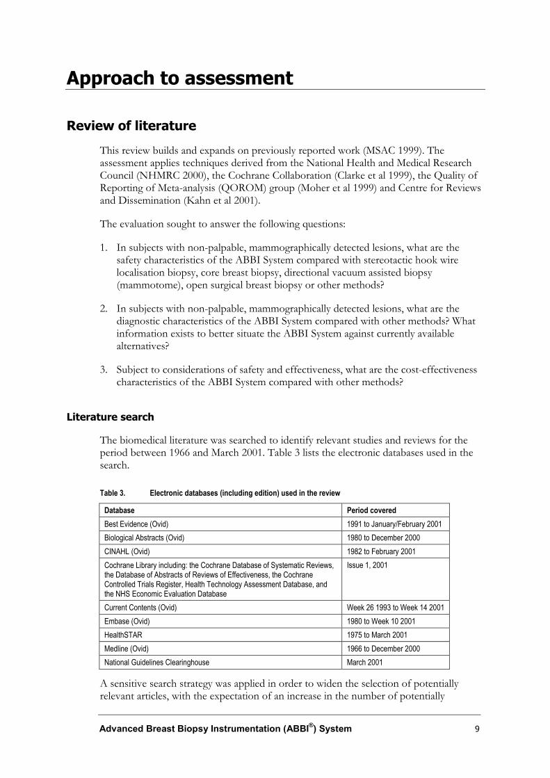
Table 3. Electronic databases (including edition) used in the review ............................... 9
Table 4. Refined search strategy and its implementation in selected electronicdatabases...................................................................................................................10
Table 5. Evidence dimensions (NHMRC 2000) ................................................................12
Table 6. Designations of levels of evidence .......................................................................12
Table 7. Study design characteristics used to assess methodologic quality....................13
Table 8. Distribution of adverse events for patients undergoing ABBI asreported in comparative studies ............................................................................14
Table 9. Distribution of adverse events for patients undergoing ABBI asreported in case series studies................................................................................15
Table 10. Technical problems arising for patients undergoing ABBI ..............................16
Table 11. Descriptive characteristics of comparative studies.............................................18
Table 12. Descriptive characteristics of case series studies ................................................20
Table 13. Summary of overall clinical results of comparative studies ..............................21
Table 14. Number of rebiopsies required following completed ABBIprocedures ................................................................................................................22
Table 15. Summary of number of completed ABBI procedures in case seriesstudies .......................................................................................................................23
Table 16. Summary of ABBI procedures that were converted to open biopsy oraborted ......................................................................................................................23
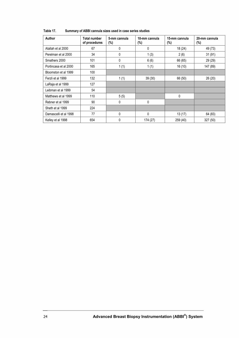
Table 17. Summary of ABBI cannula sizes used in case series studies ............................24
Table 18. Summary of overall results of case series studies ...............................................25
Table 19. Evidence summary – malignancy and type of lesions .......................................27
Table G1. Subsequent procedures following completed ABBI..........................................50
Figures
Figure 1. Schematic representation of breast biopsy techniques (adapted fromWong et al 2000) ....................................................................................................... 6
Figure 2. Flow diagram summarising the results of the literature search and theapplication of entry criteria ....................................................................................11

Advanced Breast Biopsy Instrumentation (ABBI®) System v
Executive summary
The procedure
Advanced breast biopsy instrumentation (ABBI) is a device for diagnostic biopsy ofdetected lesions of the breast. The procedure involves utilisation of a stereotactic imagingsystem.
The procedure, which is conducted by a surgeon and a diagnostic radiologist, involvesthe removal of a core of breast tissue (5–20 mm in size) using stereotactic localisationand an advanced biopsy device.
The patient is positioned prone on a table and the lesion is targeted using stereotaxis.Then, still under stereotactic guidance, a localisation needle is inserted into the lesionand, when the position is satisfactory, a wire is deployed to secure the lesion.
Medical Services Advisory Committee – role and approach
The Medical Services Advisory Committee (MSAC) is a key element of a measure takenby the Commonwealth Government to strengthen the role of evidence in healthfinancing decisions in Australia. MSAC advises the Commonwealth Minister for Healthand Ageing on the evidence relating to the safety, effectiveness and cost-effectiveness ofnew and existing medical technologies and procedures, and under what circumstancespublic funding should be supported.
A rigorous assessment of the available evidence is thus the basis of decision makingwhen funding is sought under Medicare. A team from the Monash Institute of HealthServices Research was engaged to conduct a systematic review of the literature on theABBI System for non-palpable breast lesions. A supporting committee with expertise inthis area then evaluated the evidence and provided advice to MSAC.
MSAC’s assessment of the ABBI System
Clinical need
Breast cancer is the leading cause of cancer death in women and is the greatest cause ofcancer-related mortality in Australian women between the ages of 45 and 64 years. Morethan 2,600 Australian women die from breast cancer every year, and 10,096 new cases ofbreast cancer were diagnosed in Australia in 1997.
The ABBI procedure, which is conducted by a surgeon and a diagnostic radiologist,involves the removal of a core of breast tissue (5–20 mm in size) using stereotacticlocalisation and an advanced biopsy device. The equipment involves the use of a pronestereotactic localisation table together with an ABBI device for core biopsy.

Advanced Breast Biopsy Instrumentation (ABBI®) Systemvi
Safety
Safety data differs widely as ABBI requires specialised surgical techniques and surgeonexperience differs between centres. Eleven studies reported the occurrence ofhaematoma which varied from one to 12.5 per cent. Wound infection varied from 0 to 3per cent and was reported by six studies. Three studies reported dehiscence or woundproblems ranging from 1 to 3 per cent. Bleeding was reported in three studies and rangedfrom 0.4 to 4.2 per cent. Other reported adverse events included nausea, vomiting,hypertension, bruising, anxiety attack, fainting, pneumothorax, venous thrombosis andcellulitis.
The adverse events associated with ABBI were often as a result of technical orequipment failure. Technical problems varied from 3 per cent to 32 per cent andincluded episodes such as cautery snare failure (although this has subsequently beenaddressed by manufacturers), poor precision/calibration, t-wire destabilisation,detachment of blade and computer malfunction.
Effectiveness
No randomised controlled trials comparing ABBI with any other therapeutic ordiagnostic procedure or properly designed diagnostic studies have been completed todate. Studies have compared ABBI to core needle biopsy, mammotome and wirelocalised biopsy. Sensitivity and specificity of ABBI was comparable with core needlebiopsy and mammotome in one study. Discordant rebiopsy rates appeared to be lowerfor ABBI compared with core needle biopsy and mammotome in the same study.Technical success was slightly lower for ABBI compared with core needle biopsy,mammotome and open wire localised biopsy. In another study all margins for ABBI andneedle localisation with excisional breast biopsies were positive. Mean blood loss forABBI was significantly less than for needle localisation with excisional breast biopsy.These comparative studies failed to provide detailed rebiopsy rates.
For the case series studies that reported rebiopsies (occasions when the initial ABBIfailed to obtain sufficient material for histopathological diagnosis), the rates varied from0.05 to 2.9 per cent of all ABBI procedures initially performed. Between 1 and 23 percent of ABBI procedures were converted to another form of biopsy or aborted beforeABBI was completed.
Malignancy of the ABBI biopsy varied from 11 to 44 per cent. Between 57 and 95per cent of malignant biopsies obtained using the ABBI procedure had positive margins.Mean procedure time varied from 22 to 80 minutes. Five out of 13 studies reported somemeasure of patient satisfaction or cosmetic results. Patient satisfaction outcomes weregenerally good and the procedure was generally acceptable to women.
Cost-effectiveness
There is some evidence of cost savings from using ABBI compared with other biopsyprocedures. But even if there were cost savings, this may not translate into a better cost-effectiveness ratio. Limited published evidence on the costs and effects of ABBIprecludes the provision of specific cost-effectiveness estimates.
Advanced Breast Biopsy Instrumentation (ABBI®) System vii
Recommendation
MSAC recommended that, on the strength of evidence pertaining to ABBI, publicfunding should be supported for the diagnostic use of this procedure, as long as fees aresuch that health system costs do not exceed those of comparators. There is insufficientevidence for the use of ABBI in a therapeutic role for breast cancer.
The use of the ABBI equipment is to be limited to surgeons and radiologists withsufficient training and expertise in the procedure, in order to reproduce in Australianpractice the results reported in the literature.
A costing study should be carried out to assist in the setting of the appropriate Medicarerebate.
— The Minister for Health and Aged Care accepted this recommendation on18 September 2001. —


Advanced Breast Biopsy Instrumentation (ABBI®) System 1
Introduction
The Medical Services Advisory Committee (MSAC) has reviewed the use of theAdvanced Breast Biopsy Instrumentation (ABBI) System, a device used in themanagement of non-palpable breast lesions. MSAC evaluates new and existing healthtechnologies and procedures for which funding is sought under the Medicare BenefitsSchedule (MBS) in terms of their safety, effectiveness and cost-effectiveness, while takinginto account other issues such as access and equity. MSAC adopts an evidence-basedapproach to its assessments, based on reviews of the scientific literature and otherinformation sources, including clinical expertise.
MSAC’s terms of reference and membership are at Appendix A. MSAC is amultidisciplinary expert body, comprising members drawn from such disciplines asdiagnostic imaging, pathology, surgery, internal medicine and general practice, clinicalepidemiology, health economics, consumer affairs and health administration.
This report summarises the assessment of current evidence for the ABBI System.

Advanced Breast Biopsy Instrumentation (ABBI®) System2
Background
The procedure
The ABBI System is a device for diagnostic biopsy of detected lesions of the breast. Theprocedure involves utilisation of a stereotactic imaging system.
The procedure, which is conducted by a surgeon and a diagnostic radiologist, involvesthe removal of a core of breast tissue (5–20 mm in diameter) using stereotacticlocalisation and an advanced biopsy device. The average length of tissue cores is usually25 mm or longer.
The patient is positioned prone on a table and the lesion is targeted using stereotaxis.Then, still under stereotactic guidance, a localisation needle is inserted into the lesion,and when the position is satisfactory a wire is deployed to secure the lesion.
Following stereotactic localisation of a small breast lesion, a surgical incision is made inthe breast. A rotating, cylindrical blade is inserted through the incision and advanceduntil the lesion has been included in the core, at which point an integrated diathermywire detaches the deep end of the core, and the core of tissue containing the lesion iswithdrawn from the breast. Any bleeding is stopped by dressing the wound orcauterisation, as required. Radiography of the biopsy sample is undertaken to confirm theremoval of the target tissue and the sample is submitted to a histopathologist forexamination.
ABBI is an outpatient procedure; patients are usually discharged within one hour ofcompletion and normally require one aftercare follow-up consultation.
Intended purpose
The previous MSAC application (MSAC 1999) looked at the validity of ABBI as adiagnostic tool. In this application the scope has been broadened at the request of themanufacturer to propose that ABBI be utilised primarily as a diagnostic device but withthe ability to remove small lesions, entirely and intact, with clear pathological margins asconfirmed by an assessment of the excised specimen.
ABBI potentially provides early and accurate diagnosis of breast cancer. It is indicatedfor biopsy of breast lesions (<15 mm in size) that have been detected by mammography.The lesion concerned may be an invasive carcinoma, an in situ carcinoma or, in somecases, a benign lesion.
Due to constraints of the technology, ABBI may not be suitable for a specific subset ofpatients including:
• patients with mass, asymmetry or clustered microcalcifications that cannot betargeted using digital imaging equipment (Velanovich et al 1999);
• patients unable to lie prone and still for 30 to 60 minutes (Velanovich et al 1999);
Advanced Breast Biopsy Instrumentation (ABBI®) System 3
• breasts less than 20 mm in thickness (Velanovich et al 1999);
• women on anticoagulants or currently taking aspirin;
• lesions that are too close to chest wall or lesions that are subareolar (behind thenipple) as the blood supply may be compromised (Velanovich et al 1999);
• patients weighing more than 135 kg due to possible instability of the table(ASERNIP-S 2000); and
• women with prosthetic breast implants.
ABBI: May 1999 MSAC report
The previous evaluation was undertaken by MSAC in 1999 (MSAC 1999). At that time,based upon the existing published data, it was established that there was insufficientevidence to conclude that ABBI is better than conventional stereotactic core biopsy orhookwire breast localisation. The evaluators found that there was a need to determine aspecific range of conditions for which ABBI would be applicable in the spectrum ofinvestigations available for both benign and malignant breast disease in preference to thewidespread and standard practice, particularly as it relates to conventional stereotacticcore biopsy or hookwire breast localisation needle with open biopsy.
Based upon this finding MSAC recommended that additional funding for the ABBIprocedure not be warranted at that time, and that ABBI should continue to be fundedunder the existing MBS items.
Clinical need/burden of disease
Breast cancer is the leading cause of cancer death in women and is the greatest cause ofcancer-related mortality in Australian women between the ages of 45 and 64 years. Morethan 2,600 Australian women die from breast cancer every year, and 10,096 new cases ofbreast cancer were diagnosed in Australia in 1997 (Australian Institute of Health andWelfare 2000c). In 1998 BreastScreen Australia detected a national average of 15.5 small(95% CI=14.6, 16.4) invasive cancers (≤10mm) per 10,000 women screened in the 50–69year age group (Australian Institute of Health and Welfare 2000b). Data on the numberof benign lesions identified by mammography cannot be readily located.
Breast cancer was one of the 15 leading causes of burden of disease in Australia in 1996.It is the third leading cause of burden of disease in females, accounting for 2.2 per centof the total disease adjusted life years (DALYs) (Australian Institute of Health andWelfare 2000a). The DALY is a summary measure of population health that combinesinformation on mortality and non-fatal outcomes. It uses time as a common currencyand is a measure of the years of healthy life lost due to illness or injury – one DALY isone lost year of healthy life.
Increasingly, women are participating in mammographic screening, which results inearlier detection of non-palpable lesions. The five-year relative survival rate for femalesdiagnosed with breast cancer from 1982 to 1994 was 77 per cent. The rate has increasedover time, with those diagnosed in the 1990s showing a better five-year survival rate

Advanced Breast Biopsy Instrumentation (ABBI®) System4
(79%) than those diagnosed in the 1980s. Women diagnosed with breast cancer in their40s had the best relative survival, whereas those aged in their 80s and 90s had the worstsurvival (Australian Institute of Health and Welfare 2000a).
The Australian distributor, Auto Suture Company, has advised that there are presentlyfive centres with ABBI units in Australia (Wesley Hospital, Queensland; WestmeadHospital, New South Wales; Sydney Adventist Hospital, New South Wales; RoyalWomen's Hospital, New South Wales; and Queen Elizabeth Hospital, South Australia).At this stage, the potential usage for these units is unclear due to the lack of availabledata on the total number of benign and malignant small breast lesions (<10 mm in size)biopsied in Australia annually. Table 1 shows the rate of small diameter (≤10 mm)invasive cancers detected in women screened by age in 1998, in both initial andsubsequent screening rounds.
Table 1. Age specific rate of small diameter (<10 mm) invasive cancers detected per 10,000 women screened(Australian Institute of Health and Welfare 2000b)
Age group(years)
40–44 45–49 50–54 55–59 60–64 65–69 70–74 75–79 80–84 85+ Allages:cruderate
Agestandardisedrate (Austpopulation)(95% CI)
Number ofcancersdetected
2.5 6.0 10.8 14.1 18.6 21.7 25.9 32.8 26.8 43.1 13.2 13.2(12.1, 14.4)
Statistics regarding the Medicare benefits paid on a fee-for-service basis provide anindication of the relative usage of service, as does data provided by BreastScreenAustralia. The exclusion from these statistics of services to public patients in hospital andof those paid for by Veterans’ Affairs limits the completeness of the figures.
Existing procedure and comparators
Women who are found to have a breast lesion following mammography will be referredfor further diagnostic tests. These may include additional mammography, ultrasound andneedle core biopsy or hookwire breast localisation needle for open surgical biopsy. Adifficulty is that lesions are not diagnosed with a single ‘gold standard’ test, but rather arediagnosed by a variety of tests. The extent of use, and purpose for use of thesecomparators varies between settings.
Potentially ABBI could replace core biopsy or hookwire breast localisation needle foropen surgical biopsy for breast lesions of less than 10 mm. These are thereforeappropriate comparators. It should be noted that the comparators discussed below areprimarily diagnostic in nature.
Wire localisation biopsy
The main types of localisation biopsy are hookwire or carbon fibre. This procedure isgenerally performed under general anaesthesia in Australia, although the procedure hasbeen known to be performed under local anaesthesia in other countries. The patient

Advanced Breast Biopsy Instrumentation (ABBI®) System 5
usually lies in a supine position during the wire placement procedure (although they canbe positioned supine, prone or seated depending on method of localisation).
Firstly a patient is imaged to determine the exact location of the lesion targeted forremoval. A needle is then inserted through the lesion, and its position is confirmed withanother mammograph. A small wire is then inserted through the hollow needle. When inplace the needle is withdrawn leaving the wire in place as a surgical guide to the targetlesion.
Once the end of the wire has been located internally an incision is made so both thelesion and the wire are easily accessed and removed. If the procedure is successful onlyone sample will be required for histological analysis. The scar from this procedure isusually between two to five centimetres, depending upon the depth of the lesion and thesize of the woman’s breast. The patient is usually free to leave hospital the same day.
Vacuum-assisted core biopsy
The two main forms of vacuum-assisted core biopsy (VACB) are also known as minimalinvasive breast biopsy (MIBB) and mammotome. Both of these forms of breast biopsyare in rapid evolution and different methods are used according to surgical preferenceand the availability of stereotactic tables. MSAC evaluation no. 1015, DirectionalVacuum-assisted Breast Biopsy, gives a complete report of this method.
Minimal invasive breast biopsyThis procedure is also referred to as directional vacuum-assisted biopsy in other literature(National Breast Cancer Centre 2000). The VACB probe requires a single-step placementinto the area of interest, and multiple core biopsies are made around the area where theprobe has been commissioned. Each core is evacuated from the probe by a vacuumsuction into a drainage system (Wong et al 2000). The patient is positioned either seatedor supine whilst the procedure is undertaken. The patient is usually free to leave hospitalthe same day.
MammotomeThis system works in a manner similar to that described above although the patient liesin a prone position. The mammotome procedure for mammographically detected lesionsinvolves the use of an arm attachment. Biopsies are removed manually from the samplechamber with forceps. The tissue cores may be grouped into separate tissue cassettes asrequired (eg 1–3 o’clock, 3–6 o’clock) and identified accordingly either by pencil labellingor by different coloured cassettes prior to dispatch for histopathology (Wong et al 2000).
Wounds from both vacuum methods do not require any form of suturing, and scarring,if any, is minimal due to the small incision.
Core needle biopsies
With a core needle biopsy the woman is positioned either seated, lateral or lying prone.The procedure utilises local anaesthetic and hospitalisation is not required. After carefullydisinfecting the skin, the physician uses a special fenestrated compression paddle tocompress and immobilise the breast and the lesion is accessible through a window(Heywang-Kobrunner et al 1997).

Advanced Breast Biopsy Instrumentation (ABBI®) System6
The conventional stereotactic core biopsy device comprises a spring-loaded handle,together with a disposable 14-gauge core biopsy needle. The tissue cores obtained areapproximately 1 mm in diameter (Wong et al 2000). Usually three to four cores areobtained to guarantee sufficient tissue. In the case of microcalcifications, more cores maybe necessary (Figure 1). Core biopsies can also be obtained using ultrasound guidance. Asimple adhesive bandage is sufficient to cover the site of wound following the procedure.
Microcalcification
Core
CCB 14 Gauge VACB 11 Gauge ABBI 9 French
Standard core would Needle in centre pulls ABBI aims to include allbe 3 to a maximum tissue around it into of a small cluster ofof 5 samples. core, takes biopsy and calcifications or a single
moves to the next site sample, depending onwith a view to taking the size of the cannulaconcentric biopsies. used and the degree of
widespread calcification.
Figure 1. Schematic representation of breast biopsy techniques (adapted from Wong et al 2000)
This diagram demonstrates the manner in which the same focus of mammographicmicrocalcifications is sampled by conventional core biopsy (CCB), VACB and ABBI.CCB removes some of the microcalcifications, while others are left behind. VACBremoves some of the area by multiple sampling. ABBI attempts to remove a largeportion or the whole area as a single intact core of tissue.
Marketing status of the device
ABBI has been approved by the United States Food and Drug Administration underSection 510(k), to be used when stereotactically localised large-diameter breast biopsies,identified by the placement of a needle localisation wire, are desired for diagnosticsampling of a mammographic abnormality where malignant disease is suspected (ieusually Breast Imaging Reporting and Data System class four or five). The ABBI deviceis intended to provide breast tissue for histological examination with partial or completeremoval of the imaged abnormality.

Advanced Breast Biopsy Instrumentation (ABBI®) System 7
The instrumentation is listed on the Australian Register of Therapeutic Goods numberAUST L54966. Before listing, sponsors are required to submit to the Therapeutic GoodsAdministration for assessment information such as labelling, product literature and, forcertain categories, evidence of quality systems compliance, standards compliance and testcertificates.
Current reimbursement arrangement
Breast biopsy services currently covered under the MBS and the number of servicesrendered for each in 1998–2000 are listed in Table 2. Procedures involving ABBI cancurrently be claimed under MBS using item numbers 30361 and 30363 with radiologyitem numbers 59312 (two breasts) or 59314 (one breast). Claims could also be madeunder item numbers 30345G and 30346S until May 2000, when they were deleted afterconsideration by the Medicare Benefits Consultative Committee. See Table 2 fordefinitions of item numbers.

Advanced Breast Biopsy Instrumentation (ABBI®) System8
Table 2. Breast biopsy MBS services rendered 1998–2000
Number of servicesItem no Item description
1997 1998 1999 2000
30339 Breast, benign lesion up to and including 50 mm in diameter, includingsimple cyst, fibroadenoma or fibrocystic disease, open surgical biopsyor excision of, with or without frozen section histology
2237
30343 Breast, abnormality detected by mammography or ultrasound whereguidewire or other localisation procedure is performed, excision biopsyof
1468
30344 Breast, malignant tumour, open surgical biopsy of, with or withoutfrozen section histology
228
30345G* Breast, excision of cyst, fibroadenoma or other local lesion orsegmental resection for any other reason, where frozen section biopsyis performed or where specimen radiography is used
40 56 33 13‡
30346S† Breast, excision of cyst, fibroadenoma or other local lesion orsegmental resection for any other reason, where frozen section biopsyis performed or where specimen radiography is used
7006 6819 6105 2486‡
30347 Breast, malignant tumour, complete local excision of, with or withoutfrozen section histology
2286
30358 Breast, biopsy of solid tumour or tissue of, using a vacuum-assistedbreast biopsy device under imaging guidance, for histologicalexamination, where imaging has demonstrated:(a) microcalcification oflesion; or (b) impalpable lesion less than 1 cm in diameter – includingpre-operative localisation of lesion where performed, not being aservice to which item 30363 applies
30
30360 Fine needle aspiration of an impalpable breast lesion detected bymammography or ultrasound, imaging guided – but not includingimaging
19062 26032 27816 27175
30361 Breast, preoperative localisation of lesion of, by hookwire or similardevice, using interventional techniques – but not including imaging
3679 4082 4009 4254
30363 Breast, core biopsy of solid tumour or tissue of, using mechanicalbiopsy device, for histological examination
2843 3467 4279 5327
59312 Radiographic examination of both breasts, in conjunction with a surgicalprocedure on each breast, using interventional techniques –examination and report
3 165 164 116
59314 Radiographic examination of one breast, in conjunction with a surgicalprocedure using interventional techniques – examination and report
217 2340 2634 2723
59318 Radiographic examination of excised breast tissue to confirmsatisfactory excision of one or more lesions in one breast or bothfollowing preoperative localisation in conjunction with a service underitem 30361 – examination and report
142 2139 2691 3253
* General practitioners.† Specialists.‡ January–May 2000 only.
Advanced Breast Biopsy Instrumentation (ABBI®) System 9
Approach to assessment
Review of literature
This review builds and expands on previously reported work (MSAC 1999). Theassessment applies techniques derived from the National Health and Medical ResearchCouncil (NHMRC 2000), the Cochrane Collaboration (Clarke et al 1999), the Quality ofReporting of Meta-analysis (QOROM) group (Moher et al 1999) and Centre for Reviewsand Dissemination (Kahn et al 2001).
The evaluation sought to answer the following questions:
1. In subjects with non-palpable, mammographically detected lesions, what are thesafety characteristics of the ABBI System compared with stereotactic hook wirelocalisation biopsy, core breast biopsy, directional vacuum assisted biopsy(mammotome), open surgical breast biopsy or other methods?
2. In subjects with non-palpable, mammographically detected lesions, what are thediagnostic characteristics of the ABBI System compared with other methods? Whatinformation exists to better situate the ABBI System against currently availablealternatives?
3. Subject to considerations of safety and effectiveness, what are the cost-effectivenesscharacteristics of the ABBI System compared with other methods?
Literature search
The biomedical literature was searched to identify relevant studies and reviews for theperiod between 1966 and March 2001. Table 3 lists the electronic databases used in thesearch.
Table 3. Electronic databases (including edition) used in the review
Database Period coveredBest Evidence (Ovid) 1991 to January/February 2001Biological Abstracts (Ovid) 1980 to December 2000CINAHL (Ovid) 1982 to February 2001Cochrane Library including: the Cochrane Database of Systematic Reviews,the Database of Abstracts of Reviews of Effectiveness, the CochraneControlled Trials Register, Health Technology Assessment Database, andthe NHS Economic Evaluation Database
Issue 1, 2001
Current Contents (Ovid) Week 26 1993 to Week 14 2001Embase (Ovid) 1980 to Week 10 2001HealthSTAR 1975 to March 2001Medline (Ovid) 1966 to December 2000National Guidelines Clearinghouse March 2001
A sensitive search strategy was applied in order to widen the selection of potentiallyrelevant articles, with the expectation of an increase in the number of potentially

Advanced Breast Biopsy Instrumentation (ABBI®) System10
irrelevant articles identified by the strategy (Haynes et al 1994a, Haynes et al 1994b). Asearch strategy was parsimoniously derived from numerous pilot searches of theelectronic literature and refined iteratively. The final strategy is shown in Table 4 andincorporates the search strategy of the previous report (MSAC 1999).
Table 4. Refined search strategy and its implementation in selected electronic databases*
Strategy DatabaseABBI.mp OR ((biops$.mp AND breast$.mp) AND (three-dimension$.mp OR automat$.mp ORadvance$.mp OR instrument$.mp))
Ovid databases
ABBI* OR ((biops* AND breast*) AND (three-dimension* OR automat* OR advance* OR instrument*)) Cochrane Library* Electronic databases apply different characters as “wildcard” symbols. These symbols refer to characters or groups of characters that
appear in the terminus of a word fragment. For the Ovid databases, the wildcard character is the dollar sign (“$”); the Cochrane Libraryuses the asterisk (“*”). In this case, “biops$” expands to “biopsy”, “biopsies”, etc.
Electronic searching included the Internet sites of health technology assessment groups(listed in Appendix D), professional medical organisations, medical centres and healthservice providers, and relevant national and international government agencies. Dataprovided by the manufacturer of the device was included where relevant, butconfirmation of the information was sought from independent sources.
Textbooks and book chapters were assessed, as were conference proceedings andcollections of abstracts. Reference lists of publications were scanned and relevantcitations retrieved.
Entry criteria
Collected citations were filtered through a multi-level review involving a team with skillsin clinical medicine, public health, health informatics, basic science, clinical epidemiology,and biostatistics. Articles were excluded if they met the following criteria:
• pre-clinical studies involving in vitro experiments, animals, isolated human organsor cadavers;
• studies that did not focus on the use of the ABBI System in the management ofnon-palpable breast lesions;
• studies enrolling less than 10 subjects;
• case reports, non-systematic reviews, and opinions published as editorials orletters to the editor;
• articles that included data published in later studies; and
• level IV evidence (case series) available only in abstract form.
The evaluation was restricted to studies published subsequent to the release of MSACApplication 1001 (MSAC 1999). No restrictions were placed on publication types orpopulation characteristics.

Advanced Breast Biopsy Instrumentation (ABBI®) System 11
Review profile
The search identified 298 studies. Of these, 179 (60.07%) were excluded on the basis ofthe criteria previously defined. The remaining 119 articles were retrieved for moredetailed evaluation. These included articles that did not provide enough preliminaryinformation to make a decision about inclusion or exclusion (ie for reasons such asunclear or missing abstracts and uninformative titles). Detailed evaluation of articlesnecessitated assessment of the full text. A final decision about entry was made byconsensus between two independent reviewers.
Of the 119 citations requiring full text assessment, 103 (86.55%) were excluded for thefollowing reasons: 77 were narrative reviews, 21 were expert opinions, three studiescontained data that had been published previously, and two were available only inabstract form. These 103 excluded studies are listed in Appendix E. The remaining 16studies provide the basis of this review.
Study flow is described in Figure 2.
Data extraction
The review extracted data from the included articles using a standardised instrumentcreated for this assessment. In some cases, quantitative information was poorlypresented. In these instances, every effort was made to apply statistical techniques toderive estimates of effect size or variability if enough information was available.Otherwise, a statement indicating the paucity of primary information was made.
Two independent reviewers examined each article. Discrepancies in evaluation werediscussed and resolved through consensus.
Potentially relevantstudies identified andscreened for retrieval
(n=298)
Studies excluded (n=179)
Studies retrieved for full-text evaluation
(n=119)
Studies excluded (n= 103):Narrative reviews (n=77)Expert opinions (n=21)Duplicated data (n=3)Abstract only (n=2)
Studies included in thesystematic review
(n=16)
Figure 2. Flow diagram summarising the results of the literature search and the application of entrycriteria

Advanced Breast Biopsy Instrumentation (ABBI®) System12
Dimensions of evidence
The NHMRC recommends that evidence assessment move toward an evaluation ofspecific ‘dimensions’. These dimensions (Table 5) consider important aspects of theevidence supporting a particular intervention and include three main domains: strengthof the evidence, size of the effect and relevance of the evidence. The first domain isderived directly from the literature identified as informing a particular intervention. Thelast two require expert clinical input to determine.
Table 5. Evidence dimensions (NHMRC 2000)
Type of evidence DefinitionStrength of the evidence
Level
QualityStatistical precision
The study design used, as an indicator of the degree to which bias has been eliminated bydesign.*The methods used by investigators to minimise bias within a study design.The p-value or, alternatively, the precision of the estimate of the effect. It reflects thedegree of certainty about the existence of a true effect.
Size of effect The distance of the study estimate from the ‘null’ value and the inclusion of only clinicallyimportant effects in the confidence interval.
Relevance of evidence The usefulness of the evidence in clinical practice, particularly the appropriateness of theoutcome measures used.
* See Table 6.
The strength of the evidence is composed of three sub-domains. Previous assessmentsconcentrated only on the first of these, the level of the evidence (NHMRC 1999). Table6 lists the designations recommended by the NHMRC.
Table 6. Designations of levels of evidence*
Level of evidence Study designIIIIII–1
III–2
III–3
IV
Evidence obtained from a systematic review of all relevant randomised controlled trials.Evidence obtained from at least one properly designed randomised controlled trial.Evidence obtained from well-designed pseudorandomised controlled trials (alternate allocation orsome other method).Evidence obtained from comparative studies (including systematic reviews of such studies) withconcurrent controls and allocation not randomised, cohort studies, case-control studies, orinterrupted time series with a control group.Evidence obtained from comparative studies with historical control, two or more single arm studies,or interrupted time series without a parallel control group.Evidence obtained from case series, either post-test or pre-test/post-test.
* Modified from (NHMRC 1999).
The assessment of quality, another important sub-domain, was based on characteristicsknown to reflect important aspects of study design (Schulz et al 1995, Jadad et al 1996).Table 7 summarises these characteristics and the ordinal scale used in the assessment.

Advanced Breast Biopsy Instrumentation (ABBI®) System 13
Table 7. Study design characteristics used to assess methodologic quality
RandomisationAdequate
Unclear
Inadequate
Method of allocation is random, such as computer-generated number sequences and tables ofrandom numbers.Trials in which the authors failed to describe the method of randomisation with enough detail todetermine its validity.Method of allocation is non-random, such as alternation methods or the use of case numbers.
Concealment of allocationAdequate
Unclear
Inadequate
Adequate measures to conceal allocations such as central randomisation; serially numbered,opaque, sealed envelopes; or other descriptions that contain convincing elements ofconcealment.Unclearly concealed trials in which the author failed to describe the method of concealment withenough detail to determine its validity.Method of allocation is not concealed.
Masking Masking strategy applied (single, double, etc).Participant inclusion Intention to treat analysis was performed.Losses to follow-up Losses specified.
Expert advice
A supporting committee with expertise in breast surgery, general surgery, public health,and consumer issues was established to evaluate the evidence and provide advice toMSAC from a clinical perspective. In selecting members for supporting committees,MSAC’s practice is to approach the appropriate medical colleges, specialist societies andassociations and consumer bodies for nominees. Membership of the supportingcommittee is provided at Appendix B.

Advanced Breast Biopsy Instrumentation (ABBI®) System14
Results of assessment
Is it safe?
Comparative Studies
These studies mainly reported on adverse events experienced with the ABBI procedureand do not provide detailed comparisons with the other procedures. Two studies (Yanget al 2000, Velanovich et al 1999) do not report any safety outcomes for theircomparative procedures. The study by D'Angelo et al (1997) reported no complicationsout of 23 procedures for needle localisation breast biopsy (Table 8). Yang et al (2000)report that two out of 100 ABBI patients experienced a bleeding complication (Table 8).The study by Velanovich et al (1999) compared ABBI with core needle biopsy andreported that all ABBI rebiopsies in the first 30 cases were the result of technical failure.In the third comparative study (D'Angelo et al 1997) no complications or infections werenoted for the 23 procedures.
Table 8. Distribution of adverse events for patients undergoing ABBI as reported in comparative studies
Study Outcomes Number of ABBIprocedures
Number of cases (%)
Yang et al 2000 BleedingIncomplete excision
100 2 (2.0)5 (5.0)
Velanovich et al 1999 Technical failure resulting in rebiopsy 104 All rebiopsies in first 30 cases*D’Angelo et al 1997 Complications
Infections23 0 (0)
0 (0)
* Number not stated.
Case Series
Adverse events
The adverse events associated with ABBI and reported by the appraised studies includedhaematoma, ecchymosis, bleeding, wound infection, and others. The incidence ofhaematoma ranged from 1 per cent (LaRaja et al 1999) to 12.5 per cent (Rebner et al1999) –Table 9. Bleeding was reported by only three studies and ranged from 0.4 percent (Sheth et al 1999) to 4.2 per cent (Rebner et al 1999). The incidence of woundinfection was variable and ranged from nil (Damascelli et al 1998, LaRaja et al 1999,Leibman et al 1999, Portincasa et al 2000) to 3 per cent (Rebner et al 1999, Perelman et al2000) – Table 9. The study by Bloomston et al (1999) reported that ecchymosis was notuncommon after the ABBI procedure but did not provide data. LaRaja et al (1999) statedthat bleeding varied with each patient but was controlled in every instance with cautery.
Matthews et al (1999) reported that there were no intraoperative complications in the 110biopsies performed and Sheth et al (1999) reported no cases of intolerance to the ABBIprocedure in 230 biopsies.

Advanced Breast Biopsy Instrumentation (ABBI®) System 15
Table 9. Distribution of adverse events for patients undergoing ABBI as reported in case series studies
Number ofStudypatients biopsies
Outcomes Number of cases(%)
Atallah et al 2000 65 67 Haematoma 8 (12.3)Perelman et al 2000 34 34 Haematoma
Wound infectionDehiscenceNausea/ vomiting/ hypotensionBruisingAnxiety attack
2 (6.0)1 (3.0)1 (3.0)
4 (12.0)1 (3.0)1 (3.0)
Portincasa et al 2000 165 165 HaematomaWound infectionSeromas
000
Bloomston et al 1999 100 99 Haematoma 2 (2.0)Ferzli et al 1999 135 132 Haematoma
DehiscenceFainting
2 (1.3)1 (0.9)1 (0.9)
LaRaja et al 1999 127 127 HaematomaWound infectionSeromas
1 (0.8)00
Leibman et al 1999 53 54 HaematomaWound infection
3 (5.7)0
Matthews et al 1999 107 110 HaematomaWound infection
2 (1.9)1 (0.9)
Rebner et al 1999 89 90 HaematomaBleedingWound problemsPneumothoraxVenous thrombosis
9 (12.5)3 (4.2)2 (2.8)1 (1.4)1 (1.4)
Sheth et al 1999 223 230 Bleeding 1 (0.4)Damascelli et al 1998 75 77 Haematoma
Wound infection3 (4.0)
0Kelley et al 1998 ? 654 Haematoma
Cellulitis11 (1.7)1 (0.2)
The adverse events listed in Table 9 are of low incidence and likely to be of little healthsignificance. Notably, the definition of adverse events varied across the included studies,and consistent parameters were not used to evaluate adverse events associated with theABBI procedure. This made the evaluation of adverse events across the studies difficult toquantify and collate. A clear definition of adverse events and parameters is needed in thefuture to assess the impact of ABBI.

Advanced Breast Biopsy Instrumentation (ABBI®) System16
Technical problems
The adverse events associated with ABBI were often as a result of technical orequipment failure. Technical problems varied from 3 per cent (Damascelli et al 1998) to32 per cent (Perelman et al 2000) and included episodes such as computer malfunction,poor precision, the cutting blade detaching, and t-wire destabilisation. Likewise, theoccurrence of mechanical malfunctions or equipment failure ranged from 2 per cent(Leibman et al 1999) to 23.5 per cent (Ferzli et al 1999) – Table 10. For the Bloomston etal (1999) study, equipment malfunction was a mechanical failure resulting directly frommalfunction of the ABBI device.
Table 10. Technical problems arising for patients undergoing ABBI
Number ofAuthor(s)patients biopsies
Outcomes Number of cases (%)
Perelman et al2000
34 34 Cautery snare failurePoor precision/calibrationT-wire destabilisationComputer malfunctionLesion displacementOther minor difficulties
3 (9.0)5 (15.0)2 (6.0)1 (3.0)1 (3.0)1 (3.0)
Bloomston et al1999
100 99 Equipment malfunctionConversion to open biopsy due to mechanical failure
3 (3.0)1 (1.0)
Ferzli et al 1999 135 132 Cautery snare failurePoor calibrationNeedle moved nodule changing coordinatesDeployment of the T-bar moved the neoplasmT-bar and specimen dislodged as blade withdrawn
12 (9.1)2 (1.5)7 (5.3)4 (3.0)6 (4.5)
Leibman et al 1999 53 54 T-bar fracture 3 (5.7)Damascelli et al1998
75 77 Detachment of bladeFailure of imaging computer during procedure
1 (1.3)1 (1.3)
Five out of the 12 case studies reported technical problems. Adverse events werereported for all studies although the numbers of adverse events were generally low. Thepercentages indicate that technical problems may have been more common than adverseevents. There were two centres that experienced particularly high numbers of technicalproblems, and technical problems often led to adverse events (Perelman et al 2000, Ferzliet al 1999). The results of these studies highlight the need for careful calibration ofimaging equipment. The manufacturer acknowledges that early production cannulae weremore problematic with deployment of the snare. Manufacturing processes were changedover 2–3 years ago to improve the mechanism, and the manufacturers maintain thatfrequency of the snare failure has been reduced.
A significant issue likely to affect the outcome of adverse events and technical problemsis related to the staff performing the ABBI procedure. The observed variations in thetype of adverse events across the studies may be attributed to the difference in thetraining, experience and qualification of the staff performing the ABBI procedure.Hence, the quality and experience of those undertaking the procedure must be taken intoconsideration. The adverse events related to technical or mechanical factors may also

Advanced Breast Biopsy Instrumentation (ABBI®) System 17
reflect the rapid and evolving technological advances being made in regards to the ABBIequipment.
Clinical practice guidelines
The Australian Safety and Efficacy Register of New Interventional Procedures – Surgery(ASERNIP–S) has developed guidelines for ABBI (Walsh et al 2000). The guidelines arebased on a review of the literature. A breast surgeon drafted the guidelines and a reviewgroup was formed to critique them. The guidelines have been disseminated through theRoyal Australasian College of Surgeons.
The guidelines include recommendations on lesion selection, patient selection, technicalfactors, credentials, pathological specimens and financial considerations.
Recommendations include:
• ABBI biopsy is appropriate only for impalpable breast lesions, lesions clearly visibleon a diagnostic mammogram and diagnosis of malignant lesions.
• Proven malignant lesions should not routinely undergo ABBI biopsy.
• The patient should not be anti-coagulated, must weigh less than 130 kg and must beable to lie immobile in the prone position.
• The lesion should not be too close to the chest wall, the compressed breast shouldnot be too small to allow ABBI biopsy, and mammographic lesions behind thenipple should be approached with caution.
• ABBI biopsy is a day patient procedure; orally administered premedication appearsto facilitate the procedure; post biopsy mammography and specimen radiologyshould be routinely performed.
• The ABBI room must meet appropriate radiological and surgical standards.
• The ABBI system should be operated by practitioners specifically trained andaccredited in its use and a patient database should be established at all ABBI centres.
• The ABBI technique requires both radiological and surgical skills; the procedureshould be subject to surgical audit; and ABBI biopsies should be viewed by amultidisciplinary team.
• The ABBI biopsy specimen should always be submitted for histopathologicalexamination, and a copy of the specimen X-ray should be sent with the ABBIspecimen.
• Frozen sections are not routinely recommended for ABBI specimens; thelocalisation and fixation T-bar should remain in the specimen; and orientationsutures should be placed in the specimen.

Advanced Breast Biopsy Instrumentation (ABBI®) System18
Limitations of the guidelines include a potentially narrow search strategy, unclear methodsof formulating recommendations, and recommendations that are based on low-levelevidence that is prone to bias. A review of the guidelines is expected in 2001.
Is it effective?
Comparative studies
Descriptive characteristics
The literature search uncovered a total of 16 studies that provided evidence about theeffectiveness of ABBI and that were published since the previous MSAC report (MSAC1999). The ideal study design for assessing the clinical effectiveness of a therapeuticprocedure is a randomised controlled trial. The ideal study design for assessing diagnosticaccuracy is the independent blind comparison of an appropriate spectrum of consecutivepatients, all of whom have undergone both the diagnostic test and the referencestandard. No randomised controlled trials or properly designed diagnostic studies havebeen completed to date. Three of the included studies are comparative studies (Table 11).One comparative study compared ABBI with core needle biopsy; another comparedABBI and needle localisation under general anaesthesia; and the other compared ABBIwith core needle biopsy and wire localised biopsy. The most recent publication for eachrelevant study was included.
Table 11. Descriptive characteristics of comparative studies
Population characteristics
Study Location Dates ofenrolment
Interventiongroup*
Comparison groups* Totalnumber ofpatients
Age(years)†
Length offollow-up
Yang et al2000
Korea Dec 1996 toAug 1998
ABBI (100) Core needle biopsy (59) 159 ?‡ In hospital
Velanovich etal 1999
USA Jan 1997 toMar 1998
ABBI (104) Core needle biopsy (245)Mammotome (107)Wire-localised biopsy (520)
976 ? 6 months
D'Angelo et al1997
USA Mar 1996 toJun 1996
ABBI (23) Needle localisation withexcisional breast biopsies (23)
46 Interven-tion=62(13)Compari-son=68(11)
?
*The number in brackets refers to the number of procedures that were performed.†The standard deviations for age are given in brackets.‡Data not reported or unknown.
Three reports of comparative studies were found (D'Angelo et al 1997, Velanovich et al1999, Yang et al 2000) – Table 11. All three comparative studies had concurrent controlgroups. Two studies were conducted in the USA and the other in Korea. Patients wererecruited in the late 1990s (Table 11).
The study by Velanovich et al (1999) reports that both the mammotome and ABBI werenew procedures at their institution at the commencement of their study. In this studymost patients were followed up at six months with mammograms; some had clinical

Advanced Breast Biopsy Instrumentation (ABBI®) System 19
examination only; and some were simply referred back to their primary care physicians.For the D'Angelo et al (1997) study the comparator-needle localisation with excisionalbreast biopsies was performed in the operating room under general anaesthesia. Somepatients chose in this study (D'Angelo et al 1997) not to have the newer ABBIprocedures, and those women already in need of an operation for another cause wereallocated to the comparison group.
The exclusion criteria varied between studies. The study by Yang et al (2000) excludedpatients with highly suspicious malignancies and palpable breast lesions. Another study(Velanovich et al 1999) excluded patients who underwent ultrasound-guided biopsies,and cystic lesions were excluded from biopsy in the D’Angelo et al (1997) study.
Quality
None of the studies were randomised. All three studies were comparative studies withconcurrent controls. The allocation of patients to ABBI or core needle biopsy in theYang et al (2000) study was performed according to clinical criteria (ABBI formicrocalcifications not more than 2 cm). Another study (Velanovich et al 1999) did notprovide details of the methods used to allocate patients to ABBI, core needle biopsy,mammotome or wire-localised biopsy but allocation appears to also be based on clinicalselection criteria. The study by D’Angelo et al (1997) allocated patients to ABBI orneedle localisation biopsies based on patient preferences and clinical convenience(women already scheduled for general anaesthesia had the needle localisation biopsies).Inadequate randomisation and concealment of allocation are significant potentials forbias as they have been related to a 30 per cent overestimation in the measures of effect(Schulz et al 1995).
None of the studies made mention of masking patients or investigators. There isevidence that lack of masking leads to performance bias which has been associated withup to 17 per cent overestimation of effect (Schulz et al 1995). It is unclear whetherpatients were analysed in the groups to which they were originally allocated. None of thestudies mentioned whether the results were conducted using the ‘intention-to-treat’principle.
Two studies (Yang et al 2000, D’Angelo et al 1997) had no loss to follow-up, and theother study (Velanovich et al 1999) failed to provide details of loss to follow-up. It isimportant that the two groups do not vary in their loss to follow-up. All of the studieshad a short length of follow-up, limiting the application of results to the clinical setting.
None of the studies reported sample size/power calculations. It is possible that thestudies did not have sufficient sample sizes to detect differences between groups.Although the studies were comparative in design the main results were often reportedonly for the ABBI group. There was only one outcome related to safety (complications)in one study (D’Angelo et al 1997) that was reported for ABBI and a comparator. Onestudy (Velanovich et al 1999) presented the results graphically and failed to report actualfigures.

Advanced Breast Biopsy Instrumentation (ABBI®) System20
Case series studies
Descriptive characteristics
Twelve of the 16 included studies are case series studies (Table 12). This design is highlyprone to bias and is classified as level IV evidence according to the NHMRC levels ofevidence (NHMRC 2000). Sample sizes ranged from 34 to 654 procedures (Table 12).The two largest studies were both from the USA (Kelley et al 1998, Sheth et al 1999). Instudies that reported age, participants had a mean age between 47 and 62 years. Theduration of follow-up varied between studies, with four studies assessing patients onlywhile they remained in hospital (Leibman et al 1999, Sheth et al 1999, Perelman et al2000, Smathers 2000). The longest period of follow-up was two years in the French studyby Atallah et al (2000) – Table 12.
One of the studies (Kelley et al 1998) was a multi-centre study involving eight separateinstitutions. One of the included studies also reported results independently and has beenexcluded to avoid assigning double weight to its findings. Another study was reported inthree articles at three separate stages. Only the most recent paper has been included(Ferzli et al 1999). One study was written in French (Atallah et al 2000); all the otherpapers were English publications.
Table 12. Descriptive characteristics of case series studies
Study Location Dates of enrolment
Number ofABBIprocedures
Age (years)* Length of follow-up
Atallah et al 2000 France ? 67 ?† 2 Years
Perelman et al 2000 Canada Sep 1997 to May 1998 34 ? In hospital
Smathers 2000 USA Apr 1997 to Aug 1998 101 ? In hospital
Portincasa et al 2000 Italy ? 170 51 (34–81) I week
Bloomston et al 1999 USA Apr 1996 to May 1997 100 62 (34–87) Mean of 7 months
Ferzli et al 1999 USA Apr 1996 to Apr 1997 132 52 (32–76) 1 week
LaRaja et al 1999 USA Jul 1996 to Feb 1998 127 ? 24–48 hours
Leibman et al 1999 USA Feb 1997 to Dec 1997 54 53 (32–85) In hospital
Matthews et al 1999 USA Feb 1997 to Jan 1998 110 61 (31–83) 6 months
Rebner et al 1999 USA May 1997 to Mar 1998 90 ? 11 months
Sheth et al 1999 USA Apr 1997 to Jun 1998 230 47 (30–88) In hospital
Damascelli et al 1998 Italy Jun 1997 to Jan 1998 77 ? 1 week
Kelley et al 1998 USA ? 654 ? ?
* The range for age is shown in brackets.† Data not reported or unknown.
Quality
Case series study designs are highly prone to bias. A major flaw is the failure to comparethe procedure with another procedure. There has been no attempt to assess the quality ofthese studies.

Advanced Breast Biopsy Instrumentation (ABBI®) System 21
Comparative studies
Results
Table 13. Summary of overall clinical results of comparative studies
Number of procedures (%)Study Total numberof patients
OutcomesABBI Core needle
biopsyMammotome Open wire
localisedbiopsy
Needlelocalisationwithexcisionalbreastbiopsies
Yang et al2000
159 MalignantBenignAdditional biopsyResidual cancerIncomplete excision oflesionIncomplete removal ofcalcification
9 (10.0)91 (90.0)
?*?
5 (5.0)
3 (3.0)
13 (22.0)46 (78.0)7 (18.9)4 (57.1)
?
?
Velanovichet al 1999
976 Positive margins(malignant lesions)Discordant/rebiopsyrateTechnically successful(enable diagnosis)SensitivitySpecificityResidual cancer rate
(63.6)
(5–10)‡
(92.5)‡
(90–100)‡(90–100)‡
(71.4)
?
(25–30)‡
(94.3)
(80–90)‡(90–100)‡
?
?
(20–25)‡
(96.4)
(80–90)‡(90–100)‡
?
(50.9)
?
(98.7)
??
(70.4)D'Angelo etal 1997
46 MalignantBenignPositive marginsMean procedure time(minutes)Mean blood loss (cc)Mean solitary nodulardensity (mm)Residual abnormalities(after ABBI)Residual cancer (onresection)
5 (21.7)18 (78.3)5 (100)
18 (9–38)‡
14 (9.7)†9 (3.9)†
2 (8.7)
3 (60.0)
5 (21.7)?
5 (100)?
20 (9.8)†12.8 (7.3)†
?
2 (40)
* Data not reported or unknown.† Number shown in brackets is a standard deviation.‡ Number shown in brackets is a range.
In the Yang et al (2000) study malignancy was diagnosed by ABBI in nine patients. Six ofthese nine patients were diagnosed with ductal carcinoma in situ (DCIS) and three withinvasive ductal carcinoma (IDC). Ninety patients were diagnosed by ABBI with benigndisease (58 with fibrocystic disease, 30 with fibroadenoma and two with atypicalhyperplasia). In the same study 13 patients were diagnosed with malignancy using thestereotactic core biopsy technique (11 with DCIS and two with IDC) and 46 with benigndisease (38 with fibrocystic disease and 8 with fibroadenoma). In the D’Angelo et al(1997) study five malignancies were diagnosed by ABBI (three invasive lobularcarcinomas and two invasive ductal carcinomas). The Velanovich et al (1999) studyreports the types of malignancies only in graphical format. When the stereotactic corebiopsy technique was used, 1–10 per cent of lesions were atypical ductal hyperplasia(ADH); for mammotome 10–20 per cent of lesions were ADH, for ABBI 0–10 per centof lesions were ADH; and for wire localisation biopsy 0–10 per cent of lesions wereADH. No other figures of malignancy are presented in the Velanovich et al (1999) study.

Advanced Breast Biopsy Instrumentation (ABBI®) System22
The size of ABBI cannulae varied between studies, with D’Angelo et al (1997) using a20-mm cannula, Velanovich et al (1999) a 10–20-mm cannula and Yang et al (2000) a 5–20-mm cannula.
The study by D’Angelo et al (1997) reports that patient acceptance of the ABBIprocedure was high: out of 23 cases, subjective comfort was rated excellent in 21, goodin two and poor in none. Pain was mostly due to lying prone on the ABBI table or theinjection of local anaesthetic. Cosmetic results were ‘excellent’ to both patient andsurgeon, with no breast dimpling or hollow spots seen. In comparison, patientacceptance of needle localisation breast biopsy was high, and the cosmetic appearance ofthe breast was acceptable to all women. The other two comparative studies (Yang et al2000, Velanovich et al 1999) failed to report patient satisfaction or cosmetic results.
The comparative studies have provided very little data on the primary outcome ofrebiopsy rates.
Case series studies
Results
Rebiopsy rates refer to subsequent rebiopsies that were required after the completion ofan ABBI procedure. In these cases the initial ABBI failed to obtain sufficient material forhistopathological diagnosis. Six of the 13 studies reported that rebiopsies were required(Table 14). For the studies that reported rebiopsies the rates varied from 0.05 to 2.9 percent of all ABBI procedures initially performed.
Table 14. Number of rebiopsies required following completed ABBI procedures
Study Totalnumber ofpatients
Type of rebiopsy Number ofrebiopsies resulting(%)
Perelman et al 2000 34 Needle-guided excisional biopsy 1 (2.9)Smathers 2000 101 Open surgical biopsy 2 (2.0)Bloomston et al 1999 100 Needle localisation and excisional biopsy
1 (1.0)Leibman et al 1999 54 Needle localisation 1 (1.9)Sheth et al 1999 224 Biopsies ?*Kelley et al 1998 654 Diagnostic procedure 3 (0.05)
* Data not reported or unknown.
Table 15 shows the number of ABBI procedures that were completed from the caseseries studies. It must be noted that some of the studies that report 100 per centcompleted procedures only included results for the procedures that were successfullycompleted. These figures relate to procedures that were commenced as ABBI proceduresand were completed as ABBI procedures. These figures do not reflect patients who wereexcluded prior to the commencement of the procedure because they did not meetinclusion criteria or the lesion could not be localised.

Advanced Breast Biopsy Instrumentation (ABBI®) System 23
Table 15. Summary of number of completed ABBI procedures in case series studies
Study Total number ofpatients
Total number of ABBI procedurescompleted (%)
Atallah et al 2000 67 6 (84)Perelman et al 2000 34 34 (100)Smathers 2000 101 99 (98)Portincasa et al 2000 165 165 (100)Bloomston et al 1999 100 99 (99)Ferzli et al 1999 132 101 (77)*LaRaja et al 1999 127 127 (100)Leibman et al 1999 54 54 (100)Matthews et al 1999 110 110 (100)Rebner et al 1999 90 72 (80)Sheth et al 1999 224 224 (100)Damascelli et al 1998 77 75 (97)Kelley et al 1998 654 654 (100)
* The high percentage of incomplete procedures in the Ferzli (1999) study was largely due to technical problems.
Table 16 reports a summary of conversions to other methods or procedures when theABBI procedure was initially commenced. This table does not include subsequentprocedures that were performed after the completion of the ABBI procedure. Proceduralfailure most commonly resulted in conversion to an open biopsy which was reported forfour studies (Atallah et al 2000, Bloomston et al 1999, Ferzli et al 1999, Rebner et al1999). The conversions listed in Table 16 occurred for various reasons includingproblems with imaging, technical problems or patient condition.
Table 16. Summary of ABBI procedures that were converted to open biopsy or aborted
Study Total numberof procedures
Description Number ofoccurrences (%)
Atallah et al 2000 67 Conversion to open biopsy 11 (16.4)Bloomston et al 1999 100 Conversion to open biopsy
Further freehand dissection1 (1.0)2 (2.0)
Ferzli et al 1999 132 Conversion to open biopsy 31 (23.5)*Leibman et al 1999 54 ABBI rebiopsy using larger cannula 1 (1.9)Rebner et al 1999 90 Aborted procedure, conversion to core biopsy or
failure to remove lesion18 (20.0)
Damascelli et al 1998 77 Further freehand dissectionConversion to conventional excisional biopsy
1 (1.3)1 (1.3)
* The high percentage of incomplete procedures in the Ferzli (1999) study was largely due to technical problems.
Few studies used the 5-mm ABBI cannula. The most frequently used cannulae in thesestudies were the 15-mm and 20-mm sizes.
Advanced Breast Biopsy Instrumentation (ABBI®) System24
Table 17. Summary of ABBI cannula sizes used in case series studies
Author Total numberof procedures
5-mm cannula(%)
10-mm cannula(%)
15-mm cannula(%)
20-mm cannula(%)
Atallah et al 2000 67 0 0 18 (24) 49 (73)Perelman et al 2000 34 0 1 (3) 2 (6) 31 (91)Smathers 2000 101 0 6 (6) 66 (65) 29 (29)Portincasa et al 2000 165 1 (1) 1 (1) 16 (10) 147 (89)Bloomston et al 1999 100Ferzli et al 1999 132 1 (1) 39 (30) 66 (50) 26 (20)LaRaja et al 1999 127Leibman et al 1999 54Matthews et al 1999 110 5 (5) 0Rebner et al 1999 90 0 0Sheth et al 1999 224Damascelli et al 1998 77 0 0 13 (17) 64 (83)Kelley et al 1998 654 0 174 (27) 259 (40) 327 (50)

Advanced Breast Biopsy Instrumentation (ABBI®) System 25
Table 18. Summary of overall results of case series studies
Study Number ofprocedures
Outcomes Number of patients (%)
Atallah et al 2000 67 MalignantBenignPositive marginsMean procedure time (minutes)Specificity (%)Sensitivity (%)Type of lesions – microcalcifications
12 (18)55 (82)9 (75)
60100100
42 (63)Perelman et al 2000 34 Malignant
Positive marginsMean procedure time (minutes)Mean specimen size (mL)
7 (21)4 (57)
47 (11)*17.7 (14.2)*
Smathers 2000 101 MalignantBenignPositive marginsMean length of specimen (SD) cmMean width of specimen (SD) cmMean area of specimen (mL)Mean area surrounding specimen (cm2)Mean area of lesion (cm2)
27 (27)74 (73)23 (85)
5.51 (2.18)*1.65 (0.46)*
11.110.11.05
Portincasa et al 2000 165 MalignantBenignMean procedure time (minutes)Mean Lidocaine use (mL)Type of lesion – microcalcifications– nodules– nodules with microcalcifications– distortions
64 (39)101 (61)
25 (15–45) †15 (5–30) †
89 (54)41 (25)18 (11)17 (10)
Bloomston et al 1999 100 MalignantBenignPositive marginsMean procedure time (minutes)Mean incision length (cm)Type of lesion – solid nodular density– microcalcifications– both
18 (18)81 (82)16 (89)20 (8)*
2.7 (1.6)*60 (61)27 (27)12 (12)
Ferzli et al 1999 127 MalignantBenignPositive marginsMean procedure time (minutes)Mean specimen volume (mL)
21 (17)106 (83)20 (95)
62.5 (21–130) †15.1

Advanced Breast Biopsy Instrumentation (ABBI®) System26
Table 18 (cont.). Summary of overall results of case series studies
Study Number ofprocedures
Outcomes Number of patients (%)
LaRaja et al 1999 54 MalignantBenignPositive marginMean length of specimen (cm)– obtained at biopsies for indeterminatemicrocalcifications (cm)– obtained at biopsies for breast mass(cm)– for fibroadenomas (cm)– for breast carcinoma (cm)
7/53 (13)44 (81)6 (86)
4.854.90
4.90
5.404.80
Matthews et al 1999 110 MalignantBenign
29 (26)81 (74)
Rebner et al 1999 90 MalignantBenignPositive marginsMean maximum diameter (mm)– internal calcifications (mm)– clustered calcifications (mm)– asymmetric density (mm)– area of architectural distortionType of lesions – masses– calcifications– masses and calcifications– asymmetric density– architectural distortion
11 (12)61 (68)7 (64)
7.6 (4–9) †8 (4–10) †
<10 (3–15) †8
1530 (33)53 (59)
3 (3)3 (3)1 (1)
Sheth et al 1999 224 MalignantPositive marginsMean procedure time (minutes)Mean volume of specimen (mL)
36 (17)23 (72)
65 (20–135) †12.2
Damascelli et al 1998 77 MalignantBenignPositive marginsMean procedure time (minutes)
34 (44)43 (56)24 (71)
80Kelley et al 1998 654 Mean size of scar from 10mm cannula
(mm)– 15mm cannula (mm)– 20-mm cannula (mm)
14.418.621.7
* The number in brackets is a standard deviation.† The number in brackets is a range.
The number of biopsies performed varied from 34 to 654 (Table 18). All studies werepublished between 1998 and 2000. Most studies reported the proportion of lesions thatwere malignant or benign. Malignancy varied from 11 to 44 per cent of all lesions presentin the ABBI biopsies (Table 18). The time to complete the ABBI procedure varied froman average in each study of 20–80 minutes (Table 18). The time taken to complete theprocedure, however, depends on whether investigators included the time necessary for

Advanced Breast Biopsy Instrumentation (ABBI®) System 27
localisation of the lesion, haemostasis, suturing and dressing, in addition to time to excisethe lesion. All of the studies reported high percentages of positive margins for themalignant specimens (57–95%) – Table 18. The percentage of positive margins dependson the objective of the procedure, whether it was intended to be diagnostic ortherapeutic, and the size of the cannula, although this is not specifically stated.
Table 19. Evidence summary – malignancy and type of lesions
Malignant lesionsStudy Number ofprocedures Benign lesions
(%)DCIS* ILC* IDC* L* U* Total (%)
Atallah et al 2000 67 55 (82) 3 9 12 (18)Perelman et al 2000 34 27 (79) 6 1 7 (21)Smathers 2000 101 74 (73) 27 27 (27)Portincasa et al 2000 165 101 (61) 64 64 (39)Bloomston et al 1999 100 81 (82)† 4 5 9 18 (18)Ferzli et al 1999 132 118 (89) 14 14 (11)LaRaja et al 1999 127 106 (83) 21 21 (17)Leibman et al 1999 54 44 (81)‡ 5 2 7 (13)Matthews et al 1999 110 81 (74)§ 7 2 19 1 29 (26)Rebner et al 1999 90 61 (68) 7 4 11 (12)Sheth et al 1999 224 12 4 20 36 (17)Damascelli et al 1998 77 43 (56) 11 5 17 1 34 (44)Kelley et al 1998 654 45 78 1 124 (19)
* Abbreviations: DCIS=ductal carcinoma in situ, ILC=infiltrating/invasive lobular carcinoma, IDC=invasive/infiltrating ductal carcinoma,L=lymphoma, U=unclassified.
† The 81 benign lesions include 5 fibrosis, 4 ductal epithelial hyperplasia, 4 adipose tissue, 2 papilloma, and 1 adenoma and 1 chronicinflammatory changes.
‡ The 44 benign lesions included 4 reactive lymph node, 15 fibroadenoma and 15 cystic breast disease.§ The 81 benign lesions consisted of 3 tubular adenoma, 6 fat necrosis, 1 fatty tissue, 3 intraductal papillomatosis, 7 sclerosing adenosis,
1 lymph node, 2 radial scar, 1 chronic mastitis.
Malignancy of the ABBI biopsy varied from 11 to 44 per cent (Table 19). Conversely, theproportion of benign specimens varied from 56 to 89 per cent (Table 19). One studyfailed to report the proportion of benign specimens obtained (Kelley et al 1998). Somestudies reported in detail the rates of different types of malignant and benign lesions(Table 19).
Subsequent proceduresThe number of subsequent procedures performed was reported in most papers. Detailedinformation can be found in Appendix G.
Patient satisfaction and cosmetic resultsFive studies report some measure of patient satisfaction or cosmetic result. The study byDamascelli et al (1998) did not measure cosmetic results but reported that they were‘good’ for patients who did not undergo further surgical procedures. Rebner et al (1999)reported that 3 out of 72 patients experienced scarring (the extent of the scarring was notqualified in the paper). The study by LaRaja et al (1999) followed up patients bytelephone 24–48 hours after the procedure to ask patients about their satisfaction withthe procedure. Authors report ‘excellent’ patient satisfaction, although no data isprovided. Kelley et al (1998) report that the cosmetic result was considered satisfactory inall but one case (99.8%). It is unclear whether the result was satisfactory to clinicians or

Advanced Breast Biopsy Instrumentation (ABBI®) System28
patients. The patient satisfaction outcomes were all positive, although it must be notedthat often only the patients who had ‘successful procedures’ were measured.
Perelman et al (2000) reported that 2 patients out of 34 (6.0%) experienced local painand a further two experienced positioning discomfort during the ABBI procedure. TheKelley et al (1998) study reports that information was available on postoperativeanalgesics in 488 patients out of 654. They report that 12 patients (2.4%) used aprescribed pain medication. Once again it is unclear whether these figures were obtainedfrom clinicians or patients. The study by Atallah et al (2000) reports that the procedurewas well accepted by women.
None of the studies evaluated the effect on the women of being positioneduncomfortably for significant periods of time. The studies that assessed cosmetic resultdid not follow women for more than 48 hours. None of the studies reported painexperienced by the women other than to report the surrogate measure of analgesic use.There was no mention in the papers of the procedure leading to back or neck pain due tothe positioning, and there was no evaluation of psychological effects to the womenundergoing the procedure.
Missed lesionsFour of the papers report the number of missed lesions according to follow-upprocedures (Smathers 2000, Damascelli et al 1998, Bloomston et al 1999, Sheth et al1999). Smathers (2000) reported that two lesions had been missed out of 100 biopsies,and rebiopsies were performed using the open method. Bloomston et al (1999) reportedthat, by the seven-month follow-up, one out of 55 lesions had been missed. There wasone case out of 224 that was an incorrect diagnosis of ductal carcinoma in situ and waslater found to be invasive.
There was no mention of results to follow-up of benign lesions for nine of the studies(Atallah et al 2000, Smathers 2000, Portincasa et al 2000, Ferzli et al 1999, LaRaja et al1999, Leibman et al 1999, Matthews et al 1999, Rebner et al 1999, Kelley et al 1998).
Previous health technology assessments
The Agencia de Evaluación de Techologías Sanitarias (AETS 1999) has produced ahealth technology assessment on mammography screening for breast cancer. Theassessment includes a description of techniques used to diagnose abnormalmammographic findings (core needle biopsy, fine needle aspiration, surgical biopsy,mammotome, vacuum assisted biopsy and ABBI). The assessment is based on consensusopinion and literature searches.
The assessment concludes that the available studies do not contribute sufficientinformation regarding the effectiveness of the ABBI technique and that the diagnosticaccuracy of ABBI has not been established. The AETS report also states that there aremethodological problems related to the study design of current research that limitsvalidity.ABBI was not the main focus of the AETS report and was not directly compared withother procedures.

Advanced Breast Biopsy Instrumentation (ABBI®) System 29
Ongoing studies
Three studies were found in searching the following clinical trials databases:
• National Research Register
• Current Controlled Trials
• ClinicalTrials.gov.
All three studies were listed on the National Research Register, which is based in the UK.All studies are as yet unpublished. Details of these studies are outlined in Appendix F.
What are the economic considerations?
General framework
The framework for the economic evaluation of any medical technology considered byMSAC is the comparison of the costs and benefits of that technology compared with thecurrent alternative treatment for patients. Cost-effectiveness analysis involves thecalculation of an incremental cost-effectiveness ratio (CI-CC)/(OI-OC) where CI is thetotal cost of resources used associated with the intervention, CC is the total cost ofresources used by the comparator, OI is the output associated with the intervention, andOC is the outcome associated with the comparator.
Where there are two comparators, a weighted average of cost and outcome can becalculated where the weights are the proportion of patients who are likely to receive eachof the comparator treatments.
We have undertaken a literature review of the published economic evidence.
Economic evaluation
None of the published economic evaluations quoting costs of ABBI is acost-effectiveness study. Most are cost-consequences studies, where effectiveness andcosts are considered separately, not related to each other in the form of, for example, acost per life year gained.
Liberman et al (1999, 2000) note that the ABBI system has the advantage of potentiallybeing able to remove a small lesion in its entirety as a single specimen, but also notenumerous disadvantages. These include a high frequency of lesions not suitable for ABBIbiopsy, large volume of tissue removal (compared with conventional core biopsy), highcomplication rate, and a high frequency of tumour at the margins when cancer is present.Any complications may result in higher costs.
Liberman et al (1999, 2000) also report that ABBI has higher costs for disposablesassociated with percutaneous breast biopsy tissue acquisition devices, costing US$560,compared with US$215 for directional vacuum-assisted biopsy with an 11-gauge probe(US$135 with a 14-gauge probe), and US$14–24 for 14-gauge automated core needle.Complete removal of mammographic target is also reported as possible with 14-gauge

Advanced Breast Biopsy Instrumentation (ABBI®) System30
stereotactic directional vacuum-assisted biopsy, but complete removal by whatevermethod does not ensure complete excision of the histologic process. Further research isneeded to establish which lesions, if any, it is advantageous to remove.
Damascelli et al (1998) report a cost of US$1,555, although no breakdown is given.
Bloomston et al (1999), in a study of 100 consecutive women undergoing ABBI (99successful), report an average procedural cost (based on charges to patients) ofUS$3,406.
In a comparative study of ABBI (n=23) and open excisional breast biopsy (n=23) withneedle localisation, D’Angelo et al (1997) quote costs of US$1,500 and US$2,500respectively, although no explanation or breakdown is given.
LaRaja et al (1999) quote a cost saving of US$1,000 over standard excisional biopsy withpreoperative needle localisation in a study of 139 patients, where 12 patients were unableto complete the procedure – no explanation or breakdown is given.
Matthews et al (1999) in a retrospective review of 110 procedures (109 successful) quotepatient charges of US$2,378 for ABBI and US$3,028 for open excisional needle localisedbiopsy.
Leibman et al (1999) quote costs from Ferzli et al (1997) and conclude from aretrospective review of 53 patients that ABBI has no advantages over core needle biopsyfor either malignant or benign lesions.
Ferzli et al (1997) quote the cost of ABBI at US$1,200, compared with US$2,700 for anopen biopsy. ABBI is stated to be more expensive than core biopsy (no data provided),but most of the saving is due to not needing an operating room. However, Ferzli et al(1999) state that because of the potential for technical problems with ABBI, it must beundertaken in a room with facilities approaching those of an operating room, asconversion to open procedure may be required. This means costs are greater than for alarge core biopsy, although no figures are quoted.
Velanovich et al (1999), in answer to a question recorded in the discussion section oftheir paper, quote a cost difference of US$1,800 between ABBI and wire localisedbiopsies.
To summarise, the literature suggests costs for ABBI ranging from US$1,200–3,406,giving a mean of US$1,658. This would translate to Australian costs of A$2,316–6,574,with a mean of A$3,2001.
Cost/patient
The submission suggests Australian costs of $1,726 for ABBI, and costs of $2,179 for acomparator – needle-localised wide local excision biopsy.
1 Exchange rate of US$1 to A$1.93.

Advanced Breast Biopsy Instrumentation (ABBI®) System 31
Key areas of economic uncertainty
• The evidence on costs of ABBI in the literature is mainly from the USA. Thismay not be applicable in the Australian setting and the relative health care costsmay differ.
• In the submission, the breakdown of the costs of the comparator service, needle-localised wide local excision biopsy, is not transparent.
• There are a number of comparators which may be appropriate.
• The published evidence that is presented is not based on randomised controlledtrials and, where comparator costs are quoted, it is not transparent how they werecalculated.
• There is no evidence on the cost effectiveness of the procedure.
• There may be manpower issues concerning the training of clinicians in any newprocedure. This may affect the number of adverse events, the failure rate, and theeffectiveness of the procedure.
• Any safety issues, increased complications, or the need to switch to anotherprocedure may lead to increased costs.
Likely number of patients per year
The submission suggests 5,660 procedures.
Financial cost
Based on the suggested number of procedures and costs of ABBI presented in thesubmission, the total costs would be $12.3 million. Incremental cost savings, again usingdata from the submission, would be $2.56 million if the comparator were needle-localised wide local excision biopsy.

Advanced Breast Biopsy Instrumentation (ABBI®) System32
Summary
• The evidence on the costs of the procedure varies; there is some evidence of costsavings from using ABBI compared with other biopsy procedures.
• Even if there were cost savings, this may not translate into a bettercost-effectiveness ratio. For example, if costs were lower for a certain procedurebut the procedure was less effective than a comparator (for example in terms oflife years gained), the cost per life year gained may be higher than for acomparative procedure, which may cost more but is more effective.
• Limited published evidence on the costs and effects of ABBI precludes theprovision of specific cost-effectiveness estimates.

Advanced Breast Biopsy Instrumentation (ABBI®) System 33
Conclusions
Safety
Safety data differs widely as ABBI requires specialised surgical techniques and surgeonexperience differs between centres. Eleven studies reported the occurrence ofhaematoma which varied from 1 to 12.5 per cent. Wound infection varied from 0 to 3per cent and was reported by six studies. Three studies reported dehiscence or woundproblems ranging from 1 to 3 per cent. Bleeding was reported in three studies and rangedfrom 0.4 to 4.2 per cent. Other reported adverse events included nausea, vomiting,hypertension, bruising, anxiety attack, fainting, pneumothorax, venous thrombosis andcellulitis.
The adverse events associated with ABBI were often as a result of technical orequipment failure. Technical problems varied from 3 per cent to 32 per cent andincluded episodes such as cautery snare failure (although this has subsequently beenaddressed by manufacturers), poor precision/calibration, t-wire destabilisation,detachment of blade and computer malfunction.
Effectiveness
No randomized controlled trials or properly designed diagnostic studies comparingABBI with any other therapeutic or diagnostic procedure have been completed to date.Studies have compared ABBI with core needle biopsy, and wire localised biopsy.Sensitivity and specificity of ABBI were comparable with core needle biopsy in onestudy. Discordant rebiopsy rates appeared to be lower for ABBI compared with coreneedle biopsy in the same study. Technical success was slightly lower for ABBIcompared with core needle biopsy, and open wire localised biopsy. In another study allmargins for ABBI and needle localisation with excisional breast biopsies were positive.Mean blood loss for ABBI was significantly less than for needle localisation withexcisional breast biopsy. These comparative studies failed to provide detailed rebiopsyrates.
For the case series studies that reported rebiopsies (occasions when the initial ABBIfailed to obtain sufficient material for histopathological diagnosis) the rates varied from0.05 to 2.9 per cent of all ABBI procedures initially performed. Between 1 and 23 percent of ABBI procedures were converted to another form of biopsy or aborted beforeABBI was completed.
Malignancy of the ABBI biopsy varied from 11 to 44 per cent. Between 57 and 95 percent of malignant biopsies obtained using the ABBI procedure had positive margins.Mean procedure time varied from 22 to 80 minutes. Five studies reported some measureof patient satisfaction with cosmetic results. Patient satisfaction outcomes were generallygood and the procedure was generally acceptable to women.

Advanced Breast Biopsy Instrumentation (ABBI®) System34
Cost-effectiveness
There is some evidence of cost savings from using ABBI compared with other biopsyprocedures. But even if there were cost savings, this might not translate into a bettercost-effectiveness ratio. Limited published evidence on the costs and effects of ABBIprecludes the provision of specific cost-effectiveness estimates.

Advanced Breast Biopsy Instrumentation (ABBI®) System 35
Recommendation
MSAC recommended that, on the strength of evidence pertaining to Advanced BreastBiopsy Instrumentation (ABBI), public funding should be supported for the diagnosticuse of this procedure, as long as fees are such that health system costs do not exceedthose of comparators. There is insufficient evidence for the use of ABBI in a therapeuticrole for breast cancer.
The use of the ABBI equipment is to be limited to surgeons and radiologists withsufficient training and expertise in the procedure, in order to reproduce in Australianpractice the results reported in the literature.
A costing study should be carried out to assist in the setting of the appropriate Medicarerebate.
—The Minister for Health and Aged Care accepted this recommendation on18 September 2001.—

Advanced Breast Biopsy Instrumentation (ABBI®) System36
Appendix A MSAC terms of reference andmembership
MSAC's terms of reference are to:
• advise the Commonwealth Minister for Health and Ageing on the strength ofevidence pertaining to new and emerging medical technologies and procedures inrelation to their safety, effectiveness and cost-effectiveness and under whatcircumstances public funding should be supported;
• advise the Commonwealth Minister for Health and Ageing on which newmedical technologies and procedures should be funded on an interim basis toallow data to be assembled to determine their safety, effectiveness and cost-effectiveness;
• advise the Commonwealth Minister for Health and Ageing on references relatedeither to new and/or existing medical technologies and procedures; and
• undertake health technology assessment work referred by the Australian HealthMinisters' Advisory Council (AHMAC), and report its findings to AHMAC.
The membership of MSAC comprises a mix of clinical expertise covering pathology,nuclear medicine, surgery, specialist medicine and general practice, plus clinicalepidemiology and clinical trials, health economics, consumers, and health administrationand planning:
Member ExpertiseProfessor David Weedon (Chair) pathology
Ms Hilda Bastian consumer health issues
Dr Ross Blair vascular surgery (New Zealand)
Mr Stephen Blamey general surgery
Dr Paul Hemming general practice
Dr Terri Jackson health economics
Professor Brendon Kearney health administration and planning
Mr Alan Keith Assistant Secretary, Diagnostics and Technology Branch,Commonwealth Department of Health and Ageing
Associate Professor Richard King internal medicine
Dr Michael Kitchener nuclear medicine
Professor Peter Phelan paediatrics
Dr David Robinson plastic surgery
Professor John Simes clinical epidemiology and clinical trials
Associate Professor Bryant Stokes neurological surgery, representing the Australian HealthMinisters’ Advisory Council

Advanced Breast Biopsy Instrumentation (ABBI®) System 37
Appendix B Supporting committee
Supporting committee for MSAC application 1037 Advanced Breast BiopsyInstrumentation (ABBI) System for Non-palpable Breast Lesions
Dr David Robinson (Chair)MB BS, FRACS, FRCSPresident of the Senior Medical Staff Association,Princess Alexandra Hospital, Brisbane
member of MSAC
Dr Maxwell ColemanMB BS, FRACS, FRCSSurgeon to Central and East Sydney BreastScreen,Visiting Medical Officer, St Vincent’s Hospital, Sydney
co-opted member
Mr John CollinsMB BS, FRACS, FACSSpecialist Breast and General SurgeonHead of the Breast Unit,Royal Women’s and Royal Melbourne HospitalsChairman of the Breast Study Committee of the Anti-cancer Council of Victoria;
nominated by the Royal AustralasianCollege of Surgeons
Dr John PrimroseMB, BS(Hons), FRANZCRSenior Medical Adviser,Health Access and Financing Division,Commonwealth Department of Health and Ageing
medical adviser to MSAC
Associate Professor Dr Richard West A.M.MB BS, FRACS, FRCSSurgeon to Central and East Sydney BreastScreen,Head of Department of Breast Surgery, Royal PrinceAlfred Hospital, Sydney
co-opted member
Dr Neil WetzigMB BS, FRACS, FRCSBreast and Endocrine SurgeonVisiting Surgeon to Brisbane South Breast Screeningand Assessment Service & Wesley Breast Clinic
co-opted Member
Ms Robyn WicksRN RMCounsellor BreastScreen Western, ConsumerRepresentative Breast Cancer Action Group, NSW
nominated by the Consumers’Health Forum

Advanced Breast Biopsy Instrumentation (ABBI®) System38
Appendix C Studies included in the review
Comparative studies
D'Angelo P.C., Galliano D.E. and Rosemurgy A.S. 1997, 'Stereotactic excisional breastbiopsies utilizing the advanced breast biopsy instrumentation system', American Journalof Surgery, 174: 297–302.
Velanovich V., Lewis F.R. Jr, Nathanson S.D. et al 1999, 'Comparison ofmammographically guided breast biopsy techniques', Annals of Surgery, 229: 625–630(discussion 630–3).
Yang J.H., Lee S.D. and Nam S.J. 2000, 'Diagnostic utility of ABBI™ (Advanced BreastBiopsy Instrumentation) for non-palpable breast lesions in Korea', Breast Journal, 6:257–62.
Case series studies
Atallah N., Karam R., Younane T. et al 2000, 'Stereotactic excisional biopsy (ABBItechnique) on dedicated digital prone tables. Advantages. Disadvantages. Indications.About 67 cases', Journal Medical Libanais, 48: 70–6.
Bloomston M., D'Angelo P., Galliano D. et al 1999, 'One hundred consecutive advancedbreast biopsy instrumentation procedures: complications, costs, and outcome' (seecomments), Annals of Surgical Oncology, 6: 195–9.
Damascelli B., Frigerio L.F., Lanocita R. et al 1998, 'Stereotactic excisional breast biopsyperformed by interventional radiologists using the advanced breast biopsyinstrumentation system' (see comments), British Journal of Radiology, 71: 1003–11.
Ferzli G.S., Puza T., Vanvorst-Bilotti S. et al 1999, 'Breast biopsies with ABBI™:Experience with 183 attempted biopsies', Breast Journal, 5: 26–8.
Kelley W.E. Jr, Bailey R., Bertelsen C. et al 1998, 'Stereotactic automated surgical biopsyusing the ABBI biopsy device: A multicenter study', Breast Journal, 4: 302–6.
LaRaja R.D., Saber A.A. and Sickles A. 1999, 'Early experience in the use of theAdvanced Breast Biopsy Instrumentation: a report of one hundred and twenty-sevenpatients', Surgery, 125: 380–4.
Leibman A.J., Frager D. and Choi P. 1999, 'Experience with breast biopsies using theAdvanced Breast Biopsy Instrumentation System', AJR American Journal ofRoentgenology, 172: 1409–12.
Matthews B.D. and Williams G.B. 1999, 'Initial experience with the advanced breastbiopsy instrumentation system', American Journal of Surgery, 177: 97–101.
Perelman V.S., Colapinto N.D., Lee S. et al 2000, 'Experience with the advanced breastbiopsy instrumentation system', Canadian Journal of Surgery, 43: 437–41.

Advanced Breast Biopsy Instrumentation (ABBI®) System 39
Portincasa G., Lucci E., Navarra G.G. et al 2000, 'Initial experience with breast biopsyutilizing the Advanced Breast Biopsy Instrumentation (ABBI)', Journal of SurgicalOncology, 74: 201–3.
Rebner M., Chesbrough R. and Gregory N. 1999, 'Initial experience with the advancedbreast biopsy instrumentation device' (see comments), AJR American Journal ofRoentgenology, 173: 221–6.
Sheth D., Wesen C.A., Schroder D. et al 1999, 'The advanced breast biopsyinstrumentation (ABBI) experience at a community hospital', American Surgeon 65:726–9 (discussion 729–30).
Smathers R.L. 2000, 'Advanced breast biopsy instrumentation device: percentages oflesion and surrounding tissue removed', AJR American Journal of Roentgenology, 175:801–3.

Advanced Breast Biopsy Instrumentation (ABBI®) System40
Appendix D List of health technologyagencies
• Agencia de Evaluación de Tecnologías Sanitarias (AETS)
• Agencia de Evaluación de Tecnologías Sanitarias de Andalucia (AETSA)
• Alberta Heritage Foundation for Medical Research (AHFMR)
• Agency for Healthcare Research and Quality (AHRQ)
• Australian Safety and Efficacy Register of New Interventional Procedures –Surgical (ASERNIP–S)
• L'Agence Nationale d'Accréditation et d'Evaluation en Santé (ANAES)
• L'Agence Nationale pour le Développement de l'Evaluation Medicale (ANDEM)
• British Columbia Office of Health Technology Assessment (BCOHTA)
• Catalan Agency for Health Technology Assessment
• Canadian Coordinating Office for Health Technology Assessment (CCOHTA)
• Center for Medical Technology Assessment (CMT)
• College voor Zorgverzekeringen (CVZ)
• Conseil d'évaluation des technologies de la santé du Québec (CETS)
• German Agency for Health Technology Assessment at the German Institute forMedical Documentation and Information (DAHTA@DIMDI)
• Danish Institute for Health Technology Assessment
• Danish Institute for Health Services Research (DSI)
• ECRI
• EUROSCAN
• Finnish Office for Health Care Technology Assessment (FinOHTA)
• Health Council of the Netherlands (GR)
• Minnesota Health Technology Advisory Committee (HTAC)
• Instituto Naciónal de Higiene Epidemiologia y Microbiologia (INHEM)
• Institute of Technology Assessment of the Austrian Academy of Science (ITA)

Advanced Breast Biopsy Instrumentation (ABBI®) System 41
• International Network of Agencies for Health Technology Assessment(INAHTA)
• International Society of Technology Assessment in Health Care (ISTAHC)
• Medical Technology Assessment Group (M-TAG)
• Medical Technology & Practice Patterns Institute (MTPPI)
• National Coordinating Centre for Health Technology Assessment (NCCHTA)
• National Horizon Scanning Center (NHSC)
• National Institute for Clinical Excellence (NICE)
• New Zealand Health Technology Assessment (NZHTA)
• Netherlands Organization for Scientific Research (NWO)
• Basque Office for Health Technology Assessment ( OSTEBA)
• Swedish Council on Technology Assessment in Health Care (SBU)
• The Norwegian Centre for Health Technology Assessment (SMM)
• Swiss Science Council/Technology Assessment (SWISS/TA)
• Unidad De Tecnologías De Salud (ETESA)
• TNO Prevention and Health (TNO)
• Veterans’ Affairs Technology Assessment Program (VATAP)
• WHO Health Technology Assessment Programme (Collaborating Centres)

Advanced Breast Biopsy Instrumentation (ABBI®) System42
Appendix E List of excluded studies
Narrative reviews
Amman M., Haid A., Breitfellner G. 2000, [‘Advanced breast biopsy instrumentation(ABBI). Histopathologic evaluation of a new investigation method’], Pathologe,21: 234–9.
Anonymous 1996a, 'Special stereotactic breast biopsy issue', American Surgeon, 62: 89–165.
Anonymous 1996b, 'Stereotactic breast biopsy moving to forefront', Advanced Technology inSurgical Care, 14: 40–2.
Anonymous 1998, 'New advances in breast biopsy', Point of View, 36: 14–20.
Atallah N., Karam R., Younane T., Aftimos G. 2000, 'Stereotactic excisional biopsy(ABBI technique) on dedicated digital prone tables. Advantages. Disadvantages.Indications. About 67 cases.' Journal Medical Libanais, 48: 70–6.
Azavedo E. and Svane G. 1990, 'Preoperative diagnostic and prognostic information onnonpalpable breast tumors', Recent Results in Cancer Research, 119: 114–5.
Boshaw D.L. 1997, 'New instrumentation allows for removal of non-palpable breastlesions', AORN Journal, 66: 296–8.
Buchels H.K. 1997, ['Surgical therapy of primary breast carcinoma – options andperspectives'], Langenbecks Archiv für Chirurgie, Supplement – Kongressband 114: 1207–11.
Burns R.P. 1997, 'Image-guided breast biopsy', American Journal of Surgery, 173: 9–11(discussion 2–3).
Bussieres E., Dilhuydy M.H., Barreau B. et al 2000, 'Surgical biopsies with the ABBIdevice: Diagnosis, treatment?', Sein, 10: 124–9.
Chalas E. and Valea F. 1994, 'The gynecologist and surgical procedures for breastdisease', Clinical Obstetrics & Gynecology, 37: 948–53.
Cole C.F. 1998, 'Issues in breast imaging', Lippincott's Primary Care Practice, 2: 141–8.
Dall P., Hanstein B. and Mosny D.S. 2000, 'Excision of suspicious, non-palpable,intramammary lesions by automated, stereotactic breast-biopsy techniques ',Gynakologe, 33: 309–13.
Damascelli B., Frigerio L.F., Patelli G. et al 1998, ['Diagnosis of non-palpable lesions ofthe breast with stereotactic excisional biopsy with cannula. Advanced Breast BiopsyInstrumentation (ABBI) with fine diameter of 20 mm]', Radiologia Medica, 95: 437–44.
de Paredes E.S., Langer T.G. and Cousins J. 1998, 'Interventional breast procedures',Current Problems in Diagnostic Radiology, 27: 133–84.

Advanced Breast Biopsy Instrumentation (ABBI®) System 43
Dershaw D.D. 2000, 'Equipment, technique, quality assurance, and accreditation forimaging-guided breast biopsy procedures', Radiologic Clinics of North America, 38: 773–89, ix.
Dilhuydy M.H., Henriques C., Barreau B. et al 1999, 'Advanced breast biopsyinstrumentation and minimally invasive breast biopsy', Sein, 9: 118–25.
Endo T. 1996, 'Nippon Geka Gakkai Zasshi' ['Recent improvement of mammographicdiagnosis'], Journal of Japan Surgical Society, 97: 333–7.
Foster R.S. Jr & Wood W.C. 1998, 'Alternative strategies in the management of primarybreast cancer', Archives of Surgery, 133: 1182–6.
Greenberg M.L., Camaris C., Psarianos T. et al 1997, 'Is there a role for fine-needleaspiration in radial scar/complex sclerosing lesions of the breast?', DiagnosticCytopathology, 16: 537–42.
Heywang-Kobrunner S.H., Smolny T., Schaumloffel U. et al 1998, ['New methods forminimal invasive assessment of uncertain mammography and MRI tomographyfindings'], Zentralblatt für Chirurgie, 123: 66–9.
Fisher J. 1996, 'Minimally invasive instrument streamlines breast biopsy procedure',Today's Surgical Nurse, 18: 8–11.
Kamm B.L. 1999, 'Breast procedures and imaging techniques', Radiologic Technology,71: 58–72.
Kimme-Smith C. and Solberg T. 1994, 'Acceptance testing prone stereotactic breastbiopsy units', Medical Physics, 21: 1197–201.
Kochli O.R. 2000, ['Developments in minimally invasive breast surgery – overview andour own experience: new diagnostic and therapeutic challenges in breast cancer'],Gynakologisch-Geburtshilfliche Rundschau, 40: 3–12.
Liberman L. 1999, 'Advanced Breast Biopsy Instrumentation (ABBI): analysis ofpublished experience' (comment), AJR American Journal of Roentgenology, 172: 1413–16.
Liberman L. 2000a, 'Centennial dissertation. Percutaneous imaging-guided core breastbiopsy: state of the art at the millennium', AJR American Journal of Roentgenology,174: 1191–9.
Liberman L. 2000b, 'Clinical management issues in percutaneous core breast biopsy',Radiologic Clinics of North America, 38: 791–807.
Lifrange E. and Colin C. 2000, ['Supraconservative breast surgery'], Journal de Gynecologie,Obstétrique et Biologie de la Réproduction, 29: 285–7.
MacGrogan G., Henriques C., De Mascarel I. et al 1998, ['Large core of non-palpablebreast lesions'], Archives d'Anatomie et de Cytologie Pathologiques, 46: 241–6.
March D.E., Raslavicus A., Coughlin B.F. et al 1997, 'Use of breast core biopsy in theUnited States: results of a national survey', AJR American Journal of Roentgenology,169: 697–701.

Advanced Breast Biopsy Instrumentation (ABBI®) System44
Marti W.R., Zuber M., Oertli D. et al 2000, ['The Advanced Breast BiopsyInstrumentation (ABBI) for evaluation of mammographically suspicious, non-palpablefindings of the breast: a reliable diagnosis with minor therapeutic potential'], SwissSurgery, 6: 111–15.
Mautner B.D., Schmidt K.V. and Brennan M.B. 2000, 'New diagnostic techniques andtreatments for early breast cancer', Seminars in Oncology Nursing, 16: 185–96.
Morrow M. 1995, 'When can stereotactic biopsy in diagnosing breast cancer replaceexcisional biopsy? – A clinical perspective' (see comments), Breast Cancer Research &Treatment, 36: 1–9.
Mumtaz H., Harms S.E. 1999, 'Biopsy and Intervention Working Group report', Journal ofMagnetic Resonance Imaging, 10: 1010–15.
Newman J. 1996, 'Role of stereotactic biopsy in diagnosing breast cancer' (corrected –published erratum appears in Radiologic Technology 1997 Mar–Apr, 68(4): 285), RadiologicTechnology, 68: 131–52.
Oertli D., Zuber M., Muller D. et al 1998a, ['The Advanced Breast BiopsyInstrumentation (ABBI), a system for stereotactic excision of mammographicallysuspect non-palpable findings in the breast'], Schweizerische Medizinische Wochenschrift –Journal Suisse de Médecine, 128: 811–16.
Oertli D., Zuber M., Muller D. et al 1998b, ['Initial experiences with the Advanced BreastBiopsy Instrumentation (ABBI), a system for stereotactic excision of non-palpablebreast lesions'], Langenbecks Archiv für Chirurgie – Supplement: Kongressband,115: 379–83.
Page D.L. and Simpson J.F. 1996, 'Pathology of preinvasive and excellent-prognosisbreast cancer', Current Opinion in Oncology, 8: 462–7.
Parker S.H., Dennis M.A. and Stavros A.T. 1995a, 'Critical pathways in percutaneousbreast intervention', Radiographics, 15: 946–50.
Parker S.H., Stavros A.T. and Dennis M.A. 1995b, 'Needle biopsy techniques', RadiologicClinics of North America, 33: 1171–86.
Parker S.H. and Burbank F. 1996, 'A practical approach to minimally invasive breastbiopsy', Radiology, 200: 11–20.
Parker S.H. 1998, 'The Advanced Breast Biopsy Instrumentation: another Trojan horse?'(see comments), AJR American Journal of Roentgenology, 171: 51–3.
Parker S. 2000, 'Stereotactic core biopsy: a return of the nattering nabobs of negativism'(editorial, comment), Academic Radiology, 7: 225–7.
Pastore G., Marano P., Romani M. et al 1999, ['Critical review of 215 cases of non-palpable mammary lesions'], Radiologia Medica, 97:344–8.
Peart O. 1992, 'Stereostatic localization pinpoints breast lesions', Radiologic Technology,63: 234–9.
Advanced Breast Biopsy Instrumentation (ABBI®) System 45
Poilpot S., Sebastien C., Buffet M. et al 2000, ['Stereotactic excisional non-palpable breastlesions using the advanced breast biopsy instrumentation'], Journal de Gynecologie,Obstétrique et Biologie de la Réproduction, 29: 142–7.
Robidoux A. 1992, 'Recent trends in the management of breast cancer. 2.Occult breastlesions: when and how to perform a biopsy for mammographic abnormalities',Canadian Journal of Surgery, 35: 366–70.
Roe S.M., Mathews J.A., Burns R.P. et al 1997, 'Stereotactic and ultrasound core needlebreast biopsy performed by surgeons', American Journal of Surgery, 174: 699–703(discussion, 4).
Roehrig H., Yu T. and Krupinski E. 1995, 'Image quality control for digitalmammographic systems: initial experience and outlook', Journal of Digital Imaging,8: 52–66.
Rothenberg L.N. 1993, 'Mammography instrumentation: recent developments', MedicalProgress through Technology, 19: 1–6.
Rozhkova N.I. 1995, ['Advances in the diagnosis of breast diseases'], Vestnik Rentgenologii iRadiologii, 49–50.
Russin L.D. 2000, 'New directions in breast biopsy: review of current minimally invasivemethods and presentation of a new coaxial technique', Seminars in Ultrasound CT &MRI, 21: 395–403.
Sanders L.M., Kalisher L. and Trikha S. 2000, 'Needle localization using a stereotactictable: a reassessment', AJR American Journal of Roentgenology, 174: 1689–90.
Schilling R.B., Cox J.D. and Sharma S.R. 1997, 'Advanced digital mammography', Journalof Digital Imaging, 10: 133–5.
Schillings A., Mazy S. and Mazy G. 1995, ['Breast biopsy technique'], Journal Belge deRadiologie, 78: 29–33.
Schmidt R.A. 1994, 'Stereotactic breast biopsy', Ca: a Cancer Journal for Clinicians, 44: 172–91.
Schwartz G.F. and Feig S.A. 1994, 'Management of clinically occult (non-palpable) breastlesions', Obstetrics & Gynecology Clinics of North America, 21: 621–37.
Schwartzberg B., Goates J.J. and Kelley W.E. 2000, 'Minimal Access Breast Surgery',Surgical Clinics of North America, 80: 1383–98.
Sheth D., Wesen C.A., Schroder D. et al 1999, 'The advanced breast biopsyinstrumentation (ABBI®) experience at a community hospital', American Surgeon,65: 726–9 (discussion, 9–30).
Silverstein M.J. and Silberman H. 1999, 'The breast biopsy paradigm shifts once again'(editorial, comment), Annals of Surgical Oncology, 6: 323–4.
Simpson J.F. and Page D.L. 1995, 'Pathology of preinvasive and excellent-prognosisbreast cancer', Current Opinion in Oncology, 7: 501–5.

Advanced Breast Biopsy Instrumentation (ABBI®) System46
Simpson J.F. and Page D.L. 1997, 'Pathology of preinvasive and excellent-prognosisbreast cancer', Current Opinion in Oncology, 9: 512–19.
Soo M.S., Walsh R. and Patton J. 1998, 'Prone table stereotactic breast biopsy: facilitatingbiopsy of posterior lesions using the arm-through-the-hole technique', AJR AmericanJournal of Roentgenology, 171: 615–17.
Tausch C., Konstantiniuk P., Haid A. et al 2000, 'Early experience with the AdvancedBreast Biopsy Instrumentation (ABBI®) System – A report of a multicentre study',Acta Chirurgica Austriaca, 32: 99–104.
Teh W.L., Evans A.J. and Wilson A.R. 1998, 'Definitive non-surgical breast diagnosis: therole of the radiologist' (editorial), Clinical Radiology, 53: 81–4.
Trevathan-Ramirez D. 1998, 'Innovations in breast disease diagnosis', Radiologic Technology,70: 197–203.
van Gestel R. 1998, ['Presurgical diagnostic stereotaxic biopsy is a good predictor ofbreast cancer in patients with suspicious deviations on mammograms' (letter,comment), Nederlands Tijdschrift voor Geneeskunde, 142: 1472–3.
Vega A., Nava E., Ortega E. et al 1995, ['Radiology of non-palpable breast lesions'],Revista de Medicina de la Universidad de Navarra, 39: 11–17.
Watson D.K. 1996, 'Keeping pace with the evolving management of non-palpable breastlesions' (editorial, comment), Journal of the American Osteopathic Association, 96: 397.
Winzer K.J., Filimonow S., Guski H. et al 1998, ['The ABBI system as a possibility forevaluating non-palpable lesions]', Zentralblatt für Chirurgie, 123: 57–62.
Winzer K.J., Filimonow S., Frohberg H.D. et al 1999, ['Three-dimensional breast biopsyand surgery'], Chirurg, 70: 384–93.
Wolf G. and Helbich T. 1998, ['Results of stereotactic guidance of breast excisionalbiopsy versus stereotactic core biopsy in occult breast lesions'], Wiener MedizinischeWochenschrift, 148: 316–20.
Wong A.Y., Salisbury E. and Bilous M. 2000, 'Recent developments in stereotactic breastbiopsy methodologies: an update for the surgical pathologist', Advances in AnatomicPathology, 7: 26–35.
Zannis V.J. and Aliano K.M. 1998, 'The evolving practice pattern of the breast surgeonwith disappearance of open biopsy for non-palpable lesions', American Journal ofSurgery, 176: 525–8.
Zuiani C., Bazzocchi M., Dalpiaz G. et al 1995, [‘Automatic devices for histologic biopsyof the breast: Comparison of 2 systems’], Radiologia Medica, 89: 846–8.
Expert opinionAnonymous 1993, 'New breast biopsy improves outcomes, patient satisfaction', Same Day
Surgery, 17: 95–7.

Advanced Breast Biopsy Instrumentation (ABBI®) System 47
Anonymous 1996, 'ABBI system: a new approach to breast biopsy', Minimally InvasiveSurgical Nursing, 10: 36–7.
Denton E.R., Michell M.J., Wilson A.R. et al 1999, 'Stereotactic excisional breast biopsyperformed by interventional radiologists using the ABBI system' (letter, comment),British Journal of Radiology, 72: 828–9.
Guenin M. 1999, 'Wherefore ABBI? Advanced Breast Biopsy Instrumentation' (letter,comment), AJR American Journal of Roentgenology, 173: 1410–11.
Guenin M. 2000, 'The low false-negative rate for stereotactic breast biopsy' (letter;comment), Radiology, 216: 609–10.
Head J.F. and Elliott R.L. 1996, 'Comment on "When can stereotactic core biopsyreplace excisional biopsy?"' (letter, comment), Breast Cancer Research & Treatment,39: 349–51.
Jackson G.L. 1994, 'Re: Doctors debate different breast biopsy techniques' (letter,comment), Journal of the National Cancer Institute, 86: 238–9.
Kelley W.E. Jr, Schwartzberg B.S. and Uddo J.F. 1997, 'Advanced breast biopsyinstrumentation' (letter, comment), Journal of the American College of Surgeons, 185: 604–5.
Klimberg V.S. 1999, 'Advanced breast biopsy instrumentation: not ready for prime time'(editorial, comment), Annals of Surgical Oncology, 6: 137–8.
Kuni C. 2000, 'Advanced breast biopsy instrumentation results' (letter, comment), AJRAmerican Journal of Roentgenology, 174: 570.
Needham G., Deans H.E., Gilbert F.J. et al 1993, 'Stereotactic localizationmammography' (letter, comment), Clinical Radiology, 47: 70–1.
Norton L.W. 1997, 'A stereotactic stand-off' (editorial), American Journal of Surgery,173: 151–2.
Poisson R. and Dufresne M.P. 1989, ['An evaluation of breast stereotaxis'], Union Médicaledu Canada, 118: 100–2.
Reitherman R.W. 1993, 'Non-surgical biopsy: providing a choice', Radiologic Technology,65: 121–2.
Rush J.C. 1994, 'Stereotactic breast biopsy' (letter), Ca: a Cancer Journal for Clinicians,44: 319–20.
Russin L.D. 1999, 'Trojan horses and stereotactic breast biopsy' (letter, comment), AJRAmerican Journal of Roentgenology, 172: 834–5.
Tilanus-Linthorst M.M., Obdeijn A.I., Kuenen-Boumeester V. et al 1998, ['Preoperativediagnostic stereotaxic biopsy: a good predictor of breast cancer in patients withmammographic findings indicating a possible malignancy'] (letter), Nederlands Tijdschriftvoor Geneeskunde, 142: 1167–8.
Tremper A. 1997, 'Two-minute clinic. Stereotactic breast biopsy', PMA, 30: 16–17.
Advanced Breast Biopsy Instrumentation (ABBI®) System48
van Gestel R. 1998, ['Presurgical diagnostic stereotaxic biopsy is a good predictor ofbreast cancer in patients with suspicious deviations on mammograms'] (letter,comment), Nederlands Tijdschrift voor Geneeskunde, 142: 1472–3.
Winchester D.P. 1998, 'Stereotactic breast biopsy: who should perform it?' (editorial),Journal of Surgical Oncology, 67: 213–15.
Zuurbier R.A. 1996, 'Adequacy of stereotaxic breast biopsies' (letter, comment), Surgery,119: 120–1.
Duplicated study dataFerzli G.S. and Hurwitz J.B. 1997, 'Initial experience with breast biopsy utilizing the
advanced breast biopsy instrumentation (ABBI)', Surgical Endoscopy, 11: 393–6.
Ferzli G.S., Hurwitz J.B., Puza T. et al 1997, 'Advanced breast biopsy instrumentation: Acritique', Journal of the American College of Surgeons, 185: 145–51.
Schwartzberg B.S., Goates J.J., Keeler S.A. et al 2000, 'Use of advanced breast biopsyinstrumentation while performing stereotactic breast biopsies: review of 150consecutive biopsies', Journal of the American College of Surgeons, 191: 9–15.
Abstract only dataDookeran K.A., Poller W.R., Turkas D.J. et al 1998, 'Prospective Evaluation of the
Advanced Breast Biopsy (ABBI) System as a Method for Stereotactic Breast Biopsy ofMammographic Non-palpable Abnormalities', Radiology, 209(P): 524.
Wetzig N., Furnival C. and Hirst C. 1998, 'Initial Australian experience with the ABBI',Australian and New Zealand Journal of Surgery, 68: A36.

Advanced Breast Biopsy Instrumentation (ABBI®) System 49
Appendix F Ongoing primary studies
1. Comparison of three biopsy procedures, Dr S. Reaney, South ManchesterUniversity Hospitals NHS Trust
Principal research questionCan ABBI and MIBB procedures be more effective in diagnosis of breast disease thanpresent practice?
Methodology descriptionRandomised controlled trial.
Sample group description50 patients with breast abnormalities.
Outcome measure descriptionAccuracy of ABBI and MIBI biopsy procedures.
2. Breast biopsy and ABBI way: Patient's perspectives and experiences ofundergoing breast biopsy using the ABBI system, Elaine Woodman, Castle HillHospital, East Yorkshire, UK
Principal research questionBy exploring patients’ feelings with regard to their need for a breast biopsy, its execution,and post operatively, can nursing care and support be improved?
Methodology descriptionSemi-structured interview will be conducted with each participant, at their convenienceafter the biopsy – interview to be taped then typed to form an accurate record ofinterview; data to be analysed to establish emerging themes regarding views andexperience.
Sample group descriptionWomen who have been referred to Day Surgery Unit for stereotactic localisation andexcision of breast biopsy using the ABBI system.
Outcome measure descriptionPatient satisfaction with service.
3. Prospective evaluation of a new minimally invasive system for breast biopsy:the ABBI system, Mr. Michael Kerin, Castle Hill Hospital, East Yorkshire, UK
Principal research questionIs the ABBI system an appropriate and cost-effective primary diagnostic modality forimpalpable screen detected mammographic abnormalities?
Methodology descriptionProspective controlled.
Outcome measure descriptionBiopsy quality, cost.
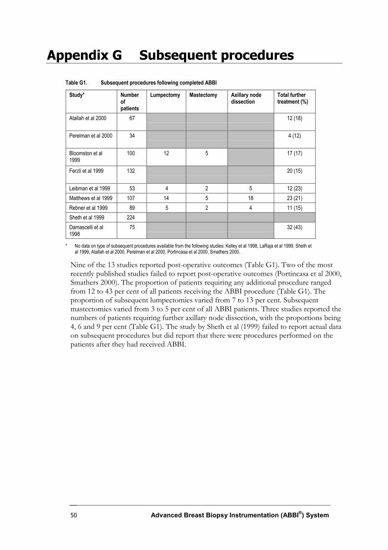
Advanced Breast Biopsy Instrumentation (ABBI®) System50
Appendix G Subsequent procedures
Table G1. Subsequent procedures following completed ABBI
Study* Numberofpatients
Lumpectomy Mastectomy Axillary nodedissection
Total furthertreatment (%)
Atallah et al 2000 67 12 (18)
Perelman et al 2000 34 4 (12)
Bloomston et al1999
100 12 5 17 (17)
Ferzli et al 1999 132 20 (15)
Leibman et al 1999 53 4 2 5 12 (23)Matthews et al 1999 107 14 5 18 23 (21)Rebner et al 1999 89 5 2 4 11 (15)Sheth et al 1999 224Damascelli et al1998
75 32 (43)
* No data on type of subsequent procedures available from the following studies: Kelley et al 1998, LaRaja et al 1999, Sheth etal 1999, Atallah et al 2000, Perelman et al 2000, Portincasa et al 2000, Smathers 2000.
Nine of the 13 studies reported post-operative outcomes (Table G1). Two of the mostrecently published studies failed to report post-operative outcomes (Portincasa et al 2000,Smathers 2000). The proportion of patients requiring any additional procedure rangedfrom 12 to 43 per cent of all patients receiving the ABBI procedure (Table G1). Theproportion of subsequent lumpectomies varied from 7 to 13 per cent. Subsequentmastectomies varied from 3 to 5 per cent of all ABBI patients. Three studies reported thenumbers of patients requiring further axillary node dissection, with the proportions being4, 6 and 9 per cent (Table G1). The study by Sheth et al (1999) failed to report actual dataon subsequent procedures but did report that there were procedures performed on thepatients after they had received ABBI.

Advanced Breast Biopsy Instrumentation (ABBI®) System 51
Abbreviations
ABBI advanced breast biopsy instrumentation
ADH atypic ductal hyperplasia
AETS Agencia de Evaluación de Tecnologías Sanitarias
AHMAC Australian Health Ministers’ Advisory Council
ASERNIP–S Australian Safety and Efficacy Register of New InterventionalProcedures – Surgical
CCB conventional core biopsy
CI confidence interval
DALY disease-adjusted life years
DCIS ductal carcinoma in situ
IDC invasive/infiltrating ductal carcinoma
ILC invasive/infiltrating lobular carcinoma
L lymphoma
MBS Medical Benefits Schedule
MIBB minimally invasive breast biopsy
MSAC Medical Services Advisory Committee
n number
NHMRC National Health and Medical Research Council
QOROM Quality of Reporting of Meta-analyses
SD standard deviation
U unclassified
VACB vacuum-assisted core biopsy
WHO World Health Organisation

Advanced Breast Biopsy Instrumentation (ABBI®) System52
References
Agencia de Evaluación de Tecnologías Sanitarias 1999, 'Diagnostico de lesiones mammariasdetectadas en cribado poblacional de cancer de mama mediante mamografia', ABBI: Nuevoinstrumento de biopsia histologica por puncion, Madrid, p. 52.
Atallah N., Karam R., Younane T. et al 2000, 'Stereotactic excisional biopsy (ABBI technique) ondedicated digital prone tables. Advantages. Disadvantages. Indications. About 67 cases',Journal Medical Libanais, 48: 70–6.
Australian Institute of Health and Welfare 2000a, Australia's Health 2000: The seventh biennnialhealth report of the Australian Institute of Health and Welfare, Australian Institute of Health andWelfare, Canberra.
Australian Institute of Health and Welfare 2000b, BreastScreen Australia Achievement Report,Australian Institute of Health and Welfare, Canberra.
Australian Institute of Health and Welfare 2000c, Cancer in Australia 1997 – Incidence and mortalitydata for 1997 and selected data for 1998 and 1999, Australian Institute of Health and Welfare,Canberra.
Bloomston M., D'Angelo P., Galliano D. et al 1999, 'One hundred consecutive advanced breastbiopsy instrumentation procedures: complications, costs, and outcome' (see comments),Annals of Surgical Oncology, 6: 195–9.
Clarke M. and Oxman A. (eds) 2000, Cochrane Reviewers’ Handbook 4.1 (updated June 2000), TheCochrane Collaboration, Oxford, England.
Cole C.F. 1998, 'Issues in breast imaging', Lippincott's Primary Care Practice, 2: 141–8.
Damascelli B., Frigerio L.F., Lanocita R. et al 1998, 'Stereotactic excisional breast biopsyperformed by interventional radiologists using the advanced breast biopsy instrumentationsystem' (see comments), British Journal of Radiology, 71: 1003–11.
D'Angelo P.C, Galliano D.E. and Rosemurgy A.S. 1997, 'Stereotactic excisional breast biopsiesutilizing the advanced breast biopsy instrumentation system', American Journal of Surgery, 174:297–302.
Ferzli G.S., Puza T., Vanvorst-Bilotti S. et al 1999, 'Breast biopsies with ABBI™: Experiencewith 183 attempted biopsies', Breast Journal, 5: 26–8.
Ferzli G.S. and Hurwitz J. 1997, 'Initial experience with breast biopsy utilising the advancedbreast biopsy instrumentation (ABBI)', Surgical Endoscopy, 11: 393–6.
Harris J.R. 1996, Diseases of the breast, Lippincott-Raven, Philadelphia.
Haynes R.B., Walker C.J., McKibbon K.A. et al 1994a, 'Performances of 27 MEDLINE systemstested by searches with clinical questions', Journal of the American Medical Informatics Association,1: 285–95.

Advanced Breast Biopsy Instrumentation (ABBI®) System 53
Haynes R.B., Wilczynski N., McKibbon K.A. et al 1994b, 'Developing optimal search strategiesfor detecting clinically sound studies in MEDLINE', Journal of the American Medical InformaticsAssociation, 1: 447–58.
Heywang-Kobrunner S.H., Schreer I. and Dershaw D.D. 1997, Diagnostic Breast Imaging –Mammography, Sonography, Magnetic Resonance Imaging, and Interventional Procedures, Thieme MedicalPublishers, New York.
Jadad A.R., Moore R.A., Carroll D. et al 1996, 'Assessing the quality of reports of randomizedclinical trials: is blinding necessary?', Controlled Clinical Trials, 17: 1–12.
Kahn K., ter Riet G., Glanville J. et al (eds) 2001, Undertaking systematic reviews of research oneffectiveness, NHS Centre for Reviews and Dissemination, University of York, York, England.
Kamm B.L. 1999, 'Breast procedures and imaging techniques', Radiologic Technology, 71: 58–72.
Kelley W.E. Jr, Bailey R., Bertelsen C. et al 1998, 'Stereotactic automated surgical biopsy usingthe ABBI biopsy device: A multicenter study', Breast Journal, 4: 302–6.
LaRaja R.D., Saber A.A. and Sickles A. 1999, 'Early experience in the use of the Advanced BreastBiopsy Instrumentation: a report of one hundred and twenty-seven patients', Surgery, 125:380–4.
Leibman A.J., Frager D. and Choi P. 1999, 'Experience with breast biopsies using the AdvancedBreast Biopsy Instrumentation system, AJR American Journal of Roentgenology, 172: 1409–12.
Liberman L. 2000, 'Clinical management issues in percutaneous core breast biopsy', BreastImaging, 38: 791–807.
Matthews B.D. and Williams G.B. 1999, 'Initial experience with the advanced breast biopsyinstrumentation system', American Journal of Surgery, 177: 97–101.
Mautner B.D., Schmidt K.V. and Brennan M.B. 2000, 'New diagnostic techniques andtreatments for early breast cancer', Seminars in Oncology Nursing, 16: 185–96.
Medicare Services Advisory Committee 1999, Advanced breast biopsy instrumentation. Application1001, Department of Health and Aged Care, Canberra.
Moher D., Cook D.J., Eastwood S. et al 1999, 'Improving the quality of reports of meta-analysesof randomised controlled trials: the QUOROM statement. Quality of Reporting of Meta-analyses', Lancet, 354: 1896–1900.
National Breast Cancer Centre 2000, New technologies in breast cancer diagnosis: Information update,online:http://www.nbcc.org.au/pages/info/resource/nbccpubs/nt_update/sept2000/lcb.htm(accessed on 23-04-2001).
National Health and Medical Research Centre 1999, A guide to the development, implementation andevaluation of clinical practice guidelines, National Health and Medical Research Council, Canberra.
National Health and Medical Research Centre 2000, How to use the evidence: assessment and applicationof scientific evidence, National Health and Medical Research Council, Canberra.

Advanced Breast Biopsy Instrumentation (ABBI®) System54
Perelman V.S., Colapinto N.D., Lee S. et al 2000, 'Experience with the advanced breast biopsyinstrumentation system', Canadian Journal of Surgery, 43: 437–41.
Portincasa G., Lucci E., Navarra G.G. et al 2000, 'Initial experience with breast biopsy utilizingthe Advanced Breast Biopsy Instrumentation (ABBI)', Journal of Surgical Oncology, 74: 201–3.
Rebner M., Chesbrough R. and Gregory N. 1999, 'Initial experience with the advanced breastbiopsy instrumentation device' (see comments), AJR American Journal of Roentgenology, 173:221–6.
Schulz K.F., Chalmers I., Hayes R.J. et al 1995, 'Empirical evidence of bias: Dimensions ofmethodological quality associated with estimates of treatment effects in controlled trials',JAMA, 273: 408–12.
Schwartzberg B.S., Goates J.J., Keeler S.A. et al 2000, 'Use of advanced breast biopsyinstrumentation while performing stereotactic breast biopsies: review of 150 consecutivebiopsies', Journal of the American College of Surgeons, 191: 9–15.
Sheth D., Wesen C.A., Schroder D. et al 1999, 'The advanced breast biopsy instrumentation(ABBI) experience at a community hospital', American Surgeon 65: 726–9 (discussion, 729–30).
Silen W., Earle Matory W.J. and Love S.M. 1996, Atlas of Techniques in Breast Surgery, Lippincott-Raven, Philadelphia.
Smathers R.L. 2000, 'Advanced breast biopsy instrumentation device: percentages of lesion andsurrounding tissue removed', AJR American Journal of Roentgenology, 175: 801–3.
Velanovich V., Lewis F.R. Jr, Nathanson S.D. et al 1999, 'Comparison of mammographicallyguided breast biopsy techniques', Annals of Surgery, 229: 625–30 (discussion, 630–3).
Walsh D., Merlin T., Humenuik V. et al 2000, 'Clinical practice guidelines for the AdvancedBreast Biopsy Instrument (ABBI), Australian Safety and Efficacy Register of New InterventionalProcedures – Surgical CPG Report No. 1, Adelaide, South Australia.
Wong A.Y., Salisbury E. and Bilous M. 2000, 'Recent developments in stereotactic breast biopsymethodologies: an update for the surgical pathologist', Advances in Anatomic Pathology, 7: 26–35.
Yang J.H., Lee S.D. and Nam S.J. 2000, 'Diagnostic utility of ABBI™ (Advanced Breast BiopsyInstrumentation) for non-palpable breast lesions in Korea', Breast Journal, 6: 257–62.