adrian moga summer school in oncology - european...
TRANSCRIPT

Testicular cancer radiotherapy Adrian Moga
Summer School in OncologyBucharest, 15th - 19th of June 2015

Emil Grube a medical student in Chicago convinced one of his professors to allow him to irradiate a cancer patient, a woman named Rose Lee, suffering from locally advanced breast cancer. By doing so, Grubbe became the World’s first Radiation Oncologist.
X-ray apparatus was first introduced at the London Hospital in 1896
X-ray treatment of tuberculosis in 1910.
Medicine has used radiation as a treatment for more than 100 years.

THERATRON Elite 100 District Clinical Hospital Sibiu
( 2013 )
The first patient to be treated with Cobalt-60 radiation was treated on October 27, 1951
Victoria Hospital in London, Ontario.

TomoTherapy combines an advanced form of intensity modulated radiation therapy (IMRT), with the accuracy of computed tomography (CT)
scanning technology, all in one machine.
100+ years later the technology has changed!


European Age-Standardised Incidence and Mortality Rates per 100,000 population, EU-27 Countries
Testicular Cancer : 2008 Estimates
http://info.cancerresearchuk.org/cancerstats/faqs

▪ Seminoma cells are extremely radiosensitive, and radiation therapy has been widely used for more than 60 years, and has an excellent long-term track record.
▪ This modality is still a standard management in pure seminomas in the United States, and in Europe it is used quite often .
▪ The available evidence suggests that 84–94% of patients who relapse will do so in the para-aortic (PA) lymph nodes
while about 10% will also have positive pelvic nodes.
▪ Isolated pelvic relapse is rare and a very small percentage of patients will relapse elsewhere in the body. Relapses remain highly salvageable and disease-specific survival approaches 100% .

▪ Historically, RT was delivered by a cobalt source using two parallel opposed anterior and posterior treatment fields, were defined with the help of bony landmarks. ▪ The dose was 30 Gy using 15 fractions. ▪ The treated areas were the para-aortic, homolateral external iliac nodes and the orchiectomy scar. This technique was known as the «dog-leg». ▪ The fields spread generally up to the superior aspect of D11 or D10 down to the inguinal ligament.
This was the standard method until the beginning of the 1980's.

Since the 1990's, following the low pelvic relapse rates reported in stage I tumors (less than 5%), the indication for pelvic irradiation was challenged.
Ommission of the pelvic irradiation :
• The results of this new approach were excellent with a low pelvic relapse rate.
• The reduced volume permits limiting the area, preserving the remaining testicular function and will hopefully decrease the secondary cancer rate .
Omission of the pelvic irradiation in Stage I testicular seminoma: A study of postorchiectomy paraaortic radiotherapy Ion Christian Kiricuta, M.D., Ph.D. Jörg Sauer, M.D.WernerBohndorf, M.D. Department of Radiation Oncology, University of Würzburg, Germany

The RT fields were a matter of debate :
The randomized study conducted by the Medical Research Council (MRC) Testicular Cancer Working Party ( Fossa et al.1999)
- in this trial 478 patients were randomized between para-aortic and pelvic RT ( dogleg ) vs. para-aortic RT alone.
- the dose was 30 Gy in 15 fractions in both arms.
- relapse –free survival /OS at 3 years 97/ 96% dogleg RT 99/100% para- aortic RT
- 3 year pelvic RFS was - 100% dogleg RT - 98% para-aortic RT

The optimal RT dose was also still a matter of debate :
• Generally, the recommended dose is between 25 and 30 Gy in 15 to 20 fractions.
• The MRC trial ( Jones et al. 2005 ) is the only randomized study that evaluates a dose de-escalation.
- it compared a dose of 20 Gy versus 30 Gy with conventional fractionation in 625 patients. - 10% of patients were treated with a « dog-leg » field - 90% with para-aortic fields.
- the relapse rates after a median follow-up of a little more than 5 years were not significantly different. ( 97% 30Gy ; 96,4% 20Gy )
- the 20 Gy arm showed a slightly lower acute toxicity rate (moderate asthenia 5% vs 20%, work incapacity 28 vs 46%).
- the only death due to the primary disease was in the 20 Gy arm .

trends
Radiation therapy plays an important role in the loco-regional treatment
The essential areas needed to be covered But as little as possible

Currently, irradiation is delivered by a high-energy linear accelerator
- with a conformational technique, allowing the shaping of the treatment fields to the expected target volume which was planned with CT-scan images and 3D reconstruction (RT3D).
- with the help of multiple irradiation beams, this technique allows a better definition of the target volumes and a maximal sparing of the neighboring critical organs such as kidneys, spinal cord.....
- computerized dosimetry and dose-volume histograms are now commonly used.

During treatment delivery, the linear accelerator completes multiple 360-degree rotations around the patient while the couch passes through the center bore of the system.

▪ The multi-leaf collimator will shape the individual radiation beams to “conform” to the shape of the tumor .
▪ IMRT uses thousands of radiation “beamlets” from many different angles to deliver a single dose of radiation.
▪ The intensity of the “beamlets” can change during the treatment session to modulate the dose, so that the tumor receives a very precise high dose of radiation, while minimizing damage to surrounding, normal tissue.

Treatment recommendation for seminomaAll patients radical inghinal orchiectomy with high ligation of spermatic cord Stage I post resection : - surveillance (relaps rate 16% ) - RT ( 20Gy to paraaortic ± pelvic Ln) - carboplatin x 1- 2 cycles Stage IIA/IIB - RT 20Gy to pelvic and paraaortic LN with boost to gross disease : 30Gy for stage IIA 36 Gy for stage IIB - consider etoposide, cilplatin ( EP) chemo x 4c for select IIB patients Stage IIC/D and III - chemo ( etoposide, cisplatinum,± bleo- mycin ) : EPx4c or BEP x 3c Handbook of Evidence – Based Radiation Oncology

target volume assessment
organs at risk assessment
dose prescription
dose fractionation
dose distribution
positioning of the patient
treatment machine settings
adjuvant therapies
Treatment planning process
Treatment planning is a process that involves the determination of treatment parameters considered optimal in the management of a patient’s disease. In radiotherapy, these parameters include :

Patient simulation and setup :
• Patients should be simulated in the supine position.
• Their arms may be placed on a wing board and a wedge may be placed under their knees.
• A clamshell shield should be used to protect the remaining testis. This reduces the dose delivered to the remaining testis, even when radiotherapy is given to the retroperitoneal nodes only.
• A clamshell is recommended even in patients who have banked sperm, given that banking is not always successful.
• The penis should be moved out of the fields.
• Tattoos should be placed at the level of the isocenter anteriorly and laterally.

The gross tumor volume ( GTV)
- is defined to be the “ gross demonstrable extent and location of the malignant growth”. - may include : primary tumor involved lymph nodes metastatic disease - is demonstrated using whatever imaging modalities are apropriate: visual observation CT, IRM, PET, SPECT, or any other method
Target volumes are like the concentric rings of an onion

Clinical target volum ( CTV ) :
- the demonstrable tumor ( GTV ) plus the microscopic disease.
- the margin surrounding the GTV that defines the CTV is delineated based on clinical experience , or in some cases, pathology studies. - in the case where surgical intervention has taken place before radiation treatment and the GTV has been removed, a CTV may exist in the absence of a GTV. - the goal of the radiation therapy is to irradiate the CTV to a dose appropriate to control the disease.
-

Planning Target Volum
The uncertainties can be classified according to : - their source - internal organ motion - patient setup uncertainties - when they occur during treatment - interfractional motion - intrafractional motion - wether they represent - errors in treatment plan preparation - systematic errors - errors in execution - random errors
• Is the final volume that must be irradiated to the tumoricidal dose to ensure that the CTV is actually irradiated to the desired dose.
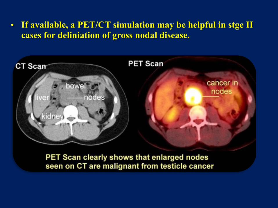
• If available, a PET/CT simulation may be helpful in stge II cases for deliniation of gross nodal disease.

Suggested target volumes for stage IA, IB
CTV Based on bony landmarks :
- superior border at T11 - inferior border at L5 - lateral border transverse processes
The kidneys, inferior vena cava, and aorta are shown (posterior view).

Computed tomographic simulation was used to create a nodal clinical target volume based on vascular anatomy. The kidneys, inferior vena cava, and aorta are shown.
Based on CT imaging :
- contour out the inferior vena cava and aorta - from 2 cm below the top of the kidney - to bifurcations inferiorly - provide a expansion -1,2cm on inferior vena cava - 1,9cm on the aorta - contour out : bone, muscle, bowel
Posterior view of para-aortic fields.

Suggested target volumes for stage IIA,IIB
CTV Based on CT anatomy : initial - the same instructions as stage I - aorta + inferior vena cava - contour out -common iliacs - proximal internal iliac vessels until takeoff of superior gluteal - external iliac vessels down to the upper border of the acetabulum - provide a expansion -1,2cm on inferior vena cava - 1,9cm on the aorta - 1,2cm on common, internal and external iliac - creat CTV vessels

-‐ contour gross nodal disease ( GTV ) - expand GTV nodes by 0,8 cm to creat CTV nodes
- merge CTV vessels and CTV nodes to creat CTV initial
PTV - expension by 0,5 cm on CTV initial initial
PTV - expension by 0,5 cm on CTV nodes conedown
PTV initial 25,5 Gy in 1,7Gy/fraction PTV conedown boost to 30 Gy/2Gy fractions for stage IIA 36Gy/2Gy fractions for stage IIB

• The 3.3-cm lateroaortic adenopathy, kidneys, inferior vena cava, aorta, ipsilateral iliac veins and arteries, and bladder are shown.
• Note that the inferior border is at the top of the acetabulum.
Multileaf collimator blocks based on nodal anatomy
Multileaf collimator blocks based on bony anatomy

Axial view showing planning target volume and isodose distribution using TomoTherapy, sparing kidneys and spinal cord in the case of stage
I seminoma.

A related technique, conformal proton beam radiation therapy
Instead of using x-rays, this technique uses proton beams. Protons are parts of atoms that cause little damage to tissues they pass through but are very effective in killing cells at the end of their path. This means that proton beam radiation can deliver more radiation to the cancer while possibly reducing damage to nearby normal tissues.

Proton therapy vs photon radiation therapy
However, the cost effectiveness of proton beam radiotherapy vs. photon radiotherapy or 3 cycles of bleomycin, etoposide, and cisplatin chemotherapy
remains controversial .
Contours and isodose lines proton plan

In many ways, RT was a victim of its own success, because given the very high cure rates and the fact that many men were diagnosed with testicular cancer at a young age (< 30 years), patients lived long enough to develop late RT toxicities
Most acute side-effects : - moderate – to – severe dyspepsia in 5 - 6% of patients - peptic ulceration in 2 - 3% - spermatogenesis may be - compromised at dose levels as low as 0,5Gy Most late side-effects : - nephrotoxicity - hypogonadism/infertility (cumulative dose above 2Gy) - prophylactic mediastinal irradiation is associated with a significant excess of cardiac and pulmonary death. - second malignancy : stomach, bladder, pancreas tumors (the actuarial risk of developing a second nontesticular malignancy is 18,2% at 25 years)

BEFORE I CAME HERE I WAS CONFUSED ABOUT THIS SUBJECT
NOW HAVING LISTENED TO YOUR LECTURE, I AM STILL CONFUSED, BUT AT A HIGHER LEVEL
Enrico Fermi , fizician italian, laureat al Premiului Nobel pentru Fizică pe anul 1938 .

The Annual 25rd Congress of the Romanian Society for Radiotherapy and Medical Oncology (RSRMO)
“Multidisciplinary Treatment of Thoracic Tumors” 15 – 17 OCTOBER 2015
Ramada Hotel, Sibiu, ROMANIA