adkinson: middleton's allergy: principles and …ad+2008.pdfskin inflammation in ad. these...
TRANSCRIPT

Adkinson: Middleton's Allergy: Principles and Practice, 7th ed. Copyright © 2008 Mosby, An Imprint of Elsevier
<< Previous | Next >> Chapter 62 – Atopic Dermatitis Mark Boguniewicz, Donald Y.M. Leung CONTENTS • Introduction 1083 • Clinical aspects 1083 • Role of the epidermal barrier 1087 • Role of allergens 1087 • Immunology 1088 • Management 1092 • Conclusion 1099 INTRODUCTION
Atopic dermatitis (AD) is a chronically relapsing inflammatory skin disease commonly associated with respiratory allergy.[1] In the 1930s, Hill and Sulzberger[2] suggested the term ‘atopic dermatitis’ to describe both the weeping eczema of infancy and childhood and the chronic xerosis and lichenified lesions more typical of older patients. This term also recognized the close relationship among AD, asthma, and allergic rhinitis. Like asthma and allergic rhinitis, AD is associated with the local infiltration of T helper type 2 (Th2) cells that secrete interleukin-4 (IL-4), IL-5, IL-13, and IL-31. More than 50% of patients with AD develop asthma, and approximately 75% develop allergic rhinitis, often as they outgrow AD.[3,4] Patients with AD can react to non-allergic as well as allergic triggers. AD can result in significant morbidity, leading to school absenteeism, occupational disability, and emotional stress. Although significant progress has been made in the understanding of AD, its cause is still unknown, and much remains to be learned about the complex interrelationship of genetic, environmental, immunologic, and epidermal factors in this disease.[5] With an increased understanding of the complex mechanisms involved, physicians can direct AD therapy from primarily symptomatic relief to more specific therapeutic measures.
SUMMARY OF IMPORTANT CONCEPTS
>> Atopic dermatitis is the most common chronic skin disease of young children, with lifetime prevalence in US schoolchildren up to 17%
>> Skin barrier/epidermal differentiation genes and immune response/host defense genes have been shown to play a key role in atopic dermatitis
>> Colonization and infection by microbial organisms including Staphylococcus aureus and herpes
simplex virus in atopic dermatitis may be due to defects in innate and adaptive immune responses
>> Treatment for most patients with chronic atopic dermatitis includes avoidance of irritants and
proven allergens, hydration and moisturizers to maintain a healthy epidermis, and topical antiinflammatory agents such as corticosteroids and calcineurin inhibitors

CLINICAL ASPECTS EPIDEMIOLOGY
A number of studies suggest an increasing prevalence of AD. Schultz Larsen[6] in Denmark demonstrated a cumulative incidence rate (up to 7 years) of 12% for twins born between 1975 and 1979, compared with a rate of 3% for twins born between 1960 and 1964. A cross-sectional questionnaire study conducted in 1992 confirmed this increased prevalence.[7] In this study of 3000 7-year-olds from Denmark, Germany, and Sweden, the frequency of AD was 15.6%. Questionnaire data from the United States in schoolchildren ages 5 to 9 years found the prevalence of AD to be 17%.[8] In a study from Japan, the authors performed skin examinations rather than relying on questionnaires to ascertain the prevalence of childhood and adolescent AD.[9] More than 7000 patients were examined, and AD was documented in 24% of those age 5 to 6 years, 19% of those age 7 to 9 years, 15% of those age 10 to 12 years, 14% of those age 13 to 15 years, and 11% of those age 16 to 18 years. The prevalence of AD in 9- to 12-year-old children was two times higher than in children of similar age examined 20 years earlier, and for 18-year-old adolescents it was five times higher. Recently, first graders age 6–7 years and sixth graders age 11–12 years were examined by dermatologists in eight prefectures of Japan randomly selected from urban and rural districts.[10] Point prevalence of AD was 11.2% in 23719 children (7.4–15.0%). Seventy-four percent of the patients were classified as mild AD, 24% as moderate AD, 1.6% as severe AD, and 0.3% as very severe AD. Prevalence in first graders was slightly higher than in sixth graders (11.8% vs 10.5%, p <0.01). No apparent difference was seen in prevalence between urban and rural districts, or between boys and girls.
Increased exposure to pollutants and indoor allergens (especially house dust mites) and a decline in breast-feeding, along with an increased awareness of AD, have been suggested as reasons for the increased frequency of AD.[11] In a prospective study, Zeiger et al[12] found that restricting the mother's diet during the third trimester of pregnancy and lactation and the child's diet during the first 2 years of life resulted in decreased prevalence of AD in the prophylaxis group compared with a control group at 12 months of age but not at 24 months. Follow-up through 7 years of age showed no difference between the prophylaxis and control groups for AD or respiratory allergy.[13] In a large study of an ethnically and socially diverse group of children in suburban Birmingham, England, Kay and coworkers[14] found that breast-feeding did not affect the lifetime prevalence rate of 20%. A study of prevalence of childhood eczema found a correlation with increased socioeconomic class that did not result from heightened parental awareness.[15]
The effects of genetic and environmental factors on allergic diseases were studied in two Japanese cities with differing climates.[16] The prevalence of allergic diseases and AD in the city with a temperate climate was significantly higher than in the one with a subtropical climate even after controlling for genetic and environmental factors. In both cities, children from atopic families had a significantly higher risk of contracting respiratory allergies and AD. In a global survey of the prevalence of asthma, allergic rhinoconjunctivitis, and AD, 463801 children ages 13 to 14 years from 155 centers in 56 countries participated.[17] The highest prevalences of AD were reported from scattered centers, including sites in Scandinavia and Africa, that were not among centers with the highest prevalences of asthma. On the other hand, the lowest prevalence rates for AD occurred in centers with the lowest prevalence of asthma and allergic rhinoconjunctivitis. Thus, the ultimate presentation of an atopic disease may depend on a complex interaction of environmental exposures with end-organ response in a genetically predisposed individual.
GENETICS
The genetics of AD is complex and an area of active research (recently reviewed in reference 18). While a number of genes are likely to be involved in the development of AD, skin barrier/epidermal differentiation genes and immune response/host defense genes have been proposed as playing a key role. Loss-of-function mutations of the epidermal barrier protein filaggrin have been demonstrated to be a major predisposing factor for a subset of patients with AD both in association with ichthyosis vulgaris[19] as well as the extrinsic subtype of AD.[20] Candidate gene approaches have also implicated variants in the SPINK5 gene, which is expressed in the uppermost epidermis, where its product, LEKT1, inhibits two serine proteases (stratum corneum tryptic enzyme and stratum corneum chymotryptic enzyme) involved in desquamation and inflammation.[5] Thus, an imbalance of protease versus protease inhibitor activity may contribute to
skin inflammation in AD. These observations establish a key role for impaired skin barrier function in the pathogenesis of AD as impaired skin barrier formation will allow increased transepidermal water loss and, importantly, increased entry of allergens, antigens, and chemicals from the environment, resulting in skin inflammatory responses. It is important to recognize that the majority of patients with AD appear to outgrow their inflammatory skin disease; thus, other gene products must also be involved in the pathogenesis of AD.
ATOPIC DIATHESIS
Most patients with AD have a genetic predisposition to develop an IgE response to common environmental allergens. Abnormal IgE responses are associated with cellular abnormalities resulting in overproduction of Th2-type cytokines, which also contribute to the eosinophilia seen in these diseases. Early onset of AD has been found to be associated with an increased risk for respiratory allergy. The highest incidence of asthma at a given age has been observed in children with onset of AD before 3 months of age, in those with severe AD and with a family history of asthma. The data showing an association of increased risk for asthma, rhinoconjunctivitis, or both with early onset of AD have been confirmed in more recent studies.[21] Respiratory allergy occurred in 50% of children who had onset of AD during the first 3 months of life and two or more atopic family members, compared with 12% of children who had onset of AD after 3 months of age and no atopic family members. In a prospective study of children with AD who were observed through 7 years of age, only 14 of 94 children in this cohort had not experienced any signs or symptoms suggestive of asthma or allergic rhinoconjunctivitis.[22] In addition, children with AD have been shown to have more severe asthma than asthmatic children without AD. This raises the possibility that allergen sensitization through the skin may predispose to more severe and persistent respiratory disease through effects on the systemic allergic response. In support of this hypothesis, murine studies have shown that epicutaneous sensitization with protein antigen can elicit a localized dermatitis, along with elevated serum IgE, airway eosinophilia, and hyperresponsiveness to methacholine.[23]
In addition, patients with AD react to both allergic and non-specific triggers, similarly to patients with asthma and allergic rhinitis. Skin hyperreactivity to irritants such as sodium lauryl sulfate (SLS) has been shown in patients with both active and inactive AD, and in patients with allergic respiratory disease even with no skin involvement, compared with non-atopic subjects.[24] An abnormal intrinsic hyperreactivity of inflammatory cells in atopic individuals may predispose them to a lower threshold of irritant responsiveness. These observations were confirmed and extended in a study showing that the stratum corneum abnormalities in non-involved AD skin were associated with increased transepidermal water loss even 7 days after application of SLS.[25] Of note, atopy can be transferred through bone marrow transplantation.[26] These observations suggest that the cutaneous abnormality in AD results from a complex interaction of resident and infiltrating cells. Furthermore, in a study of bronchial and cutaneous reactivity in asthmatic patients with and without AD, the authors found a latent predisposition for bronchial asthma in AD patients and implicated circulating activated eosinophils as the common effector cells.[27] Because the ability of eosinophils to reach their target organ depends in part on eosinophil-specific chemotactic factors, increased expression of eotaxin and monocyte chemotactic protein-4 (MCP-4), structurally homologous eosinophil chemoattractants acting through a common CCR3 receptor, has been reported both in the respiratory mucosa of patients with asthma[28] and in AD.[29] Of interest, increased numbers of IgE+ Langerhans cells have been shown in both active AD and active asthma versus inactive AD and inactive asthma, suggesting systemic regulation of active allergic disease, further aggravated by local inflammation in atopic skin lesions.[30]
NATURAL HISTORY
Atopic dermatitis typically manifests in early childhood, with onset before 5 years of age in approximately 90% of patients. In adults with new-onset dermatitis, especially without a history of childhood eczema, asthma, or allergic rhinitis, other diseases need to be considered (Box 62.1).
BOX 62.1
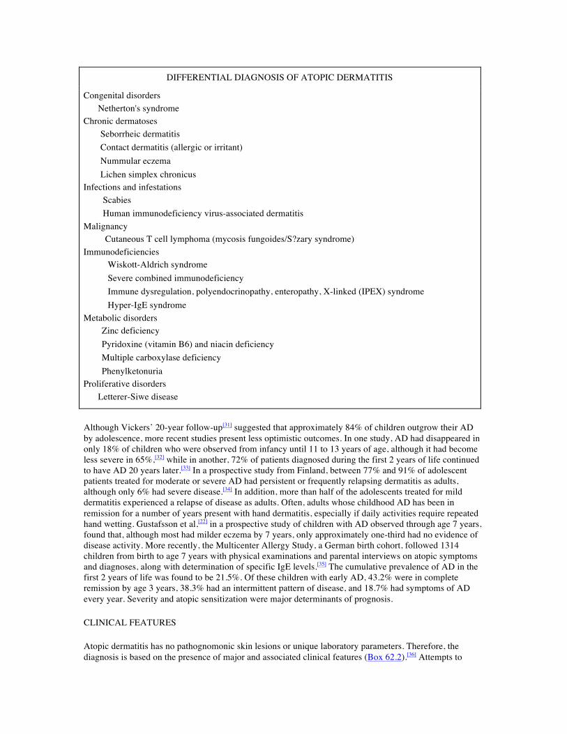
DIFFERENTIAL DIAGNOSIS OF ATOPIC DERMATITIS
Congenital disorders Netherton's syndrome Chronic dermatoses Seborrheic dermatitis Contact dermatitis (allergic or irritant) Nummular eczema Lichen simplex chronicus Infections and infestations Scabies Human immunodeficiency virus-associated dermatitis Malignancy Cutaneous T cell lymphoma (mycosis fungoides/S?zary syndrome) Immunodeficiencies Wiskott-Aldrich syndrome Severe combined immunodeficiency Immune dysregulation, polyendocrinopathy, enteropathy, X-linked (IPEX) syndrome Hyper-IgE syndrome Metabolic disorders Zinc deficiency Pyridoxine (vitamin B6) and niacin deficiency Multiple carboxylase deficiency Phenylketonuria Proliferative disorders Letterer-Siwe disease
Although Vickers’ 20-year follow-up[31] suggested that approximately 84% of children outgrow their AD by adolescence, more recent studies present less optimistic outcomes. In one study, AD had disappeared in only 18% of children who were observed from infancy until 11 to 13 years of age, although it had become less severe in 65%,[32] while in another, 72% of patients diagnosed during the first 2 years of life continued to have AD 20 years later.[33] In a prospective study from Finland, between 77% and 91% of adolescent patients treated for moderate or severe AD had persistent or frequently relapsing dermatitis as adults, although only 6% had severe disease.[34] In addition, more than half of the adolescents treated for mild dermatitis experienced a relapse of disease as adults. Often, adults whose childhood AD has been in remission for a number of years present with hand dermatitis, especially if daily activities require repeated hand wetting. Gustafsson et al,[22] in a prospective study of children with AD observed through age 7 years, found that, although most had milder eczema by 7 years, only approximately one-third had no evidence of disease activity. More recently, the Multicenter Allergy Study, a German birth cohort, followed 1314 children from birth to age 7 years with physical examinations and parental interviews on atopic symptoms and diagnoses, along with determination of specific IgE levels.[35] The cumulative prevalence of AD in the first 2 years of life was found to be 21.5%. Of these children with early AD, 43.2% were in complete remission by age 3 years, 38.3% had an intermittent pattern of disease, and 18.7% had symptoms of AD every year. Severity and atopic sensitization were major determinants of prognosis.
CLINICAL FEATURES
Atopic dermatitis has no pathognomonic skin lesions or unique laboratory parameters. Therefore, the diagnosis is based on the presence of major and associated clinical features (Box 62.2).[36] Attempts to

standardize signs and symptoms of AD include SCORAD and the Eczema Area and Severity Index (EASI); however, these indexes have been used primarily in clinical research trials.[37,38] The principal features include severe pruritus, a chronically relapsing course, typical morphology and distribution of the skin lesions, and a history of atopic disease. The presence of pruritus is critical to the diagnosis of AD, and patients with AD have a reduced threshold for pruritus.
BOX 62.2 CLINICAL FEATURES OF ATOPIC DERMATITIS
Major features Pruritus Facial and extensor involvement in infants and children Flexural lichenification in adults Chronic or relapsing dermatitis Personal or family history of atopic disease Minor features Xerosis Cutaneous infections Non-specific dermatitis of the hands or feet Ichthyosis, palmar hyperlinearity, keratosis pilaris Pityriasis alba Nipple eczema White dermatographism and delayed blanch response Anterior subcapsular cataracts Elevated serum IgE levels Positive immediate-type allergy skin tests
Modified from Hanifin JM, Rajka G. Acta Derm Venereol (Stockh) 1980; 2:44–47.[36]
Acute AD is characterized by intensely pruritic, erythematous papules associated with excoriations, vesiculations, and serous exudate. Subacute AD is characterized by erythematous, excoriated, scaling papules, whereas chronic AD is characterized by thickened skin with accentuated markings (lichenification) and fibrotic papules. Patients with chronic AD may have all three types of lesions. In addition, patients usually have dry skin. Significant differences can be observed in pH, capacitance, and transepidermal water loss between AD lesions and uninvolved skin in the same patient and on skin of normal controls.
During infancy, AD involves primarily the face, the scalp, and the extensor surfaces of the extremities. The diaper area is usually spared; if it is involved, it may be secondarily infected with Candida species, in which case the dermatitis does not spare the inguinal folds. In contrast, infragluteal involvement is a common distribution in children. In older patients with long-standing disease, the flexural folds of the extremities are the predominant location of lesions. In the Copenhagen Prospective Study on Asthma in Childhood, arm and joint involvement carried the highest predictive value for the development of AD at age 3 years.[39] Localization of AD to the eyelids may be an isolated manifestation but should be differentiated from allergic contact dermatitis.
COMPLICATING FEATURES Ocular problems

Increased numbers of IgE-bearing Langerhans cells are found in the conjunctival epithelium of patients with AD. These cells can capture aeroallergens and present them to infiltrating T cells, thus contributing to ocular inflammation. Ocular complications associated with AD can result in significant morbidity.
Atopic keratoconjunctivitis is always bilateral, and symptoms include itching, burning, tearing, and copious mucoid discharge. It is frequently associated with eyelid dermatitis and chronic blepharitis and may result in visual impairment from corneal scarring. Keratoconus is a conical deformity of the cornea that is believed to result from persistent rubbing of the eyes in patients with AD and allergic rhinitis. Anterior subcapsular cataracts may develop during adolescence or early adult life.
Hand dermatitis
Patients with AD often have non-specific hand dermatitis. This is frequently irritant in nature and aggravated by repeated wetting, especially in the occupational setting. A history of past or present AD at least doubles the effects of irritant exposure and doubles the risk in occupations where hand eczema is a common problem.
Infections
Patients with AD have an increased susceptibility to infection or colonization with a variety of organisms. These include viral infections with herpes simplex, molluscum contagiosum, and human papillomavirus. A direct relationship has been demonstrated between interferon-γ (IFN-γ)concentrations and the cytopathic effect of herpes simplex, as has an inverse relationship between IL-4 and the cytopathic effect of herpes simplex,[40] suggesting that the T cell-associated cytokine abnormalities seen in AD can enhance viral infections. Important insights into our understanding of the unique susceptibility that AD patients have to eczema herpeticum and eczema vaccinatum (a potentiallylethal complication of small pox vaccine) have recently been made with the demonstration of an acquired defect in the cutaneous antimicrobial peptide response (see also the discussion below under Immunopathologic Features).[41]
Superimposed dermatophytosis may cause AD to flare. Patients with AD appear to have a three-fold increased incidence of Trichophyton rubrum infections, compared with controls.[42] The opportunistic yeast Malassezia sympodialis, previously termed Pityrosporum ovale, has also been associated with a predominantly head and neck distribution of AD[43] and has been reported to occur in both extrinsic and intrinsic subtypes of AD.[44]
A number of studies have elucidated the importance of Staphylococcus aureus in AD. Preferential adherence of this organism in AD may be related to expression of adhesins such as fibronectin and fibrinogen in inflamed skin.[45] S. aureus can be cultured from the skin of more than 90% of patients with AD, compared with only 5% of normal subjects.[46] The higher rate of S. aureus colonization in AD lesions compared with lesions from other skin disorders may also be associated with colonization of the nares, with the hands serving as the vector of transmission.[47] Patients without obvious superinfection may have a better response to combined antistaphylococcal and topical corticosteroid therapy than to corticosteroids alone.[48] Although recurrent staphylococcal pustulosis can be a significant problem in AD, invasive S. aureus infections occur rarely and should raise the possibility of an immunodeficiency such as hyper-IgE syndrome.
DIFFERENTIAL DIAGNOSIS
A number of diseases may be confused with AD (see Box 62.1). In infants, immunodeficiency, including immune dysregulation, polyendocrinopathy, enteropathy, and X-linked (IPEX) syndrome need to be considered. IPEX is a rare disorder associated with dermatitis, enteropathy, type 1 diabetes, thyroiditis, hemolytic anemia, and thrombocytopenia.[49] IPEX results from mutations of FOXP3, a gene located on the X chromosome that encodes a DNA-binding protein required for development of regulatory T cells. Wiskott-Aldrich syndrome is an X-linked recessive disorder characterized by an eczematous rash,
associated with thrombocytopenia along with variable abnormalities in humoral and cellular immunity and severe bacterial infections. Hyper-IgE syndrome is an autosomal dominant multisystem disorder characterized by recurrent deep-seated bacterial infections, including cutaneous cold abscesses and pneumonias due to S. aureus.[50] Scabies can present as a pruritic skin disease. However, distribution in the genital and axillary areas, the presence of linear lesions, and the finding of mites, ova, and scybala in epithelial debris from skin scrapings help distinguish it from AD. An adult who has eczematous dermatitis with no history of childhood eczema and without other atopic features may have contact dermatitis, but, more importantly, cutaneous T cell lymphoma needs to be ruled out. Ideally, biopsies should be sent from three separate sites to increase the yield in identifying abnormal S?zary cells. In addition, eczematous rash suggestive of AD can be seen in patients infected with human immunodeficiency virus.
A contactant should be considered in patients whose AD does not respond to appropriate therapy. Typical distribution for a suspected contactant may be suggestive. However, allergic contact dermatitis complicating AD may appear as an acute flare of the underlying disease rather than the more typical vesiculobullous eruption. Proper diagnosis depends on confirmation of a suspected allergen with patch testing. Standardized testing with the T.R.U.E. Test is available for 23 of the most common contact allergens (see Ch. 63) although more extensive testing may be required in selected cases.
PSYCHOSOCIAL IMPLICATIONS
Patients with AD may have high levels of anxiety and problems dealing with anger and hostility. Although these emotions do not cause AD, they can exacerbate the illness. Patients often respond to stress or frustration with itching and scratching.[51] Stimulation of the central nervous system may intensify cutaneous vasomotor and sweat responses and contribute to the itch–scratch cycle. In some instances, scratching is associated with significant secondary gain or with a strong component of habit. Severe disease can have a significant impact on patients, leading to problems with social interactions and self-esteem. Of considerable importance, sleep disturbance is common in this chronic disease and significantly impacts on the quality of life of patients and family members.[52]
ROLE OF THE EPIDERMAL BARRIER
Atopic dermatitis is associated with abnormalities in skin barrier function including increased transepidermal water loss, increased levels of endogenous proteolytic enzymes, and reduced ceramide levels. Use of soaps can increase skin pH, increasing activity of endogenous proteases, and leading to breakdown of epidermal barrier function.[5] The epidermal barrier may be further damaged by exogenous proteases from house dust mites and S. aureus. This is worsened by the lack of endogenous protease inhibitors in the skin of patients with AD. These epidermal changes likely contribute to increased allergen absorption into the skin and microbial colonization. Recently, mutations in the filaggrin (FLG) gene, located in the epidermal differentiation complex on chromosome 1q21, have been shown to result in complete loss of expression of a key epidermal protein, filament-aggregating protein (filaggrin), involved in formation of the epidermal barrier.[19] However, these mutation have only been found in Caucasians of European ancestry, not in patients of African or Asian descent, making as-yet unidentified mutations or other epidermal complex abnormalities likely. Of interest, FLG mutations were shown to be a major risk factor for eczema-associated asthma. Importantly, since epicutaneous sensitization to allergen results in a greater immune response than sensitization via the airway,[23] decreased epidermal barrier function could act as a site for allergen sensitization and predispose such children to the development of respiratory allergy later in life.
ROLE OF ALLERGENS
Although elevated serum IgE levels can be demonstrated in 80–85% of patients with AD and a similar number have immediate skin test response or positive in vitro tests to food and inhalant allergens, the relationship between the course of AD and implicated allergens has been difficult to establish.
Nevertheless, a number of well-controlled studies suggest that allergens can have an impact on the course of this disease.
FOODS
May[53] first recognized that patients with AD and positive food allergen skin tests could have negative food challenges to the implicated allergen, distinguishing between symptomatic and asymptomatic hypersensitivity. Thus, triggers for clinical disease cannot be predicted simply by performing allergy testing. However, double-blinded, placebo-controlled food challenges have demonstrated that food allergens can cause exacerbations in a subset of patients with AD.[54] Approximately 33% of infants and young children with AD will show clinically relevant reactivity to a food allergen.[55] Although lesions induced by single positive challenges are usually transient, repeated challenges, more typical of real-life exposure, can result in eczematous lesions. Food-specific T cells have been cloned from lesional skin and peripheral blood of patients with AD.[56,57] Furthermore, elimination of food allergens results in amelioration of skin disease and a decrease in spontaneous basophil histamine release.[58]
AEROALLERGENS
The evidence supporting a role for aeroallergens in AD includes the finding of both allergen-specific IgE antibodies and allergen-specific T cells.[59] Exacerbation of AD can occur with exposure to allergens such as house dust mites, animal danders, and pollens. In the 1940s, Tuft[60] demonstrated that introduction of aeroallergens intranasally could exacerbate AD. Subsequently, a subgroup of patients with AD who underwent bronchoprovocation with a standardized house dust mite extract in a double-blind, randomized, placebo-controlled fashion developed unequivocal cutaneous lesions after inhalation of dust mite.[61] All the patients with dust mite-induced dermatitis had a history of asthma, and in eight of these nine patients the skin reaction was preceded by an early bronchial reaction. Therefore, the respiratory route may be important in the induction and exacerbation of AD. In addition, studies with patch testing have shown that direct contact with inhalant allergens can also result in eczematous skin eruptions.[62] Using the atopy patch test, Langeveld-Wildschut et al[63] showed that positive reactions to house dust mite were associated with IgE+ Langerhans cells in the epidermis of AD patients. In addition, the severity of AD has been correlated with the degree of sensitization to aeroallergens.[64] Most importantly, environmental control measures aimed at reducing dust mite allergen have been shown to result in clinical improvement in AD patients.[65,66] These studies suggest that inhalation or contact with aeroallergens may be involved in the pathogenesis of AD.
MICROBIAL AGENTS
In addition to their role as infectious agents, both the lipophilic yeast M. sympodialis (previously termed P. ovale)[44] and the superficial dermatophyte T. rubrum have been associated with elevated specific IgE levels in patients with AD. Patients with AD predominantly of the head and neck, compared with a group without this distribution and with a group of normal individuals, more often demonstrated positive skin tests, radioallergosorbent tests, and specific histamine release to M. sympodialis.[67] The clinical significance of these findings is suggested by clinical improvement of such patients after antifungal therapy.[43]
Leung et al[68] showed that exotoxins secreted by S. aureus are superantigens that can result in persistent inflammation or exacerbations of AD. More than half of the AD patients studied had S. aureus cultured from their skin; the S. aureus organisms secreted primarily enterotoxins A and B and toxic shock syndrome toxin-1. In addition, almost half of the patients had specific IgE antibodies directed against the staphylococcal toxins found on their skin. Basophils from patients with antitoxin IgE released histamine on exposure to the relevant toxin but not in response to toxins to which they had no specific IgE. Other investigators have confirmed these observations.[69,70] In addition, analysis of the peripheral blood skin homing (cutaneous lymphocyte antigen [CLA]-positive) T cells of superantigen-positive patients as well as their skin lesions revealed that they had undergone a T cell receptor Vβ expansion consistent with superantigenic stimulation.[71,72] A correlation also has been found between the presence of IgE against
superantigens and severity of AD.[69] Furthermore, superantigens have been shown to have an additive effect together with conventional allergens in inducing cutaneous inflammation.[73] They can also augment allergen-specific IgE synthesis,[74] subvert T regulatory (reg) cell function,[75] and induce corticosteroid resistance,[76] suggesting that several mechanisms exist by which superantigens could aggravate the severity of AD. Finally, staphylococcal enterotoxin B (SEB) applied to the skin was shown to induce erythema and induration with the infiltrating T cells selectively expanded in response to the specific superantigen.[77,78]
AUTOANTIGENS
Several groups have suggested a role for autoantigens in chronic AD. Valenta et al[79] reported that the majority of sera from patients with severe AD contain IgE antibodies directed against human proteins. One of these IgE-reactive autoantigens, a 55-kDa cytoplasmic protein in skin keratinocytes, has been cloned from a human epithelial complementary DNA (cDNA) expression library and designated Hom s 1.[80] Although the autoallergens characterized to date have mainly been intracellular proteins, they have been detected in IgE immune complexes of AD sera, suggesting that release of these autoallergens from damaged tissues could trigger IgE or T cell-mediated responses. In another study, 30% of sera from patients with AD were found to have both IgG and IgE autoantibodies that reacted with an autoantigen termed dense fine speckles 70 kD (DFS70).[81] These data suggest that skin inflammation in AD, especially in severe cases, could be maintained by endogenous human antigens. Because these autoantigens are primarily nuclear or microsomal in origin, the possibility is raised that damage to the skin by infectious organisms or by scratching could release intracellular antigens that in turn could elicit and perpetuate IgE and T cell responses in AD. Of interest, human manganese superoxide dismutase (MnSOD) may play a role as an autoallergen in a subset of patients with AD.[82] By molecular mimicry leading to cross-reactivity, such sensitization might be induced primarily by exposure to MnSOD of the skin-colonizing yeast M. sympodialis (see also discussion of M. sympodialis above under Complicating Features).
IMMUNOLOGY
The finding of elevated serum IgE concentrations and the occurrence of eczematous lesions indistinguishable from AD in patients with primary T cell immunodeficiency disorders suggest an immunologic basis for AD. In Wiskott-Aldrich syndrome, bone marrow transplantation results in correction of the immunologic defect and resolution of the dermatitis. In addition, non-atopic recipients of bone marrow transplants from atopic donors can develop atopic symptoms and positive skin tests after successful engraftment.[83] These data suggest that AD results from a bone marrow-derived cell dysfunction rather than a constitutive skin defect. Of note, the concept of intrinsic and extrinsic forms of AD, discussed earlier, has been based on low serum IgE levels and negative allergy skin tests or serum-specific IgE to food or inhalant allergens. However, the majority of patients labeled as intrinsic AD have been found to make IgE to microbial antigens, including staphylococcal toxins and Malassezia.
IMMUNOREGULATORY DYSFUNCTION
A number of immunoregulatory abnormalities have been described in AD (Box 62.3).[1] B cells from patients with AD synthesize high levels of IgE. T cells from these patients produce increased amounts of IL-4 and express abnormally high levels of IL-4 receptor. Peripheral blood mononuclear cells (PBMCs) isolated from patients with AD have a decreased capacity to make IFN-γ, which is inversely correlated with serum IgE levels. Differences that have been noted between the intrinsic and extrinsic forms of AD include the observation that skin-derived T cells from extrinsic AD interacted with B cells to support IgE synthesis, whereas T cells from the intrinsic form of AD did not.[84]
BOX 62.3 IMMUNOREGULATORY ABNORMALITIES IN ATOPIC DERMATITIS
Increased synthesis of IgE

Increased levels of specific IgE to multiple allergens, including foods, aeroallergens, microorganisms, and enterotoxins
Increased expression of CD23 on B cells and monocytes Increased surface expression of FcεRI on antigen-presenting cells in the skin
Increased levels of cutaneous T cell-attracting chemokine and thymus and activation-regulated chemokine
Increased secretion of interleukin-4 (IL-4), IL-5, and IL-13 by T helper type 2 (Th2) cells Decreased secretion of interferon-γ by T helper type 1 (Th1) cells Decreased CD4+ CD25+ T reg cell immunosuppressive activity after superantigen stimulation Decreased secretion of antimicrobial peptides by keratinocytes
Increased levels of monocyte cyclic adenosine monophosphate phosphodiesterase, with increased IL-10 and prostaglandin E2
A number of studies have shown an increased frequency of both circulating[85,86] and lesional allergen-specific Th2 cells secreting IL-4, IL-5, and IL-13 in patients with AD.[84,87] Furthermore, an increased frequency of circulating skin homing (CLA+) type 2 cytokine-producing cells and decreased frequency of CLA+ type 1 cytokine-producing cells have been reported in the peripheral blood of patients with AD.[88] In addition to acting as an IgE isotype-specific switch, IL-4 also inhibits the production of IFN-γ and downregulates the differentiation of Th1 cells.[89] IFN-γ production is also inhibited by prostaglandin E2 (PGE2) and by IL-10, both of which are secreted in increased amounts by monocytes from patients with AD.[90,91] In one study, no defect in the capacity of cells from AD patients to produce IL-12, an important inducer of IFN-γ, was detected.[92] However, neutralization of IL-10 and IL-4 was able to correct production of IFN-γ. Therefore, the activation of Th2-type cells and monocytes may be central to the immune dysregulation in AD.
A role for the co-stimulatory molecules CD80/CD86 has been investigated in AD. CD86 was shown to be predominantly expressed on Langerhans cells in both the epidermis and the dermis in AD with almost complete inhibition of antigen-specific T cell proliferation with an anti-CD86 monoclonal antibody.[93] Studies have also suggested that these accessory molecules differ in their capacity to generate Th1 versus Th2 T cell responses, and the expression of CD86 on B cells of AD patients was found to be significantly higher than on B cells from patients with psoriasis or from normal controls, whereas there was no significant difference in CD80 expression among the three subject groups.[94] Interestingly, total serum IgE from AD patients and normal subjects correlated significantly with CD86 expression on B cells, suggesting a role for CD86+ B cells in IgE synthesis. Purified CD86+ B cells produced significantly more IgE than did CD86− B cells in vitro, and anti-CD86, but not CD80, monoclonal antibody (mAb) significantly decreased IgE production by peripheral blood mononuclear cells stimulated with IL-4 and anti-CD40 mAb. Furthermore, CD86+ B cells had a significantly higher level of IL-4R and CD23 expression than did CD80+ B cells. These data demonstrate the predominant expression of CD86 in AD and suggest a role in IgE synthesis.
Recently, the role of T reg cells in AD has been reviewed.[49] Superantigens have been shown to subvert T reg cell function[75] and dysregulation of disease-causing effector T cells has been observed in AD lesions, in association with an impaired CD4+ CD25+ FOXP3+ T-cell infiltration, despite the expression of type 1 regulatory cells in the dermis.[95]
IMMUNOPATHOLOGIC FEATURES
Routine histologic examination of clinically normal-appearing skin in AD reveals mild epidermal hyperplasia and a sparse, predominantly lymphocytic infiltrate in the dermis.[1] Acute eczematous lesions are characterized by both intercellular edema of the epidermis (spongiosis) and intracellular edema. T cell-derived IFN-γ was shown to increase expression of Fas receptor (CD95) on keratinocytes, leading to

acantholysis (loss of intercellular cohesion) with subsequent intercellular edema resulting in the characteristic histology.[96] A sparse lymphocytic infiltrate may be observed in the epidermis, whereas a marked perivenular infiltrate consisting of lymphocytes and some monocytes with rare eosinophils, basophils, and neutrophils is seen in the dermis. In chronic lichenified lesions, the epidermis has prominent hyperkeratosis with increased numbers of epidermal Langerhans cells and predominantly monocytes/macrophages in the dermal infiltrate. Mast cells are usually increased in number but are not degranulated. Of note, mast cells have been shown to be the predominant CD30 ligand-positive cell in the inflammatory milieu in AD lesions, leading to degranulation-independent chemokine secretion.[97]
Immunohistochemical staining of acute and chronic skin lesions in AD shows that the lymphocytes are predominantly CD3, CD4, and CD45RO memory T cells; that is, they have previously encountered antigen.[1] These cells also express CD25 and human leukocyte antigen (HLA)-DR on their surface, indicative of intralesional activation. In addition, almost all of the T cells infiltrating into atopic skin lesions express high levels of the skin lymphocyte homing receptor, CLA, a ligand for the vascular adhesion molecule, E-selectin.[98]
Vascular endothelial cells from atopic skin lesions express abnormally high levels of E-selectin, as well as vascular cell adhesion molecule-1 (VCAM-1) and CD54.[99] Mast cells, monocytes, Langerhans cells, and keratinocytes are all potential sources of IL-1 and tumor necrosis factor-α (TNF-α), which induce E-selectin, a molecule critical to the targeting of CLA-expressing T cells to sites of cutaneous inflammation.[98] Furthermore, migration of skin-homing T cells into atopic skin lesions also involves interaction between VCAM-1 and very late antigen-4 as well as CD54 and leukocyte function-associated antigen-1.[100] In addition, VCAM-1, which can be induced by IL-4 and IL-13, is involved in eosinophil and mononuclear cell movement into sites of allergic inflammation.
In contrast to epidermal Langerhans cells from normal subjects, Langerhans cells found in the epidermis and dermis of patients with chronic AD express CD1b, CD36, and HLA-DR surface antigens and are potent activators of autologous resting CD4 T cells.[101] Furthermore, both Langerhans cells and macrophages infiltrating into the AD skin lesion have surface-bound IgE.[102,103] A distinct population of CD1a inflammatory dendritic epidermal cells in cutaneous lesions in AD has been described.[104] These cells are subjected to specific signals leading to the upregulation of the high-affinity IgE receptor, FcεRI, in AD skin. In addition, while the number of CD1a+ epidermal dendritic cells has been shown to be similar in the inflammatory milieu in lesions from both intrinsic and extrinsic AD, intrinsic AD was characterized by decreased expression of FcεRI on the CD1a+ epidermal dendritic cells.[105]
Activated eosinophils are present in significantly greater numbers in chronic compared with acute lesions.[106] These eosinophils undergo cytolysis with release of granule protein contents into the upper dermis of lesional skin.[107] Deposition of eosinophil major basic protein can be detected throughout the upper dermis, and to a lesser extent deeper in the dermis.[108] Major basic protein deposition is more prominent in involved areas compared with uninvolved skin. It may contribute to the pathogenesis of AD through its cytotoxic properties and its capacity to induce basophil and mast cell degranulation.
A striking finding in the skin biopsy specimens of patients with AD is the absence of polymorphonuclear neutrophils (PMNs), even in the setting of intense scratching or increased S. aureus colonization and infection. PMN defects in subjects with AD include impaired phagocytic function and a reduced capacity to produce reactive oxygen species (reviewed in reference 109). Patients with AD have defective PMN chemotactic activity, which correlated with disease severity and the presence of cutaneous infections. The chemotactic defects are due in part to decreased expression of relevant chemoattractant receptors, as well as ligand-binding defects, ligand-signalingdefects, or both. It is likely that the defect in cutaneous PMN recruitment observed in patients with AD is also a function of defective tissue signals from the skin (e.g., reduced LL-37 production), as otherwise these patients would be susceptible to infections at other mucosal surfaces.

The role of keratinocytes in skin inflammation in AD has been increasingly recognized.[1] Keratinocytes from AD patients secrete high levels of RANTES following stimulation with TNF-α and IFN-γ. They are also an important source of thymic stromal lymphopoietin (TSLP), which activates dendritic cells (DC) to prime naive T cells to produce IL-4 and IL-13 (Th2 cell differentiation). Of note, mice genetically engineered to overexpress TSLP in the skin develop AD-like skin inflammation.[110] Besides producing proinflammatory cytokines, keratinocytes also play a vital role in the cutaneous innate immune responses by secreting antimicrobial peptides, including human beta defensins and cathelicidins, in response to microbial insult or tissue injury. An important insight into understanding why patients with AD are frequently colonized and infected by various microbes was the demonstration that their keratinocytes produce reduced amounts of antimicrobial peptides and this may predispose them to colonization and infection with S. aureus, viruses, and fungi.[111] Of note, this is not a primary keratinocyte defect, but rather an acquired one due to IL-4, IL-13, and IL-10 mediated inhibition of TNF and interferon-γ-induced antimicrobial peptide generation.[112]
CYTOKINE EXPRESSION
Cytokine expression in AD lesions reflects the nature of the underlying inflammation. Hamid et al[106] used in situ hybridization to study IL-4, IL-5, and IFN-γ messenger ribonucleic acid (mRNA) expression in acute and chronic skin lesions as well as uninvolved skin of patients with AD. Biopsies from uninvolved atopic skin showed a significant increase in the number of cells expressing IL-4, but not IL-5 or IFN-γ, mRNA. Both acute and chronic lesions had significantly greater numbers of cells that were positive for IL-4 and IL-5 compared with uninvolved or normal skin. Neither acutely involved nor uninvolved atopic skin showed significant numbers of IFN-γ mRNA-expressing cells. In contrast, chronic AD skin lesions, when compared with acute lesions, had significantly fewer IL-4 mRNA-expressing cells and significantly more IL-5 mRNA-expressing cells. T cells comprised the majority of IL-5-expressing cells in both acute and chronic lesions. Activated eosinophils were found in significantly greater numbers in chronic compared with acute lesions. These data suggest that although both acute and chronic lesions in AD are associated with increased IL-4 and IL-5 gene activation, acute skin inflammation is associated with predominantly IL-4 expression, whereas chronic inflammation is associated with IL-5 expression and eosinophil infiltration.
Interleukin-13 expression was also found to be higher in acute AD lesions, compared with chronic AD or psoriatic lesions.[113] These data suggest that IL-13 may be involved in the pathogenesis of AD and further support the hypothesis that acute inflammation in AD is mediated by Th2-type cytokines. Chronic lesions had increased numbers of IL-12 mRNA-positive cells compared with acute or uninvolved skin. IL-12 is a potent inducer of IFN-γ synthesis, and, consistent with this observation, increased IFN-γ expression has been reported in chronic AD lesions.[114] At a clonal level, T cells from AD patients with cow's milk allergy showed significantly greater production of IL-4, whereas IFN-γ production was greater in the milk-tolerant patients.[115] IL-5 and IL-13 cytokine production strongly correlated with IL-4 production.
Consistent with the observation that the activity of cytokines is dependent on the expression of their receptors, acute AD lesions have been shown to contain a significantly higher number of cells expressing mRNA of the α subunit of the IL-4 receptor (IL-4Rα), compared with chronic AD lesions, uninvolved skin, or normal control skin.[116] In contrast, chronic AD lesions contained significantly more cells expressing the IL-5Rα mRNA and GM-CSFRα mRNA than did acute AD lesions, uninvolved skin, or normal control skin.
Differences in cytokine profiles have been suggested between the intrinsic and extrinsic forms of AD. In this respect, Akdis et al[84] showed lower levels of IL-5 and IL-13 in the supernatants from T cells of patients with intrinsic AD. Of note, neutralization of IL-13 had no effect on the low levels of IgE production by the patients with intrinsic AD.
Pruritus is a hallmark of AD and the underlying processes involved are complex.[117] Mice that overexpress the T cell-derived cytokine IL-31 develop intense pruritus and dermatitis and patients with AD have been shown to have CLA+ T cells that produce higher levels of IL-31.[118] In patients with AD as well as ACD,

another pruritic dermatosis, expression of IL-31 is associated with expression of IL-4 and IL-13, Th2 cytokines that characterize the atopic phenotype.[119] In addition, S. aureus superantigen has been shown to rapidly induce IL-31 expression in atopic individuals and since patients with AD are heavily colonized with toxin-producing S. aureus (as discussed above), this can further contribute to their pruritus.[120] Of note, calcineurin inhibitors and other agents that target T cells are effective at reducing pruritus in AD patients and new insights into the role of IL-31 in AD may point to new targets for antipruritic therapy.
CHEMOKINES
The CC family chemokines RANTES, MCP-4, and eotaxin have all been found to be increased in AD skin lesions and probably contribute to the chemotaxis of eosinophils and Th2-type lymphocytes into the skin.[121,122] IL-16, a chemoattractant for CD4+ T cells, is more highly expressed in acute than in chronic AD skin lesions.[123] Studies suggest a role for cutaneous T cell-attracting chemokine (CTACK/ CCL27) in the preferential attraction of CLA+ T cells to the skin.[124] The chemokine receptor CCR3, which is found on eosinophils and Th2-type lymphocytes and can mediate the actions of eotaxin, RANTES, and MCP-4, has been reported to be increased in both lesional and non-lesional skin of AD patients.[122] Leukotriene B4 is also released on exposure of AD skin to allergens and may act as a chemoattractant for the initial influx of inflammatory cells.[125]
An increasing number of chemokines have been described as participating in the immunopathogenesis of AD.[126] CCL18 has been shown to be expressed by DCs in the dermis and LCs and IDECs in the epidermis of patients with AD but not in normal or psoriatic skin. CCL18 binds to CLA+ T cells in peripheral blood, suggesting a role in homing of T cells to the skin. In addition, expression of CCL18 appears to be induced by exposure to allergens and S. aureus enterotoxin B. CCL1 has also been shown to be selectively upregulated in AD compared with other inflammatory skin diseases in response to allergens and staphylococcal products, binding to CCR8 on a subset of T cells and DCs. This suggests a role for CCL1–CCR8 interactions in the cutaneous inflammatory response in AD. Importantly, CTACK and thymus and activation-regulated chemokine levels have been shown to be specific for AD, increasing with disease severity and decreasing with successful treatment, suggesting a role as biochemical markers for clinical management of this disease.[127]
IMMUNOPHARMACOLOGIC ABNORMALITIES
Leukocytes from patients with AD have genetically determined increased cyclic adenosine monophosphate (cAMP)-phosphodiesterase (PDE) enzyme activity. Cellular abnormalities associated with this finding include increased IgE synthesis by B cells, increased IL-4 production by T cells, and increased histamine releasability in basophils.[128] The greatest PDE abnormality is seen in AD monocytes that have a unique, highly active isoenzyme.
Monocytes from patients with AD can modulate T cell dysfunction through the inhibition of IFN-γ production. This is mediated in part by increased monocyte PGE2 production associated with elevated PDE activity[90] and by monocyte-associated IL-10.[91] In addition, enhanced survival or decreased apoptosis of circulating and infiltrating monocytes, in association with increased production of GM-CSF in AD, may play an important role in the establishment of chronic inflammation.[129]
ROLE OF IMMUNOGLOBULIN E IN CUTANEOUS INFLAMMATION
In AD, IgE may play an important role in allergen-induced, cell-mediated reactions involving Th2-type cells that are distinct from conventional delayed-type hypersensitivity reactions mediated by Th1-type cells.[130] IgE-dependent biphasic reactions are frequently associated with clinically significant allergic reactions and may contribute to the inflammatory process of AD. Immediate-type reactions related to mediator release by mast cells bearing allergen-specific IgE may result in the pruritus and erythema that occur after exposure to relevant allergens. IgE-dependent late-phase reactions can then lead to more persistent symptoms. The T cell infiltrate in cutaneous allergen-induced late-phase reactions has increased

mRNA for IL-3, IL-4, IL-5, and GM-CSF but not for IFN-γ. These cells are therefore similar to the Th2-type cells found in AD lesions. In addition, the cutaneous late-phase reaction is associated with a pattern of adhesion molecule expression similar to that in AD. Therefore, a sustained IgE-dependent late-phase reaction may be part of the chronic inflammatory process in AD.
Furthermore, epidermal Langerhans cells in AD skin express IgE on their cell surface and are significantly more efficient than IgE-negative Langerhans cells at presenting allergen to T cells.[131] In addition, Langerhans cells from atopic individuals have a much higher level of FcεRI expression.[132] Efficient allergen capture and presentation to Th2 cells in atopic skin may be an important mechanism for sustaining local T cell activation.
SKIN-DIRECTED Th2-LIKE CELL RESPONSE
A number of studies have demonstrated important similarities between the allergic inflammation of asthma and AD. Common features include local infiltration of Th2-type cells in response to allergens, development of specific IgE to allergens, a chronic inflammatory process, and organ-specific hyperreactivity. In both diseases, IL-4- and IL-5-secreting memory Th2-type cells have a central role in the induction of local IgE responses and recruitment of eosinophils.[106] The recognition of T cell heterogeneity based on expression of tissue-selective homing receptors suggests that an individual's propensity for specific allergic disease may be a function of end-organ targeting by their effector T cells. In this respect, T cells migrating to the skin express CLA, whereas most memory/effector T cells isolated from asthmatic airways do not.
In a study of patients with milk-induced AD, casein-reactive T cells expressed significantly higher levels of CLA than did Candida albicans-reactive T cells from these patients or casein-reactive T cells from patients with milk-induced enterocolitis or eosinophilic gasteroenteritis.[133] Additional evidence for selective end-organ targeting by T cell subsets in allergic inflammation includes data showing that dust mite-specific T cell proliferation in mite-sensitized patients with AD was localized to the CLA-expressing fraction of T cells.[134] In contrast, T cells isolated from mite-allergic asthmatics that proliferated on exposure to the relevant allergen were CLA negative. Furthermore, CLA-expressing T cells isolated from patients with AD, but not from normal controls, showed evidence of activation (HLA-DR expression) and also spontaneously produced IL-4 but not IFN-γ. This suggests that T cell effector function in AD is closely linked to CLA expression.
IMMUNOLOGIC BASIS FOR CHRONIC ALLERGIC SKIN INFLAMMATION
The mechanisms responsible for chronic inflammation in AD have not been fully elucidated. However, several interdependent factors are probably involved (Fig. 62.1). One important factor is likely to be repeated exposure to allergens such as foods, aeroallergens, and microorganisms. This can lead to chronic allergic responses and Th2-type cell expansion. Consistent with this concept, specific allergen avoidance can result in clinical improvement or clearing of AD. In addition, clinical improvement after treatment with antistaphylococcal antibiotics may be related to the reduction of S. aureus exotoxin levels on the skin.
Fig. 62.1 Immunologic abnormalities in the progression of atopic dermatitis. (Reproduced from J Allergy Clin Immunol 2006; 118.)
Toxins acting as superantigens can induce CLA expression by stimulating IL-12 production.[135] In addition, by stimulating epidermal Langerhans cells and macrophages to secrete IL-1 and TNF-α, staphylococcal exotoxins can induce vascular endothelial E-selectin expression. This, in turn, would facilitate migration of CLA+ T cells to the area. Furthermore, toxin-stimulated Langerhans cells migrating to regional skin-associated lymph nodes act as antigen-presenting cells and could produce IL-12 locally, thus influencing

the skin-homing capability of antigen-stimulated T cells. Toxins also stimulate a high number of T cells via the variable domain of the T cell receptor β-chain to proliferate and secrete the cytokines implicated in tissue inflammation. By eliciting an IgE response, staphylococcal toxins could exacerbate AD by activating mast cells, basophils, or other FcεRI-bearing cells. In addition, other staphylococcal proteins such as protein A and α-toxin can participate in the induction of local inflammation in AD by releasing TNF-α from epidermal keratinocytes.[136] Such amplifying pathways could then lead to persistent cutaneous inflammation.
Furthermore, cytokines secreted by Th2-type cells after exposure to allergen inhibit IFN-γ production by Th1-type cells, increase IgE synthesis, and promote the migration, differentiation, and proliferation of eosinophils.[1] Mast cells can also produce IL-4 after allergen stimulation. Monocytes from AD patients express high levels of IL-10 and PGE2, which antagonize Th1-type cells.[91] Therefore, conditions favoring a persistent Th2-type cell response may be established in AD. Monocytes from AD patients also have a lower incidence of spontaneous apoptosis associated with increased production of GM-CSF.[137] Together with IL-5, GM-CSF contributes to increased survival and infiltration of eosinophils in chronic AD. Finally, allergen-induced inflammation can alter corticosteroid receptor binding affinity, thus bluntingthe antiinflammatory effects of corticosteroids.[138]
The itch–scratch cycle contributes to skin inflammation in AD as well. Recent studies demonstrating that keratinocytes are an important source of cytokines have provided new insights into the mechanisms by which scratching could promote inflammation. In this regard, scratching can injure or stimulate keratinocytes, leading to the release of cytokines, such as IL-1 and TNF-α, that are necessary for the induction of adhesion molecules that attract cells into cutaneous sites of inflammation. Both resident and infiltrating cells could then perpetuate the inflammatory process by secreting additional cytokines and mediators.
In summary, antigen or superantigen exposure, allergen-induced IgE synthesis and Th2-type cell expansion, mast cell degranulation, and repeated keratinocyte injury may all contribute to chronic AD skin inflammation and possibly to non-specific cutaneous hyperresponsiveness as well.
MANAGEMENT CONVENTIONAL THERAPY
The current understanding of the pathophysiology of AD supports the concept that assessing the role of allergens, infectious agents, irritants, physical environment, and emotional stressors is equal in importance to initiating therapy with first-line agents. The acute and chronic aspects of AD need to be considered when designing an individual treatment plan (Fig. 62.2). Patients should understand that therapy is not curative but that avoidance of exacerbating factors together with proper daily skin care can result in control of symptoms and improve the long-term outcome. Management of patients with AD has been the subject of several comprehensive reviews.[139–141]
Fig. 62.2 Approach to the patient with atopic dermatitis (AD). ∗Per black box warning, second-line, intermittent therapy for patients ≥2 years. R/o MRSA, Rule out methicillin-resistant Staphylococcus aureus; UV, ultraviolet.
IDENTIFICATION AND ELIMINATION OF EXACERBATING FACTORS Irritants

Patients with AD have a lowered threshold of irritant responsiveness. Therefore, recognition and avoidance of irritants are integral to successful management of this disease. These include detergents, soaps, chemicals, pollutants, and abrasive materials, as well as extremes of temperature and humidity. Cleansers with minimal defatting activity and a neutral pH should be used rather than soaps. A number of mild cleansers are available in sensitive skin formulations. New clothing should be laundered before it is worn to reduce the content of formaldehyde and other chemicals. Residual laundry detergent in clothing may be irritating, and, although changing to a milder detergent can be helpful, using a liquid rather than a powder detergent and adding an extra rinse cycle are more beneficial. Occlusive clothing should be avoided, and cotton or cotton blends should be used. Ideally, the temperature in the home and work environments should be temperate to minimize sweating. Swimming is usually well tolerated; however, because swimming pools are treated with chlorine or bromine, it is important for patients to shower and use a mild soap immediately afterward, to remove these potentially irritating chemicals, and then to apply moisturizers or occlusives. Although sunlight may be beneficial to some patients with AD, non-sensitizing sunscreens should be used to avoid sunburn. Products developed for use on the face are often best tolerated by patients with AD. Prolonged sun exposure can cause evaporative losses, overheating, and sweating, which can be irritating.
Allergens
Identification of allergens involves taking a careful history and doing selective immediate hypersensitivity skin tests or in vitro tests when appropriate. Negative skin tests with proper controls have a high predictive value for ruling out a suspected allergen. Positive skin tests have a lower correlation with clinical symptoms in suspected food allergen-induced AD and should be confirmed with double-blind, placebo-controlled food challenge, unless there is a history of anaphylaxis to the suspected food. In children who have undergone such a challenge, milk, egg, peanut, soy, wheat, and fish account for approximately 90% of the food allergens found to exacerbate AD. More important, avoidance of foods implicated in controlled challenges results in clinical improvement.[54,58] Extensive elimination diets, which may be both extremely burdensome and at times nutritionally unsound, are almost never warranted, because even patients with multiple positive allergy tests are rarely clinically sensitive to more than three foods on challenge. Specific IgE concentrations in response to four food allergens measured by the Pharmacia CAP assay (egg, 7kUA/L [2kUA/L age =2 years]; milk, 15kUA/L [5kUA/L age =2 years]; peanut, 14kUA/L; fish, 20kUA/L) have been shown to be associated with a greater than 95% chance of clinical reaction, although the levels do not identify the type or severity of reaction.[142] The atopy patch test has been shown to reveal sensitization in some patients with AD, but remains an investigational tool at present.[143]
Environmental control measures aimed at reducing dust mite load may improve AD in patients who demonstrate specific IgE to dust mite allergen.[65] These measures include using dust mite-proof encasings on pillows, mattresses, and box springs; washing linens in hot water weekly; removing bedroom carpeting; and decreasing indoor humidity levels. Of interest, in a study of adult AD patients, those not sensitized to house dust mite benefited from the use of allergy-proof covers as much as sensitized patients did, suggesting that impermeable covers may reduce exposure to other allergens, irritants, or infectious organisms.[144]
Psychosocial factors
Recognizing and addressing sleep disturbance problems in both patients and caregivers is critical in a chronic, relapsing disease such as AD.[52] Counseling is often helpful in dealing with the frustrations associated with AD. Relaxation, behavioral modification, and biofeedback may all be of benefit, especially for patients with habitual scratching. Long-lasting benefits of insight-oriented psychotherapy have been described in selected patients.[145] Massage therapy can be considered an adjunct treatment for AD, with one study in children showing significant benefit.[146]
Patient education

Learning about the chronic nature of AD, exacerbating factors, and appropriate treatment options is important for both patients and family members. The International Study of Life with Atopic Eczema (ISOLATE) found that patients and caregivers often delay initiation of treatment for AD flares and have concerns about their prescribed medications.[147] Physicians should provide patients and their families with both general disease information and detailed written skin care recommendations and should review this information on follow-up. Educational pamphlets and videotapes may be obtained from the National
Eczema Association (800-818-7546 or www.eczema-assn.org ), a national non-profit, patient-oriented organization. In addition, patients and their families should be counseled about the natural history and prognosis of the disease and receive appropriate vocational counseling.
Hydration
Atopic dry skin shows enhanced transepidermal water loss and reduced water-binding capacity. Patients may also have decreased ceramide levels in their skin, resulting in reduced water-binding capacity, higher transepidermal water loss, and decreased water content.[148] Therefore, skin hydration is an essential component of therapy. The best way to reestablish the skin's barrier function is to soak the affected area or bathe for approximately 10 minutes in warm (not lukewarm!) water and then apply an occlusive agent to retain the absorbed water. Substances such as oatmeal or baking soda added to the bath water may feel soothing to certain patients but do not affect water absorption. Hydration of the face or neck can be achieved by applying a wet facecloth or towel to the involved area. A wet washcloth may be more readily accepted if holes are cut out for the eyes and mouth, allowing the patient to remain functional. Hand or foot dermatitis can be treated by soaking the limb in a basin. Baths may need to be taken several times a day during flares of AD, whereas showers may be adequate for patients with mild disease. It is essential to use an occlusive preparation within a few minutes after hydrating the skin to prevent evaporation, which is damaging to the epidermis. Patients and their families need to understand proper hydration techniques. Bathing may also remove allergens from the skin surface and reduce colonization by S. aureus. Balneotherapy in acidic hot springs has been shown to help patients with refractory atopic dermatitis.[149] While bleach baths with dilute sodium hypochlorite have been recommended to reduce skin infections (⅛–? cup of household bleach per full tub of water), this approach may lead to severe skin irritation if the wrong proportion of bleach to bath water is used, and should be used with caution.
Moisturizers and occlusives
The use of an effective emollient, especially when combined with hydration therapy, helps to restore and preserve the stratum corneum barrier and can result in a decreased need for topical corticosteroids.[150] Moisturizers are available as lotions, creams, and ointments. Lotions contain more water than creams and may be more drying because of an evaporative effect. Both lotions and creams can cause skin irritation secondary to added preservatives and fragrances. Because moisturizers usually need to be applied several times daily on a long-term basis, they should be obtained in 1-pound (0.45kg) jars if available. Crisco shortening can be used if an inexpensive moisturizer is needed. Petroleum jelly (Vaseline) is an effective occlusive when used to seal in water after bathing.
Alpha-hydroxy acids have an impact on keratinization by affecting corneocyte cohesion and stratum corneum formation, and they increase dermal mucopolysaccharides and collagen formation. A 12% ammonium lactate emulsion has been assessed by clinical criteria and by non-invasive methods, including electrical capacitance of stratum corneum, skin surface lipids, transepidermal water loss, skin surface topography, and biomechanical properties of the skin.[151] All patients showed a significant increase in electrical capacitance, skin surface lipids, extensibility and firmness of the skin, and improvement in the skin barrier function and skin surface topography. Of potential clinical importance, 12% ammonium lactate has been shown to mitigate the epidermal and dermal atrophy associated with application of a potent topical corticosteroid.[152]

Recent findings suggest that, in contrast to changes in sphingolipid metabolism caused by aging, a newly discovered enzyme, SM deacylase, is highly expressed in the epidermis of AD patients and competes with sphingomyelinase or β-glucocerebrosidase for the common substrate SM or glucosylceramide.[153] This, in turn, leads to ceramide deficiency of the stratum corneum in AD. Of potential practical significance, whereas an equimolar ratio of ceramides, cholesterol, and either the essential fatty acid linoleic acid or the non-essential palmitic or stearic fatty acids allows normal repair of damaged human skin, further acceleration of barrier repair occurs as the ratio of any of these ingredients is increased up to three-fold.[154] Several new non-steroidal creams (Atopiclair, MimyX) marketed as medical devices have also become available.[155] Although they are not regulated by the US Food and Drug Administration (FDA) they do require prescriptions.
Corticosteroids
Corticosteroids reduce inflammation and pruritus and are effective for both the acute and chronic components of AD. They affect multiple resident and infiltrating cells primarily through suppression of inflammatory genes, reducing inflammation and pruritus. Topical corticosteroids are available in a wide variety of formulations, ranging from extremely high- to low-potency preparations (Box 62.4). The vehicle in which the product is formulated can alter the potency of the corticosteroid and move it up or down in this classification. Generic formulations of topical corticosteroids are required to have the same active ingredient and the same concentration as the original product. However, many generics do not have the same vehicle formulation, and the bioequivalence of the product can vary significantly. Choice of a particular product depends on the severity and distribution of skin lesions. In general, an effective topical corticosteroid of the lowest potency should be used. However, choosing a preparation that is too weak may result in persistent or worsening AD. Resistant lesions may respond to a potent topical corticosteroid under occlusion, although this needs to be used cautiously to prevent irreversible atrophic changes. In addition, when treating pediatric patients, clinicians should be aware of age-appropriate indications. Examples include fluticasone 0.05% cream for up to 28 days in children 3 months of age or older, fluticasone lotion for children 12 months and older, and mometasone cream and ointment for children ages 2 years and older.
BOX 62.4
SELECTED TOPICAL CORTICOSTEROID PREPARATIONS[∗]
Group 1 Clobetasol propionate (Temovate) 0.05% ointment/cream Betamethasone dipropionate (Diprolene) 0.05% ointment/cream Group 2 Mometasone furoate (Elocon) 0.1% ointment Halcinonide (Halog) 0.1% cream Fluocinonide (Lidex) 0.05% ointment/cream Desoximetasone (Topicort) 0.25% ointment/cream Group 3 Fluticasone propionate (Cutivate) 0.005% ointment Halcinonide (Halog) 0.1% ointment Betamethasone valerate (Valisone) 0.1% ointment Group 4 Mometasone furoate (Elocon) 0.1% cream Triamcinolone acetonide (Kenalog) 0.1% ointment/cream Fluocinolone acetonide (Synalar) 0.025% ointment Group 5 Fluocinolone acetonide (Synalar) 0.025% cream
Hydrocortisone valerate (Westcort) 0.2% ointment Group 6 Desonide (DesOwen) 0.05% ointment/cream/lotion Alclometasone dipropionate (Aclovate) 0.05% ointment/cream Group 7 Hydrocortisone (Hytone) 2.5% & 1% ointment/cream
Adapted from Stoughton RB. Vasoconstrictor assay-specific applications. In: Maibach HI, Surber C, eds. Topical corticosteroids. Basel, Switzerland: Karger; 1992:42–53.
∗ Representative corticosteroids are listed by group from 1 (superpotent) through 7 (least potent).
With appropriately used low- to medium-potency topical corticosteroids, side effects are infrequent. Thinning of the skin, with telangiectasias, bruising, hypopigmentation, acne, striae, and secondary infections, may occur. The face, particularly the eyelids, and the intertriginous areas are especially sensitive to these adverse effects, and only low-potency preparations should be used routinely on these areas. Perioral dermatitis, characterized by erythema, scaling, and follicular papules and pustules that occur around the mouth, in the alar creases, and sometimes on the upper lateral eyelids, can occur with the use of topical corticosteroids on the face. ‘Steroid addiction’ describes an adverse effect primarily of the face of adult women treated with topical corticosteroids, who complain of a burning sensation. Patients improve with total discontinuation of the corticosteroid therapy.[156] High-potency topical corticosteroids must be used cautiously, especially under occlusion, because they may lead to significant atrophic changes and systemic side effects.
Topical corticosteroids are available in a variety of bases, including ointments, creams, lotions, solutions, gels, sprays, oil, and even tape (see Box 62.4). There is, therefore, no need to compound these medications. Ointments are most occlusive and as a rule provide better delivery of the medication while preventing evaporative losses. In addition, ointments have been shown to spread more evenly when compared with other formulations such as creams and solutions. In a humid environment, creams may be better tolerated than ointments, because the increased occlusion can cause itching or even folliculitis. In general, however, creams and lotions, although easier to spread, are less effective and can contribute to skin dryness and irritation. Solutions can be used on the scalp and hirsute areas, although their alcohol content can be quite irritating, especially if used on inflamed or open lesions. In addition to their irritant potential, additives used to formulate the different bases can cause sensitization. Furthermore, allergic contact dermatitis to the corticosteroid molecule is being recognized with increasing frequency.[157] This diagnosis is often difficult to establish on clinical grounds as it can present as acute or chronic eczema. Patch testing has been done primarily with tixocortol pivalate and budesonide. Expanded testing has been associated with both false-positive and false-negative reactions.
An inadequate prescription size often contributes to suboptimally controlled AD, especially when patients have widespread, chronic disease. Approximately 30g of medication is needed to cover the entire body of an average adult. The fingertip unit (FTU) has been proposed as a measure for applying topical corticosteroids and has been studied in children with AD.[158] This is the amount of topical medication that extends from the tip to the first joint on the palmar aspect of the index finger. It takes approximately 1FTU to cover the hand or groin, 2FTUs for the face or foot, 3FTUs for an arm, 6FTUs for the leg, and 14FTUs for the trunk. Patients need to be instructed in the proper use of topical corticosteroids. Application of an emollient immediately before or over a topical corticosteroid preparation may decrease the effectiveness of the corticosteroid. Patients often assume that the potency of their prescribed corticosteroid is based solely on the percentage noted after the compound name (e.g., they believe that hydrocortisone 2.5% is more potent than clobetasol 0.05%) and therefore may apply the preparations incorrectly. In addition, patients are often given a high-potency corticosteroid and told to discontinue it after a period of time without being given a lower-potency corticosteroid; this can result in rebound flaring of the AD, similar to what is often seen with oral corticosteroid therapy for AD. A step-care approach with a mid-range or high-potency

preparation (although usually not to the face, axillae, or groin), followed by low-potency preparations, may be more successful.
Once-daily treatment, which may help with patient adherence to the regimen, has been shown to be effective for fluticasone propionate, a molecule with an increased binding affinity for the corticosteroid receptor.[159] Topical mometasone has been studied in children with AD and is also approved for once-daily use.[160] Topical corticosteroids usually have been discontinued after the inflammation resolves, while hydration and moisturizers are continued. However, because even normal-appearing skin in AD shows evidence of immunologic dysregulation, the use of topical corticosteroids as ‘maintenance therapy’ has been suggested. In several studies with fluticasone, once control of AD with a once-daily regimen was achieved, long-term control could be maintained with twice-weekly applications of the topical corticosteroid to areas that had previously been involved, but now appeared normal.[161] This approach has resulted in fewer relapses and less need for topical corticosteroids compared to treating eczema in a reactive manner.
In addition to their antiinflammatory properties, topical corticosteroids can decrease S. aureus colonization in patients with AD. In a double-blind, randomized, 1-week trial of desonide compared with a vehicle in children with AD, clinical scores improved and S. aureus density significantly decreased within the desonide group but not in the vehicle group.[162] Finally, a number of patients with AD may not show a clinical improvement with topical corticosteroids. Reasons for this result may include complication by superinfection or inadequate potency of the preparation used (discussed earlier). In addition, allergen-induced immune activation can alter the T cell response to glucocorticoids by inducing cytokine-dependent abnormalities in glucocorticoid receptor binding affinity.[163] PBMCs from patients with chronic AD have reduced glucocorticoid receptor binding affinity, which can be sustained with the combination of IL-2 and IL-4 in vitro. In addition, corticosteroid unresponsiveness may contribute to treatment failure in some patients.[134] Endogenous cortisol levels have been found to control the magnitude of cutaneous allergic inflammatory responses, suggesting that an impaired response to corticosteroids could contribute to chronic AD.[164] Alternatively, Blotta et al[165] suggested that chronic corticosteroid therapy can have deleterious, albeit insidious, immunologic effects in allergic patients. These results are based on in vitro data that may not recreate the complex milieu in allergic inflammation. A much more frequent reason for failure of corticosteroid therapy is non-adherence to the treatment regimen. Patients or parents often expect a quick and permanent resolution of the AD and become disillusioned by the lack of cure with current therapy. A significant number of patients and caregivers also admit to non-adherence to prescribed topical corticosteroid therapy due to fear of using this class of medications.[147,166] These findings emphasize the need for both education and alternative therapies.
Systemic corticosteroids, including oral prednisone, should be avoided in the management of a chronic, relapsing disorder such as AD.[140] Often, patients or parents demand immediate improvement of the disease and find systemic corticosteroids more convenient to use than topical therapy. However, the dramatic improvement observed with systemic corticosteroids may be associated with an equally dramatic flaring of AD after discontinuation. If a short course of oral corticosteroids is given, it is usually best to prescribe a tapering dose. Topical skin care, particularly with topical corticosteroids, should be intensified during the taper to suppress rebound flaring of AD.
Topical calcineurin inhibitors
The approval of the topical calcineurin inhibitors (TCIs) tacrolimus ointment 0.03% and 0.1% and pimecrolimus cream 1% as non-steroidal agents for the treatment of AD has represented a milestone in the management of this disease.[167] Both drugs have proven to be effective with a good safety profile for treatment up to 4 years with tacrolimus ointment[168] and up to 2 years with pimecrolimus cream.[169] A fairly common side effect with TCIs is a transient burning sensation of the skin, although a few patients may complain of more prolonged burning or stinging. Treatment with TCIs is not associated with skin atrophy; thus, they are particularly useful for the treatment of areas such as the face and intertriginous regions. Ongoing surveillance and recent reports have not shown a trend for increased frequency of viral superinfections, especially eczema herpeticum, or any problems with response to childhood

vaccinations.[170] Currently, tacrolimus ointment 0.03% is approved for intermittent treatment of moderate–severe AD in children aged 2 years and older, tacrolimus ointment 0.1% for intermittent treatment of moderate–severe AD in adults, and pimecrolimus cream 1% is approved for intermittent treatment of patients aged 2 years and older with mild–moderate AD. While there is no evidence of a causal link of cancer and the use of TCIs, the US FDA has issued a ‘black box’ warning for tacrolimus ointment 0.03% and 0.1% (Protopic, Astellas) and pimecrolimus cream 1% (Elidel, Novartis) because of a lack of long-term safety data (see US package inserts for Protopic, Astellas and Elidel, Novartis). Further, the new labeling states that these drugs are recommended as second-line treatments and that their use in children under the age of 2 years is currently not recommended. Long-term safety studies with TCIs in patients with AD, including infants and children, are ongoing. Of note, a Joint Task Force of the American College of Allergy, Asthma and Immunology and the American Academy of Allergy, Asthma and Immunology reviewed the available data and concluded that the risk:benefit ratios of tacrolimus ointment 0.03% and 0.1% and pimecrolimus cream 1% are similar to those of most conventional therapies for the treatment of chronic relapsing eczema.[171]
Ongoing studies are being performed with topical calcineurin inhibitors and several studies have shown that pimecrolimus cream 1% is well tolerated and effective in infants age 3–23 months with AD.[172,173] Given the chronic and relapsing nature of atopic dermatitis, the question of whether treatment of early signs or symptoms of disease with a topical calcineurin inhibitor could influence long-term outcomes was addressed in clinical trials up to 1 year in duration with pimecrolimus cream 1%.[174] The primary efficacy parameter was the incidence of flares and need for topical corticosteroid rescue. In the infant study, 64% of pimecrolimus compared to 35% of vehicle-treated patients did not require any topical corticosteroids during the study.[173] Of interest, subgroup analysis showed that there were significantly fewer flares in the pimecrolimus-treated children of all clinical severities, including severe AD. These studies suggest that earlier use of a topical calcineurin inhibitor can lead to better long-term disease control with fewer flares and significantly less need for topical corticosteroid rescue therapy.
Tar preparations
Crude coal tar extracts have antiinflammatory properties that are not as pronounced as those of topical corticosteroids. Nevertheless, in a study using the atopy patch test, tar performed similarly to a topical corticosteroid in its ability to inhibit the influx of a number of proinflammatory cells and in the expression of adhesion molecules in response to epicutaneous allergen challenge.[175] Tar preparations used with topical corticosteroids in chronic AD may reduce the need for more potent corticosteroid preparations. Tar shampoos are often beneficial for scalp involvement. The use of tar preparations on acutely inflamed skin should be avoided because it may result in skin irritation. Other than dryness or irritation, side effects associated with tar products are rare but include photosensitivity reactions and a pustular folliculitis.
Wet dressings
Wet-wrap dressings reduce pruritus and inflammation by cooling the skin, act as a barrier to trauma associated with scratching, and improve penetration of topical corticosteroids.[176] In one study, children with severe AD showed significant clinical improvement after 1 week of treatment using tubular bandages applied over diluted topical corticosteroids.[137] Of note, no significant differences were demonstrated among several dilutions of a mid-potency corticosteroid, suggesting that clinical benefit can be achieved with this approach in more severely affected patients even with the use of lower-potency corticosteroids. Although long-term studies with this therapy are lacking, most of the improvement in the latter study occurred during the first week. An alternative approach employs clothing, using wet pajamas or long underwear, with dry pajamas or a sweatsuit on top.[176] Hands and feet can be covered by wet tube socks under dry tube socks. Alternatively, the face, trunk, or extremities can be covered by wet gauze with dry gauze over it and secured in place with an elastic bandage or pieces of tube socks. Dressings may be removed when they dry, or they may be re-wetted. They are often best tolerated at bedtime. Overuse of wet dressings can result in chilling, maceration of the skin, or, infrequently, secondary infection. Because this approach can be somewhat labor-intensive, it may be best reserved for acute exacerbations of AD along

with selective use in areas of resistant dermatitis. Of note, the package inserts for topical calcineurin inhibitors recommend that they not be used under any occlusive dressing.
Antiinfective therapy
Systemic antibiotic therapy may be necessary to treat AD when a secondary infection with S. aureus is present. Semisynthetic penicillins or first- or second-generation cephalosporins for 7 to 10 days are usually effective. Erythromycin-resistant organisms are fairly common, making macrolides less useful alternatives. Unfortunately, recolonization after a course of antistaphylococcal therapy occurs rapidly.[177] Maintenance antibiotic therapy, however, should be avoided, because it may result in colonization by methicillin-resistant organisms. The topical antistaphylococcal antibiotic mupirocin (Bactroban), applied three times daily to affected areas for 7 to 10 days, may be effective for treating localized areas of involvement. Twice-daily treatment for 5 days with a nasal preparation of mupirocin may reduce nasal carriage of S. aureus, which may result in clinical benefit in AD. Although antibacterial cleansers are effective in reducing bacterial skin flora, they can cause significant skin irritation. However, a double-blind, placebo-controlled study found that daily bathing with an antimicrobial soap containing 1.5% triclocarban resulted in reductions in S. aureus colonization and significantly greater clinical improvement than with the placebo soap.[178] See also the discussion regarding bleach baths above.
Patients with disseminated eczema herpeticum, also referred to as Kaposi's varicelliform eruption, usually require treatment with systemic acyclovir. Recurrent cutaneous herpetic infections can be controlled with daily prophylactic oral acyclovir. Superficial dermatophytosis and M. sympodialis infections can be treated with topical or, rarely, with systemic antifungal drugs.[43]
Antipruritic agents
Pruritus is the most common and usually the least tolerated symptom of AD. Even partial reduction of pruritus can result in significant improvement in quality of life for patients with severe AD. The participation of histamine in the pruritus of AD has been questioned, and in a dermal microdialysis study looking at mast cell degranulation, the authors concluded that mediators other than histamine cause pruritus.[179] Neuropeptides or cytokines may be important mediators, because centrally acting agents such as opioid receptor antagonists have been shown to be effective against the itch of AD.[180] It is noteworthy that use of cyclosporin A, which results in decreased transcription of a number of proinflammatory cytokines, leads to rapid improvement in pruritus for many AD patients.[181]
Systemic antihistamines and anxiolytics may be most useful through their tranquilizing and sedative effects and can be used primarily in the evening to avoid daytime drowsiness. The tricyclic antidepressant doxepin, which has both histamine1 (H1) and H2 receptor binding affinity as well as a long half-life, may be given as a single 10- to 50-mg dose in the evening in adults. If nocturnal pruritus remains severe, short-term use of a sedative to allow adequate rest may be appropriate. Although second-generation antihistamines have been reported to be ineffective in treating the pruritus associated with AD, they have shown modest clinical benefit in at least some AD patients.[182]
Treatment of AD with topical antihistamines and topical anesthetics should be avoided because of potential sensitization. Although in a 1-week study, topical 5% doxepin cream resulted in significant reduction of pruritus and no sensitization,[183] rechallenge with the drug after the 7-day course of therapy was not evaluated. Later case reports have documented reactions to topical doxepin.[184]
RECALCITRANT DISEASE Hospitalization
Atopic dermatitis patients who are erythrodermic or who appear toxic may need to be hospitalized. Hospitalization may also be appropriate for patients with severe disseminated disease resistant to first-line therapy. Often, removing the patient from environmental allergens or stressors, together with intense

education and assurance of compliance with therapy, results in marked clinical improvement. In this setting, the patient can also undergo appropriately controlled provocative challenges to help identify potential triggering factors. This can be done in a day hospital model.[185]
Cyclosporin A
The benefit of oral cyclosporin A in severe AD in adults has been demonstrated in placebo-controlled studies.[186,187] A 1-year study of cyclosporin A (5mg/kg/day) in a pediatric population using either intermittent or continuous treatment showed no significant differences between these two approaches with respect to efficacy or safety parameters, and a subset of patients remained in remission after treatment was stopped.[188] In addition, children as young as 22 months of age were shown to respond to low-dose (2.5mg/kg/day) cyclosporin A.[189]
Short-term oral cyclosporin A therapy can result in increased serum urea, creatinine, and bilirubin concentrations, but these values normalize after treatment is discontinued. Because of the concern for progressive or irreversible nephrotoxicity with extended treatment, few patients have been evaluated on maintenance therapy. In one study,[190] patients with severe AD treated with oral cyclosporin A, 5mg/kg per day for 6 weeks, were monitored until relapse and then were treated with a second 6-week course. Although this treatment regimen did not result in lasting remission for the majority of patients, a subset of patients appeared to get extended clinical benefit. In another prospective, open, multicenter study of 100 adults with severe AD, subjects were treated for up to 48 weeks.[191] For the first 8 weeks, cyclosporin A was administered at 2.5mg/kg per day, then adjusted according to clinical response. Cyclosporin A produced rapid and highly significant improvements in all indices of disease activity, including signs and symptoms, body surface area, pruritus, and sleep disturbance.
Phototherapy and photochemotherapy
Ultraviolet (UV) light therapy can be a useful treatment modality for chronic recalcitrant AD.[140] It may exert its therapeutic effect in part by decreasing the expression of activation markers on CLA+ T cells. Patients who do not experience photoexacerbations of their AD and who are not fair complexioned may benefit from moderate amounts of natural sunlight. However, they should be cautioned not to sunburn and to avoid perspiring, which can induce pruritus. Sunlamp treatment at home usually is not recommended because of the danger of overexposure. Under medical supervision, UVB is effective in the treatment of AD, although narrow-band UVB may be a safer alternative. The addition of UVA to UVB can increase the therapeutic response. Alternatively, high-dose UVA1 is a fast-acting and effective phototherapeutic approach in patients with acute exacerbations of AD. Unlike traditional UVA-UVB phototherapy, which appears less effective for acute exacerbations and acts primarily in the epidermis, high-dose UVA1 therapy has been shown to significantly decrease dermal IgE-binding cells, including mast cells and dendritic cells. UVA1 may also exert antiinflammatory effects indirectly by downregulating proinflammatory cytokines or directly by inducing apoptosis in skin-infiltrating CD4+ T cells.
Photochemotherapy with oral methoxypsoralen therapy followed by UVA (PUVA) may be indicated in patients with severe AD, especially with failure of topical therapy in patients with significant corticosteroid side effects. Short-term adverse effects may include erythema, pruritus, and pigmentation, whereas long-term adverse effects include premature skin aging and cutaneous malignancies. Topical psoralens combined with UVA may be equally effective. PUVA therapy in children with severe AD and growth suppression has resulted in accelerated growth.[192] However, the long-term risk of cutaneous malignancies has usually precluded treatment of children with PUVA.
Recombinant human interferon-γ
Interferon-γ suppresses IgE synthesis and inhibits Th2 cell function. Treatment with subcutaneous recombinant human interferon-γ (rhIFN-γ) results in reduced clinical severity and decreased total circulating eosinophil counts in patients with AD.[193] Clinical improvement has also been shown to

correlate with reduction in white blood cells, eosinophil, and lymphocyte counts and normalization of the CD4:CD8 ratio among large lymphocytes. Patients may show persistent improvement several months after discontinuation of therapy. Two open, long-term studies showed clinical efficacy in AD patients treated for a minimum of 22 months with 50 μg/m2 rhIFN-γ given daily or every other day.[194,195] These studies demonstrated that patients with AD can be treated on a chronic basis with rhIFN-γ without deterioration of their disease or significant adverse effects. This is noteworthy, because IFN-γ has been shown to have proinflammatory effects in some clinical settings. Importantly, effective dosing with rhIFN-γ is associated with a decrease in eosinophil counts, suggesting that rhIFN-γ acts primarily on the allergic inflammatory response, as opposed to IgE synthesis. Therefore, it is possible that a subset of patients treated with rhIFN-γ would respond to individualized titration of their treatment dose.[196]
Azathioprine
Azathioprine is a systemic immunosuppressive agent affecting purine nucleotide synthesis and metabolism, which has been shown to be effective for a number of dermatological diseases including severe recalcitrant AD (reviewed in reference 141). Azathioprine has a number of side effects, including myelosuppression, hepatotoxicity, gastrointestinal disturbances, increased susceptibility for infections, and possible development of skin cancer. Azathioprine is metabolized by thiopurine methyl transferase (TPMT) and deficiency of this enzyme should be excluded before starting an oral immunosuppression with azathioprine. The recommended dosage of azathioprine for dermatological indications is 1–3mg/kg daily, but should be adjusted based on TPMT levels, and routine screening blood tests should be performed. The onset of action is usually slow, and benefit may not be apparent for several months after starting treatment.
EXPERIMENTAL AND UNPROVEN THERAPIES Allergen desensitization
Uncontrolled trials have suggested that desensitization to specific allergens may improve AD. In a controlled study, patients with AD were treated with either allergen extract or placebo in a blinded fashion for longer than 24 months.[197] Some 81% of patients in the active treatment group improved, compared with 40% in the placebo group. However, patients were not treated with the same extract, and standardized allergens were not available. In a double-blind, controlled trial of desensitization with tyrosine-adsorbed Dermatophagoides pteronyssinus extract, children with AD and immediate hypersensitivity to D. pteronyssinus failed to demonstrate any clinical benefit for desensitization compared with placebo after an 8-month course of treatment.[198] In a second phase, children to whom D. pteronyssinus extract was initially administered were randomly assigned to continue on active treatment or placebo for an additional 6 months. The clinical scores suggested that extended desensitization was more effective than placebo, but the numbers were too small to permit confident conclusions. A high placebo effect may have concealed any additional therapeutic effect from active treatment. A recent randomized multicenter trial investigated the efficacy of dust mite-specific immunotherapy in AD patients sensitized to dust mite allergens for 1 year and revealed a dose-dependent effect on disease symptoms.[199] However, further well-controlled trials with standardized extracts of relevant allergens are needed before this form of therapy can be recommended for AD.
Intravenous gammaglobulin
Because chronic inflammation and T cell activation appear to play a critical role in the pathogenesis of AD, high-dose intravenous immunoglobulin (IVIG) could have immunomodulatory effects in this disease. IVIG could also interact directly with infectious organisms or toxins involved in the pathogenesis of AD. IVIG has been shown to contain high concentrations of staphylococcal toxin-specific antibodies that inhibit the in vitro activation of T cells by staphylococcal toxins.[200] The mechanism of inhibition by IVIG was shown to be direct blocking of toxin binding to, or presentation by, antigen-presenting cells. In addition, IVIG has been shown to reduce IL-4 protein expression in AD.[201]

Treatment of severe refractory AD with IVIG has yielded conflicting results. Studies have not been controlled and have involved small numbers of patients. In a study of nine patients with severe AD treated with IVIG (Venoglobulin-I, Alpha Therapeutic Corporation), 2g/kg monthly for seven infusions, skin disease improved slightly in six patients, while their average daily prednisone dosage did not change significantly.[202] Mean serum IgE levels did not decrease significantly during IVIG therapy, and in vitro IgE production by PBMCs after IL-4 and anti-CD40 stimulation was not significantly reduced. In contrast, a review of 32 AD patients treated with high-dose IVIG found clinical improvement in 61% of the patients.[203] Adults were less likely to respond (48%) than children (90%) and the duration of response was also more prolonged in children. Adjunctive therapy in adults was more effective than monotherapy (59% vs 0%), whereas monotherapy was effective in 90% of children. Additional controlled studies are needed to answer the question of efficacy in a more definitive manner.
Omalizumab
Anecdotal reports suggest clinical benefit in some patients with AD treated for their asthma with monoclonal anti-IgE injections. While treatment of three adult patients with severe AD and elevated serum IgE levels resulted in lack of efficacy,[204] significant improvement in three adolescent patients has recently been reported.[205]
Antifungal therapy
The clinical benefit of topical or systemic antifungal therapy for AD remains uncertain, despite the potential role of M. sympodialis discussed above.[43] A subgroup of patients appear to benefit, although drugs such as itraconazole may have an antiinflammatory effect.
Traditional Chinese herbal therapy
Traditional Chinese herbal therapy (TCHT) in the form of decoctions (boiled and strained extracts) has been used in children and adults with AD.[140] Clinical benefit with TCHT may be short lived, and effectiveness may diminish despite continued treatment. The specific ingredients of TCHT that may be responsible for clinical improvement in AD remain to be elucidated. Hepatic toxicity associated with long-term use, idiosyncratic reactions, and adulteration with corticosteroids remain a concern, and TCHT for AD remains an investigational therapy.
Probiotics
Lactobacilli and bifidobacteria are gut microorganisms hypothesized to educate the neonatal immune system by converting the Th2-biased prenatal responses into balanced immune responses. Recently, lactobacilli were shown to prime monocyte-derived DCs to drive the development of T reg cells.[206] These T reg cells produced increased levels of IL-10 and were capable of inhibiting the proliferation of bystander T cells in an IL-10-dependent fashion. The mechanism was shown to be binding of the C-type lectin DC-specific intercellular adhesion molecule 3-grabbing non-integrin (DC-SIGN). Blocking antibodies to DC-SIGN inhibited the induction of the T reg cells by these probiotic bacteria, stressing that ligation of DC-SIGN can actively prime DCs to induce T reg cells and might explain their beneficial effect in AD. A number of clinical trials have been done in patients with AD with varying results, and in addition, these supplements are currently not FDA regulated.[207–209]
Essential fatty acids
Since disturbances in the metabolism of essential fatty acids (EFAs) have been reported in patients with AD, clinical trials with either fish oil as a source of n-3 series EFAs or oil extracted from the seeds of Oenothera biennis (evening primrose) as a source of n-6 series EFAs have been conducted, with conflicting results. A double-blind, placebo-controlled, parallel-group, randomized study that avoided the
methodologic and analytic problems of a number of previous studies found no clinical benefit of either evening primrose oil or fish oil in AD.[210]
Mycophenolate mofetil
Mycophenolate mofetil (MMF), a purine biosynthesis inhibitor, has been used as an immunosuppressant in organ transplantation and, more recently, for inflammatory skin disorders.[140] Clinical improvement has been demonstrated in the majority of the patients, and responders showed lasting remission. The drug has been well tolerated, although herpes retinitis has been reported. In addition, lack of benefit has been reported in a small number of patients with AD treated with 1–1.25g MMF twice daily for up to 12 weeks. Dose-finding and controlled studies need to be performed.
Leukotriene receptor antagonists
The role of cysteinyl leukotrienes has not been well defined in AD, although one study demonstrated antigen-specific release of leukotriene B4 in vivo in non-lesional skin from patients with AD using skin chambers.[125] A pilot study with montelukast in children with moderate-to-severe AD showed statistical improvement with active drug, compared with placebo, in the 11 patients completing the protocol, when montelukast was added to conventional therapy that included a topical corticosteroid.[211] In contrast, a randomized, double-blind, placebo-controlled, 4-week trial of 59 adults with moderate to severe AD treated with montelukast 10 mg daily showed no difference in efficacy between patients who received montelukast and the placebo group.[212]
CONCLUSION
Although the diagnosis of AD continues to be based on the recognition of characteristic signs and symptoms, significant advances have been made in understanding the role of epidermal barrier defects and immune abnormalities in this increasingly prevalent disease. These studies have identified new mutations of key stratum corneum proteins and pointed out a deficiency in antimicrobial peptide synthesis by keratinocytes, contributing to skin colonization and infection in AD. Other studies have revealed a multifunctional role for IgE in atopic skin inflammation. Furthermore, Th2-type cells with skin-homing capability, mast cells, Langerhans cells, keratinocytes, and eosinophils all contribute to the complex inflammatory process in AD. These observations have provided the rationale for development of immunomodulatory and antiinflammatory agents in the treatment of chronic AD. Identification of a specific biochemical or genetic marker could not only improve diagnostic capabilities but also lead to more specific strategies for studying the epidemiology and genetics of AD. Undoubtedly, the new insights into pathogenesis of AD will lead to more specific therapeutic agents and, perhaps eventually, to prevention of this disease.