adenine n3 is a main alkylation site of styrene oxide in double-stranded dna
TRANSCRIPT

Chemico-Biological Interactions 124 (2000) 13–27
Adenine N3 is a main alkylation site of styreneoxide in double-stranded DNA
Mikko Koskinen a,*, Pavel Vodicka b, Kari Hemminki a
a Center for Nutrition and Toxicology, Department of Biosciences at No6um, Karolinska Institute,SE-141 57 Huddinge, Sweden
b Institute of Experimental Medicine, Czech Academy of Science, Vıdenska 1083,14220 Prague 4, Czech Republic
Received 2 June 1999; received in revised form 14 July 1999; accepted 30 August 1999
Abstract
Styrene 7,8-oxide (SO), a major metabolite of styrene, is classified as a probable humancarcinogen. In the present work, salmon testis DNA was reacted with SO and the alkylationproducts were analysed after sequential depurination in neutral or acidic conditions followedby HPLC separation and UV-detection. A novel finding was that the N-3 position of adeninewas the next most reactive alkylation site in double-stranded DNA, comprising 4% of thetotal alkylation, as compared to alkylation at the N-7 position of guanine, 93% of the totalalkylation. Both a- and b-products of SO were formed at these two sites. Other modified siteswere N2-guanine (1.5%, a-isomer), 1-adenine (0.4%, both isomers) and N6-adenine (0.7%,both isomers) as well as 1-hypoxanthine (0.1%, a-isomer), formed by deamination of thecorresponding 1-adenine adduct. The results indicated that in double-stranded DNA N-7 ofguanine and N-3 of adenine account for 97% of alkylation by SO. However, these abundantadducts are not stable, the half-life of depurination in DNA for 3-substituted adenines being�10 and �20 h, for a- and b-isomers, respectively, and 51 h for both isomers of7-substituted guanines. © 2000 Elsevier Science Ireland Ltd. All rights reserved.
Keywords: Styrene oxide; DNA adducts; Purine alkylation
www.elsevier.com/locate/chembiont
Abbre6iations: Ade, adenine; AMP, 2%-deoxyadenosine 5%-monophosphate; ESI-MS, electrospray ion-ization mass spectrometry; GMP, 2%-deoxyguanosine 5%-monophosphate; Gua, guanine; SO, styrene-7,8-oxide.
* Corresponding author. Tel.: +46-8-6089245; fax: +46-8-6081501.E-mail address: [email protected] (M. Koskinen)
0009-2797/00/$ - see front matter © 2000 Elsevier Science Ireland Ltd. All rights reserved.
PII: S0009 -2797 (99 )00137 -4

M. Koskinen et al. / Chemico-Biological Interactions 124 (2000) 13–2714
1. Introduction
Styrene, a widely used industrial chemical, is a possible human carcinogen [1,2].Its principal intermediary metabolite is styrene 7,8-oxide (SO)1, a direct actingmutagen and carcinogen in experimental animals [1]. SO is a versatile electrophilebeing able to react with various positions of nucleic acid constituents [3–14] eitherthrough the a- or b-carbon of the epoxide, both resulting in two differentdiastereomeric forms. In studies with nucleosides in vitro, SO has been shown toreact at 7-, N2-, O6-positions of deoxyguanosine, 1- and N6-positions of de-oxyadenosine, N4-, 3- and O2-positions of deoxycytidine and 3-position ofthymidine [7]. The mechanism of alkylation of guanosine and adenosine has beenstudied using optically active SO as a stereochemical probe [9,14]. The reactions atexocyclic N2- and O6-sites in guanosine and N6-site in adenosine were found toproceed through the a-carbon via an ionized substrate (SN1-mechanism) while thereactions at ring nitrogens, i.e. 7-guanosine and 1-adenosine, proceeded through theb-carbon by SN2-mechanism [9,14].
In cultured human lymphocytes styrene oxide induces hypoxanthine–guaninephosphoribosyl transferase (hprt) mutations only at A–T nucleotide pairs [15]which has been puzzling because only guanine alkylation products have beendetected in DNA [7,10,12,13,16,17]. However, recently 1- and N6-adenine adductshave been described in double-stranded DNA [18], but no relationship to theguanine adducts was given. In the present paper the authors wanted to establish therelative amounts of all the positional isomers of SO-DNA adducts using step-wisehydrolysis, which should recover all the purine adducts except for those bound toO-atoms. The novel aspects of the study were that DNA was maintained inphysiological salt concentration throughout the study to maintain the double-stranded DNA structure and that the adducts were assayed for by UV-absorbanceto allow quantification of all the products.
2. Materials and methods
2.1. Chemicals
Chemicals were used as purchased from the manufacturers. Adenine, 2%-de-oxyguanosine 5%-monophosphate (GMP), 2%-deoxyadenosine 5%-monophosphate(AMP) and salmon testis DNA (sodium salt) were from Sigma (St. Louis, MO),racemic styrene oxide (\97% pure) from Aldrich Chemie (Steinheim, Germany),methanol was gradient grade from Merck (Darmstadt, Germany). All other chem-icals were either from Sigma or Merck.
2.2. Reactions of adenine and AMP with SO
Adenine (1 mg/ml) and AMP (3 mg/ml) were treated with 100 mM SO in 50 mMTris buffer (pH 7.4) and 30% methanol by incubating at 37°C for 23 h. The excess

M. Koskinen et al. / Chemico-Biological Interactions 124 (2000) 13–27 15
SO was extracted with ethyl acetate, and the mixtures were evaporated to drynessin freeze dryer. The dried mixtures were redissolved in water and the products wereseparated by HPLC (Beckman, System Gold with 168 diode array detector)equipped with a C-18 column (Kromasil, 250×4.5 mm, 5 mm). A binary gradientconsisting of 50 mM ammonium formate pH 4.6 and methanol at flow-rate 0.7ml/min was used. For the adenine adducts, initial elution was with 10% methanolin the ammonium formate buffer for 10 min, followed by increase of proportion ofmethanol to 40% in 60 min and further to 100% in 20 min. 100% methanol wasmaintained for 5 min and decreased to 10% in 10 min. For AMP adducts thegradient had initial elution with 2% of methanol in 50 mM ammonium formatebuffer pH 4.6 for 10 min, followed by steps decried above. The peaks were detectedby UV-absorption at 254 nm with diode-array detector. The collected reactionproducts were characterized by UV-spectroscopy (Beckman DU-640 spectrophoto-meter) in water, 0.1 M NaOH and 0.1 M HCl (Table 1) [19], and by massspectrometry (see below). The adenine adducts detected were those reacted at 1-, 3-,7-, 9- and N6-positions (Figs. 1 and 2). For all of the adenine adducts, except forthat of N6, two products were detected indicating that the nucleophile had openedthe epoxide both at the a- and b-carbons. For N6-adenine only the a-isomer of theadduct was observed. AMP was found to be modified at the 1- and N6-positions ofthe adenine moiety as well as in the phosphate group (Fig. 2). The HPLC and MSdata of some of the adenine adducts and those depurinated from AMP adducts arecollected in Table 2.
The 1-hypoxanthine adducts were prepared from 1-adenine adducts by treating atpH 7.4, 90°C for 30 min. An HPLC separation using gradient for adenine adductsshowed deaminated products at retention times 61 and 62 min for b- and a-isomers,respectively. In the case of the a-isomer, �66% of 1-adenine adduct was deami-nated while the extent of the reaction for b-isomer was only �7%. This is inagreement with a previous report of the deamination of adenosine adducts byshowing that deamination is much slower for the b-isomer of 1-adenine adduct,while for the a-isomer the reaction is relatively facile [14,18,20]. In the course of
Table 1UV absorption data of the styrene 7,8-oxide (SO)-modified adenine and 2%-deoxyadenosine 5%-monophosphate (AMP)
lmax (nm) lmax (nm) lmax (nm)Neutral 0.1 M HCl0.1 M NaOH
260 2612621-AMP268267 270N6-AMP
259 258PO4-AMP 260260 2601-Ade 271
2752753-Ade 274270 2737-Ade 271260 2599-Ade 261
276276267N6-Ade

M. Koskinen et al. / Chemico-Biological Interactions 124 (2000) 13–2716
Fig. 1. Structural formulas and interconversions of adenine adducts of styrene 7,8-oxide (SO).

M. Koskinen et al. / Chemico-Biological Interactions 124 (2000) 13–27 17
incubation at pH 7.4, the Dimroth rearrangement of 1- to N6-adenine adducts tookplace, for b-isomers about 6% and for a-isomer about 4% being transformed.
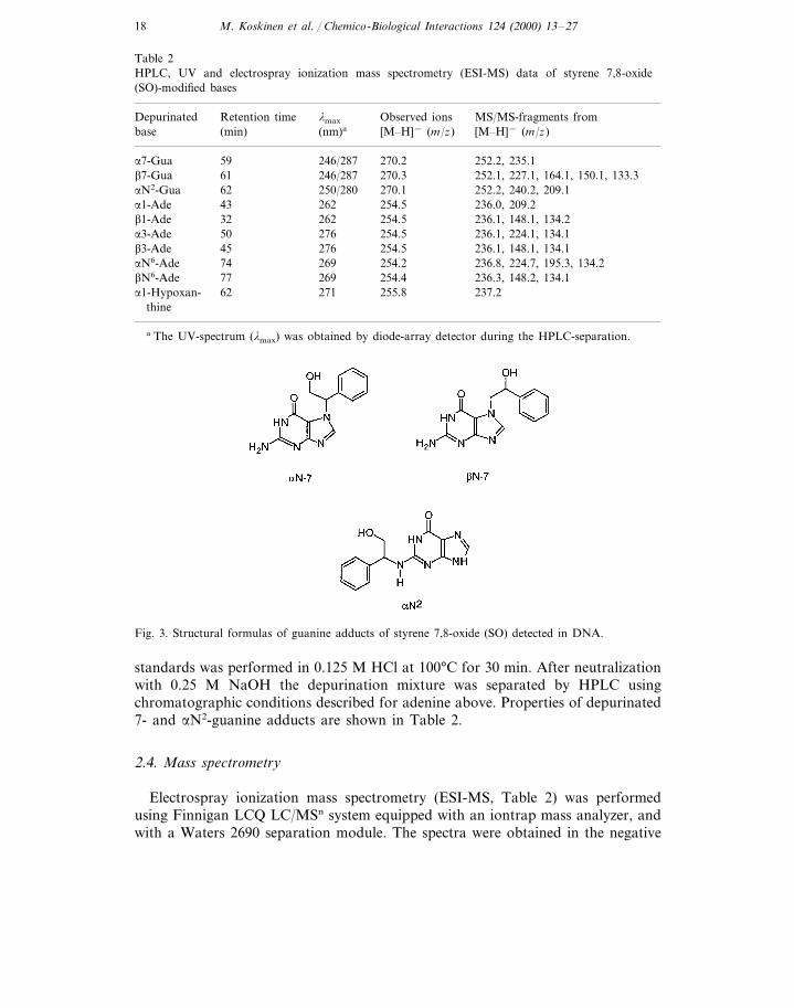
2.3. Preparation of 7- and aN2-substituted guanines
To prepare 7- and N2-alkylated guanine standards (Fig. 3) we first reacted GMP(2 mg/ml) with SO and separated the products, as previously described [21]. Thereaction was made at elevated pH, in 20 mM NH3HCO3/NH3, pH 10.5 and 30%methanol, since this has shown to favour the alkylation at the exocyclic N2-position[12]. The diastereomeric pair of the b-isomers of 7-subtituted GMP eluted at 32 and37 min, and those of the a-isomer at 35 and 43 min. The diastereomeric pair of theaN2-GMP adducts eluted at 52 and 56 min. The identity of these products wasverified by MS and UV-spectroscopy and by depurination, as described[5,7,12,16,21]. Depurination of the nucleotide adducts to obtain 7- and aN2-guanine
Fig. 2. HPLC separations of adenine and 2%-deoxyadenosine 5%-monophosphate (AMP) adducts. PanelA: separation of adenine adducts Peaks: 1, ade; 2, bN-1; 3, aN-1; 4, bN-3; 5, aN-3; 6 and 7, N-7; 8 and9, N-9; 10, N6. The peaks eluting after 10 are obviously bis substituted adenines. Panel B: separationsof AMP adducts. Peaks: 1, AMP; 2 and 3, bN-1; 4 and 5, aN-1; 6, 7, 8 and 9, PO4; 10, aN6; 11, bN6;12, bis-adduct of aN6 and PO4.
M. Koskinen et al. / Chemico-Biological Interactions 124 (2000) 13–2718
Table 2HPLC, UV and electrospray ionization mass spectrometry (ESI-MS) data of styrene 7,8-oxide(SO)-modified bases
lmax Observed ionsRetention time MS/MS-fragments fromDepurinated[M–H]− (m/z)base [M–H]− (m/z)(nm)a(min)
a7-Gua 246/287 270.2 252.2, 235.159270.3 252.1, 227.1, 164.1, 150.1, 133.3246/28761b7-Gua270.1 252.2, 240.2, 209.1aN2-Gua 62 250/280254.5 236.0, 209.2262a1-Ade 43254.5b1-Ade 236.1, 148.1, 134.232 262254.5 236.1, 224.1, 134.127650a3-Ade
27645 254.5 236.1, 148.1, 134.1b3-Ade26974 254.2 236.8, 224.7, 195.3, 134.2aN6-Ade
254.4 236.3, 148.2, 134.1269bN6-Ade 7762a1-Hypoxan- 271 255.8 237.2
thine
a The UV-spectrum (lmax) was obtained by diode-array detector during the HPLC-separation.
Fig. 3. Structural formulas of guanine adducts of styrene 7,8-oxide (SO) detected in DNA.
standards was performed in 0.125 M HCl at 100°C for 30 min. After neutralizationwith 0.25 M NaOH the depurination mixture was separated by HPLC usingchromatographic conditions described for adenine above. Properties of depurinated7- and aN2-guanine adducts are shown in Table 2.
2.4. Mass spectrometry
Electrospray ionization mass spectrometry (ESI-MS, Table 2) was performedusing Finnigan LCQ LC/MSn system equipped with an iontrap mass analyzer, andwith a Waters 2690 separation module. The spectra were obtained in the negative
M. Koskinen et al. / Chemico-Biological Interactions 124 (2000) 13–27 19
ion mode. The samples (�10–100 ng), dissolved in the eluent of water/acetonitrile/NH3 (49/50/1%), were applied by loop injection (10 ml) into the running solvent, ata flow-rate of 0.2 ml/min. For MS/MS experiments precurser ions were selected inthe iontrap analyzer and fragmented. The full scan data were acquired for 70–1000m/z and for MS/MS data 70–500 m/z. Electrospray voltage was 5.0 kV andcapillary temperature 250°C.
For the 1-, 3- and N6-substituted adenines the isomerism of the hydrox-yphenylethyl moiety was determined by ESI-MS (Table 2). The pseudomolecularion [M–H]− for the adducts was observed at m/z 254. For the a-isomer, theMS/MS experiment on [M–H]− resulted in product ion m/z 224 indicating thecleavage of the CH2O. The characteristic fragment for the b-isomer is that of m/z148, which was interpreted to be related to the cleavage of the C6H5CHO. Theassignments are in line with the previously reported MS studies for SO substitutedguanines [5,12,13], in which the cleaved fragments are CH2OH or C6H5CH2OH fora- or b-isomers, respectively. Phosphate alkylation of AMP was identified byMS/MS-experiment in which the cleavage of the glycosidic bond gave a fragment ofm/z 315 that corresponded to SO modified phosphate with deoxyribose moiety.
2.5. Reaction of salmon testis DNA with SO, hydrolysis of modified DNA, test ofhyperchromicity
SO-modified DNA was prepared by incubating 4–6 mg of salmon testis DNA in2 ml of either, 10 mM Tris buffer (pH 7.4), 0.15 M NaCl or in 50 mM Tris buffer(pH 7.4) with 23 ml SO at 37°C for 23 h. After incubation, the excess SO wasextracted with ethyl acetate and DNA was precipitated with ethanol. The DNA waswashed with 70% ethanol and redissolved in water. The neutral hydrolysis wasperformed by keeping the DNA in water solution in boiling water bath for 30 min,followed by ethanol precipitation, as described [21]. The HPLC separation wascarried out as for the adenine products above, except the peaks were detected byUV-absorption at 284 nm.
After the neutral hydrolysis, the remaining DNA was depurinated in 1 ml of 0.1M HCl at 72°C either for 20 or 100 min followed by neutralization with 5M NaOHand 1 M Tris (pH 7.4). The hydrolysis in 0.1 M HCl for 20 min did not depurinateall of the aN2-guanine adducts. Thus, when the incubation time at 72°C wasincreased to 100 min, the aN2-guanine adduct level increased 5-fold and aN6-adenine level 2-fold. Modest increase was observed also with 1-adenine and1-hypoxanthine adducts. However, the UV-spectra showed presence of unknownimpurities for the latter products. Therefore the levels of the N-1 adducts reportedhere are based on the 20 min acid hydrolysis. The released purine adducts wereextracted with water saturated 1-butanol. The butanol phase was evaporated todryness in vacuum centrifuge and was reconstituted in water prior the HPLCanalysis. The efficiency of extraction was for b1-adenine 41%, a1-adenine 46%,aN2-guanine 80%, a1-hypoxanthine 80% and for aN6-adenine 90%. The HPLCseparation was carried out as for the adenine products above.

M. Koskinen et al. / Chemico-Biological Interactions 124 (2000) 13–2720
The alkylation products were identified from their UV-spectra obtained by diodearray detector and retention times, which were compared to those of the standardcompounds. For quantification of the adducts, the concentration of standardcompound was measured by UV using extinction coefficients available in literaturefor similar adducts [22–25]. The known amounts of each adduct (at similarconcentrations as the expected adducts) were then analysed by HPLC at the sameconditions as for the DNA hydrolysate to obtain the coefficient that relates the areaunits to the amount of the analyte. A linear relationship was assumed between theresponse and the concentration of the adduct.
Double-strandedness of DNA was tested by hyperchromic effect observed whentreating unmodified salmon testis DNA in 50 mM NaOH and recording the changein absorbance at 260 nm [26]. Thus, for the DNA in 10 mM Tris buffer and 0.15M NaCl an increase of 28% was observed by the alkaline treatment, verifying theDNA to be double-stranded. For the DNA in 50 mM Tris (pH 7.4) the hyper-chromic effect was 12% indicating partial denaturation. The double-stranded DNAwas estimated to be opened to single-stranded in the proportion of ca. 57% on thatbasis of the decrease of the hyperchromic effect observed.
The stabilities of N-3 adenine and N-7 guanine products in DNA were studied in10 mM Tris (pH 7.4), 0.15 M NaCl. Separate samples of SO-DNA were incubatedat 37°C 0, 2, 18, 24, 48, 96h after which DNA was precipitated and the supernatantwas analysed for spontaneously depurinated products. For comparison also theremaining DNA was depurinated in neutral conditions and analysed as above.
3. Results
To prepare standards for adenine adducts formed in DNA adenine was reactedwith SO at pH 7.4 and, for comparison, AMP was also reacted at similarconditions. For adenine we detected previously unreported isomeric pairs ofSO-adducts reacted at 3-, 7- and 9-positions. For N6-adenine, only the a-isomer wasdetected (Fig. 2). The adducts were characterised by UV and ESI-MS (Tables 1 and2). The latter being able to distinguish the a- and b-isomers of the SO-adduct (seeSection 2). For AMP, both isomers of N6-adducts were observed, the b-isomerbeing obviously formed by the Dimroth rearrangement, as suggested [14]. Anothermarked difference in reactions between adenine and AMP was that in AMP no N-3adducts were detected, not even those depurinated from the nucleotide.
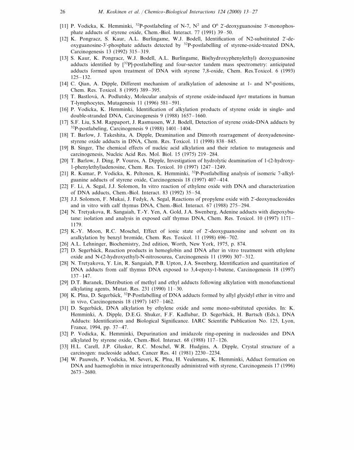
To ensure double-stranded structure of DNA, the incubation of salmon testisDNA with SO was performed in 0.15 M NaCl and 10 mM Tris buffer (pH 7.4). Itwas shown by testing for hyperchromic effect that DNA in these conditionsretained the double-stranded structure. HPLC analysis of the neutral hydrolysate ofSO modified DNA showed the highest modified position to be 7-guanine (Fig. 4,Table 3), as expected [7,16]. The products were verified by collecting the fractions,and the MS analysis showed the [M–H]− at m/z 270, as found for the standards(Table 2). The two peaks eluting before the 7-guanine adducts had not beenpreviously detected and they were now identified as the b- and a- isomers of

M. Koskinen et al. / Chemico-Biological Interactions 124 (2000) 13–27 21
3-adenine adducts of SO, respectively. The deprotonated molecular ion [M–H]− forthese products was found by MS at m/z 254, as expected for adenine adducts ofSO. The identities of these peaks were verified by comparing the retention times andUV-spectra obtained by diode-array detector to those of the synthesized 3-adeninestandards.
In mild acidic hydrolysis, the remaining purines were liberated and extracted tobutanol. The HPLC analysis of the hydrolysate showed the presence of the bothisomers of 1-adenine, aN2-guanine and aN6-adenine adducts (Fig. 4, Table 3). Abackground peak arising from salmon testis DNA disturbed the quantification ofb-isomer of N6-adenine. However, as an upper limit of the alkylation, the level ofthe bN6 was estimated to be 2.8/106 nucleotides. A peak corresponding to thedeaminated a1-adenine adduct, i.e. a1-hypoxanthine, was detected eluting after theaN2-guanine. The b-isomer of the hypoxanthine adduct had the same retention timeas that of aN2-guanine. But, since the deamination of b1-adenine is expected to bemuch slower than that of the a isomer (Section 2) it was assumed that itcontributed only a minor fraction to the N2-guanine peak. The assignment of thefractions was based on coelution with the standards and on the UV-spectraobtained by diode array detector. The absence of the peaks in similarly treatedunexposed salmon testis DNA was verified. The adenine adducts were furtherconfirmed by treating SO-DNA either in 0.1 M NaOH at 37°C for 20 h or in waterat 97°C for 40 min. When analysed by acidic depurination, the alkaline-treatedSO-DNA was devoid of N-1 adenine adducts. Instead, the peaks corresponding
Table 3Alkylation levels of DNA modified by styrene 7,8-oxide (SO) in 10 mM Tris 0.15 M NaCl (pH 7.4)a,determined by neural and acidic hydrolysis, and the amounts of adducts released during the incubationof salmon testis DNA with SO
Alkylation levelb Proportion of totalProductalkylation (%)
a7-Gua 2360 31.48Neutral hydrolysisb7-Gua 3380 45.19
1.36113a3-Adeb3-Ade 102 1.51aN2-GuaAcid hydrolysis 112 1.50
0.3224.2b1-Adea1-Ade 7.6 0.10
5.1 0.07a-1 hypoxanthineaN6-Ade 52.9 0.71bN6-Ade B2.8 B0.04a7-Gua 9.29Incubation solutionc 696
549 7.34b7-Guab3-Ade 83.9 1.12
a Alkylation level as mean of at least two experiments.b The alkylation levels are expressed as adducted nucleotides/106 normal nucleotides.c Adducts spontaneously released during the preparation of SO-modified DNA, analysed from
incubation solution after precipitation of DNA.

M. Koskinen et al. / Chemico-Biological Interactions 124 (2000) 13–2722
Fig. 4. Analysis of DNA treated by styrene 7,8-oxide (SO) in 10mM Tris–HCl, 0.15 M NaCl, pH 7.4.Panel A: neutral hydrolysis of SO-modified DNA. Peaks: 1, bN-3 ade; 2, aN-3 ade; 3, aN-7 gua; 4, bN-7gua. Panel B: Acidic hydrolysis of SO-modified DNA. Peaks: 1, bN-1 ade; 2, aN-1 ade; 3, aN2 gua; 4,aN-1 hypoxanthine; 5, aN6 ade; 6, bN6 ade.
both a- and b-isomers of N6-adenines were markedly increased due to the Dimrothrearrangement [14]. Also in the heat treatment the N-1 adenine product seemed tobe completely removed, while especially the aN-1 hypoxanthine and bN6-adeninefractions were increased. The O6-guanine products are also obviously formed in thereaction between SO and DNA [16], but due to their acid lability [8] they could notbe detected.
During the incubation of SO with DNA a considerable proportion of the mostlabile adducts i.e. 7-guanines and 3-adenines were liberated (Table 3). Thus, 17% ofall 7-guanines formed and 28% of all 3-adenines formed were released during thepreparation of SO-modified DNA. As the spontaneously depurinated adducts aretaken into account, the total proportion of 7-guanine adducts is 93% and that ofthe 3-adenine adducts 4%. The rate of depurination was studied by incubatingSO-treated DNA at 37°C in 10 mM Tris (pH 7.4) containing 0.15 M NaCl. The3-adenine adducts were found to be more labile when compared to the 7-guanineadducts, the half-lives of a and b3-adenines being �10 and �20 h, respectively.

M. Koskinen et al. / Chemico-Biological Interactions 124 (2000) 13–27 23
For both a- and b-isomers of N-7 guanine adducts the half-lives were found to be51 h.
When DNA was incubated with SO in 50 mM Tris buffer (pH 7.4) without 0.15M NaCl, the reactions, especially at the N2-guanine and N6-adenine positions, weremarkedly increased. This indicates that the double-stranded DNA has partlydenaturated and the position involved in base-pairing had become accessible foralkylation. By hyperchromicity measurements over 50% of DNA was renderedsingle-stranded. The level of aN2-guanine was found to be 650 adducts/106 normalnucleotides and that of aN6-adenine 310/106, the levels being 6-fold higher thanthose obtained for DNA modified by SO in 0.15 NaCl. The increase of 7-alkylationwas much more modest, being ca. 23%. The alkylation levels obtained by neutraldepurination were 3110/106 and 4310/106, for a7-and b7-guanines, respectively.Unexpected results were that in the case of 3-adenine adducts only the a-isomer wasobserved at the level of 700/106 and that there was almost 7-fold increase inalkylation as compared to the alkylation in 0.15 M NaCl.
4. Discussion
The major novel aspects of the present study were that several SO induced DNAadducts were determined by UV analysis, and during the handling of DNA saltconcentration of was maintained at 0.15 M to ensure the double-stranded structure.A major finding was that the 3-adenine was the most abundant SO-alkylationposition after that of 7-guanine. Even though several studies have earlier beencarried out on the modification of DNA by SO [7,10,12,13,16–18], the alkylation atthe 3-position of adenine has remained unidentified, probably due to the highlability of these adducts [19] and the impossibility to detect them by 32P-postlabel-ing. For some other epoxides, i.e. ethylene oxide [22,27], propylene oxide [23],diepoxybutane [28] and 3,4-epoxy-1-butane [24] as well as for methyl methanesulfonate [29] and allyl glycidyl ether [30], the alkylation at N-3 adenine is found atrelatively high levels, that is ca. 4-14% of 7-guanine alkylation [31]. Thus, theproportion of 4% for SO alkylation found in the present study is comparable to theother related DNA alkylating agents. The N-3 adenine adducts of SO were foundto be unstable being prone to depurination, as has been observed for the other3-adenine adducts [19]. The depurination half-life of 20 h for b3-adenine adduct issimilar to the other 3-modified adenine adducts [30], while the depurination of thea-isomer, with half-life of ca. 10 h, can be considered relatively fast. The depurina-tion of both isomers of the 7-guanine adducts of SO at pH 7.4 was found to be 51h, 3-fold faster that earlier reported for double-stranded SO-modified DNA at pH4.2 [32].
In reaction between SO and adenine, the 3-position was alkylated to almost ashigh extent as the 1-position, whereas in AMP no N-3-adducts were observed. Thelack of 3-alkylation is also noted for SO-treated adenosine and deoxyadenosine[7,14] and the above mentioned epoxides [22–24,27–29]. The reason for the lack ofthe N-3 adducts in nucleosides may be due to an internal hydrogen bond between

M. Koskinen et al. / Chemico-Biological Interactions 124 (2000) 13–2724
the deoxyribose 5%-hydroxyl group and the N-3 atom of the adenine residue [33]. InAMP similar intramolecular (or intermolecular) hydrogen bonding can take placebetween a hydroxyl group in the phosphate moiety and N-3 atom of adenine. InDNA the conformation of the base and the sugar is obviously twisted, compared toa free nucleoside or nucleotide, preventing such hydrogen bonding, and allowingthe entrance of the epoxide to the nucleophilic 3-position of adenine. Surprisingfinding was that in partly denaturated DNA the 3-alkylation was almost 7-foldhigher as in the double-stranded DNA. This suggests that 3-position is even moreopen to alkylation in single-stranded DNA.
One of the major results of the present study was that, by using sequentialdepurination, it was possible to determine relative proportions of the purineadducts. An overwhelming proportion, 93% of the total alkylation of double-stranded DNA by SO occurred at the 7-position of guanine. Thus, only a smallfraction of other positions, in addition to 3-adenine, i.e. N2-guanine as well as 1-and N6-adenine, were alkylated. These corresponded to less than 3% of the totalalkylation. Thus, the positions involved in hydrogen bonding of base-pairing werefar less susceptible to alkylation than those of 7-guanine and 3-adenine. It shouldbe noted that the current hydrolysis method did not allow the detection ofO6-guanine or pyrimidine adducts. However, based on the previous studies, theexpected O6-alkylation level is relatively low compared to that at N2-guanine [16],and the thymidine adducts are not expected to be formed in detectable amounts [7].3-Cytosine adducts of SO are formed with deoxynucleosides [7], and the prelimi-nary results indicate that the adducts derived from 3-cytosine alkylation can alsobeen found in SO modified DNA using the 32P-postlabeling technique, but at ca.0.2% of total alkylation (M. Koskinen et al., unpublished). Thus, the relativeproportions given are not markedly affected by the mentioned adducts. In aprevious study, in which the reaction of double-stranded DNA was performed in 10mM Tris (pH 7.4) buffer without salt, 95% of alkylation was found to take placein the guanine, being in accordance with the present study [16]. However, thealkylation at guanine was found in proportions of 74:23:3.7 at the 7-, N2- andO6-positions, respectively, the alkylation at N2-position being drastically higherthan in the present study. Because of the low ionic strength, it cannot be excludedthat a considerable proportion of single-stranded DNA was present. In single-stranded DNA the positions involved in base-pairing are more accessible foralkylation [16]. In the present study, SO was reacted with salmon testis DNA eitherin 50 mM Tris (pH 7.4) or in 10 mM Tris (pH 7.4) containing 0.15 M NaCl. In 50mM Tris (pH 7.4) alkylation at N2-guanine and N6-adenine positions were in-creased markedly, especially when compared to 7-guanine position, indicating thatsome of the double-stranded DNA had denaturated. This was supported byhyperchromicity measurements. In 0.15 M NaCl, it might, however, be that thedouble-stranded structure cannot be completely assured since the excess of SO ispresent as droplets in the reaction mixture and in the interphase between thedroplet and aqueous phase the DNA might be locally denaturated.
The 1-adenosine adducts of SO are unstable being prone to deamination to1-inosine adducts, and to rearrangement to corresponding N6-adenine adducts

M. Koskinen et al. / Chemico-Biological Interactions 124 (2000) 13–27 25
[14,18,20]. Therefore, the adduct levels reported here do not tell the real levelsinitially formed with the reaction of SO and the adenine residues in DNA. In arecent study, Barlow et al. [18], using [3H]deoxyadenosine labelled calf thymusDNA, detected only the stable adenosine-derived adducts, i.e. 1-inosine and N6-adenosine. The study with shorter incubation times and higher salt concentrations,however, showed that it was possible to detect the unstable 1-adenine adducts in invitro exposed DNA. Yet, in chronic in vivo studies it would be likely that the stable1-hypoxanthine and N6-adenine adducts would be the predominant species. Theseadducts are also main candidates for the mutagenic lesions of SO, because incultured cells mutations only at the A/T sites have been observed [15].
Regarding the possibility for human monitoring of in vivo exposure to styrene,the present study suggests that the 7-guanine adducts should be the main target.This can be accomplished either by the 32P-postlabeling method [21,34–36] orperhaps by LC/MS. 3-adenine adducts are also abundant, but their analysis by32P-postlabeling might be very difficult due to their instability as well as due to thedifficulty of preparing nucleotide standards.
Acknowledgements
We gratefully acknowledge Dr Elke K.H. Schweda for her help with the massspectrometric analyses. The study was supported by the Swedish Work-environ-ment Fund and by the grant GACR, 313/99/1460.
References
[1] IACR Monographs on the Evaluation of Carcinogenic Risks of Chemicals to Humans, vol. 60,International Agency for Research on Cancer, Lyon, France, 1994.
[2] M. Sorsa, K. Peltonen, H. Vainio, K. Hemminki (Eds.), Butadiene and Styrene: Assessment ofHealth Hazard. International Agency for Research on Cancer, Lyon, France, 1993.
[3] D.H. Phillips, P.B. Farmer, Evidence for DNA and protein binding by styrene and styrene oxide,Crit. Rev. Toxicol. 19 (1994) 227–249.
[4] K. Hemminki, J. Paasivirta, T. Kurkirinne, L. Virkki, Alkylation products of DNA bases by simpleepoxides, Chem.-Biol. Intreact. 30 (1980) 259–270.
[5] K. Hemminki, A. Hesso, Reaction products of styrene oxide with guanosine in aqueous media,Carcinogenesis 5 (1984) 601–607.
[6] K. Hemminki, R. Suni, Formation of phosphodiesters in thymidine monophosphate by styreneoxide, Toxicol. Lett. 21 (1984) 59–63.
[7] K. Savela, A. Hesso, K. Hemminki, Characterization of reaction products between styrene anddeoxynucleosides and DNA, Chem.-Biol. Interact. 60 (1986) 235–246.
[8] R.C. Moschel, K. Hemminki, A. Dipple, Hydrolysis and rearrangement of O6-substitutedguanosine products resulting from reaction of guanosine with styrene oxide, J. Org. Chem. 51(1986) 2952–2955.
[9] F. Latif, R.C. Moschel, K. Hemminki, A. Dipple, Styrene oxide as a stereochemical probe for themechanism of aralkylation at different sites on guanosine, Chem. Res. Toxicol. 1 (1988) 364–369.
[10] K. Pongracz, S. Kaur, A.L. Burlingame, W.J. Bodell, O6-substituted 2%-deoxyguanoine-3%-de-oxyphosphate adducts detected by 32P-postlabeling of styrene oxide treated DNA, Carcinogenesis10 (1989) 1009–1013.
M. Koskinen et al. / Chemico-Biological Interactions 124 (2000) 13–2726
[11] P. Vodicka, K. Hemminki, 32P-postlabeling of N-7, N2 and O6 2%-deoxyguanosine 3%-monophos-phate adducts of styrene oxide, Chem.-Biol. Interact. 77 (1991) 39–50.
[12] K. Pongracz, S. Kaur, A.L. Burlingame, W.J. Bodell, Identification of N2-substituted 2%-de-oxyguanosine-3%-phosphate adducts detected by 32P-postlabelling of styrene-oxide-treated DNA,Carcinogenesis 13 (1992) 315–319.
[13] S. Kaur, K. Pongracz, W.J. Bodell, A.L. Burlingame, Bis(hydroxyphenylethyl) deoxyguanosineadducts identified by [32P]-postlabelling and four-sector tandem mass spectrometry: anticipatedadducts formed upon treatment of DNA with styrene 7,8-oxide, Chem. Res.Toxicol. 6 (1993)125–132.
[14] C. Qian, A. Dipple, Different mechanism of aralkylation of adenosine at 1- and N6-positions,Chem. Res. Toxicol. 8 (1995) 389–395.
[15] T. Bastlova, A. Podlutsky, Molecular analysis of styrene oxide-induced hprt mutations in humanT-lymphocytes, Mutagenesis 11 (1996) 581–591.
[16] P. Vodicka, K. Hemminki, Identification of alkylation products of styrene oxide in single- anddouble-stranded DNA, Carcinogenesis 9 (1988) 1657–1660.
[17] S.F. Liu, S.M. Rappaport, J. Rasmussen, W.J. Bodell, Detection of styrene oxide-DNA adducts by32P-postlabeling, Carcinogenesis 9 (1988) 1401–1404.
[18] T. Barlow, J. Takeshita, A. Dipple, Deamination and Dimroth rearragement of deoxyadenosine-styrene oxide adducts in DNA, Chem. Res. Toxicol. 11 (1998) 838–845.
[19] B. Singer, The chemical effects of nucleic acid alkylation and their relation to mutagenesis andcarcinogenesis, Nucleic Acid Res. Mol. Biol. 15 (1975) 219–284.
[20] T. Barlow, J. Ding, P. Vouros, A. Dipple, Investigation of hydrolytic deamination of 1-(2-hydroxy-1-phenylethyl)adenosine, Chem. Res. Toxicol. 10 (1997) 1247–1249.
[21] R. Kumar, P. Vodicka, K. Peltonen, K. Hemminki, 32P-Postlabelling analysis of isomeric 7-alkyl-guanine adducts of styrene oxide, Carcinogenesis 18 (1997) 407–414.
[22] F. Li, A. Segal, J.J. Solomon, In vitro reaction of ethylene oxide with DNA and characterizationof DNA adducts, Chem.-Biol. Interact. 83 (1992) 35–54.
[23] J.J. Solomon, F. Mukai, J. Fedyk, A. Segal, Reactions of propylene oxide with 2%-deoxynucleosidesand in vitro with calf thymus DNA, Chem.-Biol. Interact. 67 (1988) 275–294.
[24] N. Tretyakova, R. Sangaiah, T.-Y. Yen, A. Gold, J.A. Swenberg, Adenine adducts with diepoxybu-tane: isolation and analysis in exposed calf thymus DNA, Chem. Res. Toxicol. 10 (1997) 1171–1179.
[25] K.-Y. Moon, R.C. Moschel, Effect of ionic state of 2%-deoxyguanosine and solvent on itsaralkylation by benzyl bromide, Chem. Res. Toxicol. 11 (1998) 696–702.
[26] A.L. Lehninger, Biochemistry, 2nd edition, Worth, New York, 1975, p. 874.[27] D. Segerback, Reaction products in hemoglobin and DNA after in vitro treatment with ethylene
oxide and N-(2-hydroxyethyl)-N-nitrosourea, Carcinogenesis 11 (1990) 307–312.[28] N. Tretyakova, Y. Lin, R. Sangaiah, P.B. Upton, J.A. Swenberg, Identification and quantitation of
DNA adducts from calf thymus DNA exposed to 3,4-epoxy-1-butene, Carcinogenesis 18 (1997)137–147.
[29] D.T. Baranek, Distribution of methyl and ethyl adducts following alkylation with monofunctionalalkylating agents, Mutat. Res. 231 (1990) 11–30.
[30] K. Plna, D. Segerback, 32P-Postlabelling of DNA adducts formed by allyl glycidyl ether in vitro andin vivo, Carcinogenesis 18 (1997) 1457–1462.
[31] D. Segerback, DNA alkylation by ethylene oxide and some mono-subtituted epoxides. In: K.Hemminki, A. Dipple, D.E.G. Shuker, F.F. Kadlubar, D. Segerback, H. Bartsch (Eds.), DNAAdducts: Identification and Biological Significance. IARC Scientific Publication No. 125, Lyon,France, 1994, pp. 37–47.
[32] P. Vodicka, K. Hemminki, Depurination and imidazole ring-opening in nucleosides and DNAalkylated by styrene oxide, Chem.-Biol. Interact. 68 (1988) 117–126.
[33] H.L. Carell, J.P. Glusker, R.C. Moschel, W.R. Hudgins, A. Dipple, Crystal structure of acarcinogen: nucleoside adduct, Cancer Res. 41 (1981) 2230–2234.
[34] W. Pauwels, P. Vodicka, M. Severi, K. Plna, H. Veulemans, K. Hemminki, Adduct formation onDNA and haemoglobin in mice intraperitoneally administred with styrene, Carcinogenesis 17 (1996)2673–2680.

M. Koskinen et al. / Chemico-Biological Interactions 124 (2000) 13–27 27
[35] P. Vodicka, R. Stetina, P. Kumar, K. Plna, K. Hemminki, 7-alkylguanine adducts of styrene oxidedetermined by 32P-postlabelling in DNA and human embryonal lung fibroblasts (HEL), Carcino-genesis 17 (1996) 801–808.
[36] P. Vodicka, T. Tvrdik, S. Osterman-Golkar, L. Vodickova, K. Peterkova, P. Soucek, J. Sarmanova,P.B. Farmer, B. Lambert, K. Hemminki, An evaluation of styrene genotoxicity using severalbiomarkers in a three-year follow up study of hand lamination workers, Mutat. Res. (in press).
.