adaptive thresholding method through digital image processing · the detection of cervical cancer...
TRANSCRIPT
TAF Journal of Advances in Health and Medical Sciences
2015, 1(1): 30-36 JAHMS 1
7
Content from this work is copyrighted by TAF Publishing, which permits restricted commercial use, distribution and reproduction in any medium under a written permission. Users may print articles for educational and research uses only, provided the original author and source are credited. Any further utilization of this work must maintain attribution to the author(s), the title of the work and journal citation in the form of a proper scientific referencing.
PRIMARY RESEARCH The detection of cervical cancer disease using an adaptive thresholding method through digital image processing Eggi I. Putri 1,*, Rita Magdalena 2,*, Ledya Novamizanti 3,*
1, 2, 3 Telkom Engineering School (TES)–Telkom University (Tel-U), Bandung, Jawa Barat 40257, Indonesia
Abstract-Cervical cancer is a kind of cancer diseases caused by human papilloma virus
type 16 and 18, attacking woman cervix. To detect a cervical cancer, the frequently-used
method is Pap-Smear; however, errors often occur when the method is taken to diagnose
the level of cervical cancer. Thus, a proper system is required, which is supposed for being
able to help identifying the result of Pap-Smear. This study aims at designing a system to
detect the symptoms of cervical cancer using MATLAB to solve these errors. The image
processing begins with converting the type of an image, which is followed by a
thresholding, and a noise removal using filters until the image has become ready to be
detected. For a thresholding process, an Adaptive Thresholding method is taken, in which
the thresholding focuses on local threshold values. The system is considerably able to
classify images into two types, i.e. normal and abnormal (precancerous). Abnormal type is
divided into three subtypes, i.e. mild, moderate and severe. An experiment is conducted on
the proposed system, in which it is supposed to analyze 500 test images, including 250 for
training and 250 for testing. Based on the testing process, a perfect 100% accuracy rate is
obtained, while the average processing time is 25.4 seconds with a WS value at 10 and C
value at -2.
© 2015 TAF Publishing. All rights reserved.
INTRODUCTION
The reproductive system in woman body consists of several parts, in which
one of them is uterus. Uterus consists of cervix. The cervix connects the uterus
with female reproductive system. In fact, cervical cancer is kind of cancer
diseases caused by human papilloma virus type 16 and 18 attacking woman
cervix. Cervical cancer is a frightening specter for every woman, because its
symptoms is not clearly observable before an advanced stage. Thus, an early
detection is regularly required to preventing and treating cervical cancer * Corresponding author: E.I. Putri, R. Magdalena and L. Novamizanti
E-mail: [email protected]; [email protected]; [email protected]
Keywords: Cervical cancer Pap smear Digital image processing Thresholding
Received: 15 January 2015 Accepted: 26 July 2015 Published: 15 October 2015
2015 J. Adv. Health. Med. Sci. 31
ISSN: 2517-9616 TAF
DOI: 10.20474/jahms-1.1.4 Publishing
symptoms. The current cervical cancer detection method that is frequently
used is Pap-Smear. However, errors often occur in diagnosing the level of
cervical cancer. Therefore, a system that is able to help identifying the result
of Pap-Smear is required. This research aims at providing a digital alternative
in the early examination of cervical cancer by using software to help medical
examiners to interpret Pap-smear results. Prior researches have attempted to
use an evolving ANN technique [1], highly efficient second-order neural
network training algorithms [2], and a neuro-fuzzy classification that is able
to classify two classes, i.e. normal and abnormal, with a 75% average
accuracy. On the other hand, the current research uses Adaptive Thresholding
and K-Nearest Neighbor for classifying the cell types of cervical cancer. The
classification is divided into two classes, i.e. normal and abnormal. The
normal class is divided into four classes, i.e. columnar, parabasal,
intermediate, and superficial. The abnormal class is divided into three classes,
e.g. mild, moderate, and severe.
METHODOLOGY
Cervix
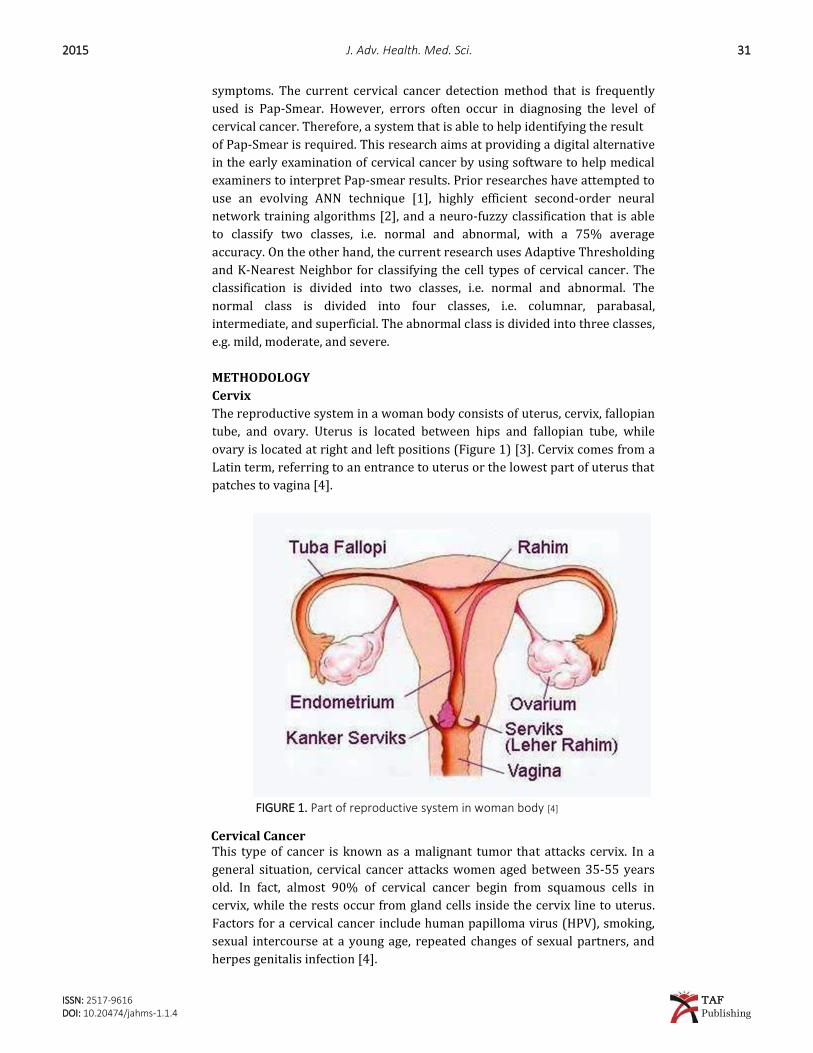
The reproductive system in a woman body consists of uterus, cervix, fallopian
tube, and ovary. Uterus is located between hips and fallopian tube, while
ovary is located at right and left positions (Figure 1) [3]. Cervix comes from a
Latin term, referring to an entrance to uterus or the lowest part of uterus that
patches to vagina [4].
FIGURE 1. Part of reproductive system in woman body [4]
Cervical Cancer
This type of cancer is known as a malignant tumor that attacks cervix. In a
general situation, cervical cancer attacks women aged between 35-55 years
old. In fact, almost 90% of cervical cancer begin from squamous cells in
cervix, while the rests occur from gland cells inside the cervix line to uterus.
Factors for a cervical cancer include human papilloma virus (HPV), smoking,
sexual intercourse at a young age, repeated changes of sexual partners, and
herpes genitalis infection [4].
32 E. I. Putri, R. Magdalena, L. Novamizanti – Detection of cervical cancer … 2015
ISSN: 2517-9616 TAF
DOI: 10.20474/jahms-1.1.4 Publishing
Types of Cervix Cells
Cells changing in cervix occurs slowly and it may not produce cancer cells. In
fact, there is a pre-cancer/cervix dysplasia condition, in which there is a
change from normal cells to abnormal ones before transform into a cervical
cancer. In general, dysplasia cells have a big size, dark colored and stick with
other cells in cervix.
The existence of pre-cancer cells must be carefully treated during the pre-
cancer condition because a good treatment may possibly prevent pre-cancer
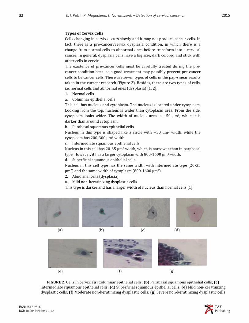
cells to be cancer cells. There are seven types of cells in the pap-smear results
taken in the current research (Figure 2). Besides, there are two types of cells,
i.e. normal cells and abnormal ones (dysplasia) [1, 2]:
1. Normal cells
a. Columnar epithelial cells
This cell has nucleus and cytoplasm. The nucleus is located under cytoplasm.
Looking from the top, nucleus is wider than cytoplasm area. From the side,
cytoplasm looks wider. The width of nucleus area is ~50 µm2, while it is
darker than around cytoplasm.
b. Parabasal squamous epithelial cells
Nucleus in this type is shaped like a circle with ~50 µm2 width, while the
cytoplasm has 200-300 µm2 width.
c. Intermediate squamous epithelial cells
Nucleus in this cell has 20-35 µm2 width, which is narrower than in parabasal
type. However, it has a larger cytoplasm with 800-1600 µm2 width.
d. Superficial squamous epithelial cells
Nucleus in this cell type has the same width with intermediate type (20-35
µm2) and the same width of cytoplasm (800-1600 µm2).
2. Abnormal cells (dysplasia)
a. Mild non-keratinizing dysplastic cells
This type is darker and has a larger width of nucleus than normal cells [1].
(a) (b) (c) (d)
(e) (f) (g)
FIGURE 2. Cells in cervix: (a) Columnar epithelial cells; (b) Parabasal squamous epithelial cells; (c) intermediate squamous epithelial cells; (d) Superficial squamous epithelial cells; (e) Mild non-keratinizing dysplastic cells; (f) Moderate non-keratinizing dysplastic cells; (g) Severe non-keratinizing dysplastic cells
2015 J. Adv. Health. Med. Sci. 33
ISSN: 2517-9616 TAF
DOI: 10.20474/jahms-1.1.4 Publishing
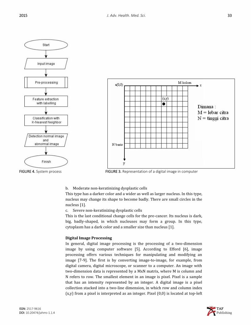
FIGURE 4. System process FIGURE 3. Representation of a digital image in computer
b. Moderate non-keratinizing dysplastic cells
This type has a darker color and a wider as well as larger nucleus. In this type,
nucleus may change its shape to become badly. There are small circles in the
nucleus [1].
c. Severe non-keratinizing dysplastic cells
This is the last conditional change cells for the pre-cancer. Its nucleus is dark,
big, badly-shaped, in which nucleuses may form a group. In this type,
cytoplasm has a dark color and a smaller size than nucleus [1].
Digital Image Processing
In general, digital image processing is the processing of a two-dimension
image by using computer software [5]. According to Efford [6], image
processing offers various techniques for manipulating and modifying an
image [7-9]. The first is by converting image-to-image, for example, from
digital camera, digital microscope, or scanner to a computer. An image with
two-dimension data is represented by a MxN matrix, where M is column and
N refers to row. The smallest element in an image is pixel. Pixel is a sample
that has an intensity represented by an integer. A digital image is a pixel
collection stacked into a two-line dimension, in which row and column index
(x,y) from a pixel is interpreted as an integer. Pixel (0,0) is located at top-left
34 E. I. Putri, R. Magdalena, L. Novamizanti – Detection of cervical cancer … 2015
ISSN: 2517-9616 TAF
DOI: 10.20474/jahms-1.1.4 Publishing
corner, while x-index moves to right and y-index moves to lower position.
Figure 3 exhibits a representation of digital image in a computer.
Adaptive Thresholding Method
The thresholding process produces binary images, which have two grayscale
values, i.e. black and white. Meanwhile, the adaptive thresholding is a
threshold process that utilizes a local threshold, which is calculated based on
neighboring pixel statistics. At least, there are three types of adaptive
thresholding [7], in which the current research
utilizes the first type (mean concerning local intensity).
1. Mean concerning local intensity
where:
W = size for window in image
Nw = pixel in window
C = constant
2. Median approximation = median (( , ), ( , ) ) – (E.2)
3. Maximum and minimum statistical approximation
RESULTS
System Design
The input is a cell image of cervical cancer from Pap-Smear results, in which
all are in color and BMP format (goo.gl/zJms0O). This research uses 500
images, in which 250 are training images and 250 are testing images. Figure 4
describes the process.
Pre-Processing
RGB to Grayscale. The input images are sampled into RGB format. The first
process includes changing image format from RGB to grayscale. It is intended
to enhance the
density of information for next processes. The images are transformed from
RGB (3
Grayscale= (0.299 + 0.587 + 0.114 ) (E.4)
layers) to grayscale (1 layer) based on the following formula:
Adaptive Thresholding. This research uses adaptive thresholding, because
there is a lighting difference on certain area between images. The method
suggests a threshold value from sub-images, which acts as the local threshold.
The threshold value is valid for sub-images in which the area in an image is
given by window size value (WS) and constant (C). To achieve a better
accuracy, WS value and C value are adjusted over certain areas.
Filtering. The current research uses a filter (bwareaopen) to remove noises
within an image. In the proposed system, noises are removed with a
restriction of wide white pixel value in image at less than 100. The value is set
by considering that a value less than 100 may not produce the best result, in
which there are noises such as small circles. However, a value bigger than 100
2015 J. Adv. Health. Med. Sci. 35
ISSN: 2517-9616 TAF
DOI: 10.20474/jahms-1.1.4 Publishing
may make the edge of an image to fade and break, and any object in the image
cannot be correctly determined.
Labelling. Labelling is taken to check white pixels in an image. The checking
is taken from left to right and upper to lower. It produces easy detection and
classification. The labelling process chooses 30 larger wide cells in an image. To do the detection process, this research uses K-Nearest Neighbor
(KNN) with a Euclidean Distance rule through the following formula:
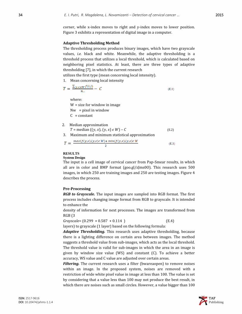
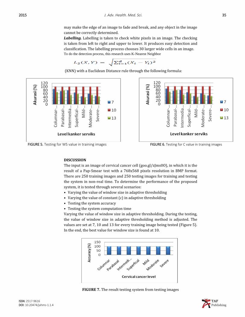
FIGURE 5. Testing for WS value in training images FIGURE 6. Testing for C value in training images
DISCUSSION
The input is an image of cervical cancer cell (goo.gl/zJms0O), in which it is the
result of a Pap-Smear test with a 768x568 pixels resolution in BMP format.
There are 250 training images and 250 testing images for training and testing
the system in non-real time. To determine the performance of the proposed
system, it is tested through several scenarios:
• Varying the value of window size in adaptive thresholding
• Varying the value of constant (c) in adaptive thresholding
• Testing the system accuracy
• Testing the system computation time
Varying the value of window size in adaptive thresholding. During the testing,
the value of window size in adaptive thresholding method is adjusted. The
values are set at 7, 10 and 13 for every training image being tested (Figure 5).
In the end, the best value for window size is found at 10.
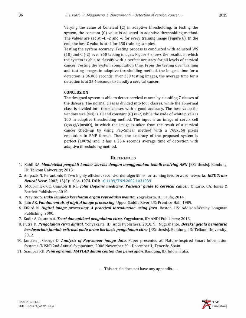
FIGURE 7. The result testing system from testing images
36 E. I. Putri, R. Magdalena, L. Novamizanti – Detection of cervical cancer … 2015
ISSN: 2517-9616 TAF
DOI: 10.20474/jahms-1.1.4 Publishing
Varying the value of Constant (C) in adaptive thresholding. In testing the
system, the constant (C) value is adjusted in adaptive thresholding method.
The values are set at -4, -2 and -6 for every training image (Figure 6). In the
end, the best C value is at -2 for 250 training samples.
Testing the system accuracy. Testing process is conducted with adjusted WS
(10) and C (-2) over 250 testing images. Figure 7 shows the results, in which
the system is able to classify with a perfect accuracy for all levels of cervical
cancer. Testing the system computation time. From the testing over training
and testing images in adaptive thresholding method, the longest time for a
detection is 36.063 seconds. Over 250 testing images, the average time for a
detection is at 25.4 seconds to classify a cervical cancer.
CONCLUSION
The designed system is able to detect cervical cancer by classifing 7 classes of
the disease. The normal class is divided into four classes, while the abnormal
class is divided into three classes with a good accuracy. The best value for
window size (ws) is 10 and constant (C) is -2, while the wide of white pixels is
100 in adaptive thresholding method. The input is an image of cervix cell
(goo.gl/zJms0O), in which the image is taken from the result of a cervical
cancer check-up by using Pap-Smear method with a 768x568 pixels
resolution in BMP format. Then, the accuracy of the proposed system is
perfect (100%) and it has a 25.4 seconds average time of detection with
adaptive thresholding method.
REFERENCES 1. Kahfi RA. Mendeteksi penyakit kanker serviks dengan menggunakan teknik evolving ANN [BSc thesis]. Bandung,
ID: Telkom University; 2013.
2. Ampazis N, Perantonis S. Two highly efficient second-order algorithms for training feedforward networks. IEEE Trans
Neural Netw. 2002; 13(5): 1064-1074. DOI: 10.1109/TNN.2002.1031939
3. McCormick CC, Giuntoli II RL. John Hopkins medicine: Patients' guide to cervical cancer. Ontario, CA: Jones &
Bartlett Publishers; 2010.
4. Prayitno S. Buku lengkap kesehatan organ reproduksi wanita. Yogyakarta, ID: Saufa; 2014.
5. Jain AK. Fundamentals of digital image processing. Upper Saddle River, US: Prentice-Hall; 1989.
6. Efford N. Digital image processing: A practical introduction using Java. Boston, US: Addison-Wesley Longman
Publishing; 2000.
7. Kadir A, Susanto A. Teori dan aplikasi pengolahan citra. Yogyakarta, ID: ANDI Publishers; 2013.
8. Putra D. Pengolahan citra digital. Yohyakarta, ID: Andi Publishers; 2010. 9. Nugrahanto. Deteksi gejala hematuria
berdasarkan jumlah eritrosit pada urine berbasis pengolahan citra [BSc thesis]. Bandung, ID: Telkom University;
2012.
10. Jantzen J, George D. Analysis of Pap-smear image data. Paper presented at: Nature-Inspired Smart Information
Systems (NISIS) 2nd Annual Symposium; 2006 November 29 - December 1; Tenerife, Spain.
11. Sianipar RH. Pemrograman MATLAB dalam contoh dan penerapan. Bandung, ID: Informatika.
— This article does not have any appendix. —